EP2987868A1 - Elektrochemischer Nachweis von Einzelnukleotidpolymorphismen - Google Patents
Elektrochemischer Nachweis von Einzelnukleotidpolymorphismen Download PDFInfo
- Publication number
- EP2987868A1 EP2987868A1 EP14181411.1A EP14181411A EP2987868A1 EP 2987868 A1 EP2987868 A1 EP 2987868A1 EP 14181411 A EP14181411 A EP 14181411A EP 2987868 A1 EP2987868 A1 EP 2987868A1
- Authority
- EP
- European Patent Office
- Prior art keywords
- temperature
- capture probe
- nucleic acid
- target
- snp
- Prior art date
- Legal status (The legal status is an assumption and is not a legal conclusion. Google has not performed a legal analysis and makes no representation as to the accuracy of the status listed.)
- Withdrawn
Links
- 239000002773 nucleotide Substances 0.000 title claims abstract description 28
- 125000003729 nucleotide group Chemical group 0.000 title claims abstract description 26
- 102000054765 polymorphisms of proteins Human genes 0.000 title abstract description 7
- 238000000835 electrochemical detection Methods 0.000 title description 4
- 238000000034 method Methods 0.000 claims abstract description 44
- 238000001514 detection method Methods 0.000 claims abstract description 35
- 201000010099 disease Diseases 0.000 claims abstract description 28
- 208000037265 diseases, disorders, signs and symptoms Diseases 0.000 claims abstract description 28
- 239000003550 marker Substances 0.000 claims abstract description 17
- 229910000489 osmium tetroxide Inorganic materials 0.000 claims abstract description 15
- ROFVEXUMMXZLPA-UHFFFAOYSA-N Bipyridyl Chemical compound N1=CC=CC=C1C1=CC=CC=N1 ROFVEXUMMXZLPA-UHFFFAOYSA-N 0.000 claims abstract description 14
- 201000006417 multiple sclerosis Diseases 0.000 claims abstract description 6
- 239000000523 sample Substances 0.000 claims description 84
- 150000007523 nucleic acids Chemical class 0.000 claims description 70
- 108020004707 nucleic acids Proteins 0.000 claims description 69
- 102000039446 nucleic acids Human genes 0.000 claims description 69
- 230000001681 protective effect Effects 0.000 claims description 45
- 238000002844 melting Methods 0.000 claims description 36
- 230000008018 melting Effects 0.000 claims description 36
- 230000000295 complement effect Effects 0.000 claims description 25
- 230000000052 comparative effect Effects 0.000 claims description 17
- 101001063392 Homo sapiens Lymphocyte function-associated antigen 3 Proteins 0.000 claims description 14
- 102100030984 Lymphocyte function-associated antigen 3 Human genes 0.000 claims description 14
- 101001135589 Homo sapiens Tyrosine-protein phosphatase non-receptor type 22 Proteins 0.000 claims description 13
- 102100033138 Tyrosine-protein phosphatase non-receptor type 22 Human genes 0.000 claims description 13
- 102100031618 HLA class II histocompatibility antigen, DP beta 1 chain Human genes 0.000 claims description 12
- 108010045483 HLA-DPB1 antigen Proteins 0.000 claims description 12
- 238000002372 labelling Methods 0.000 claims description 6
- 238000006243 chemical reaction Methods 0.000 claims description 4
- 238000005698 Diels-Alder reaction Methods 0.000 claims description 3
- 230000004069 differentiation Effects 0.000 claims description 3
- 229910052762 osmium Inorganic materials 0.000 claims description 3
- SYQBFIAQOQZEGI-UHFFFAOYSA-N osmium atom Chemical group [Os] SYQBFIAQOQZEGI-UHFFFAOYSA-N 0.000 claims description 3
- 125000000714 pyrimidinyl group Chemical group 0.000 claims description 3
- 230000004044 response Effects 0.000 claims description 3
- 238000002560 therapeutic procedure Methods 0.000 claims description 3
- 230000003321 amplification Effects 0.000 claims description 2
- 238000003199 nucleic acid amplification method Methods 0.000 claims description 2
- 239000012285 osmium tetroxide Substances 0.000 abstract description 6
- 238000002848 electrochemical method Methods 0.000 abstract description 5
- 230000002068 genetic effect Effects 0.000 abstract description 4
- 238000003745 diagnosis Methods 0.000 abstract description 3
- 238000009396 hybridization Methods 0.000 description 65
- 239000000243 solution Substances 0.000 description 31
- LENZDBCJOHFCAS-UHFFFAOYSA-N tris Chemical compound OCC(N)(CO)CO LENZDBCJOHFCAS-UHFFFAOYSA-N 0.000 description 18
- 239000007983 Tris buffer Substances 0.000 description 17
- 238000005259 measurement Methods 0.000 description 17
- PCHJSUWPFVWCPO-UHFFFAOYSA-N gold Chemical compound [Au] PCHJSUWPFVWCPO-UHFFFAOYSA-N 0.000 description 15
- 229910052737 gold Inorganic materials 0.000 description 15
- 239000010931 gold Substances 0.000 description 15
- 238000004365 square wave voltammetry Methods 0.000 description 14
- 108020004414 DNA Proteins 0.000 description 12
- 230000003252 repetitive effect Effects 0.000 description 12
- 239000013256 coordination polymer Substances 0.000 description 11
- 108700028369 Alleles Proteins 0.000 description 10
- 238000004458 analytical method Methods 0.000 description 6
- 238000000502 dialysis Methods 0.000 description 6
- RWQNBRDOKXIBIV-UHFFFAOYSA-N thymine Chemical compound CC1=CNC(=O)NC1=O RWQNBRDOKXIBIV-UHFFFAOYSA-N 0.000 description 5
- QAOWNCQODCNURD-UHFFFAOYSA-N Sulfuric acid Chemical compound OS(O)(=O)=O QAOWNCQODCNURD-UHFFFAOYSA-N 0.000 description 4
- 230000015572 biosynthetic process Effects 0.000 description 4
- 125000004119 disulfanediyl group Chemical group *SS* 0.000 description 4
- 239000003814 drug Substances 0.000 description 4
- 238000010438 heat treatment Methods 0.000 description 4
- 108091028043 Nucleic acid sequence Proteins 0.000 description 3
- 239000003795 chemical substances by application Substances 0.000 description 3
- 230000001419 dependent effect Effects 0.000 description 3
- 229940079593 drug Drugs 0.000 description 3
- 238000002474 experimental method Methods 0.000 description 3
- 230000007614 genetic variation Effects 0.000 description 3
- 238000004393 prognosis Methods 0.000 description 3
- XLYOFNOQVPJJNP-UHFFFAOYSA-N water Substances O XLYOFNOQVPJJNP-UHFFFAOYSA-N 0.000 description 3
- 102100029470 Apolipoprotein E Human genes 0.000 description 2
- IJGRMHOSHXDMSA-UHFFFAOYSA-N Atomic nitrogen Chemical compound N#N IJGRMHOSHXDMSA-UHFFFAOYSA-N 0.000 description 2
- 239000000872 buffer Substances 0.000 description 2
- 239000008364 bulk solution Substances 0.000 description 2
- 229910052799 carbon Inorganic materials 0.000 description 2
- 210000004027 cell Anatomy 0.000 description 2
- 238000011880 melting curve analysis Methods 0.000 description 2
- BASFCYQUMIYNBI-UHFFFAOYSA-N platinum Chemical compound [Pt] BASFCYQUMIYNBI-UHFFFAOYSA-N 0.000 description 2
- 230000008569 process Effects 0.000 description 2
- 108090000623 proteins and genes Proteins 0.000 description 2
- 239000000126 substance Substances 0.000 description 2
- 238000003786 synthesis reaction Methods 0.000 description 2
- 229940113082 thymine Drugs 0.000 description 2
- -1 vaccines Substances 0.000 description 2
- GEYOCULIXLDCMW-UHFFFAOYSA-N 1,2-phenylenediamine Chemical class NC1=CC=CC=C1N GEYOCULIXLDCMW-UHFFFAOYSA-N 0.000 description 1
- LLVWLCAZSOLOTF-UHFFFAOYSA-N 1-methyl-4-[1,4,4-tris(4-methylphenyl)buta-1,3-dienyl]benzene Chemical compound C1=CC(C)=CC=C1C(C=1C=CC(C)=CC=1)=CC=C(C=1C=CC(C)=CC=1)C1=CC=C(C)C=C1 LLVWLCAZSOLOTF-UHFFFAOYSA-N 0.000 description 1
- FWMNVWWHGCHHJJ-SKKKGAJSSA-N 4-amino-1-[(2r)-6-amino-2-[[(2r)-2-[[(2r)-2-[[(2r)-2-amino-3-phenylpropanoyl]amino]-3-phenylpropanoyl]amino]-4-methylpentanoyl]amino]hexanoyl]piperidine-4-carboxylic acid Chemical compound C([C@H](C(=O)N[C@H](CC(C)C)C(=O)N[C@H](CCCCN)C(=O)N1CCC(N)(CC1)C(O)=O)NC(=O)[C@H](N)CC=1C=CC=CC=1)C1=CC=CC=C1 FWMNVWWHGCHHJJ-SKKKGAJSSA-N 0.000 description 1
- UGZAJZLUKVKCBM-UHFFFAOYSA-N 6-sulfanylhexan-1-ol Chemical compound OCCCCCCS UGZAJZLUKVKCBM-UHFFFAOYSA-N 0.000 description 1
- 101150037123 APOE gene Proteins 0.000 description 1
- 208000024827 Alzheimer disease Diseases 0.000 description 1
- 101710095339 Apolipoprotein E Proteins 0.000 description 1
- 102000013918 Apolipoproteins E Human genes 0.000 description 1
- 108010025628 Apolipoproteins E Proteins 0.000 description 1
- 201000003883 Cystic fibrosis Diseases 0.000 description 1
- 239000003298 DNA probe Substances 0.000 description 1
- 238000001712 DNA sequencing Methods 0.000 description 1
- 206010061818 Disease progression Diseases 0.000 description 1
- 101001053339 Homo sapiens Inositol polyphosphate 4-phosphatase type II Proteins 0.000 description 1
- 102100024366 Inositol polyphosphate 4-phosphatase type II Human genes 0.000 description 1
- 208000024556 Mendelian disease Diseases 0.000 description 1
- 241000699670 Mus sp. Species 0.000 description 1
- 239000007832 Na2SO4 Substances 0.000 description 1
- 108091034117 Oligonucleotide Proteins 0.000 description 1
- 208000001132 Osteoporosis Diseases 0.000 description 1
- 208000018020 Sickle cell-beta-thalassemia disease syndrome Diseases 0.000 description 1
- 229910021607 Silver chloride Inorganic materials 0.000 description 1
- 108091027568 Single-stranded nucleotide Proteins 0.000 description 1
- PMZURENOXWZQFD-UHFFFAOYSA-L Sodium Sulfate Chemical compound [Na+].[Na+].[O-]S([O-])(=O)=O PMZURENOXWZQFD-UHFFFAOYSA-L 0.000 description 1
- 206010043391 Thalassaemia beta Diseases 0.000 description 1
- JLCPHMBAVCMARE-UHFFFAOYSA-N [3-[[3-[[3-[[3-[[3-[[3-[[3-[[3-[[3-[[3-[[3-[[5-(2-amino-6-oxo-1H-purin-9-yl)-3-[[3-[[3-[[3-[[3-[[3-[[5-(2-amino-6-oxo-1H-purin-9-yl)-3-[[5-(2-amino-6-oxo-1H-purin-9-yl)-3-hydroxyoxolan-2-yl]methoxy-hydroxyphosphoryl]oxyoxolan-2-yl]methoxy-hydroxyphosphoryl]oxy-5-(5-methyl-2,4-dioxopyrimidin-1-yl)oxolan-2-yl]methoxy-hydroxyphosphoryl]oxy-5-(6-aminopurin-9-yl)oxolan-2-yl]methoxy-hydroxyphosphoryl]oxy-5-(6-aminopurin-9-yl)oxolan-2-yl]methoxy-hydroxyphosphoryl]oxy-5-(6-aminopurin-9-yl)oxolan-2-yl]methoxy-hydroxyphosphoryl]oxy-5-(6-aminopurin-9-yl)oxolan-2-yl]methoxy-hydroxyphosphoryl]oxyoxolan-2-yl]methoxy-hydroxyphosphoryl]oxy-5-(5-methyl-2,4-dioxopyrimidin-1-yl)oxolan-2-yl]methoxy-hydroxyphosphoryl]oxy-5-(4-amino-2-oxopyrimidin-1-yl)oxolan-2-yl]methoxy-hydroxyphosphoryl]oxy-5-(5-methyl-2,4-dioxopyrimidin-1-yl)oxolan-2-yl]methoxy-hydroxyphosphoryl]oxy-5-(5-methyl-2,4-dioxopyrimidin-1-yl)oxolan-2-yl]methoxy-hydroxyphosphoryl]oxy-5-(6-aminopurin-9-yl)oxolan-2-yl]methoxy-hydroxyphosphoryl]oxy-5-(6-aminopurin-9-yl)oxolan-2-yl]methoxy-hydroxyphosphoryl]oxy-5-(4-amino-2-oxopyrimidin-1-yl)oxolan-2-yl]methoxy-hydroxyphosphoryl]oxy-5-(4-amino-2-oxopyrimidin-1-yl)oxolan-2-yl]methoxy-hydroxyphosphoryl]oxy-5-(4-amino-2-oxopyrimidin-1-yl)oxolan-2-yl]methoxy-hydroxyphosphoryl]oxy-5-(6-aminopurin-9-yl)oxolan-2-yl]methoxy-hydroxyphosphoryl]oxy-5-(4-amino-2-oxopyrimidin-1-yl)oxolan-2-yl]methyl [5-(6-aminopurin-9-yl)-2-(hydroxymethyl)oxolan-3-yl] hydrogen phosphate Polymers Cc1cn(C2CC(OP(O)(=O)OCC3OC(CC3OP(O)(=O)OCC3OC(CC3O)n3cnc4c3nc(N)[nH]c4=O)n3cnc4c3nc(N)[nH]c4=O)C(COP(O)(=O)OC3CC(OC3COP(O)(=O)OC3CC(OC3COP(O)(=O)OC3CC(OC3COP(O)(=O)OC3CC(OC3COP(O)(=O)OC3CC(OC3COP(O)(=O)OC3CC(OC3COP(O)(=O)OC3CC(OC3COP(O)(=O)OC3CC(OC3COP(O)(=O)OC3CC(OC3COP(O)(=O)OC3CC(OC3COP(O)(=O)OC3CC(OC3COP(O)(=O)OC3CC(OC3COP(O)(=O)OC3CC(OC3COP(O)(=O)OC3CC(OC3COP(O)(=O)OC3CC(OC3COP(O)(=O)OC3CC(OC3COP(O)(=O)OC3CC(OC3CO)n3cnc4c(N)ncnc34)n3ccc(N)nc3=O)n3cnc4c(N)ncnc34)n3ccc(N)nc3=O)n3ccc(N)nc3=O)n3ccc(N)nc3=O)n3cnc4c(N)ncnc34)n3cnc4c(N)ncnc34)n3cc(C)c(=O)[nH]c3=O)n3cc(C)c(=O)[nH]c3=O)n3ccc(N)nc3=O)n3cc(C)c(=O)[nH]c3=O)n3cnc4c3nc(N)[nH]c4=O)n3cnc4c(N)ncnc34)n3cnc4c(N)ncnc34)n3cnc4c(N)ncnc34)n3cnc4c(N)ncnc34)O2)c(=O)[nH]c1=O JLCPHMBAVCMARE-UHFFFAOYSA-N 0.000 description 1
- 239000002253 acid Substances 0.000 description 1
- 150000004056 anthraquinones Chemical class 0.000 description 1
- 238000013459 approach Methods 0.000 description 1
- 239000007864 aqueous solution Substances 0.000 description 1
- 238000007846 asymmetric PCR Methods 0.000 description 1
- 230000008901 benefit Effects 0.000 description 1
- 210000000988 bone and bone Anatomy 0.000 description 1
- 238000009395 breeding Methods 0.000 description 1
- 230000001488 breeding effect Effects 0.000 description 1
- 230000019522 cellular metabolic process Effects 0.000 description 1
- 210000000349 chromosome Anatomy 0.000 description 1
- 238000001567 chrono-coulometry Methods 0.000 description 1
- 238000007796 conventional method Methods 0.000 description 1
- 238000001816 cooling Methods 0.000 description 1
- 229910052593 corundum Inorganic materials 0.000 description 1
- 239000010431 corundum Substances 0.000 description 1
- 238000002484 cyclic voltammetry Methods 0.000 description 1
- 238000007405 data analysis Methods 0.000 description 1
- 239000008367 deionised water Substances 0.000 description 1
- 150000005205 dihydroxybenzenes Chemical class 0.000 description 1
- 238000010790 dilution Methods 0.000 description 1
- 239000012895 dilution Substances 0.000 description 1
- 230000005750 disease progression Effects 0.000 description 1
- 238000002651 drug therapy Methods 0.000 description 1
- 239000012634 fragment Substances 0.000 description 1
- 229910021397 glassy carbon Inorganic materials 0.000 description 1
- 150000002343 gold Chemical class 0.000 description 1
- 244000144972 livestock Species 0.000 description 1
- 229950010248 loretin Drugs 0.000 description 1
- 238000013507 mapping Methods 0.000 description 1
- 238000004949 mass spectrometry Methods 0.000 description 1
- 239000000463 material Substances 0.000 description 1
- 230000004060 metabolic process Effects 0.000 description 1
- 238000012986 modification Methods 0.000 description 1
- 230000004048 modification Effects 0.000 description 1
- 230000035772 mutation Effects 0.000 description 1
- NTNWKDHZTDQSST-UHFFFAOYSA-N naphthalene-1,2-diamine Chemical class C1=CC=CC2=C(N)C(N)=CC=C21 NTNWKDHZTDQSST-UHFFFAOYSA-N 0.000 description 1
- 150000002791 naphthoquinones Chemical class 0.000 description 1
- 229910052757 nitrogen Inorganic materials 0.000 description 1
- 230000003287 optical effect Effects 0.000 description 1
- 244000052769 pathogen Species 0.000 description 1
- 230000003285 pharmacodynamic effect Effects 0.000 description 1
- 230000002974 pharmacogenomic effect Effects 0.000 description 1
- 150000004986 phenylenediamines Chemical class 0.000 description 1
- 108010043629 phosphatidylinositol-3,4-bisphosphate 4-phosphatase Proteins 0.000 description 1
- 229910052697 platinum Inorganic materials 0.000 description 1
- 238000002360 preparation method Methods 0.000 description 1
- 150000004053 quinones Chemical class 0.000 description 1
- 239000011541 reaction mixture Substances 0.000 description 1
- 238000006479 redox reaction Methods 0.000 description 1
- 238000011160 research Methods 0.000 description 1
- 238000007894 restriction fragment length polymorphism technique Methods 0.000 description 1
- 230000002441 reversible effect Effects 0.000 description 1
- 229920006395 saturated elastomer Polymers 0.000 description 1
- 208000007056 sickle cell anemia Diseases 0.000 description 1
- 230000011664 signaling Effects 0.000 description 1
- HKZLPVFGJNLROG-UHFFFAOYSA-M silver monochloride Chemical compound [Cl-].[Ag+] HKZLPVFGJNLROG-UHFFFAOYSA-M 0.000 description 1
- 229910052938 sodium sulfate Inorganic materials 0.000 description 1
- 125000006850 spacer group Chemical group 0.000 description 1
- 241000894007 species Species 0.000 description 1
- 238000011282 treatment Methods 0.000 description 1
- 238000000108 ultra-filtration Methods 0.000 description 1
- 229960005486 vaccine Drugs 0.000 description 1
Images



Classifications
-
- C—CHEMISTRY; METALLURGY
- C12—BIOCHEMISTRY; BEER; SPIRITS; WINE; VINEGAR; MICROBIOLOGY; ENZYMOLOGY; MUTATION OR GENETIC ENGINEERING
- C12Q—MEASURING OR TESTING PROCESSES INVOLVING ENZYMES, NUCLEIC ACIDS OR MICROORGANISMS; COMPOSITIONS OR TEST PAPERS THEREFOR; PROCESSES OF PREPARING SUCH COMPOSITIONS; CONDITION-RESPONSIVE CONTROL IN MICROBIOLOGICAL OR ENZYMOLOGICAL PROCESSES
- C12Q1/00—Measuring or testing processes involving enzymes, nucleic acids or microorganisms; Compositions therefor; Processes of preparing such compositions
- C12Q1/68—Measuring or testing processes involving enzymes, nucleic acids or microorganisms; Compositions therefor; Processes of preparing such compositions involving nucleic acids
- C12Q1/6813—Hybridisation assays
- C12Q1/6827—Hybridisation assays for detection of mutation or polymorphism
Definitions
- the present invention relates to the field of DNA diagnosis, in particular, to detection of single nucleotide polymorphisms.
- Single nucleotide polymorphisms play an important role in many diseases having a genetic component, e.g., in multiple sclerosis.
- the invention provides an electrochemical method of detection of SNP, which exploits the different temperature optima of detection of DNA of varying sequences labeled with a redox marker such as osmium tetroxide bipyridine, wherein the detection is preferably carried out at a directly heated electrode.
- the invention also relates to kits for carrying out said method, and a method of diagnosing a disease associated with a SNP.
- Detection of DNA by electrochemical means is taught, e.g., in WO 2007020093 A2 or US 8,216,446 (derived therefrom). It teaches a method of labelling and analysing nucleic acids, more specifically, a method of analysing nucleic acids, which comprises contacting the nucleic acids to be detected, which are in the form of single-stranded nucleic acid strands partially (i.e.
- nucleic acid strands hybridized with protective strands, with substances which specifically react with nucleobases of the single-stranded sections of the nucleic acid strands, and which are capable of subsequently participating in a reversible redox reaction in electroanalytical processes known to the skilled worker, e.g., osmium tetroxide.
- a Single Nucleotide Polymorphism is a DNA sequence variation occurring commonly within a population (e.g. 1%) in which a single nucleotide -A, C, T or G - in the genome (or other shared sequence) differs between members of a biological species or paired chromosomes. For example, two sequenced DNA fragments from different individuals contain a difference in a single nucleotide. In this case we say that there are two alleles. Almost all common SNPs have only two alleles. These genetic variations between individuals (particularly in non-coding parts of the genome) are exploited in DNA fingerprinting, which is used in forensic science. Also, these genetic variations underlie differences in our susceptibility to disease. The severity of illness and the way our body responds to treatments are also manifestations of genetic variations. For example, a single base mutation in the APOE (apolipoprotein E) gene is associated with a higher risk for Alzheimer disease (wikipedia).
- APOE apolipoprotein E
- SNPs are also critical for personalized medicine. However, their greatest importance in biomedical research may be for comparing regions of the genome between cohorts (such as with matched cohorts with and without a disease) in genome-wide association studies. The study of SNPs is also important in crop and livestock breeding programs.
- SNPs are usually biallelic and thus easily assayed. A single SNP may cause a Mendelian disease. For complex diseases, SNPs do not usually function individually, rather, they work in coordination with other SNPs to manifest a disease condition. As of 23 July 2013, dbSNP listed 62,676,337 SNPs in humans.
- SNPs have been used in genome-wide association studies (GWAS), e.g. as high-resolution markers in gene mapping related to diseases or normal traits.
- GWAS genome-wide association studies
- PK pharmacokinetics
- pharmacodynamics i.e. how drugs act in individuals with different genetic variants.
- Diseases with different SNPs may become relevant pharmacogenomic targets for drug therapy.
- Some SNPs are associated with the metabolism of different drugs. SNPs without an observable impact on the phenotype are still useful as genetic markers in genome-wide association studies, because of their quantity and the stable inheritance over generations (wikipedia).
- Exemplary SNPs are associated, e.g., with osteoporosis ( Ferron et al., 2011, Inositol Polyphosphate 4-Phosphatase B as a Regulator of Bone Mass in Mice and Humans. Cell Metabolism 14, 466-477 ) or with multiple sclerosis, e.g., INPP4B, which has been found to be relevant for diagnosis, prognosis and prognosis of disease progression, as well as therapy response.
- osteoporosis Ferron et al., 2011, Inositol Polyphosphate 4-Phosphatase B as a Regulator of Bone Mass in Mice and Humans. Cell Metabolism 14, 466-477
- multiple sclerosis e.g., INPP4B
- Detection of SNP is usually carried out by DNA sequencing, mass spectrometry, single-strand conformation polymorphism (SSCP), restriction fragment length polymorphism or hybridization analysis.
- SSCP single-strand conformation polymorphism
- SSCP single-strand conformation polymorphism
- restriction fragment length polymorphism or hybridization analysis.
- the invention provides a method for detection of a Single Nucleotide Polymorphism (SNP) in a target nucleic acid, comprising
- the inventors have investigated different sequence-dependent hybridization behaviour of full and mismatched DNA targets at gold electrodes influenced by the hybridization parameters temperature, time and target concentration.
- the targets were modified with [OsO 4 (bipy)] (osmium tetroxide bipyridine) labels using the approach of protective strands.
- [OsO 4 (bipy)] osmium tetroxide bipyridine
- Different DNA sequences containing SNPs that are supposed to be relevant for multiple sclerosis were investigated as examples of the method of the invention.
- the expected hybridization behaviour that the full match target always delivers a higher signal than a single base-mismatched strand was not observed at all hybridization temperatures.
- the target nucleic acid may be RNA, but, preferably, it is DNA.
- the SNP in the target nucleic acid is typically biallelic.
- the allele of the target nucleic acid is typically unknown and is to be analyzed, but the two possible alleles are known.
- the target nucleic acid after generating single strands, e.g., by asymmetric PCR or by heating to a temperature above the melting temperature, e.g., 1-5 min at 90-95 °C, optionally followed by rapid cooling according to WO 2013/045417 A1 , which is also fully incorporated herein by reference, is first hybridized with a single stranded nucleotide, the protective strand, which overlaps with the position of the SNP.
- the position of the SNP is approximately central in the protective strand, i.e., the SNP's position may, independently, be located about 5, 6, 7, 8, 9, 10, 11, 12, 13, 14, or 15 nucleotides away from each end of the protective strand.
- the protective strand is either matched to the target strand in the position of the SNP or mismatched, which, for an unknown target, is to be determined by the method of the invention.
- Hybridization to the protective strand may be carried out at room temperature, e.g., preferably, 20 °C-25 °C, more preferably, about 20 °C. In the context of the invention, about means +/- 10%. Hybridization may be carried out for 30 minutes - 4 hours, 60 minutes-3 hours, or about 2 hours.
- the unprotected, i.e., single stranded parts of target-strands are labeled with a redox marker as taught in WO 2007020093 A2 .
- the redox marker only binds to thymin bases in single strands, so that the protected sequence adjacent to the SNP is not labeled, and keeps its capability of formation of a double strand.
- the redox marker preferably is an osmium (VIII) complex, preferably, [OsO 4 (bipy)].
- the redox marker may be a molecule consisting of an electrochemically active component and a 1,3-diene component, which reacts with C-C-double bonds of the pyrimidine rings via a Diels-Alder reaction, mimicking the osmium tetroxide bipyridine complex both sterically and electrochemically.
- the electrochemically active component may be selected from the group consisting of quinones, diaminobenzenes, naphtha quinones, diaminonaphthalenes, anthraquinones, phenolthia-zones, dihydroxybenzenes, phenylenediamines, tetrathiazanes, tetrathiafulvalenes and in each case the corresponding redox partners.
- the labeling reaction may be carried out for 30 minutes - 4 hours, 60 minutes-3 hours, or about 2 hours.
- the excess redox markers may be removed from the analytical solution by dialysis or ultrafiltration prior to the next step. Dialysis may be carried out overnight or for about 2-4 hours, wherein, optionally, the buffer is exchanged after 30 minutes -2 hours or after about 1 hour. Alternatively, excess redox markers remain in the analytical solution after labeling, and the electrochemical signals of the redox markers later bound on the surface of the electrode are separated from the signals of the redox markers present in solution during detection by suitable electrochemical analytical methods such as chronocoulometry.
- the protective strand has to be replaced by the immobilized capture probe at the electrode surface.
- a gold electrode is used, but another suitable electrode, e.g., a platinum electrode, can also be employed.
- the capture probe may be immobilized on the electrode by a conventional method, e.g., via a dithiol-linker.
- Hybridization to the capture probe is carried out at a specific temperature. In the context of the invention, "a” is understood as “one or more", unless explicitly specified otherwise. Hybridization is carried out for about 10 min to about 1 hour, preferably, for about 15 min to about 30 min, most preferably, for about 15 min. A shorter time allows a higher throughput of analysis, however, the inventors showed that longer hybridization time allows for better detection, in particular, better detection of signals of matched target sequences.
- each protective strand sequence e.g., 2, 3, 4, 5, 6, 7, 8, 9 or 10 mismatches.
- 4-6 most preferably, 5 mismatches are inserted in each protective strand sequence.
- the SNP mismatch is located approximately in the middle of the target sequence of the protective strand complementary to the target, the mismatches in the protective strand are located near the ends of each sequence, i.e., there are at least 3, preferably, 4, 5, 6 or 7 complementary nucleotides between the location of the (potential) mismatch and the next mismatch.
- the length of the protective strand may be between 10 and 40 nucleotides, preferably, between 14 and 30 nucleotides, between 16 and 25 nucleotides, between 18 and 22 nucleotides or about 20 nucleotides.
- the capture probe preferably does not contain any mismatches to the target nucleic acids, except, potentially, at the position of the SNP.
- the capture probe is further modified with a dithiol-linker at the 3'-end.
- the complementary sequence (including the SNP's position) may have a length of between 10 and 40 nucleotides, preferably, between 14 and 30 nucleotides, between 16 and 25 nucleotides, between 18 and 22 nucleotides or about 20 nucleotides.
- the signal is detected by voltammetric analysis.
- the method of the invention comprises
- the method further comprises
- steps (a) -(d) are performed at a plurality of temperatures. This may be performed either consecutively or in parallel. Parallel analysis is preferably carried out on an array of heated electrodes, preferably, directly heated electrodes. The capture probes are immobilized on the heated electrodes.
- a suitable array is, e.g., disclosed in DE 10 2011 109 402 . This allows for simultaneous detection of signals at different temperatures for each target.
- the specific temperature is varied in the course of the detection of the signal, i.e., a temperature curve is generated on a single electrode for each target nucleic acid (and comparative nucleic acid.
- the temperature preferably ranges from a temperature below or at the melting temperature of the protective strand with the mismatched target sequence to a temperature above the melting temperature of the protective strand with the mismatched or matched target sequence, preferably, to at least the melting temperature of the capture probe with the mismatched target sequence.
- the temperature may be raised continuously, or in intervals of, e.g., 1 °C, 2 °C, 3 °C, 4 °C, 5 °C, 10 °C, 15 °C or 20 °C or any intermediate interval.
- the rate of heating may be, e.g., about 2 minutes per 5 °C to about 15 min per 5 °C, or about 5 min per 5°C to about 10 min per 5°C.
- the method of the invention may comprise obtaining a temperature curve of signal for the target nucleic acid, and comparative temperature curves for mismatch-t and match-t or for the comparative capture probe, at a plurality of temperatures.
- the temperature curve of the target nucleic acid is then compared to the comparative temperature curves. If it matches the temperature curve for the mismatch, it can be concluded that the target nucleic acid comprises a mismatch, and if it matches the temperature curve of the match, it can be concluded that the target nucleic acid comprises a match.
- the plurality of temperatures at which the hybridization with the capture probe is performed may comprise at least a temperature below or at the melting temperature of the protective strand to the mismatched or matched target sequence and a temperature of at least the melting temperature of the capture probe to the mismatched or matched target sequence, and, optionally, a temperature between the melting temperature of the protective strand to the mismatched target sequence and the melting temperature of the capture probe to the mismatched target sequence.
- the plurality of temperatures at which the hybridization with the capture probe is performed may alternatively comprise at least a temperature below or at the melting temperature of the protective strand to the mismatched or matched target sequence and a temperature between the melting temperature of the protective strand to the mismatched target sequence and the melting temperature of the capture probe to the mismatched target sequence, and, optionally, a temperature of at least the melting temperature of the capture probe to the mismatched or matched target sequence.
- the plurality of temperatures comprises two, three, four, five, six, seven, eight, nine, ten, 11, 12, 13, 14, 15, 16, 17, 18, 19, 20 or more temperatures, preferably, about 4-8 or 5-7 or 6 temperatures, e.g., with intervals or 1 ° C, 2° C, 3° C, 4° C, 5° C, 6° C, 7° C, 8° C, 9 °C, 10° C, 15 °C or 20 °C, preferably, intervals of about 5 °C-10 ° C.
- the range may be, e.g., between 30°C and 70°C, 35 and 65°C, 40 and 60°C, 45 and 55°C or 50 and 55°C. As described below, specific temperatures and ranges are preferred for specific SNPs.
- the melting temperatures of protective strands and capture probes may serve as guidance in the selection of appropriate temperatures.
- Melting temperature of a double strand consisting of two nucleic acid strands acid may be determined as known in the state of the art. For example, they may be calculated by summing up 2°C for each complementary T or A and 4°C for each complementary G or C in the sequence of one of the two nucleic acids.
- Melting curve analysis is a possible experimental method to find the melting temperature due to Surkus and Flechsig, Electrochemical detection of DNA melting curves by means of heated biosensors, Electroanalysis 21 (2009) 1119 ; or Yang et al., 2014 (Accurate Zygote-Specific Discrimination of Single-Nucleotide Polymorphisms Using Microfluidic Electrochemical DNA Melting Curves. Angew. Chem. Int. Ed. 53:3163-3167 ). It is essential to perform the melting curve analysis at an electrode surface, because in bulk solution the Tm values can be 30° C higher.
- the melting temperatures for the protective strand and the target nucleic acid are usually in the range of about 38-50°C, preferably, about 40-44°C (with higher melting temperatures for the matched than for the mismatched allele of the SNP).
- the melting temperatures for the capture probe to the target nucleic acid are typically about 52-68°C, preferably about 54-60°C (again, with higher melting temperatures for the matched than for the mismatched allele of the SNP).
- the signal of a mismatched target sequence may be higher than the signal of a matched target sequence. This may be the case for hybridization at or below the melting temperature of the protective strand to the mismatched target sequence. For example, this applies if the SNP is selected from the group comprising PTPN22, CD58, and HSChr16.
- the signal of a matched target sequence may be higher than the signal of a mismatched target sequence. This may be the case at the melting temperature of the capture probe to the mismatched target sequence, or higher than that temperature. For example, this applies if the SNP is selected from the group comprising HLA-DPB1, PTPN22, CD58, and HSChr16.
- the hybridization may also be carried out at said one or two specific temperature(s) without analyzing a full temperature range.
- the temperature is about 50°C
- the temperature is about 30°C and/or about 55°C
- the specific temperature is about 40°C and/or about 50-55°C
- the temperature is about 50-55°C and/or about 65°C. If two temperatures are provided for each SNP, the lower temperature of these leads to a higher signal for a mismatch, and the higher temperature leads to a higher signal for a match.
- the temperature of the hybridization to the capture probe in step (d) is a defined temperature, which, usually, is higher than room temperature. This temperature may be achieved by heating the complete reaction mix. It may also be achieved by heating the electrode at which the capture probe is immobilized.
- the electrode is a directly heated electrode. In an array, each directly heated electrode may be heated individually. Alternatively, the array may be heated in total, e.g., for generating a temperature curve with the one or more target nucleic acids and the comparative nucleic acids (mismatch-t and match-t).
- the method of the invention is carried out after amplification of the target nucleic acid or a region thereof comprising the SNP.
- the nucleic acid is amplified by PCR.
- the PCR may be symmetric or asymmetric, and may yield a high number of single stranded target nucleic acid sequences.
- the concentration of the target nucleic acid may be known.
- the concentration may be standardized after PCR.
- the concentration can, e.g., be detected by optical means, or it can also be determined electrochemically, e.g., after dilution, as the inventors have shown that the signal is concentration dependent in certain ranges. A standard curve for determination of the concentration may be employed.
- Electrochemical detection of the concentration may be carried out with the same protective strand/capture probe as detection of the SNP, or can be carried out based on a different sequence present in the PCR sequence, which has the advantage of being independent on the SNP comprised in the analyzed target nucleic acid. If so, it is advantageous to determine signals for a concentration of comparative respective nucleic acids of known concentration in parallel.
- the presence or absence of the mismatch in the target nucleic acid may also be determined if the concentration of the target nucleic acid is unknown.
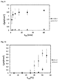
- the method of the invention is carried out at a plurality of temperatures, e.g., 5-7 temperatures with an interval of about 5 °C, as shown, e.g., in the figures.
- the signal can then be normalized, e.g., with regard to the lowest or highest signal detected; preferably, the highest signal. Accordingly, the form of the signal-temperature curves is characteristic for match or mismatch in the target nucleic acid.
- the signals at the selected temperatures are simultaneously determined on an array of individually heated electrodes, preferably, directly heated electrodes, as disclosed above, which makes detection of SNPs much quicker than the prior art methods, e.g., in Yang et al. 2014.
- the invention also relates to a method of diagnosing a disease associated with a SNP in a patient, or a method of assessing the risk of a patient for developing a disease, or for determining the risk of progression of a disease, wherein the disease is associated with one or more SNP, the method comprising detecting presence or absence of the one or more SNP in a patient by the method of the invention.
- the SNPs may be selected from the group comprising HLA-DPB1, PTPN22, CD58, INPP4b and HSChr16, preferably; HLA-DPB1, PTPN22, CD58, INPP4b and HSChr16.
- the signal detected will be an overlay of the two signals of the respective alleles.
- a distinct, two-peaked signal will be obtained in a temperature curve.
- Data analysis may allow for differentiation of heterozygous from homozygous patients.
- the present invention also provides a kit comprising:
- the electrodes may be heatable or heated, preferably, directly heatable or directly heated.
- the capture probe may be immobilized on the electrode or electrodes, or it may be suitable for immobilization.
- the redox marker preferably is [OsO 4 (bipy)].
- the protective strand and capture probe may be one or more, e.g., all of the capture probes provided in the examples below.
- the respective target nucleic acids (with match and mismatch) provided in the examples below can also be incorporated in the kit and used as comparative targets.
- the invention also provides use of a kit of the invention for detecting a SNP, or for diagnosing a disease, assessing the risk of a patient for developing a disease (prognosis of a disease), or for determining the risk of progression of a disease, or for determining the probable response to a therapy, wherein the disease is associated with one or more SNP.
- 2,2'-Bipyridine was delivered by Merck and osmium tetroxide was obtained as a 2% aqueous solution from Fluka.
- the Tris-buffer containing 10mM tris-(hydroxymethyl)-aminomethane and 0.5 M Na 2 SO 4 was adjusted to pH 7.5 using sulfuric acid. All DNA oligonucleotides were delivered by Friz Biochem (Neuried, Germany).
- the probe strand contained 15 thymine bases as a spacer to allow high hybridization efficiency on the electrode surface.
- the DNA-Targets were labeled with [OsO 4 (bipy)] by means of protective strands.
- 25 ⁇ l unlabelled DNA-Target solution and 25 ⁇ l protective strand solution were mixed together and left for 2 hours at room temperature to allow hybridization.
- 12.5 ⁇ l of 10 mM [OsO 4 (bipy)] were added to label the five unprotected 5'-terminal thymine bases and left again for 2 hours at room temperature. This way, the recognition site of the signaling strand remained unmodified.
- the gold disc electrode was polished using corundum 0.3 ⁇ m (Buehler) and thoroughly rinsed with deionised water. After this, the electrode was cleaned electrochemically by using cyclic voltammetry (25 scans, -0.3 to 1.7 V, scan rate 100 mV/s) in 0.5 M sulfuric acid. After rinsing the electrode with water, the electrode was dried under a stream of nitrogen. In the next step, a 4 ⁇ l or 10 ⁇ l drop of the probe solution (30 ⁇ M in Tris-buffer) was placed onto the top of the cleaned electrode surface in order to form the capture probe SAM. This volume was sufficient to ensure complete coverage.
- the electrode was immersed in 1 mM aqueous 6-mercapto-1-hexanol solution for one hour to obtain a well ordered surface with immobilized DNA probes in an upright position. After rinsing this ssDNA-SAM-modified-electrode with water, it was used for hybridization experiments with the target strands.
Landscapes
- Chemical & Material Sciences (AREA)
- Organic Chemistry (AREA)
- Life Sciences & Earth Sciences (AREA)
- Zoology (AREA)
- Wood Science & Technology (AREA)
- Proteomics, Peptides & Aminoacids (AREA)
- Health & Medical Sciences (AREA)
- Engineering & Computer Science (AREA)
- Microbiology (AREA)
- Immunology (AREA)
- Physics & Mathematics (AREA)
- Molecular Biology (AREA)
- Biotechnology (AREA)
- Biophysics (AREA)
- Analytical Chemistry (AREA)
- Biochemistry (AREA)
- Bioinformatics & Cheminformatics (AREA)
- General Engineering & Computer Science (AREA)
- General Health & Medical Sciences (AREA)
- Genetics & Genomics (AREA)
- Measuring Or Testing Involving Enzymes Or Micro-Organisms (AREA)
Priority Applications (1)
| Application Number | Priority Date | Filing Date | Title |
|---|---|---|---|
| EP14181411.1A EP2987868A1 (de) | 2014-08-19 | 2014-08-19 | Elektrochemischer Nachweis von Einzelnukleotidpolymorphismen |
Applications Claiming Priority (1)
| Application Number | Priority Date | Filing Date | Title |
|---|---|---|---|
| EP14181411.1A EP2987868A1 (de) | 2014-08-19 | 2014-08-19 | Elektrochemischer Nachweis von Einzelnukleotidpolymorphismen |
Publications (1)
| Publication Number | Publication Date |
|---|---|
| EP2987868A1 true EP2987868A1 (de) | 2016-02-24 |
Family
ID=51357835
Family Applications (1)
| Application Number | Title | Priority Date | Filing Date |
|---|---|---|---|
| EP14181411.1A Withdrawn EP2987868A1 (de) | 2014-08-19 | 2014-08-19 | Elektrochemischer Nachweis von Einzelnukleotidpolymorphismen |
Country Status (1)
| Country | Link |
|---|---|
| EP (1) | EP2987868A1 (de) |
Citations (3)
| Publication number | Priority date | Publication date | Assignee | Title |
|---|---|---|---|---|
| WO2007020093A2 (de) | 2005-08-19 | 2007-02-22 | Universität Rostock | Verfahren zur markierung und analyse von nukleinsäuren |
| DE102011109402A1 (de) | 2011-08-04 | 2013-02-07 | Universität Rostock | Elektrochemischer Sensor |
| WO2013045417A1 (de) | 2011-09-28 | 2013-04-04 | Universität Rostock | Sequenzspezifische analyse von nukleinsäuren |
-
2014
- 2014-08-19 EP EP14181411.1A patent/EP2987868A1/de not_active Withdrawn
Patent Citations (5)
| Publication number | Priority date | Publication date | Assignee | Title |
|---|---|---|---|---|
| WO2007020093A2 (de) | 2005-08-19 | 2007-02-22 | Universität Rostock | Verfahren zur markierung und analyse von nukleinsäuren |
| US8216446B1 (en) | 2005-08-19 | 2012-07-10 | Universität Rostock | Method for labelling and analysing nucleic acids |
| DE102011109402A1 (de) | 2011-08-04 | 2013-02-07 | Universität Rostock | Elektrochemischer Sensor |
| WO2013045417A1 (de) | 2011-09-28 | 2013-04-04 | Universität Rostock | Sequenzspezifische analyse von nukleinsäuren |
| US20140228247A1 (en) * | 2011-09-28 | 2014-08-14 | Universitat Rostock | Sequence-specific analysis of nucleic acids |
Non-Patent Citations (8)
| Title |
|---|
| ALLEN H. J. YANG ET AL: "Accurate Zygote-Specific Discrimination of Single-Nucleotide Polymorphisms Using Microfluidic Electrochemical DNA Melting Curves", ANGEWANDTE CHEMIE INTERNATIONAL EDITION, vol. 53, no. 12, 12 February 2014 (2014-02-12), pages 3163 - 3167, XP055153661, ISSN: 1433-7851, DOI: 10.1002/anie.201310059 * |
| DUWENSEE H ET AL: "Electrochemical product detection of an asymmetric convective polymerase chain reaction", BIOSENSORS AND BIOELECTRONICS, ELSEVIER BV, NL, vol. 25, no. 2, 15 October 2009 (2009-10-15), pages 400 - 405, XP026600441, ISSN: 0956-5663, [retrieved on 20090803], DOI: 10.1016/J.BIOS.2009.07.025 * |
| FERRON ET AL.: "Inositol Polyphosphate 4-Phosphatase B as a Regulator of Bone Mass in Mice and Humans", CELL METABOLISM, vol. 14, 2011, pages 466 - 477, XP028308482, DOI: doi:10.1016/j.cmet.2011.08.013 |
| FLECHSIG GERD-UWE ET AL: "Electrochemical detection of DNA hybridization by means of osmium tetroxide complexes and protective oligonucleotides", ANALYTICAL CHEMISTRY, AMERICAN CHEMICAL SOCIETY, US, vol. 79, no. 5, 1 March 2007 (2007-03-01), pages 2125 - 2130, XP002425409, ISSN: 0003-2700, DOI: 10.1021/AC062075C * |
| HEIKO DUWENSEE ET AL: "Electrochemical competitive hybridization assay for DNA detection using osmium tetroxide-labelled signalling strands", THE ANALYST, vol. 134, no. 5, 1 January 2009 (2009-01-01), pages 899, XP055037127, ISSN: 0003-2654, DOI: 10.1039/b819283a * |
| MARTIN JACOBSEN ET AL: "Hybridization Detection of Osmium Tetroxide Bipyridine-Labeled DNA and RNA on Heated Gold Wire Electrodes", ELECTROANALYSIS, vol. 25, no. 2, 23 February 2013 (2013-02-23), pages 373 - 379, XP055153967, ISSN: 1040-0397, DOI: 10.1002/elan.201200460 * |
| RESKE ET AL: "Electrochemical detection of osmium tetroxide-labeled PCR-products by means of protective strands", TALANTA, ELSEVIER, AMSTERDAM, NL, vol. 74, no. 3, 11 December 2007 (2007-12-11), pages 393 - 397, XP022384832, ISSN: 0039-9140, DOI: 10.1016/J.TALANTA.2007.09.004 * |
| YANG ET AL.: "Accurate Zygote-Specific Discrimination of Single-Nucleotide Polymorphisms Using Microfluidic Electrochemical DNA Melting Curves", ANGEW. CHEM. INT. ED., vol. 53, 2014, pages 3163 - 3167, XP055153661, DOI: doi:10.1002/anie.201310059 |
Similar Documents
| Publication | Publication Date | Title |
|---|---|---|
| KR102018122B1 (ko) | 한우육의 동일성 확인용 바이오 마커 및 이의 용도 | |
| EP1975248B1 (de) | Nukleotid-Primer-Set und Nukleotidsonde zur Detektion des Methylentetrahydrofolatreduktase (MTHFR)-Genotyps | |
| EP3447152A1 (de) | Präzise und massiv parallele quantifizierung von nukleinsäure | |
| EP2253713A2 (de) | Verfahren zur messung von kopiezahlen von chromosomen, genen oder speziellen nukleotidsequenzen mithilfe eines snp-arrays | |
| HK1217112A1 (zh) | 单链多核苷酸扩增方法 | |
| JP5662293B2 (ja) | 注意欠陥多動性障害の診断用snpとそれを含むマイクロアレイ及びキット | |
| KR20170033844A (ko) | 미백 피부 타입 유전자 다형성 마커 및 이의 용도 | |
| CN101522911A (zh) | 用于确定细胞色素p450 1a2、2a6和2d6、pxr以及udp-葡萄糖醛酸基转移酶1a基因的基因型的单倍型标签单核苷酸多态性及其用于多重基因分型的方法 | |
| US7919611B2 (en) | Nucleotide primer set and nucleotide probe for detecting genotype of N-acetyltransferase-2 (NAT2) | |
| CN102277437B (zh) | 用于确定UGT1A基因的基因型的htSNP及其用于多重基因分型的方法 | |
| EP2984183B1 (de) | Genetische tests mit nanosonden | |
| Ortiz et al. | Generic platform for the multiplexed targeted electrochemical detection of osteoporosis-associated single nucleotide polymorphisms using recombinase polymerase solid-phase primer elongation and ferrocene-modified nucleoside triphosphates | |
| WO2018172348A1 (en) | Easy one-step amplification and labeling (eosal) | |
| EP3348650B1 (de) | Kit und verfahren zum nachweis von einzelnukleotid-polymorphismus | |
| Chiba et al. | Electrochemical detection of insertion/deletion mutations based on enhanced flexibility of bulge-containing duplexes on electrodes | |
| JP3499795B2 (ja) | 遺伝子解析法 | |
| EP3455376B1 (de) | Verfahren zur herstellung einer vielzahl an dna-sonden und verfahren zur analyse von genomischer dna unter verwendung der dna-sonden | |
| US20100285970A1 (en) | Methods of sequencing nucleic acids | |
| KR20170049768A (ko) | 피부 색상 및 흑화 민감도 진단용 단일염기다형성 마커 및 이의 용도 | |
| KR100477766B1 (ko) | 염기변이 분석 프라이머 및 그 방법 | |
| KR101187317B1 (ko) | 산재성 위암 감수성 예측용 다형성 마커 및 이를 이용한 산재성 위암 감수성 예측 방법 | |
| RU2738752C1 (ru) | Способ идентификации личности и установления родства с помощью InDel полиморфизмов и набор синтетических олигонуклеотидов для их генотипирования | |
| JP4399025B1 (ja) | N−アセチルトランスフェラーゼ2(nat2)の遺伝子型を検出するための核酸プライマーセット、キット、及び該プライマーセットを用いた検出方法 | |
| KR20110041668A (ko) | 돼지의 단일뉴클레오타이드다형성 마커 및 이를 이용한 국내산 돈육의 원산지 판별방법 | |
| US20150031565A1 (en) | Determination of the identities of single nucleotide polymorphisms, point mutations and characteristic nucleotides in dna |
Legal Events
| Date | Code | Title | Description |
|---|---|---|---|
| PUAI | Public reference made under article 153(3) epc to a published international application that has entered the european phase |
Free format text: ORIGINAL CODE: 0009012 |
|
| AK | Designated contracting states |
Kind code of ref document: A1 Designated state(s): AL AT BE BG CH CY CZ DE DK EE ES FI FR GB GR HR HU IE IS IT LI LT LU LV MC MK MT NL NO PL PT RO RS SE SI SK SM TR |
|
| AX | Request for extension of the european patent |
Extension state: BA ME |
|
| STAA | Information on the status of an ep patent application or granted ep patent |
Free format text: STATUS: THE APPLICATION IS DEEMED TO BE WITHDRAWN |
|
| 18D | Application deemed to be withdrawn |
Effective date: 20160825 |