EP2995338A1 - Device for placing a catheter in a point of application of a drug with direct control via ultrasound - Google Patents
Device for placing a catheter in a point of application of a drug with direct control via ultrasound Download PDFInfo
- Publication number
- EP2995338A1 EP2995338A1 EP15382443.8A EP15382443A EP2995338A1 EP 2995338 A1 EP2995338 A1 EP 2995338A1 EP 15382443 A EP15382443 A EP 15382443A EP 2995338 A1 EP2995338 A1 EP 2995338A1
- Authority
- EP
- European Patent Office
- Prior art keywords
- catheter
- placing
- drug
- point
- application
- Prior art date
- Legal status (The legal status is an assumption and is not a legal conclusion. Google has not performed a legal analysis and makes no representation as to the accuracy of the status listed.)
- Withdrawn
Links
- 239000003814 drug Substances 0.000 title claims abstract description 36
- 229940079593 drug Drugs 0.000 title claims abstract description 30
- 238000002604 ultrasonography Methods 0.000 title claims abstract description 26
- 239000000463 material Substances 0.000 claims abstract description 13
- 238000003780 insertion Methods 0.000 claims description 4
- 230000037431 insertion Effects 0.000 claims description 4
- 239000007787 solid Substances 0.000 claims description 4
- 229920002635 polyurethane Polymers 0.000 claims description 2
- 239000004814 polyurethane Substances 0.000 claims description 2
- 230000001681 protective effect Effects 0.000 claims description 2
- 229910001220 stainless steel Inorganic materials 0.000 claims description 2
- 239000010935 stainless steel Substances 0.000 claims description 2
- 210000005036 nerve Anatomy 0.000 description 14
- 239000012634 fragment Substances 0.000 description 6
- 238000000034 method Methods 0.000 description 6
- 208000002193 Pain Diseases 0.000 description 4
- 238000006073 displacement reaction Methods 0.000 description 4
- 208000000094 Chronic Pain Diseases 0.000 description 2
- 101100536354 Drosophila melanogaster tant gene Proteins 0.000 description 2
- 230000001154 acute effect Effects 0.000 description 2
- 208000005298 acute pain Diseases 0.000 description 2
- 230000003444 anaesthetic effect Effects 0.000 description 2
- 238000013461 design Methods 0.000 description 2
- 208000015181 infectious disease Diseases 0.000 description 2
- 238000013508 migration Methods 0.000 description 2
- 230000005012 migration Effects 0.000 description 2
- 230000000007 visual effect Effects 0.000 description 2
- 208000031968 Cadaver Diseases 0.000 description 1
- 239000004429 Calibre Substances 0.000 description 1
- 238000013459 approach Methods 0.000 description 1
- 230000003416 augmentation Effects 0.000 description 1
- 239000004566 building material Substances 0.000 description 1
- 230000000694 effects Effects 0.000 description 1
- 230000003902 lesion Effects 0.000 description 1
- 239000003589 local anesthetic agent Substances 0.000 description 1
- 239000002184 metal Substances 0.000 description 1
- 230000000149 penetrating effect Effects 0.000 description 1
- 239000004033 plastic Substances 0.000 description 1
- 229920003023 plastic Polymers 0.000 description 1
- 108090000623 proteins and genes Proteins 0.000 description 1
- 230000000717 retained effect Effects 0.000 description 1
- 231100000241 scar Toxicity 0.000 description 1
Images


Classifications
-
- A—HUMAN NECESSITIES
- A61—MEDICAL OR VETERINARY SCIENCE; HYGIENE
- A61M—DEVICES FOR INTRODUCING MEDIA INTO, OR ONTO, THE BODY; DEVICES FOR TRANSDUCING BODY MEDIA OR FOR TAKING MEDIA FROM THE BODY; DEVICES FOR PRODUCING OR ENDING SLEEP OR STUPOR
- A61M25/00—Catheters; Hollow probes
- A61M25/01—Introducing, guiding, advancing, emplacing or holding catheters
- A61M25/06—Body-piercing guide needles or the like
-
- A—HUMAN NECESSITIES
- A61—MEDICAL OR VETERINARY SCIENCE; HYGIENE
- A61B—DIAGNOSIS; SURGERY; IDENTIFICATION
- A61B17/00—Surgical instruments, devices or methods
- A61B17/34—Trocars; Puncturing needles
- A61B17/3415—Trocars; Puncturing needles for introducing tubes or catheters, e.g. gastrostomy tubes, drain catheters
-
- A—HUMAN NECESSITIES
- A61—MEDICAL OR VETERINARY SCIENCE; HYGIENE
- A61M—DEVICES FOR INTRODUCING MEDIA INTO, OR ONTO, THE BODY; DEVICES FOR TRANSDUCING BODY MEDIA OR FOR TAKING MEDIA FROM THE BODY; DEVICES FOR PRODUCING OR ENDING SLEEP OR STUPOR
- A61M19/00—Local anaesthesia; Hypothermia
-
- A—HUMAN NECESSITIES
- A61—MEDICAL OR VETERINARY SCIENCE; HYGIENE
- A61M—DEVICES FOR INTRODUCING MEDIA INTO, OR ONTO, THE BODY; DEVICES FOR TRANSDUCING BODY MEDIA OR FOR TAKING MEDIA FROM THE BODY; DEVICES FOR PRODUCING OR ENDING SLEEP OR STUPOR
- A61M25/00—Catheters; Hollow probes
- A61M25/01—Introducing, guiding, advancing, emplacing or holding catheters
- A61M25/0194—Tunnelling catheters
-
- A—HUMAN NECESSITIES
- A61—MEDICAL OR VETERINARY SCIENCE; HYGIENE
- A61M—DEVICES FOR INTRODUCING MEDIA INTO, OR ONTO, THE BODY; DEVICES FOR TRANSDUCING BODY MEDIA OR FOR TAKING MEDIA FROM THE BODY; DEVICES FOR PRODUCING OR ENDING SLEEP OR STUPOR
- A61M25/00—Catheters; Hollow probes
- A61M25/01—Introducing, guiding, advancing, emplacing or holding catheters
- A61M25/06—Body-piercing guide needles or the like
- A61M25/065—Guide needles
-
- A—HUMAN NECESSITIES
- A61—MEDICAL OR VETERINARY SCIENCE; HYGIENE
- A61B—DIAGNOSIS; SURGERY; IDENTIFICATION
- A61B17/00—Surgical instruments, devices or methods
- A61B17/34—Trocars; Puncturing needles
- A61B17/3401—Puncturing needles for the peridural or subarachnoid space or the plexus, e.g. for anaesthesia
-
- A—HUMAN NECESSITIES
- A61—MEDICAL OR VETERINARY SCIENCE; HYGIENE
- A61M—DEVICES FOR INTRODUCING MEDIA INTO, OR ONTO, THE BODY; DEVICES FOR TRANSDUCING BODY MEDIA OR FOR TAKING MEDIA FROM THE BODY; DEVICES FOR PRODUCING OR ENDING SLEEP OR STUPOR
- A61M25/00—Catheters; Hollow probes
- A61M2025/0007—Epidural catheters
-
- A—HUMAN NECESSITIES
- A61—MEDICAL OR VETERINARY SCIENCE; HYGIENE
- A61M—DEVICES FOR INTRODUCING MEDIA INTO, OR ONTO, THE BODY; DEVICES FOR TRANSDUCING BODY MEDIA OR FOR TAKING MEDIA FROM THE BODY; DEVICES FOR PRODUCING OR ENDING SLEEP OR STUPOR
- A61M25/00—Catheters; Hollow probes
- A61M2025/0008—Catheters; Hollow probes having visible markings on its surface, i.e. visible to the naked eye, for any purpose, e.g. insertion depth markers, rotational markers or identification of type
-
- A—HUMAN NECESSITIES
- A61—MEDICAL OR VETERINARY SCIENCE; HYGIENE
- A61M—DEVICES FOR INTRODUCING MEDIA INTO, OR ONTO, THE BODY; DEVICES FOR TRANSDUCING BODY MEDIA OR FOR TAKING MEDIA FROM THE BODY; DEVICES FOR PRODUCING OR ENDING SLEEP OR STUPOR
- A61M25/00—Catheters; Hollow probes
- A61M25/01—Introducing, guiding, advancing, emplacing or holding catheters
- A61M25/06—Body-piercing guide needles or the like
- A61M25/0662—Guide tubes
- A61M2025/0687—Guide tubes having means for atraumatic insertion in the body or protection of the tip of the sheath during insertion, e.g. special designs of dilators, needles or sheaths
-
- A—HUMAN NECESSITIES
- A61—MEDICAL OR VETERINARY SCIENCE; HYGIENE
- A61M—DEVICES FOR INTRODUCING MEDIA INTO, OR ONTO, THE BODY; DEVICES FOR TRANSDUCING BODY MEDIA OR FOR TAKING MEDIA FROM THE BODY; DEVICES FOR PRODUCING OR ENDING SLEEP OR STUPOR
- A61M25/00—Catheters; Hollow probes
- A61M25/01—Introducing, guiding, advancing, emplacing or holding catheters
- A61M25/0105—Steering means as part of the catheter or advancing means; Markers for positioning
- A61M25/0108—Steering means as part of the catheter or advancing means; Markers for positioning using radio-opaque or ultrasound markers
Definitions
- Device for placing a catheter in a point of application of a drug with direct control by means of ultrasound.
- the present invention relates to a device for placing a catheter in a point of application of a medicament, such as a local anesthetic for the control of acute or chronic pain along a nerve, with direct control by means of an echo.
- a medicament such as a local anesthetic for the control of acute or chronic pain along a nerve
- the sanitary hollow needles can pass through its light guides and / or catheters of different materials (plastics, metal etc.), having different lengths, diameters of the inner lights, outer diameters, the geometry of the tip double triple openings, etc. located in the tip or elsewhere in its length, the building materials, and external markings to evaluate the insertion depth, but all these needles are straight and they can not, have a use as expected in the invention .
- catheters are of different material, more or less rigid, and a variable length; but all have in common that the openings, where the medicated result, are located in the tip or close to it, which also prevents its use as expected in the invention.
- the device comprises a curved hollow needle of material detectable by means of ultrasound, and a formal catheter of a size adapted to be introduced by said needle, the placement technique being carried out under an ultrasound view. Because the ultrasound is able to show the position of the needle, the nerve and the distribution among the tissues of the drug administered.
- this needle can externally incorporate graduated marking to allow direct visual control of the depth of the needle and nerve.
- the tip of the needle preferably has its blunt end, it being understood that blunt view and say that is not very sharp to damage the sensitive or functional tissues in case of accidental contact with the same, while remaining sharp enough to allow easy progression through tissues. The sharpness of the peak is determined in each case, because it is necessary to combine these two variables.
- the catheter consists of: a tubular part for the introduction of the drug, endowed with a few lateral openings to end the same, and completed by a closed end; a filiform extension of manipulation; and positional references with respect to the needle.
- the positional references of the catheter with respect to the needle will be used to determine the exact position of the lateral perforations near the nerve or the area to which the drug is to be supplied - with the help of the needle and the ultrasound.
- the ecography, as he made an appointment, will allow the realization of the technique under a direct vision, with a complete and effective realization.
- the filiform extension is solid or blind, and this difference from the configuration of the hollow tubular section, it can also be detectable by the reporter, which provides an additional positional reference visible in the art of perform the precise placement of the catheter.
- the curved needle makes it possible to penetrate through the skin and tissues and, following its curvature and passing next to the nerve, continue in the same direction until it exits to another point of the skin.
- the catheter is inserted through the needle inlet, which coats the inside until its tip appears through the tip of the needle.
- the point of this one, element detectable by the ecographer is located so that, with the references of the position of the catheter, it is then possible to determine the placement of the openings, just at the point which is the nerve, it is then pulled from the tip of the catheter previously seized, which makes the catheter is introduced into the tissues of the patient.
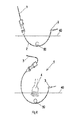
- the device (1) for positioning a catheter (3) at a point of application of a medicament according to the invention comprises a needle (2) hollow and curved of a material detectable by means of ecography (4), and a catheter (3) which is formally and dimensionally capable of being introduced into the interior of said needle (2), which needs, for optimum performance, the participation of the aforementioned ultrasound (4) which detects the position of the needle ( 2), as illustrated by figure 6 .
- the material of the needle will be mainly stainless steel.
- the needle (2) may externally incorporate a graduated marking (16) to allow direct visual control of the needle and nerve depth.
- this graduated marking (16) comprises a few reference lines (17) 1 centimeter away from each other. This graduation ideally covers the entire length of the needle (2).
- the catheter (3) (see figure 1 ) comprises a tubular section (5) for introducing the medicament, provided with lateral openings (6) and completed at a closed end (7), and a filiform extension (8) which allows handling during its deployment, and also some position references of the needle (2).
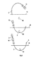
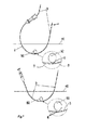
- the needle (2) (see Figures 2 to 4 ) comprises an anatomical manipulation handle (10) for its insertion, which is effected by penetrating into the tissues (40) of the patient, and approximates the desired area for the application of the drugs, for example a nerve (50) as one see it in the Figures 5 to 7 , returning to exit outside the tissues (40) of the patient by its curved shape.
- the catheter (3) is inserted through the needle, as seen in the bottom view of the figure 5 , for which the needle (2) ideally comprises a feed housing (12) of the catheter (3) at its inlet, the feed housing being in this example of the non-limiting invention (12), materialized in the handle (10) itself.
- the needle (2) may comprise a durable protective cover (20) in a socket (21) of the handle (10) (see FIG. Figures 3 and 4 ).
- the tip of the needle (2) has its blunt end (14), (see a detail of form figure 2 ) so that it is possible to advance through the tissues (40), but without damaging the nerve (50) in the event of accidental contact.
- the tubular portion (5) of the catheter (3) comprises three lateral openings (6), the first of which is adjacent to the closed end (7) of said tubular portion (5) and the others distant from each other consecutively from the previous in the main axial direction of the catheter (3); these three lateral openings (6) are located radically and in phase opposition of 120 degrees, and being spaced in the axial direction of the catheter, (3), preferably three millimeters.
- the filamentary extension (8) of the catheter is solid, and has a length of at least 14 centimeters, whereas the position references comprise visible marks (23, 24) which are made outside.
- the catheter (3) (see figure 1 ), ideally comprising a first visible mark (23) and differentiated from the rest of the other marks, located at a closed end distance (7) for the tubular portion (5) of the catheter (3) equivalent to the length of the needle (2) including the handle, and additional visible marks (24) inter-spaced from and between the first visible mark, being ideally the distance between the visible marks (23, 24) by one centimeter.
- the catheter (3) is preferably made of a material detectable by the scribe, such as polyurethane or any other material in the future would improve its ecographic guidance, always to maintain the design.
- the needle (2) and the catheter (3) can be manufactured in various sizes, given by way of non-limiting example, a needle assembly (2) of internal diameter of 17 gauge, 18 or 19 gauges respectively associated with a catheter (3) of diameter 18 gauge, 19 or 20 gauges, with lengths of the needle between 90 and 150 millimeters and the catheter between 400 and 500 millimeters.
Landscapes
- Health & Medical Sciences (AREA)
- Life Sciences & Earth Sciences (AREA)
- Veterinary Medicine (AREA)
- Engineering & Computer Science (AREA)
- Biomedical Technology (AREA)
- Heart & Thoracic Surgery (AREA)
- Animal Behavior & Ethology (AREA)
- General Health & Medical Sciences (AREA)
- Public Health (AREA)
- Anesthesiology (AREA)
- Hematology (AREA)
- Surgery (AREA)
- Biophysics (AREA)
- Pulmonology (AREA)
- Gastroenterology & Hepatology (AREA)
- Pathology (AREA)
- Nuclear Medicine, Radiotherapy & Molecular Imaging (AREA)
- Medical Informatics (AREA)
- Molecular Biology (AREA)
- Media Introduction/Drainage Providing Device (AREA)
Abstract
Un dispositif (1) pour placer un cathéter (3) dans un point d'application d'un médicament avec un contrôle direct au moyen de l'échographie qui comprend une aiguille (2) creuse courbe dans un matériau détectable par échographie (4), et un cathéter (3) aux formes et dimensions adéquates pour être introduit à l'intérieur de cette aiguille (2); où le cathéter (3) comprend une section tubulaire (5) pour l'introduction du médicament douée de quelques perforations latérales (6) et achevée par une extrémité fermée (7), et une prolongation filiforme (8) de manipulation, et des références de position par rapport à l'aiguille (2)A device (1) for placing a catheter (3) in a point of application of a drug with direct control by means of ultrasound which comprises a curved hollow needle (2) in a material detectable by ultrasound (4) and a catheter (3) of adequate shape and size to be inserted into the needle (2); wherein the catheter (3) comprises a tubular section (5) for introducing the medicament having a few lateral perforations (6) and completed by a closed end (7), and a filiform extension (8) of manipulation, and references position relative to the needle (2)
Description
Dispositif pour placer un cathéter dans un point d'application d'un médicament avec un contrôle direct au moyen de l'écographie.Device for placing a catheter in a point of application of a drug with direct control by means of ultrasound.
L' invention présente se rapporte à un dispositif pour placer un cathéter dans un point d'application d'un médicament, comme un anesthésique local pour le contrôle de la douleur aigüe ou chronique le long d'un nerf, avec un contrôle direct au moyen d'un ecôgraphe.The present invention relates to a device for placing a catheter in a point of application of a medicament, such as a local anesthetic for the control of acute or chronic pain along a nerve, with direct control by means of an echo.
Il existe un ample éventail de dispositifs (des aiguilles, des trocars) sur le marché depuis des années pour placer un cathéter le long d'un nerf, et administrer un anesthésique à travers du dit cathéter.There is a wide range of devices (needles, trocars) on the market for years to place a catheter along a nerve, and administer an anesthetic through said catheter.
Les aiguilles creuses sanitaires permettent de passer à travers de sa lumière des guides et/ou des cathéters de différents matériaux (plastiques, métalliques etc.), ayant différentes les longueurs, les diamètres des lumières intérieures, les diamètres extérieurs, les géométrie de la pointe, de doubles triples ouvertures, etc. situés dans la pointe ou ailleurs dans sa longueur, les matériaux de construction, et des marquages extérieurs pour évaluer la profondeur d'insertion, mais toutes ces aiguilles sont droites et elles ne peuvent, pas avoir une utilisation comme il est attendu dans l'invention.The sanitary hollow needles can pass through its light guides and / or catheters of different materials (plastics, metal etc.), having different lengths, diameters of the inner lights, outer diameters, the geometry of the tip double triple openings, etc. located in the tip or elsewhere in its length, the building materials, and external markings to evaluate the insertion depth, but all these needles are straight and they can not, have a use as expected in the invention .
Également on connaît aussi des aiguilles solides d'une multitude de tailles et de formes, droites ou avec des courbes, avec pointes d'infinité de formes, de diamètres différents, mais aucune ne permet le passage d'autres matiéraux pour son placement, ce qui empêche également son utilisation comme il est attendu dans l'invention.Also known are solid needles of a multitude of sizes and shapes, straight or with curves, with points of infinity of shapes, of different diameters, but none allows the passage of other materials for its placement, this which also prevents its use as expected in the invention.
À l'égard des cathéters, ils sont de différents matériau, plus ou moins rigides, et une longueur variable; mais tous ont en commun que les ouvertures, où abouti la médication, sont situées dans la pointe ou tout près d'elle, ce qui empêche aussi son utilisation comme il est attendu dans l'invention.With regard to the catheters, they are of different material, more or less rigid, and a variable length; but all have in common that the openings, where the medicated result, are located in the tip or close to it, which also prevents its use as expected in the invention.
Le placement des cathéters par le système traditionnel (jusqu'à présent utilisé) implique divers problèmes dérivés du manque de contrôle de la pointe du cathéter, parmi lesquels il faut énumérer :
- 1.- Le déplacement involontaire du cathéter qui est très fréquent (20 à 40 % des applications) et il augmente avec le passage des jours. Il se produit, ou bien au moment du retrait de l'aiguille introductive ou par la suite, avec la perte résultante d'effectivité, puisque la médication ne reste pas appliquée dans le lieu correct
- 2.-Impossible un replacement en cas de déplacement du cathéter, puisque nous n'avons pas de contrôle de la pointe après être introduit sous la peau ce qu'il oblige son retrait complet et à répéter toute la technique au complet depuis le début, autant de fois que nécessaire. Ce qui implique gène pour le patient et une augmentation du coût économique des matériaux et du temps employé.
- 3.-Si une rupture du cathéter survient (1 %), effet inhabituelle, quand elle se produit, le fragment qui reste retenu sous la peau, oblige à décider sur les deux options possibles : ou bien le laisser avec le risque que comporte laisser un corps étrange (infection, une migration en pouvant blesser des structures), ou bien chirurgicalement l'extraire que en fonction de leur emplacement implique un risque important de blesser des tissus pour le chercher, et même de, ne pas le trouver, donné son petit calibre et longueur du fragment. La décision à prendre parfois est complexe et coûteuse, tant dans des moyens matériels comme des séquelles physiques (une lésions internes, une cicatrice).
- 1.- The involuntary displacement of the catheter which is very frequent (20 to 40% of the applications) and increases with the passage of the days. It occurs, either at the time of removal of the introducer needle or thereafter, with the resultant loss of effectiveness, since the medication does not remain applied in the correct place
- 2.-Impossible a replacement in case of displacement of the catheter, since we have no control of the tip after being introduced under the skin what it forces its complete withdrawal and to repeat all the technique in full since the beginning, as many times as necessary. This involves gene for the patient and an increase in the economic cost of the materials and time employed.
- 3.-If a rupture of the catheter occurs (1%), unusual effect, when it occurs, the fragment that remains retained under the skin, forces to decide on the two possible options: or leave it with the risk that involves leaving a strange body (infection, migration that can injure structures), or surgically extract it as a function of their location involves a significant risk of injuring tissue to seek it, and even not to find it, given its small caliber and fragment length. The decision to be taken sometimes is complex and expensive, as well in material means as physical sequelae (an internal lesions, a scar).
Le dispositif de l'invention permet :
- 1.- Placer le cathéter avec précision, on faisant se rencontrer exactement les ouvertures du même dans le point d'application (typiquement a côté du nerf sélectionné), tout sous contrôle échographique. Cela produit l'application du médicament dans l'endroit parfait pour obtenir leurs résultats.
- 2.-En plus, permet la fixation des deux extrémités à la peau, en diminuant le risque de déplacement involontaire, tant au moment du placement comme a posteriori, tout en maintenant son effectivité pendant plus de temps, de façon a gérer avec présicion un médicament par moyen du dit cathéter.
- 3.-En plus, pendant son emplacement initial, ou par la suite, si s'est produit un déplacement de l' emplacement des ouvertures, ou si on a besoin de déplacer le cathéter pour atteindre une autre zone, ayant les deux côtés du cathéter contrôlés, permet de le déplacer dans les deux sens selon la nécessité et l 'emplacer à un nouveau emplacement, facilement, sans la nécessité de réaliser tout le procédé depuis le début.
- 4.-En plus, il est extrêmement avantageux qu'en cas d'une rupture du cathéter, les deux extrémités sont sur la peau, aucun fragment ne reste jamais sous la peau, c'est suffisant étirer simplement de ces deux extrémités.
- 1.- Place the catheter precisely, making the openings of the same exactly meet in the point of application (typically next to the selected nerve), all under ultrasound control. This produces the application of the drug in the perfect place to get their results.
- 2.-In addition, allows the attachment of both ends to the skin, reducing the risk of involuntary displacement, both at the time of placement as a posteriori, while maintaining its effectiveness for more time, so as to manage presicion a drug by means of said catheter.
- 3.-In addition, during its initial location, or afterwards, if there has been a displacement of the location of the openings, or if it is necessary to move the catheter to reach another area, having both sides of the Controlled catheter, allows to move it back and forth as needed and to place it at a new location, easily, without the need to perform the entire process from the beginning.
- 4.-In addition, it is extremely advantageous that in case of rupture of the catheter, the two ends are on the skin, no fragment never remains under the skin, it is sufficient to simply stretch these two ends.
Selon l'invention, le dispositif comprend une aiguille creuse courbe de matériau détectable au moyen de l' écographie, et un cathéter formel et d'une taille adaptée pour être introduit par la dite aiguille, la technique de placement étant réalisée sous une vision échographique, puisque l'écographie est capable d'affiicher la position de l'aiguille, du nerf et de la distribution parmi les tissus, du médicament administrée.According to the invention, the device comprises a curved hollow needle of material detectable by means of ultrasound, and a formal catheter of a size adapted to be introduced by said needle, the placement technique being carried out under an ultrasound view. because the ultrasound is able to show the position of the needle, the nerve and the distribution among the tissues of the drug administered.
Dans ce document comme aiguille creuse et courbe on entend n'importe quelle aiguille creuse avec différente trajectoire de la droite, de façon à ce que sa pointe s'approche à son entrée, en pouvant aussi être en forme de U, V et similaires. De plus, cette aiguille peut extérieurement incorporer un marquage gradué pour permettre un contrôle visuel direct sur la profondeur de l'aiguille et du nerf. En outre, la pointe de l'aiguille possède de préférence son extrémité émoussée, étant entendu que émoussée vuet dire qui n'est pas très aiguisé pour abîmer les tissus sensibles ou fonctionnelles en cas de contact accidentel avec les mêmes, tout en restant suffisamment pointu pour permettre une progression facile à travers des tissus. L'acuité de la pointe est déterminé dans chaque cas, car il faut combiner ces deux variables.In this document like hollow needle and curve one hears any hollow needle with different trajectory of the right, so that its tip approaches to its entrance, being able also to be in the form of U, V and similar. In addition, this needle can externally incorporate graduated marking to allow direct visual control of the depth of the needle and nerve. In addition, the tip of the needle preferably has its blunt end, it being understood that blunt view and say that is not very sharp to damage the sensitive or functional tissues in case of accidental contact with the same, while remaining sharp enough to allow easy progression through tissues. The sharpness of the peak is determined in each case, because it is necessary to combine these two variables.
Selon l'invention, le cathéter se compose : d'une partie tubulaire pour l'introduction du médicament, doué de quelques ouvertures latéraux pour aboutir du même, et achevé par une extrémité fermée; une prolongation filiforme de manipulation; et des références de position par rapport à l'aiguille. Les références de position du cathéter par rapport à l'aiguille seront utiler pour déterminer la position exacte des perforations latérales près du nerf ou la zone à laquelle on désire fournir le médicament - avec l'aide de l'aiguille et de l'écographie. L'écographie, comme il a pris rendez-vous, va permettre la réalisation de la technique sous une vision directe, avec une complète et effective réalisation.According to the invention, the catheter consists of: a tubular part for the introduction of the drug, endowed with a few lateral openings to end the same, and completed by a closed end; a filiform extension of manipulation; and positional references with respect to the needle. The positional references of the catheter with respect to the needle will be used to determine the exact position of the lateral perforations near the nerve or the area to which the drug is to be supplied - with the help of the needle and the ultrasound. The ecography, as he made an appointment, will allow the realization of the technique under a direct vision, with a complete and effective realization.
On préférence, le prolongement filiforme est solide ou aveugle, et cette différence de la par rapport a la configuration de la section tubulaire creux, elle peut également être détectable par l'écographe, qui fourni une référence de position supplémentaire visible dans l'art de réaliser le placement précis du cathéter.Preferably, the filiform extension is solid or blind, and this difference from the configuration of the hollow tubular section, it can also be detectable by the reporter, which provides an additional positional reference visible in the art of perform the precise placement of the catheter.
L'utilisation est la suivante: l'aiguille courbe fait possible pénétrer à travers la peau et des tissus et, suivant sa courbure et en passant à côté du nerf, de continuer dans la même direction jusqu'à ce qu'elle sorte à un autre point de la peau. Une fois que les deux extrémités sont en dehors de la peau, le cathéter est inséré a travers l'entrée de l'aiguille, qui lange l'intérieur jusqu'à ce que sa pointe apparaisse par le biais de la pointe de l'aiguille, moment ou est saisie cette pointe du cathéter et de commencer à extraire l'aiguille avec surveillance par l'écographie jusqu'à, ce que la pointe de celle-ci, élément détectable par l'ecographe, est situé de sorte que, avec les références de la position du cathéter, l'on puisse déterminer ensuite le placement des ouvertures, tout juste au point qui est le nerf, on tire alors de la pointe du cathéter au préalable saisie, ce qui rend que le cathéter s'introduit dans les tissus du patient. Avec les marques de position du cathéter - et donc de leurs ouvertures latéraux au regard de l'aiguille, et en sachant où la pointe de celle-ci est, déjà les dites ouvertures se trouvent près du nerf affecté; ensuite on fixe fermement la pointe de cathéter pour le reste pour qu'il ne soit pas déplacé, et finalement l'aiguille est retiré définitivement, le cathéter reste placé et l'on peut connecter la section tubulaire à la fourniture du médicament, généralement un anesthésique.The use is as follows: the curved needle makes it possible to penetrate through the skin and tissues and, following its curvature and passing next to the nerve, continue in the same direction until it exits to another point of the skin. Once both ends are out of the skin, the catheter is inserted through the needle inlet, which coats the inside until its tip appears through the tip of the needle. when this point of the catheter is seized and to start to extract the needle with surveillance by the ecography until, the point of this one, element detectable by the ecographer, is located so that, with the references of the position of the catheter, it is then possible to determine the placement of the openings, just at the point which is the nerve, it is then pulled from the tip of the catheter previously seized, which makes the catheter is introduced into the tissues of the patient. With marks of position of a catheter - and therefore of their lateral openings with regard to a needle, and knowing where the tip of this one is, already the said openings are located near the affected nerve; then the catheter tip is firmly fixed for the rest so that it is not displaced, and finally the needle is removed permanently, the catheter remains placed and the tubular section can be connected to the supply of the drug, usually a anesthetic.
Avec cette simple technique, réalisable grâce au dispositif de l'invention, on obtient une avancée majeure dans le traitement de la douleur aiguë et chronique puisque tous les inconvénient sont résolus et les complications qui surviennent lors la forme traditionnelle visée au début. Les avantages sont très importantes par rapport a la simplicité du design, des avantages des coûts et du confort pour le patient ainsi que le gain en temps et le coût des matériaux.With this simple technique, achievable thanks to the device of the invention, we obtain a major advance in the treatment of acute and chronic pain since all the disadvantages are solved and the complications that arise in the traditional form referred to at the beginning. The benefits are very important in terms of simplicity of design, cost advantages and patient comfort, as well as time and cost savings.
-
La
figure 1 .-Montre une vue du cathéter du dispositif de l'invention.Thefigure 1 Shows a view of the catheter of the device of the invention. -
La
figure 2 .-Montre une vue de détail en vue de face et de côté de la pointe de aiguille du dispositif de l'invention.Thefigure 2 Shows a detailed view in front and side view of the needle tip of the device of the invention. -
La
figure 3 .-Montre une vue en coupe de l'aiguille du dispositif de l'invention.Thefigure 3 Shows a sectional view of the needle of the device of the invention. -
La
figure 4 .-Montre une vue générale de l'aiguille du dispositif de l'invention.Thefigure 4 Shows a general view of the needle of the device of the invention. -
Les
figures 5 à 7 .- montrent une séquence d'utilisation du dispositif de l'invention, dans laquelle lafigure 5 montre trois vue respectivement de: démarrage de l'introduction de l'aiguille, l'achèvement de l'introduction de l'aiguille dans les tissus du patient, et le cathéter est inséré dans l'aiguille; où lafigure 6 montre deux vues respectivement de: maintenir la pointe du cathéter et le retrait de l'aiguille avec guidage écographique jusqu'à ce que sa pointe se trouve joint au nerf; et où lafigure 7 montre deux vues respectivement de : l'insertion du cathéter jusqu'à ce que les ouvertures latéraux de la section tubulaire se trouvent joint au nerf et de l'application du médicament, après le retrait de l'aiguille.TheFigures 5 to 7 show a sequence of use of the device of the invention, in which thefigure 5 shows three views respectively of: starting the introduction of the needle, completing the introduction of the needle into the patient's tissue, and the catheter is inserted into the needle; where thefigure 6 shows two views respectively of: holding the tip of the catheter and retracting the needle with ecographic guidance until its tip is attached to the nerve; and where thefigure 7 shows two views respectively of: the insertion of the catheter until the lateral openings of the tubular section are joined to the nerve and the application of the drug, after removal of the needle.
Le dispositif (1) pour positioner un cathéter (3) en un point d'application d'un médicament selon l'invention comprend une aiguille (2) creux et courbe d'un matériau détectable au moyen d'écographie (4), et un cathéter (3) formel et dimensionalmente apte à être introduit à l'inténieur de la dite aiguille (2), ayant besoin pour une performance optimale de la participation de l'écographie précitée (4) qui détecte la position de l'aiguille (2), comme illustre la
L'aiguille (2) peut incorporer extérieurement un marquage gradué (16) pour permettre le contrôle visuel direct sur la profondeur de aiguille et du nerf. Dans cet exemple de réalisation de l'invention ce marquage gradué (16) comprend quelques traits (17) de référence éloignés 1 centimètre entre eux. Cette graduation couvre idéalement toute la longueur de l'aiguille (2).The needle (2) may externally incorporate a graduated marking (16) to allow direct visual control of the needle and nerve depth. In this exemplary embodiment of the invention, this graduated marking (16) comprises a few reference lines (17) 1 centimeter away from each other. This graduation ideally covers the entire length of the needle (2).
Le cathéter (3) (voir
L'aiguille (2) (voir
La pointe de l'aiguille (2) a son extrémité émoussée (14), (voir un détail de forme
Pour sa part, la partie tubulaire (5) du cathéter (3) comprend trois ouvertures latérales (6), dont la première est adjacente à l'extrémité fermé (7) de la dite partie tubulaire (5) et les autres éloignées entre elles consécutivement du précédent dans le sens axiale principal du cathéter (3); se trouvent ces trois ouvertures laterales (6) disposées radicalement et en opposition de phase de 120 degrés, et étant l'espacemente dans la direction axiale du cathéter,(3), de préférence trois millimètres.For its part, the tubular portion (5) of the catheter (3) comprises three lateral openings (6), the first of which is adjacent to the closed end (7) of said tubular portion (5) and the others distant from each other consecutively from the previous in the main axial direction of the catheter (3); these three lateral openings (6) are located radically and in phase opposition of 120 degrees, and being spaced in the axial direction of the catheter, (3), preferably three millimeters.
Dans cet exemple de réalisation, la prolongation filiforme (8) du cathéter est solide, et a une longueur d'au moins 14 centimètres, alors que les références de position comprennent marques visibles (23, 24) qui se trouvent réalisées à l'extérieur du cathéter (3) (voir
Le cathéter (3) est de préférence constitué dun matériau détectable par l'écographe, tel que du polyuréthane ou tout autre matièraux au l'avenir parmettrait d'ameliorer son guidage ecographique, toujours permettant maintenir la conception.The catheter (3) is preferably made of a material detectable by the scribe, such as polyurethane or any other material in the future would improve its ecographic guidance, always to maintain the design.
À la fois, l'aiguille (2) et le cathéter (3) peuvent être fabriqués en différentes tailles, citées à titre d'exemple non limitatif, un ensemble d'aiguille (2) de diamètre intérieur de calibre 17, 18 ou 19 gauges respectivement associé avec un cathéter (3) de diamètre calibre 18, 19 ou 20 gauges, avec des longueurs de l'aiguille comprises entre 90 et 150 milimetres et du cathéter entre 400 et 500 millimètres.At the same time, the needle (2) and the catheter (3) can be manufactured in various sizes, given by way of non-limiting example, a needle assembly (2) of internal diameter of 17 gauge, 18 or 19 gauges respectively associated with a catheter (3) of diameter 18 gauge, 19 or 20 gauges, with lengths of the needle between 90 and 150 millimeters and the catheter between 400 and 500 millimeters.
Avoir décrit suffisamentt la nature de l'invention, indique que la description de celle-ci et de son mode de réalisation préféré ne devrait pas être interprétée comme limitant, et qui couvre toutes les variantes possibles de réalisation qui suit inmédiatement le contenu d'ici et ses revendications.Having sufficiently described the nature of the invention, indicates that the description thereof and of its preferred embodiment should not be interpreted as limiting, and which covers all the possible variants of realization which follows immediately the contents of here and his claims.
Claims (19)
Applications Claiming Priority (1)
| Application Number | Priority Date | Filing Date | Title |
|---|---|---|---|
| ES201431321A ES2528069B1 (en) | 2014-09-12 | 2014-09-12 | Device for placing a catheter at a point of application of a drug with direct control by ultrasound |
Publications (1)
| Publication Number | Publication Date |
|---|---|
| EP2995338A1 true EP2995338A1 (en) | 2016-03-16 |
Family
ID=52396870
Family Applications (1)
| Application Number | Title | Priority Date | Filing Date |
|---|---|---|---|
| EP15382443.8A Withdrawn EP2995338A1 (en) | 2014-09-12 | 2015-09-09 | Device for placing a catheter in a point of application of a drug with direct control via ultrasound |
Country Status (2)
| Country | Link |
|---|---|
| EP (1) | EP2995338A1 (en) |
| ES (1) | ES2528069B1 (en) |
Citations (4)
| Publication number | Priority date | Publication date | Assignee | Title |
|---|---|---|---|---|
| US5234438A (en) * | 1991-04-16 | 1993-08-10 | Neal Semrad | Process and device for creating new tunnels in tissue |
| US5735829A (en) * | 1996-03-22 | 1998-04-07 | Cherian; George | Intercostal anesthetic infusion catheter |
| EP1201263A2 (en) * | 2000-10-24 | 2002-05-02 | Vivek Thappa | Closed-end infusion catheter and an introducer |
| WO2014033812A1 (en) * | 2012-08-27 | 2014-03-06 | テルモ株式会社 | Medical tube, medical tube assembly and puncture needle |
Family Cites Families (5)
| Publication number | Priority date | Publication date | Assignee | Title |
|---|---|---|---|---|
| US20080058702A1 (en) * | 2005-12-12 | 2008-03-06 | Cook Critical Care Incorporated | Continuous nerve block assembly |
| US8235944B2 (en) * | 2009-02-17 | 2012-08-07 | Morteza Gharib | Medical device for placement of continuous regional anesthesia catheters |
| US20110112511A1 (en) * | 2009-11-09 | 2011-05-12 | Singer Jonathan E | Method and apparatus for administering anesthetics to peripheral nerve regions |
| US9265897B2 (en) * | 2011-01-26 | 2016-02-23 | Avent, Inc. | Method and corresponding kit for administering a paravertebral block |
| WO2014074237A1 (en) * | 2012-11-09 | 2014-05-15 | Solodex Llc | Continuous anesthesia nerve conduction apparatus, system and method thereof |
-
2014
- 2014-09-12 ES ES201431321A patent/ES2528069B1/en not_active Expired - Fee Related
-
2015
- 2015-09-09 EP EP15382443.8A patent/EP2995338A1/en not_active Withdrawn
Patent Citations (4)
| Publication number | Priority date | Publication date | Assignee | Title |
|---|---|---|---|---|
| US5234438A (en) * | 1991-04-16 | 1993-08-10 | Neal Semrad | Process and device for creating new tunnels in tissue |
| US5735829A (en) * | 1996-03-22 | 1998-04-07 | Cherian; George | Intercostal anesthetic infusion catheter |
| EP1201263A2 (en) * | 2000-10-24 | 2002-05-02 | Vivek Thappa | Closed-end infusion catheter and an introducer |
| WO2014033812A1 (en) * | 2012-08-27 | 2014-03-06 | テルモ株式会社 | Medical tube, medical tube assembly and puncture needle |
Also Published As
| Publication number | Publication date |
|---|---|
| ES2528069B1 (en) | 2015-11-11 |
| ES2528069A1 (en) | 2015-02-03 |
Similar Documents
| Publication | Publication Date | Title |
|---|---|---|
| US8801766B2 (en) | Devices for deploying intraocular shunts | |
| US8828070B2 (en) | Devices for deploying intraocular shunts | |
| AU2011275551B2 (en) | Device for deploying a flexible implant | |
| US8585629B2 (en) | Systems for deploying intraocular shunts | |
| US8663303B2 (en) | Methods for deploying an intraocular shunt from a deployment device and into an eye | |
| JP6552538B2 (en) | Intraocular shunt placement device | |
| US9095411B2 (en) | Devices for deploying intraocular shunts | |
| US9649223B2 (en) | Inserter for tubular medical implant devices | |
| US8308701B2 (en) | Methods for deploying intraocular shunts | |
| US20120123434A1 (en) | Devices for deploying intraocular shunts | |
| JP2016507333A5 (en) | Device for forming an anastomosis | |
| AU2009274128A1 (en) | Devices and methods for forming tracts in tissue | |
| KR20180082484A (en) | Needle and related assemblies and methods | |
| EP0653193A1 (en) | Subcutaneous locating device and related medical prosthesis | |
| CN106998992A (en) | Jacket for endoscope and endoscope-use injection locating apparatus | |
| FR2946865A1 (en) | DEVICE FOR TREATING A BLOOD CIRCULATION CONDUIT | |
| EP3488810A1 (en) | Device for anchoring and locating suspected lesions on multiple anchoring means | |
| IL269108B2 (en) | Device and method for creating a channel in soft tissue | |
| FR3012317A1 (en) | SURGICAL INSTRUMENT FOR THE INSTALLATION OF A UROGENITAL IMPLANT | |
| JP2019510583A5 (en) | ||
| KR102358916B1 (en) | Medical needle structure | |
| FR3010319A1 (en) | DEVICE FOR FIXING AND STABILIZING A CATHETER THROUGH A PATIENT'S DERMAT | |
| HK1189179B (en) | Intraocular shunt deployment devices | |
| FR2969478A1 (en) | Instrument for exposing carpal ligament during orthopedic surgery for treating carpal tunnel syndrome, has connecting fin provided with flanges for connecting flanges with one of support edges of blade by rim portion in heel |
Legal Events
| Date | Code | Title | Description |
|---|---|---|---|
| PUAI | Public reference made under article 153(3) epc to a published international application that has entered the european phase |
Free format text: ORIGINAL CODE: 0009012 |
|
| AK | Designated contracting states |
Kind code of ref document: A1 Designated state(s): AL AT BE BG CH CY CZ DE DK EE ES FI FR GB GR HR HU IE IS IT LI LT LU LV MC MK MT NL NO PL PT RO RS SE SI SK SM TR |
|
| AX | Request for extension of the european patent |
Extension state: BA ME |
|
| PUAJ | Public notification under rule 129 epc |
Free format text: ORIGINAL CODE: 0009425 |
|
| 32PN | Public notification |
Free format text: NOTIFICATION AU TITRE DES REGLES 59 ET 163(2) CBE (OEB FORM 1111 DATEE 13.04.2016) |
|
| STAA | Information on the status of an ep patent application or granted ep patent |
Free format text: STATUS: THE APPLICATION IS DEEMED TO BE WITHDRAWN |
|
| 18D | Application deemed to be withdrawn |
Effective date: 20160917 |