EP3013941B1 - Systèmes et procédés améliorés permettant de produire des cellules souches et des cellules différenciées - Google Patents
Systèmes et procédés améliorés permettant de produire des cellules souches et des cellules différenciées Download PDFInfo
- Publication number
- EP3013941B1 EP3013941B1 EP14817538.3A EP14817538A EP3013941B1 EP 3013941 B1 EP3013941 B1 EP 3013941B1 EP 14817538 A EP14817538 A EP 14817538A EP 3013941 B1 EP3013941 B1 EP 3013941B1
- Authority
- EP
- European Patent Office
- Prior art keywords
- cells
- cell
- samples
- automated
- ipscs
- Prior art date
- Legal status (The legal status is an assumption and is not a legal conclusion. Google has not performed a legal analysis and makes no representation as to the accuracy of the status listed.)
- Active
Links
Images





























Classifications
-
- C—CHEMISTRY; METALLURGY
- C12—BIOCHEMISTRY; BEER; SPIRITS; WINE; VINEGAR; MICROBIOLOGY; ENZYMOLOGY; MUTATION OR GENETIC ENGINEERING
- C12N—MICROORGANISMS OR ENZYMES; COMPOSITIONS THEREOF; PROPAGATING, PRESERVING, OR MAINTAINING MICROORGANISMS; MUTATION OR GENETIC ENGINEERING; CULTURE MEDIA
- C12N5/00—Undifferentiated human, animal or plant cells, e.g. cell lines; Tissues; Cultivation or maintenance thereof; Culture media therefor
- C12N5/06—Animal cells or tissues; Human cells or tissues
- C12N5/0602—Vertebrate cells
- C12N5/0696—Artificially induced pluripotent stem cells, e.g. iPS
-
- G—PHYSICS
- G01—MEASURING; TESTING
- G01N—INVESTIGATING OR ANALYSING MATERIALS BY DETERMINING THEIR CHEMICAL OR PHYSICAL PROPERTIES
- G01N33/00—Investigating or analysing materials by specific methods not covered by groups G01N1/00 - G01N31/00
- G01N33/48—Biological material, e.g. blood, urine; Haemocytometers
- G01N33/50—Chemical analysis of biological material, e.g. blood, urine; Testing involving biospecific ligand binding methods; Immunological testing
- G01N33/5005—Chemical analysis of biological material, e.g. blood, urine; Testing involving biospecific ligand binding methods; Immunological testing involving human or animal cells
-
- C—CHEMISTRY; METALLURGY
- C12—BIOCHEMISTRY; BEER; SPIRITS; WINE; VINEGAR; MICROBIOLOGY; ENZYMOLOGY; MUTATION OR GENETIC ENGINEERING
- C12N—MICROORGANISMS OR ENZYMES; COMPOSITIONS THEREOF; PROPAGATING, PRESERVING, OR MAINTAINING MICROORGANISMS; MUTATION OR GENETIC ENGINEERING; CULTURE MEDIA
- C12N2503/00—Use of cells in diagnostics
Definitions
- Embryonic stem (ES) cells can be derived from the blastocyst of an early stage embryo and have the potential to develop into endoderm, ectoderm, and mesoderm (the three germ layers) (i.e., they are "pluripotent"). In vitro, ES cells tend to spontaneously differentiate into various types of tissues, and the control of their direction of differentiation can be challenging. There are unresolved ethical concerns that are associated with the destruction of embryos in order to harvest human ES cells. These problems limit their availability for research and therapeutic applications.
- AS cells are found among differentiated tissues. Stem cells obtained from adult tissues typically have the potential to form a more limited spectrum of cells (i.e., "multipotent"), and typically only differentiate into the cell types of the tissues in which they are found, though recent reports have shown some plasticity in certain types of AS cells. They also generally have a limited proliferation potential.
- iPSC or iPSCs Induced pluripotent stem cells
- iPSCs are produced by laboratory methods from differentiated adult cells.
- iPSCs are widely recognized as important tools, e.g., for conducting medical research.
- the technology for producing iPSCs has been time-consuming and labor-intensive.
- Differentiated adult cells, e.g., fibroblasts are reprogrammed, cultured, and allowed to form individual colonies which represent unique clones.
- identifying these types of cells has been difficult because the majority of the cells are not fully-reprogrammed iPSC clones.
- iPSC clones are selected based on the morphology of the cells, with desirable colonies possessing sharply demarcated borders containing cells with a high nuclear-to-cytoplasmic ratio.
- clones are manually-picked by micro-thin glass tools and cultured on "feeder" layers of cells typically, murine embryonic fibroblasts (MEFs). This step is performed typically at 14 - 21 days post-infection with a reprograming vector. Then the clones are expanded for another 14 - 21 days or more, prior to undergoing molecular characterization.
- MEFs murine embryonic fibroblasts
- stem cells are an attractive source of cells for therapeutic applications, medical research, pharmaceutical testing, and the like.
- an automated system for rapidly producing and isolating reproducible iPSC cell lines under standard conditions in order to meet these and other needs There also there remains in the art a need for methods of making panels of iPSC cell lines, and differentiated cells produced therefrom, that are derived from multiple different individuals in a "population of interest.”
- the present invention provides various improved systems and methods as defined in the claims for obtaining, generating, culturing, and handling cells, such as stem cells (including induced pluripotent stem cells or iPSCs) and differentiated cells. Aspects of the invention for which protection is sought are as defined the appended claims.
- the present invention builds upon, and provides certain improvements over, the systems and methods described previously in WO/2013/082509 and U.S. 2013-0345094 . Building on the work described in these prior patent applications, the Applicants have now developed certain improved methods and systems that can significantly reduce variability in obtaining, generating, culturing, and handling a variety of cell types, including differentiated cells and stem cells. Such improved systems and methods are particularly advantageous for automated and/or high-throughput applications - greatly facilitating the ability to work with large numbers of cells in a parallel manner.
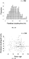
- the Applicants have discovered that variability in the generation and culture and differentiated cells and stem cells can be reduced, allowing for more efficient parallel processing, if individual samples are selected and grouped according to various characteristics (such as growth characteristics or donor age), such that cells having similar characteristics are grown and handled together.
- various characteristics such as growth characteristics or donor age
- such methods may be referred to as “binning” methods, or “batching” methods, or “binning and batching” methods, or “data-driven batching” methods.
- Applicants also discovered that the efficiency of cellular reprograming could be improved, and the variability in reprogramming efficiency reduced, when cells were grown in low serum or serum free medium for a certain amount of time prior to, and optionally also during, reprogramming.
- Applicants also found that differences in methods used to generate embryoid bodies (EBs) from pluripotent stem cells had significant and surprising effects on differentiation. For example, Applicants found that "hanging drop" methods for EB generation led to a bias towards endoderm differentiation, while the generation of EBs in V-bottom plates led to a more uniform differentiation potential, as well we being better suited to automated and high-throughput systems.
- EBs embryoid bodies
- the present invention provides a method for the automated generation and/or manipulation of stem cells, the method comprising: (a) culturing multiple different samples of cells, wherein the cells comprise adult somatic cells or induced pluripotent stem cells, (b) determining the proliferation rate or cell doubling time of individual cell samples from among the multiple different samples of cells, (c) freezing individual cell samples from among the multiple different samples of cells, (d) selecting from the individual cell samples a subset of samples having similar proliferation rates or cell doubling times, (e) thawing the subset of samples selected in step (d), (f) plating the subset of samples in a multi-well plate, (g) culturing the subset of samples until they reach a desired confluency, and (h) where the cells comprise adult somatic cells, contacting the somatic cells with one or more reprogramming factors in order to produce iPSCs, or, where the cells comprise iPSCs, treating the iPSCs in order to produce differentiated cells.
- the cells may be derived from human donors.
- the adult somatic cells may be fibroblasts.
- Each of the steps may be automated.
- the individual cell samples in the subset of samples may have proliferation rates or cell doubling times that are within 10% of one another.
- the culturing of step (g) may be performed in low serum medium.
- the step of selecting a subset of samples may further comprise selecting samples based the age, sex, race, ethnicity, diagnosis, genotype, phenotype, blood type, HLA type, treatment history, or drug response profile of the cell sample or of the donor from which the cell sample was obtained.
- the step of treating the iPSCs in order to produce differentiated cells may comprise contacting the iPSCs with one or more differentiation factors.
- the step of treating the iPSCs in order to produce differentiated cells may comprise generating embryoid bodies (EBs), for example wherein the step of generating embryoid bodies (EBs) comprises plating iPSCs in V-bottom plates.
- Also described herein is a method for the efficient generation, culture and/or handling of multiple cell samples in parallel, the method comprising: (a) culturing multiple different cell samples, (b) determining, or obtaining information regarding, one or more characteristics or properties of individual samples from among the multiple different cell samples (or of the donor/subject from which such samples were obtained), (c) selecting from the multiple different cell samples a subset of cell samples having a desired characteristic or property, (d) plating the subset of cell samples in a multi-well plate, and (e) culturing the subset of cell samples in the multi-well plate.
- Also described herein is a method for the efficient culture of multiple cell samples in parallel, the method comprising: (a) culturing multiple different cell samples, (b) determining, or obtaining information regarding, one or more characteristics or properties of individual samples from among the multiple different cell samples (or of the donor/subject from which such samples were obtained), (c) freezing individual cell samples from among the multiple different cell samples, (d) selecting from the multiple different cell samples a subset of cell samples having a desired characteristic or property, (e) thawing the subset of cell samples selected in step (d) plating the subset of cell samples in a multi-well plate, and (f) culturing the subset of cell samples in the multi-well plate.
- the characteristic or property is the cellular proliferation rate or cell doubling time of a cell sample.
- the characteristic or property relates to the age, sex, race, ethnicity, diagnosis (e.g. for a disease or a disorder), genotype, phenotype, blood type, HLA type, treatment history, or drug response profile of the cell sample or of the donor/subject from which the cell sample was obtained.
- cell samples are selected on the basis of at least two characteristics, including (i) the cellular proliferation rate or cell doubling time of the cell sample, and (ii) the age, sex, race, ethnicity, diagnosis (e.g.
- the cell samples are adult somatic cells, such as adult somatic fibroblasts.
- the cell samples are induced pluripotent stem cell (iPSCs).
- iPSCs induced pluripotent stem cell
- the cell samples are differentiated cells derived from pluripotent stem cells.
- one or more of the steps is automated.
- each of the steps is automated.
- the step of selecting from the multiple different cell samples a subset of the cell samples is automated and/or performed by a computer.
- the selected samples have proliferation rates or cell doubling times that vary by less than 30% between samples, or by less than 25% between samples, or by less than 20% between samples, or by less than 15% between samples, or by less than 10% between samples, or by less than 5% between samples, or by less than 2% between samples.
- Also described herein is a method for the efficient generation of induced pluripotent stem cells from differentiated adult somatic cells, the method comprising: (a) culturing multiple different samples of differentiated adult somatic cells, (b) determining the proliferation rate or cell doubling time of individual samples from among the multiple different samples of differentiated adult somatic cells, (c) selecting from the multiple different samples of differentiated adult somatic cells a subset of samples having similar proliferation rates or cell doubling times, (d) plating the subset of the samples selected in a multi-well plate, such that each of the samples in the multi-well plate has a similar proliferation rate or cell doubling time, (e) culturing the cell samples in the multi-well plate until they reach a desired confluency, and (f) contacting the cell samples with one or more reprogramming factors in order to produce iPSCs.
- a method for the automated generation of induced pluripotent stem cells from differentiated adult somatic cells comprising: (a) culturing multiple different samples of differentiated adult somatic cells, (b) determining the proliferation rate or cell doubling time of individual samples from among the multiple different samples of differentiated adult somatic cells, (c) freezing individual cell samples from among the multiple different cell samples, (d) selecting from the multiple different samples of differentiated adult somatic cells a subset of the samples having similar proliferation rates or cell doubling times, (e) thawing and plating the subset of the samples selected in step (d) into a multi-well plate, such that each of the samples in the multi-well plate has a similar proliferation rate or cell doubling time, (f) culturing the cell samples in the multi-well plate until they reach a desired confluency, and (g) contacting the cell samples with one or more reprogramming factors in order to produce iPSCs.
- the differentiated adult somatic cells are fibroblasts.
- the fibroblasts are derived from human donors/subjects.
- one or more of the steps is automated.
- each of the steps is automated.
- the step of selecting from the multiple different cell samples a subset of the cell samples having similar proliferation rates or cell doubling times is performed by a computer.
- the selected samples have proliferation rates or cell doubling times that vary by less than 30% between samples, or by less than 25% between samples, or by less than 20% between samples, or by less than 15% between samples, or by less than 10% between samples, or by less than 5% between samples, or by less than 2% between samples.
- the culturing of the somatic cells prior to contacting with reprogramming factors is performed in low serum medium or in serum free medium.
- the step of selecting a subset of the cell samples further comprises selecting cell samples on the basis of the age, sex, race, ethnicity, diagnosis (e.g.
- the method further comprises producing differentiated cells from the pluripotent stem cells, for example by contacting the pluripotent stem cells with one or more differentiation factors or by generating embryoid bodies (EBs).
- EBs embryoid bodies
- the EBs are generated in V-bottom plates.
- Also described herein is a method for the efficient generation of human induced pluripotent stem cells (iPSCs) from human donor fibroblasts, the method comprising: (a) culturing multiple different samples of human donor fibroblasts, (b) determining the proliferation rate or cell doubling time of individual samples from among the multiple different samples of human donor fibroblasts, (c) freezing individual cell samples from among the multiple different samples of human donor fibroblasts, (d) selecting from the multiple different samples of human donor fibroblasts a subset of the samples having similar proliferation rates or cell doubling times, (e) thawing the subset of samples selected in step (d), (f) culturing the subset of cell samples in a multi-well plate, such that each of the samples in the multi-well plate has a similar proliferation rate or cell doubling time, wherein the culturing comprises contacting the cell samples with low serum medium for at least 3 days, and (g) subsequently contacting the cell samples with one or more reprogramming factors in order to produce
- one or more of the steps is automated.
- each of the steps is automated.
- the step of selecting from the multiple different cell samples a subset of the cell samples having similar proliferation rates or cell doubling times is automated and/or performed by a computer.
- the selected samples have proliferation rates or cell doubling times that vary by less than 30% between samples, or by less than 25% between samples, or by less than 20% between samples, or by less than 15% between samples, or by less than 10% between samples, or by less than 5% between samples, or by less than 2% between samples.
- the step of selecting a subset of the cell samples further comprises selecting cell samples on the basis of the age, sex, race, ethnicity, diagnosis (e.g. for a disease or a disorder), genotype, phenotype, blood type, HLA type, treatment history, or drug response profile of the cell sample or the donor/subject from which the cell sample was obtained.
- Also described herein is a method for the automated generation of differentiated cells from pluripotent stem cells, the method comprising: (a) culturing multiple different samples of pluripotent stem cells, (b) determining the proliferation rate or cell doubling time for individual samples from among the multiple different samples of pluripotent stem cells, (c) selecting from the multiple different samples of pluripotent stem cells a subset of samples having similar proliferation rates or cell doubling times, (d) plating the subset of the samples selected in step (c) into a multi-well plate, such that each of the samples in the multi-well plate has a similar proliferation rate or cell doubling time, (e) culturing the cell samples in the multi-well plate until they reach a desired passage number and/or confluency, and (f) producing differentiated cells from the pluripotent stem cells.
- a method for the automated generation of differentiated cells from pluripotent stem cells comprising: (a) culturing multiple different samples of pluripotent stem cells, (b) determining the proliferation rate or cell doubling time for each of the multiple different samples of pluripotent stem cells, (c) freezing individual cell samples from among the multiple different cell samples, (d) selecting from the multiple different samples of pluripotent stem cells a subset of the samples having similar proliferation rates or cell doubling times, (e) thawing and plating the subset of the samples selected in step (x) into a multi-well plate, such that each of the samples in the multi-well plate has a similar proliferation rate or cell doubling time, (f) culturing the cell samples in the multi-well plate until they reach a desired passage number and/or confluency, and (g) producing differentiated cells from the pluripotent stem cells.
- the pluripotent stem cells are induced the pluripotent stem cells (iPSCs).
- one or more of the steps is automated.
- the each of the steps is automated.
- the step of selecting from the multiple different cell samples a subset of the cell samples having similar proliferation rates or cell doubling times is automated and/or performed by a computer.
- the selected samples have proliferation rates or cell doubling times that vary by less than 30% between samples, or by less than 25% between samples, or by less than 20% between samples, or by less than 15% between samples, or by less than 10% between samples, or by less than 5% between samples, or by less than 2% between samples.
- the step of producing differentiated cells from the iPSCs comprises contacting the iPSCs with one or more differentiation factor.
- the step of producing differentiated cells from the iPSCs comprises generating embryoid bodies (EBs).
- the step of producing differentiated cells from the iPSCs comprises generating embryoid bodies (EBs) in V-bottom plates.
- the step of selecting a subset of the cell samples further comprises selecting cell samples on the basis of the age, sex, race, ethnicity, diagnosis (e.g. for a disease or a disorder), genotype, phenotype, blood type, HLA type, treatment history, or drug response profile of the cell sample or the donor/subject from which the cell sample was obtained.
- automated "data-driven batching systems” that can be used to select and group (or “bin and/or batch”) cell samples based on one or more properties or characteristics, as described above.
- Such automated data-driven batching systems systems can be used, for example, to select and group somatic cells (such as fibroblasts) for analysis or for subsequent iPSC generation, to select and group stem cells (such as iPSCs) for analysis or for subsequent differentiation, and/or to select and group cells (such as somatic cells obtained from donors, iPSCs, or differentiated cells derived from iPSCs) for inclusion in a cell panel.
- the components of the data-driven batching systems described here can comprise, or can be modified from, or can be used in conjunction with, the other automated systems, and components thereof, described herein and/or those described in WO 2013/082509 and U.S 2013-345094 . Further, one of skill in the art will appreciate where and how the automated systems described herein (and in WO 2013/082509 and U.S. 2013-345094 ) can be modified to provide or include such data-driven batching systems.
- an automated data-driven batching system that comprises: (a) a component/system for determining the cell proliferation rate or cell doubling time of a cell sample, (b) a component/system for selecting and/or retrieving cell samples having a desired cell proliferation rate or cell doubling time, and (c) a component/system for plating the selected samples in a multi-well plate.
- the present invention provides an automated system for growing cells for parallel processing, the system comprising: (a) a component for determining the cell proliferation rates or cell doubling times of multiple cell samples, (b) a component for cryopreserving the cell samples, (c) a component for selecting cell samples from among the multiple cell samples having a similar proliferation rates or cell doubling times, (d) a component for thawing the selected cell samples, and (e) a component for plating the selected samples in a multi-well plate.
- the component for determining the cell proliferation rates or cell doubling times may comprise a confluency checking unit.
- the automated system may further comprise an induction unit which contacts the cells in the multi-well plate with one or more reprogramming factors, for example, wherein the induction unit contacts the cells in the multi-well plate with one or more reprogramming factors after the cells have been cultured in low serum medium for 5-7 days.
- the component/system of the data-driven batching system used to determine the cell proliferation rate or cell doubling time of a cell sample may comprise an automated cell imager (such as a Celigo imager) or other automated device that can be used to measure cell confluency or cell numbers, or some other parameter from which cell proliferation rate or cell doubling time can be calculated (such as a confluency checking unit).
- the component/system for cryopreserving the cell sample may comprise a system for robotically transferring cell samples into cryopreservation tubes (such as barcoded cryopreservation tubes) and may comprise a 80°C freezer.
- the component/system for selecting cell samples having a desired cell proliferation rate or cell doubling time may comprise a computer system programmed with desired selection criteria, and/or may comprise an automated sample access system, such as a -80°C Sample Access Manager or "SAM" (Hamilton Storage Technologies).
- an automated sample access system may comprise an inventory database that allows for flexible recall and downstream process batching of cell samples, for example based on one or more factors such as cell proliferation rate, cell doubling time, or any other desired property or characteristic.
- an automated system for generating and isolating iPSCs comprising: (a) a somatic cell plating unit for placing somatic cells on a plate; (b) a data-driven batching system used to select somatic cell samples for reprogramming, and (c) an induction unit for automated reprogramming of the somatic cells by contacting the somatic cells on the somatic cell plating unit with reprogramming factors to produce iPSCs.
- the system further comprises a sorting unit for selectively sorting and isolating the iPSCs produced by the induction unit, e.g., by identifying iPSC specific markers, including, e.g., surface markers on the cells.
- the somatic cells are fibroblasts.
- an automated system for generating and isolating differentiated adult cells from stem cells e.g., iPSCs, embryonic stem (ES) cells or mesenchymal stem (MS) cells
- stem cells e.g., iPSCs, embryonic stem (ES) cells or mesenchymal stem (MS) cells
- a stem cell plating unit for placing stem cells on a plate
- ES embryonic stem
- MS mesenchymal stem
- an induction unit for automated differentiation of stem cells for example by contacting the cells on the with one or more differentiation factors to produce differentiated cells.
- the system further comprises a sorting unit for selectively sorting and isolating the differentiated cells produced by the induction unit.
- iPSCs induced pluripotent stem cells
- the method comprising: (a) obtaining adult somatic cells, (b) culturing the adult somatic cells in low serum medium days, and (c) subsequently contacting the adult somatic cells with one or more reprogramming factors, in order to produce iPSCs.
- the population of adult somatic cells obtained in step (a) had previously been frozen.
- the population of adult somatic cells had been thawed in low serum medium.
- the adult somatic cells are fibroblasts.
- the adult somatic cells are cultured in low serum medium for more than 1 day prior to contacting the adult somatic cells with one or more reprogramming factors. In some such methods described herein the adult somatic cells are cultured in low serum medium for more than 2 days prior to contacting the adult somatic cells with one or more reprogramming factors. In some such methods described herein the adult somatic cells are cultured in low serum medium for more than 3 days prior to contacting the adult somatic cells with one or more reprogramming factors. In some such methods described herein the adult somatic cells are cultured in low serum medium for more than 4 days prior to contacting the adult somatic cells with one or more reprogramming factors.
- the adult somatic cells are cultured in low serum medium for more than 5 days prior to contacting the adult somatic cells with one or more reprogramming factors. In some such methods described herein the adult somatic cells are cultured in low serum medium for more than 6 days prior to contacting the adult somatic cells with one or more reprogramming factors. In some such methods described herein the adult somatic cells are cultured in low serum medium for more than 7 days prior to contacting the adult somatic cells with one or more reprogramming factors. In some such methods described herein the adult somatic cells are cultured in low serum medium for 5-7 days prior to contacting the adult somatic cells with one or more reprogramming factors.
- the adult somatic cells are cultured in low serum medium for 4-8 days prior to contacting the adult somatic cells with one or more reprogramming factors. In some such methods described herein the adult somatic cells are cultured in low serum medium for 3-9 days prior to contacting the adult somatic cells with one or more reprogramming factors. In some such methods described herein the adult somatic cells are cultured in low serum medium for 2-10 days prior to contacting the adult somatic cells with one or more reprogramming factors. In some such methods described herein the step of contacting the adult somatic cells with one or more reprogramming factors is performed while the adult somatic cells are in low serum medium. In some such methods described herein the low serum medium comprises less than 5% serum.
- the low serum medium comprises less than 4% serum. In some such methods described herein the low serum medium comprises less than 3% serum. In some such methods described herein the low serum medium comprises less than 2% serum. In some such methods described herein the low serum medium comprises less than 1% serum. In some such methods described herein the low serum medium is serum free. In some such methods described herein the low serum medium comprises a serum replacement. Several different serum replacements are known in the art and any such serum replacement can be used.
- cells such as somatic cells (e.g. donor-derived fibroblasts), pluripotent stem cells (such as iPSCs), differentiated cells produced from pluripotent stem cells (such as hematopoetic cells, muscle cells, cardiac muscle cells, liver cells, cartilage cells, epithelial cells, urinary tract cells, and neuronal cells), and transdifferentiated cells, such as those produced using the methods and systems described herein.
- pluripotent stem cells such as iPSCs
- differentiated cells produced from pluripotent stem cells such as hematopoetic cells, muscle cells, cardiac muscle cells, liver cells, cartilage cells, epithelial cells, urinary tract cells, and neuronal cells
- transdifferentiated cells such as those produced using the methods and systems described herein.
- arrays or “panels” or “banks” comprising such cells.
- Such cell arrays, panels or banks may comprise cells derived from multiple different individuals, for example multiple different individuals in a "population of interest.”
- the population of interest can be any population desired, including, but not limited to, the world population, the population of a particular country, the population of a particular continent, the population of a particular geographic region, the population of a particular racial or ethnic group, a population of a particular age, a population of a particular sex (male or female), a population having a particular disease or disorder, a population having a particular mutation, a population having a particular genotype, a population having a particular phenotype, a population having a particular blood type, a population having a particular HLA type, a population having a particular drug response profile, and the like.
- the individuals from whom cells are derived are selected in order to be representative of the variation in the particular population of interest. For example, if the population of interest is the U.S. population, the individuals from whom cells are derived are preferably selected to be representative of the U.S. population (e.g. in terms of race/ethnicity and/or any other desired characteristic), for example based on census data or some other suitable criteria.
- the cell panels comprise isogenic control cells. For example, where the cell panels contain cells having a certain mutation, the panels may comprise control cells in which that mutation is not present (for example if it has been corrected) but where the cells are otherwise genetically identical.
- the cells in the cell panels comprise a reporter gene, such as a reporter gene that can be used to report expression of a gene or gene product that is or may be involved in a disease, or is in a pathway that is or may be involved in a disease.
- the cell panels comprise both sporadic and familial lines.
- the panel may comprise samples from subjects in which that disease arose as the result of a sporadic mutation, as well as samples from subjects in which that disease was inherited.
- the cell panels comprise 3 or more cell lines/clones from each subject, in order to provide replicates of samples from each subject.
- populations of stem cells such as iPSCs
- differentiated cells wherein the populations of cells are derived from at least 96, or at least 384, or at least 1596 different individuals from the population of interest.
- Cells from different individuals are provided in separate vessels, such as separate wells of a 96-well, 384-well, or 1596-well microtiter plate.
- the cells, arrays, panels and banks described herein may be useful in a variety of different applications, for example in studying or determining the efficacy, toxicity, teratogenicity or safety of, one or more candidate drugs on cells of different individuals in a population of interest.
- the cell panels can be used to perform "clinical trials in a dish.”
- gene sets and probe sets that may be useful for detection of iPSCs or differentiated cells, such as those made using the automated systems of the present invention.
- Such gene and probe sets can be used in as part of one of the automated systems of the invention or in other applications, as desired.
- a "Pluri25" gene/probe set comprising the following genes, or probes for detecting expression of the following genes: retroviral tOct4, retroviral tSox2, retroviral tKlf4, retroviral tC-Myc, Sendai tOct4, Sendai tSox2, Sendai tKlf4, Sendai tC-Myc, Sendai vector (SeV), POU5F1 (Oct4), SOX2, KLF4, MYC, LIN28, NANOG, ZFP42, SOX17, AFP, NR2F2, ANPEP (CD13), ACTB, POLR2A, ALAS1, SRY and XIST.
- a gene/probe set for detection of iPSCs comprising the following genes, or probes for detecting expression of the following genes: Sendai tOct4, Sendai tSox2, Sendai tKlf4, Sendai tC-Myc, Sendai vector (SeV), POU5F1 (Oct4), SOX2, KLF4, MYC, LIN28, NANOG, ZFP42, SOX17, AFP, NR2F2, ANPEP (CD13), ACTB, POLR2A, ALAS1, SRY and XIST.
- a gene/probe set for detection of iPSCs comprising the following genes, or probes for detecting expression of the following genes: retroviral tOct4, retroviral tSox2, retroviral tKlf4, retroviral tC-Myc, POU5F1 (Oct4), SOX2, KLF4, MYC, LIN28, NANOG, ZFP42, SOX17, AFP, NR2F2, ANPEP (CD13), ACTB, POLR2A, ALAS1, SRY and XIST.
- a gene/probe set for detection of iPSCs comprising the following genes, or probes for detecting expression of the following genes: POU5F1 (Oct4), SOX2, KLF4, MYC, LIN28, NANOG, ZFP42, SOX17, AFP, NR2F2, ANPEP (CD13), ACTB, POLR2A, ALAS 1, SRY and XIST.
- 3GLSC100 a gene/probe set referred to as the "3GLSC100" gene/probe set, which is useful for detection of iPSCs and which comprises the genes, or probes for detecting expression of the genes, listed in Table 2 in the Detailed Description section of the present application.
- gene/probe sets for detection of cells that have begun to differentiate into cardiomyocytes.
- One such gene/probe set referred to as the "cardiac 1" gene/probe set, comprises the following genes, or probes for detecting expression of the following genes: ACTN1, BMP4, GATA4, GJA1, IRX-4, ISL1, KDR, MEF2A, MEF2C, MESP1, MYH6, MYH7, MYL2, MYL7, NKX2-5, NPPA, PDGFRa, SIRPA, TBX20, TBX5, TNNI3, TNNT2, VCAM1, VWF, MIXL1, NANOG, OCT4, SOX17, Brachury T and KCNJ2.
- Another such gene/probe set comprises the following genes, or probes for detecting expression of the following genes: ACTN1, BMP4, GATA4, GJA1, IRX-4, ISL1, KDR, MEF2A, MEF2C, MESP1, MYH6, MYH7, MYL2, MYL7, NKX2-5, NPPA, PDGFRa, SIRPA, TBX20, TBX5, TNNI3, TNNT2, VCAM1, VWF, MIXL1, NANOG, OCT4, SOX17, Brachury T, KCNJ2, GAPDH, GUSB, HPRT1, and TBP.
- Such methods utilize fluorescence activated cell sorting (FACS).
- the present invention provides various improved systems and methods as defined in the claims for obtaining, generating, culturing, and handling cells, such as stem cells (including induced pluripotent stem cells or iPSCs) and differentiated cells.
- stem cells including induced pluripotent stem cells or iPSCs
- differentiated cells such as stem cells (including induced pluripotent stem cells or iPSCs) and differentiated cells.
- the present invention builds upon, and provides certain improvements over, the systems and methods described previously WO/2013/082509 and U.S. 2013-0345094 .
- the Applicants have now developed certain improved methods and systems that can significantly reduce variability in obtaining, generating, culturing, and handling a variety of cell types, including differentiated cells and stem cells.
- Such improved systems and methods are particularly advantageous for automated and/or high-throughput applications- greatly facilitating the ability to work with large numbers of cells in a parallel manner.
- adult means post-fetal, i.e., an organism from the neonate stage through the end of life, and includes, for example, cells obtained from delivered placenta tissue, amniotic fluid and/or cord blood.
- the term "adult differentiated cell” encompasses a wide range of differentiated cell types obtained from an adult organism, that are amenable to producing iPSCs using the instantly described automation system.
- the adult differentiated cell is a "fibroblast.”
- Fibroblasts also referred to as “fibrocytes” in their less active form, are derived from mesenchyme. Their function includes secreting the precursors of extracellular matrix components including, e.g., collagen. Histologically, fibroblasts are highly branched cells, but fibrocytes are generally smaller and are often described as spindle-shaped. Fibroblasts and fibrocytes derived from any tissue may be employed as a starting material for the automated workflow system on the invention.
- induced pluripotent stem cells or, iPSCs
- stem cells are produced from differentiated adult cells that have been induced or changed, i.e., reprogrammed into cells capable of differentiating into tissues of all three germ or dermal layers: mesoderm, endoderm, and ectoderm.
- the iPSCs produced do not refer to cells as they are found in nature.
- Mammalian "somatic cells” useful in the present invention include, by way of example, adult stem cells, sertoli cells, endothelial cells, granulosa epithelial cells, neurons, pancreatic islet cells, epidermal cells, epithelial cells, hepatocytes, hair follicle cells, keratinocytes, hematopoietic cells, melanocytes, chondrocytes, lymphocytes (B and T lymphocytes), erythrocytes, macrophages, monocytes, mononuclear cells, fibroblasts, cardiac muscle cells, other known muscle cells, and generally any live somatic cells.
- fibroblasts are used.
- somatic cell is also intended to include adult stem cells.
- An adult stem cell is a cell that is capable of giving rise to all cell types of a particular tissue.
- Exemplary adult stem cells include hematopoietic stem cells, neural stem cells, and mesenchymal stem cells.
- totipotency refers to a cell with a developmental potential to make all of the cells in the adult body as well as the extra-embryonic tissues, including the placenta.
- the fertilized egg zygote
- the fertilized egg is totipotent, as are the cells (blastomeres) of the morula (up to the 16-cell stage following fertilization).
- pluripotent refers to a cell with the developmental potential, under different conditions, to differentiate to cell types characteristic of all three germ cell layers, i.e., endoderm (e.g., gut tissue), mesoderm (including blood, muscle, and vessels), and ectoderm (such as skin and nerve).
- endoderm e.g., gut tissue
- mesoderm including blood, muscle, and vessels
- ectoderm such as skin and nerve.
- a pluripotent cell has a lower developmental potential than a totipotent cell.
- the ability of a cell to differentiate to all three germ layers can be determined using, for example, a nude mouse teratoma formation assay.
- pluripotency can also evidenced by the expression of embryonic stem (ES) cell markers, although the preferred test for pluripotency of a cell or population of cells generated using the compositions and methods described herein is the demonstration that a cell has the developmental potential to differentiate into cells of each of the three germ layers.
- ES embryonic stem
- a pluripotent cell is termed an "undifferentiated cell.” Accordingly, the terms "pluripotency” or a "pluripotent state” as used herein refer to the developmental potential of a cell that provides the ability for the cell to differentiate into all three embryonic germ layers (endoderm, mesoderm and ectoderm).
- a cell in a pluripotent state typically has the potential to divide in vitro for a long period of time, e.g., greater than one year or more than 30 passages.
- multipotent when used in reference to a “multipotent cell” refers to a cell that has the developmental potential to differentiate into cells of one or more germ layers, but not all three. Thus, a multipotent cell can also be termed a “partially differentiated cell.” Multipotent cells are well known in the art, and examples of multipotent cells include adult stem cells, such as for example, hematopoietic stem cells and neural stem cells. “Multipotent” indicates that a cell may form many types of cells in a given lineage, but not cells of other lineages. For example, a multipotent hematopoietic cell can form the many different types of blood cells (red, white, platelets, etc.), but it cannot form neurons. Accordingly, the term “multipotency” refers to a state of a cell with a degree of developmental potential that is less than totipotent and pluripotent.
- stem cell or "undifferentiated cell” as used herein, refer to a cell in an undifferentiated or partially differentiated state that has the property of self-renewal and has the developmental potential to differentiate into multiple cell types, without a specific implied meaning regarding developmental potential (i.e., totipotent, pluripotent, multipotent, etc.).
- a stem cell is capable of proliferation and giving rise to more such stem cells while maintaining its developmental potential.
- self-renewal can occur by either of two major mechanisms. Stem cells can divide asymmetrically, which is known as obligatory asymmetrical differentiation, with one daughter cell retaining the developmental potential of the parent stem cell and the other daughter cell expressing some distinct other specific function, phenotype and/or developmental potential from the parent cell.
- the daughter cells themselves can be induced to proliferate and produce progeny that subsequently differentiate into one or more mature cell types, while also retaining one or more cells with parental developmental potential.
- a differentiated cell may derive from a multipotent cell, which itself is derived from a multipotent cell, and so on. While each of these multipotent cells may be considered stem cells, the range of cell types each such stem cell can give rise to, i.e., their developmental potential, can vary considerably.
- some of the stem cells in a population can divide symmetrically into two stem cells, known as stochastic differentiation, thus maintaining some stem cells in the population as a whole, while other cells in the population give rise to differentiated progeny only.
- stem cell refers to any subset of cells that have the developmental potential, under particular circumstances, to differentiate to a more specialized or differentiated phenotype, and which retain the capacity, under certain circumstances, to proliferate without substantially differentiating.
- stem cell refers generally to a naturally occurring parent cell whose descendants (progeny cells) specialize, often in different directions, by differentiation, e.g., by acquiring completely individual characters, as occurs in progressive diversification of embryonic cells and tissues. Some differentiated cells also have the capacity to give rise to cells of greater developmental potential. Such capacity may be natural or may be induced artificially upon treatment with various factors.
- embryonic stem cell refers to naturally occurring pluripotent stem cells of the inner cell mass of the embryonic blastocyst (see, for e.g., U.S. Pat. Nos. 5,843,780 ; 6,200,806 ; 7,029,913 ; 7,584,479 ). Such cells can similarly be obtained from the inner cell mass of blastocysts derived from somatic cell nuclear transfer (see, for example, U.S. Pat. Nos. 5,945,577 , 5,994,619 , 6,235,970 ).
- Embryonic stem cells are pluripotent and give rise during development to all derivatives of the three primary germ layers: ectoderm, endoderm and mesoderm. In other words, they can develop into each of the more than 200 cell types of the adult body when given sufficient and necessary stimulation for a specific cell type. They do not contribute to the extra-embryonic membranes or the placenta, i.e., are not totipotent.
- the distinguishing characteristics of an embryonic stem cell define an "embryonic stem cell phenotype.” Accordingly, a cell has the phenotype of an embryonic stem cell if it possesses one or more of the unique characteristics of an embryonic stem cell, such that that cell can be distinguished from other cells not having the embryonic stem cell phenotype.
- Exemplary distinguishing embryonic stem cell phenotype characteristics include, without limitation, expression of specific cell-surface or intracellular markers, including protein and microRNAs, gene expression profiles, methylation profiles, deacetylation profiles, proliferative capacity, differentiation capacity, karyotype, responsiveness to particular culture conditions, and the like.
- the determination of whether a cell has an "embryonic stem cell phenotype" is made by comparing one or more characteristics of the cell to one or more characteristics of an embryonic stem cell line cultured within the same laboratory.
- somatic stem cell is used herein to refer to any pluripotent or multipotent stem cell derived from non-embryonic tissue, including fetal, juvenile, and adult tissue.
- Natural somatic stem cells have been isolated from a wide variety of adult tissues including blood, bone marrow, brain, olfactory epithelium, skin, pancreas, skeletal muscle, and cardiac muscle. Each of these somatic stem cells can be characterized based on gene expression, factor responsiveness, and morphology in culture.
- Exemplary naturally occurring somatic stem cells include, but are not limited to, neural stem cells, neural crest stem cells, mesenchymal stem cells, hematopoietic stem cells, and pancreatic stem cells.
- a "somatic pluripotent cell” refers to a somatic cell, or a progeny cell of the somatic cell, that has had its developmental potential altered, i.e., increased, to that of a pluripotent state by contacting with, or the introduction of, one or more reprogramming factors using the compositions and methods described herein.
- progenitor cell is used herein to refer to cells that have greater developmental potential, i.e., a cellular phenotype that is more primitive (e.g., is at an earlier step along a developmental pathway or progression) relative to a cell which it can give rise to by differentiation. Often, progenitor cells have significant or very high proliferative potential. Progenitor cells can give rise to multiple distinct cells having lower developmental potential, i.e., differentiated cell types, or to a single differentiated cell type, depending on the developmental pathway and on the environment in which the cells develop and differentiate.
- germline cells also known as “gametes” are the spermatozoa and ova which fuse during fertilization to produce a cell called a zygote, from which the entire mammalian embryo develops.
- the somatic cell Every other cell type in the mammalian body--apart from the sperm and ova, the cells from which they are made (gametocytes) and undifferentiated, pluripotent, embryonic stem cells--is a somatic cell: internal organs, skin, bones, blood, and connective tissue are all made up of somatic cells.
- the somatic cell is a "non-embryonic somatic cell,” by which is meant a somatic cell that is not present in or obtained from an embryo and does not result from proliferation of such a cell in vitro.
- the somatic cell is an "adult somatic cell,” by which is meant a cell that is present in or obtained from an organism other than an embryo or a fetus or results from proliferation of such a cell in vitro.
- an "adult somatic cell” by which is meant a cell that is present in or obtained from an organism other than an embryo or a fetus or results from proliferation of such a cell in vitro.
- the compositions and methods for reprogramming a somatic cell described herein can be performed both in vivo and in vitro (where in vivo is practiced when a somatic cell is present within a subject, and where in vitro is practiced using an isolated somatic cell maintained in culture).
- differentiated cell encompasses any somatic cell that is not, in its native form, pluripotent, as that term is defined herein.
- a differentiated cell also encompasses cells that are partially differentiated, such as multipotent cells, or cells that are stable, non-pluripotent partially reprogrammed, or partially differentiated cells, generated using any of the compositions and methods described herein.
- a differentiated cell is a cell that is a stable intermediate cell, such as a non-pluripotent, partially reprogrammed cell. It should be noted that placing many primary cells in culture can lead to some loss of fully differentiated characteristics.
- differentiated or somatic cells does not render these cells non-differentiated cells (e.g. undifferentiated cells) or pluripotent cells.
- the transition of a differentiated cell (including stable, non-pluripotent partially reprogrammed cell intermediates) to pluripotency requires a reprogramming stimulus beyond the stimuli that lead to partial loss of differentiated character upon placement in culture.
- Reprogrammed and, in some embodiments, partially reprogrammed cells also have the characteristic of having the capacity to undergo extended passaging without loss of growth potential, relative to parental cells having lower developmental potential, which generally have capacity for only a limited number of divisions in culture.
- the term "differentiated cell” also refers to a cell of a more specialized cell type (i.e., decreased developmental potential) derived from a cell of a less specialized cell type (i.e., increased developmental potential) (e.g., from an undifferentiated cell or a reprogrammed cell) where the cell has undergone a cellular differentiation process.
- reprogramming refers to a process that reverses the developmental potential of a cell or population of cells (e.g., a somatic cell). Stated another way, reprogramming refers to a process of driving a cell to a state with higher developmental potential, i.e., backwards to a less differentiated state.
- the cell to be reprogrammed can be either partially or terminally differentiated prior to reprogramming.
- reprogramming encompasses a complete or partial reversion of the differentiation state, i.e., an increase in the developmental potential of a cell, to that of a cell having a pluripotent state.
- reprogramming encompasses driving a somatic cell to a pluripotent state, such that the cell has the developmental potential of an embryonic stem cell, i.e., an embryonic stem cell phenotype.
- reprogramming also encompasses a partial reversion of the differentiation state or a partial increase of the developmental potential of a cell, such as a somatic cell or a unipotent cell, to a multipotent state.
- Reprogramming also encompasses partial reversion of the differentiation state of a cell to a state that renders the cell more susceptible to complete reprogramming to a pluripotent state when subjected to additional manipulations, such as those described herein.
- reprogramming of a cell using the synthetic, modified RNAs and methods thereof described herein causes the cell to assume a multipotent state (e.g., is a multipotent cell).
- reprogramming of a cell (e.g. a somatic cell) using the synthetic, modified RNAs and methods thereof described herein causes the cell to assume a pluripotent-like state or an embryonic stem cell phenotype.
- reprogrammed cells The resulting cells are referred to herein as "reprogrammed cells,” “somatic pluripotent cells,” and “RNA-induced somatic pluripotent cells.”
- the term "partially reprogrammed somatic cell” as referred to herein refers to a cell which has been reprogrammed from a cell with lower developmental potential by the methods as disclosed herein, such that the partially reprogrammed cell has not been completely reprogrammed to a pluripotent state but rather to a non-pluripotent, stable intermediate state.
- Such a partially reprogrammed cell can have a developmental potential lower that a pluripotent cell, but higher than a multipotent cell, as those terms are defined herein.
- a partially reprogrammed cell can, for example, differentiate into one or two of the three germ layers, but cannot differentiate into all three of the germ layers.
- a "reprogramming factor,” as used herein, refers to a developmental potential altering factor, as that term is defined herein, such as a gene, protein, RNA, DNA, or small molecule, the expression of which contributes to the reprogramming of a cell, e.g. a somatic cell, to a less differentiated or undifferentiated state, e.g. to a cell of a pluripotent state or partially pluripotent state.
- a reprogramming factor can be, for example, transcription factors that can reprogram cells to a pluripotent state, such as SOX2, OCT3/4, KLF4, NANOG, LIN-28, c-MYC, and the like, including as any gene, protein, RNA or small molecule, that can substitute for one or more of these in a method of reprogramming cells in vitro.
- exogenous expression of a reprogramming factor using the synthetic modified RNAs and methods thereof described herein, induces endogenous expression of one or more reprogramming factors, such that exogenous expression of one or more reprogramming factors is no longer required for stable maintenance of the cell in the reprogrammed or partially reprogrammed state.
- Reprogramming to a pluripotent state in vitro is used herein to refer to in vitro reprogramming methods that do not require and/or do not include nuclear or cytoplasmic transfer or cell fusion, e.g., with oocytes, embryos, germ cells, or pluripotent cells.
- a reprogramming factor can also be termed a "de-differentiation factor,” which refers to a developmental potential altering factor, as that term is defined herein, such as a protein or RNA, that induces a cell to de-differentiate to a less differentiated phenotype, that is a de-differentiation factor increases the developmental potential of a cell.
- differentiation factor refers to a developmental potential altering factor, as that term is defined herein, such as a protein, RNA, or small molecule, that induces a cell to differentiate to a desired cell-type, i.e., a differentiation factor reduces the developmental potential of a cell.
- a differentiation factor can be a cell-type specific polypeptide, however this is not required. Differentiation to a specific cell type can require simultaneous and/or successive expression of more than one differentiation factor.
- the developmental potential of a cell or population of cells is first increased via reprogramming or partial reprogramming using synthetic, modified RNAs, as described herein, and then the cell or progeny cells thereof produced by such reprogramming are induced to undergo differentiation by contacting with, or introducing, one or more synthetic, modified RNAs encoding differentiation factors, such that the cell or progeny cells thereof have decreased developmental potential.
- a reprogrammed cell as the term is defined herein, can differentiate to a lineage-restricted precursor cell (such as a mesodermal stem cell), which in turn can differentiate into other types of precursor cells further down the pathway (such as a tissue specific precursor, for example, a cardiomyocyte precursor), and then to an end-stage differentiated cell, which plays a characteristic role in a certain tissue type, and may or may not retain the capacity to proliferate further.
- a lineage-restricted precursor cell such as a mesodermal stem cell
- precursor cells such as a mesodermal stem cell
- end-stage differentiated cell which plays a characteristic role in a certain tissue type, and may or may not retain the capacity to proliferate further.
- the term "without the formation of a pluripotent intermediate cell” refers to the transdifferentiation of one cell type to another cell type, preferably, in one step; thus a method that modifies the differentiated phenotype or developmental potential of a cell without the formation of a pluripotent intermediate cell does not require that the cell be first dedifferentiated (or reprogrammed) and then differentiated to another cell type. Instead, the cell type is merely "switched" from one cell type to another without going through a less differentiated phenotype.
- transdifferentiation refers to a change in the developmental potential of a cell whereby the cell is induced to become a different cell having a similar developmental potential, e.g., a liver cell to a pancreatic cell, a pancreatic alpha cell into a pancreatic beta cell, etc.
- the system and methods of the invention are well suited for transdifferentiation of cells.
- RNA transcribed from a gene and polypeptides obtained by translation of mRNA transcribed from a gene.
- an expression product is transcribed from a sequence that does not encode a polypeptide, such as a microRNA.
- transcription factor refers to a protein that binds to specific parts of DNA using DNA binding domains and is part of the system that controls the transcription of genetic information from DNA to RNA.
- small molecule refers to a chemical agent which can include, but is not limited to, a peptide, a peptidomimetic, an amino acid, an amino acid analog, a polynucleotide, a polynucleotide analog, an aptamer, a nucleotide, a nucleotide analog, an organic or inorganic compound (e.g., including heterorganic and organometallic compounds) having a molecular weight less than about 10,000 grams per mole, organic or inorganic compounds having a molecular weight less than about 5,000 grams per mole, organic or inorganic compounds having a molecular weight less than about 1,000 grams per mole, organic or inorganic compounds having a molecular weight less than about 500 grams per mole, and salts, esters, and other pharmaceutically acceptable forms of such compounds.
- organic or inorganic compound e.g., including heterorganic and organometallic compounds
- exogenous refers to a nucleic acid (e.g., a synthetic, modified RNA encoding a transcription factor), or a protein (e.g., a transcription factor) that has been introduced by a process involving the hand of man into a biological system such as a cell or organism in which it is not normally found, or in which it is found in lower amounts.
- a factor e.g. a synthetic, modified RNA encoding a transcription factor, or a protein, e.g., a polypeptide
- endogenous refers to a factor or expression product that is native to the biological system or cell (e.g., endogenous expression of a gene, such as, e.g., SOX2 refers to production of a SOX2 polypeptide by the endogenous gene in a cell).
- introduction of one or more exogenous factors to a cell e.g., a developmental potential altering factor, using the compositions and methods comprising synthetic, modified RNAs described herein, induces endogenous expression in the cell or progeny cell(s) thereof of a factor or gene product necessary for maintenance of the cell or progeny cell(s) thereof in a new developmental potential.
- isolated cell refers to a cell that has been removed from an organism in which it was originally found, or a descendant of such a cell.
- the cell has been cultured in vitro, e.g., in the presence of other cells.
- the cell is later introduced into a second organism or re-introduced into the organism from which it (or the cell or population of cells from which it descended) was isolated.
- isolated population refers to a population of cells that has been removed and separated from a mixed or heterogeneous population of cells.
- an isolated population is a "substantially pure” population of cells as compared to the heterogeneous population from which the cells were isolated or enriched.
- the isolated population is an isolated population of pluripotent cells which comprise a substantially pure population of pluripotent cells as compared to a heterogeneous population of somatic cells from which the pluripotent cells were derived.
- synthetic, modified RNA refers to an RNA molecule produced in vitro, which comprise at least one modified nucleoside as that term is defined herein below. Methods of the invention do not require modified RNA.
- the synthetic, modified RNA composition does not encompass mRNAs that are isolated from natural sources such as cells, tissue, organs etc., having those modifications, but rather only synthetic, modified RNAs that are synthesized using in vitro techniques.
- composition encompasses a plurality of different synthetic, modified RNA molecules (e.g., at least 2, at least 3, at least 4, at least 5, at least 6, at least 7, at least 8, at least 9, at least 10, at least 11, at least 12, at least 13, at least 14, at least 15, at least 16, at least 17, at least 18, at least 19, at least 20, at least 25, at least 30, at least 40, at least 50, at least 75, at least 90, at least 100 synthetic, modified RNA molecules or more).
- a synthetic, modified RNA composition can further comprise other agents (e.g., an inhibitor of interferon expression or activity, a transfection reagent, etc.).
- Such a plurality can include synthetic, modified RNA of different sequences (e.g., coding for different polypeptides), synthetic, modified RNAs of the same sequence with differing modifications, or any combination thereof.
- polypeptide refers to a polymer of amino acids comprising at least 2 amino acids (e.g., at least 5, at least 10, at least 20, at least 30, at least 40, at least 50, at least 60, at least 70, at least 80, at least 90, at least 100, at least 125, at least 150, at least 175, at least 200, at least 225, at least 250, at least 275, at least 300, at least 350, at least 400, at least 450, at least 500, at least 600, at least 700, at least 800, at least 900, at least 1000, at least 2000, at least 3000, at least 4000, at least 5000, at least 6000, at least 7000, at least 8000, at least 9000, at least 10,000 amino acids or more).
- protein and “polypeptide” are used interchangeably herein.
- peptide refers to a relatively short polypeptide, typically between about 2 and 60 amino acids in length.
- Microarrays and particularly "cell arrays” or “cell panels” are currently needed for screening of large biomolecule libraries, such as RNAs, DNAs, proteins and small molecules with respect to their biological functions and for fundamental investigation of cell and gene-functions.
- Many research facilities both in academia and in industry need advanced high-density arrays to improve their screening-efficiency, velocity and quality.
- Many screens will first become possible or significantly more affordable with the development of next generation microarrays and cell arrays / cell panels, respectively.
- An invention array or cell panel should typically fit onto a customary microtiter scaled plate to ensure the usability of conventional microplate handling robots and microscopes.
- cell arrays or cell panels can comprise any collection of cell lines that need to be assayed as a unit under identical conditions, for example where the only variable is the genotype of the cell lines.
- An example could be a collection of normal and disease specific iPSC lines, or their differentiated derivatives, plated in microtiter plates in wells adjacent to each other.
- the cell panels may comprise cells derived from multiple individuals in any "population of interest" and can be selected such that the cells (or individuals from whom the cells are derived) are representative of the diversity of that population of interest.
- the cells in the cell panels may be stem cells, differentiated cells made from such stem cells, or differentiated cells made by trans-differentiation from cells of another type (e.g. by trans-differentiation).
- Such cell panels can be used, for example, to probe the activity of a single factor (e.g. small molecule) on multiple genotypes simultaneously to discover genotype specific effects of that factor using the appropriate assays.
- One advantage of the present invention is that it provides methods and systems for generating an essentially limitless supply of isogenic or synegenic human cells (such as iPSCs and differentiated cells derived therefrom) that may be suitable for transplantation, use in drug discovery assays, and/or for disease modeling.
- Such cells may be tailored specifically to the patient, therefore, potentially obviating the significant problem associated with current transplantation methods, such as, rejection of the transplanted tissue, which may occur because of host versus graft or graft versus host rejection.
- the cells demonstrate each person's response to chemicals when used in drug discovery or their individual manifestation of diseases in disease models.
- iPSCs or fully differentiated somatic cells prepared from iPSCs derived from somatic cells derived from humans can be stored in an iPSC bank as a library of cells, and one kind or more kinds of the iPSCs in the library can be used for preparation of somatic cells, tissues, or organs that are free of rejection by a patient to be subjected to stem cell therapy.
- Such cell panels can be made, for example, using the automated systems of the present invention or other suitable systems known the art.
- the cells in the cell panels may be somatic cells, stem cells, differentiated cells made from such stem cells, or differentiated cells made by trans-differentiation from cells of another type (e.g. by trans-differentiation).
- Such cell panels can be made in multiple formats and can be frozen and stored to form frozen banks of cell panels.
- Such cell panels may be useful for a varierty of different applications, including, but not limited to, use in assays designed to screen for new drugs that might be effective in a given population of interest, and/or to test the efficacy, safety, and/or toxicity of drugs in a given population of interest.
- the cell panels described herein may include samples obtained from, and/or be designed to be representative of, any "population of interest" desired, including, but not limited to, the world population, the population of a particular country, the population of a particular continent, the population of a particular geographic region (e.g. Northern Italian, Indian sub-continent, etc.), the population of a particular racial or ethnic group (e.g. Ashkenazi Jews, Maoris, etc.), a population of a particular age, a population of a particular sex (male or female), a population having a particular disease or disorder (e.g.
- a specific cancer metastatic cancer, Huntington disease, Parkinson's disease, psoriasis, asthma, post-traumatic stress disorder, traumatic brain injury, autism, or any other disease or disorder of interest
- a population having a particular mutation a population having a particular genotype, a population having a particular phenotype, a population having a particular HLA type, a population having a particular blood group, a population having a particular drug response profile, and the like.
- the panels may be designed to be representative of the population of interest (e.g. in terms or race, ethnicity, sex, age, genotype, phenotype or any other desired characteristic), for example based on population Census data.
- the cell panels may comprise engineered lines, such as those created to test for the effects of particular mutations.
- control panels of cells or panels comprising “control” cells.
- Such controls can be used for comparison to cells from the populations of interest. For example, if a panel comprises cell samples having a particular mutation (such as a mutation related to a particular disease) it may desirable to have a control panel comprising control cell samples, or to include control cell samples in the panel.
- control cells samples may comprise isogenic control cell samples, such as cell samples in which a mutation has been corrected.
- the stem cells may comprise induced pluripotent stem cells (iPSCs).
- iPSCs induced pluripotent stem cells
- Such panels can be made, for example, by obtaining differentiated somatic cells from an adult or child and using iPSC methods known in the art to convert those cells to pluripotent stem cells, for example using the automated systems of the invention.
- differentiated cells that are derived from multiple individuals in a population of interest.
- Types of differentiated cells that may be provided using in the format of a panel according to the invention include, but are not limited to: oligodendrocytes, beta cells, cortical neurons, dopaminergic neurons, cardiomyocytes, and cells of certain mesenchymal lineages (osteoblasts).
- the cell panels described herein are made in, and/or provided in, the form of tissue culture vessels, such as tissue culture plates, bottles, or vials.
- the cell panels described herein are made in, and/or provided in, microtiter plates, such as those having 6, 24, 96, 384, 1536, 3456 or 9600 wells in one plate.
- Each well in such a microtiter plate may comprise cells derived from one individual (such as one human individual) with every well containing cells from different individuals.
- 96 different individuals may be represented in one 96-well plate
- 384 different individuals may be represented in one 384-well plate
- 1596 different individuals may be represented in one 1596-well plate within the population group of interest.
- Multiple wells within one plate may comprise cells from the same individual.
- the panels of the present invention are made using an automation platform, such as that described herein or in U.S 2013-0345094 .
- these cell panels may be made manually or by any other suitable means known in the art.
- the cell panels described herein have a variety of uses.
- the cell panels can be used for disease modeling, drug screening, toxicology testing (e.g. for testing the toxixicity of drugs on specific populations of interest), efficacy studies (e.g. for testing efficacy of drugs on specific populations of interest), studying basic biology, studying developmental biology, for generating cell products (e.g. materials generated using cells as "factories"), or identifying groups of individuals similarly affected by drugs.
- the cell panels can be used in methods that resemble clinical trials but that are performed in vitro, allowing drugs to be tested on cells from large cohorts of different individuals. In this way it may be possible, for example, to identify subgroups of individuals that respond in a particular way to drugs before the drugs are used in clinical trials or are approved and used in the population at large.
- the cell panels described herein can be provided in various forms. In one embodiment they can be provided as growing/living cells, for example in the form of a microtiter plate, or plates, of living cells. Such plates of living cells can be passaged as needed to maintain, continue or expand the cell panel(s). The plates of cells can be passaged using an automated system such as that described herein.
- the cell panels described herein are provided as frozen cells, for example in one or more plates or vials.
- the panels of the present invention may be frozen and/or thawed using an automated system such as that described herein.
- Described herein are automated systems suitable for generating, maintaining and handling a variety types, such as iPSCs and differentiated cells produced therefrom.
- the system greatly improves the efficiency and reproducibility of making and handling standardized iPSC lines and other cell lines.
- researchers generate iPSCs by hand, which limits the cells utility due to researcher variability and an inability to generate large numbers of cells.
- the system circumvents these problems with a completely automated system from receipt of the tissue or cell sample to banking of large stocks of well-defined iPSC lines and/or differentiated cells produced therefrom.
- the system allows for consistency in the generation of large numbers of cells from many donors, which will facilitate the use of iPSC technology to discover treatments and cures for many diseases.
- Various components of the automated systems are described herein.
- each of such systems can be modified by inclusion of a data-driven batching system, or a component thereof, as described in other sections of this patent application.
- the workflow system described herein includes an automated system for generating and isolating iPSCs, comprising: a somatic cell, e.g., fibroblast, plating unit for placing cells on a plate; and an induction unit for automated reprogramming of cells by contacting the cells on the plating unit with reprogramming factors to produce iPSCs.
- the system further comprises a data-driven batching system, or a component thereof.
- the invention system includes a sorting unit for selectively sorting and isolating the iPSCs produced by the induction unit by identifying iPSC specific markers, including, e.g., surface markers or green fluorescent proteins inserted by a transfection vector.
- Somatic cells can be obtained from cell lines, biopsy or other tissue samples, including blood, and the like.
- an automated system for generating and isolating differentiated adult cells from stem cells e.g., iPSCs, embryonic stem (ES) cells or mesenchymal stem (MS) cells
- stem cells e.g., iPSCs, embryonic stem (ES) cells or mesenchymal stem (MS) cells
- a stem cell plating unit for placing cells, e.g., iPSCs, ES or MS cells, on a plate
- an induction unit for automated reprogramming of cells by contacting the cells on the stem cell plating unit with reprogramming factors to produce differentiated adult cells.
- the system further comprises a data-driven batching system, or a component thereof.
- the system further includes a sorting unit for selectively sorting and isolating the differentiated adult cells produced by the induction unit by identifying markers specific to the differentiated adult cells.
- an automated system for generating and isolating differentiated adult cells from induced pluripotent stem cells comprising:an iPSC plating unit for placing iPSCs on a plate; and an induction unit for automated reprogramming of iPSCs by contacting the iPSCs on the iPSC plating unit with reprogramming factors to produce differentiated adult cells.
- the system further comprises a data-driven batching system, or a component thereof.
- the system further includes a sorting unit for selectively sorting and isolating the differentiated adult cells produced by the induction unit by identifying markers specific to the differentiated adult cells.
- the inventive workflow system provides a new workflow system that starts with adult differentiated cells (e.g., isolated or tissue samples) and results in either iPSCs or adult cells derived from pluripotent cells.
- the workflow system comprises a data-driven batching system, or a component thereof.
- the adult differentiated cells are preferably fibroblasts obtained, e.g., from skin biopsies.
- the adult fibroblasts are converted into induced pluripotent stem cells (iPSCs) by the inventive workflow that incorporates automation and robotics.
- the inventive workflow system is capable of generating thousands of iPSCs in parallel resulting in an accelerated timeframe, in a period of months instead of the years, which would have previously been required.
- the inventive workflow system can be adapted to any cell isolation system for starting material and be applied to direct or indirect reprogramming and transdifferentiation, for example.
- the inventive workflow system will allow production employing cellular arrays of cells from 6, 24, 96, 384, 1536 sized arrays, or greater (such as 3456 or 9600 sized arrays).
- the inventive workflow system is flexible and will allow for multiple iterations and flexibility in cell type and tissue.
- the description herein is shown with fibroblasts as an illustrative somatic cell. As noted herein, other cell types are used in the system. The example is not meant to be limited in this way.
- the workflow system is broken down into four independently-operated units:
- an automated -80 storage and retrieval system for storing fibroblasts and final clones in 1.4mL Matrix screw cap tubes is part of the system.
- the systems, and the steps and operations that each unit will perform, will be described below.
- the system components that may be used to perform these automated steps include by way of example, STARlet Manual Load, a Modular Arm for 4/8/12 ch./MPH, 8 channels with 1000 ⁇ l Pipetting Channels and an iSWAP Plate Handler, all available from Hamilton Science Robotics. If centerfuging is needed or desired, an Agilent VSpin Microplate Centerfuge can be used.
- the software may be Celigo API Software.
- the incubator may be a Cytomat Incubator. For plate handling a Cytomat 24 Barcode Reader, Cytomat 23mm Stackers, and a Cytomat 400mm transfer station may be used. For plate tilting, one may use a MultiFlex Tilt Module.
- the system controller may be a Dell PG with a Windows XP operating system.
- the carrier package may be a Q Growth Carrier Package.
- the system components that may be used to perform these automated steps may be selected from the same components used in the Quarantine Growth Workflow, except a STARlet Auto Load may be used.
- a Spectramax L Reader may be used as a spectral acquisition device.
- the system components that may be used to perform these automated steps include STARlet Manual Load, a Modular Arm for 4/8/12 ch./MPH, 8 channels with 1000 ⁇ l Pipetting Channels and an iSWAP Plate Handler, all available from Hamilton Science Robotics.
- the system controller may be a Dell PG with a Windows XP operating system.
- the carrier package may be a Q Growth Carrier Package.
- the system components that may be used to perform these automated steps may be selected from the same components used in the Quarantine Growth Workflow with the addition of one or more CORE 96 PROBEHEAD II 1000 ⁇ l model probe heads.
- Systems 6, 7, and 8 Maintenance, QC, Expansion, and Freezing
- the system components that may be used to perform these automated steps may be selected from the same components used in the Quarantine Growth Workflow.
- iPSCs described herein may be differentiated into a number of different cell types to treat a variety of disorders by methods known in the art.
- iPSCs may be induced to differentiate into hematopoetic stem cells, muscle cells, cardiac muscle cells, liver cells, cartilage cells, epithelial cells, urinary tract cells, neuronal cells, and the like.
- the differentiated cells may then be transplanted back into the patient's body to prevent or treat a condition or used to advance medical research or in to develop drug discovery assays.
- the methods of the present invention may be used to as a treatment or to develop a treatment for a subject having a myocardial infarction, congestive heart failure, stroke, ischemia, peripheral vascular disease, alcoholic liver disease, cirrhosis, Parkinson's disease, Alzheimer's disease, diabetes, cancer, arthritis, wound healing, immunodeficiency, aplastic anemia, anemia, Huntington's disease, amyotrophic lateral sclerosis (ALS), lysosomal storage diseases, multiple sclerosis, spinal cord injuries, genetic disorders, and similar diseases, where an increase or replacement of a particular cell type/tissue or cellular de-differentiation is desirable.
- ALS amyotrophic lateral sclerosis
- the inventive system can also be used to obtain cell populations enriched in fully reprogrammed cells, from among cells that have undergone differentiation in established iPSC cell lines that were cultured under both murine embryonic fibroblast (MEF) feeder layer, as well as feeder reconditions.
- the inventive system further enables the live-sorting of defined subpopulations of fully-reprogrammed, or differentiated, iPSC cells into 96-well plates for use in high-throughput screening campaigns.
- Figure 1 shows the steps performed by System 1, including plating of a biopsy (2), outgrowth and passaging (4) (rolling production on liquid handling robot), QC (6) (automated testing for mycoplasma), and (8) automated freezing on liquid handling robot.
- FIG. 2 shows the steps performed by Systems 2, 3, and 4. Fibroblasts are plated by the automated system (10), reprogramming factors are introduced by the automated system (12), iPSCs are isolated by automated sorting and isolation (14), desired clones are selected and expanded by the automated system (16), automated quality checks (QC) for pluripotent status by marker assays and embryoid body assays (18), followed by automated freezing and storage of desired cells (20).
- reprogramming factors are introduced by the automated system (12)
- iPSCs are isolated by automated sorting and isolation (14)
- desired clones are selected and expanded by the automated system (16)
- automated quality checks QC for pluripotent status by marker assays and embryoid body assays (18)
- FIG. 3 is a flowchart showing the step (22) through (60) involved in System 1.
- Figure 3 illustrates an example of the workflow and decision tree for production of fibroblasts from biopsies.
- the workflow is divided into Quarantine (58) and Clean phases (60).
- a technician plates biopsies in 6-well plates (22) and logs the plates into the automated incubator (24).
- the liquid handling robot retrieves the plates from the automated incubator to feed and check confluency of the outgrowths on an automated microscope (26).
- the plates are returned to the incubator and allowed to outgrow (28).
- the liquid handler removes the plate from the incubator and exchanges the media for antibiotic and antimycotic free media (30).
- the robot moves the plate to the incubator for another five days (32).
- the robot then removes the plate and retrieves media to daughter plates for mycoplasma test (34).
- the daughter plates are moved to the Quarantine Assay system for mycoplasma testing (36).
- a choice is then made based on a positive signal from the assay (38). If all wells of a 6-well plate fail with a positive mycoplasma assay result (40) they are discarded. If all wells of a 6-well plate are negative and free of mycoplasma, they are transferred out of quarantine into the clean growth system (46). If some wells are positive and some wells are negative, the negative wells are maintained in quarantine (42). The negative wells are passaged (44) to new plates, transferred to the incubator, and the source plates containing positive wells are discarded. These cultures proceed through steps to retest for mycoplasma (24, 26, 28, 30, 32, 34, 36, 38). Clean cultures are monitored for growth (50), passaged (52) and frozen in cryovials (54, 56).
- FIGS 4A , 4B1 , 4B2 , and 4C illustrate an example of the flow of patient samples through multi-well tissue culture plates during the automated reprogramming process.
- a flowchart describes the flow of procedures performed at each step of the workflow (70, 88, 98).
- multi-well cell culture plates are shown with platemaps for example samples represented by shaded wells or groups of wells marked with sample labels (61-68, 72-86, 88-96). Transfer of a sample from plate-to-plate or well-to-well through the procedure is shown from left to right as indicated by arrows.
- the automated iPSC derivation process begins when patient samples and control fibroblast samples (61) are plated in individual wells of a 6-well plate (62). These are passaged at defined cell number into individual wells of a 24-well plate (64) for infection using viruses encoding reprogramming factors or other means of introducing reprogramming factors to the cells.
- reprogrammed samples are depleted of non-reprogrammed cells by cell sorting or, as is preferred, using magnetic bead based enrichment and plated at clonal density in multiple wells in 96-well plates (66). Two such plates are shown in this example.
- 6 wells as indicated by wells with a dot in the middle (66) are identified containing a single clone positive for a pluripotency surface marker as assayed by immunofluorescent analysis on automated imager.
- These clones are passaged and cherry picked to reformat the clones into a minimum number of 96- well plates (68).
- the example figure shows six clones per individual starting sample and indicates that clones from 16 starting sample can be arrayed onto a 96-well plate. To facilitate plate processing, this cherry picking step can be performed over multiple passages to consolidate the clones onto a minimum number of plates.
- these clones are serially passaged until confluence of stem cell colonies within a well is achieved for each starting sample (72).
- Each plates' samples are then replicated onto duplicate plates (74-86), to allow for the quality control (6) and selection of clones that demonstrate appropriate stem cell characteristics.
- one plate is generated by the system for a Pluripotency quality control assay needed to determine pluripotent status of the individual clones (74) and one plate is generated for carrying forward in subsequent passages (76).
- the plate that is carried forward is passaged again into three plates (78, 80, 82) for further quality control and expansion.
- One plate is harvested for QC assays to characterize Karyotype and genetic diversity (78).
- a second plate (82) is passaged onto v-bottom plates to form embryoid bodies (84) for a QC assay that assesses differentiation capability of the iPS clones.
- the final plate (80) is carried forward for further expansion. Individual clones that do not pass quality control from previous pluripotency QC assays are not carried forward as shown by the "X" in the wells indicated in Figure 4 .
- the consolidated plate (86) will contain iPS lines (or differentiated lines) from up to 32 individuals represented by 3 iPS clones per individual on a single 96 well plate or up to 96 individuals if represented by a single clone each.
- Remaining clones are consolidated onto as few plates as possible until one to three clones remain (86-92). As shown in Figure 4C , these are expanded for cryopreservation while attached to the plate (88) or further expanded (92-94) and cryopreserved in cryovials (96). Any or all information from the pluripotency marker screen shown in figure 4A (70), and the quality control assays shown in Figure 4B1 can be used alone or in combination to decide which clones to select for consolidation and arraying in the automated process.
- iPSC are induced from somatic cells with reprogramming factors.
- Reprogramming factors are contemplated to include, e.g., transcription factors.
- the method for reprogramming adult cells includes, e.g., introducing and expressing a combination of specific transcription factors, e.g., a combination of Oct3/4, Sox2, Klf4 and c-Myc genes.
- transcription factors may be employed in transforming or reprogramming adult cells.
- These other transcription factors include, e.g., Lin28, Nanog, hTert and SV40 large T antigen as described, for example, by Takahashi et al., 2006 Cell, 126: 663-676 and Huiqun Yin, et al. 2009, Front. Agric. China 3(2): 199-208 .
- iPSCs can be generated using direct introduction of RNAs into a cell, which, when translated, provide a desired protein or proteins.
- Higher eukaryotic cells have evolved cellular defenses against foreign, "non-self," RNA that ultimately result in the global inhibition of cellular protein synthesis, resulting in cellular toxicity.
- This response involves, in part, the production of Type I or Type II interferons, and is generally referred to as the "interferon response" or the "cellular innate immune response.”
- the cellular defenses normally recognize synthetic RNAs as foreign, and induce this cellular innate immune response.
- RNAs that are modified in a manner that avoids or reduces the response. Avoidance or reduction of the innate immune response permit sustained expression from exogenously introduced RNA necessary, for example, to modify the developmental phenotype of a cell.
- sustained expression is achieved by repeated introduction of synthetic, modified RNAs into a target cell or its progeny.
- inventive methods include natural or synthetic RNAs.
- the natural, modified, or synthetic RNAs in one aspect can be introduced to a cell in order to induce exogenous expression of a protein of interest in a cell.
- the ability to direct exogenous expression of a protein of interest using the modified, synthetic RNAs described herein is useful, for example, in the treatment of disorders caused by an endogenous genetic defect in a cell or organism that impairs or prevents the ability of that cell or organism to produce the protein of interest. Accordingly, compositions and methods comprising the RNAs described herein can be used for the purposes of gene therapy.
- RNAs described can advantageously be used in the alteration of cellular fates and/or developmental potential.
- the ability to express a protein from an exogenous RNA permits either the alteration or reversal of the developmental potential of a cell, i.e., the reprogramming of the cell, and the directed differentiation of a cell to a more differentiated phenotype.
- a critical aspect in altering the developmental potential of a cell is the requirement for sustained and prolonged expression of one or more developmental potential altering factors in the cell or its immediate progeny. Traditionally, such sustained expression has been achieved by introducing DNA or viral vectors to a cell. These approaches have limited therapeutic utility due to the potential for insertional mutagenesis.
- RNAs encoding a reprogramming factor or factors are used to reprogram cells to a less differentiated phenotype, i.e., having a greater developmental potential.
- a major goal of stem cell technology is to make the stem cell differentiate into a desired cell type, i.e., directed differentiation or produce cells via transdifferentiation.
- a desired cell type i.e., directed differentiation or produce cells via transdifferentiation.
- compositions and methods described herein useful for reprogramming cells they are also applicable to this directed differentiation and transdifferentiation of cells to a desired phenotype. That is, the same technology described herein for reprogramming is directly applicable to the differentiation of the reprogrammed cell, or any other stem cell or precursor cell, for that matter, to a desired cell type.
- the synthetic, modified RNA molecule comprises at least two modified nucleosides.
- the two modified nucleosides are selected from the group consisting of 5-methylcytidine (5mC), N6-methyladenosine (m6A), 3,2'-O-dimethyluridine (m4U), 2-thiouridine (s2U), 2' fluorouridine, pseudouridine, 2'-O-methyluridine (Um), 2' deoxy uridine (2' dU), 4-thiouridine (s4U), 5-methyluridine (m5U), 2'-O-methyladenosine (m6A), N6,2'-O-dimethyladenosine (m6Am), N6,N6,2'-O-trimethyladenosine (m62Am), 2'-O-methylcytidine (Cm), 7-methylguanosine (m7G), 2'-O-methylguanosine (Gm), N2,7-dimethylguanosine (m2,7G),
- Genes, proteins or RNA used in the methods described herein include but are not limited to OCT4, SOX1, SOX 2, SOX 3, SOX15, SOX 18, NANOG, KLF1, KLF 2, KLF 4, KLF 5, NR5A2, c-MYC, 1-MYC, n-MYC, REM2, TERT, and LIN28.
- a single transcription factor may be employed in reprogramming adult fibroblasts to iPSCs with the addition of certain small molecule pathway inhibitors.
- pathway inhibitors include e.g., the transforming growth factor-beta (TGFb) pathway inhibitors, SB431542 (4-[4-(1,3-benzodioxol-5-yl)-5-(2-pyridinyl)-1H-imidazol-2-yl]-benzamide), and A-83-01 [3-(6-Methyl-2-pyridinyl)- N -phenyl-4-(4-quinoliny1)-1 H -pyrazole-1-carbothioamide], the extracellular signal-regulated kinases (ERK) and microtubule-associated protein kinase (MAPK/ERK) pathway inhibitor PD0325901 (N-[(2R)-2,3-dihydroxypropoxy]-3,4-difluoro-2-[(2-fluoro
- a general reprogramming protocol consists of expanding differentiated adult cells from tissue samples, e.g., skin biopsies and contacting them with reprogramming factors as discussed above, e.g., infecting them, i.e., transfecting, with e.g., expression vectors, such as viral constructs containing transcripts for pluripotent transcription factors.
- the fibroblasts are obtained by art-known methods, e.g., by mechanically disrupting the tissue followed by enzymatic dissociation to release the fibroblasts, and culturing the fibroblasts by art-known methods, e.g., as described by Dimos et. al., 2008, Science Vol. 321 (5893): 1218-1221 .
- vectors e.g., viral vectors, plasmid vectors
- vectors are not required for transfection techniques, including those transferring mRNA molecules to cells.
- Transfection of the fibroblasts with an expression vector is carried out according to instructions provided with the desired vector.
- a time e.g., ranging from about 2 to about 10 days post-transfection
- the cells are dissociated and contacted with fluorescent tagged antibodies raised against the CD13 NEG , SSEA4 POS and Tra-1-60 POS surface markers.
- the dissociated and antibody-labeled cells are then resuspended in a phosphate buffered saline solution and moved to an automated sorting and isolation of iPSC clones.
- Surface marker positive cells are sorted by tag color or absence thereof directly into sterile tubes containing tissue culture media or multi-well (6-96 well) tissue culture plates coated with MEFs or cell free biological matrices and cultured until formation of visible colonies occurs.
- Colonies are then further confirmed as iPSC by light microscopic inspection of the resulting clones or optionally by microscopic fluorescence inspection of clones labeled with fluorescent tagged antibodies.
- one or more of the vectors also insert a green fluorescence protein (GFP) expression marker, for convenience in sorting and identification.
- GFP green fluorescence protein
- cells are subjected to analysis to provide early confirmation and identification of iPSCs.
- analysis is conducted by Southern blot, or other art-known methods which include, but are not limited, to MicroArray, NanoString, quantitative real time PCR (qPCR), whole genome sequencing, immunofluorescence microscopy, flow cytometry, and fluorescence activated cell sorting.
- qPCR quantitative real time PCR
- Positive expression of the cell membrane surface markers SSEA4 and Tra-1-60 and negative expression of CD13 provides an improved method for identifying reprogrammed human fibroblasts and confirming that a clone is an iPSC.
- This improved system is described in more detail in Example 3, whereby fluorescence activated cell sorting (FACS) is used to identify and isolate cells/clones that are CD13-negative, SSEA4-positive and Tra-1-60-positive resulting in improved yield/selection of reprogrammed IPSCs and depletion of both parental and contaminating partially reprogrammed cells.
- FACS fluorescence activated cell sorting
- genes comprising genes whose expression can be used to detect, or confirm the presence or generation of, particular cells such as iPSCs or other pluripotent cells or differentiated cells derived from such iPSCs or other pluripotent cells.
- Such gene sets, and methods and compositions (such as probes and/or other detection agents) that allow detection of the expression of genes from such gene sets, can be used in a variety of different situations.
- pluripotent stem cells such as iPSCs, or differentiated cells produced therefrom.
- Pluri25 gene set and nucleic acid probes or other agents (such as antibodies) capable of detecting expression of genes in the Pluri25 gene set (which may be referred to as a Pluri25 probe set).
- a Pluri25 probe set Such a gene/probe set may be used to detect, or confirm the presence or generation of, iPSCs.
- the Pluri25 gene/probe set comprises the following genes, or probes or other agents for detection of the expression of the following genes: four retrovial transgenes (tOct4, tSox2, tKlf4, and tC-Myc), four Sendai transgenes (tOct4, tSox2, tKlf4, tC-Myc) plus Sendai vector marker (SeV), seven pluripotency markers (POU5F1 (Oct4), SOX2, KLF4, MYC, LIN28, NANOG, and ZFP42), three spontaneous differentiation markers (SOX17, AFP, and NR2F2), one fibroblast marker (ANPEP (CD13)), three house-keeping markers (ACTB, POLR2A, and ALAS1) and two sex markers (SRY and XIST) - for a total of 25 markers.
- four retrovial transgenes tOct4, tSox2, tKlf4, and tC-
- the genes in the Pluri25 gene set are also listed in Table 1, below: Table 1: Pluri25 Gene Set Retroviral Sendai transgenes Pluripotency Markers Spontaneous Differentiation Fibroblasts Housekeeping tOct4 S-tOct4 POU5F1 (OCT4) SOX17 ANPEP (CD13) ACTB tSox2 S-tKlf4 SOX2 AFP POLR2A tKlf4 S-tC-myc KLF4 NR2F2 ALAS1 tC-Myc S-tSox2 MYC SeV LIN28 NANOG ZFP42
- a Pluri25 probe set may comprise nucleic acid probes or other agents (such as antibodies) capable of detecting expression or expression products of each of the genes in the Pluri25 gene set.
- Individual probes or detection agents may be used for each gene or, where appropriate, single probes or detection agents spanning several of the genes or gene expression products may be used. For example, a single nucleic acid probe spanning all of the four retroviral transgenes may be used.
- the sequences of each of the genes in the Pluri25 gene set are known in the art, and nucleic acid probes or other detection agents (such as antibodies) capable of detecting expression of each of these genes may be available in the art or may be made using standard methods known in the art.
- the Pluri25 gene set or probe set can be used to monitor pluripotency in human stem cell cultures, analyze contamination with differentiated cells or human fibroblasts, monitoring the sex of the cells, and monitor expression of retroviral and/or Sendai transgenes or vector components.
- the Pluri25 gene or probe set also contains a probe (SeV) for monitoring Sendai virus expression independent of expression of any transgenes. Further description of the Pluri25 gene/probe set, including validation studies and other data generated using the Pluri25 gene/probe set, is provided in Example 3, and Table 1.
- the Pluri25 gene or probe set may be modified so as to exclude the retroviral transgene markers but keep the Sendai transgene and/or Sendai vector markers, or the Pluri25 gene or probe set may be modified so as to exclude the Sendai transgene and/or Sendai vector markers but keep the retrovial transgene markers, or the Pluri25 gene or probe set may be modified so as to exclude both the Sendai transgene/vector markers and the retrovial transgene markers.
- Described herein are a gene or probe set comprising: seven pluripotency markers (POU5F1 (Oct4), SOX2, KLF4, MYC, LIN28, NANOG, and ZFP42), three spontaneous differentiation markers (SOX17, AFP, and NR2F2), one fibroblast marker (ANPEP (CD13)), three house-keeping markers (ACTB, POLR2A, and ALAS1) and two sex markers (SRY and XIST) - for a total of 16 markers.
- 3GLSC100 gene set
- nucleic acid probes or other agents capable of detecting expression products of genes in the 3GLSC100 gene set
- iPSCs similarly to the Pluri25 gene set
- embryoid body assays either directed or undirected
- the 3GLSC100 gene set comprises 83 genes selected from among the published germlayer scorecard of Bock et al. and 17 additional genes that are a subset of the Pluri25 gene set.
- the genes in the 3GLSC100 gene set are listed in Table 2 below.
- Table 2 3GLSC100Gene Set Mesoderm Ectoderm Endoderm Retroviral Sendai Pluripotent Other Housekeeping ABCG2 ABCG2 APOE tOct4 tOct4 POU5F1 SRY ACTB ADIPOQ APOE CD44 tSox2 tSox2 NANOG XIST POLR2A
- ANPEP CD44 CDH2 tKlf4 tKlf4 ZFP42 ALAS1 CD34 CDH2 CDX2 tC-Myc tC-Myc CD36 CRABP2 CTNNB1 SeV CD4 EN1 FOXA2 CD44 FAS GATA4 CDH1 FGFR2 GATA6 CDH2 FUT4 GCG CDH5
- a 3GLSC100 probe set may comprise nucleic acid probes or other agents (such as antibodies) capable of detecting expression or expression products of each of the genes in the 3GLSC100 gene set.
- Individual probes or detection agents may be used for each gene or, where appropriate, single probes or detection agents spanning several of the genes or gene expression products may be used.
- a single nucleic acid probe spanning all of the four retroviral transgenes in the 3GLSC100 gene set may be used.
- the sequences of each of the genes in the 3GLSC100 gene set are known in the art, and nucleic acid probes or other detection agents (such as antibodies) capable of detecting expression of each of these genes may be available in the art or may be made using standard methods known in the art.
- the 3GLSC100 gene set or probe set can be used in the same ways that the Pluri25 gene set is used and can also be used (as described above). Further description of the 3GLSC100 gene/probe set, including validation studies and other data generated using the 3GLSC100 gene/probe set, is provided in Example 3.
- the 3GLSC100 gene or probe set may be modified so as to exclude the retroviral transgene markers but keep the Sendai transgene and/or Sendai vector markers.
- the 3GLSC100 gene or probe set may be modified so as to exclude the Sendai transgene and/or Sendai vector markers but keep the retrovial transgene markers.
- the 3GLSC100 gene or probe set may be modified so as to exclude both the Sendai transgene/vector markers and the retrovial transgene markers.
- cardiomy 1 or cardiac 2 gene set (collectively the “cardiac gene sets"), and in particular nucleic acid probes or other agents (such as antibodies) capable of detecting expression products of genes in such gene sets (which may be referred to as a cardiac probe sets) may be used to detect, or confirm the presence or generation of, cells that are on a path towards differentiation into cardiomyocytes, such as cells that are have been derived from iPSCs or other pluiropotent cells and have been treated to encourage differentiation down a cardiomyocyte lineage.
- cardiomyocytes such as cells that are have been derived from iPSCs or other pluiropotent cells and have been treated to encourage differentiation down a cardiomyocyte lineage.
- the "cardiac 1" gene set comprises the following genes: ACTN1, BMP4, GATA4, GJA1, IRX-4, ISL1, KDR, MEF2A, MEF2C, MESP1, MYH6, MYH7, MYL2, MYL7, NKX2-5, NPPA, PDGFRa, SIRPA, TBX20, TBX5, TNNI3, TNNT2, VCAM1, VWF, MIXL1, NANOG, OCT4, SOX17, Brachury T and KCNJ2 - for a total of 30 genes.
- the "cardiac 2" gene set comprises the all of the genes in the cardiac 1 gene set and the following four additional genes: GAPDH, GUSB, HPRT1, and TBP - for a total of 34 genes.
- a cardiac probe set may comprise nucleic acid probes or other agents (such as antibodies) capable of detecting expression or expression products of each of the genes in the cardiac gene set. Individual probes or detection agents may be used for each gene or, where appropriate, single probes or detection agents spanning several of the genes or gene expression products may be used. The sequences of each of the genes in the cardiac gene set are known in the art, and nucleic acid probes or other detection agents (such as antibodies) capable of detecting expression of each of these genes may be available in the art or may be made using standard methods known in the art.
- the cardiac gene sets or probe sets can be used to establish the differentiation stage of pluripotent stem cells when pushed to differentiate towards a cardiomyocyte phenotype.
- the cardiac gene sets comprise pluripotency markers, cardiac mesoderm markers, cardiac progenitor markers, immature cardiomyocyte markers, and mature cardiomyocyte markers. They also include vascular markers and surface markers expressed by cardiomyocytes during differentiation to facilitate purification, for example by via flow cytometry or by a method using magnetic beads. Variations on the cardiac 1 and cardiac 2 gene or probe sets may be used.
- any art-known transfection vector may be employed as a reprogramming factor, including, e.g., an RNA such as mRNA, microRNA, siRNA, antisense RNA and combinations thereof.
- Other expression vectors that may be employed include, e.g., a retrovirus, a lentivirus, an adenovirus, an adeno associated virus, a herpes virus, a Sindbis virus, a pox virus, a bacula virus, a bacterial phage, a Sendai virus and combinations thereof.
- an employed vector is a non-replicative vector such as, e.g., Sendai virus vectors engineered to be nonreplicative.
- the preferred Sendai virus vector while incapable of replication, remains capable of productive expression of nucleic acids encoding protein(s) carried by the vector, thereby preventing any potential uncontrolled spread to other cells or within the body of a vaccinee.
- This type of Sendai vector is commercially available as a CytoTuneTM-iPSC Sendai viral vector kit (DNAVEC, DV-0301).
- transfection method may be employed to insert such vectors into the adult fibroblasts, including, e.g., electroporation, gene gun, and the like.
- Chemical transfection is optionally conducted by means of a transfecting agent e.g., a polymer, calcium phosphate, a cationic lipid, e.g., for lipofection, and the like.
- Cell penetrating peptides are also optionally employed to carry vectors or other agents into the adult fibroblast cells.
- cell-penetrating peptides include those derived from proteins, e.g., protein transduction domains and/or amphipathic peptides that can carry vectors or other agents into the cell include peptides.
- cell-penetrating peptides The subject of cell-penetrating peptides has been reviewed, e.g., by Heitz et al., 2009 British Journal of Pharmacology, 157: 195-206 .
- Other cell penetrating peptides are art-known, and are disclosed by Heitz, Id .
- Other cell-penetrating technologies including, e.g., liposomes and nanoparticles, are also contemplated to be employed in the methods of the present invention. Liposomes and nanoparticles are also described by Heitz, Id .
- Antibodies can be employed in order to identify the transformed cells.
- Four antibodies against stem cell specific surface proteins are commonly used to identify and characterize human pluripotent stem cell populations; SSEA3, SSEA4, Tra-1-60 and Tra-1-81.
- the Stage Specific Embryonic Antigens 3 and 4 (SSEA3 and SSEA4) are two monoclonal antibodies which recognize sequential regions of a ganglioside present on human 2102Ep cells ( Henderson et al., 2002 Stem Cells 20: 329-337 ; Kannagi et al., 1983, Embo J 2: 2355-2361 ).
- the Tra-1-60 and Tra-1-81 antibodies were originally raised against human embryonal carcinoma (EC) cells ( PW et al., 1984, Hybridoma 3: 347-361 ) and have been shown to specifically recognize a carbohydrate epitope on a keratan sulfated glycoprotein identified as podocalyxin, a member of the CD34-related family of sialomucins ( Badcock et al., 1999, Cancer Research 59: 4715-4719 ; Nielsen et al., 2007, PLoS ONE 2: e237 ; Schopperle and DeWolf, 2007, Stem Cells 25: 723-730 ).
- EC human embryonal carcinoma
- CD326 or EpCam Sundberg et al., 2009, Stem Cell Res 2: 113-124
- CD24 Heat Stable Antigen
- CD133 Barraud et al., 2007, Journal of Neuroscience Research 85, 250-259
- Gang et al., 2007, Blood 109: 1743-1751 Chan et al., 2009, Id .
- compositions comprising iPSCs, e.g., compositions employed as research tools, or as pharmaceutical compositions, comprising effective amounts of iPSCs prepared by the inventive automated system.
- iPSCs e.g., methods of treatment and/or tissue/organ repair by administering iPSCs produced by the inventive automated system, or differentiated cells derived therefrom.
- Appropriate differentiated cells may be derived from iPSCs produced by the inventive methods.
- the mode of administration can be determined by a person of skill in the art depending on the type of organ/injury to be treated.
- iPSCs or differentiated cells derived therefrom may be administered by injection (as a suspension) or implanted on a biodegradable matrix.
- iPSCs transdifferentiated, or differentiated cells derived therefrom
- pharmaceutical agent(s) are applied to a battery of iPSCs, or differentiated cells derived therefrom.
- the cells can vary in tissue source, in differentiated cell type, or allelic source, to allow identification of cells or tissue types that react favorably or unfavorably to one or more pharmaceutical agents of interest.
- the iPSCs produced by the inventive automated system may be used as a vehicle for introducing genes to correct genetic defects, such as osteogenesis imperfecta, diabetes mellitus, neurodegenerative diseases such as, for instance, Alzheimer's disease, Parkinson's disease, the various motor neuron diseases (MND), e.g., amyotrophic lateral sclerosis (ALS), primary lateral sclerosis (PLS), progressive muscular atrophy (PMA) and the like.
- MND motor neuron diseases
- ALS amyotrophic lateral sclerosis
- PLS primary lateral sclerosis
- PMA progressive muscular atrophy
- iPSCs produced by the inventive automated system may also be employed to provide specific cell types for biomedical research, as well as directly, or as precursors, to produce specific cell types for cell-based assays, e.g., for cell toxicity studies (to determine the effect of test compounds on cell toxicity), to determine teratogenic or carcinogenic effects of test compounds by treating the cells with the compound and observing and/or recording the compound's effects on the cells, e.g. effect on cellular differentiation.
- Figure 5A , 5B , 5C illustrate an example of the equipment configuration needed to accomplish the workflow.
- Figure 5A shows a system configuration for the automated expansion and quality control of a fibroblast bank.
- Figure 5B shows a system configuration for the automated thawing of patient samples, such as fibroblasts, automated introduction of reprogramming factors with the patient samples, such as fibroblasts, automated cell sorting with MultiMACS, and automated colony identification and reformatting.
- Figure 5C shows a system configuration for the automated expansion of iPS clones, automated Embryoid Body production, and automated freezing.
- the hardware configuration used to accomplish the derivation of a fibroblast bank consists of a Hamilton STARlet liquid handling robot (100) connected to the following hardware components: a Cytomat 24C GLS automated incubator (108) that allows for the incubation of cell cultures, a Cyntellect Celigo cytometer (102) for automated image acquisition and analysis, an Agilent V-Spin automated centrifuge (106) for the centrifugation of cells in plates or tubes, and a Hamilton Capper DeCapper (104) for the automated capping and decapping of cryotubes.
- a Cytomat 24C GLS automated incubator that allows for the incubation of cell cultures
- a Cyntellect Celigo cytometer for automated image acquisition and analysis
- an Agilent V-Spin automated centrifuge 10
- a Hamilton Capper DeCapper (104) for the automated capping and decapping of cryotubes.
- the controller software further communicates with scheduling software (120) to link System interactions.
- the Hamilton STARlet (100) is equipped with a Modular Arm for 4/8/12 channel pipetting, 8 pipetting channels, iSWAP plate handler, CO-RE Gripper for plate and lid handling, MultiFlex tilt Module for tilting plates during media exchanges, Hamilton Heated Shaker 2.0, as well as a Carrier Package for flexible layout of the liquid handling platform with plate and lid parks, pipette stackers, daughter plate stackers and troughs for holding media.
- the Cyntellect Celigo (102) is comprised of an imaging unit and programmable software on a PC for control of image acquisition and image analysis.
- the Celigo is preferred because it does not move the cell culture plates during imaging thereby reducing agitation of plated biopsies.
- the Hamilton Capper Decapper (104) and the Agilent V-Spin centrifuge (106) are contained with the Hamilton STARlet within a NuAire BSL II biosafety cabinet (110) to maintain a sterile operating environment during manipulation of cell culture plates.
- MICROLAB STAR VENUS TWO Base Pack 4.3 software (118) with VENUS Dynamic Scheduler 5.1 (120) are used in conjunction with individual attached hardware component drivers for the centrifuge (106), Capper Decapper (104), Celigo (102), and Cytomat 24 (108) and Cytomat transfer station to integrate the operation of the system.
- the following methods programmed using the provided controller software (118) are needed for functionality of the system and can be combined in defined sequence to accomplish the derivation of fibroblast lines from patient skin biopsies:
- An independent hardware configuration is used to accomplish the mycoplasma testing of a fibroblast bank and consists of a Hamilton STARlet liquid handling robot (112) connected to a BioTek Synergy HT Reader (114). These components are further controlled by programmable software (116) on a PC that communicates with all instruments and controls the manipulation of cell culture-ware and cells between the hardware components.
- the Hamilton STARlet (112) is equipped with a Modular Arm for 4/8/12 channel pipetting, 8 pipetting channels, iSWAP plate handler, CO-RE Gripper for plate and lid handling, as well as a Carrier Package for flexible layout of the liquid handling platform with plate and lid parks, pipette stackers, daughter plate stackers and plate parks and troughs for holding reagents needed for the assay.
- MICROLAB STAR VENUS TWO Base Pack 4.3 software (116) is used in conjunction with the attached hardware component drivers for the BioTek Synergy HT Reader (114) to integrate the operation of the system.
- a method is programmed using this software that allows execution of the MycoAlert Mycoplasma Detection assay (36) and data analysis to determine assay result (38).
- the hardware configuration needed to thaw fibroblasts, infect fibroblasts with reprogramming viruses, magnetic sort of reprogrammed cells, and identification of stem cell colonies is composed of three Hamilton STAR liquid handling units (122, 136, 138), two Cytomat 48C incubators (132), one Cytomat 2C 425 incubator (142), two Cyntellect Celigo cytometers (124, 140), Hamilton Capper DeCapper (126), Agilent V-Spin (128), Miltenyi MultiMACS magnetic separation device (136).
- the liquid handlers, a Celigo, the Hamilton Capper Decapper and Agilent V-Spin are all connected by a Hamilton Rack Runner robotic rail (130).
- Each Hamilton STAR is equipped with a Modular Arm for 4/8/12 channel pipetting, 8 pipetting channels, iSWAP plate handler, CO-RE Gripper for plate and lid handling, one or more MultiFlex tilt Module for tilting plates during media exchanges, one or more Hamilton Heated Shaker 2.0, as well as carrier packages for flexible layout of the liquid handling platform with plate and lid parks, pipette stackers, daughter plate stackers and troughs for holding media.
- One of the Hamilton STAR liquid handlers (122) is also equipped with a 96-well pipetting head.
- One Celigo (140) and the Cytomat 2C incubator (142) are connected directly to one of the Hamilton STARs (138) to facilitate automated cell sorting.
- the Hamilton STARs are contained within NuAire BSL II biosafety cabinets (144, 146,148) to maintain a sterile operating environment during manipulation of cell culture plates.
- the remaining components are enclosed in a Hepa filtered hood to maintain a sterile operating environment during transportation of cell culture plates among the devices.
- the Cytomat 48C incubator (132) is connected to the other components of the system by the Rack Runner transport rail (130).
- MICROLAB STAR VENUS TWO Base Pack 4.3 software controllers (150, 152, 154) with VENUS Dynamic Scheduler 5.1 (156) are used in conjunction with individual attached hardware component drivers for all of the Hamilton STARs (122, 134, 138), the centrifuge (128), the Capper/decapper (126), the two Celigos (140, 124), the Rack Runner (130), and Cytomat 24 (132), the Cytomat 2C (142), and associated Cytomat transfer stations to integrate the operation of the system.
- the following methods programmed using the provided controller software (150, 152, 154) are needed for functionality of the system and can be combined in a defined sequence to accomplish derivation of iPS colonies from fibroblasts:
- the hardware configuration needed to expand reprogrammed Stem Cell Colonies, generate plates of colonies for quality control assays and generate plates and tubes for cryostorage is composed of three Hamilton STAR liquid handling units (150, 154, 160), Cytomat 24C incubator (172), one Cytomat 2C 425 incubator (174), one Cyntellect Celigo cytometer (166), Hamilton Capper DeCapper (170), Agilent V-Spin (168), and Agilent PlateLoc plate sealer (164).
- the liquid handlers, a Celigo, the Hamilton Capper Decapper, Agilent V-Spin, and Agilent PlateLoc plate sealer are all connected by a Hamilton Rack Runner robotic rail (162).
- the Hamilton STARs and STARlet are equipped with Modular Arms for 4/8/12 channel pipetting, 8 pipetting channels, iSWAP plate handlers, CO-RE Grippers for plate and lid handling, one or more MultiFlex tilt Modules for tilting plates during media exchanges, one or more Hamilton Heated Shaker 2.0s, as well as a carrier packages for flexible layout of the liquid handling platforms with plate and lid parks, pipette stackers, daughter plate stackers and troughs for holding media.
- One of the STARs (160) also has a 96 channel Multichannel pipetting head to facilitate media exchanges and passaging.
- the Cytomat 2C and Cytomat 24C incubators are connected to the Hamilton STARs by a Hamilton Rack Runner transport rail (162) to facilitate automated media exchanges.
- the Hamilton STARs are contained within a NuAire BSL II biosafety cabinet (176, 178, 180) to maintain a sterile operating environment during manipulation of cell culture plates.
- the remaining components are enclosed in a Hepa filtered hood to maintain a sterile operating environment during transportation of cell culture plates among the devices.
- VENUS TWO Base Pack 4.3 software controllers (182, 184, 186) with VENUS Dynamic Scheduler 5.1 (188) are used in conjunction with individual attached hardware component drivers for the centrifuge, decapper, plate sealer, Celigo, and Cytomat incubators and Cytomat transfer station to integrate the operation of the system.
- the following methods are needed for functionality of the system and can be combined in a defined sequence to expand cell cultures in plates for quality control assays and freezing in plates or cryovials:
- the first step in the workflow to derive iPSCs from patient samples is to obtain and expand adult cells. This is accomplished, for example, by obtaining a skin punch biopsy or discarded dermal tissue, then isolating and expanding cultures of fibroblasts from the tissue. In the workflow described in the present Example, this is accomplished by the automated system comprised of Systems 1 and 2.
- the automated components of System 1 and 2 (100-120) and System 3 (122-132, 154, 190) perform the steps needed to derive a fibroblast bank stored in cryotubes (61) from patient samples, including plating of a patient biopsy (2, 22-24), outgrowth and passaging (4, 26-32) (rolling production on liquid handling robot), QC (6, 34-46) (automated testing for mycoplasma), and automated freezing on the liquid handling robot (8, 48-56).
- the workflow and decision tree for production of fibroblasts from biopsies is divided into Quarantine (58) and Clean phases (60). As biopsies enter the facility, a technician plates biopsies in 6-well plates (22) and logs the plates into the automated incubator (24) to begin the quarantine workflow.
- the liquid handling robot retrieves the plates from the automated incubator to feed and check confluency of the outgrowth of adult fibroblasts from the plated tissue on an automated microscope (26).
- the plates are returned to the incubator and allowed to continue to outgrow (28).
- the liquid handler removes the plate from the incubator and exchanges the media for antibiotic and antimycotic free media (30) to prepare for mycoplasma testing.
- the robot moves the plate to the incubator for another five days (32).
- the robot then removes the plate and retrieves media to daughter plates for mycoplasma test (34).
- the daughter plates are moved to the Quarantine Assay system for mycoplasma testing (36). A choice is then made based on a positive signal from the assay (38).
- Fibroblasts in cryotubes (61) are plated by the automated system (10), reprogramming factors are introduced by the automated system (12), iPSCs are isolated by automated sorting and isolation in System (14), desired clones are selected by the automated system (16), and expanded by the automated system (16), automated quality checks by the automated system (QC) for pluripotent status by marker assays and embryoid body assays (18), followed by automated freezing and storage of desired cells by the automated system (20). These steps are accomplished on the automated systems 3, 4, 5, 6, 7, and 8 (122-192).
- the automated iPS derivation process begins when 96 patient and control fibroblast samples in cryotubes (61) are plated in individual wells of a 6-well plate (62). These are passaged at defined cell number into individual wells of a 24-well plate for infection using viruses encoding reprogramming factors (64).
- reprogrammed samples are depleted of non-reprogrammed cells by cell sorting or magnetic bead-based enrichment and plated at clonal density in multiple wells in 96-well plates (66).
- 6 wells (66) are identified containing a single clone positive for a pluripotency surface marker. These clones are cherry picked and consolidated into a minimum number of 96-well plates (68).
- clones are serially passaged until confluence within a well is achieved for each starting sample (72).
- Each plates' samples are then replicated onto duplicate plates (74, 76), one plate for a Pluripotency quality control assay needed to determine pluripotent status of the individual clones (74) and one plate for carrying forward in subsequent passages (76).
- the plate that is carried forward is passaged again into three plates (78, 80, 82).
- One plate is harvested for QC assay that assesses Karyotype and genetic diversity (78), one plate (82) is passaged onto v-bottom plates to form embryoid bodies (84) for a QC assay that assesses differentiation capability of the iPS clones, and the final plate (80) is carried forward for further expansion.
- ES cells tend to spontaneously differentiate into various types of tissues, and the control of their direction of differentiation can be challenging.
- some progress has been achieved in the directed differentiation of ES cells to particular types of differentiated daughter cells.
- it is now possible to direct the differentiation of human ES cells to functional midbrain dopaminergic neurons using defined factors added to the cell cultures at defined stages of their stepwise differentiation see, e.g., Kriks et al., 2011 Nature, Nov. 6. doi: 10.1038/nature10648 (Epub)).
- stepwise differentiation see, e.g., Kriks et al., 2011 Nature, Nov. 6. doi: 10.1038/nature10648 (Epub)
- As differentiation is not homogenous, it remains necessary to isolate populations of interest for further study or manipulation.
- the process and instrumentation described here could be used to first derive and expand pluripotent embryonic stem cells and also isolate subpopulations of their differentiated derivatives by automated methods including automated magnetic cell isolation.
- differentiated derivatives can be isolated using the described workflow and instrumentation.
- the differentiated derivatives can be obtained by directed application of defined factors required to induce a cell fate change or after spontaneous differentiation.
- inhibitors of the TGF beta pathway can be used to induce neural cell fates from pluripotent stem cells.
- Neural cells can be subsequently isolated from non-neural by magnetic bead immunolabeling of surface antigens, such as NCAM.
- the described workflow and instrumentation can be used to magnetically isolate, select, culture and expand differentiated cells like neurons. This process is also applicable to other differentiated cell types, like cardiac cells, for which there exist antibodies that recognize cell surface antigens specific to the cell type of interest.
- Multipotent stems cells are also contemplated to be used with the automated systems of the invention to generate differentiated adult cells.
- mesenchymal stem (MS) cells can be employed to generate differentiated adult cells using the automated systems of the invention.
- MS cells are the formative pluripotent blast or embryonic-like cells found in bone marrow, blood, dermis, and periosteum and placenta that are capable of differentiating into specific types of mesenchymal or connective tissues including adipose, osseous, cartilaginous, elastic, muscular, and fibrous connective tissues.
- the specific differentiation pathway which these cells enter depends upon various influences from mechanical influences and/or endogenous bioactive factors, such as growth factors, cytokines, and/or local microenvironmental conditions established by host tissues. Examples include differentiation of MS cells into differentiated cells with the properties of chondrocytes for cartilage repair, e.g., see U.S. patent No. 8,048,673 .
- the Nanostring nCounter Plex2 Assay Kit is used to target the 400 genomic loci, often known to be invariant among the population, allows for integrated molecular karyotype analysis coupled with "fingerprint" tracking of cell line identity.
- the molecular karyotype analysis utilizes an average of 8 probes per chromosome arm to verify genomic stability during the course of cell culture derivation and expansion of iPSC lines. Identity analysis will also be performed on all lines based on 30 common copy number varations (CNVs) of polymorphic loci, which allows for unambiguous identification of individual genomes.
- CNVs common copy number varations
- surface marker staining is performed to show that cells are positive for Tra-1-60 surface marker, which is monitored e.g., with the Celigo automated imager.
- PSC lines must show a significant level of the pluripotency genes.
- a probe set of 100 gene makers (described below) was utilized that includes the six markers for pluripotency (Oct4, Klf4, cMyc, Nanog, Lin28, ZFP42, and Sox2). To perform this analysis a sample of cells was lysed and RNA was harvested. The nCounter Plex2 Assay Kit was used to analyze expression levels in multiple samples and hundreds of gene targets simultaneously enabling the high-throughput approach to PSC characterization.
- nCounter gene expression assays are quantitative, selection criteria is based on expression levels falling within a range relative to a control panel of established hESC lines analyzed grown under identical conditions. Lines that pass pluripotency gene expression criteria will be further expanded and differentiated in vitro in embryoid body (EB) assays.
- EB embryoid body
- the panels of gene markers includes: 83 different gene markers selected from each of the 3 germ layers (83), 5 retrovirus transgene (4 factors with single detection probe, 1 probe), 5 sendai transgenes (4 factors + vector only, 5), Oct4, Klf4, cMyc, Nanog, Lin28, ZFP42 (pluripotency, Sox2 is in germlayer group, 6 probes), sex markers (SRY, XIST (2) - donor sex must match or lines will be rejected), .and housekeeping genes, ACTB, POLR2A, ALAS1 (3 probes).
- Cell lines are expanded through plating of the initial cells into 2 separate wells of a 6-well plates then placing them within a CO2 incubator and allowing them to grow up to a maximum of 95% confluence.
- the vials are first placed within the SAM -80 freezer to perform the initial slow cool. This system has automated monitoring of temperature and logs of time the system is accessed.
- each vial is individually marked with a unique 2D barcode and inventory is tracked within the LIMS.
- Pluripotency gene expression - iPSC clones must show a significant level of the pluripotency genes.
- a probe set of 100 gene makers (described below) was used that includes the six markers for pluripotency (Oct4, Klf4, cMyc, Nanog, Lin28, ZFP42, and Sox2).
- Oct4, Klf4, cMyc, Nanog, Lin28, ZFP42, and Sox2 markers for pluripotency
- nCounter gene expression assays are quantitative, selection criteria is based on expression levels falling within a range relative to a control panel of established hESC lines analyzed grown under identical conditions. Selected clones that pass pluripotency gene expression criteria will be further expanded and differentiated in vitro in embryoid body assays.
- EB formation gene expression assay - In order to firmly establish the nature and magnitude of epigenetic variation that exists among human pluripotent stem cell lines, three genomic assays were applied to 20 established embryonic stem cell (ESC) lines and 12 iPSC lines that were recently derived and functionally characterized. As a step toward lowering the experimental burden of comprehensive cell line characterization, and to improve the accuracy over standard existing assays, all of the data from these studies are combined using the three genomic assays into a bioinformatics scorecard, which enables high-throughput prediction of the quality and utility of any pluripotent cell line. This scorecard was used to analyze gene expression data from the EBs formed from each clone of the iPSC lines.
- the automated system was used to generate EBs in 96-well v-bottom plates and ends in RNA harvest for Nanostring nCounter Plex2 Assay Kit.
- the score card comprised 83 different gene markers selected from each of the 3 germ layers (83), 5 retrovirus transgenes (4 factors with single detection probe, 1 probe), 5 sendai transgenes (4 factors + vector only, 5), Oct4, Klf4, cMyc, Nanog, Lin28, ZFP42 (pluripotency, Sox2 is in germlayer group, 6 probes), sex markers (SRY, XIST (2)), and housekeeping genes (ACTB, POLR2A, ALAS1 (3 probes).
- the Nanostring nCounter Plex2 Assay Kit Prior to accepting a line and at the end of each expansion, the Nanostring nCounter Plex2 Assay Kit was used to target the 400 genomic loci allowed for integrated molecular karyotype analysis coupled with "fingerprint" tracking of cell line identity.
- the molecular karyotype analysis utilizes an average of 8 probes per chromosome arm to verify genomic stability during the course of cell culture derivation and expansion of iPSC lines.
- the "fingerprint" identity tracking analysis will rely on a combinatorial signature based on 30 common copy number variations (CNVs) of polymorphic loci, which allows for unambiguous identification of individual genomes.
- CNVs common copy number variations
- tissue donors known to be relatives will not be processed in the same batch, as it is theoretically possible they will have similar CNVs.
- the data from the identity analysis will be cross-referenced with the initial CNV data to ensure that the LIMS system properly tracked all cell lines.
- Freeze-Thaw Analysis one vial is thawed after cryopreservation. Cells are counted following recovery and plated in one well of a 6-well plate. Cultures are observed daily. Colonies are photographed on the first day of appearance and then 5 days later. Colonies must at least double in diameter within 5 days after first observation.
- biopsies or discarded tissue are plated in multiple wells of a 6-well dish and maintained by an automated system that feeds, images, passages, and freezes fibroblast outgrowths. Examples of the image analysis interface is shown for a typical sample. A single plate is used per donated sample to minimize cross contamination.
- B Cell numbers are extrapolated from confluence measurements based on linear regression from a standard curve generated independently.
- C An example of cell counts for a typical biopsy outgrowth maintained on the automated system. Extrapolated cell numbers per patient sample are plotted for each well independently (top) allowing calculation of total output from the sample (bottom).
- Figure 7 shows FACS analyses and graphs showing automated iPSC reprogramming. Expression levels of pluripotent surface markers on reprogrammed human fibroblasts were followed over a 3 week period to observe reprogramming kinetics and determine optimal time points at which to isolate defined cell populations.
- A FACS gating scheme used for analysis.
- B A substantial proportion of cells co-expressing traditional pluripotency surface markers SSEA4 & TRA-1-60 retain the fibroblast marker CD13 at all timepoints during reprogramming using either retroviral or Sendai vectors to introduce reprogramming factors Oct4, Sox2, Klf4 and c-Myc.
- Figure 8 shows FACs pre-sort analyses and a part of the automated system to demonstrate enrichment and clone selection of iPSCs.
- A Non-reprogrammed cell populations can be depleted from cultures of iPSCs by negative selection by a fibroblast marker. This strategy leaves iPSCs untouched. In the example, fibroblasts are efficiently removed from the culture containing 2% established iPSCs leaving TRA-1-60 positive iPSCs untouched.
- B A Miltenyi MultiMACS system integrated into Hamilton liquid handler can sort 24 samples in parallel.
- C An example colony of newly derived iPSCs derived by negative selection using anti-fibroblast antibody conjugated magnetic beads on the MultiMACS system.
- iPSC induction has also been demonstrated by automated transfection of modified mRNA. iPSC colonies from BJ fibroblasts were efficiently recovered after 10 days of automated delivery of a transfection mix containing modified mRNA. After an additional two days culture, the same well was stained with TRA-1-60 to identify undifferentiated cells. iPSCs in the well demonstrate that these are undifferentiated iPSCs. iPSC colonies isolated by purification away from non-reprogrammed cells using magnetic bead depletion on the automated system were efficiently recovered.
- High throughput scorecard assays for gene expression have been generated.
- the first stage of a quality control screen uses a panel of pluripotency differentiation and transgene markers to choose an initial set of three clones.
- Figure 9A shows transcript counts after normalization to HK gene expression for two HESC lines, Sendai positive control, fibroblast negative control, and iPS lines derived by FACS sorting assayed at passage 5 and 10. All assays are run relative to a panel of normal HESC and iPS lines maintained under similar conditions. Not shown was an example image of an Embryoid body generated on the system in 96-well V-bottom plates. The arrow points to the EB.
- Figure 9C illustrates the second stage of a quality control screen uses an additional 83 germ layer/lineage markers to monitor differentiation capability in embryoid body assays.
- Single EBs are generated and pooled to generate RNA for expression analysis of germ layer markers in the embryoid body scorecard assay. Shown is a cluster dendrogram analysis of gene expression in EBs collected from nine different embryonic stem cells lines. After normalization, data generated from direct lysis of six EBs compares favorably to data generated from total RNA extracted and purified from EBs prepared from bulk culture.
- Figure 10 demonstrates high throughput karyotyping of iPSCs based on Nanostring nCounter assays for CNVs.
- Figure 10A is an example of the nCounter Karyotype assay on BC1 iPSCs;
- Figure 10B is an example of the nCounter Karyotype assay on 1016 fibroblasts with partial gain and loss of chromosome arms.
- Comparison to Affymetrix SNP 6.0 chip data demonstrating copy number gains on a portion of the q arm of Chr1 (top track, 1q21.2 - 1q43) and loss of part of the long arm of Chr6 (bottom track, 6q16.3 - 6q26).
- iPSC induced pluripotent stem cell
- FACS derived iPSC lines express common markers of pluripotency, and possess spontaneous differentiation potential in vitro and in vivo.
- 228 individual iPSC lines were derived using either integrating (retroviral) or non- integrating (Sendai virus) reprogramming vectors and performed extensive characterization on a subset of those lines.
- the iPSC lines used in this study were derived from 76 unique samples from a variety of tissue sources, including fresh or frozen fibroblasts generated from biopsies harvested from healthy or disease patients.
- This method removes contaminating cells at an early stage and can be used with a variety of cell populations including: cells reprogrammed with DNA-integrating or non-integrating viruses; fibroblasts harvested from healthy or specific disease patients.
- 228 iPSC lines have been generated and characterized from 76 fibroblast lines obtained from multiple sources including fresh biopsies, frozen stocks, and cell line repositories.
- Fibroblast medium Dulbecco's modified Eagle's medium (DMEM), supplemented with 10% fetal bovine serum, GlutamaxTM, and penicillin/streptomycin (Invitrogen, Carlsbad, CA)] was added, and dishes incubated at 37°C in a humidified 5% CO2 atmosphere with media exchange every 5 days. Fibroblast outgrowths were harvested by trypsinization, expanded in a T25 flask in fibroblast medium, and allowed to reach ⁇ 90% confluence prior to freezing or splitting for reprogramming as described below.
- DMEM Dulbecco's modified Eagle's medium
- fibroblasts were used within the first three passages from biopsy or within one passage after a thawing. All parent fibroblast and reprogrammed lines were subjected to cytogenetic analysis (Cell Line Genetics) and for DNA fingerprinting by short tandem repeat (STR) analysis.
- cytogenetic analysis Cell Line Genetics
- STR short tandem repeat
- Fibroblasts were reprogrammed using high titer stocks of vesicular stomatitis virus G (VSVG)-coated retroviruses containing Oct4, Sox2, cMyc, and Klf4 (Harvard Gene Therapy Initiative at Harvard Medical School) as previously described [12], or the non-integrating CytoTuneTM - Sendai viral vector kit (Life Technologies, A13780).
- VSVG vesicular stomatitis virus G
- Klf4 Hard Gene Therapy Initiative at Harvard Medical School
- Fibroblasts reprogrammed with retroviruses were infected at 1 ⁇ 104 cells/well in 1 ml of human ESC medium (HUESM) [KnockoutTM DMEM supplemented with 20% KnockoutTM serum replacement (Invitrogen), 10 ng/ml bFGF (Invitrogen), nonessential amino acids (Invitrogen), ⁇ -mercaptoethanol (Invitrogen), L-glutamine, and penicillin/streptomycin (Invitrogen)].
- HUESM human ESC medium
- KnockoutTM serum replacement Invitrogen
- 10 ng/ml bFGF Invitrogen
- nonessential amino acids Invitrogen
- ⁇ -mercaptoethanol Invitrogen
- L-glutamine penicillin/streptomycin
- the medium was exchanged on day 2 to HUESM containing ALK5 inhibitor SB431542 (2 ⁇ M; Stemgent), MEK inhibitor PD0325901 (0.5 ⁇ M; Stemgent), and ROCK inhibitor [13] Thiazovivin (0.5 ⁇ M; Stemgent)] and changed daily thereafter.
- ALK5 inhibitor SB431542 (2 ⁇ M; Stemgent)
- MEK inhibitor PD0325901 0.5 ⁇ M; Stemgent
- ROCK inhibitor [13] Thiazovivin
- cells were subjected to FACS analysis or passaged onto feeder cells by enzymatic dissociation using Dispase (GIBCO) and/or Accutase (Sigma-Aldrich) then passaged onto ⁇ -irradiated murine embryonic fibroblasts (MEFs; Globalstem) or MatrigelTM (BD Biosciences) coated plates in HUESM at 2 ⁇ 103 cells/cm2.
- GEBCO Dispase
- Accutase Sigma-Aldrich
- Cells enzymatically harvested as described above were filtered through a 35 ⁇ m cell strainer (BD biosciences) to obtain a single cell suspension prior to resuspension in 100 ⁇ l of a sterile iPSC staining buffer [DPBS containing 0.5% bovine serum albumin fraction V (BSA; Invitrogen), 100 U/ml penicillin/streptomycin (Invitrogen), 2 mM EDTA (Invitrogen), and 20 mM glucose (Sigma)].
- BSA bovine serum albumin fraction V
- Invitrogen 100 U/ml penicillin/streptomycin
- 2 mM EDTA Invitrogen
- 20 mM glucose Sigma
- a cocktail of fluorescence-conjugated antibodies [1 ⁇ l each anti-CD13 (555394) anti-SSEA4 (560219) and anti-Tra-1-60 (560173), obtained from BD, CA] was added to the cells and incubated at room temperature (RT) for 15 minutes shielded from light. Stained cells were washed once with iPSC staining buffer and sorted immediately on a 5 laser BD Biosciences ARIA-IIuTM SOU Cell Sorter using "gentle FACS" sorting conditions based on the work of Pruszak et al. (100 ⁇ m ceramic nozzle, 20 psi) [14].
- Target cell populations were sorted directly onto MEF layers (ARIA plate holder at 37°C) at between 2 ⁇ 103 and 5 ⁇ 104 cells/well in a 6-well plate containing HUESM plus 20 ⁇ M Y-27632 (ROCK inhibitor; Calbiochem).
- ROCK inhibitor was removed from the medium and replaced with SB431542 (2 ⁇ M), PD0325901 (0.5 ⁇ M), and Thiazovivin (0.5 ⁇ M) [13] with daily media exchange.
- Several individual colonies were picked 7-10 days after sorting and expanded for characterization.
- Probes for human Oct4, Sox2, and KLF4 were generated by PCR using the digoxigenin (DIG) probe synthesis kit (Roche) and Southern blotting was performed using DIG System detection reagents (Roche).
- Genomic DNA was isolated from human ESCs, parent fibroblast cells, and iPSCs using the Qiagen DNA Mini kit.
- DNA samples (5-10 ⁇ g) were digested overnight with BglII to generate a single cut in the integrated viral backbone of each transgene, and digests were resolved on a 0.8% agarose gel (without ethidium bromide), which was then denatured with 0.5% NaOH and neutralized. The gel was transferred to a nylon membrane by overnight capillary transfer.
- the pluripotency codeset contains 25 probes for detection of the Sendai and retroviral transgenes, pluripotency and spontaneous differentiation markers, and housekeeping genes (Table 5).
- the lineage codeset is derived from a previous study [15] and contains 85 probes for the three germ layers in addition to probes for retroviral and Sendai transgenes, and housekeeping genes (Table 6).
- RNA from a retrovirus-positive or Sendai-positive control line, a fibroblast line (1043), and two human ESC lines (HUES42 and Hues16) was included in each run.
- Data were analyzed using the nSolver Analysis Software v1.0 (NanoString) and plotted using Prism (Graphpad Software, La Jolla, CA). Data quality control and normalization to geometric mean for both internal positive controls, and subsequently for housekeeping genes, was performed in the nSolver analysis software.
- Table 5 Pluripotency Codeset (S3) Retroviral Sendai transgenes Pluripotency Markers Spontaneous Differentiation Fibroblasts Housekeeping tOct4 S-tOct4 POU5F1 (OCT4) SOX17 ANPEP (CD13) ACTB tSox2 S-tKlf4 SOX2 AFP POLR2A tKlf4 S-tC-myc KLF4 NR2F2 ALAS1 tC-Myc S-tSox2 MYC SeV LIN28 NANOG ZFP42 Table 6: Lineage Codeset (S4) Mesoderm Ectoderm Endoderm Retroviral Sendai Pluripotent Other Housekeeping ABCG2 ABCG2 APOE tOct4 tOct4 POU5F1 SRY ACTB ADIPOQ APOE CD44 tSox2 tSox2 NANOG XIST POLR2A ANPEP CD44 CDH2 tKlf4
- Embryoid bodies were formed by placing clumps of iPSCs in 96-well non-tissue culture treated V-bottom plates (Evergreen 222-8031-01V) containing HUESM. After 5 weeks of culture, EBs were harvested, fixed in 4% paraformaldehyde (PFA) for 30 min at RT and processed in 15% and 30% sucrose solutions for one day each prior embedding in O.C.T., freezing, sectioning into 10 ⁇ m slices and mounting on glass slides. EB sections were immunostained with antibodies against the markers shown in Table 7, below.
- PFA paraformaldehyde
- EB sections were incubated with blocking solution 10% donkey serum in PBST (PBS with 0.1% Triton-100) for 1 h at RT, followed by an overnight incubation at 4°C with primary antibodies. After washing three times with PBST, sections were incubated for 1 h at RT with appropriate secondary antibodies obtained from Molecular Probes. Finally, sections were washed and counterstained with DAPI (1:1000 in PBS) for 15 min at RT.
- PBST PBS with 0.1% Triton-100
- Table 7 Primary Antibodies for Immunofluorescence (S5) Antibody Company Catalog # Dilution Oct4 Stemgent 09-0023 1:250 Sox2 Stemgent 09-0024 1:250 Tra-1-60 Millipore MAB4381 1:250 SSEA4 R&D Systems MAB1435 1:250 Nanog R&D Systems AF1997 1:250 SSEA3 R&D Systems MAB1434 1:250 Smooth Muscle DAKO M085101 1:500 Alpha-1-Fetoprotein DAKO A0502 1:500 TUJ1 Covance MMS-435P 1:500 Nestin Millipore AB5922 1:500 MAP2 Abcam ab32454 1:500
- CD13NEGSSEA4POSTra-1-60POS Tra-1-60POS
- CD13NEGSSEA4POSTra-1-60NEG Tra-1-60NEG
- the cultures originating from the enriched Tra-1-60POS population contained fewer Tra-1-60NEG differentiated cells and no detectable CD13POS parental fibroblasts.
- the Tra-1-60POS population was present in these cultures at approximately double the proportion found in the Tra-1-60NEG enriched culture (30% vs. 14%).
- the culture originating from the Tra-1-60NEG enriched population contained a Tra-1-60POS population at a similar proportion to the originally sorted culture (18%T vs. 14%T).
- CD13 expression was then analyzed within the SSEA4POSTra-1-60POS population of reprogrammed fibroblasts at 7 dpi. Roughly one quarter of the SSEA4POSTra-1-60POS population expressed CD13 indicating the presence of a heterogeneous population of fully reprogrammed, transformed, or transitioning cells (23% CD13POS, 66% CD13-; Figure 11D ), some of which expressed both Nanog and CD13 Figure 11E .
- SSEA4POSTra-1-60POS cells were detectable as early as 7 dpi, and their proportion increased up to 21 dpi, then remained constant as marker negative cells outgrew the reprogrammed cells Table 3.
- the SSEA4POSTra-1-60POS population also expressed the SSEA3 and CD326 pluripotency markers [16], [17], [18], [19].
- SSEA4POSCD13POS cells appeared by 7 dpi and while most of this population disappeared by 14 dpi, a proportion remained in the culture.
- the sorting strategy shown in Figure 12B was developed which omits the contaminating partially reprogrammed CD13POSSSEA4POS population by selecting the highest Tra-1-60POS expressing cells within the CD13NEGSSEA4POS population.
- FACS was used to derive 228 individual iPSC lines from over 75 fresh or frozen fibroblast lines generated from biopsies harvested from healthy or disease patients using either integrating (retroviral) or non-integrating (Sendai virus) reprogramming vectors and extensive characterization was performed on a subset of those lines which is described in the data that follows.
- the first 2 weeks of the reprogramming process was further characterized on 128 FACS derived iPSC lines using the analysis structure shown in Figure 11D .
- the phenotype and function of the FACS-derived iPSC clones were compared to manually picked clones.
- the 0825, 1018, and 1023 fibroblast lines shown in Figure 12A reprogrammed using the 4-factor retroviral protocol were subjected to either FACS derivation at 7 dpi or standard picking techniques.
- FACS derivation at 7 dpi or standard picking techniques For each method, one clone from each line was randomly selected for expansion and further characterization by a standard battery of assays, including karyotypic analysis, DNA fingerprint, pluripotent surface marker expression, qRT-PCR, and Embryoid body (EB) and teratoma formation.
- EB Embryoid body
- Both the manually and FACS-derived iPSC lines expressed common markers of pluripotency, including the surface marker Tra-1-60 and the transcription factor Nanog, and generated compact colonies morphologically consistent with normal hESCs, Figure 19 (A-B).
- the cell lines were expanded for ten passages and the expression of endogenous Nanog, Oct4, Sox2, cMyc, and Klf4, and silencing of the viral transgenes Oct4, Sox2, Klf4, and cMyc were assayed.
- a probe-based Nanostring nCounter transcript quantification assay was used to assess pluripotency by detecting both activation of endogenous gene expression Figure 13A and silencing of retroviral transgenes Figure 13B .
- Modified pluripotent scorecard assay was performed on manually and FACS derived clones to demonstrate (A) activation of endogenous gene expression and (B) silencing of gene expression and presence of unreprogrammed and transformed fibroblasts CD13POS in manually derived clones.
- C Three sorted and three picked lines from patient 1023 were used to compare the ability of both methods to generate independent clones. 10 ⁇ of genomic DNA were cut overnight with BglII and submitted to Southern blotting. The HUES line HES2 was used as a positive control for endogenous KLF4/OCT4, and as a negative control for transgene insertions. Samples were first blotted for KLF4, then stripped and reblotted for OCT4.
- EBs were collected and assayed for markers of three embryonic germ layers endoderm, mesoderm, and ectoderm by immunohistochemistry for ⁇ 1-fetoprotein (AFP), smooth muscle actin ( ⁇ SMA), and beta III tubulin (Tuj1), respectively.
- AFP ⁇ 1-fetoprotein
- ⁇ SMA smooth muscle actin
- Tuj1 beta III tubulin
- RNA from the EBs were collected after two weeks of differentiation and tested against a panel of lineage-specific nCounter probes Table S4 previously validated to detect expression of genes commonly found in the three germ layers [15] Figure 14C .
- Table S4 previously validated to detect expression of genes commonly found in the three germ layers [15] Figure 14C .
- all lines expressed comparable levels of the germ layer-associated genes, indicating they have similar potential to spontaneously differentiate in vitro into any germ layer.
- Table S8 See Kahler et al, 2013).
- Embryoid bodies were derived from FACS (A) or manually derived clones (B) and stained for expression of alpha fetoprotein, smooth muscle actin and beta III tubulin (Tuj 1) to demonstrate differentiation potential in vitro potential. 10 ⁇ Magnification (C) Differentiation potential of the derived lines for expression of germ layer genes present in the Lineage scorecard assay. (D) Teratomas from FACS (D) or manually derived (E) clones of 1023 fibroblast line indicating in vitro differentiation potential by formation of three germ layers.
- a foreskin fibroblast line (0819) was retrovirally reprogrammed using both FACS and manual derivation methods.
- Three individual clones were chosen from each derivation method and expanded on MEFs to asses pluripotent surface marker expression by FCM at later passages.
- cultures of all clones (C1-C6) resulting from each derivation method possessed populations of cells positively expressing both SSEA4 and Tra-1-60 at varying proportions indicating stability at between 12-14 passages.
- Clones C3 and C6 were adapted to matrigel and mTser media (*C3 and *C6 in Fig. 15A,B ) following 11 passages on MEFS and expanded for 3-5 passages to demonstrate the stability of FACS derived iPS lines following changes in substrate and media conditions.
- FCM analysis of Matrigel adapted iPS lines show stable surface marker expression with less SSEA4POSTra-1-60NEG populations than the manually derived clone C6.
- Clones C3 and C6 were adapted to Matrigel and mTSER media around passage11 and expanded for several passages prior to surface marker analysis by flow cytometry to demonstrate stability following changes in culture conditions. Events displayed in the 2D scatter plots are "live" cells as defined by forward and side scatter properties expressing indicated surface markers.
- fibroblasts were reprogrammed using non-integrating Sendai viral constructs carrying the four Yamanaka reprogramming factors and compared the FACS and manual derivation methods to determine if there were differences between the integrating and non-integrating reprogramming systems.
- an adult fibroblast line (131) was infected and subjected to either FACS sorting at 11 dpi or to manual derivation.
- the fraction of SSEA4POSTRA-1-60POS cells that were also CD13POS was significantly lower (1-2%) than with retroviral reprogramming (37-54%, Figure 12C ), suggesting an accelerated rate of reprogramming.
- retroviral lines several clones from each derivation technique were selected and expanded for characterization following confirmation that the parent fibroblast line possessed a normal karyotype and DNA fingerprint, and was free of contaminating cell lines Figure 18 .
- Table 8 Summary of FACS Derived hIPSC Lines (S6) Model* Cell Lines Total Derivations Retro Sendai Alzheimer 11 20 19 1 Parkinsons 4 8 2 6 FTD 2 2 0 2 GAN 5 14 14 0 Cardiac LMNA 3 7 6 1 Cardiac_LongQT 6 14 14 0 MODY 11 34 24 10 T1D 3 24 17 7 T2D 1 8 8 0 MS_RR 1 2 0 2 MS_SP 1 1 0 1 Control 28 94 51 43 Totals 76 228 155 73 *MS_RR Multiple Sclerosis Relapsing Remitting, MS_SP Multiple Sclerosis Secondary Progressive, FTD Frontal Temporal Dementia, GAN Giant Axonal Neuropathy, LMNA Lamin A/C, MODY Mature Onset Diabetes of the Young
- FACS Fluorescence Activated Cell Sorting
- Negative selection against CD13POS cells significantly reduces the appearance of transformed cells in ipsc cultures and suggests that negative selection for a marker present on the starting somatic cells can be used to exclude non-reprogrammed or transformed cells from the cultures. Future studies will be needed to determine if this strategy applies to derivation from other somatic cell types or reprogramming methods.
- a tissue procurement process has been established through which over 500 genetically diverse skin samples have been collected representing approximately 79% of the ethnic diversity needed to model the U.S. population (based on U.S. census data).
- 500 fibroblast lines have been generated from these samples and 300 iPSC lines using an automated system as described herein, with work continuing to generate more lines and to expand the sample set further.
- the cell lines produced are provided in 96-well microtiter plates with each well containing cells derived from a different individual.
- Somatic cells are isolated, expanded, and analyzed for copy number variations (CNVs) to determine karyotype and a genomic identifier.
- High-content imaging is used to collect data cellular growth rates, cell counts, and morphology that guide reprogramming and differentiation.
- Non-genomic integrating reprogramming is accomplished using transduction of five individual mRNAs expressing Oct4, Sox2, Klf4, Lin28, and cMyc (see, for example, Warren et al., 2010). The process is performed in a chemically-defined, xeno-free, media to minimize potential sources of variability in resulting cell populations.
- Clonal selection is achieved using a multi-stage process including a magnetic bead based enrichment scheme where non-reprogrammed cells are depleted from the cultures of iPSCs by negative selection of CD13 and high-content imaging of surface marker Tra-1-60.
- the resulting iPSCs are transferred to a 96-well plate where colony growth is monitored using the automated high-content imager.
- transcriptional analysis is performed by direct mRNA measurements of a set of 100 gene probes covering pluripotency, all three germ layers, sex, and housekeeping pathways. This analysis is performed on the iPSCs as well as embryoid bodies (EBs) generated from them.
- iPSCs Induced pluripotent stem cells
- the present Example describes a robotic platform for iPSC reprogramming that enables high-throughput conversion of skin biopsies into iPSC lines with minimal human intervention.
- iPSC lines manufactured by this automated system exhibited significantly less variation than those produced manually. This robotic platform thus enables the application of iPSCs to population-scale biomedical problems including the study of complex genetic disease and the development of personalized medicines.
- iPSCs induced pluripotent stem cells
- the present Example describes the development of three integrated robotic systems that automate the entire process of deriving iPSC lines.
- This integrated reprogramming platform was used to systematically explore several variables that have been reported to be important sources of variation in the reprogramming process. Importantly, automation alone was found to eliminate a significant portion of the variation in differentiation propensity exhibited between iPS cell lines.
- a robotic system As a first step in developing an integrated and automated process for deriving iPSCs ( Figure 20A ), a robotic system has been established for isolating skin fibroblasts from volunteer biopsies and tissue samples ( Figure 20A , Stage 1). This system processes tissue under quarantine conditions prior to mycoplasma testing, allowing for the expansion and freezing of fibroblasts before being transferred into a cleaner culture environment.
- the system is housed in a BSL II biosafety hood ( Figure 20B ) and integrates a robotic liquid handler with an automated centrifuge, microscope and incubator.
- the biopsy processing system is complemented by a second, independent liquid handling robot connected to a luminescence plate reader, used to conduct mycoplasma detection assays.
- fibroblast derivation biopsies and tissue samples were manually dissected and placed into individual plates filled with a low serum culture media selected to eliminate batch to batch variation in serum.
- low serum medium is more compatible with downstream reprogramming using modified RNAs (Warren et al., 2010). All further maintenance, including passaging and feeding, was performed via automation. Every five days biopsy outgrowth was measured through automated image acquisition and quantification ( Figure 20C and D ). Media exchanges and media collection for mycoplasma testing were also handled in an automated fashion. Multi-well plates of media collected for mycoplasma testing were then placed onto the second platform for automated testing ( Figure 27A ). Following completion of mycoplasma testing, fibroblast outgrowths were enzymatically passaged by the system into new plates for further expansion.
- fibroblasts were frozen into multiple 2D barcoded cryovials and banked to produce low passage frozen stocks.
- the automated imaging system first identified wells with outgrowth ( Figure 20Di ) and area of the culture plate occupied to calculate a confluence value ( Figure 20Dii ).
- DNA staining and algorithms were used for identifying and counting nuclei to extrapolate cell counts from confluence values using standard curves ( Figure 20Diii and 20Div ). This allowed non-invasive calculation of growth rates of the fibroblasts.
- an integrated robotic platform was constructed with the capacity to thaw fibroblast samples, deliver reprogramming factors via Sendai virus transduction (Fusaki et al., 2009) or modified mRNA transfection (Warren et al., 2010), perform magnetic selection of reprogrammed cells, and finally image cultures to identify nacent stem cell colonies after surface marker staining ( Figure 20A , Stage 2, and Figure 21A ).
- transfection methods were adapted to deliver modified mRNA (Warren et al., 2010) to reprogram fibroblasts by automation. After automated passaging, fibroblasts were treated via automated media exchange with B18R to block interferon response. The liquid handling system was also used to deliver transfection mix containing miRNA (Miyoshi et al., 2011) followed by 10 daily transfections of base modified mRNA encoding Oct4, Klf4, Sox2, c-Myc, Lin-28, and nuclear GFP (nGFP) ( Figure 21B ). The miRNA was also included together with the fourth mRNA transfection. Human fibroblast conditioned media was replaced daily by automated media exchanges before each transfection.
- Reprogramming efficiency was between .001% and .16% for successful attempts per plated somatic cell, consistent with previous results obtained under feeder free conditions (Warren et al., 2012), and was slightly higher for control BJ fibroblasts (0.43%) than for adult fibroblast cultures (0.014%).
- the next analysis investigated the factors that correlated with reprogramming success rates.
- the number of colonies produced per well was determined after mRNA mediated reprogramming production runs and several parameters were compared during fibroblast recovery and reprogramming.
- the number of colonies was determined by automated image based colony identification and counting after TRA-1-60 live cell surface staining of the cultures ( Figure 21D ).
- fibroblasts from older donors were successfully reprogrammed, increasing donor age negatively influenced the number of colonies produced.
- slower proliferation rates during reprogramming as well as the presence of serum during fibroblast recovery and passaging all had a negative impact on the number of colonies produced, when controlling for other variables included in the analysis ( Figure 21F-I ). This is consistent with a transition from serum containing media to serum-free media reducing proliferation rates important for reprogramming success. Together, these results define conditions that allow for the derivation of iPSCs using this automated process.
- FACS methods have previously been described for enriching reprogrammed iPSCs using a standard panel of cell surface markers (Example 3 above, and Kahler et al., 2013).
- incompletely reprogrammed cells often retain surface expression of fibroblast specific markers.
- reprogrammed iPS cells were enriched using automated magnetic bead based negative selection to remove incompletely reprogrammed cells ( Figure 22A ).
- negative selection was used to, in parallel process 24 lines at a time ( Figure 22B ).
- the population flow-through fraction of iPS cells by MACS was then plated into 96-well plates.
- the flow-through fraction was plated on an extracellular matrix in serum and feeder free conditions in a three point, two-fold serial dilution in 12 wells per initial reprogrammed well to recover colonies ( Figure 22D ).
- TRA-1-60+colonies formed within 7-10 days ( Figure 22C ) and had a typical ES cell morphology (cells with large nuclei, small amount of cytoplasm, with compact monolayer colony morphology).
- the sorted cultures were further screened by gene expression assays using a panel of pluripotency (NANOG, POU5F1, LIN28, ZFP42, SOX2) and differentiation (ANPEP, NR2F2, AFP, SOX17) marker genes.
- a panel of pluripotency NANOG, POU5F1, LIN28, ZFP42, SOX2
- differentiation ANPEP, NR2F2, AFP, SOX17 marker genes.
- performance of candidate iPSC lines were compared to an established reference panel of hESC lines ( Figure 23A, B ).
- a set of newly reprogrammed and sorted iPSC lines were tested using this assay and 9 of 11 tested lines had scores consistent with the hESC reference panel.
- the primary aim for developing the automated process was to allow parallel derivation and culture of multiple iPS cell lines in parallel.
- the knowledge of growth rates and cell density at the time of consolidation combined with the ability to bin and batch cell lines and/or adjust plating characteristics at various steps was hypothesized to further enable parallel culture of cell lines with diverse growth characteristics at early passages ( Figure 22G ).
- automated and informatic processes were developed for cryopreservation of nascent iPSCs with similar growth characteristics ( Figure 20A Stage 3, and Figure 24A and 31A ). After freezing in cryotubes followed by subsequent thaw, iPSCs recovered to show a normal morphology ( Figure 24B ).
- Enzymatically robotically passaged cultures formed morphologically ideal colonies with well defined edges and expressed pluripotency markers POU5F1, Oct-4, Sox2, Nanog, SSEA-4, Tra1-60 and Tra 1-81 ( Figure 31C ).
- the cultures growing in 96-well plates could be successfully maintained over 5-7 days growth and before requiring passaging ( Figure 24F ).
- Figure 31B Following expansion of a single 96-well plate into three plates, between-plate variation remained low ( Figure 31B ). Additionally, the wells did not lose SSEA-4+/Tra1-60+ pluripotency marker expression ( Figure 31D ). Passaging rations between 1:1 and 1:15 have been successfully performed on the automated system.
- a robotic platform was developed to generate EBs and the ability of iPSCs to spontaneously differentiate into the three germ layers was measured.
- Several automation compatible methods for reproducibly forming EBs were tested followed by an automated process to harvest and lyse EBs before using a Nanostring gene expression assay to quantitatively assess germ layer differentiation as previously described (Bock et al, 2011).
- the previous assay was modified to include the 83 germ layer lineage genes divided into ectoderm (EC), mesoderm (ME), and endoderm (EN) gene sets as well as probes for transgene silencing and Sendai vector elimination, sex (SRY and XIST), pluripotency (Oct4, POU5F1, Sox2, Nanog, ZFP42), and housekeeping gene expression (ACTB, POLR2A, ALAS1).
- EC ectoderm
- ME mesoderm
- EN endoderm
- ACTB housekeeping gene expression
- hierarchical clustering of EB gene expression suggested that that samples group together according to the type of method used for the EB formation assay . EBs formed by hanging drop methods clustered separately from those produced by plating in V-bottom plates.
- EB formation was optimized using EGFP marked iPSC lines, to seed cells from a single well of a 96-well plate into six wells of 96 well V-bottom plates ( Figure 25B-C ).
- plates were imaged using Celigo to monitor EBs formation ( Figure 25B ).
- These automated methods allowed the iPSCs to aggregate and form embryoid bodies at the bottom of the well ( Figure 25C ). While the differentiation propensity scorecard values for all samples showed excellent correlation with those previously published (Bock et al., 2011), a slight decrease was noted in correlation with previously published data from identical reference lines grown under different culture conditions ( Figure 25D-F ), suggesting that the assay is sensitive to differentiation culture conditions. Therefore comparisons were made using reference data generated from hESCs adapted to the culture conditions used to derive iPSCs on the automated system.
- This Example describes an embodiment of a fully automated platform for reprogramming easily obtainable skin cells into iPSCs.
- the described platform achieves reproducibility and population scale iPSC derivation and differentiation, using an automated approach based on recent advances in reprogramming and characterization methods (Bock et al., 2011; Kahler et al, 2013; Warren et al, 2010).
- a fully-robotic process has been established for the generation of fibroblast banks, iPSC reprogramming, as well as automated assays for assessing differentiation potential.
- the creation of this platform allowed assessment of the sources of functional variability between iPS cells. Importantly, sue to the use of robotics, this has been achieved with both the precision and scale that could previously not have been considered.
- iPSCs had a substantial effect on cell line properties.
- aspects of iPSC production using both mRNA delivery and Sendai virus infection could be automated, incomplete eviction of Sendai virus led to a substantial change in the signature of gene expression in pluripotent cells.
- a modified mRNA reprogramming method as astandard protocol.
- the flexibility of the system allows for the future adoption of other reprogramming methods as these become available.
- the integrated system has a capacity to produce approximately 384 iPSC lines per month. Running a second personnel shift using the current system could allow capacity to double, resulting in the ability to produce 768 iPSC lines per month.
- the advantage to this approach of automating production over a manual process is that capacity can be scaled with additional systems with only a minimal increase in personnel time.
- the timeline for producing seed stocks of characterized iPSC lines from fibroblasts is approximately 12 weeks.
- Dermal fibroblasts were derived from donor tissue samples collected in biopsy collection medium (see extended methods below for all media formulations). Samples were washed in Biopsy Plating Media, cut into 1-2 mm pieces, added to a 6 well plate and dried for 15 minutes after which, biopsy plating medium was added to wells dropwise. Plates were left undisturbed for 10 days to allow for initial outgrowth before being transferred to the robotic system for automated culture and expansion, growth rate analysis, mycoplasma testing and freezing.
- Reprogramming of fibroblasts was performed using automated transfection and feeding methods to deliver modified mRNA (Stemgent, #00-0071) and conditioned Pluriton reprogramming medium supplemented with B18R (200 ng/mL) as per manufacturer's instructions. After 15-20 days following the initiation of reprogramming, cultures were transitioned to Freedom media (Life Technologies, #A14577SA) for an additional 5-10 days, after which cells were live stained with TRA-1-60 (Life Technologies, #A13828) to identify iPS colonies by the automated imaging system. Sendai virus infections were performed using an automated infection protocol with virus preparations kept cold on a chiller block located within the robotic platform.
- iPS cells were enriched by automated depletion of non-reprogrammed fibroblasts using a MultiMACSTM system (Miltenyi Biotec, #130-050-601) integrated into a Hamilton STAR liquid handling system using anti-human fibroblast microbeads (Miltenyi Biotec, #130-050-601).
- the iPSCs were collected, seeded into 4 wells of a 96-well imaging plate (BD Biosciences, #353219), and serially diluted 3-fold in adjacent wells.
- samples were selected and robotically cherry-picked after bulk dissociation with 0.05 mM EDTA (Life Technologies, #15575-020), and consolidated into a new Geltrex-coated 96-well plate (Corning, #3599).
- Cells were dissociated and plated for gravity aggregation using the automated liquid handling systems in 96 well V-bottom plates (Greiner, #651161) in the presence of human ES culture medium without bFGF (see extended methods for formulation) supplemented with 1 ⁇ M Thiazovivin for the first 24 hrs after passaging.
- Cell aggregates (EBs) were allowed to grow for a total of 16 days with media refreshed every 48 hrs by automated methods. EBs were imaged before being harvested and lysed using automated methods and cell lysate analyzed with custom NanoString codesets (Pluri25 and 3GL (Kahler et al., 2013 on the Nanostring nCounter system according to manufacturer's instructions.
- Pluripotency of iPSCs and hESCs was verified by immunofluorescence for the markers: Nanog (Cell Signalling Technology, #4903), Oct4 (#09-0023), Sox2 (#09-0024), SSEA4 (#09-0006), Tra 1-81 (#09-0011), Tra 1-60 (#09-00110; all Stemgent). Images were acquired using a Celigo, Nikon Eclipse TE 2000-U or Olympus BX41 fluorescent microscopes. Pluripotency was also analyzed by FACS analysis for CD-13, SSEA4 and Tra 1-60 on an ARIA-IIuTM SOU Cell Sorter as previously described (Kahler et al., 2013).
- DNA and RNA were isolated from cells using High Pure PCR Template Preparation kits (Roche, #11796828001), and RNeasy Micro kits (Qiagen, #74004) respectively. DNA/RNA was analyzed using a NanoString nCounter system following using NanoString's recommended procedures.
- the first platform for fibroblast banking consists of a Hamilton Starlet liquid handler with a plate shuttle directly connecting a Cytomat C24 incubator. Additional devices such as a Celigo cell imager, an Agilent Vspin centrifuge, and a Hamilton Decapper were integrated to facilitate fibroblast growth tracking, passaging and freezing processes.
- the second platform for iPSC generation is a cluster of three independent liquid handling systems connected by a Hamilton Rack Runner robotic arm and rail. This format allows parallel processing on multiple systems.
- Each system has been customized for its intended purpose with a combination of channel pipettors, plate heaters, shakers, tilters, and cooling modules. Usage of shared automated devices such as the Hamilton Rack Runner, Cytomat C48 incubator, Celigo cell imager, Agilent Vspin, and Hamilton Decapper are controlled by a reservation system.
- the third platform for iPSC characterization and banking is a mirror cluster with a slightly different device configuration for optimized 96-well plate handling.
- Control, scheduling and inventory software integrate with method scripts for fully automated operation of the systems. Each method outputs detailed log and mapping files of processing steps, and Dropcam video monitoring cameras record system activity. Consumable usage and reagent barcodes are also automatically tracked on a database.
- Somatic cell lines were derived from patient tissue samples collected at collaborating clinics in Complete M106 media which contains Medium 106 (Life Technologies, #M-106-500), 50X Low Serum Growth Supplement (Life Technologies, #S-003-10) and 100X Antibiotic-Antimycotic (Life Technologies, #154240-062). Samples were de-identified and assigned an internal barcode for tracking identity and passage number.
- Biopsy Plating Media contains KnockoutTM-DMEM (Life Technologies #10829-018), 10% FBS (Life Technologies, #100821-147), 2 mM GlutaMAX (Life Technologies, #35050-061), 0.1 mM MEM Non-Essential Amino Acids (Life Technologies, #11140-050), 1X Antibiotic-Antimycotic, 0.1 mM 2-Mercaptoethanol (Life Technologies, #21985-023) and 1% Nucleosides (Millipore, #ES-008-D).
- tissue culture plate (Corning, #3516) and allowed to dry down for 15 minutes. After drying, 3 mL of biopsy plating media were added dropwise to each well containing tissue pieces and placed in a quarantine incubator for 10 days to allow for initial outgrowth undisturbed. Plates were then transferred to an automated incubator (Cytomat, Thermo Fisher) for routine cell culture on the automated system. All reagents used for automated methods were assigned internal barcodes encoding media aliquots and reagent lot numbers and scanned into individual methods.
- Fibroblasts were maintained in Complete M106 media for one week and monitored by a Celigo automated imager (Nexcelom) for outgrowth before being changed into antibiotic free M106 media for 3 days.
- a separate liquid handling robot was used to perform a mycoplasma luminescent assay using the MycoAlert Mycoplasma Detection kit (Lonza, #LT107-318) with the accompanying MycoAlert Assay Control Set (Lonza, #LT07-518) and read on an integrated BioTek Synergy HT imaging system.
- Fibroblasts frozen in matrix tubes, stored within the SAM were removed in batches of 20 and manually counted to determine cell number and viability.
- Cells were manually resuspended into matrix tubes at known cell numbers, and frozen using the Biocision CoolBox.
- matrix tubes typically consisting of duplicates of 20 cell lines and 8 BJ fibroblast controls, were removed and placed onto System 3.
- Cells were thawed in a 37 °C water bath for 30 seconds, before being placed on the robot deck.
- fibroblast growth medium consisting of DMEM (#11965), 10% FBS, Glutamax, 2-Mercaptoethanol (all Life Technologies) was added to each vial, recapped and automatically centrifuged.
- the supernatant was subsequently removed, and the fibroblasts resuspended in fresh media before being transferred to 4, pre-barcoded, 12 well plates (Corning, #3513). Mapping files, traced through a centralized monitoring system, were automatically generated through the use of user-generated worklists. Cells were automatically transferred, via the RackRunner, to a cytomat where cells were housed. Each 12 well plate was fed every three days, with automated imaging occurring at least three times over a 10 day growth period.
- the cell counts were auto-exported with the liquid handling software automatically calculating the exact volume of cell suspension required for transfer into daughter wells of a 24 well plate (Corning, #3526).
- the Dead/Total cell count and confluence readout were recorded in each method run. Following the passage, cells remaining the in the original 12 well plate were re-fed and allowed to re-expand for downstream DNA isolation.
- GeltrexTM plate coating 1 mL of GeltrexTM was diluted into 99 mL of pre-chilled DMEM-F12 (Life Technologies, #10565-018) and kept at 4°C on a module in System 7. Pre-chilled plates in either 96 well or 24 well plate formats were automatically coated with 100 ⁇ L or 500 ⁇ L of the pre-chilled GeltrexTM solution respectively. Coated plates were sealed and stored for a maximum for 2 weeks at 4°C for later use to avoid premature gelling. Prior to use, plates were incubated at 37°C for 1 hour.
- Sendai virus Life Technologies, #A138001
- a method was established to allow automated addition of the Sendai virus to the passaged fibroblasts. Following a medium exchange into fresh fibroblast growth medium the virus, kept chilled on a cooling block on system 3, was added dropwise into each well of the 24 well plate. Cells were briefly shaken for 10 seconds, before being returned to the cytomat incubator via the RackRunner. Cells were medium exchanged daily and monitored for the presence of colonies with automated imaging via the Celigo.
- an mRNA reprogramming kit (Stemgent, #00-0071) was used.
- the automated iPS cell sorting method was based on a previously developed FACS method ( Kahler et al, 2013, PloS one ).
- the worklist defined the 24-well source plate to be sorted and the 96-well destination plates that the sorted iPS cells should be seeded into.
- the 24-well plate was called from the Cytomat, and half of the samples were processed at a time.
- Cells from 12 wells were dissociated with Accutase and transferred into half of a 24-deep well harvest plate (E&K Scientific, #EK-2053-S). After a 2 minute centrifugation step, the supernatant was removed, and cell pellets were resuspended with FACS buffer.
- the collection plate was centrifuged for 2 minutes, supernatant was removed, and cell pellets were resuspended in 400 ⁇ L Freedom medium supplemented with 1 uM Thiazovivin (Stemgent, #04-0017).
- Quadruplicate aliquots of the mixture containing 100 ul of cells were seeded into 4 wells of a Geltrex pre-coated, 96-well BD black imaging plate and serially diluted over a 3-fold range.
- the automated method looped through again to process the second half of the 24-well source plate.
- Wells in 96-well sorted plates were identified, and a cherry-picking worklist was created to dictate source and destination transfer patterns. Per run, pairs of 96-well source plates were called from the Cytomat and processed together, until the destination plate was filled. Selected wells were washed and incubated with 75 ⁇ L of 0.05mM EDTA for 6 minutes.
- a worklist was created, indicating source and destination plates. All liquid-handling steps herein occurred in the entire plate at once.
- the source plate was placed on the deck and cell media was aspirated. Cells were washed once with Accutase before a further addition of 25 ⁇ L per well. Accutase incubation was for 5 minutes at 37 °C on a heated shaker. Cells were neutralized with 175 ⁇ L Freedom media containing 1 ⁇ M thiazovivin, and transferred to an intermediate 96-well V-bottom plate (Evergreen, #222-8031-01V).
- a worklist was created, indicating which 96-well plates were to be frozen into 2D barcoded Matrix tubes in Matrix racks. All liquid-handling steps use a 96-head. Media was aspirated and cells were washed with 50 ⁇ L Accutase before a further addition of 50 ⁇ L of Accutase was added per well and cells were incubated at room temperature for 12 minutes. Enzyme neutralization was performed by the addition of Freedom media containing 1 ⁇ M Thiazovivin. Cell suspensions were transferred to an intermediate 96-well V-bottom plate and centrifuged for 5 minutes at 300 RCF. The Matrix rack was automatically de-capped and replaced onto the deck.
- a Geltrex coated 96 well plate was retrieved from the Cytomat incubator. Liquid handling steps were performed with a 96-head. Tubes in the Matrix rack were capped and de-capped when necessary. 700 ⁇ L of Freedom media with 1 ⁇ M Thiazovivin was added to each vial. The tubes in the Matrix rack were centrifuged for 5 minutes at 300 RCF. Supernatant was removed and cells were resuspended in 125 ⁇ L of Freedom media with thiazovivin; 100 ⁇ L was transferred to the 96 well plate. The plate was placed in the Cytomat incubator. A 10 ⁇ L volume of cell suspension remaining in each tube was used for Dead/Total cell count by the Celigo imager.
- EBs were imaged using a Celigo to determine their presence prior to collection by the liquid handler workstation. EBs were lysed through the addition of Lysis buffer using a Bravo Automated Liquid Handling Platform (Agilent Technologies).
- Lysis buffer 2x contained (0.5% N-Lauroylsarcosine Sodium salt (Sigma-Aldrich, #61747), 4M Guanidine Thiocyanate (Sigma-Aldrich, #50983), 200 mM 2-mercaptoethanol (Sigma-Aldrich, #63689), 0.02 Sodium Citrate (Sigma-Aldrich, #C8532), 2% DMSO (Sigma-Aldrich, #D2650).
- Cell extracts were quantified with Quant-iT TM RNA Assay Kit (Life Technologies, #Q-33140). Subsequently, 100 ng of cell extract was used for gene expression analysis on the NanoString nCounter system following manufacturer's protocol. A custom codeset was used which covers 98 genes representing early differentiation markers of the three germ layers (Kahler, DJ et al., 2013).
- PBST Triton X-100
- DNA was isolated from both iPSCs and fibroblasts. Following the passage of cells from a 12 well to a 24 well, the fibroblasts remaining within the 12 well plate were robotically cultured for 10-12 days before being manually passaged to 6 well plates. Upon reaching ⁇ 90% confluence, as monitored through the Celigo, each 6-well plate was manually treated with TypLE Select CTS and the resulting cell pellet collected in a 96 deep well plate (Corning, #3960). The trough was sealed and frozen at -80 °C until DNA extraction. iPSCs were robotically passaged from 96 well plates into 24 well plates before being robotically harvested into 24 well plates and sealed before being stored at -80 °C.
- DNA isolation from the cell pellets was achieved using the High Pure Template PCR Template Preparation Kit (Roche, #11796828001) as per the manufacturer's instructions with the following modifications: 1) cells were treated with 4 ⁇ L of RNase (Qiagen, #19101) for 2 minutes whilst resuspended in PBS; 2) DNA was eluted in 30 ⁇ L of water.
Landscapes
- Health & Medical Sciences (AREA)
- Life Sciences & Earth Sciences (AREA)
- Engineering & Computer Science (AREA)
- Biomedical Technology (AREA)
- Chemical & Material Sciences (AREA)
- Immunology (AREA)
- Biotechnology (AREA)
- General Health & Medical Sciences (AREA)
- Hematology (AREA)
- Genetics & Genomics (AREA)
- Microbiology (AREA)
- Wood Science & Technology (AREA)
- Zoology (AREA)
- Cell Biology (AREA)
- Biochemistry (AREA)
- Urology & Nephrology (AREA)
- Molecular Biology (AREA)
- Organic Chemistry (AREA)
- Bioinformatics & Cheminformatics (AREA)
- Physics & Mathematics (AREA)
- Developmental Biology & Embryology (AREA)
- Transplantation (AREA)
- General Engineering & Computer Science (AREA)
- Food Science & Technology (AREA)
- Medicinal Chemistry (AREA)
- Tropical Medicine & Parasitology (AREA)
- Analytical Chemistry (AREA)
- General Physics & Mathematics (AREA)
- Pathology (AREA)
- Micro-Organisms Or Cultivation Processes Thereof (AREA)
- Apparatus Associated With Microorganisms And Enzymes (AREA)
- Measuring Or Testing Involving Enzymes Or Micro-Organisms (AREA)
Claims (12)
- Procédé permettant de produire et/ou de manipuler automatiquement des cellules souches, le procédé consistant à :(a) cultiver de multiples échantillons cellulaires différents, les cellules comprenant des cellules somatiques adultes ou des cellules souches pluripotentes induites,(b) déterminer le taux de prolifération ou le temps de doublement cellulaire des échantillons cellulaires individuels parmi les multiples échantillons cellulaires différents,(c) congeler des échantillons cellulaires individuels parmi les multiples échantillons cellulaires différents,(d) sélectionner parmi les échantillons cellulaires individuels un sous-ensemble d'échantillons ayant des taux de prolifération ou des temps de doublement de cellules similaires,(e) décongeler le sous-ensemble d'échantillons sélectionnés à l'étape (d),(f) placer le sous-ensemble d'échantillons dans une plaque multi-puits,(g) cultiver le sous-ensemble d'échantillons jusqu'à ce qu'ils atteignent une confluence souhaitée, et(h) lorsque les cellules comprennent des cellules somatiques adultes, mettre en contact les cellules somatiques avec un ou plusieurs facteurs de reprogrammation afin de produire des iPSC, ou, lorsque les cellules comprennent des iPSC, traiter les iPSC afin de produire des cellules différenciées.
- Procédé selon la revendication 1, dans lequel les cellules proviennent de donneurs humains.
- Procédé selon la revendication 1, dans lequel les cellules somatiques adultes sont des fibroblastes.
- Procédé selon la revendication 1, dans lequel chacune des étapes est automatisée.
- Procédé selon la revendication 1, dans lequel les échantillons cellulaires individuels du sous-ensemble d'échantillons ont des taux de prolifération ou des temps de doublement cellulaire qui se situent à moins de 10 % les uns des autres.
- Procédé selon la revendication 1, dans lequel la culture de l'étape (g) est effectuée dans un milieu à faible teneur en sérum.
- Procédé selon la revendication 1, dans lequel l'étape de sélection d'un sous-ensemble d'échantillons comprend en outre la sélection d'échantillons en fonction de l'âge, du sexe, de la race, de l'origine ethnique, du diagnostic, du génotype, du phénotype, du groupe sanguin, du type de HLA, des antécédents de traitement ou du profil de réponse aux médicaments de l'échantillon cellulaire ou du donneur dont on a obtenu l'échantillon cellulaire.
- Procédé selon la revendication 1, dans lequel l'étape de traitement des iPSC visant à produire des cellules différenciées comprend la mise en contact des iPSC avec un ou plusieurs facteurs de différenciation.
- Procédé selon la revendication 1, dans lequel l'étape de traitement des iPSC visant à produire des cellules différenciées comprend la production de corps embryoïdes (EB), par exemple l'étape consistant à produire des corps embryoïdes (EB) comprend le placement d'iPSC dans des plaques à fond en V.
- Système automatisé de regroupement de cellules pour traitement parallèle, le système comprenant :(a) un composant permettant de déterminer les taux de prolifération cellulaire ou les temps de doublement cellulaire de multiples échantillons cellulaires,(b) un composant permettant de cryoconserver les échantillons cellulaires,(c) un composant permettant de sélectionner des échantillons cellulaires parmi les multiples échantillons cellulaires ayant des taux de prolifération ou des temps de doublement cellulaire similaires,(d) un composant permettant de décongeler les échantillons cellulaires sélectionnés, et(e) un composant permettant de placer les échantillons sélectionnés dans une plaque multi-puits.
- Système automatisé selon la revendication 10, dans lequel le composant permettant de déterminer les taux de prolifération cellulaire ou les temps de doublement cellulaire comprend une unité de vérification de confluence.
- Système automatisé selon la revendication 10, comprenant en outre une unité d'acheminement qui met en contact les cellules de la plaque multi-puits avec un ou plusieurs facteurs de reprogrammation, l'unité d'acheminement mettant en contact, par exemple, les cellules de la plaque multi-puits avec un ou plusieurs facteurs de reprogrammation après que les cellules ont été cultivées dans un milieu à faible teneur en sérum pendant 5 à 7 jours.
Applications Claiming Priority (2)
| Application Number | Priority Date | Filing Date | Title |
|---|---|---|---|
| US201361840271P | 2013-06-27 | 2013-06-27 | |
| PCT/US2014/044702 WO2014210533A1 (fr) | 2013-06-27 | 2014-06-27 | Systèmes et procédés améliorés permettant de produire des cellules souches et des cellules différenciées |
Publications (3)
| Publication Number | Publication Date |
|---|---|
| EP3013941A1 EP3013941A1 (fr) | 2016-05-04 |
| EP3013941A4 EP3013941A4 (fr) | 2017-06-07 |
| EP3013941B1 true EP3013941B1 (fr) | 2018-10-31 |
Family
ID=52142734
Family Applications (1)
| Application Number | Title | Priority Date | Filing Date |
|---|---|---|---|
| EP14817538.3A Active EP3013941B1 (fr) | 2013-06-27 | 2014-06-27 | Systèmes et procédés améliorés permettant de produire des cellules souches et des cellules différenciées |
Country Status (2)
| Country | Link |
|---|---|
| EP (1) | EP3013941B1 (fr) |
| WO (1) | WO2014210533A1 (fr) |
Families Citing this family (3)
| Publication number | Priority date | Publication date | Assignee | Title |
|---|---|---|---|---|
| US11268070B2 (en) * | 2018-04-16 | 2022-03-08 | Cellular Engineering Technologies, Inc. | Methods for creating integration-free, virus-free, exogenous oncogene-free IPS cells and compositions for use in such methods |
| JPWO2019240008A1 (ja) * | 2018-06-13 | 2021-06-24 | 富士フイルム株式会社 | 情報処理装置、導出方法、及び導出プログラム |
| CN109706114A (zh) * | 2019-02-21 | 2019-05-03 | 吉林大学 | 一种牛睾丸支持细胞诱导多能干细胞的方法 |
Family Cites Families (5)
| Publication number | Priority date | Publication date | Assignee | Title |
|---|---|---|---|---|
| US20110171185A1 (en) * | 1999-06-30 | 2011-07-14 | Klimanskaya Irina V | Genetically intact induced pluripotent cells or transdifferentiated cells and methods for the production thereof |
| JP2008307007A (ja) * | 2007-06-15 | 2008-12-25 | Bayer Schering Pharma Ag | 出生後のヒト組織由来未分化幹細胞から誘導したヒト多能性幹細胞 |
| CN101580816B (zh) * | 2009-04-23 | 2012-02-29 | 中国科学院广州生物医药与健康研究院 | 诱导多能性干细胞快速高效产生的新型无血清培养基以及使用其的方法 |
| EP2481795A1 (fr) * | 2011-01-28 | 2012-08-01 | Universität für Bodenkultur Wien | Procédé de génération de cellules souches pluripotentes induites et des cellules différentiées |
| AU2012345676B2 (en) * | 2011-12-01 | 2018-10-04 | New York Stem Cell Foundation, Inc. | Automated system for producing induced pluripotent stem cells or differentiated cells |
-
2014
- 2014-06-27 EP EP14817538.3A patent/EP3013941B1/fr active Active
- 2014-06-27 WO PCT/US2014/044702 patent/WO2014210533A1/fr not_active Ceased
Non-Patent Citations (1)
| Title |
|---|
| None * |
Also Published As
| Publication number | Publication date |
|---|---|
| EP3013941A4 (fr) | 2017-06-07 |
| EP3013941A1 (fr) | 2016-05-04 |
| HK1224339A1 (en) | 2017-08-18 |
| WO2014210533A1 (fr) | 2014-12-31 |
Similar Documents
| Publication | Publication Date | Title |
|---|---|---|
| US20260117194A1 (en) | Systems and methods for producing stem cells and differentiated cells | |
| US11732242B2 (en) | Automated system for producing induced pluripotent stem cells or differentiated cells | |
| US20160222355A1 (en) | Systems and methods for producing stem cells differentiated cells, and genetically edited cells | |
| WO2014210533A1 (fr) | Systèmes et procédés améliorés permettant de produire des cellules souches et des cellules différenciées | |
| HK1224339B (en) | Improved systems and methods for producing stem cells and differentiated cells | |
| HK40029445A (en) | Automated system for producing induced pluripotent stem cells or differentiated cells | |
| HK40029445B (en) | Automated system for producing induced pluripotent stem cells or differentiated cells | |
| HK1202584A1 (en) | Automated system for producing induced pluripotent stem cells or differentiated cells | |
| KR20260069768A (ko) | 유도 다능성 줄기 세포 또는 분화된 세포를 제조하기 위한 자동화 시스템 |
Legal Events
| Date | Code | Title | Description |
|---|---|---|---|
| PUAI | Public reference made under article 153(3) epc to a published international application that has entered the european phase |
Free format text: ORIGINAL CODE: 0009012 |
|
| 17P | Request for examination filed |
Effective date: 20160127 |
|
| AK | Designated contracting states |
Kind code of ref document: A1 Designated state(s): AL AT BE BG CH CY CZ DE DK EE ES FI FR GB GR HR HU IE IS IT LI LT LU LV MC MK MT NL NO PL PT RO RS SE SI SK SM TR |
|
| AX | Request for extension of the european patent |
Extension state: BA ME |
|
| DAX | Request for extension of the european patent (deleted) | ||
| RIC1 | Information provided on ipc code assigned before grant |
Ipc: C12N 5/02 20060101AFI20170130BHEP |
|
| RIC1 | Information provided on ipc code assigned before grant |
Ipc: C12N 5/02 20060101AFI20170207BHEP |
|
| RAP1 | Party data changed (applicant data changed or rights of an application transferred) |
Owner name: NEW YORK STEM CELL FOUNDATION, INC. |
|
| A4 | Supplementary search report drawn up and despatched |
Effective date: 20170511 |
|
| RIC1 | Information provided on ipc code assigned before grant |
Ipc: C12N 5/02 20060101AFI20170504BHEP |
|
| REG | Reference to a national code |
Ref country code: HK Ref legal event code: DE Ref document number: 1224339 Country of ref document: HK |
|
| GRAP | Despatch of communication of intention to grant a patent |
Free format text: ORIGINAL CODE: EPIDOSNIGR1 |
|
| STAA | Information on the status of an ep patent application or granted ep patent |
Free format text: STATUS: GRANT OF PATENT IS INTENDED |
|
| INTG | Intention to grant announced |
Effective date: 20180628 |
|
| GRAS | Grant fee paid |
Free format text: ORIGINAL CODE: EPIDOSNIGR3 |
|
| GRAA | (expected) grant |
Free format text: ORIGINAL CODE: 0009210 |
|
| STAA | Information on the status of an ep patent application or granted ep patent |
Free format text: STATUS: THE PATENT HAS BEEN GRANTED |
|
| AK | Designated contracting states |
Kind code of ref document: B1 Designated state(s): AL AT BE BG CH CY CZ DE DK EE ES FI FR GB GR HR HU IE IS IT LI LT LU LV MC MK MT NL NO PL PT RO RS SE SI SK SM TR |
|
| REG | Reference to a national code |
Ref country code: CH Ref legal event code: EP Ref country code: GB Ref legal event code: FG4D |
|
| REG | Reference to a national code |
Ref country code: AT Ref legal event code: REF Ref document number: 1059401 Country of ref document: AT Kind code of ref document: T Effective date: 20181115 |
|
| REG | Reference to a national code |
Ref country code: DE Ref legal event code: R096 Ref document number: 602014035251 Country of ref document: DE |
|
| REG | Reference to a national code |
Ref country code: IE Ref legal event code: FG4D |
|
| REG | Reference to a national code |
Ref country code: NL Ref legal event code: MP Effective date: 20181031 |
|
| REG | Reference to a national code |
Ref country code: LT Ref legal event code: MG4D |
|
| REG | Reference to a national code |
Ref country code: AT Ref legal event code: MK05 Ref document number: 1059401 Country of ref document: AT Kind code of ref document: T Effective date: 20181031 |
|
| PG25 | Lapsed in a contracting state [announced via postgrant information from national office to epo] |
Ref country code: HR Free format text: LAPSE BECAUSE OF FAILURE TO SUBMIT A TRANSLATION OF THE DESCRIPTION OR TO PAY THE FEE WITHIN THE PRESCRIBED TIME-LIMIT Effective date: 20181031 Ref country code: LT Free format text: LAPSE BECAUSE OF FAILURE TO SUBMIT A TRANSLATION OF THE DESCRIPTION OR TO PAY THE FEE WITHIN THE PRESCRIBED TIME-LIMIT Effective date: 20181031 Ref country code: BG Free format text: LAPSE BECAUSE OF FAILURE TO SUBMIT A TRANSLATION OF THE DESCRIPTION OR TO PAY THE FEE WITHIN THE PRESCRIBED TIME-LIMIT Effective date: 20190131 Ref country code: AT Free format text: LAPSE BECAUSE OF FAILURE TO SUBMIT A TRANSLATION OF THE DESCRIPTION OR TO PAY THE FEE WITHIN THE PRESCRIBED TIME-LIMIT Effective date: 20181031 Ref country code: IS Free format text: LAPSE BECAUSE OF FAILURE TO SUBMIT A TRANSLATION OF THE DESCRIPTION OR TO PAY THE FEE WITHIN THE PRESCRIBED TIME-LIMIT Effective date: 20190228 Ref country code: PL Free format text: LAPSE BECAUSE OF FAILURE TO SUBMIT A TRANSLATION OF THE DESCRIPTION OR TO PAY THE FEE WITHIN THE PRESCRIBED TIME-LIMIT Effective date: 20181031 Ref country code: FI Free format text: LAPSE BECAUSE OF FAILURE TO SUBMIT A TRANSLATION OF THE DESCRIPTION OR TO PAY THE FEE WITHIN THE PRESCRIBED TIME-LIMIT Effective date: 20181031 Ref country code: LV Free format text: LAPSE BECAUSE OF FAILURE TO SUBMIT A TRANSLATION OF THE DESCRIPTION OR TO PAY THE FEE WITHIN THE PRESCRIBED TIME-LIMIT Effective date: 20181031 Ref country code: ES Free format text: LAPSE BECAUSE OF FAILURE TO SUBMIT A TRANSLATION OF THE DESCRIPTION OR TO PAY THE FEE WITHIN THE PRESCRIBED TIME-LIMIT Effective date: 20181031 Ref country code: NO Free format text: LAPSE BECAUSE OF FAILURE TO SUBMIT A TRANSLATION OF THE DESCRIPTION OR TO PAY THE FEE WITHIN THE PRESCRIBED TIME-LIMIT Effective date: 20190131 |
|
| PG25 | Lapsed in a contracting state [announced via postgrant information from national office to epo] |
Ref country code: PT Free format text: LAPSE BECAUSE OF FAILURE TO SUBMIT A TRANSLATION OF THE DESCRIPTION OR TO PAY THE FEE WITHIN THE PRESCRIBED TIME-LIMIT Effective date: 20190301 Ref country code: NL Free format text: LAPSE BECAUSE OF FAILURE TO SUBMIT A TRANSLATION OF THE DESCRIPTION OR TO PAY THE FEE WITHIN THE PRESCRIBED TIME-LIMIT Effective date: 20181031 Ref country code: RS Free format text: LAPSE BECAUSE OF FAILURE TO SUBMIT A TRANSLATION OF THE DESCRIPTION OR TO PAY THE FEE WITHIN THE PRESCRIBED TIME-LIMIT Effective date: 20181031 Ref country code: AL Free format text: LAPSE BECAUSE OF FAILURE TO SUBMIT A TRANSLATION OF THE DESCRIPTION OR TO PAY THE FEE WITHIN THE PRESCRIBED TIME-LIMIT Effective date: 20181031 Ref country code: SE Free format text: LAPSE BECAUSE OF FAILURE TO SUBMIT A TRANSLATION OF THE DESCRIPTION OR TO PAY THE FEE WITHIN THE PRESCRIBED TIME-LIMIT Effective date: 20181031 Ref country code: GR Free format text: LAPSE BECAUSE OF FAILURE TO SUBMIT A TRANSLATION OF THE DESCRIPTION OR TO PAY THE FEE WITHIN THE PRESCRIBED TIME-LIMIT Effective date: 20190201 |
|
| PG25 | Lapsed in a contracting state [announced via postgrant information from national office to epo] |
Ref country code: DK Free format text: LAPSE BECAUSE OF FAILURE TO SUBMIT A TRANSLATION OF THE DESCRIPTION OR TO PAY THE FEE WITHIN THE PRESCRIBED TIME-LIMIT Effective date: 20181031 Ref country code: CZ Free format text: LAPSE BECAUSE OF FAILURE TO SUBMIT A TRANSLATION OF THE DESCRIPTION OR TO PAY THE FEE WITHIN THE PRESCRIBED TIME-LIMIT Effective date: 20181031 Ref country code: IT Free format text: LAPSE BECAUSE OF FAILURE TO SUBMIT A TRANSLATION OF THE DESCRIPTION OR TO PAY THE FEE WITHIN THE PRESCRIBED TIME-LIMIT Effective date: 20181031 |
|
| REG | Reference to a national code |
Ref country code: DE Ref legal event code: R097 Ref document number: 602014035251 Country of ref document: DE |
|
| PG25 | Lapsed in a contracting state [announced via postgrant information from national office to epo] |
Ref country code: SK Free format text: LAPSE BECAUSE OF FAILURE TO SUBMIT A TRANSLATION OF THE DESCRIPTION OR TO PAY THE FEE WITHIN THE PRESCRIBED TIME-LIMIT Effective date: 20181031 Ref country code: EE Free format text: LAPSE BECAUSE OF FAILURE TO SUBMIT A TRANSLATION OF THE DESCRIPTION OR TO PAY THE FEE WITHIN THE PRESCRIBED TIME-LIMIT Effective date: 20181031 Ref country code: SM Free format text: LAPSE BECAUSE OF FAILURE TO SUBMIT A TRANSLATION OF THE DESCRIPTION OR TO PAY THE FEE WITHIN THE PRESCRIBED TIME-LIMIT Effective date: 20181031 Ref country code: RO Free format text: LAPSE BECAUSE OF FAILURE TO SUBMIT A TRANSLATION OF THE DESCRIPTION OR TO PAY THE FEE WITHIN THE PRESCRIBED TIME-LIMIT Effective date: 20181031 |
|
| PLBE | No opposition filed within time limit |
Free format text: ORIGINAL CODE: 0009261 |
|
| STAA | Information on the status of an ep patent application or granted ep patent |
Free format text: STATUS: NO OPPOSITION FILED WITHIN TIME LIMIT |
|
| 26N | No opposition filed |
Effective date: 20190801 |
|
| PG25 | Lapsed in a contracting state [announced via postgrant information from national office to epo] |
Ref country code: SI Free format text: LAPSE BECAUSE OF FAILURE TO SUBMIT A TRANSLATION OF THE DESCRIPTION OR TO PAY THE FEE WITHIN THE PRESCRIBED TIME-LIMIT Effective date: 20181031 |
|
| PG25 | Lapsed in a contracting state [announced via postgrant information from national office to epo] |
Ref country code: MC Free format text: LAPSE BECAUSE OF FAILURE TO SUBMIT A TRANSLATION OF THE DESCRIPTION OR TO PAY THE FEE WITHIN THE PRESCRIBED TIME-LIMIT Effective date: 20181031 |
|
| REG | Reference to a national code |
Ref country code: CH Ref legal event code: PL |
|
| REG | Reference to a national code |
Ref country code: BE Ref legal event code: MM Effective date: 20190630 |
|
| PG25 | Lapsed in a contracting state [announced via postgrant information from national office to epo] |
Ref country code: TR Free format text: LAPSE BECAUSE OF FAILURE TO SUBMIT A TRANSLATION OF THE DESCRIPTION OR TO PAY THE FEE WITHIN THE PRESCRIBED TIME-LIMIT Effective date: 20181031 |
|
| PG25 | Lapsed in a contracting state [announced via postgrant information from national office to epo] |
Ref country code: IE Free format text: LAPSE BECAUSE OF NON-PAYMENT OF DUE FEES Effective date: 20190627 |
|
| PG25 | Lapsed in a contracting state [announced via postgrant information from national office to epo] |
Ref country code: BE Free format text: LAPSE BECAUSE OF NON-PAYMENT OF DUE FEES Effective date: 20190630 Ref country code: CH Free format text: LAPSE BECAUSE OF NON-PAYMENT OF DUE FEES Effective date: 20190630 Ref country code: LU Free format text: LAPSE BECAUSE OF NON-PAYMENT OF DUE FEES Effective date: 20190627 Ref country code: LI Free format text: LAPSE BECAUSE OF NON-PAYMENT OF DUE FEES Effective date: 20190630 |
|
| PG25 | Lapsed in a contracting state [announced via postgrant information from national office to epo] |
Ref country code: CY Free format text: LAPSE BECAUSE OF FAILURE TO SUBMIT A TRANSLATION OF THE DESCRIPTION OR TO PAY THE FEE WITHIN THE PRESCRIBED TIME-LIMIT Effective date: 20181031 |
|
| PG25 | Lapsed in a contracting state [announced via postgrant information from national office to epo] |
Ref country code: MT Free format text: LAPSE BECAUSE OF FAILURE TO SUBMIT A TRANSLATION OF THE DESCRIPTION OR TO PAY THE FEE WITHIN THE PRESCRIBED TIME-LIMIT Effective date: 20181031 Ref country code: HU Free format text: LAPSE BECAUSE OF FAILURE TO SUBMIT A TRANSLATION OF THE DESCRIPTION OR TO PAY THE FEE WITHIN THE PRESCRIBED TIME-LIMIT; INVALID AB INITIO Effective date: 20140627 |
|
| PG25 | Lapsed in a contracting state [announced via postgrant information from national office to epo] |
Ref country code: MK Free format text: LAPSE BECAUSE OF FAILURE TO SUBMIT A TRANSLATION OF THE DESCRIPTION OR TO PAY THE FEE WITHIN THE PRESCRIBED TIME-LIMIT Effective date: 20181031 |
|
| P01 | Opt-out of the competence of the unified patent court (upc) registered |
Effective date: 20230507 |
|
| PGFP | Annual fee paid to national office [announced via postgrant information from national office to epo] |
Ref country code: DE Payment date: 20250627 Year of fee payment: 12 |
|
| PGFP | Annual fee paid to national office [announced via postgrant information from national office to epo] |
Ref country code: GB Payment date: 20250627 Year of fee payment: 12 |
|
| PGFP | Annual fee paid to national office [announced via postgrant information from national office to epo] |
Ref country code: FR Payment date: 20250625 Year of fee payment: 12 |