EP3028638A1 - Procédé et appareil à ultrasons pour le traitement d'image ultrasonore - Google Patents
Procédé et appareil à ultrasons pour le traitement d'image ultrasonore Download PDFInfo
- Publication number
- EP3028638A1 EP3028638A1 EP15165356.5A EP15165356A EP3028638A1 EP 3028638 A1 EP3028638 A1 EP 3028638A1 EP 15165356 A EP15165356 A EP 15165356A EP 3028638 A1 EP3028638 A1 EP 3028638A1
- Authority
- EP
- European Patent Office
- Prior art keywords
- measuring device
- image
- measuring
- ultrasound
- adjusting
- Prior art date
- Legal status (The legal status is an assumption and is not a legal conclusion. Google has not performed a legal analysis and makes no representation as to the accuracy of the status listed.)
- Granted
Links
Images









Classifications
-
- A—HUMAN NECESSITIES
- A61—MEDICAL OR VETERINARY SCIENCE; HYGIENE
- A61B—DIAGNOSIS; SURGERY; IDENTIFICATION
- A61B8/00—Diagnosis using ultrasonic, sonic or infrasonic waves
- A61B8/46—Ultrasonic, sonic or infrasonic diagnostic devices with special arrangements for interfacing with the operator or the patient
- A61B8/467—Ultrasonic, sonic or infrasonic diagnostic devices with special arrangements for interfacing with the operator or the patient characterised by special input means
-
- A—HUMAN NECESSITIES
- A61—MEDICAL OR VETERINARY SCIENCE; HYGIENE
- A61B—DIAGNOSIS; SURGERY; IDENTIFICATION
- A61B8/00—Diagnosis using ultrasonic, sonic or infrasonic waves
- A61B8/46—Ultrasonic, sonic or infrasonic diagnostic devices with special arrangements for interfacing with the operator or the patient
-
- A—HUMAN NECESSITIES
- A61—MEDICAL OR VETERINARY SCIENCE; HYGIENE
- A61B—DIAGNOSIS; SURGERY; IDENTIFICATION
- A61B8/00—Diagnosis using ultrasonic, sonic or infrasonic waves
- A61B8/46—Ultrasonic, sonic or infrasonic diagnostic devices with special arrangements for interfacing with the operator or the patient
- A61B8/461—Displaying means of special interest
-
- A—HUMAN NECESSITIES
- A61—MEDICAL OR VETERINARY SCIENCE; HYGIENE
- A61B—DIAGNOSIS; SURGERY; IDENTIFICATION
- A61B8/00—Diagnosis using ultrasonic, sonic or infrasonic waves
- A61B8/46—Ultrasonic, sonic or infrasonic diagnostic devices with special arrangements for interfacing with the operator or the patient
- A61B8/461—Displaying means of special interest
- A61B8/465—Displaying means of special interest adapted to display user selection data, e.g. icons or menus
-
- A—HUMAN NECESSITIES
- A61—MEDICAL OR VETERINARY SCIENCE; HYGIENE
- A61B—DIAGNOSIS; SURGERY; IDENTIFICATION
- A61B8/00—Diagnosis using ultrasonic, sonic or infrasonic waves
- A61B8/46—Ultrasonic, sonic or infrasonic diagnostic devices with special arrangements for interfacing with the operator or the patient
- A61B8/467—Ultrasonic, sonic or infrasonic diagnostic devices with special arrangements for interfacing with the operator or the patient characterised by special input means
- A61B8/469—Ultrasonic, sonic or infrasonic diagnostic devices with special arrangements for interfacing with the operator or the patient characterised by special input means for selection of a region of interest
-
- A—HUMAN NECESSITIES
- A61—MEDICAL OR VETERINARY SCIENCE; HYGIENE
- A61B—DIAGNOSIS; SURGERY; IDENTIFICATION
- A61B8/00—Diagnosis using ultrasonic, sonic or infrasonic waves
- A61B8/52—Devices using data or image processing specially adapted for diagnosis using ultrasonic, sonic or infrasonic waves
- A61B8/5207—Devices using data or image processing specially adapted for diagnosis using ultrasonic, sonic or infrasonic waves involving processing of raw data to produce diagnostic data, e.g. for generating an image
-
- A—HUMAN NECESSITIES
- A61—MEDICAL OR VETERINARY SCIENCE; HYGIENE
- A61B—DIAGNOSIS; SURGERY; IDENTIFICATION
- A61B8/00—Diagnosis using ultrasonic, sonic or infrasonic waves
- A61B8/52—Devices using data or image processing specially adapted for diagnosis using ultrasonic, sonic or infrasonic waves
- A61B8/5215—Devices using data or image processing specially adapted for diagnosis using ultrasonic, sonic or infrasonic waves involving processing of medical diagnostic data
-
- A—HUMAN NECESSITIES
- A61—MEDICAL OR VETERINARY SCIENCE; HYGIENE
- A61B—DIAGNOSIS; SURGERY; IDENTIFICATION
- A61B8/00—Diagnosis using ultrasonic, sonic or infrasonic waves
- A61B8/52—Devices using data or image processing specially adapted for diagnosis using ultrasonic, sonic or infrasonic waves
- A61B8/5215—Devices using data or image processing specially adapted for diagnosis using ultrasonic, sonic or infrasonic waves involving processing of medical diagnostic data
- A61B8/5223—Devices using data or image processing specially adapted for diagnosis using ultrasonic, sonic or infrasonic waves involving processing of medical diagnostic data for extracting a diagnostic or physiological parameter from medical diagnostic data
-
- A—HUMAN NECESSITIES
- A61—MEDICAL OR VETERINARY SCIENCE; HYGIENE
- A61B—DIAGNOSIS; SURGERY; IDENTIFICATION
- A61B8/00—Diagnosis using ultrasonic, sonic or infrasonic waves
- A61B8/54—Control of the diagnostic device
-
- G—PHYSICS
- G06—COMPUTING OR CALCULATING; COUNTING
- G06F—ELECTRIC DIGITAL DATA PROCESSING
- G06F3/00—Input arrangements for transferring data to be processed into a form capable of being handled by the computer; Output arrangements for transferring data from processing unit to output unit, e.g. interface arrangements
- G06F3/01—Input arrangements or combined input and output arrangements for interaction between user and computer
- G06F3/03—Arrangements for converting the position or the displacement of a member into a coded form
- G06F3/041—Digitisers, e.g. for touch screens or touch pads, characterised by the transducing means
-
- G—PHYSICS
- G06—COMPUTING OR CALCULATING; COUNTING
- G06F—ELECTRIC DIGITAL DATA PROCESSING
- G06F3/00—Input arrangements for transferring data to be processed into a form capable of being handled by the computer; Output arrangements for transferring data from processing unit to output unit, e.g. interface arrangements
- G06F3/01—Input arrangements or combined input and output arrangements for interaction between user and computer
- G06F3/048—Interaction techniques based on graphical user interfaces [GUI]
- G06F3/0484—Interaction techniques based on graphical user interfaces [GUI] for the control of specific functions or operations, e.g. selecting or manipulating an object, an image or a displayed text element, setting a parameter value or selecting a range
-
- G—PHYSICS
- G06—COMPUTING OR CALCULATING; COUNTING
- G06F—ELECTRIC DIGITAL DATA PROCESSING
- G06F3/00—Input arrangements for transferring data to be processed into a form capable of being handled by the computer; Output arrangements for transferring data from processing unit to output unit, e.g. interface arrangements
- G06F3/01—Input arrangements or combined input and output arrangements for interaction between user and computer
- G06F3/048—Interaction techniques based on graphical user interfaces [GUI]
- G06F3/0484—Interaction techniques based on graphical user interfaces [GUI] for the control of specific functions or operations, e.g. selecting or manipulating an object, an image or a displayed text element, setting a parameter value or selecting a range
- G06F3/04842—Selection of displayed objects or displayed text elements
-
- G—PHYSICS
- G06—COMPUTING OR CALCULATING; COUNTING
- G06F—ELECTRIC DIGITAL DATA PROCESSING
- G06F3/00—Input arrangements for transferring data to be processed into a form capable of being handled by the computer; Output arrangements for transferring data from processing unit to output unit, e.g. interface arrangements
- G06F3/01—Input arrangements or combined input and output arrangements for interaction between user and computer
- G06F3/048—Interaction techniques based on graphical user interfaces [GUI]
- G06F3/0484—Interaction techniques based on graphical user interfaces [GUI] for the control of specific functions or operations, e.g. selecting or manipulating an object, an image or a displayed text element, setting a parameter value or selecting a range
- G06F3/04845—Interaction techniques based on graphical user interfaces [GUI] for the control of specific functions or operations, e.g. selecting or manipulating an object, an image or a displayed text element, setting a parameter value or selecting a range for image manipulation, e.g. dragging, rotation, expansion or change of colour
-
- G—PHYSICS
- G06—COMPUTING OR CALCULATING; COUNTING
- G06F—ELECTRIC DIGITAL DATA PROCESSING
- G06F3/00—Input arrangements for transferring data to be processed into a form capable of being handled by the computer; Output arrangements for transferring data from processing unit to output unit, e.g. interface arrangements
- G06F3/01—Input arrangements or combined input and output arrangements for interaction between user and computer
- G06F3/048—Interaction techniques based on graphical user interfaces [GUI]
- G06F3/0484—Interaction techniques based on graphical user interfaces [GUI] for the control of specific functions or operations, e.g. selecting or manipulating an object, an image or a displayed text element, setting a parameter value or selecting a range
- G06F3/0486—Drag-and-drop
-
- G—PHYSICS
- G06—COMPUTING OR CALCULATING; COUNTING
- G06F—ELECTRIC DIGITAL DATA PROCESSING
- G06F3/00—Input arrangements for transferring data to be processed into a form capable of being handled by the computer; Output arrangements for transferring data from processing unit to output unit, e.g. interface arrangements
- G06F3/01—Input arrangements or combined input and output arrangements for interaction between user and computer
- G06F3/048—Interaction techniques based on graphical user interfaces [GUI]
- G06F3/0487—Interaction techniques based on graphical user interfaces [GUI] using specific features provided by the input device, e.g. functions controlled by the rotation of a mouse with dual sensing arrangements, or of the nature of the input device, e.g. tap gestures based on pressure sensed by a digitiser
- G06F3/0488—Interaction techniques based on graphical user interfaces [GUI] using specific features provided by the input device, e.g. functions controlled by the rotation of a mouse with dual sensing arrangements, or of the nature of the input device, e.g. tap gestures based on pressure sensed by a digitiser using a touch-screen or digitiser, e.g. input of commands through traced gestures
-
- G—PHYSICS
- G06—COMPUTING OR CALCULATING; COUNTING
- G06F—ELECTRIC DIGITAL DATA PROCESSING
- G06F3/00—Input arrangements for transferring data to be processed into a form capable of being handled by the computer; Output arrangements for transferring data from processing unit to output unit, e.g. interface arrangements
- G06F3/01—Input arrangements or combined input and output arrangements for interaction between user and computer
- G06F3/048—Interaction techniques based on graphical user interfaces [GUI]
- G06F3/0487—Interaction techniques based on graphical user interfaces [GUI] using specific features provided by the input device, e.g. functions controlled by the rotation of a mouse with dual sensing arrangements, or of the nature of the input device, e.g. tap gestures based on pressure sensed by a digitiser
- G06F3/0488—Interaction techniques based on graphical user interfaces [GUI] using specific features provided by the input device, e.g. functions controlled by the rotation of a mouse with dual sensing arrangements, or of the nature of the input device, e.g. tap gestures based on pressure sensed by a digitiser using a touch-screen or digitiser, e.g. input of commands through traced gestures
- G06F3/04886—Interaction techniques based on graphical user interfaces [GUI] using specific features provided by the input device, e.g. functions controlled by the rotation of a mouse with dual sensing arrangements, or of the nature of the input device, e.g. tap gestures based on pressure sensed by a digitiser using a touch-screen or digitiser, e.g. input of commands through traced gestures by partitioning the display area of the touch-screen or the surface of the digitising tablet into independently controllable areas, e.g. virtual keyboards or menus
-
- G—PHYSICS
- G16—INFORMATION AND COMMUNICATION TECHNOLOGY [ICT] SPECIALLY ADAPTED FOR SPECIFIC APPLICATION FIELDS
- G16H—HEALTHCARE INFORMATICS, i.e. INFORMATION AND COMMUNICATION TECHNOLOGY [ICT] SPECIALLY ADAPTED FOR THE HANDLING OR PROCESSING OF MEDICAL OR HEALTHCARE DATA
- G16H50/00—ICT specially adapted for medical diagnosis, medical simulation or medical data mining; ICT specially adapted for detecting, monitoring or modelling epidemics or pandemics
- G16H50/30—ICT specially adapted for medical diagnosis, medical simulation or medical data mining; ICT specially adapted for detecting, monitoring or modelling epidemics or pandemics for calculating health indices; for individual health risk assessment
-
- A—HUMAN NECESSITIES
- A61—MEDICAL OR VETERINARY SCIENCE; HYGIENE
- A61B—DIAGNOSIS; SURGERY; IDENTIFICATION
- A61B8/00—Diagnosis using ultrasonic, sonic or infrasonic waves
- A61B8/44—Constructional features of the ultrasonic, sonic or infrasonic diagnostic device
- A61B8/4405—Device being mounted on a trolley
-
- A—HUMAN NECESSITIES
- A61—MEDICAL OR VETERINARY SCIENCE; HYGIENE
- A61B—DIAGNOSIS; SURGERY; IDENTIFICATION
- A61B8/00—Diagnosis using ultrasonic, sonic or infrasonic waves
- A61B8/52—Devices using data or image processing specially adapted for diagnosis using ultrasonic, sonic or infrasonic waves
- A61B8/5292—Devices using data or image processing specially adapted for diagnosis using ultrasonic, sonic or infrasonic waves using additional data, e.g. patient information, image labeling, acquisition parameters
-
- G—PHYSICS
- G06—COMPUTING OR CALCULATING; COUNTING
- G06F—ELECTRIC DIGITAL DATA PROCESSING
- G06F2203/00—Indexing scheme relating to G06F3/00 - G06F3/048
- G06F2203/048—Indexing scheme relating to G06F3/048
- G06F2203/04804—Transparency, e.g. transparent or translucent windows
-
- G—PHYSICS
- G06—COMPUTING OR CALCULATING; COUNTING
- G06T—IMAGE DATA PROCESSING OR GENERATION, IN GENERAL
- G06T7/00—Image analysis
- G06T7/0002—Inspection of images, e.g. flaw detection
- G06T7/0012—Biomedical image inspection
Definitions
- Ultrasound diagnosis apparatuses transmit ultrasound signals generated by transducers of a probe to an object and receive echo signals reflected from the object, thereby obtaining at least one image of an internal part of the object.
- ultrasound diagnosis apparatuses are used for medical purposes including observation of the interior of an object, detection of foreign substances, and diagnosis of damage to the object.
- Such ultrasound diagnosis apparatuses provide high stability, display images in real time, and are safe due to the lack of radioactive exposure, compared to X-ray apparatuses. Therefore, ultrasound imaging apparatuses are widely used together with other image diagnosis apparatuses.
- a user has to measure a size, angle, area, or volume of an internal organ of a patient from an ultrasound image. Also, there is an occasion when a user needs to extract a characteristic of a graph or a figure indicated in an ultrasound image.
- a user may passively obtain a measurement value with respect to a measurement item that is to be measured, by using a measuring device, such as a caliper displayed in an ultrasound image.
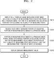
- the displaying of the measuring device image on the ultrasound image may include displaying the measuring device image in a half-transparent way so that an area of the ultrasound image, which overlaps the measuring device image, is not covered by the measuring device image.
- the ultrasound apparatus may display on the ultrasound image the measuring device image indicating two measuring points in the ultrasound image. Also, the ultrasound apparatus may receive a user input configuring the position of the measuring point via the measuring device image. When receiving the user input configuring the position of the measuring point, the ultrasound apparatus may measure a distance based on the configured position of the measuring point.
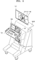
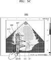
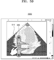
- the display unit 141 of the ultrasound apparatus 1000 may display an ultrasound image 130 on the touch screen 106. Also, the ultrasound apparatus 1000 may display on the ultrasound image 130 a measuring device image 110 for measuring an object included in the ultrasound image 130.
- the measuring device image 110 may include a measuring point which indicates a point on the ultrasound image that is to be measured. Also, the measuring device image 110 may include an adjusting portion for receiving a user input adjusting a position of the measuring point. In this case, the measuring point may be disposed apart from the adjusting portion. Accordingly, the user may precisely configure the measuring portion, without covering with a finger the measuring portion on the ultrasound image.
- the ultrasound apparatus 1000 may determine the position of the measuring point based on the position of the adjusting portion. Also, when the position of the measuring point is determined, the ultrasound apparatus 1000 may display the measuring point by shifting the measuring point in the measuring device image. In this case, the ultrasound apparatus 1000 may shift the measuring point by changing at least one of the shape and the position of the measuring device image 110.
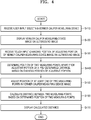
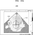
- the ultrasound apparatus 1000 may determine one from among the plurality of measuring devices, based on a pattern of a user input. For example, when the ultrasound apparatus 1000 receives a user input touching two points on a touch screen and dragging the two points in opposite directions, the ultrasound apparatus 1000 may determine an oval measuring device as the measuring device. Also, when the ultrasound apparatus 1000 receives a user input touching two points on the touch screen and rotating one point, the ultrasound apparatus 1000 may determine a degree measuring device as the measuring device.
- the ultrasound apparatus 1000 may display a measuring device image corresponding to the determined measuring device, on the ultrasound image.
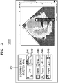
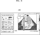
- the measuring device selection menu 340 may include icons 341 through 346 for selecting a measuring device.
- the ultrasound apparatus 1000 may store the changed position of the first adjusting portion 531 and the second adjusting portion 533 and the calculated position of the measuring points 511 and 513.
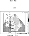
- the vernier caliper measuring device image 510 may include two measuring points 511 and 513 which indicate two points in an ultrasound image that are to be measured. Also, the vernier caliper measuring device image 510 may include an adjusting portion 555 indicating an adjusting area.
- the adjusting portion 555 may include a first adjusting portion 557 indicating a position of a first adjusting area and a second adjusting portion 559 indicating a position of a second adjusting area.
- the measuring points are located apart from the adjusting portion 555 by a pre-determined distance, and thus, the adjusting portion 555 indicating the adjusting area may be displayed at a location that is apart from the measuring points by the predetermined distance.
- the ultrasound apparatus 1000 may change the position of the first adjusting portion 557 by increasing or decreasing the length of the vernier caliper measuring device image 510.
- the point in the ultrasound image that is indicated by the measuring points 511 and 513 in the vernier caliper measuring device image 510 may be a point that is to be measured.
- the ultrasound apparatus 1000 may display a measurement value obtained via the pincette measuring device image 710.
- FIG. 8 is a flowchart of a method of providing a measuring function via a scissors measuring device, via the ultrasound apparatus 1000, according to an embodiment.
- the scissors measuring device image may include an adjusting portion indicating an adjusting area.
- the adjusting area may be an area on the ultrasound image for receiving a user's touch input for changing a position of the measuring points.
- the adjusting portion may be a handle portion of the scissors.
- the ultrasound apparatus 1000 may receive a touch input changing a position of the adjusting portion of the scissors measuring device image, on the ultrasound image, in operation S830.
- the ultrasound apparatus 1000 may determine a point that is apart from the adjusting point by a predetermined distance along a direction of a straight line connecting the adjusting point and at least one reference point determined on the ultrasound image, as the measuring point.
- the ultrasound apparatus 1000 may calculate and display a measurement value with respect to the circle, in operation S860.
- the ultrasound apparatus 1000 may calculate at least one of a diameter, a circumferential length, and an area of the circle.
- the ultrasound apparatus 1000 may display the scissors measuring device image corresponding to the scissors measuring device, on the ultrasound image.
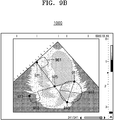
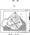
- the ultrasound apparatus 1000 may determine, based on the changed position of the first adjusting point 921, a point that is apart from the first adjusting point 921 by a pre-determined distance, along a direction of the straight line connecting the first adjusting point 921 and the cross point 925, as the second measuring point 913.
- the ultrasound apparatus 1000 may simultaneously move the first adjusting area 961, the second adjusting area 963, and the cross point 925, along a parallel direction.
- the measuring device image 910 corresponding to the scissors measuring device may include two measuring points 911 and 913 which indicate points in an ultrasound image that are to be measured. Also, the measuring device image 910 corresponding to the scissors measuring device may include two adjusting portions 991 and 993 which indicate adjusting areas. Also, the measuring device image 910 may display an image 953 indicating a circle having the two measuring points 911 and 913 as a diameter.
- the ultrasound apparatus 1000 may change the configured measuring area. Also, the ultrasound apparatus 1000 may calculate a diameter, a circumferential length, and an area of a circle based on the changed measuring area.
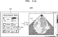
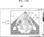
- the ultrasound apparatus 1000 may determine a segment connecting the trace point 1111.
- the ultrasound apparatus 1000 may adjust a size of the pen measuring device image 1110. For example, when the ultrasound apparatus receives a long touch input with respect to the adjusting portion 1123 and a touch input dragging the adjusting portion 1123 in a lengthwise direction or the pen measuring device image 1110 or in the direction opposite thereto, the ultrasound apparatus 1000 may adjust the length of the pen measuring device image 1110. For example, when receiving the touch input extending the pen measuring device image 1110 in the lengthwise direction of the pen measuring device image 1110, the ultrasound apparatus 1000 may enlarge the size of the pen measuring device image 1110 by extending the pen measuring device image 1110 along the dragged area.
- the ultrasound apparatus 1000 may decrease the pen measuring device image 1110 along the dragged area.
- the ultrasound apparatus 1000 may change the pen measuring device image 1110 from 10cm to 5cm.
- the ultrasound apparatus 1000 may display a button image 1140 for storing information with respect to the configured line 1120.
- the information with respect to the configured line 1120 may include the position of the trace points 1111 forming the configured line 1120 and a measurement value with respect to the line 1120.
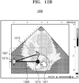
- the ultrasound apparatus 1000 may measure the angle of the two straight lines 1221 and 1223.
- the ultrasound apparatus 1000 may measure the angle of the two straight lines 1221 and 1223 based on a position of the three adjusting portions 1211, 1213, and 125.
- the ultrasound apparatus 1000 may measure the angle of the two straight lines 1221 and 1223 based on a position of the central point of the three adjusting portions 1211, 1213, and 1215.
- FIG. 12B is a view for describing a method of providing an angle measuring function via the ultrasound apparatus 1000, according to an embodiment.
- the ultrasound apparatus 1000 may measure the angle formed by the two straight lines 1321 and 1323. For example, the ultrasound apparatus 1000 may determine a cross point at which the two straight lines 1321 and 1323 meet each other when the two straight lines 1321 and 1323 extend, based on a position of the end points of the two straight lines 1321 and 1323. When the cross point is determined, the ultrasound apparatus 1000 may calculate the angle formed by the two straight lines 1321 and 1323 at the cross point.
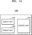
- the ultrasound apparatus 1000 may include a display unit 1100, a user input unit 1200, and a control unit 1300. However, not all of the illustrated components are essential. The ultrasound apparatus 1000 may be realized by more or less components that the illustrated components.
- the display unit 1100 may display on the ultrasound image a measuring device image including a plurality of measuring points indicating points in the ultrasound image that are to be measured, and adjusting portions for adjusting the plurality of measuring points.
- the measuring points may be disposed apart from the adjusting portions.
- the control unit 1300 may adjust a position of at least one of the plurality of measuring points based on the changed position of the adjusting portion and may obtain a measurement value based on a position of the plurality of measuring points including the at least one measuring point, the position of which is changed. Also, the control unit 1300 may adjust the position of the at least one of the plurality of measuring points by changing at least one of a position and a shape of the measuring device image.
- the display unit 1100 may display the obtained measurement value on the measuring device image.
- the display unit 1100 may display the measuring device image half-transparently so that an area of the ultrasound image, which overlaps the measuring device image, is not covered by the measuring device image.
- the display unit 1100 may display a button image for storing the measurement value, corresponding to the ultrasound image, on the ultrasound image.
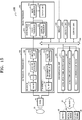
- a transmitter 110 supplies a driving signal to the probe 20.
- the transmitter 110 includes a pulse generator 112, a transmission delaying unit 114, and a pulser 116.
- the pulse generator 112 generates pulses for forming transmission ultrasound waves based on a predetermined pulse repetition frequency (PRF), and the transmission delaying unit 114 delays the pulses by delay times necessary for determining transmission directionality.
- the pulses which have been delayed correspond to a plurality of piezoelectric vibrators included in the probe 20, respectively.
- the pulser 116 applies a driving signal (or a driving pulse) to the probe 20 based on timing corresponding to each of the pulses which have been delayed.
- the memory 400 stores various data processed by the ultrasound apparatus 1000.
- the memory 400 may store medical data related to diagnosis of an object, such as ultrasound data and an ultrasound image that are input or output, and may also store algorithms or programs which are to be executed in the ultrasound apparatus 1000.
- All or some of the probe 20, the ultrasound transceiver 100, the image processor 200, the communication module 300, the memory 400, the user input unit 1200, and the controller 1300 may be implemented as software modules. However, embodiments of the present invention are not limited thereto, and some of the components stated above may be implemented as hardware modules. Furthermore, at least one selected from the ultrasound transceiver 100, the image processor 200, and the communication module 300 may be included in the controller 1300. However, embodiments of the present invention are not limited thereto.
Landscapes
- Engineering & Computer Science (AREA)
- Health & Medical Sciences (AREA)
- Life Sciences & Earth Sciences (AREA)
- Physics & Mathematics (AREA)
- General Engineering & Computer Science (AREA)
- Theoretical Computer Science (AREA)
- Medical Informatics (AREA)
- Public Health (AREA)
- Biomedical Technology (AREA)
- General Health & Medical Sciences (AREA)
- Pathology (AREA)
- Animal Behavior & Ethology (AREA)
- Veterinary Medicine (AREA)
- Heart & Thoracic Surgery (AREA)
- Radiology & Medical Imaging (AREA)
- Molecular Biology (AREA)
- Surgery (AREA)
- Biophysics (AREA)
- Nuclear Medicine, Radiotherapy & Molecular Imaging (AREA)
- Human Computer Interaction (AREA)
- General Physics & Mathematics (AREA)
- Computer Vision & Pattern Recognition (AREA)
- Physiology (AREA)
- Data Mining & Analysis (AREA)
- Databases & Information Systems (AREA)
- Epidemiology (AREA)
- Primary Health Care (AREA)
- Ultra Sonic Daignosis Equipment (AREA)
Priority Applications (1)
| Application Number | Priority Date | Filing Date | Title |
|---|---|---|---|
| EP22162754.0A EP4052653A1 (fr) | 2014-12-05 | 2015-04-28 | Procédé et appareil à ultrasons pour le traitement d'image ultrasonore |
Applications Claiming Priority (1)
| Application Number | Priority Date | Filing Date | Title |
|---|---|---|---|
| KR1020140174277A KR102293915B1 (ko) | 2014-12-05 | 2014-12-05 | 초음파 이미지 처리 방법 및 이를 위한 초음파 장치 |
Related Child Applications (1)
| Application Number | Title | Priority Date | Filing Date |
|---|---|---|---|
| EP22162754.0A Division EP4052653A1 (fr) | 2014-12-05 | 2015-04-28 | Procédé et appareil à ultrasons pour le traitement d'image ultrasonore |
Publications (2)
| Publication Number | Publication Date |
|---|---|
| EP3028638A1 true EP3028638A1 (fr) | 2016-06-08 |
| EP3028638B1 EP3028638B1 (fr) | 2022-04-06 |
Family
ID=53015621
Family Applications (2)
| Application Number | Title | Priority Date | Filing Date |
|---|---|---|---|
| EP22162754.0A Pending EP4052653A1 (fr) | 2014-12-05 | 2015-04-28 | Procédé et appareil à ultrasons pour le traitement d'image ultrasonore |
| EP15165356.5A Active EP3028638B1 (fr) | 2014-12-05 | 2015-04-28 | Procédé et appareil à ultrasons pour le traitement d'image ultrasonore |
Family Applications Before (1)
| Application Number | Title | Priority Date | Filing Date |
|---|---|---|---|
| EP22162754.0A Pending EP4052653A1 (fr) | 2014-12-05 | 2015-04-28 | Procédé et appareil à ultrasons pour le traitement d'image ultrasonore |
Country Status (4)
| Country | Link |
|---|---|
| US (4) | US11000261B2 (fr) |
| EP (2) | EP4052653A1 (fr) |
| KR (5) | KR102293915B1 (fr) |
| CN (3) | CN116746961A (fr) |
Cited By (2)
| Publication number | Priority date | Publication date | Assignee | Title |
|---|---|---|---|---|
| EP3682812A4 (fr) * | 2017-09-14 | 2020-11-04 | FUJIFILM Corporation | Dispositif de diagnostic par ultrasons et procédé de commande d'un dispositif de diagnostic par ultrasons |
| EP3766430A4 (fr) * | 2018-03-16 | 2021-04-21 | FUJIFILM Corporation | Dispositif de diagnostic d'onde acoustique et procédé de commande du dispositif de diagnostic d'onde acoustique |
Families Citing this family (14)
| Publication number | Priority date | Publication date | Assignee | Title |
|---|---|---|---|---|
| KR102250094B1 (ko) * | 2013-12-23 | 2021-05-10 | 삼성전자주식회사 | 모바일 장치의 기능 수행 방법 및 모바일 장치 |
| KR102293915B1 (ko) * | 2014-12-05 | 2021-08-26 | 삼성메디슨 주식회사 | 초음파 이미지 처리 방법 및 이를 위한 초음파 장치 |
| JP6784115B2 (ja) * | 2016-09-23 | 2020-11-11 | コニカミノルタ株式会社 | 超音波診断装置及びプログラム |
| CN108498118B (zh) * | 2017-02-28 | 2021-08-03 | 佳能医疗系统株式会社 | 超声波图像诊断装置、医用图像处理装置以及医用图像处理程序 |
| KR101984005B1 (ko) * | 2017-04-28 | 2019-05-30 | 연세대학교 산학협력단 | 초음파 영상 내 선택 지점의 위치를 조절하는 방법 및 그 장치 |
| JP7095261B2 (ja) * | 2017-11-06 | 2022-07-05 | コニカミノルタ株式会社 | 画像表示処理システム及び画像処理装置 |
| CN107992692B (zh) * | 2017-12-08 | 2021-07-20 | 北京汽车集团越野车有限公司 | 一种车身测点文件的生成方法及终端 |
| CN110507358B (zh) * | 2018-05-21 | 2022-01-11 | 珠海艾博罗生物技术股份有限公司 | 一种从超声图像上测量胎儿颈背透明物厚度的图像处理方法及系统 |
| WO2020086811A1 (fr) * | 2018-10-25 | 2020-04-30 | Butterfly Network, Inc. | Procédés et appareil pour effectuer des mesures sur une image ultrasonore |
| TWI776013B (zh) * | 2019-01-25 | 2022-09-01 | 佳世達科技股份有限公司 | 用於觸控顯示裝置的操作方法 |
| CN113116383B (zh) * | 2019-12-30 | 2023-05-12 | 无锡祥生医疗科技股份有限公司 | 超声设备的快速测量方法、系统和存储介质 |
| CN111513757A (zh) * | 2020-04-23 | 2020-08-11 | 无锡祥生医疗科技股份有限公司 | 测量方法、装置与存储介质 |
| KR102490676B1 (ko) * | 2021-09-30 | 2023-01-27 | 주식회사 소노티엑스 | 초음파를 이용한 장치 및 방법 |
| EP4445846B1 (fr) * | 2023-04-10 | 2026-03-25 | Samsung Medison Co., Ltd. | Dispositif d'imagerie ultrasonore comprenant un écran tactile et son procédé de commande |
Citations (4)
| Publication number | Priority date | Publication date | Assignee | Title |
|---|---|---|---|---|
| US20130324850A1 (en) * | 2012-05-31 | 2013-12-05 | Mindray Ds Usa, Inc. | Systems and methods for interfacing with an ultrasound system |
| US20140121524A1 (en) * | 2012-03-26 | 2014-05-01 | Alice M. Chiang | Tablet ultrasound system |
| EP2742868A1 (fr) * | 2012-12-12 | 2014-06-18 | Samsung Medison Co., Ltd. | Appareil à ultrasons et procédé de saisie d'informations dans celui-ci |
| EP2777506A1 (fr) * | 2013-03-13 | 2014-09-17 | Samsung Electronics Co., Ltd. | Procédé de fourniture d'image de copie et appareil à ultrasons correspondant |
Family Cites Families (51)
| Publication number | Priority date | Publication date | Assignee | Title |
|---|---|---|---|---|
| JPH03261813A (ja) | 1990-03-12 | 1991-11-21 | Terumo Corp | 画像処理装置のキャリパ計測装置 |
| JPH0453546A (ja) | 1990-06-20 | 1992-02-21 | Toshiba Corp | 画像表示装置 |
| JPH06125893A (ja) | 1992-10-20 | 1994-05-10 | Fujitsu Ltd | 超音波診断装置 |
| JP3351564B2 (ja) | 1992-12-15 | 2002-11-25 | フクダ電子株式会社 | キャリパ表示方式 |
| JP4149015B2 (ja) | 1997-10-08 | 2008-09-10 | 株式会社日立メディコ | 超音波診断装置 |
| US7093005B2 (en) * | 2000-02-11 | 2006-08-15 | Terraspring, Inc. | Graphical editor for defining and creating a computer system |
| US6692436B1 (en) | 2000-04-14 | 2004-02-17 | Computerized Screening, Inc. | Health care information system |
| US20060116577A1 (en) * | 2004-11-30 | 2006-06-01 | Siemens Medical Solutions Usa, Inc. | Direct image measurement editing mode for ultrasound reports |
| US9492240B2 (en) | 2009-06-16 | 2016-11-15 | Intuitive Surgical Operations, Inc. | Virtual measurement tool for minimally invasive surgery |
| US8303502B2 (en) * | 2007-03-06 | 2012-11-06 | General Electric Company | Method and apparatus for tracking points in an ultrasound image |
| WO2009090686A1 (fr) | 2008-01-17 | 2009-07-23 | Shimadzu Corporation | Appareil et programme de mesure des images |
| CN102006828B (zh) * | 2008-03-03 | 2014-08-27 | 柯尼卡美能达株式会社 | 超声波诊断装置 |
| JP2009297072A (ja) * | 2008-06-10 | 2009-12-24 | Toshiba Corp | 超音波診断装置、及び医用画像処理装置 |
| US8096949B2 (en) * | 2008-07-02 | 2012-01-17 | U-Systems, Inc. | User interface for ultrasound mammographic imaging |
| US8538103B2 (en) * | 2009-02-10 | 2013-09-17 | Hitachi Medical Corporation | Medical image processing device, medical image processing method, medical image diagnostic apparatus, operation method of medical image diagnostic apparatus, and medical image display method |
| KR101167248B1 (ko) * | 2009-05-22 | 2012-07-23 | 삼성메디슨 주식회사 | 터치 인터랙션을 사용하는 초음파 진단 장치 |
| US20140082542A1 (en) * | 2010-07-19 | 2014-03-20 | Qview, Inc. | Viewing and correlating between breast ultrasound and mammogram or breast tomosynthesis images |
| US8502789B2 (en) * | 2010-01-11 | 2013-08-06 | Smart Technologies Ulc | Method for handling user input in an interactive input system, and interactive input system executing the method |
| KR101117930B1 (ko) * | 2010-05-13 | 2012-02-29 | 삼성메디슨 주식회사 | 슬라이스 영상과 함께 부가 정보를 제공하는 초음파 시스템 및 방법 |
| EP2387949A1 (fr) * | 2010-05-17 | 2011-11-23 | Samsung Medison Co., Ltd. | Système ultrasonore pour mesurer une image à l'aide d'un modèle de figure et procédé de fonctionnement du système ultrasonore |
| US20130072797A1 (en) * | 2010-05-31 | 2013-03-21 | Samsung Medison Co., Ltd. | 3d ultrasound apparatus and method for operating the same |
| JP2012019824A (ja) | 2010-07-12 | 2012-02-02 | Hitachi Aloka Medical Ltd | 超音波診断装置 |
| US9152306B2 (en) * | 2011-03-29 | 2015-10-06 | Intel Corporation | Techniques for touch and non-touch user interaction input |
| GB2489709B (en) * | 2011-04-05 | 2013-07-31 | Mirada Medical Ltd | Measurement system for medical images |
| JP5392794B2 (ja) | 2011-09-29 | 2014-01-22 | 株式会社セイコーアイ・インフォテック | 計測装置および計測プログラム |
| BR112014013935A8 (pt) * | 2011-12-12 | 2017-06-13 | Koninklijke Philips Nv | dispositivo de processamento de imagem, e, meio legível por computador |
| KR20130080311A (ko) * | 2012-01-04 | 2013-07-12 | 삼성메디슨 주식회사 | 의료 영상 표시 방법 및 의료 영상 표시 장치 |
| KR101501515B1 (ko) * | 2012-01-04 | 2015-03-11 | 삼성메디슨 주식회사 | 진단 영상 장치 및 그 동작 방법 |
| US20130201210A1 (en) * | 2012-01-13 | 2013-08-08 | Qualcomm Incorporated | Virtual ruler |
| KR101520613B1 (ko) * | 2012-02-06 | 2015-05-15 | 삼성메디슨 주식회사 | 초음파 영상 데이터 제공 방법 및 초음파 영상 데이터 제공 장치 |
| WO2013137276A1 (fr) * | 2012-03-12 | 2013-09-19 | 株式会社東芝 | Dispositif de diagnostic ultrasonore |
| US10667790B2 (en) * | 2012-03-26 | 2020-06-02 | Teratech Corporation | Tablet ultrasound system |
| JP5907780B2 (ja) * | 2012-04-02 | 2016-04-26 | 富士フイルム株式会社 | 超音波診断装置 |
| WO2013170053A1 (fr) * | 2012-05-09 | 2013-11-14 | The Regents Of The University Of Michigan | Transducteur linéaire à entraînement magnétique pour imagerie ultrasonore |
| US20130321286A1 (en) * | 2012-05-31 | 2013-12-05 | Mindray Ds Usa, Inc. | Systems and methods for interfacing with an ultrasound system |
| US8777856B2 (en) * | 2012-06-26 | 2014-07-15 | General Electric Company | Diagnostic system and method for obtaining an ultrasound image frame |
| JP6013051B2 (ja) * | 2012-07-02 | 2016-10-25 | 東芝メディカルシステムズ株式会社 | 超音波診断装置及びその操作支援方法 |
| KR20140038777A (ko) * | 2012-09-21 | 2014-03-31 | 한국디지털병원수출사업협동조합 | 터치 기반 3차원 초음파 진단기 |
| WO2014058929A1 (fr) * | 2012-10-08 | 2014-04-17 | Fujifilm Sonosite, Inc. | Systèmes et procédés d'entrée tactile sur des appareils à ultrasons |
| JP6019362B2 (ja) * | 2012-11-12 | 2016-11-02 | 本多電子株式会社 | 医療画像測定装置及び医療画像測定方法 |
| CN102920451B (zh) * | 2012-11-23 | 2014-08-27 | 北京谷山丰生物医学技术有限公司 | 以多点触摸的方式测量心电图的方法 |
| KR101468419B1 (ko) | 2012-12-04 | 2014-12-03 | 삼성메디슨 주식회사 | 3차원 캘리퍼를 이용하여 측정 정보를 제공하는 의료 시스템 및 방법 |
| US10799209B2 (en) * | 2012-12-26 | 2020-10-13 | Philips Image Guided Therapy Corporation | Measurement navigation in a multi-modality medical imaging system |
| JP6070228B2 (ja) | 2013-02-01 | 2017-02-01 | コニカミノルタ株式会社 | 超音波画像診断装置 |
| JP2014233522A (ja) * | 2013-06-04 | 2014-12-15 | セイコーエプソン株式会社 | 超音波測定装置および超音波測定方法 |
| KR102243032B1 (ko) * | 2014-03-18 | 2021-04-21 | 삼성메디슨 주식회사 | 초음파 장치 및 초음파 장치의 측정 방법 |
| US9538985B2 (en) * | 2014-04-18 | 2017-01-10 | Fujifilm Sonosite, Inc. | Hand-held medical imaging system with improved user interface for deploying on-screen graphical tools and associated apparatuses and methods |
| US10579207B2 (en) * | 2014-05-14 | 2020-03-03 | Purdue Research Foundation | Manipulating virtual environment using non-instrumented physical object |
| WO2016007673A2 (fr) * | 2014-07-09 | 2016-01-14 | Edan Instruments, Inc. | Interface utilisateur de système à ultrasons portable, et systèmes et procédés de gestion de ressources |
| KR102293915B1 (ko) * | 2014-12-05 | 2021-08-26 | 삼성메디슨 주식회사 | 초음파 이미지 처리 방법 및 이를 위한 초음파 장치 |
| US20160179326A1 (en) * | 2014-12-23 | 2016-06-23 | General Electric Company | Medical imaging apparatus and method for managing touch inputs in a touch based user interface |
-
2014
- 2014-12-05 KR KR1020140174277A patent/KR102293915B1/ko active Active
-
2015
- 2015-04-28 EP EP22162754.0A patent/EP4052653A1/fr active Pending
- 2015-04-28 EP EP15165356.5A patent/EP3028638B1/fr active Active
- 2015-06-15 US US14/739,734 patent/US11000261B2/en active Active
- 2015-12-03 CN CN202310748579.9A patent/CN116746961A/zh active Pending
- 2015-12-03 CN CN202310754434.XA patent/CN116763341A/zh active Pending
- 2015-12-03 CN CN201510882396.1A patent/CN105662460B/zh active Active
-
2021
- 2021-05-07 US US17/314,577 patent/US11717266B2/en active Active
- 2021-05-07 US US17/314,501 patent/US11857371B2/en active Active
- 2021-08-19 KR KR1020210109747A patent/KR102423916B1/ko active Active
-
2022
- 2022-07-18 KR KR1020220088568A patent/KR102607204B1/ko active Active
-
2023
- 2023-06-13 US US18/209,015 patent/US20230320699A1/en active Pending
- 2023-11-22 KR KR1020230163781A patent/KR102798675B1/ko active Active
-
2025
- 2025-04-16 KR KR1020250049692A patent/KR20250055497A/ko active Pending
Patent Citations (4)
| Publication number | Priority date | Publication date | Assignee | Title |
|---|---|---|---|---|
| US20140121524A1 (en) * | 2012-03-26 | 2014-05-01 | Alice M. Chiang | Tablet ultrasound system |
| US20130324850A1 (en) * | 2012-05-31 | 2013-12-05 | Mindray Ds Usa, Inc. | Systems and methods for interfacing with an ultrasound system |
| EP2742868A1 (fr) * | 2012-12-12 | 2014-06-18 | Samsung Medison Co., Ltd. | Appareil à ultrasons et procédé de saisie d'informations dans celui-ci |
| EP2777506A1 (fr) * | 2013-03-13 | 2014-09-17 | Samsung Electronics Co., Ltd. | Procédé de fourniture d'image de copie et appareil à ultrasons correspondant |
Cited By (4)
| Publication number | Priority date | Publication date | Assignee | Title |
|---|---|---|---|---|
| EP3682812A4 (fr) * | 2017-09-14 | 2020-11-04 | FUJIFILM Corporation | Dispositif de diagnostic par ultrasons et procédé de commande d'un dispositif de diagnostic par ultrasons |
| US11036376B2 (en) | 2017-09-14 | 2021-06-15 | Fujifilm Corporation | Ultrasound diagnosis apparatus and method of controlling ultrasound diagnosis apparatus |
| EP3766430A4 (fr) * | 2018-03-16 | 2021-04-21 | FUJIFILM Corporation | Dispositif de diagnostic d'onde acoustique et procédé de commande du dispositif de diagnostic d'onde acoustique |
| US11490879B2 (en) | 2018-03-16 | 2022-11-08 | Fujifilm Corporation | Acoustic wave diagnostic apparatus and control method of acoustic wave diagnostic apparatus |
Also Published As
| Publication number | Publication date |
|---|---|
| EP4052653A1 (fr) | 2022-09-07 |
| KR102607204B1 (ko) | 2023-11-29 |
| US20160157825A1 (en) | 2016-06-09 |
| KR20210105865A (ko) | 2021-08-27 |
| KR102423916B1 (ko) | 2022-07-22 |
| US20210267575A1 (en) | 2021-09-02 |
| KR20250055497A (ko) | 2025-04-24 |
| KR102293915B1 (ko) | 2021-08-26 |
| CN105662460B (zh) | 2023-07-14 |
| US11717266B2 (en) | 2023-08-08 |
| KR20230163988A (ko) | 2023-12-01 |
| CN105662460A (zh) | 2016-06-15 |
| US20210271381A1 (en) | 2021-09-02 |
| US11857371B2 (en) | 2024-01-02 |
| US20230320699A1 (en) | 2023-10-12 |
| CN116746961A (zh) | 2023-09-15 |
| CN116763341A (zh) | 2023-09-19 |
| KR20220104671A (ko) | 2022-07-26 |
| KR102798675B1 (ko) | 2025-04-23 |
| EP3028638B1 (fr) | 2022-04-06 |
| KR20160068468A (ko) | 2016-06-15 |
| US11000261B2 (en) | 2021-05-11 |
Similar Documents
| Publication | Publication Date | Title |
|---|---|---|
| US11857371B2 (en) | Ultrasound method and apparatus for processing ultrasound image to obtain measurement information of an object in the ultrasound image | |
| US10013768B2 (en) | Method and apparatus for displaying image showing object | |
| US10806433B2 (en) | Ultrasound apparatus and method of operating the same | |
| EP3034005B1 (fr) | Procédé, appareil et système de génération de marqueur de corps indicateur d'objet | |
| KR102388130B1 (ko) | 의료 영상 표시 방법 및 장치 | |
| KR102273831B1 (ko) | 의료 영상을 디스플레이 하는 방법 및 그 의료 영상 장치 | |
| KR102310976B1 (ko) | 초음파 진단 장치, 그에 따른 초음파 진단 방법 및 그에 따른 컴퓨터 판독 가능한 저장매체 | |
| US10004478B2 (en) | Method and apparatus for displaying ultrasound image | |
| EP3199108A1 (fr) | Procédé et appareil d'affichage d'image ultrasonore | |
| KR102297148B1 (ko) | 3차원 영상을 표시하는 초음파 시스템 및 그 방법 | |
| US10809878B2 (en) | Method and apparatus for displaying ultrasound image | |
| KR102270718B1 (ko) | 초음파 진단장치, 그에 따른 초음파 진단 장치의 동작 방법 및 그에 따른 컴퓨터 판독 가능한 저장매체 |
Legal Events
| Date | Code | Title | Description |
|---|---|---|---|
| PUAI | Public reference made under article 153(3) epc to a published international application that has entered the european phase |
Free format text: ORIGINAL CODE: 0009012 |
|
| AK | Designated contracting states |
Kind code of ref document: A1 Designated state(s): AL AT BE BG CH CY CZ DE DK EE ES FI FR GB GR HR HU IE IS IT LI LT LU LV MC MK MT NL NO PL PT RO RS SE SI SK SM TR |
|
| AX | Request for extension of the european patent |
Extension state: BA ME |
|
| STAA | Information on the status of an ep patent application or granted ep patent |
Free format text: STATUS: REQUEST FOR EXAMINATION WAS MADE |
|
| 17P | Request for examination filed |
Effective date: 20161206 |
|
| RBV | Designated contracting states (corrected) |
Designated state(s): AL AT BE BG CH CY CZ DE DK EE ES FI FR GB GR HR HU IE IS IT LI LT LU LV MC MK MT NL NO PL PT RO RS SE SI SK SM TR |
|
| STAA | Information on the status of an ep patent application or granted ep patent |
Free format text: STATUS: EXAMINATION IS IN PROGRESS |
|
| 17Q | First examination report despatched |
Effective date: 20200709 |
|
| GRAP | Despatch of communication of intention to grant a patent |
Free format text: ORIGINAL CODE: EPIDOSNIGR1 |
|
| STAA | Information on the status of an ep patent application or granted ep patent |
Free format text: STATUS: GRANT OF PATENT IS INTENDED |
|
| INTG | Intention to grant announced |
Effective date: 20211021 |
|
| INTG | Intention to grant announced |
Effective date: 20211026 |
|
| GRAS | Grant fee paid |
Free format text: ORIGINAL CODE: EPIDOSNIGR3 |
|
| GRAA | (expected) grant |
Free format text: ORIGINAL CODE: 0009210 |
|
| STAA | Information on the status of an ep patent application or granted ep patent |
Free format text: STATUS: THE PATENT HAS BEEN GRANTED |
|
| AK | Designated contracting states |
Kind code of ref document: B1 Designated state(s): AL AT BE BG CH CY CZ DE DK EE ES FI FR GB GR HR HU IE IS IT LI LT LU LV MC MK MT NL NO PL PT RO RS SE SI SK SM TR |
|
| REG | Reference to a national code |
Ref country code: GB Ref legal event code: FG4D |
|
| REG | Reference to a national code |
Ref country code: CH Ref legal event code: EP |
|
| REG | Reference to a national code |
Ref country code: AT Ref legal event code: REF Ref document number: 1480549 Country of ref document: AT Kind code of ref document: T Effective date: 20220415 |
|
| REG | Reference to a national code |
Ref country code: IE Ref legal event code: FG4D |
|
| REG | Reference to a national code |
Ref country code: DE Ref legal event code: R096 Ref document number: 602015077992 Country of ref document: DE |
|
| REG | Reference to a national code |
Ref country code: LT Ref legal event code: MG9D |
|
| REG | Reference to a national code |
Ref country code: NL Ref legal event code: MP Effective date: 20220406 |
|
| REG | Reference to a national code |
Ref country code: AT Ref legal event code: MK05 Ref document number: 1480549 Country of ref document: AT Kind code of ref document: T Effective date: 20220406 |
|
| PG25 | Lapsed in a contracting state [announced via postgrant information from national office to epo] |
Ref country code: NL Free format text: LAPSE BECAUSE OF FAILURE TO SUBMIT A TRANSLATION OF THE DESCRIPTION OR TO PAY THE FEE WITHIN THE PRESCRIBED TIME-LIMIT Effective date: 20220406 |
|
| PG25 | Lapsed in a contracting state [announced via postgrant information from national office to epo] |
Ref country code: SE Free format text: LAPSE BECAUSE OF FAILURE TO SUBMIT A TRANSLATION OF THE DESCRIPTION OR TO PAY THE FEE WITHIN THE PRESCRIBED TIME-LIMIT Effective date: 20220406 Ref country code: PT Free format text: LAPSE BECAUSE OF FAILURE TO SUBMIT A TRANSLATION OF THE DESCRIPTION OR TO PAY THE FEE WITHIN THE PRESCRIBED TIME-LIMIT Effective date: 20220808 Ref country code: NO Free format text: LAPSE BECAUSE OF FAILURE TO SUBMIT A TRANSLATION OF THE DESCRIPTION OR TO PAY THE FEE WITHIN THE PRESCRIBED TIME-LIMIT Effective date: 20220706 Ref country code: LT Free format text: LAPSE BECAUSE OF FAILURE TO SUBMIT A TRANSLATION OF THE DESCRIPTION OR TO PAY THE FEE WITHIN THE PRESCRIBED TIME-LIMIT Effective date: 20220406 Ref country code: HR Free format text: LAPSE BECAUSE OF FAILURE TO SUBMIT A TRANSLATION OF THE DESCRIPTION OR TO PAY THE FEE WITHIN THE PRESCRIBED TIME-LIMIT Effective date: 20220406 Ref country code: GR Free format text: LAPSE BECAUSE OF FAILURE TO SUBMIT A TRANSLATION OF THE DESCRIPTION OR TO PAY THE FEE WITHIN THE PRESCRIBED TIME-LIMIT Effective date: 20220707 Ref country code: FI Free format text: LAPSE BECAUSE OF FAILURE TO SUBMIT A TRANSLATION OF THE DESCRIPTION OR TO PAY THE FEE WITHIN THE PRESCRIBED TIME-LIMIT Effective date: 20220406 Ref country code: ES Free format text: LAPSE BECAUSE OF FAILURE TO SUBMIT A TRANSLATION OF THE DESCRIPTION OR TO PAY THE FEE WITHIN THE PRESCRIBED TIME-LIMIT Effective date: 20220406 Ref country code: BG Free format text: LAPSE BECAUSE OF FAILURE TO SUBMIT A TRANSLATION OF THE DESCRIPTION OR TO PAY THE FEE WITHIN THE PRESCRIBED TIME-LIMIT Effective date: 20220706 Ref country code: AT Free format text: LAPSE BECAUSE OF FAILURE TO SUBMIT A TRANSLATION OF THE DESCRIPTION OR TO PAY THE FEE WITHIN THE PRESCRIBED TIME-LIMIT Effective date: 20220406 |
|
| PG25 | Lapsed in a contracting state [announced via postgrant information from national office to epo] |
Ref country code: RS Free format text: LAPSE BECAUSE OF FAILURE TO SUBMIT A TRANSLATION OF THE DESCRIPTION OR TO PAY THE FEE WITHIN THE PRESCRIBED TIME-LIMIT Effective date: 20220406 Ref country code: PL Free format text: LAPSE BECAUSE OF FAILURE TO SUBMIT A TRANSLATION OF THE DESCRIPTION OR TO PAY THE FEE WITHIN THE PRESCRIBED TIME-LIMIT Effective date: 20220406 Ref country code: LV Free format text: LAPSE BECAUSE OF FAILURE TO SUBMIT A TRANSLATION OF THE DESCRIPTION OR TO PAY THE FEE WITHIN THE PRESCRIBED TIME-LIMIT Effective date: 20220406 Ref country code: IS Free format text: LAPSE BECAUSE OF FAILURE TO SUBMIT A TRANSLATION OF THE DESCRIPTION OR TO PAY THE FEE WITHIN THE PRESCRIBED TIME-LIMIT Effective date: 20220806 |
|
| REG | Reference to a national code |
Ref country code: CH Ref legal event code: PL |
|
| REG | Reference to a national code |
Ref country code: BE Ref legal event code: MM Effective date: 20220430 |
|
| REG | Reference to a national code |
Ref country code: DE Ref legal event code: R097 Ref document number: 602015077992 Country of ref document: DE |
|
| PG25 | Lapsed in a contracting state [announced via postgrant information from national office to epo] |
Ref country code: SM Free format text: LAPSE BECAUSE OF FAILURE TO SUBMIT A TRANSLATION OF THE DESCRIPTION OR TO PAY THE FEE WITHIN THE PRESCRIBED TIME-LIMIT Effective date: 20220406 Ref country code: SK Free format text: LAPSE BECAUSE OF FAILURE TO SUBMIT A TRANSLATION OF THE DESCRIPTION OR TO PAY THE FEE WITHIN THE PRESCRIBED TIME-LIMIT Effective date: 20220406 Ref country code: RO Free format text: LAPSE BECAUSE OF FAILURE TO SUBMIT A TRANSLATION OF THE DESCRIPTION OR TO PAY THE FEE WITHIN THE PRESCRIBED TIME-LIMIT Effective date: 20220406 Ref country code: MC Free format text: LAPSE BECAUSE OF FAILURE TO SUBMIT A TRANSLATION OF THE DESCRIPTION OR TO PAY THE FEE WITHIN THE PRESCRIBED TIME-LIMIT Effective date: 20220406 Ref country code: LU Free format text: LAPSE BECAUSE OF NON-PAYMENT OF DUE FEES Effective date: 20220428 Ref country code: LI Free format text: LAPSE BECAUSE OF NON-PAYMENT OF DUE FEES Effective date: 20220430 Ref country code: EE Free format text: LAPSE BECAUSE OF FAILURE TO SUBMIT A TRANSLATION OF THE DESCRIPTION OR TO PAY THE FEE WITHIN THE PRESCRIBED TIME-LIMIT Effective date: 20220406 Ref country code: DK Free format text: LAPSE BECAUSE OF FAILURE TO SUBMIT A TRANSLATION OF THE DESCRIPTION OR TO PAY THE FEE WITHIN THE PRESCRIBED TIME-LIMIT Effective date: 20220406 Ref country code: CZ Free format text: LAPSE BECAUSE OF FAILURE TO SUBMIT A TRANSLATION OF THE DESCRIPTION OR TO PAY THE FEE WITHIN THE PRESCRIBED TIME-LIMIT Effective date: 20220406 Ref country code: CH Free format text: LAPSE BECAUSE OF NON-PAYMENT OF DUE FEES Effective date: 20220430 |
|
| PLBE | No opposition filed within time limit |
Free format text: ORIGINAL CODE: 0009261 |
|
| STAA | Information on the status of an ep patent application or granted ep patent |
Free format text: STATUS: NO OPPOSITION FILED WITHIN TIME LIMIT |
|
| PG25 | Lapsed in a contracting state [announced via postgrant information from national office to epo] |
Ref country code: BE Free format text: LAPSE BECAUSE OF NON-PAYMENT OF DUE FEES Effective date: 20220430 |
|
| 26N | No opposition filed |
Effective date: 20230110 |
|
| GBPC | Gb: european patent ceased through non-payment of renewal fee |
Effective date: 20220706 |
|
| PG25 | Lapsed in a contracting state [announced via postgrant information from national office to epo] |
Ref country code: AL Free format text: LAPSE BECAUSE OF FAILURE TO SUBMIT A TRANSLATION OF THE DESCRIPTION OR TO PAY THE FEE WITHIN THE PRESCRIBED TIME-LIMIT Effective date: 20220406 |
|
| PG25 | Lapsed in a contracting state [announced via postgrant information from national office to epo] |
Ref country code: IE Free format text: LAPSE BECAUSE OF NON-PAYMENT OF DUE FEES Effective date: 20220428 |
|
| PG25 | Lapsed in a contracting state [announced via postgrant information from national office to epo] |
Ref country code: SI Free format text: LAPSE BECAUSE OF FAILURE TO SUBMIT A TRANSLATION OF THE DESCRIPTION OR TO PAY THE FEE WITHIN THE PRESCRIBED TIME-LIMIT Effective date: 20220406 Ref country code: GB Free format text: LAPSE BECAUSE OF NON-PAYMENT OF DUE FEES Effective date: 20220706 |
|
| PG25 | Lapsed in a contracting state [announced via postgrant information from national office to epo] |
Ref country code: HU Free format text: LAPSE BECAUSE OF FAILURE TO SUBMIT A TRANSLATION OF THE DESCRIPTION OR TO PAY THE FEE WITHIN THE PRESCRIBED TIME-LIMIT; INVALID AB INITIO Effective date: 20150428 |
|
| PG25 | Lapsed in a contracting state [announced via postgrant information from national office to epo] |
Ref country code: MK Free format text: LAPSE BECAUSE OF FAILURE TO SUBMIT A TRANSLATION OF THE DESCRIPTION OR TO PAY THE FEE WITHIN THE PRESCRIBED TIME-LIMIT Effective date: 20220406 Ref country code: CY Free format text: LAPSE BECAUSE OF FAILURE TO SUBMIT A TRANSLATION OF THE DESCRIPTION OR TO PAY THE FEE WITHIN THE PRESCRIBED TIME-LIMIT Effective date: 20220406 |
|
| PG25 | Lapsed in a contracting state [announced via postgrant information from national office to epo] |
Ref country code: TR Free format text: LAPSE BECAUSE OF FAILURE TO SUBMIT A TRANSLATION OF THE DESCRIPTION OR TO PAY THE FEE WITHIN THE PRESCRIBED TIME-LIMIT Effective date: 20220406 |
|
| PG25 | Lapsed in a contracting state [announced via postgrant information from national office to epo] |
Ref country code: MT Free format text: LAPSE BECAUSE OF FAILURE TO SUBMIT A TRANSLATION OF THE DESCRIPTION OR TO PAY THE FEE WITHIN THE PRESCRIBED TIME-LIMIT Effective date: 20220406 |
|
| PG25 | Lapsed in a contracting state [announced via postgrant information from national office to epo] |
Ref country code: BG Free format text: LAPSE BECAUSE OF FAILURE TO SUBMIT A TRANSLATION OF THE DESCRIPTION OR TO PAY THE FEE WITHIN THE PRESCRIBED TIME-LIMIT Effective date: 20220406 |
|
| PG25 | Lapsed in a contracting state [announced via postgrant information from national office to epo] |
Ref country code: BG Free format text: LAPSE BECAUSE OF FAILURE TO SUBMIT A TRANSLATION OF THE DESCRIPTION OR TO PAY THE FEE WITHIN THE PRESCRIBED TIME-LIMIT Effective date: 20220406 |
|
| PGFP | Annual fee paid to national office [announced via postgrant information from national office to epo] |
Ref country code: FR Payment date: 20250307 Year of fee payment: 11 |
|
| PGFP | Annual fee paid to national office [announced via postgrant information from national office to epo] |
Ref country code: IT Payment date: 20250306 Year of fee payment: 11 |
|
| PGFP | Annual fee paid to national office [announced via postgrant information from national office to epo] |
Ref country code: DE Payment date: 20250305 Year of fee payment: 11 |