EP3049818B1 - Correction d'atténuation en imagerie tep/irm basée sur l'irm avec séquence d'impulsions de dixon - Google Patents
Correction d'atténuation en imagerie tep/irm basée sur l'irm avec séquence d'impulsions de dixon Download PDFInfo
- Publication number
- EP3049818B1 EP3049818B1 EP14767008.7A EP14767008A EP3049818B1 EP 3049818 B1 EP3049818 B1 EP 3049818B1 EP 14767008 A EP14767008 A EP 14767008A EP 3049818 B1 EP3049818 B1 EP 3049818B1
- Authority
- EP
- European Patent Office
- Prior art keywords
- image
- subject
- echo time
- nuclear
- data
- Prior art date
- Legal status (The legal status is an assumption and is not a legal conclusion. Google has not performed a legal analysis and makes no representation as to the accuracy of the status listed.)
- Active
Links
Images
Classifications
-
- G—PHYSICS
- G01—MEASURING; TESTING
- G01R—MEASURING ELECTRIC VARIABLES; MEASURING MAGNETIC VARIABLES
- G01R33/00—Arrangements or instruments for measuring magnetic variables
- G01R33/20—Arrangements or instruments for measuring magnetic variables involving magnetic resonance
- G01R33/44—Arrangements or instruments for measuring magnetic variables involving magnetic resonance using nuclear magnetic resonance [NMR]
- G01R33/48—NMR imaging systems
- G01R33/4808—Multimodal MR, e.g. MR combined with positron emission tomography [PET], MR combined with ultrasound or MR combined with computed tomography [CT]
- G01R33/481—MR combined with positron emission tomography [PET] or single photon emission computed tomography [SPECT]
-
- G—PHYSICS
- G01—MEASURING; TESTING
- G01R—MEASURING ELECTRIC VARIABLES; MEASURING MAGNETIC VARIABLES
- G01R33/00—Arrangements or instruments for measuring magnetic variables
- G01R33/20—Arrangements or instruments for measuring magnetic variables involving magnetic resonance
- G01R33/44—Arrangements or instruments for measuring magnetic variables involving magnetic resonance using nuclear magnetic resonance [NMR]
- G01R33/48—NMR imaging systems
- G01R33/4816—NMR imaging of samples with ultrashort relaxation times such as solid samples, e.g. MRI using ultrashort TE [UTE], single point imaging, constant time imaging
-
- G—PHYSICS
- G01—MEASURING; TESTING
- G01R—MEASURING ELECTRIC VARIABLES; MEASURING MAGNETIC VARIABLES
- G01R33/00—Arrangements or instruments for measuring magnetic variables
- G01R33/20—Arrangements or instruments for measuring magnetic variables involving magnetic resonance
- G01R33/44—Arrangements or instruments for measuring magnetic variables involving magnetic resonance using nuclear magnetic resonance [NMR]
- G01R33/48—NMR imaging systems
- G01R33/4828—Resolving the MR signals of different chemical species, e.g. water-fat imaging
-
- G—PHYSICS
- G01—MEASURING; TESTING
- G01R—MEASURING ELECTRIC VARIABLES; MEASURING MAGNETIC VARIABLES
- G01R33/00—Arrangements or instruments for measuring magnetic variables
- G01R33/20—Arrangements or instruments for measuring magnetic variables involving magnetic resonance
- G01R33/44—Arrangements or instruments for measuring magnetic variables involving magnetic resonance using nuclear magnetic resonance [NMR]
- G01R33/48—NMR imaging systems
- G01R33/54—Signal processing systems, e.g. using pulse sequences ; Generation or control of pulse sequences; Operator console
- G01R33/56—Image enhancement or correction, e.g. subtraction or averaging techniques, e.g. improvement of signal-to-noise ratio and resolution
- G01R33/5608—Data processing and visualization specially adapted for MR, e.g. for feature analysis and pattern recognition on the basis of measured MR data, segmentation of measured MR data, edge contour detection on the basis of measured MR data, for enhancing measured MR data in terms of signal-to-noise ratio by means of noise filtering or apodization, for enhancing measured MR data in terms of resolution by means for deblurring, windowing, zero filling, or generation of gray-scaled images, colour-coded images or images displaying vectors instead of pixels
-
- G—PHYSICS
- G01—MEASURING; TESTING
- G01R—MEASURING ELECTRIC VARIABLES; MEASURING MAGNETIC VARIABLES
- G01R33/00—Arrangements or instruments for measuring magnetic variables
- G01R33/20—Arrangements or instruments for measuring magnetic variables involving magnetic resonance
- G01R33/44—Arrangements or instruments for measuring magnetic variables involving magnetic resonance using nuclear magnetic resonance [NMR]
- G01R33/48—NMR imaging systems
- G01R33/54—Signal processing systems, e.g. using pulse sequences ; Generation or control of pulse sequences; Operator console
- G01R33/56—Image enhancement or correction, e.g. subtraction or averaging techniques, e.g. improvement of signal-to-noise ratio and resolution
- G01R33/561—Image enhancement or correction, e.g. subtraction or averaging techniques, e.g. improvement of signal-to-noise ratio and resolution by reduction of the scanning time, i.e. fast acquiring systems, e.g. using echo-planar pulse sequences
- G01R33/5615—Echo train techniques involving acquiring plural, differently encoded, echo signals after one RF excitation, e.g. using gradient refocusing in echo planar imaging [EPI], RF refocusing in rapid acquisition with relaxation enhancement [RARE] or using both RF and gradient refocusing in gradient and spin echo imaging [GRASE]
-
- G—PHYSICS
- G01—MEASURING; TESTING
- G01R—MEASURING ELECTRIC VARIABLES; MEASURING MAGNETIC VARIABLES
- G01R33/00—Arrangements or instruments for measuring magnetic variables
- G01R33/20—Arrangements or instruments for measuring magnetic variables involving magnetic resonance
- G01R33/44—Arrangements or instruments for measuring magnetic variables involving magnetic resonance using nuclear magnetic resonance [NMR]
- G01R33/48—NMR imaging systems
- G01R33/54—Signal processing systems, e.g. using pulse sequences ; Generation or control of pulse sequences; Operator console
- G01R33/56—Image enhancement or correction, e.g. subtraction or averaging techniques, e.g. improvement of signal-to-noise ratio and resolution
- G01R33/561—Image enhancement or correction, e.g. subtraction or averaging techniques, e.g. improvement of signal-to-noise ratio and resolution by reduction of the scanning time, i.e. fast acquiring systems, e.g. using echo-planar pulse sequences
- G01R33/5615—Echo train techniques involving acquiring plural, differently encoded, echo signals after one RF excitation, e.g. using gradient refocusing in echo planar imaging [EPI], RF refocusing in rapid acquisition with relaxation enhancement [RARE] or using both RF and gradient refocusing in gradient and spin echo imaging [GRASE]
- G01R33/5616—Echo train techniques involving acquiring plural, differently encoded, echo signals after one RF excitation, e.g. using gradient refocusing in echo planar imaging [EPI], RF refocusing in rapid acquisition with relaxation enhancement [RARE] or using both RF and gradient refocusing in gradient and spin echo imaging [GRASE] using gradient refocusing, e.g. EPI
Definitions
- the following relates generally to medical imaging. It finds particular application in conjunction with magnetic resonance (MR) imaging to provide attenuation correction for positron emission tomography (PET) imaging, and will be described with particular reference thereto. However, it will be understood that it also finds application in other usage scenarios and is not necessarily limited to the aforementioned application.
- MR magnetic resonance
- PET positron emission tomography
- a subject is injected with a radiopharmaceutical which targets particular tissues typically through absorption based on a metabolic activity.
- positrons are emitted which annihilate in contact with an electron to form a pair of photons emitted 180° opposite along a line of response (LOR).
- LOR line of response
- the emitted gamma photons are recorded by PET detectors surrounding the subject.
- the locations of the annihilation events are computed from the recorded gamma photons which provide an image of the tissues targeted by the radiopharmaceutical.
- the emitted photons are affected by the density of tissues between the point of the annihilation event and the detector by either absorption or deflection.
- the recorded amount of radiopharmaceutical present in tissue is attenuated from the actual amount by the tissue density along the LORs.
- Correction for attenuation of PET image data seeks to accurately identify the tissue density at each voxel with an attenuation correction map for the subject being imaged.
- MR based attenuation correction - "MRAC” magnetic resonance systems
- Water and fat images are used to segment tissues for tissue classification.
- Current MR techniques such as Dixon or mDixon, for concurrently obtaining water and fat images are typically optimized to produce the best possible spatial resolution in a given data acquisition time.
- an in-phase image (0 0 ) and an out-of-phase image (180 0 ) are reconstructed from data collected at two echo times in a pulse sequence timed such that the phase difference between water and fat is usually 180 0 at the first echo time and 0 0 (or 360°) at the second echo time.
- Typical PET systems provide a resolution of ⁇ 2 mm, whereas typical MR systems achieve a resolution of ⁇ 1 mm.
- the MR system usually classifies each voxel of the tissue at a higher resolution in the attenuation map, which is then later aggregated, averaged, interpolated, etc. to map to the coarser resolution of the PET system to perform the attenuation correction.
- Y Berker et al describe in "MRI-Based Attenuation Correction for Hybrid PET/MRI Systems: A 4-Class Tissue Segmentation Technique Using a Combined Ultrashort-Echo-Time/Dixon MRI Sequence ", published in J Nucl Med, vol. 53 no. 5 796-804 (2012 ), a UTE triple-echo (UTILE) MRI sequence.
- Berker et al combine UTE sampling for bone detection and gradient echoes for Dixon water-fat separation in a radial 3-dimensional acquisition. Air masks are derived mainly from the phase information of the first echo. Cortical bone is segmented using a dual-echo technique. Soft-tissue and adipose-tissue decomposition is achieved using a 3-point Dixon-like decomposition.
- WO 2013/001399 describes a medical apparatus including a magnetic resonance imaging system for acquiring magnetic resonance data from an imaging volume, a processor for controlling the medical apparatus, and a memory containing machine executable instructions and a pulse sequence.
- the magnetic resonance data acquired using the pulse sequence comprises free induction decay data and multiple gradient echo data.
- Execution of the instructions causes the processor to acquire the magnetic resonance data using the magnetic resonance imaging system in accordance with the pulse sequence, and reconstruct an in-phase image, a fat- saturated image, a water-saturated image, and an ultra-short echo time image from the magnetic resonance data, wherein the ultra-short echo time image comprises bone image data.
- the following discloses a new and improved MR timing optimization for MR based attenuation correction in PET/MR imaging which addresses the above referenced issues, and others.
- a medical imaging system is provided as defined in independent claim 1.
- One advantage is minimizing or eliminating motion artifacts.
- Another advantage resides in shorter TRs and data acquisition times.
- Another advantage includes accurate attenuation corrected PET images.
- Another advantage resides in flexibility in the choice of echo times. Another advantage resides in the ability to acquire MR data at one station within a patient's breath hold.
- Another advantage includes the applicability to various MR scanner types.
- Another advantage resides in elimination of signal cancellation, as occurring in out-of-phase images.
- the mDixon technique permits reducing echo times, repetition time, and thus scan time substantially compared to the Dixon technique, since the second echo time can be chosen much shorter than the first in-phase echo time.
- the mDixon technique allows dispensing with the requirement (as per the traditional Dixon technique) for the phase differences between water and fat to be approximately equal to 0° and 180° because this phase difference requirement often leads to widely spaced echo times and thus long repetition times of pulse sequences.
- phase differences other than 180° and/or 0° allows avoiding signal cancellations in voxels with fat and water components, which in turn allows constructing more accurate attenuation maps for MR based attenuation correction in PET.
- the two echoes do not have to be exactly 0° and 180°, they can be spaced more closely together. This allows capturing even fast-decaying components (short T2*) which would have otherwise gone undetected in the traditional Dixon scheme by the time the second echo is acquired.
- the invention may take form in various components and arrangements of components, and in various steps and arrangement of steps.
- the drawings are only for purposes of illustrating the preferred embodiments and are not to be construed as limiting the invention.
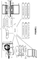
- the MR/PET system 10 with timing optimization and MR based PET attenuation correction maps is schematically shown.
- the system 10 includes a MR scanner (or subsystem) 12 illustrated in partial cross section with a static B 0 main magnetic field such as a horizontal bore scanner, open system scanner, c-type scanner, vertical field scanner, hybrid scanner and the like.
- the MR scanner includes a subject support 14 such as a horizontal bed or couch which supports a subject 16 and moves the subject into the MR scanner bore, static main magnetic field, and field of view during imaging.
- the subject support moves the subject to one or more stations which positions predetermine anatomical portions of the subject in the field of view for imaging.
- the MR scanner 14 includes a main magnet 18 which generates the static main magnetic field (B 0 ) such as a horizontal main magnetic field.
- the MR scanner further includes one or more gradient coils 20 for applying gradient magnetic fields, each of a particular area, strength, and timing to manipulate resonance in the tissues of the subject.
- the MR scanner includes one or more RF coils 22 which generate RF pulses to excite magnetic resonance in the subject 16 in a transmit mode and which can receive resonance signals from the subject in a receive mode.
- the RF coil 22 can include a local coil.
- the system 10 includes a sequence controller 24 and a RF receiver unit 26.
- the sequence controller controls the operation of the imaging sequence which includes a RF transmitter unit 28 controlling the operation of the RF coils 22.
- a gradient controller 30 controls the operation of the gradient coils 20.
- the communication between the controlling unit and the corresponding coils can be wired or wireless.
- the sequence controller provides the instructions for the RF transmitter unit 28 and the gradient controller 30 to apply a selected mDixon pulse sequence to the subject.
- the RF receiver 26 receives MR data 32 which can be stored in storage 34, e.g. MR signals indicative of the magnetic resonance excited in the tissue of the subject.
- the sequence controller 24 is configured to apply ultra-short echo time sequences (UTE).
- UTE pulse sequences are characterized by having very short TEs (echo times) in the range of 0.05-0.20 milliseconds (ms). UTEs can be produced for instance (but this is not limiting) by use of half radio frequency (rf) excitations with radial mapping from the center of k-space.
- rf radio frequency
- the echo times TE in an mDixon pulse sequence is longer than the UTE echo times, for instance 2.3 ms and 4.6 ms (at 1.5T), but, again, these numbers are examplary only and not limiting.
- a timing optimization unit 36 selects a first echo time and a second echo time of the mDixon pulse sequence that minimizes or optimizes a repetition time (TR), with the phase angle difference between water and fat at the first and the second echo time being unequal to 0° and 180°.
- the water-fat phase angle of the first echo, Q 1 is selected such that the angle is greater than 0° or not exactly in-phase.
- the water-fat phase angle of the second echo, Q 2 is selected to minimize the difference of Q 2 - Q 1 for the selected spatial resolution and the available gradient magnetic field strength and rise time of the MR scanner.
- the TR is correspondingly shortened, which reduces the data acquisition time per station, preferably to within a breath hold to reduce respiratory motion artifacts.
- a typically breath hold is 15-20 seconds.
- the first echo time can be selected to be less than 1.2ms and the second echo time can be selected to be less than 2.0 ms, leading to a TR in the order of 3 ms.
- the second echo time is thus substantially shorter than the first in-phase echo time of 4.6 ms and 2.3 ms, respectively.
- the increment in the phase angle difference between water and fat from the first to the second echo time is less than 180°, allowing an unambiguous resolution of a broader range of static main magnetic field strength offsets.
- phase angle differences between water and fat at the first and/or at the second echo time is substantially greater than 0° (eg, greater than 5°) and/or substantially less than 180° (eg, less than 175°), respectively.
- 0° eg, greater than 5°
- 180° eg, less than 175°
- UTE-mDIXON In an embodiment of the invention (referred to herein as "UTE-mDIXON") it is proposed herein to combine the mDixon technique with a UTE pulse sequencing.
- the first echo time (“in-phase image” acquisition)
- the water and fat phase angle at the second echo time (out-of-phase image) is required to be substantially different from 180° and 0°, respectively, in particular, the phase angle at the second echo time is substantially less than 180° (eg 145°-155°) as above in the "pure" mDixon example outside the scope of the invention without UTE.
- the mDixon sequence for the first echo time is replaced by an UTE pulse to acquire a UTE image.
- the scan time in one embodiment, is adapted to the patient.
- the time that the patient can hold his or her breath is estimated or measured.
- the scan time is set to this estimated or measured breath hold time.
- the number of repetitions is selected to provide sufficient data to reconstruct an image with a resolution at least as good as the resolution of the PET or other nuclear image.
- the timing optimization unit 36 calculates the first and the second echo time and the repetition time which optimize the phase angle differences between water and fat at the two echo times given the scan time constraint. If the echo times allow a higher spatial resolution of the MR image than of the PET image, the scan time can be shorted to reduce the breath hold times.
- the data near the center of k-space, which is most sensitive to motion is collected first during the breath hold and the data near the periphery of k-space, which is less sensitive to motion, is collected last or zero filled when motion becomes too great.
- An MR reconstruction unit 38 embodied as a configured processor such as a processor 40 of a workstation 42, receives the RF data, e.g. the demodulated MR signals 32 from the RF receiver 26 and reconstructs at least one anatomical image 44 which differentiates water and fat.
- the processor can construct a separate water image 45 and fat image 46.
- the images can be stored in the memory 34 which can include local and/or solid state memory, local and/or remote disk storage, and the like.
- the memory can include a storage management system such as a Picture Archiving and Communication System (PACS), Departmental Radiology Information System (RIS), and the like.
- PPS Picture Archiving and Communication System
- RIS Departmental Radiology Information System
- An MR tissue classification unit 48 classifies tissues based on the anatomical image 44 with water and fat differentiation and/or the water image 45 and the fat image 46. The classifications are by voxel.
- the MR tissue classification unit 48 assigns attenuation values to the classified tissues.
- An MR anatomical image is generated in which regions attributable to water, fat, bone, and air are denoted and separated. The bone and air regions are differentiated and assigned attenuation values.
- the air and water regions represent soft tissue, the voxels of which are classified based on the relative amounts of water and fat and assigned a corresponding attenuation value. In this manner, various organs, muscles, tendons, and other soft tissues are assigned more accurate attenuation coefficients.
- An attenuation map unit 50 constructs an attenuation map 52 based on the classified tissues and pre-assigned attenuation values for each type of tissue.
- the attenuation map 52 can be stored in the memory 34.
- the attenuation map is constructed using the anatomical image and/or water and fat images without respiratory motion artifacts, which improves the accuracy of the attenuation map.
- the phase angle difference (at at least one of the two echo times) is substantially different from 0° and/or from 180° so as to avoid signal cancellation in particular in fat + water voxels
- the attenuation map can be produced with great fidelity which in turn allows increasing the fidelity of the PET imagery when using the attenuation map for correction as will be explained in more detail below.
- the system includes a PET scanner 54 which is shown as a separate unit for simplicity of illustration. Coupled PET and MR units which share the patient support are contemplated to reduce misalignment errors between the PET image and the MR attenuation map.
- a hybrid PET/MR system e.g.
- an MR system with the gradient coil 20 split to define a gap adjacent the center of the examination region and the PET detectors disposed in the gap, enables the PET and MR data to be collected concurrently.
- Other nuclear imaging devices which utilize attenuation maps, such as Single Proton Emission Computed Tomography (SPECT) devices, are also contemplated.
- the data acquisition of the PET scanner (or nuclear medicine device) and MR scanner 12 can operate simultaneously or sequentially.
- the PET scanner 54 includes a subject support 56, preferably the same as the subject support 14.
- the PET scanner includes detectors located circumferentially around the examination region which receives the subject supported by the subject support 56.
- a PET data acquisition unit 58 receives nuclear decay data, e.g.
- the received nuclear decay data are recorded in list mode, in a data store 60.
- the list mode data are time stamped based on detection time.
- a respiration monitor 61 monitors the patient's respiration cycle and the respiration phase and time are recorded in the list mode data store.
- the nuclear imaging data can be temporally segmented based on respiratory phase, e.g. within a breath hold. In another embodiment, images in two or more respiration phases are generated and transformed into the breath hold phase.
- a PET reconstruction unit 62 reconstructs or generates one or more PET images 64 of the subject with the received nuclear decay data and the constructed attenuation map 52.
- the workstation 42 includes the electronic processor or electronic processing device 40, a display 66, and at least one input device 68.
- the display displays the reconstructed MR images, reconstructed PET images, menus, panels, and user controls, and the like.
- the 'display' 66 or 'display device' as used herein encompasses an output device or a user interface adapted for displaying the images or data.
- a display may output visual, audio, and or tactile data.
- Examples of a display include, but are not limited to: a computer monitor, a television screen, a touch screen, a tactile electronic display, a Braille screen, a cathode ray tube (CRT), a storage tube, a bistable display, electronic paper, a vector display, a flat panel display, a vacuum fluorescent display (VF), a light-emitting diode (LED) display, an electroluminescent display (ELD), a plasma display panel (PDP), a liquid crystal display (LCD), an organic light-emitting diode display (OLED), a projector, and a head-mounted display.
- the input device 68 inputs healthcare practitioner selections.
- the workstation 42 can be a desktop computer, a laptop, a tablet, a mobile computing device, a smartphone, and the like.
- the input device 68 can be a keyboard, a mouse, a microphone, and the like.
- the various units 36, 38, 48, 50, 58, 62 are suitably embodied by an electronic data processing device, such as the electronic processor or electronic processing device 40 of the workstation 42, or by a network-based server computer operatively connected with the workstation 42 by a network 70, or so forth.
- the reconstruction techniques, derivation techniques, and map construction are suitably implemented using a non-transitory storage medium storing instructions (e.g., software) readable by an electronic data processing device and executable by the electronic data processing device.
- the electronic processing device 40 can include a video processor which overlays the MR anatomical image 44, water image 45 and/or fat image 46 and the reconstructed PET image 64.
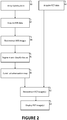
- a first echo time and a second echo time of a modified Dixon (mDixon) pulse sequence are selected that minimize or optimize a repetition time (TR).
- the scan time is adapted to the subject by being matched to the subject's breath hold capability while still having sufficient repetitions, e.g. TRs, to generate an image with a resolution as good as that of the PET image.
- the phase angle difference between water and fat at the first and the second echo time are optimized for the given TR and are unequal to 0° and 180°.
- the phase angle differences are substantially less than 180°,eg is less than 175°.
- the phase angle difference is between 145°-155°. More particularly, the phase angle difference is approximately 150°.
- the phase angle difference at at least one echo time is at least 90°. Said more generally, a phase angle difference (at at least one of the echo times) is ensured so as to substantially avoid signal cancellation for fat + water voxels.
- both the phase angles at the respective echo times are substantially different from 180° and 360° (or 0°), for instance the respective phase angle differences are about 150° (+/- 10° or +/- 15° or +/- 20°) (eg for the second echo time) and/or are about 330°(+/- 10° or +/- 15° or +/- 20°) (eg for the first echo time).
- the UTE-mDIXON pulse sequence where a UTE pulse sequence is used together with the mDixon technique, the phase angle difference of the first measurement (Free Induction Decay), that is at first echo time, may be allowed to be close to 0°, whilst the phase angle difference of the second measurement, that is at second echo time, would still be required to be substantially different from 180° and 0°(or 360°), respectively.
- the phase difference at the second measurement is ensured (as previously mentioned) to be about 150°, or at least to be in the range 150° (+/- 10°).
- UTE pulse sequencing with mDixon in this manner allows distinguishing rapid signal decay due to very short T2*, as in bone structures, from water-fat de-phasing and potential signal cancellation.
- the mDixon pulse sequence is applied to the subject and MR data representing at least a portion of the subject are received in a step 82.
- the MR data is representative of a 3D portion of the subject as MR scanning in a 3D portion allows a more accurate reconstruction of the attenuation map at step 88 below.
- the pulse sequence can be applied during a breath hold of the subject positioned at a predetermined position or station within the MR scanner. For example, first and second echo times of 2ms or less can be applied using a 1.5 T or 3T MR scanner.
- the received MR data are reconstructed into at least an anatomical image in which water and fat are differentiated based on the phase angle differences between water and fat at the first and second echo time.
- Tissues are segmented and classified based on the reconstructed anatomical image in a step 86.
- Voxels corresponding to bone, air, and soft tissue are identified. Based on the relative water and fat content of each soft tissue voxel, the soft tissue voxels are further classified. Attenuation values are assigned to the classified tissue in each voxel.
- An attenuation map is constructed in a step 88.
- nuclear imaging or PET data are acquired for the portion of the subject's anatomy corresponding to the MR data, e.g. in the same field of view.
- the nuclear decay data are generated from gamma photon emissions detected from the portion of the subject in the detector region of the PET scanner.
- the data can be acquired during a breath hold and/or mapped or registered to the attenuation map.
- the acquired PET data and the constructed attenuation map are used to reconstruct one or more PET images in a step 92.
- the reconstructed PET image can include an overlay of the MR anatomical image.
- the reconstructed PET image can include an overlap display of the classified tissues.
- the image can be displayed by the display device and/or stored in the memory for later retrieval and display and/or analysis in a step 94.
- a non-transitory computer-readable storage medium carrying software can be used which controls one or more electronic data processing devices or processors 40 to perform the steps described.
- the acquisition of the nuclear imaging (eg PET) data at step 90 may occur at any stage before step 92.
- the mDixon method with the two echo times is supplemented by an additional readout with an ultra-short echo time (UTE) acquired prior to the two echoes acquired for the mDIXON reconstruction.
- UTE ultra-short echo time
- the systems and related methods as proposed herein may be used in any form of nuclear medicine not merely PET (eg SPECT, and radiation therapy are also envisaged), where MR image data are available and are to be used for attenuation correction purposes.
- the systems and related methods are particularly useful for generation of whole-body attenuation maps, which (at least partially) require data acquisition during breath holding. It is also useful in the context of integrated PET/MR systems as shown above as it imposes less stringent requirements on the MR gradient system.
Landscapes
- Physics & Mathematics (AREA)
- Nuclear Medicine, Radiotherapy & Molecular Imaging (AREA)
- High Energy & Nuclear Physics (AREA)
- Condensed Matter Physics & Semiconductors (AREA)
- General Physics & Mathematics (AREA)
- Health & Medical Sciences (AREA)
- Engineering & Computer Science (AREA)
- General Health & Medical Sciences (AREA)
- Radiology & Medical Imaging (AREA)
- Signal Processing (AREA)
- Pulmonology (AREA)
- Theoretical Computer Science (AREA)
- Artificial Intelligence (AREA)
- Computer Vision & Pattern Recognition (AREA)
- Magnetic Resonance Imaging Apparatus (AREA)
Claims (14)
- Système (10) d'imagerie médicale, comprenant :une unité d'optimisation de synchronisation (40) conçue pour sélectionner i) un premier temps d'écho correspondant à un temps d'écho d'une décroissance d'induction libre mesurée comportant une séquence d'impulsions UTE et un second temps d'écho d'une séquence d'impulsions Dixon, mDixon modifiée, etii) un nombre suffisant de répétitions pour générer une image par résonance magnétique, RM, d'un sujet (16) imagé d'au moins une première résolution, la différence d'angle de phase entre l'eau et la graisse au second temps d'écho étant différente de 0 ° et 180 ° entraînant ainsi la sélection d'une séquence d'impulsions UTE-mDixon ;un balayeur à résonance magnétique RM (12) conçu pour appliquer la séquence d'impulsions UTE-mDixon sélectionnée au sujet (16) et pour recevoir des données RM (32) du sujet, le balayeur RM (12) comprenanti) une commande de gradient (30) conçue pour commander l'application d'au moins un champ magnétique à gradient pour produire un premier et un second écho au premier et au second temps d'écho, etii) au moins une bobine de gradient (20) conçue pour appliquer un champ de gradient au sujet ;une unité de reconstruction RM (38) conçue pour reconstruire l'au moins une image RM (44) en fonction des données RM (32) ;une unité de classification RM (48) conçue pour segmenter et/ou pour classer des tissus en fonction de l'au moins une image RM reconstruite ;une unité de carte d'atténuation (50) conçue pour construire la carte d'atténuation (52) en fonction des tissus segmentés et/ou classés de l'au moins une image RM ; etun système d'imagerie nucléaire (62) conçu pour recevoir des données de désintégration nucléaire et pour générer au moins une image nucléaire (64) de la première résolution en fonction des données de désintégration nucléaire reçues d'un sujet (16) imagé et de la carte d'atténuation (52), dans lequel la génération de l'image nucléaire (64) comprend l'enregistrement de la carte d'atténuation sur les données de désintégration nucléaire reçues, dans lequel la différence d'angle de phase entre l'eau et la graisse au second temps d'écho est dans la plage de 150 ° +/- 10 °.
- Système (10) selon la revendication 1, dans lequel le premier temps d'écho et le second temps d'écho sont respectivement inférieurs à 2,0 ms à 3 T ou inférieurs à 4,0 ms à 1,5 T.
- Système (10) selon l'une quelconque des revendications 1 et 2, le système d'imagerie médicale comprenant un support de sujet (14) pour déplacer le sujet (16) vers au moins une station, dont au moins une position prédétermine au moins une des parties du sujet dans le champ de vision pour l'imagerie, dans lequel le nombre de répétitions définit un temps de balayage par position le long du sujet (16) imagé inférieur ou égal à une durée d'apnée du sujet (16) imagé.
- Système (10) selon l'une quelconque des revendications 1 à 3, dans lequel le système d'imagerie nucléaire comprend :
un balayeur TEP (54) conçu pour générer l' au moins une image nucléaire à partir des émissions de photons gamma détectées chez le sujet. - Système (10) selon les revendications 1 à 4, comprenant en outre :
un processeur vidéo (40) conçu pour superposer l'au moins une image nucléaire (64) générée et l'au moins une image RM (44). - Système selon l'une quelconque des revendications 1 à 5, comprenant en outre :
un dispositif d'affichage (66) conçu pour afficher l'image nucléaire (64) générée. - Système selon l'une quelconque des revendications 1 à 6, dans lequel l'au moins un processeur vidéo (40) est conçu pour superposer le tissu segmenté et/ou classé et l'au moins une image nucléaire (64) générée.
- Procédé d'imagerie médicale, comprenant :la sélection (80) i) d'un premier temps d'écho correspondant à un temps d'écho d'une décroissance d'induction libre mesurée comportant une séquence d'impulsions UTE et d'un second temps d'écho d'une séquence d'impulsions Dixon, mDixon modifiée, etii) d'un nombre suffisant de temps de répétition (TR) pour générer une image du sujet (16) d'au moins une première résolution, la différence d'angle de phase entre l'eau et la graisse au second temps d'écho étant inégal à 0 ° et 180 °;la sélection ainsi d'une séquence d'impulsions UTE-mDixon ;l'application (82) de la séquence d'impulsions UTE-mDixon au sujet (16) et la réception des données RM du sujet, dans lequel l'application (82) comprend la commande de l'application d'au moins un champ magnétique à gradient pour produire un premier et un second écho au premier et au second temps d'écho ;la reconstruction (84) d'au moins une image RM (44) anatomique à partir des données RM (32) reçues ;la segmentation et/ou la classification des tissus en fonction de l'au moins une image RM (44) reconstruite ;la construction (88) d'une carte d'atténuation (52) en fonction des tissus segmentés et/ou classés ;la réception (90) des données de désintégration nucléaire du sujet (16) imagé ;l'enregistrement de la carte d'atténuation construite comportant les données de désintégration nucléaire reçues ;la génération (92) d'au moins une image de la première résolution en fonction des données de désintégration nucléaire reçues et de la carte d'atténuation (52), dans lequel la différence d'angle de phase entre l'eau et la graisse au second temps d'écho est dans la plage de 150 ° +/- 10 °.
- Procédé selon la revendication 8, dans lequel le premier temps d'écho et le second temps d'écho sont respectivement inférieurs à 2,0 ms à 3 T ou inférieurs à 4,0 ms à 1,5 T.
- Procédé selon l'une quelconque des revendications 8 et 9, dans lequel la réception comprend en outre :
la génération (90) des données de désintégration nucléaire à partir des émissions de photons gamma détectées chez le sujet. - Procédé selon l'une quelconque des revendications 8 à 10, dans lequel la génération (92) comprend en outre :
la superposition de l'image TEP reconstruite et de l'au moins une image RM (44) anatomique reconstruite. - Procédé selon l'une quelconque des revendications 8 à 11, comprenant en outre le déplacement du sujet (16) vers au moins une station, dont au moins une position prédétermine au moins une partie anatomique du sujet dans le champ de vision pour l'imagerie, dans lequel le nombre de répétitions définit un temps de balayage par position le long du sujet (16) imagé inférieur ou égal à une durée d'apnée du sujet (16) imagé.
- Support d'enregistrement non transitoire lisible par ordinateur portant un logiciel, lequel, lorsqu'il est exécuté sur au moins un dispositif de traitement des données électroniques (40) d'un balayeur à résonance magnétique, RM, (12) commande le balayeur RM pour mettre en œuvre le procédé selon l'une quelconque des revendications 8 à 12.
- Dispositif de traitement des données électroniques (40) d'un balayeur à résonance magnétique, RM, (12) conçu pour commander le balayeur RM pour mettre en œuvre le procédé selon l'une quelconque des revendications 8 à 12.
Priority Applications (1)
| Application Number | Priority Date | Filing Date | Title |
|---|---|---|---|
| EP14767008.7A EP3049818B1 (fr) | 2013-09-23 | 2014-09-18 | Correction d'atténuation en imagerie tep/irm basée sur l'irm avec séquence d'impulsions de dixon |
Applications Claiming Priority (4)
| Application Number | Priority Date | Filing Date | Title |
|---|---|---|---|
| US201361881018P | 2013-09-23 | 2013-09-23 | |
| EP13189802 | 2013-10-23 | ||
| PCT/EP2014/069910 WO2015040120A1 (fr) | 2013-09-23 | 2014-09-18 | Correction d'atténuation basée sur la rm en imagerie tep/rm avec séquence d'impulsions dixon |
| EP14767008.7A EP3049818B1 (fr) | 2013-09-23 | 2014-09-18 | Correction d'atténuation en imagerie tep/irm basée sur l'irm avec séquence d'impulsions de dixon |
Publications (2)
| Publication Number | Publication Date |
|---|---|
| EP3049818A1 EP3049818A1 (fr) | 2016-08-03 |
| EP3049818B1 true EP3049818B1 (fr) | 2021-08-11 |
Family
ID=49515196
Family Applications (1)
| Application Number | Title | Priority Date | Filing Date |
|---|---|---|---|
| EP14767008.7A Active EP3049818B1 (fr) | 2013-09-23 | 2014-09-18 | Correction d'atténuation en imagerie tep/irm basée sur l'irm avec séquence d'impulsions de dixon |
Country Status (4)
| Country | Link |
|---|---|
| US (1) | US10274562B2 (fr) |
| EP (1) | EP3049818B1 (fr) |
| CN (1) | CN105579861B (fr) |
| WO (1) | WO2015040120A1 (fr) |
Families Citing this family (8)
| Publication number | Priority date | Publication date | Assignee | Title |
|---|---|---|---|---|
| EP3025163A2 (fr) * | 2013-07-23 | 2016-06-01 | Koninklijke Philips N.V. | Différenciation de tissu par imagerie par résonance magnétique |
| JP6280881B2 (ja) * | 2015-02-17 | 2018-02-14 | ジーイー・メディカル・システムズ・グローバル・テクノロジー・カンパニー・エルエルシー | スキャン条件決定装置、磁気共鳴装置、スキャン条件決定方法、およびプログラム |
| KR101844514B1 (ko) * | 2016-09-02 | 2018-04-02 | 삼성전자주식회사 | 자기 공명 영상 장치 및 자기 공명 영상 획득 방법 |
| US10325409B2 (en) * | 2017-06-16 | 2019-06-18 | Microsoft Technology Licensing, Llc | Object holographic augmentation |
| EP3511728A1 (fr) * | 2018-01-12 | 2019-07-17 | Koninklijke Philips N.V. | Technique de dixon en un seul point pour la séparation graisse/eau dans l'imagerie par résonance magnétique du transfert de saturation par échange chimique |
| CN110215226B (zh) * | 2019-05-28 | 2023-10-03 | 上海联影医疗科技股份有限公司 | 图像衰减校正方法、装置、计算机设备和存储介质 |
| CN117110960B (zh) * | 2023-04-06 | 2026-02-27 | 浙江大学 | 基于主磁场图的灵活回波时间水脂分离方法、系统及设备 |
| CN119887993B (zh) * | 2025-03-27 | 2025-06-20 | 上海交通大学医学院附属仁济医院 | 一种生成磁共振质子密度脂肪分数与r2星定量图的方法 |
Family Cites Families (8)
| Publication number | Priority date | Publication date | Assignee | Title |
|---|---|---|---|---|
| EP1139109A1 (fr) * | 2000-03-28 | 2001-10-04 | Bracco International B.V. | Procédé pour l'imagerie des poumons par résonance magnétique |
| JP4251763B2 (ja) * | 2000-08-11 | 2009-04-08 | 株式会社日立メディコ | 磁気共鳴イメージング装置 |
| WO2010018478A1 (fr) * | 2008-08-15 | 2010-02-18 | Koninklijke Philips Electronics, N.V. | Correction d'atténuation pour des systèmes d'imagerie nucléaire pet et spect (tomographie d'émission monophotonique), à l'aide de données d'images spectroscopiques obtenues par résonance magnétique |
| DE102008044844B4 (de) | 2008-08-28 | 2018-08-30 | Siemens Healthcare Gmbh | Verfahren zur Ermittlung einer Schwächungskarte zur Verwendung in der Positronenemissionstomographie und von Homogenitätsinformationen des Magnetresonanzmagnetfeldes |
| BRPI1006422A2 (pt) * | 2009-03-30 | 2020-01-28 | Koninl Philips Electronics Nv | método de diferenciação entre água e tecido adiposo nos dados de imagem de ressonância magnética (irm), meio legível por computador, e, sistema que ultiliza uma técnica de dixon modificada para diferenciar entre água e tecido adiposo nos dados de imagem de ressonância magnética (irm) |
| EP2724172A2 (fr) | 2011-06-27 | 2014-04-30 | Koninklijke Philips N.V. | Imagerie par résonance magnétique d'un tissu osseux |
| US9204817B2 (en) * | 2012-04-19 | 2015-12-08 | General Electric Company | Attenuation correction in positron emission tomography using magnetic resonance imaging |
| JP5966112B1 (ja) | 2013-04-02 | 2016-08-10 | コーニンクレッカ フィリップス エヌ ヴェKoninklijke Philips N.V. | 磁気共鳴画像法を使用した骨組織の検出 |
-
2014
- 2014-09-18 WO PCT/EP2014/069910 patent/WO2015040120A1/fr not_active Ceased
- 2014-09-18 CN CN201480052249.5A patent/CN105579861B/zh not_active Expired - Fee Related
- 2014-09-18 US US14/916,297 patent/US10274562B2/en active Active
- 2014-09-18 EP EP14767008.7A patent/EP3049818B1/fr active Active
Non-Patent Citations (1)
| Title |
|---|
| XIANG QING-SAN: "Two-point water-fat imaging with partially-opposed-phase (POP) acquisition: an asymmetric Dixon method", MAGNETIC RESONANCE IN MEDICINE, JOHN WILEY & SONS, INC, US, vol. 56, no. 3, 1 September 2006 (2006-09-01), pages 572 - 584, XP002586397, ISSN: 0740-3194, [retrieved on 20060807] * |
Also Published As
| Publication number | Publication date |
|---|---|
| CN105579861B (zh) | 2019-07-12 |
| EP3049818A1 (fr) | 2016-08-03 |
| CN105579861A (zh) | 2016-05-11 |
| US10274562B2 (en) | 2019-04-30 |
| US20160202334A1 (en) | 2016-07-14 |
| WO2015040120A1 (fr) | 2015-03-26 |
Similar Documents
| Publication | Publication Date | Title |
|---|---|---|
| EP3049818B1 (fr) | Correction d'atténuation en imagerie tep/irm basée sur l'irm avec séquence d'impulsions de dixon | |
| JP6371776B2 (ja) | 磁気共鳴の拡散強調画像(dwi)のための動作検出および修正方法 | |
| US10698062B2 (en) | Diffusion MRI method for generating a synthetic diffusion image at a high B-value | |
| EP3077836B1 (fr) | Imagerie à résonance magnétique utilisant des séquences d'impulsions avec temps d'écho égal à zéro | |
| US10185013B2 (en) | Magnetic resonance imaging (MRI) apparatus and method of generating MR image | |
| US10215820B2 (en) | Differentiating tissues with MR imaging | |
| EP3229689B1 (fr) | Estimation d'activité hors du champ de vision à l'aide d'une survisualisation et des données antérieures des patients en tomographie par émission de positrons | |
| CN104736062B (zh) | Mr接收线圈定位和基于mr的衰减校正 | |
| JP2014064889A (ja) | 医療映像装置及びその制御方法 | |
| US10274563B2 (en) | Magnetic resonance imaging apparatus and method | |
| US10386441B2 (en) | Method and apparatus for attenuation correction of emission tomography scan data | |
| CN112204411A (zh) | Cest磁共振成像中的运动检测 | |
| CN102749602B (zh) | 采集磁共振图像数据的方法和相应的组合的装置 | |
| US11972857B2 (en) | Automated subject monitoring for medical imaging | |
| US9427206B2 (en) | Method and apparatus for gate specific MR-based attenuation correction of timed-gated pet studies | |
| EP3470866A1 (fr) | Imagerie par résonance magnétique résolue par signal de mouvement | |
| Levin Klausen et al. | Hardware and software approaches to multi-modality imaging |
Legal Events
| Date | Code | Title | Description |
|---|---|---|---|
| PUAI | Public reference made under article 153(3) epc to a published international application that has entered the european phase |
Free format text: ORIGINAL CODE: 0009012 |
|
| 17P | Request for examination filed |
Effective date: 20160425 |
|
| AK | Designated contracting states |
Kind code of ref document: A1 Designated state(s): AL AT BE BG CH CY CZ DE DK EE ES FI FR GB GR HR HU IE IS IT LI LT LU LV MC MK MT NL NO PL PT RO RS SE SI SK SM TR |
|
| AX | Request for extension of the european patent |
Extension state: BA ME |
|
| DAX | Request for extension of the european patent (deleted) | ||
| RAP1 | Party data changed (applicant data changed or rights of an application transferred) |
Owner name: KONINKLIJKE PHILIPS N.V. |
|
| STAA | Information on the status of an ep patent application or granted ep patent |
Free format text: STATUS: EXAMINATION IS IN PROGRESS |
|
| 17Q | First examination report despatched |
Effective date: 20200619 |
|
| GRAP | Despatch of communication of intention to grant a patent |
Free format text: ORIGINAL CODE: EPIDOSNIGR1 |
|
| STAA | Information on the status of an ep patent application or granted ep patent |
Free format text: STATUS: GRANT OF PATENT IS INTENDED |
|
| INTG | Intention to grant announced |
Effective date: 20210302 |
|
| RIN1 | Information on inventor provided before grant (corrected) |
Inventor name: BOERNERT, PETER Inventor name: HU, ZHIQIANG Inventor name: STEHNING, CHRISTIAN Inventor name: EGGERS, HOLGER Inventor name: HU, LINGZHI |
|
| GRAS | Grant fee paid |
Free format text: ORIGINAL CODE: EPIDOSNIGR3 |
|
| GRAA | (expected) grant |
Free format text: ORIGINAL CODE: 0009210 |
|
| STAA | Information on the status of an ep patent application or granted ep patent |
Free format text: STATUS: THE PATENT HAS BEEN GRANTED |
|
| AK | Designated contracting states |
Kind code of ref document: B1 Designated state(s): AL AT BE BG CH CY CZ DE DK EE ES FI FR GB GR HR HU IE IS IT LI LT LU LV MC MK MT NL NO PL PT RO RS SE SI SK SM TR |
|
| REG | Reference to a national code |
Ref country code: GB Ref legal event code: FG4D |
|
| REG | Reference to a national code |
Ref country code: CH Ref legal event code: EP |
|
| REG | Reference to a national code |
Ref country code: DE Ref legal event code: R096 Ref document number: 602014079379 Country of ref document: DE |
|
| REG | Reference to a national code |
Ref country code: IE Ref legal event code: FG4D Ref country code: AT Ref legal event code: REF Ref document number: 1419927 Country of ref document: AT Kind code of ref document: T Effective date: 20210915 |
|
| REG | Reference to a national code |
Ref country code: LT Ref legal event code: MG9D |
|
| REG | Reference to a national code |
Ref country code: NL Ref legal event code: MP Effective date: 20210811 |
|
| REG | Reference to a national code |
Ref country code: AT Ref legal event code: MK05 Ref document number: 1419927 Country of ref document: AT Kind code of ref document: T Effective date: 20210811 |
|
| PG25 | Lapsed in a contracting state [announced via postgrant information from national office to epo] |
Ref country code: HR Free format text: LAPSE BECAUSE OF FAILURE TO SUBMIT A TRANSLATION OF THE DESCRIPTION OR TO PAY THE FEE WITHIN THE PRESCRIBED TIME-LIMIT Effective date: 20210811 Ref country code: SE Free format text: LAPSE BECAUSE OF FAILURE TO SUBMIT A TRANSLATION OF THE DESCRIPTION OR TO PAY THE FEE WITHIN THE PRESCRIBED TIME-LIMIT Effective date: 20210811 Ref country code: RS Free format text: LAPSE BECAUSE OF FAILURE TO SUBMIT A TRANSLATION OF THE DESCRIPTION OR TO PAY THE FEE WITHIN THE PRESCRIBED TIME-LIMIT Effective date: 20210811 Ref country code: PT Free format text: LAPSE BECAUSE OF FAILURE TO SUBMIT A TRANSLATION OF THE DESCRIPTION OR TO PAY THE FEE WITHIN THE PRESCRIBED TIME-LIMIT Effective date: 20211213 Ref country code: NO Free format text: LAPSE BECAUSE OF FAILURE TO SUBMIT A TRANSLATION OF THE DESCRIPTION OR TO PAY THE FEE WITHIN THE PRESCRIBED TIME-LIMIT Effective date: 20211111 Ref country code: ES Free format text: LAPSE BECAUSE OF FAILURE TO SUBMIT A TRANSLATION OF THE DESCRIPTION OR TO PAY THE FEE WITHIN THE PRESCRIBED TIME-LIMIT Effective date: 20210811 Ref country code: FI Free format text: LAPSE BECAUSE OF FAILURE TO SUBMIT A TRANSLATION OF THE DESCRIPTION OR TO PAY THE FEE WITHIN THE PRESCRIBED TIME-LIMIT Effective date: 20210811 Ref country code: LT Free format text: LAPSE BECAUSE OF FAILURE TO SUBMIT A TRANSLATION OF THE DESCRIPTION OR TO PAY THE FEE WITHIN THE PRESCRIBED TIME-LIMIT Effective date: 20210811 Ref country code: BG Free format text: LAPSE BECAUSE OF FAILURE TO SUBMIT A TRANSLATION OF THE DESCRIPTION OR TO PAY THE FEE WITHIN THE PRESCRIBED TIME-LIMIT Effective date: 20211111 Ref country code: AT Free format text: LAPSE BECAUSE OF FAILURE TO SUBMIT A TRANSLATION OF THE DESCRIPTION OR TO PAY THE FEE WITHIN THE PRESCRIBED TIME-LIMIT Effective date: 20210811 |
|
| PG25 | Lapsed in a contracting state [announced via postgrant information from national office to epo] |
Ref country code: PL Free format text: LAPSE BECAUSE OF FAILURE TO SUBMIT A TRANSLATION OF THE DESCRIPTION OR TO PAY THE FEE WITHIN THE PRESCRIBED TIME-LIMIT Effective date: 20210811 Ref country code: LV Free format text: LAPSE BECAUSE OF FAILURE TO SUBMIT A TRANSLATION OF THE DESCRIPTION OR TO PAY THE FEE WITHIN THE PRESCRIBED TIME-LIMIT Effective date: 20210811 Ref country code: GR Free format text: LAPSE BECAUSE OF FAILURE TO SUBMIT A TRANSLATION OF THE DESCRIPTION OR TO PAY THE FEE WITHIN THE PRESCRIBED TIME-LIMIT Effective date: 20211112 |
|
| PG25 | Lapsed in a contracting state [announced via postgrant information from national office to epo] |
Ref country code: NL Free format text: LAPSE BECAUSE OF FAILURE TO SUBMIT A TRANSLATION OF THE DESCRIPTION OR TO PAY THE FEE WITHIN THE PRESCRIBED TIME-LIMIT Effective date: 20210811 |
|
| PG25 | Lapsed in a contracting state [announced via postgrant information from national office to epo] |
Ref country code: DK Free format text: LAPSE BECAUSE OF FAILURE TO SUBMIT A TRANSLATION OF THE DESCRIPTION OR TO PAY THE FEE WITHIN THE PRESCRIBED TIME-LIMIT Effective date: 20210811 |
|
| REG | Reference to a national code |
Ref country code: CH Ref legal event code: PL |
|
| REG | Reference to a national code |
Ref country code: DE Ref legal event code: R097 Ref document number: 602014079379 Country of ref document: DE |
|
| REG | Reference to a national code |
Ref country code: BE Ref legal event code: MM Effective date: 20210930 |
|
| PG25 | Lapsed in a contracting state [announced via postgrant information from national office to epo] |
Ref country code: SM Free format text: LAPSE BECAUSE OF FAILURE TO SUBMIT A TRANSLATION OF THE DESCRIPTION OR TO PAY THE FEE WITHIN THE PRESCRIBED TIME-LIMIT Effective date: 20210811 Ref country code: SK Free format text: LAPSE BECAUSE OF FAILURE TO SUBMIT A TRANSLATION OF THE DESCRIPTION OR TO PAY THE FEE WITHIN THE PRESCRIBED TIME-LIMIT Effective date: 20210811 Ref country code: RO Free format text: LAPSE BECAUSE OF FAILURE TO SUBMIT A TRANSLATION OF THE DESCRIPTION OR TO PAY THE FEE WITHIN THE PRESCRIBED TIME-LIMIT Effective date: 20210811 Ref country code: MC Free format text: LAPSE BECAUSE OF FAILURE TO SUBMIT A TRANSLATION OF THE DESCRIPTION OR TO PAY THE FEE WITHIN THE PRESCRIBED TIME-LIMIT Effective date: 20210811 Ref country code: EE Free format text: LAPSE BECAUSE OF FAILURE TO SUBMIT A TRANSLATION OF THE DESCRIPTION OR TO PAY THE FEE WITHIN THE PRESCRIBED TIME-LIMIT Effective date: 20210811 Ref country code: CZ Free format text: LAPSE BECAUSE OF FAILURE TO SUBMIT A TRANSLATION OF THE DESCRIPTION OR TO PAY THE FEE WITHIN THE PRESCRIBED TIME-LIMIT Effective date: 20210811 Ref country code: AL Free format text: LAPSE BECAUSE OF FAILURE TO SUBMIT A TRANSLATION OF THE DESCRIPTION OR TO PAY THE FEE WITHIN THE PRESCRIBED TIME-LIMIT Effective date: 20210811 |
|
| PLBE | No opposition filed within time limit |
Free format text: ORIGINAL CODE: 0009261 |
|
| STAA | Information on the status of an ep patent application or granted ep patent |
Free format text: STATUS: NO OPPOSITION FILED WITHIN TIME LIMIT |
|
| 26N | No opposition filed |
Effective date: 20220512 |
|
| PG25 | Lapsed in a contracting state [announced via postgrant information from national office to epo] |
Ref country code: LU Free format text: LAPSE BECAUSE OF NON-PAYMENT OF DUE FEES Effective date: 20210918 Ref country code: IT Free format text: LAPSE BECAUSE OF FAILURE TO SUBMIT A TRANSLATION OF THE DESCRIPTION OR TO PAY THE FEE WITHIN THE PRESCRIBED TIME-LIMIT Effective date: 20210811 Ref country code: IE Free format text: LAPSE BECAUSE OF NON-PAYMENT OF DUE FEES Effective date: 20210918 Ref country code: BE Free format text: LAPSE BECAUSE OF NON-PAYMENT OF DUE FEES Effective date: 20210930 |
|
| PG25 | Lapsed in a contracting state [announced via postgrant information from national office to epo] |
Ref country code: SI Free format text: LAPSE BECAUSE OF FAILURE TO SUBMIT A TRANSLATION OF THE DESCRIPTION OR TO PAY THE FEE WITHIN THE PRESCRIBED TIME-LIMIT Effective date: 20210811 Ref country code: LI Free format text: LAPSE BECAUSE OF NON-PAYMENT OF DUE FEES Effective date: 20210930 Ref country code: CH Free format text: LAPSE BECAUSE OF NON-PAYMENT OF DUE FEES Effective date: 20210930 |
|
| PG25 | Lapsed in a contracting state [announced via postgrant information from national office to epo] |
Ref country code: FR Free format text: LAPSE BECAUSE OF NON-PAYMENT OF DUE FEES Effective date: 20211011 |
|
| PG25 | Lapsed in a contracting state [announced via postgrant information from national office to epo] |
Ref country code: HU Free format text: LAPSE BECAUSE OF FAILURE TO SUBMIT A TRANSLATION OF THE DESCRIPTION OR TO PAY THE FEE WITHIN THE PRESCRIBED TIME-LIMIT; INVALID AB INITIO Effective date: 20140918 |
|
| PG25 | Lapsed in a contracting state [announced via postgrant information from national office to epo] |
Ref country code: CY Free format text: LAPSE BECAUSE OF FAILURE TO SUBMIT A TRANSLATION OF THE DESCRIPTION OR TO PAY THE FEE WITHIN THE PRESCRIBED TIME-LIMIT Effective date: 20210811 |
|
| PGFP | Annual fee paid to national office [announced via postgrant information from national office to epo] |
Ref country code: GB Payment date: 20230926 Year of fee payment: 10 |
|
| PGFP | Annual fee paid to national office [announced via postgrant information from national office to epo] |
Ref country code: DE Payment date: 20230928 Year of fee payment: 10 |
|
| REG | Reference to a national code |
Ref country code: DE Ref legal event code: R084 Ref document number: 602014079379 Country of ref document: DE |
|
| PG25 | Lapsed in a contracting state [announced via postgrant information from national office to epo] |
Ref country code: MK Free format text: LAPSE BECAUSE OF FAILURE TO SUBMIT A TRANSLATION OF THE DESCRIPTION OR TO PAY THE FEE WITHIN THE PRESCRIBED TIME-LIMIT Effective date: 20210811 |
|
| PG25 | Lapsed in a contracting state [announced via postgrant information from national office to epo] |
Ref country code: TR Free format text: LAPSE BECAUSE OF FAILURE TO SUBMIT A TRANSLATION OF THE DESCRIPTION OR TO PAY THE FEE WITHIN THE PRESCRIBED TIME-LIMIT Effective date: 20210811 |
|
| REG | Reference to a national code |
Ref country code: GB Ref legal event code: 746 Effective date: 20240904 |
|
| PG25 | Lapsed in a contracting state [announced via postgrant information from national office to epo] |
Ref country code: MT Free format text: LAPSE BECAUSE OF FAILURE TO SUBMIT A TRANSLATION OF THE DESCRIPTION OR TO PAY THE FEE WITHIN THE PRESCRIBED TIME-LIMIT Effective date: 20210811 |
|
| REG | Reference to a national code |
Ref country code: DE Ref legal event code: R119 Ref document number: 602014079379 Country of ref document: DE |
|
| GBPC | Gb: european patent ceased through non-payment of renewal fee |
Effective date: 20240918 |
|
| PG25 | Lapsed in a contracting state [announced via postgrant information from national office to epo] |
Ref country code: DE Free format text: LAPSE BECAUSE OF NON-PAYMENT OF DUE FEES Effective date: 20250401 |
|
| PG25 | Lapsed in a contracting state [announced via postgrant information from national office to epo] |
Ref country code: GB Free format text: LAPSE BECAUSE OF NON-PAYMENT OF DUE FEES Effective date: 20240918 |