EP3054894B1 - Ein verfahren zur herstellung einer bioposthetischen herzklappe - Google Patents
Ein verfahren zur herstellung einer bioposthetischen herzklappe Download PDFInfo
- Publication number
- EP3054894B1 EP3054894B1 EP14852216.2A EP14852216A EP3054894B1 EP 3054894 B1 EP3054894 B1 EP 3054894B1 EP 14852216 A EP14852216 A EP 14852216A EP 3054894 B1 EP3054894 B1 EP 3054894B1
- Authority
- EP
- European Patent Office
- Prior art keywords
- ridges
- tissue
- depressions
- biological tissue
- heart valve
- Prior art date
- Legal status (The legal status is an assumption and is not a legal conclusion. Google has not performed a legal analysis and makes no representation as to the accuracy of the status listed.)
- Active
Links
Images


Classifications
-
- A—HUMAN NECESSITIES
- A61—MEDICAL OR VETERINARY SCIENCE; HYGIENE
- A61F—FILTERS IMPLANTABLE INTO BLOOD VESSELS; PROSTHESES; DEVICES PROVIDING PATENCY TO, OR PREVENTING COLLAPSING OF, TUBULAR STRUCTURES OF THE BODY, e.g. STENTS; ORTHOPAEDIC, NURSING OR CONTRACEPTIVE DEVICES; FOMENTATION; TREATMENT OR PROTECTION OF EYES OR EARS; BANDAGES, DRESSINGS OR ABSORBENT PADS; FIRST-AID KITS
- A61F2/00—Filters implantable into blood vessels; Prostheses, i.e. artificial substitutes or replacements for parts of the body; Appliances for connecting them with the body; Devices providing patency to, or preventing collapsing of, tubular structures of the body, e.g. stents
- A61F2/02—Prostheses implantable into the body
- A61F2/24—Heart valves ; Vascular valves, e.g. venous valves; Heart implants, e.g. passive devices for improving the function of the native valve or the heart muscle; Transmyocardial revascularisation [TMR] devices; Valves implantable in the body
- A61F2/2412—Heart valves ; Vascular valves, e.g. venous valves; Heart implants, e.g. passive devices for improving the function of the native valve or the heart muscle; Transmyocardial revascularisation [TMR] devices; Valves implantable in the body with soft flexible valve members, e.g. tissue valves shaped like natural valves
- A61F2/2415—Manufacturing methods
-
- A—HUMAN NECESSITIES
- A61—MEDICAL OR VETERINARY SCIENCE; HYGIENE
- A61F—FILTERS IMPLANTABLE INTO BLOOD VESSELS; PROSTHESES; DEVICES PROVIDING PATENCY TO, OR PREVENTING COLLAPSING OF, TUBULAR STRUCTURES OF THE BODY, e.g. STENTS; ORTHOPAEDIC, NURSING OR CONTRACEPTIVE DEVICES; FOMENTATION; TREATMENT OR PROTECTION OF EYES OR EARS; BANDAGES, DRESSINGS OR ABSORBENT PADS; FIRST-AID KITS
- A61F2/00—Filters implantable into blood vessels; Prostheses, i.e. artificial substitutes or replacements for parts of the body; Appliances for connecting them with the body; Devices providing patency to, or preventing collapsing of, tubular structures of the body, e.g. stents
- A61F2/02—Prostheses implantable into the body
- A61F2/24—Heart valves ; Vascular valves, e.g. venous valves; Heart implants, e.g. passive devices for improving the function of the native valve or the heart muscle; Transmyocardial revascularisation [TMR] devices; Valves implantable in the body
- A61F2/2412—Heart valves ; Vascular valves, e.g. venous valves; Heart implants, e.g. passive devices for improving the function of the native valve or the heart muscle; Transmyocardial revascularisation [TMR] devices; Valves implantable in the body with soft flexible valve members, e.g. tissue valves shaped like natural valves
-
- A—HUMAN NECESSITIES
- A61—MEDICAL OR VETERINARY SCIENCE; HYGIENE
- A61L—METHODS OR APPARATUS FOR STERILISING MATERIALS OR OBJECTS IN GENERAL; DISINFECTION, STERILISATION OR DEODORISATION OF AIR; CHEMICAL ASPECTS OF BANDAGES, DRESSINGS, ABSORBENT PADS OR SURGICAL ARTICLES; MATERIALS FOR BANDAGES, DRESSINGS, ABSORBENT PADS OR SURGICAL ARTICLES
- A61L27/00—Materials for grafts or prostheses or for coating grafts or prostheses
- A61L27/36—Materials for grafts or prostheses or for coating grafts or prostheses containing ingredients of undetermined constitution or reaction products thereof, e.g. transplant tissue, natural bone, extracellular matrix
- A61L27/3604—Materials for grafts or prostheses or for coating grafts or prostheses containing ingredients of undetermined constitution or reaction products thereof, e.g. transplant tissue, natural bone, extracellular matrix characterised by the human or animal origin of the biological material, e.g. hair, fascia, fish scales, silk, shellac, pericardium, pleura, renal tissue, amniotic membrane, parenchymal tissue, fetal tissue, muscle tissue, fat tissue, enamel
- A61L27/3625—Vascular tissue, e.g. heart valves
-
- A—HUMAN NECESSITIES
- A61—MEDICAL OR VETERINARY SCIENCE; HYGIENE
- A61L—METHODS OR APPARATUS FOR STERILISING MATERIALS OR OBJECTS IN GENERAL; DISINFECTION, STERILISATION OR DEODORISATION OF AIR; CHEMICAL ASPECTS OF BANDAGES, DRESSINGS, ABSORBENT PADS OR SURGICAL ARTICLES; MATERIALS FOR BANDAGES, DRESSINGS, ABSORBENT PADS OR SURGICAL ARTICLES
- A61L27/00—Materials for grafts or prostheses or for coating grafts or prostheses
- A61L27/36—Materials for grafts or prostheses or for coating grafts or prostheses containing ingredients of undetermined constitution or reaction products thereof, e.g. transplant tissue, natural bone, extracellular matrix
- A61L27/3683—Materials for grafts or prostheses or for coating grafts or prostheses containing ingredients of undetermined constitution or reaction products thereof, e.g. transplant tissue, natural bone, extracellular matrix subjected to a specific treatment prior to implantation, e.g. decellularising, demineralising, grinding, cellular disruption/non-collagenous protein removal, anti-calcification, crosslinking, supercritical fluid extraction, enzyme treatment
-
- A—HUMAN NECESSITIES
- A61—MEDICAL OR VETERINARY SCIENCE; HYGIENE
- A61L—METHODS OR APPARATUS FOR STERILISING MATERIALS OR OBJECTS IN GENERAL; DISINFECTION, STERILISATION OR DEODORISATION OF AIR; CHEMICAL ASPECTS OF BANDAGES, DRESSINGS, ABSORBENT PADS OR SURGICAL ARTICLES; MATERIALS FOR BANDAGES, DRESSINGS, ABSORBENT PADS OR SURGICAL ARTICLES
- A61L27/00—Materials for grafts or prostheses or for coating grafts or prostheses
- A61L27/36—Materials for grafts or prostheses or for coating grafts or prostheses containing ingredients of undetermined constitution or reaction products thereof, e.g. transplant tissue, natural bone, extracellular matrix
- A61L27/3683—Materials for grafts or prostheses or for coating grafts or prostheses containing ingredients of undetermined constitution or reaction products thereof, e.g. transplant tissue, natural bone, extracellular matrix subjected to a specific treatment prior to implantation, e.g. decellularising, demineralising, grinding, cellular disruption/non-collagenous protein removal, anti-calcification, crosslinking, supercritical fluid extraction, enzyme treatment
- A61L27/3691—Materials for grafts or prostheses or for coating grafts or prostheses containing ingredients of undetermined constitution or reaction products thereof, e.g. transplant tissue, natural bone, extracellular matrix subjected to a specific treatment prior to implantation, e.g. decellularising, demineralising, grinding, cellular disruption/non-collagenous protein removal, anti-calcification, crosslinking, supercritical fluid extraction, enzyme treatment characterised by physical conditions of the treatment, e.g. applying a compressive force to the composition, pressure cycles, ultrasonic/sonication or microwave treatment, lyophilisation
-
- A—HUMAN NECESSITIES
- A61—MEDICAL OR VETERINARY SCIENCE; HYGIENE
- A61L—METHODS OR APPARATUS FOR STERILISING MATERIALS OR OBJECTS IN GENERAL; DISINFECTION, STERILISATION OR DEODORISATION OF AIR; CHEMICAL ASPECTS OF BANDAGES, DRESSINGS, ABSORBENT PADS OR SURGICAL ARTICLES; MATERIALS FOR BANDAGES, DRESSINGS, ABSORBENT PADS OR SURGICAL ARTICLES
- A61L27/00—Materials for grafts or prostheses or for coating grafts or prostheses
- A61L27/50—Materials characterised by their function or physical properties, e.g. injectable or lubricating compositions, shape-memory materials, surface modified materials
- A61L27/507—Materials characterised by their function or physical properties, e.g. injectable or lubricating compositions, shape-memory materials, surface modified materials for artificial blood vessels
-
- B—PERFORMING OPERATIONS; TRANSPORTING
- B23—MACHINE TOOLS; METAL-WORKING NOT OTHERWISE PROVIDED FOR
- B23K—SOLDERING OR UNSOLDERING; WELDING; CLADDING OR PLATING BY SOLDERING OR WELDING; CUTTING BY APPLYING HEAT LOCALLY, e.g. FLAME CUTTING; WORKING BY LASER BEAM
- B23K26/00—Working by laser beam, e.g. welding, cutting or boring
- B23K26/0006—Working by laser beam, e.g. welding, cutting or boring taking account of the properties of the material involved
-
- B—PERFORMING OPERATIONS; TRANSPORTING
- B23—MACHINE TOOLS; METAL-WORKING NOT OTHERWISE PROVIDED FOR
- B23K—SOLDERING OR UNSOLDERING; WELDING; CLADDING OR PLATING BY SOLDERING OR WELDING; CUTTING BY APPLYING HEAT LOCALLY, e.g. FLAME CUTTING; WORKING BY LASER BEAM
- B23K26/00—Working by laser beam, e.g. welding, cutting or boring
- B23K26/02—Positioning or observing the workpiece, e.g. with respect to the point of impact; Aligning, aiming or focusing the laser beam
- B23K26/06—Shaping the laser beam, e.g. by masks or multi-focusing
- B23K26/062—Shaping the laser beam, e.g. by masks or multi-focusing by direct control of the laser beam
- B23K26/0622—Shaping the laser beam, e.g. by masks or multi-focusing by direct control of the laser beam by shaping pulses
- B23K26/0624—Shaping the laser beam, e.g. by masks or multi-focusing by direct control of the laser beam by shaping pulses using ultrashort pulses, i.e. pulses of 1 ns or less
-
- B—PERFORMING OPERATIONS; TRANSPORTING
- B23—MACHINE TOOLS; METAL-WORKING NOT OTHERWISE PROVIDED FOR
- B23K—SOLDERING OR UNSOLDERING; WELDING; CLADDING OR PLATING BY SOLDERING OR WELDING; CUTTING BY APPLYING HEAT LOCALLY, e.g. FLAME CUTTING; WORKING BY LASER BEAM
- B23K26/00—Working by laser beam, e.g. welding, cutting or boring
- B23K26/08—Devices involving relative movement between laser beam and workpiece
- B23K26/082—Scanning systems, i.e. devices involving movement of the laser beam relative to the laser head
-
- B—PERFORMING OPERATIONS; TRANSPORTING
- B23—MACHINE TOOLS; METAL-WORKING NOT OTHERWISE PROVIDED FOR
- B23K—SOLDERING OR UNSOLDERING; WELDING; CLADDING OR PLATING BY SOLDERING OR WELDING; CUTTING BY APPLYING HEAT LOCALLY, e.g. FLAME CUTTING; WORKING BY LASER BEAM
- B23K26/00—Working by laser beam, e.g. welding, cutting or boring
- B23K26/352—Working by laser beam, e.g. welding, cutting or boring for surface treatment
- B23K26/355—Texturing
-
- A—HUMAN NECESSITIES
- A61—MEDICAL OR VETERINARY SCIENCE; HYGIENE
- A61F—FILTERS IMPLANTABLE INTO BLOOD VESSELS; PROSTHESES; DEVICES PROVIDING PATENCY TO, OR PREVENTING COLLAPSING OF, TUBULAR STRUCTURES OF THE BODY, e.g. STENTS; ORTHOPAEDIC, NURSING OR CONTRACEPTIVE DEVICES; FOMENTATION; TREATMENT OR PROTECTION OF EYES OR EARS; BANDAGES, DRESSINGS OR ABSORBENT PADS; FIRST-AID KITS
- A61F2/00—Filters implantable into blood vessels; Prostheses, i.e. artificial substitutes or replacements for parts of the body; Appliances for connecting them with the body; Devices providing patency to, or preventing collapsing of, tubular structures of the body, e.g. stents
- A61F2/0095—Packages or dispensers for prostheses or other implants
-
- A—HUMAN NECESSITIES
- A61—MEDICAL OR VETERINARY SCIENCE; HYGIENE
- A61F—FILTERS IMPLANTABLE INTO BLOOD VESSELS; PROSTHESES; DEVICES PROVIDING PATENCY TO, OR PREVENTING COLLAPSING OF, TUBULAR STRUCTURES OF THE BODY, e.g. STENTS; ORTHOPAEDIC, NURSING OR CONTRACEPTIVE DEVICES; FOMENTATION; TREATMENT OR PROTECTION OF EYES OR EARS; BANDAGES, DRESSINGS OR ABSORBENT PADS; FIRST-AID KITS
- A61F2/00—Filters implantable into blood vessels; Prostheses, i.e. artificial substitutes or replacements for parts of the body; Appliances for connecting them with the body; Devices providing patency to, or preventing collapsing of, tubular structures of the body, e.g. stents
- A61F2/02—Prostheses implantable into the body
- A61F2/24—Heart valves ; Vascular valves, e.g. venous valves; Heart implants, e.g. passive devices for improving the function of the native valve or the heart muscle; Transmyocardial revascularisation [TMR] devices; Valves implantable in the body
- A61F2/2412—Heart valves ; Vascular valves, e.g. venous valves; Heart implants, e.g. passive devices for improving the function of the native valve or the heart muscle; Transmyocardial revascularisation [TMR] devices; Valves implantable in the body with soft flexible valve members, e.g. tissue valves shaped like natural valves
- A61F2/2418—Scaffolds therefor, e.g. support stents
-
- A—HUMAN NECESSITIES
- A61—MEDICAL OR VETERINARY SCIENCE; HYGIENE
- A61F—FILTERS IMPLANTABLE INTO BLOOD VESSELS; PROSTHESES; DEVICES PROVIDING PATENCY TO, OR PREVENTING COLLAPSING OF, TUBULAR STRUCTURES OF THE BODY, e.g. STENTS; ORTHOPAEDIC, NURSING OR CONTRACEPTIVE DEVICES; FOMENTATION; TREATMENT OR PROTECTION OF EYES OR EARS; BANDAGES, DRESSINGS OR ABSORBENT PADS; FIRST-AID KITS
- A61F2220/00—Fixations or connections for prostheses classified in groups A61F2/00 - A61F2/26 or A61F2/82 or A61F9/00 or A61F11/00 or subgroups thereof
- A61F2220/0025—Connections or couplings between prosthetic parts, e.g. between modular parts; Connecting elements
- A61F2220/0075—Connections or couplings between prosthetic parts, e.g. between modular parts; Connecting elements sutured, ligatured or stitched, retained or tied with a rope, string, thread, wire or cable
-
- A—HUMAN NECESSITIES
- A61—MEDICAL OR VETERINARY SCIENCE; HYGIENE
- A61L—METHODS OR APPARATUS FOR STERILISING MATERIALS OR OBJECTS IN GENERAL; DISINFECTION, STERILISATION OR DEODORISATION OF AIR; CHEMICAL ASPECTS OF BANDAGES, DRESSINGS, ABSORBENT PADS OR SURGICAL ARTICLES; MATERIALS FOR BANDAGES, DRESSINGS, ABSORBENT PADS OR SURGICAL ARTICLES
- A61L2430/00—Materials or treatment for tissue regeneration
- A61L2430/20—Materials or treatment for tissue regeneration for reconstruction of the heart, e.g. heart valves
-
- A—HUMAN NECESSITIES
- A61—MEDICAL OR VETERINARY SCIENCE; HYGIENE
- A61L—METHODS OR APPARATUS FOR STERILISING MATERIALS OR OBJECTS IN GENERAL; DISINFECTION, STERILISATION OR DEODORISATION OF AIR; CHEMICAL ASPECTS OF BANDAGES, DRESSINGS, ABSORBENT PADS OR SURGICAL ARTICLES; MATERIALS FOR BANDAGES, DRESSINGS, ABSORBENT PADS OR SURGICAL ARTICLES
- A61L2430/00—Materials or treatment for tissue regeneration
- A61L2430/40—Preparation and treatment of biological tissue for implantation, e.g. decellularisation, cross-linking
-
- B—PERFORMING OPERATIONS; TRANSPORTING
- B23—MACHINE TOOLS; METAL-WORKING NOT OTHERWISE PROVIDED FOR
- B23K—SOLDERING OR UNSOLDERING; WELDING; CLADDING OR PLATING BY SOLDERING OR WELDING; CUTTING BY APPLYING HEAT LOCALLY, e.g. FLAME CUTTING; WORKING BY LASER BEAM
- B23K2103/00—Materials to be soldered, welded or cut
- B23K2103/30—Organic materials
- B23K2103/32—Material from living organisms, e.g. skins
Definitions
- the invention relates to a method of treating biological tissue and, more particularly, to a method for imparting topographical changes onto a surface of a bioprosthetic tissue to direct or control patterns of cellular migration and tissue formation on the tissue.
- Bio tissue such as animal pericardium (e.g., bovine, porcine), has come into common use in connection with medical devices and implantable bioprostheses, such as bioprosthetic heart valves and vascular patches.
- implantable bioprostheses such as bioprosthetic heart valves and vascular patches.
- the biological tissue Once implanted in the body, the biological tissue may become calcified or may stimulate the formation of scar or other types of tissue, such as pannus.
- the biological tissue may also stimulate cells to migrate into and integrate the implanted biological tissue into the host body.
- pannus is a membrane of granulation tissue, rich in fibroblasts, that forms in response to healing.
- the body may produce pannus where the native valve has been removed and a prosthetic heart valve has been implanted.
- pannus growth does not encroach the valve orifice or chamber space, but occasionally the hanging edges can hit or obstruct a leaflet.
- pannus overgrowth interferes with valve functioning, surgery is the only option to remove the pannus overgrowth. There is no reliable way of predicting whether a particular patient will be more or less susceptible to pannus overgrowth.
- Pannus overgrowth is estimated to be the cause of obstructive heart valve failure in a significant number of cases. In the aortic position, pannus formation occurs mainly on the inflow or ventricular side, while in the mitral position, it occurs both on the atrial and ventricular sides.
- prosthetic valves In order to prevent tissue ingrowth into the clearance of leaflets, prosthetic valves have been designed with longer housing cylinders with the goal of creating an ingrowth barrier.
- Such valve designs are often impractical, infeasible or undesirable for minimally-invasive or percutaneously-deliverable heart valves, as an important design goal for such valves is to reduce the delivery profile as much as practically possible. Thus, the inclusion of additional features or structures which add to the material bulk of the valves is generally avoided.
- US 2002/0173842 A1 discloses a valve prosthesis which includes a plurality of flexible leaflets and a stent having a central lumen.
- a slit in the stent extends from the central lumen to an outer surface.
- An occluding portion of the flexible leaflets extends across the central lumen and an attachment portion extends from the central lumen through the slit and forms a sewing cuff for attachment to a patient's tissue.
- the attachment portion forms a sewing cuff for attachment to native heart tissue.
- WO 98/32400 A1 discloses a prosthetic valve having a generally annular frame with three posts and three scallops.
- the frame is tri-symmetric with an axis of symmetry defined by the axis of blood flow through the valve.
- the external surface of the frame is generally cylindrical with diameter D.
- Each leaflet has a truncated spherical surface adjacent to its free edge.
- the spherical surface is joined tangentially to a truncated conical surface.
- the half angle of the truncated cone is approximately 37.5°.
- the radius of the sphere is approximately D/2 - 0.5 (mm).
- the leaflet surface is axi-symmetrical with the axis of symmetry being perpendicular to the axis of the valve frame and blood flow.
- US 2007/0233228 A1 discloses a prosthetic stented heart valve which includes a compressible and expandable stent structure having first and second opposite ends, an expanded outer periphery, and a compressed outer periphery that is at least slightly smaller than the expanded outer periphery when subjected to an external radial force.
- the valve further includes a valve segment comprising a dual-layer sheet formed into a generally tubular shape having at least one longitudinally extending seam, and a plurality of leaflets formed by attachment of an outer layer of the dual-layer sheet to an inner layer of the dual-layer sheet in a leaflet defining pattern.
- the valve segment is at least partially positioned within the stent structure.
- the valve may further include at least one opening in the outer layer of the dual-layer sheet that is spaced from both the first and second ends of the stent structure.
- WO 2007/016251 A2 discloses medical devices for implantation within a body vessel comprising a thromboresistant material.
- the thromboresistant material preferably comprises a biocompatible polyurethane, a remodelable material, a bioactive agent or any combination thereof.
- the medical device can be a prosthetic valve comprising a thromboresistant material.
- the medical device can also comprise a support frame with one or more valve leaflets attached to the support frame.
- bioprosthetic heart valves which control pannus formation or which reduce or eliminate the likelihood of pannus overgrowth that interferes with proper valve functioning.
- the present invention is defined by the appended claims. More specifically, the invention relates to a contoured biological tissue for forming a leaflet of a bioprosthetic heart valve, and a method of preparing the contoured tissue.
- a predetermined pattern is provided on the tissue, comprising a plurality of ridges or depressions that are configured to facilitate cellular migration in a first direction and discourage cellular migration in a second direction.
- the biological tissue can be used in connection with a bioprosthetic heart valve comprising a biological tissue leaflet structure coupled to a supporting frame.
- a method for manufacturing a bioprosthetic heart valve comprises contouring a portion of a biological tissue valve leaflet with a predetermined pattern.
- the predetermined pattern comprises a plurality of ridges or depressions oriented in the same or different directions.
- One or more leaflets are coupled to a support frame. Adjacent ridges or depressions are at least 10 microns apart to prevent or at least impede the migration of fibroblasts transversely across the ridges or depressions.
- the contouring can be performed with a laser, preferably a femtosecond laser.
- the biological tissue can be at least partially crosslinked, and/or at least partially dehydrated, such as with a glycerin-based treatment solution, before the contouring.
- the biological tissue can be treated with a capping agent after the crosslinking, after the contouring, or both.
- the method can further comprise packaging the bioprosthetic heart valve in a package that does not contain a liquid storage solution in contact with the bioprosthetic heart valve.
- a contoured bioprosthetic heart valve comprises one or a plurality of leaflets formed from a biological tissue and coupled to a support frame.
- a contoured pattern is provided on the leaflets.
- the contoured pattern comprises a plurality of ridges or depressions, the distance between adjacent ridges or depressions being at least 10 microns.
- the plurality of ridges or depressions can comprise a first set of parallel depressions or ridges, and can further comprise a second set of parallel ridges or grooves transversing the first set of parallel ridges or grooves at an angle.
- the bioprosthetic heart valve can further comprise sutures coupling the one or more leaflets to a support structure or a skirt.
- the predetermined pattern can be disposed on the leaflets adjacent to at least a portion of the sutures.
- the one or plurality of heart valve leaflets can each comprise a straight free edge and an arcuate cusp edge and the contoured pattern is provided substantially along the arcuate cusp edge.
- Another contoured bioprosthetic heart valve for implantation within an arterial wall of a patient comprises a biological tissue leaflet structure coupled to a supporting frame.
- the biological tissue leaflet structure has a circumferential outer peripheral surface facing the arterial wall.
- a contoured pattern is provided around the circumferential outer peripheral surface.
- the contoured pattern comprises a plurality of ridges or depressions, spaced and/or sized to prevent or impede cellular migration across the ridges or depressions.
- the adjacent ridges or depressions are preferably at least 10 microns apart.
- the contoured pattern can be provided along an entire length of sutures coupling the biological tissue leaflet structure to the supporting frame.
- Yet another contoured bioprosthetic heart valve comprises one or a plurality of leaflets formed from a biological tissue.
- the leaflets each comprise a straight free edge and an arcuate cusp edge.
- a contoured pattern is provided on the leaflets.
- the contoured pattern comprises a plurality of ridges or depressions extending radially from the straight free edge to the arcuate cusp edge. Cellular migration is promoted along the ridges or depressions.
- a method for treating a biological tissue comprises providing a tissue having a planar surface, treating the tissue to reduce surface irregularities, and contouring the tissue surface with a predetermined pattern comprising a plurality of ridges or depressions.
- the ridges and depressions are provided to encourage cellular migration in a first direction and discourage cellular migration in a second direction.
- the distance between adjacent ridges or depressions is at least 10 microns.
- the tissue can be reduced in thickness to a range of about 250-500 microns by compressing the tissue or by removing material from the tissue.
- the contouring can be performed by a laser, preferably a femtosecond laser.
- the tissue can be at least partially dried or dehydrated before the contouring, such as by using a glycerin-based treatment solution.
- the tissue can then be packaged in a package that does not contain a liquid storage solution in contact with the biological tissue.
- Yet another embodiment comprises a biological tissue for forming a leaflet of a bioprosthetic heart valve, the tissue having a surface contoured with a predetermined pattern comprising a plurality of ridges or depressions configured to encourage cellular migration in a first direction and discourage cellular migration in a second direction.
- the distance between adjacent ridges or depressions is at least 10 microns.
- the biological tissue is treated such that the tissue is thinned (by compression or by removal of tissue material) and/or the tissue surface is smoothed (by compression and/or crosslinking), as described in U.S. Pub. No. 2011/0238167, published September 29, 2011 , and U.S. Pat. No. 7,141,064, issued November 28, 2006 .
- the biological tissue is thinned to a thickness of about 100-800 microns, preferably about 250-500 microns and most preferably about 100-300 microns.
- FIGS. 1A-1B depict a bioprosthetic heart valve leaflet 100A having a plurality of parallel grooves 120.
- the leaflet 100A is depicted herein as comprising an arcuate cusp edge 104, a generally straight free edge 102 opposite the cusp edge 104, and a pair of oppositely-directed tabs 106 at either end of the free edge 102.
- a plurality of depressions, e.g., grooves, or raised ridges 120 is provided on one side of the leaflet surface and is patterned to follow the shape of the arcuate cusp edge 104.
- the parallel grooves 120 can be provided on substantially the entire surface of the leaflet 100A or, as shown in FIG.
- the parallel grooves can be provided only along the area adjacent the cusp edge 104 of the leaflet 100B.
- the patterns of parallel grooves 120 depicted in FIGS. 1A-1B are provided to discourage cellular migration in a transverse direction originating from the periphery or central region of the leaflet towards the central region or periphery of the leaflet, respectively.
- parallel refers to two paths that are substantially identical but are offset so as to never intersect. Accordingly, “parallel” is broadly understood to encompass lines, curves and wavy lines that follow the same path but are offset from one another by a constant or variable distance. In a preferred embodiment, adjacent paths are offset by a constant distance.
- transversely refers to a path that intersects the contoured pattern, e.g., grooves or ridges, and is not limited to a particular angle at which the path intersects the contoured pattern.
- Fibroblasts are a class of cells that synthesize the extracellular matrix and collagen, the structural framework for animal tissues. Fibroblasts play a critical role in wound healing. Implantation of bioprosthetic heart valves at a valve annulus stimulates fibroblasts to migrate and form a pannus around the heart valve and, more specifically, in the areas surrounding the suture lines of a bioprosthetic heart valve. It is therefore believed that directing the migration of cells (e.g., fibroblasts) responsible for pannus formation will similarly help control the extent and location of pannus growth.
- cells e.g., fibroblasts
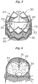
- FIG. 5 depicts an embodiment of a bioprosthetic heart valve 50 as generally comprising a structural frame or stent 52, a flexible leaflet structure 54 supported by the frame 52 and a flexible skirt 56 secured to the outer surface of the leaflet structure 54.
- the valve 50 can be implanted in the annulus of the native aortic valve, but also can be adapted to be implanted in other native valves of the heart or in various other ducts or orifices of the body.
- Valve 50 has an inflow end 60 and an outflow end 62.
- Valve 50 and frame 52 are configured to be radially collapsible to a collapsed or crimped state for introduction into the body on a delivery catheter and radially expandable to an expanded state for implanting the valve at a desired location in the body, such as the native aortic valve.
- Leaflet structure 54 desirably comprises three separate connected leaflets 70, which are individually depicted in FIGS. 1-3 .
- the individual leaflets can be arranged together to coapt in a tricuspid arrangement, as best shown in FIGS. 5 and 6 .
- the leaflets 70 attach to one another at their adjacent sides to form commissures 80 of the leaflet structure 54.
- the leaflets are made of biological tissue, preferably bovine or porcine pericardium, and have a thickness in the range of about 100 to about 800 microns.
- the curved cusp edge forms a single scallop in the leaflet structure 54 and when secured to two other leaflets 70 to form the leaflet structure 54, the curved cusp edges collectively form a scallop-shaped lower edge of the leaflet structure.
- a suture line 84 visible on the exterior of the skirt 56 tracks the scalloped shape of the leaflet structure 54.
- the skirt 56 can be formed, for example, of polyethylene terephthalate (PET) or pericardium, preferably bovine pericardium, ribbon.
- PET polyethylene terephthalate
- the leaflet structure 54 attaches to the skirt via a thin PET reinforcing strip 88 or sleeve.
- the leaflet structure 54 is sandwiched between skirt 56 and the reinforcing strip 88.
- the suture 84, which secures the reinforcing strip and the leaflet structure 54 to skirt 56 can be any suitable suture, and desirably tracks the curvature of the bottom edge of the leaflet structure 54 as seen on the exterior of the skirt 56 in FIG. 5 .
- the skirt 56 and leaflet structure 54 preferably reside inside the frame 52.
- pannus growth on bioprosthetic heart valves typically occurs along the suture lines.
- pannus growth would most likely originate along the suture line 84 which corresponds to the cusp edge 104 of the leaflets 100A and 100B in FIGS. 1A and 1B , typically along the outwardly facing external surface or the circumferential outer peripheral surface of the valve 50 that is in contact with the arterial wall or body lumen.
- a plurality of grooves or ridges 120 in a pattern that follows the suture line 84 will discourage or reduce the extent to which pannus growth occurs in a transverse direction to the suture line 84 and reduce the extent to which pannus overgrowth will reach the inflow or outflow orifices or otherwise interfere with the coaptation of the leaflets.
- adjacent grooves or ridges 120 are both dimensioned and offset at a distance that is greater than the average size of a fibroblast or other cell associated with pannus formation.
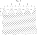
- FIG. 4 is a cross-sectional view along 4-4 of FIG. 1B depicting the spacing B between adjacent grooves and the width A and depth C of each groove 120.
- Fibroblasts are believed to have a size in the range of 10 to 40 microns and more typically from 10 to 20 microns.
- adjacent grooves 120 are offset at a distance B of at least 10 microns, preferably at least 20 microns, more preferably at least 30 microns and most preferably at least 40 microns.
- one or both of the width A and depth C of the grooves 120 are dimensioned to be at least as large as, if not larger than, the average size of a fibroblast or other cell associated with pannus formation.
- adjacent ridges or depressions are at least 10 microns apart to prevent or at least impede the migration of fibroblasts transversely across the ridges or depressions
- one or both of the width A and depth C of each individual groove 120 is at least 10 microns, preferably at least 20 microns, more preferably at least 30 microns and most preferably at least 40 microns.
- the depth C of the grooves 120 is selected so as to not compromise the strength of the tissue leaflets.
- the grooves 120 are provided on only the one side of the leaflet that faces the arterial or cardiac wall when the valve is implanted.
- the depth C of the grooves 120 preferably does not exceed 25%, preferably 10% and more preferably 5% and most preferably 2% of the average thickness of the leaflet.
- the depth C of the grooves preferably does not exceed 62.5 microns, preferably 25 microns, more preferably 12.5 microns, and most preferably 5 microns.
- a width of less than 5 microns it is understood that at least one of the other parameters, e.g., the distance B between grooves 120 or the width A of the grooves, is preferably selected to exceed the average cell size of at least 10 microns.
- the values for A, B and C are selected to sequester or trap the fibroblasts or cells within grooves and/or between adjacent grooves 120.
- the consideration with respect to selecting the distance between ridges and height of individual ridges is analogous to the distance between grooves and the depth of the grooves.
- FIG. 2 depicts a bioprosthetic heart valve leaflet 200 having a grid pattern 210 disposed along an area adjacent the cusp edge 204 opposite the free edge 202.
- the grid pattern is provided by two sets of intersecting parallel grooves or ridges which define a plurality of square-shaped cells.
- grooves are employed, the same considerations as to the distance between adjacent grooves and the width and depth of the grooves as discussed in relation to FIGS. 1A and 1B apply.
- ridges it is the dimension of the individual cells formed by the intersecting ridges that are operative in preventing cellular migration.
- the two sets of intersecting parallel ridges need not be at right angles to one another so long as the individual cells themselves are sized or dimensioned to be larger than the average size of a fibroblast or other cell associated with pannus formation.
- the individual cells formed in the tissue have at least a width and a length of at least 10 microns, preferably at least 20 microns, more preferably at least 30 microns and most preferably at least 40 microns.
- FIG. 3 depicts a bioprosthetic heart valve leaflet 300 in which radial grooves 310 are provided as originating from a point 312 on the free edge 302 of the leaflet and extending to the arcuate cusp edge 304. While not depicted in FIG. 3 , the radial grooves 310 can also extend into the tab portions 306 in a similar manner. The radial grooves 310 function to direct the cellular migration along their length. Because cellular interaction is desired, the distance between adjacent grooves can be no larger than the average size of fibroblasts or other cells associated with pannus formation as measured from the cusp edge.
- this distance is less than 40 microns, preferably less than 30 microns, more preferably less than 20 microns and most preferably less than 10 microns. Smaller distances between radial grooves 310 will permit migration transversely across the radial grooves 310.
- contouring of the biological tissue has been described and depicted herein with respect to heart valve leaflets, it is understood that such contouring can be performed on any biological tissue that is intended for implantation in the body.
- the contouring can be provided in connection with biological tissue patches which are used for repair in cardiac and vascular reconstruction, soft tissue deficiency repair, valve leaflet repair, carotid repair, closure of pericardial defects, and suture line reinforcement during general surgical procedures.
- the dimensions and the location of the contouring would depend upon the intended result: to stimulate cellular migration along a specified area of the tissue or to deter cellular migration and thus tissue formation in specified areas of the tissue.
- a groove being dimensioned with one or both of a width and depth that is greater than the average diameter of a cell (e.g., 10, 20, 30, or 40 microns) would likely trap the cells, prevent cellular migration transversely across the groove and thus hinder the cells from forming a network necessary to create unwanted tissue formation outside of the groove.
- a ridge can be configured to have a height that effectively prevents cellular migration across the ridge and thus accomplish the same result.
- the laser is a femtosecond laser.
- the laser includes a dual axis scanning lens, 2x beam expansion, 1550 nm wavelength, 31.5 ⁇ J pulse energy on target; 1.6 W average power, 50 Hz repetition rate, 650 fs pulse width (ref); 30 ⁇ m laser spot size, elliptical polarization, 112 mm focal length, 400 mm/s coarse milling speed (20 ⁇ m fill spacing in cross hatch pattern), and 800 mm/s fine milling speed (20 ⁇ m fill spacing in cross hatch pattern).
- the laser is coupled to a guiding device.
- a substantial amount of technology has been developed for guiding lasers and ablating tissue with great precision. Corneal ablation has been widely practiced and excimer lasers have become common. Reference is made to U.S. Pat. No. 4,840,175 . Recent work with mode locking lasers having very short pulse lengths in picosecond and femtosecond ranges with reduced heating is also suitable.
- Milling machines for precisely guiding lasers are also available.
- Milling machines employing a laser having the above specifications as the operative tool found to be useful for conveniently processing pericardium membranes have a 2-axis scanning laser head, tissue holders to facilitate loading the work into the machine, an X-Y table to increase working area of the laser and an automatic tissue holder loading mechanism.
- Mechanisms as described can be employed to selectively ablate a mounted pericardium membrane to generate patterns of different distances and dimensions as described herein. Further description of such mechanisms can be found, for example, in U.S. Pub. No. 2011/0238167, published September 29, 2011 .
- the operation of the milling machine is automated according to input data defining the pattern and the coarseness of the ablation.
- Such machines are arranged to control the depth of the ablation based on the specific height of the surface being worked on.
- the tissue is compressed or contoured to a substantially uniform thickness and height.
- a fixed reference can be used rather than the height of the tissue surface being cut.
- Laser ablation to contour the tissue is understood to be advantaged if performed on a substantially dehydrated or dry tissue. This can be accomplished by first fixing the tissue with a glycerin-based treatment.
- the tissue can first be cross-linked using glutaraldehyde or other suitable fixative.
- the tissue can also be at least partially dehydrated or dried by other chemical or non-chemical means to permit storage of the contoured tissue in a non-fluid environment.
- the tissue can be at least partially dehydrated or dried prior to contouring.
- Methods of treating tissue to at least partially dehydrate or dry the tissue, as compared to its native state are disclosed in U.S. Pat. No. 8,007,992, issued August 30, 2011 to Edwards Lifesciences, Corp . and U.S. Pat. No. 6,534,004, issued March 18, 2003 to The Cleveland Clinic Foundation .
- the tissue can then be mechanically compressed, cut into leaflets and contoured via laser ablation.
- One contemplated sequence for contouring the biological tissue includes first cross-linking the tissue (e.g., bovine or porcine pericardium) with a glutaraldehyde-buffered solution.
- the tissue can be heat treated using a process such as that disclosed in U.S. Pat. No. 5,931,969 to Carpentier, issued Aug. 3, 1999 .
- the thickness of the tissue can be reduced by compression or by removing tissue material by laser or mechanical means such as by using a dermatome.
- the tissue can be treated with a capping and/or reducing agent to mitigate later in vivo calcification, which can also include treating with a glycerol/ethanol solution, as is described for example, in U.S. Pat. No. 7,972,376, issued July 5, 2011 to Edwards Lifesciences Corp.
- the tissue can also be at least partially dehydrated or dried by other chemical or non-chemical means to permit storage of the contoured tissue in a non-fluid environment.
- cross-linking the tissue results in a somewhat easier to handle work piece, the contouring can occur prior to cross-linking as well.
- bulk tissue sheet can be compressed and contoured first before or after fixing, or leaflets can first be cut from the bulk membrane which are then compressed and contoured before or after fixing.
- Cross-linking the collagenous matrix provides stability prior to implantation to retard degeneration.
- the fixation process generally operates by blocking reactive molecules on the surface of and within the donor tissue, thereby rendering it substantially non-antigenic and suitable for implantation.
- Fixing bioprosthetic tissue typically involves contacting the tissue with a cross-linking agent, normally a solution.
- Exemplary fixing solutions for bioprosthetic tissue such as bovine or porcine pericardium include glutaraldehyde, formaldehyde, other aldehydes, EDC, polyethylene glycol, etc. Other ways to fix tissue exist, including heating, irradiating, etc.
- the fixing step can help maintain the pericardium in a particular three-dimensional form if undertaken after the membrane is otherwise prepared.
- the contoured leaflets are attached to a surrounding heart valve support frame or other such components, and sterilized such as with ethylene oxide.
- calcification nucleation sites e.g., aldehydes and Schiff bases
- a capping agent e.g., ethanolamine
- a reducing agent e.g., sodium borohydride
- a collagen preserving agent e.g. glycerol
Landscapes
- Health & Medical Sciences (AREA)
- Engineering & Computer Science (AREA)
- Life Sciences & Earth Sciences (AREA)
- Biomedical Technology (AREA)
- Physics & Mathematics (AREA)
- Optics & Photonics (AREA)
- Chemical & Material Sciences (AREA)
- Public Health (AREA)
- Veterinary Medicine (AREA)
- Oral & Maxillofacial Surgery (AREA)
- General Health & Medical Sciences (AREA)
- Animal Behavior & Ethology (AREA)
- Transplantation (AREA)
- Cardiology (AREA)
- Vascular Medicine (AREA)
- Medicinal Chemistry (AREA)
- Epidemiology (AREA)
- Mechanical Engineering (AREA)
- Dermatology (AREA)
- Plasma & Fusion (AREA)
- Heart & Thoracic Surgery (AREA)
- Botany (AREA)
- Molecular Biology (AREA)
- Chemical Kinetics & Catalysis (AREA)
- Manufacturing & Machinery (AREA)
- Urology & Nephrology (AREA)
- Zoology (AREA)
- Prostheses (AREA)
- Materials For Medical Uses (AREA)
- Gastroenterology & Hepatology (AREA)
- Pulmonology (AREA)
Claims (15)
- Verfahren zur Herstellung einer bioprothetischen Herzklappe (50), dadurch gekennzeichnet, dass das Verfahren umfasst:Konturieren von mindestens einem Abschnitt eines Klappensegels (100A, 100B), das aus einem biologischen Gewebe gebildet ist, mit einem vorbestimmten Muster, wobei das vorbestimmte Muster eine Vielzahl von Kämmen (120) oder Vertiefungen umfasst, die in derselben Richtung oder unterschiedlichen Richtungen angeordnet sind; undKoppeln von einem oder mehreren Segeln an einen Stützrahmen;wobei benachbarte Kämme (120) oder Vertiefungen mindestens 10 Mikrometer auseinander liegen, um die Migration von Fibroblasten quer über die Kämme (120) oder Vertiefungen zu verhindern oder mindestens zu erschweren.
- Verfahren nach Anspruch 1, wobei das Konturieren mit einem Laser durchgeführt wird.
- Verfahren nach Anspruch 1 oder Anspruch 2, des Weiteren umfassend mindestens teilweise Vernetzen des biologischen Gewebes.
- Verfahren nach Anspruch 3, des Weiteren umfassend mindestens teilweise Dehydratisieren des biologischen Gewebes vor dem Konturieren, wobei das mindestens teilweise Dehydratisieren des biologischen Gewebes vorzugsweise mit einer Behandlungslösung auf Glycerinbasis durchgeführt wird.
- Verfahren nach Anspruch 3 oder Anspruch 4, des Weiteren umfassend Behandeln des biologischen Gewebes mit einem Verkappungsmittel nach dem mindestens teilweisen Vernetzen, nach dem Konturieren, oder von beiden.
- Verfahren nach Anspruch 4, des Weiteren umfassend Verpacken der bioprothetischen Herzklappe (50) in einer Packung, die keine flüssige Lagerungslösung in Kontakt mit der bioprothetischen Herzklappe (50) enthält.
- Konturierte bioprothetische Herzklappe (50), umfassend:ein oder eine Vielzahl von Segeln (70; 100A, 100B), die aus einem biologischen Gewebe gebildet und an einen Stützrahmen gekoppelt sind;gekennzeichnet durch ein konturiertes Muster, das auf dem einen oder einer Vielzahl von Segeln (70; 100A, 100B) bereitgestellt wird, wobei das konturierte Muster eine Vielzahl von Kämmen (120) oder Vertiefungen umfasst, wobei der Abstand zwischen benachbarten Kämmen (120) oder Vertiefungen mindestens 10 Mikrometer beträgt.
- Konturierte bioprothetische Herzklappe (50) nach Anspruch 7, des Weiteren umfassend Nähte, welche das eine oder eine Vielzahl von Segeln (70; 100A, 100B) an eine Stützstruktur (54) oder eine Schürze (56) koppeln, und wobei das vorbestimmte Muster auf dem einen oder einer Vielzahl von Segeln (70; 100A, 100B) benachbart zu mindestens einem Abschnitt der Nähte angeordnet ist.
- Konturierte bioprothetische Herzklappe (50) nach Anspruch 7 oder Anspruch 8, wobei das eine oder eine Vielzahl von Segeln (70; 100A, 100B) jeweils einen geraden freien Rand (102, 302) und einen bogenförmigen Kuspenrand (104, 304) umfassen und wobei das konturierte Muster im Wesentlichen entlang des bogenförmigen Kuspenrands (104, 304) bereitgestellt wird.
- Konturierte bioprothetische Herzklappe (50) nach einem der Ansprüche 7 bis 9, wobei die Vielzahl der Kämme (120) oder Vertiefungen einen ersten Satz von parallelen Kämmen (120) oder Rillen (310) umfasst, wobei die Vielzahl der Kämme (120) oder Vertiefungen vorzugsweise einen zweiten Satz von parallelen Kämmen (120) oder Rillen (310) umfasst, die den ersten Satz der parallelen Kämme (120) oder Rillen (310) in einem Winkel queren.
- Biologisches Gewebe zum Bilden eines Segels einer bioprothetischen Herzklappe, dadurch gekennzeichnet, dass das Gewebe eine Oberfläche hat, die mit einem vorbestimmten Muster konturiert ist, das eine Vielzahl von Kämmen (120) oder Vertiefungen umfasst, die ausgestaltet sind, um zelluläre Migration in eine erste Richtung zu begünstigen und zelluläre Migration in eine zweite Richtung ungünstig zu machen, wobei der Abstand zwischen benachbarten Kämmen (120) oder Vertiefungen mindestens 10 Mikrometer beträgt.
- Verfahren zum Behandeln eines biologischen Gewebes zum Bilden eines Segels einer bioprothetischen Herzklappe, wobei das Verfahren umfasst:Bereitstellen eines Gewebes mit einer planaren Oberfläche; undBehandeln des Gewebes, um Oberflächenunregelmäßigkeiten zu reduzieren;dadurch gekennzeichnet, dass das Verfahren des Weiteren umfasst:Konturieren der Gewebeoberfläche mit einem vorbestimmten Muster, das eine Vielzahl von Kämmen (120) oder Vertiefungen umfasst;wobei die Vielzahl der Kämme (120) und Vertiefungen bereitgestellt wird, um zelluläre Migration in eine erste Richtung zu begünstigen und zelluläre Migration in eine zweite Richtung ungünstig zu machen, wobei der Abstand zwischen benachbarten Kämmen (120) oder Vertiefungen mindestens 10 Mikrometer beträgt.
- Verfahren nach Anspruch 12, wobei das Konturieren mit einem Laser durchgeführt wird, wobei der Laser vorzugsweise ein Femtosekunden-Laser ist.
- Verfahren nach einem der Ansprüche 12 bis 13, des Weiteren umfassend mindestens teilweise Dehydratisieren des biologischen Gewebes vor dem Konturieren, wobei das mindestens teilweise Dehydratisieren des biologischen Gewebes vorzugsweise mit einer Behandlungslösung auf Glycerinbasis durchgeführt wird.
- Verfahren nach Anspruch 14, des Weiteren umfassend Verpacken des biologischen Gewebes in einer Packung, die keine flüssige Lagerungslösung in Kontakt mit dem biologischen Gewebe enthält.
Applications Claiming Priority (3)
| Application Number | Priority Date | Filing Date | Title |
|---|---|---|---|
| US201361888080P | 2013-10-08 | 2013-10-08 | |
| US14/495,621 US10959839B2 (en) | 2013-10-08 | 2014-09-24 | Method for directing cellular migration patterns on a biological tissue |
| PCT/US2014/059706 WO2015054393A1 (en) | 2013-10-08 | 2014-10-08 | Method for directing cellular migration patterns on a biological tissue |
Publications (3)
| Publication Number | Publication Date |
|---|---|
| EP3054894A1 EP3054894A1 (de) | 2016-08-17 |
| EP3054894A4 EP3054894A4 (de) | 2017-06-28 |
| EP3054894B1 true EP3054894B1 (de) | 2023-10-25 |
Family
ID=52777560
Family Applications (1)
| Application Number | Title | Priority Date | Filing Date |
|---|---|---|---|
| EP14852216.2A Active EP3054894B1 (de) | 2013-10-08 | 2014-10-08 | Ein verfahren zur herstellung einer bioposthetischen herzklappe |
Country Status (7)
| Country | Link |
|---|---|
| US (2) | US10959839B2 (de) |
| EP (1) | EP3054894B1 (de) |
| CN (1) | CN105338926B (de) |
| AU (1) | AU2014331957B2 (de) |
| CA (1) | CA2910603C (de) |
| SG (1) | SG11201508886QA (de) |
| WO (1) | WO2015054393A1 (de) |
Families Citing this family (13)
| Publication number | Priority date | Publication date | Assignee | Title |
|---|---|---|---|---|
| WO2008089365A2 (en) * | 2007-01-19 | 2008-07-24 | The Cleveland Clinic Foundation | Method for implanting a cardiovascular valve |
| US9730790B2 (en) | 2009-09-29 | 2017-08-15 | Edwards Lifesciences Cardiaq Llc | Replacement valve and method |
| US9681951B2 (en) | 2013-03-14 | 2017-06-20 | Edwards Lifesciences Cardiaq Llc | Prosthesis with outer skirt and anchors |
| US10959839B2 (en) * | 2013-10-08 | 2021-03-30 | Edwards Lifesciences Corporation | Method for directing cellular migration patterns on a biological tissue |
| CN111712216B (zh) * | 2018-01-07 | 2024-01-26 | 苏州杰成医疗科技有限公司 | 心脏瓣膜假体和输送 |
| CA3092153A1 (en) * | 2018-03-05 | 2019-09-12 | Edwards Lifesciences Corporation | Optical tissue measurement |
| CN113573668B (zh) | 2019-02-04 | 2024-11-22 | 爱德华兹生命科学公司 | 强化再生心脏瓣膜 |
| ES3033413T3 (en) * | 2019-03-11 | 2025-08-04 | Corcym S R L | A method of providing features on an implantable material involving the use of laser, implantable cardiovascular prostheses and implantable materials processed according to said method |
| US11406494B2 (en) * | 2019-07-29 | 2022-08-09 | Shinka MIYAMOTO | Prosthetic valve forming template and prosthetic valve |
| EP4221634A1 (de) * | 2020-09-30 | 2023-08-09 | Boston Scientific Limited | Einkanalherzklappe mit einheitlichem segel und schürze |
| CN115531039A (zh) * | 2021-06-30 | 2022-12-30 | 中国医学科学院阜外医院 | 表面图案化的人工机械心脏瓣膜及提高血液相容性的方法 |
| CN120018869A (zh) * | 2022-08-25 | 2025-05-16 | 爱德华兹生命科学公司 | 用于材料改性的方法和系统 |
| WO2025059408A1 (en) * | 2023-09-14 | 2025-03-20 | Edwards Lifesciences Corporation | Valves with reduced tissue ingrowth and pannus formation |
Citations (1)
| Publication number | Priority date | Publication date | Assignee | Title |
|---|---|---|---|---|
| US20130018454A1 (en) * | 2009-11-25 | 2013-01-17 | Drexel University | Small Diameter Vascular Graft Produced by a Hybrid Method |
Family Cites Families (240)
| Publication number | Priority date | Publication date | Assignee | Title |
|---|---|---|---|---|
| US2393580A (en) | 1942-03-31 | 1946-01-22 | Edwin C Weiskopf | Method of preparing tissue |
| IL47062A (en) | 1975-04-10 | 1979-07-25 | Yeda Res & Dev | Process for diminishing antigenicity of tissues to be usedas transplants by treatment with glutaraldehyde |
| US4222126A (en) * | 1978-12-14 | 1980-09-16 | The United States Of America As Represented By The Secretary Of The Department Of Health, Education & Welfare | Unitized three leaflet heart valve |
| US4378224A (en) | 1980-09-19 | 1983-03-29 | Nimni Marcel E | Coating for bioprosthetic device and method of making same |
| US4323358A (en) | 1981-04-30 | 1982-04-06 | Vascor, Inc. | Method for inhibiting mineralization of natural tissue during implantation |
| US4372743A (en) | 1981-06-22 | 1983-02-08 | American Hospital Supply Corp. | Low-pressure fixation of valvular tissue intended for implantation |
| US4350492A (en) | 1981-08-24 | 1982-09-21 | Vascor, Inc. | Method for preparing tissue heart valve |
| FR2523810B1 (fr) | 1982-03-23 | 1988-11-25 | Carpentier Alain | Tissu biologique greffable et procede pour sa preparation |
| US4481009A (en) | 1982-05-13 | 1984-11-06 | American Hospital Supply Corporation | Polymer incorporation into implantable biological tissue to inhibit calcification |
| US4405327A (en) | 1982-08-25 | 1983-09-20 | Extracorporeal Medical Specialties, Inc. | Method for inhibiting mineralization of natural tissue during implantation |
| US4402697A (en) | 1982-08-25 | 1983-09-06 | Extracorporeal Medical Specialties, Inc. | Method for inhibiting mineralization of natural tissue during implantation |
| US4885005A (en) | 1982-11-12 | 1989-12-05 | Baxter International Inc. | Surfactant treatment of implantable biological tissue to inhibit calcification |
| WO1984001894A1 (en) | 1982-11-12 | 1984-05-24 | American Hospital Supply Corp | Chemical sterilization of implantable biological tissue |
| US5215541A (en) | 1982-11-12 | 1993-06-01 | Baxter International Inc. | Surfactant treatment of implantable biological tissue to inhibit calcification |
| IT1159433B (it) | 1983-07-25 | 1987-02-25 | Sorin Biomedica Spa | Procedimento ed apparecchiatura per la fabbricazione di lembi valvolari per protesi valvolari cardiache e protesi valvolare cardiaca provvista di tali lembi |
| US4865871A (en) | 1983-08-23 | 1989-09-12 | Board Of Regents The University Of Texas System | Method for cryopreparing biological tissue |
| GB8413319D0 (en) | 1984-05-24 | 1984-06-27 | Oliver Roy Frederick | Biological material |
| EP0169259A1 (de) | 1984-07-25 | 1986-01-29 | Surgical Patent Products Inc. Ltd. | Gefässprothesen für trockene Aufbewahrung, Verfahren zur Behandlung und ihre Verwendung in Chirurgie |
| US4553974A (en) | 1984-08-14 | 1985-11-19 | Mayo Foundation | Treatment of collagenous tissue with glutaraldehyde and aminodiphosphonate calcification inhibitor |
| US4770665A (en) | 1985-11-05 | 1988-09-13 | American Hospital Supply Corporation | Elastomeric polymer incorporation into implantable biological tissue to inhibit calcification |
| GB8618374D0 (en) | 1986-07-28 | 1986-09-03 | Hsc Res Dev Corp | Biological vascular prostheses |
| US4786287A (en) | 1986-10-10 | 1988-11-22 | Baxter Travenol Laboratories | Process for decreasing residual aldehyde levels in implantable bioprosthetic tissue |
| US4840175A (en) | 1986-12-24 | 1989-06-20 | Peyman Gholam A | Method for modifying corneal curvature |
| US4838888A (en) | 1987-04-17 | 1989-06-13 | Baxter Travenol Laboratories, Inc. | Calcification mitigation of implantable bioprostheses |
| FR2617855B1 (fr) | 1987-07-08 | 1992-02-21 | Bioetica Sa | Procede de reticulation du collagene par introduction de groupes azides ainsi que les tissus et biomateriaux obtenus par mise en oeuvre du procede |
| US5147514A (en) | 1987-08-02 | 1992-09-15 | University Of North Carolina | Process for cross-linking collagenous material and resulting product |
| JP2529112B2 (ja) | 1987-08-31 | 1996-08-28 | 株式会社 高研 | 生体弁 |
| US4976733A (en) | 1988-02-03 | 1990-12-11 | Biomedical Design, Inc. | Prevention of prosthesis calcification |
| US5746775A (en) | 1988-04-01 | 1998-05-05 | The Board Of Regent6S Of The University Of Michigan | Method of making calcification-resistant bioprosthetic tissue |
| US5094661A (en) | 1988-04-01 | 1992-03-10 | The University Of Michigan | Calcification-resistant materials and methods of making same through use of trivalent aluminum |
| DE3834545A1 (de) | 1988-10-11 | 1990-04-12 | Rau Guenter | Flexibles schliessorgan, insbesondere herzklappe, und verfahren zur herstellung desselben |
| US5002566A (en) | 1989-02-17 | 1991-03-26 | Baxter International Inc. | Calcification mitigation of bioprosthetic implants |
| US5051401A (en) | 1989-04-07 | 1991-09-24 | University Of South Alabama | Inhibition of mineral deposition by phosphorylated and related polyanionic peptides |
| AU626686B2 (en) | 1989-06-09 | 1992-08-06 | Medtronic, Inc. | Dynamic fixation of porcine aortic valves |
| US5154007A (en) | 1989-08-17 | 1992-10-13 | Board Of Regents University Of Texas System | Method and apparatus for cryopreparing biological tissue |
| US8067149B2 (en) | 1990-09-12 | 2011-11-29 | Lifecell Corporation | Acellular dermal matrix and method of use thereof for grafting |
| US5200399A (en) | 1990-09-14 | 1993-04-06 | Boyce Thompson Institute For Plant Research, Inc. | Method of protecting biological materials from destructive reactions in the dry state |
| US5104405A (en) | 1991-02-21 | 1992-04-14 | The University Of Southern California | Process for improving the biostability of tissue implant devices and bioprosthetic implants so produced |
| IT1245750B (it) | 1991-05-24 | 1994-10-14 | Sorin Biomedica Emodialisi S R | Protesi valvolare cardiaca, particolarmente per sostituzione della valvola aortica |
| US5329846A (en) | 1991-08-12 | 1994-07-19 | Bonutti Peter M | Tissue press and system |
| US6503277B2 (en) | 1991-08-12 | 2003-01-07 | Peter M. Bonutti | Method of transplanting human body tissue |
| US5998483A (en) | 1991-09-20 | 1999-12-07 | Camiener; Gerald W. | Glyoxal-containing preservative compositions |
| US5489297A (en) * | 1992-01-27 | 1996-02-06 | Duran; Carlos M. G. | Bioprosthetic heart valve with absorbable stent |
| US5476516A (en) | 1992-03-13 | 1995-12-19 | Albert Einstein College Of Medicine Of Yeshiva University | Anticalcification treatment for aldehyde-tanned biological tissue |
| IN175399B (de) * | 1992-03-26 | 1995-06-10 | Kalke Mhatre Associates | |
| US5437287A (en) | 1992-08-17 | 1995-08-01 | Carbomedics, Inc. | Sterilization of tissue implants using iodine |
| US5509932A (en) | 1993-04-08 | 1996-04-23 | Keogh; James R. | Fixed tissue medical devices comprising albumin-binding dyes |
| US5460962A (en) | 1994-01-04 | 1995-10-24 | Organogenesis Inc. | Peracetic acid sterilization of collagen or collagenous tissue |
| US5447536A (en) | 1994-02-17 | 1995-09-05 | Biomedical Design, Inc. | Method for fixation of biological tissue |
| US6203755B1 (en) | 1994-03-04 | 2001-03-20 | St. Jude Medical, Inc. | Electron beam sterilization of biological tissues |
| EP0871414B1 (de) | 1994-03-14 | 2004-04-28 | Cryolife, Inc | Herstellungsverfahren von gewebe zur implantation |
| US5595571A (en) | 1994-04-18 | 1997-01-21 | Hancock Jaffe Laboratories | Biological material pre-fixation treatment |
| US5558875A (en) | 1994-06-06 | 1996-09-24 | Wang; Su | Method of preparing collagenous tissue |
| AU2863495A (en) | 1994-06-15 | 1996-01-05 | St. Jude Medical Inc. | Enhanced cross-linking of natural tissues |
| KR0131046B1 (ko) | 1994-07-13 | 1998-04-14 | 김은영 | 항석회화 생체 조직 인공 심장 판막 |
| USRE40570E1 (en) | 1994-07-29 | 2008-11-11 | Edwards Lifesciences Corporation | Apparatuses and methods for treating biological tissue to mitigate calcification |
| US5931969A (en) | 1994-07-29 | 1999-08-03 | Baxter International Inc. | Methods and apparatuses for treating biological tissue to mitigate calcification |
| DE69523074T2 (de) | 1994-07-29 | 2002-06-06 | Edwards Lifesciences Corp., Irvine | Verfahren zur behandlung von implantierbaren biologischen geweben zur verringerung von verkalkung |
| US5549666A (en) | 1994-09-02 | 1996-08-27 | Baxter International Inc. | Natural tissue valve prostheses having variably complaint leaflets |
| US5674298A (en) | 1994-10-21 | 1997-10-07 | The Board Of Regents Of The University Of Michigan | Calcification-resistant bioprosthetic tissue and methods of making same |
| ZA963151B (en) | 1995-04-19 | 1997-04-24 | St Jude Medical | Matrix substrate for a viable body tissue-derived prosthesis and method for making the same |
| US6231608B1 (en) | 1995-06-07 | 2001-05-15 | Crosscart, Inc. | Aldehyde and glycosidase-treated soft and bone tissue xenografts |
| US5861028A (en) * | 1996-09-09 | 1999-01-19 | Shelhigh Inc | Natural tissue heart valve and stent prosthesis and method for making the same |
| US5720894A (en) | 1996-01-11 | 1998-02-24 | The Regents Of The University Of California | Ultrashort pulse high repetition rate laser system for biological tissue processing |
| US6302909B1 (en) | 1996-07-31 | 2001-10-16 | St. Jude Medical, Inc. | Calcification-resistant biomaterials |
| US6193749B1 (en) | 1996-02-05 | 2001-02-27 | St. Jude Medical, Inc. | Calcification-resistant biomaterials |
| IT1285308B1 (it) | 1996-03-12 | 1998-06-03 | Sorin Biomedica Cardio Spa | Procedimento per la preparazione di materiale biologico per impianto |
| US5919472A (en) | 1996-03-19 | 1999-07-06 | Medtronic, Inc. | Treatment of aldehyde-fixed tissue |
| US5925552A (en) | 1996-04-25 | 1999-07-20 | Medtronic, Inc. | Method for attachment of biomolecules to medical devices surfaces |
| US5928916A (en) | 1996-04-25 | 1999-07-27 | Medtronic, Inc. | Ionic attachment of biomolecules with a guanidino moiety to medical device surfaces |
| US5945319A (en) | 1996-04-25 | 1999-08-31 | Medtronic, Inc. | Periodate oxidative method for attachment of biomolecules to medical device surfaces |
| IL118149A0 (en) | 1996-05-05 | 1996-09-12 | Rdc Rafael Dev Corp | Method for producing heart valves and heart valves produced by the method |
| US5766520A (en) | 1996-07-15 | 1998-06-16 | Universal Preservation Technologies, Inc. | Preservation by foam formation |
| US5645587A (en) | 1996-06-05 | 1997-07-08 | Chanda; Jyotirmay | Prevention of calcification and degeneration of biological tissue grafts for implantation in humans |
| US5782931A (en) | 1996-07-30 | 1998-07-21 | Baxter International Inc. | Methods for mitigating calcification and improving durability in glutaraldehyde-fixed bioprostheses and articles manufactured by such methods |
| US6121041A (en) | 1996-07-31 | 2000-09-19 | St. Jude Medical, Inc. | Use of microorganisms for decellularizing bioprosthetic tissue |
| AU3950297A (en) | 1996-08-21 | 1998-03-06 | Sulzer Vascutek Limited | Method of sterilising material for implantation |
| US6764509B2 (en) | 1996-09-06 | 2004-07-20 | Carbomedics Inc. | Prosthetic heart valve with surface modification |
| US5782914A (en) | 1996-11-29 | 1998-07-21 | Bio-Vascular, Inc. | Method for preparing heterogeneous tissue grafts |
| GB9701479D0 (en) * | 1997-01-24 | 1997-03-12 | Aortech Europ Ltd | Heart valve |
| US6506339B1 (en) | 1997-02-10 | 2003-01-14 | Biomedical Design, Inc. | Method of sterilization |
| CA2279625C (en) | 1997-02-10 | 2008-01-29 | Biomedical Design, Inc. | Sterilization of organs and prostheses with coupling agents |
| US5928281A (en) | 1997-03-27 | 1999-07-27 | Baxter International Inc. | Tissue heart valves |
| US5961549A (en) * | 1997-04-03 | 1999-10-05 | Baxter International Inc. | Multi-leaflet bioprosthetic heart valve |
| ATE279163T1 (de) | 1997-04-11 | 2004-10-15 | Cryolife Inc | Gewebedezellularization |
| US6206917B1 (en) | 1997-05-02 | 2001-03-27 | St. Jude Medical, Inc. | Differential treatment of prosthetic devices |
| US5993844A (en) | 1997-05-08 | 1999-11-30 | Organogenesis, Inc. | Chemical treatment, without detergents or enzymes, of tissue to form an acellular, collagenous matrix |
| US6156030A (en) | 1997-06-04 | 2000-12-05 | Y-Beam Technologies, Inc. | Method and apparatus for high precision variable rate material removal and modification |
| AUPO736797A0 (en) | 1997-06-16 | 1997-07-10 | Lions Eye Institute Of Western Australia Incorporated, The | Large beam scanning laser ablation |
| US6117979A (en) | 1997-08-18 | 2000-09-12 | Medtronic, Inc. | Process for making a bioprosthetic device and implants produced therefrom |
| US6166184A (en) | 1997-08-18 | 2000-12-26 | Medtronic Inc. | Process for making a bioprosthetic device |
| US5862806A (en) | 1997-10-30 | 1999-01-26 | Mitroflow International, Inc. | Borohydride reduction of biological tissues |
| US6267782B1 (en) | 1997-11-20 | 2001-07-31 | St. Jude Medical, Inc. | Medical article with adhered antimicrobial metal |
| US6008292A (en) | 1997-12-02 | 1999-12-28 | Baxter International Inc. | Method for inhibiting calcification of aldehyde-fixed bioprosthetic materials |
| US5921980A (en) | 1997-12-03 | 1999-07-13 | University Of Kentucky Research Foundation | Laser skin graft harvesting apparatus and related method |
| US5910170A (en) * | 1997-12-17 | 1999-06-08 | St. Jude Medical, Inc. | Prosthetic heart valve stent utilizing mounting clips |
| US6254635B1 (en) | 1998-02-02 | 2001-07-03 | St. Jude Medical, Inc. | Calcification-resistant medical articles |
| US6093530A (en) | 1998-02-06 | 2000-07-25 | Sulzer Carbomedics Inc. | Non-calcific biomaterial by glutaraldehyde followed by oxidative fixation |
| US6251579B1 (en) | 1998-02-13 | 2001-06-26 | Sulzer Carbomedics Inc. | Oxidative stabilization of collagen containing materials |
| US6383732B1 (en) | 1999-02-11 | 2002-05-07 | Crosscart, Inc. | Method of preparing xenograft heart valves |
| CA2335152C (en) | 1998-05-14 | 2008-11-18 | The Cleveland Clinic Foundation | Processing of implantable animal tissues for dry storage |
| US6630001B2 (en) | 1998-06-24 | 2003-10-07 | International Heart Institute Of Montana Foundation | Compliant dehyrated tissue for implantation and process of making the same |
| US7063726B2 (en) | 1998-06-30 | 2006-06-20 | Lifenet | Plasticized bone grafts and methods of making and using same |
| US6293970B1 (en) | 1998-06-30 | 2001-09-25 | Lifenet | Plasticized bone and soft tissue grafts and methods of making and using same |
| US6734018B2 (en) | 1999-06-07 | 2004-05-11 | Lifenet | Process for decellularizing soft-tissue engineered medical implants, and decellularized soft-tissue medical implants produced |
| US6394096B1 (en) | 1998-07-15 | 2002-05-28 | Corazon Technologies, Inc. | Method and apparatus for treatment of cardiovascular tissue mineralization |
| US6156531A (en) | 1998-07-20 | 2000-12-05 | Sulzer Carbomedics Inc. | Cross-linking tissue with a compound having a C8 to C40 aliphatic chain |
| US6448076B2 (en) | 1998-09-15 | 2002-09-10 | The Regents Of The University Of Michigan | Method for chemically acellularizing a biological tissue sample |
| US6214054B1 (en) | 1998-09-21 | 2001-04-10 | Edwards Lifesciences Corporation | Method for fixation of biological tissues having mitigated propensity for post-implantation calcification and thrombosis and bioprosthetic devices prepared thereby |
| US6334873B1 (en) | 1998-09-28 | 2002-01-01 | Autogenics | Heart valve having tissue retention with anchors and an outer sheath |
| JP4663120B2 (ja) | 1998-09-30 | 2011-03-30 | メドトロニック,インコーポレイテッド | 移植で使用される組織の無機質化を減少させる方法 |
| US6214055B1 (en) | 1998-10-30 | 2001-04-10 | Mures Cardiovascular Research, Inc. | Method and kit for rapid preparation of autologous tissue medical devices |
| GB9825938D0 (en) | 1998-11-27 | 1999-01-20 | Univ Sheffield | Skin composites |
| US6375680B1 (en) | 1998-12-01 | 2002-04-23 | St. Jude Medical, Inc. | Substrates for forming synthetic tissues |
| US6106555A (en) | 1998-12-15 | 2000-08-22 | Av Healing Llc | Method for tissue fixation |
| US6364905B1 (en) | 1999-01-27 | 2002-04-02 | Sulzer Carbomedics Inc. | Tri-composite, full root, stentless valve |
| US6267786B1 (en) | 1999-02-11 | 2001-07-31 | Crosscart, Inc. | Proteoglycan-reduced soft tissue xenografts |
| US6287338B1 (en) | 1999-03-10 | 2001-09-11 | Sulzer Carbomedics Inc. | Pre-stressing devices incorporating materials subject to stress softening |
| US6177514B1 (en) | 1999-04-09 | 2001-01-23 | Sulzer Carbomedics Inc. | Blocked functional reagants for cross-linking biological tissues |
| US6322593B1 (en) | 1999-04-09 | 2001-11-27 | Sulzer Carbomedics Inc. | Method for treating cross-linked biological tissues |
| US6132986A (en) | 1999-04-23 | 2000-10-17 | Sulzer Carbomedics Inc. | Tissue crosslinking for bioprostheses using activated difunctional or polyfunctional acids |
| US6328762B1 (en) | 1999-04-27 | 2001-12-11 | Sulzer Biologics, Inc. | Prosthetic grafts |
| US6644642B1 (en) | 1999-05-25 | 2003-11-11 | Silverbrook Research Pty Ltd | Printed media parallel binder |
| US6790229B1 (en) | 1999-05-25 | 2004-09-14 | Eric Berreklouw | Fixing device, in particular for fixing to vascular wall tissue |
| US6174331B1 (en) | 1999-07-19 | 2001-01-16 | Sulzer Carbomedics Inc. | Heart valve leaflet with reinforced free margin |
| US6312474B1 (en) | 1999-09-15 | 2001-11-06 | Bio-Vascular, Inc. | Resorbable implant materials |
| US6352708B1 (en) | 1999-10-14 | 2002-03-05 | The International Heart Institute Of Montana Foundation | Solution and method for treating autologous tissue for implant operation |
| US6479079B1 (en) | 1999-12-13 | 2002-11-12 | Sulzer Carbomedics Inc. | Anticalcification treatments for fixed biomaterials |
| US6376244B1 (en) | 1999-12-29 | 2002-04-23 | Children's Medical Center Corporation | Methods and compositions for organ decellularization |
| US6471723B1 (en) | 2000-01-10 | 2002-10-29 | St. Jude Medical, Inc. | Biocompatible prosthetic tissue |
| US6872226B2 (en) | 2001-01-29 | 2005-03-29 | 3F Therapeutics, Inc. | Method of cutting material for use in implantable medical device |
| DE60134625D1 (de) | 2000-01-27 | 2008-08-14 | 3F Therapeutics Inc | Herzklappenprothese |
| US6391538B1 (en) | 2000-02-09 | 2002-05-21 | The Children's Hospital Of Philadelphia | Stabilization of implantable bioprosthetic tissue |
| AU2001257095A1 (en) * | 2000-04-19 | 2001-11-07 | Iowa State University Research Foundation Inc. | Patterned substrates and methods for nerve regeneration |
| US6610088B1 (en) | 2000-05-03 | 2003-08-26 | Shlomo Gabbay | Biologically covered heart valve prosthesis |
| US6653062B1 (en) | 2000-07-26 | 2003-11-25 | Wisconsin Alumni Research Foundation | Preservation and storage medium for biological materials |
| US6605667B1 (en) | 2000-09-08 | 2003-08-12 | Closure Medical Corporation | Antioxidant enriched adhesive compositions and storage containers therefor |
| US6696074B2 (en) | 2000-12-04 | 2004-02-24 | Tei Biosciences, Inc. | Processing fetal or neo-natal tissue to produce a scaffold for tissue engineering |
| US20020111532A1 (en) | 2000-12-19 | 2002-08-15 | Pathak Chandrashekhar P. | Tris(hydroxymethyl)phosphino compounds as tissue crosslinking agents |
| US6468660B2 (en) | 2000-12-29 | 2002-10-22 | St. Jude Medical, Inc. | Biocompatible adhesives |
| US20020091441A1 (en) | 2001-01-05 | 2002-07-11 | Guzik Donald S. | Focused beam cutting of materials |
| US8038708B2 (en) | 2001-02-05 | 2011-10-18 | Cook Medical Technologies Llc | Implantable device with remodelable material and covering material |
| US20070027535A1 (en) | 2005-07-28 | 2007-02-01 | Cook Incorporated | Implantable thromboresistant valve |
| US7078163B2 (en) | 2001-03-30 | 2006-07-18 | Medtronic, Inc. | Process for reducing mineralization of tissue used in transplantation |
| US6936067B2 (en) | 2001-05-17 | 2005-08-30 | St. Jude Medical Inc. | Prosthetic heart valve with slit stent |
| GB2375771A (en) | 2001-05-24 | 2002-11-27 | Univ Leeds | Decellularisation of tissue implant material |
| US20030069635A1 (en) * | 2001-05-29 | 2003-04-10 | Cartledge Richard G. | Prosthetic heart valve |
| US6939378B2 (en) | 2001-06-01 | 2005-09-06 | The Board Of Trustees Of The Leland Stanford Junior University | Microfabricated tissue as a substrate for pigment epithelium transplantation |
| US6589591B1 (en) | 2001-07-10 | 2003-07-08 | Baylor College Of Medicine | Method for treating medical devices using glycerol and an antimicrobial agent |
| US6562069B2 (en) * | 2001-09-19 | 2003-05-13 | St. Jude Medical, Inc. | Polymer leaflet designs for medical devices |
| US6726715B2 (en) * | 2001-10-23 | 2004-04-27 | Childrens Medical Center Corporation | Fiber-reinforced heart valve prosthesis |
| US20030093147A1 (en) * | 2001-11-13 | 2003-05-15 | Ogle Matthew F. | Medical devices that stimulate growth factor production |
| US7201771B2 (en) | 2001-12-27 | 2007-04-10 | Arbor Surgical Technologies, Inc. | Bioprosthetic heart valve |
| US7189258B2 (en) | 2002-01-02 | 2007-03-13 | Medtronic, Inc. | Heart valve system |
| US6878168B2 (en) | 2002-01-03 | 2005-04-12 | Edwards Lifesciences Corporation | Treatment of bioprosthetic tissues to mitigate post implantation calcification |
| US8308797B2 (en) | 2002-01-04 | 2012-11-13 | Colibri Heart Valve, LLC | Percutaneously implantable replacement heart valve device and method of making same |
| US7918899B2 (en) | 2002-01-25 | 2011-04-05 | Biomedical Design, Inc. | Variably crosslinked tissue |
| US20100254900A1 (en) * | 2002-03-18 | 2010-10-07 | Campbell Phil G | Biocompatible polymers and Methods of use |
| US7141064B2 (en) | 2002-05-08 | 2006-11-28 | Edwards Lifesciences Corporation | Compressed tissue for heart valve leaflets |
| US20030229394A1 (en) * | 2002-06-06 | 2003-12-11 | Ogle Matthew F. | Processed tissue for medical device formation |
| US7578843B2 (en) | 2002-07-16 | 2009-08-25 | Medtronic, Inc. | Heart valve prosthesis |
| US6908591B2 (en) | 2002-07-18 | 2005-06-21 | Clearant, Inc. | Methods for sterilizing biological materials by irradiation over a temperature gradient |
| US7008763B2 (en) | 2002-09-23 | 2006-03-07 | Cheung David T | Method to treat collagenous connective tissue for implant remodeled by host cells into living tissue |
| EP1551569B1 (de) | 2002-09-26 | 2017-05-10 | Advanced Bio Prosthetic Surfaces, Ltd. | Implantierbare materialien mit bearbeiteten oberflächen und herstellungsverfahren dafür |
| US7189259B2 (en) | 2002-11-26 | 2007-03-13 | Clemson University | Tissue material and process for bioprosthesis |
| ATE401843T1 (de) | 2003-03-20 | 2008-08-15 | Aortech Internat Plc | Ventil |
| US20050107870A1 (en) * | 2003-04-08 | 2005-05-19 | Xingwu Wang | Medical device with multiple coating layers |
| US20050211680A1 (en) | 2003-05-23 | 2005-09-29 | Mingwei Li | Systems and methods for laser texturing of surfaces of a substrate |
| IL156626A (en) | 2003-06-24 | 2009-12-24 | Yeda Res & Dev | System for selective cell destruction |
| US7367969B2 (en) | 2003-08-11 | 2008-05-06 | Raydiance, Inc. | Ablative material removal with a preset removal rate or volume or depth |
| US7143769B2 (en) | 2003-08-11 | 2006-12-05 | Richard Stoltz | Controlling pulse energy of an optical amplifier by controlling pump diode current |
| US9022037B2 (en) | 2003-08-11 | 2015-05-05 | Raydiance, Inc. | Laser ablation method and apparatus having a feedback loop and control unit |
| DE602004021022D1 (de) | 2003-10-28 | 2009-06-18 | Medtronic Inc | Verfahren zur herstellung von vernetzten materialien und bioprothetischen vorrichtungen |
| US7955788B2 (en) | 2003-10-30 | 2011-06-07 | Medtronic, Inc. | Bioprosthetic tissue preparation with synthetic hydrogels |
| US7901461B2 (en) * | 2003-12-05 | 2011-03-08 | Ethicon, Inc. | Viable tissue repair implants and methods of use |
| US8657881B2 (en) * | 2004-04-20 | 2014-02-25 | Depuy Mitek, Llc | Meniscal repair scaffold |
| US7648676B2 (en) | 2004-04-20 | 2010-01-19 | Rti Biologics, Inc. | Process and apparatus for treating implants comprising soft tissue |
| WO2006026325A2 (en) | 2004-08-26 | 2006-03-09 | Pathak Chandrashekhar P | Implantable tissue compositions and method |
| US7566343B2 (en) * | 2004-09-02 | 2009-07-28 | Boston Scientific Scimed, Inc. | Cardiac valve, system, and method |
| US20060085063A1 (en) * | 2004-10-15 | 2006-04-20 | Shastri V P | Nano- and micro-scale engineering of polymeric scaffolds for vascular tissue engineering |
| US20060110370A1 (en) | 2004-11-23 | 2006-05-25 | Pathak Chandrashenkhar P | Treatments for reduction of cytotoxicity and viral contamination of implantable medical devices |
| US7410497B2 (en) * | 2004-12-14 | 2008-08-12 | Boston Scientific Scimed, Inc. | Stimulation of cell growth at implant surfaces |
| ES2569494T3 (es) | 2004-12-24 | 2016-05-11 | Admedus Regen Pty Ltd. | Un biomaterial implantable y un método de producción del mismo |
| IE20050018A1 (en) * | 2005-01-19 | 2006-11-01 | Nat Univ Ireland | Tissue graft scaffold |
| JP4975013B2 (ja) | 2005-03-11 | 2012-07-11 | ウエイク・フオレスト・ユニバーシテイ・ヘルス・サイエンシズ | 組織が工作された心臓弁の製造 |
| US20060217804A1 (en) | 2005-03-25 | 2006-09-28 | Dove Jeffrey S | Treatment of bioprosthetic tissues to mitigate post implantation calcification |
| US7579381B2 (en) | 2005-03-25 | 2009-08-25 | Edwards Lifesciences Corporation | Treatment of bioprosthetic tissues to mitigate post implantation calcification |
| US7722666B2 (en) * | 2005-04-15 | 2010-05-25 | Boston Scientific Scimed, Inc. | Valve apparatus, system and method |
| US7914569B2 (en) | 2005-05-13 | 2011-03-29 | Medtronics Corevalve Llc | Heart valve prosthesis and methods of manufacture and use |
| US7238200B2 (en) | 2005-06-03 | 2007-07-03 | Arbor Surgical Technologies, Inc. | Apparatus and methods for making leaflets and valve prostheses including such leaflets |
| US8470022B2 (en) | 2005-08-31 | 2013-06-25 | Cook Biotech Incorporated | Implantable valve |
| AU2006295080A1 (en) | 2005-09-21 | 2007-04-05 | Medtronic, Inc. | Composite heart valve apparatus manufactured using techniques involving laser machining of tissue |
| EP1996013A4 (de) * | 2005-09-21 | 2010-03-10 | Dask Technologies Llc | Verfahren und zusammensetzungen für die organ- und gewebefunktionalität |
| US8075615B2 (en) | 2006-03-28 | 2011-12-13 | Medtronic, Inc. | Prosthetic cardiac valve formed from pericardium material and methods of making same |
| US20080299408A1 (en) | 2006-09-29 | 2008-12-04 | University Of Rochester | Femtosecond Laser Pulse Surface Structuring Methods and Materials Resulting Therefrom |
| US10876193B2 (en) | 2006-09-29 | 2020-12-29 | University Of Rochester | Nanostructured materials, methods, and applications |
| CN103933612B (zh) | 2006-10-27 | 2016-06-22 | 爱德华兹生命科学公司 | 用于外科植入的生物组织 |
| WO2008089365A2 (en) | 2007-01-19 | 2008-07-24 | The Cleveland Clinic Foundation | Method for implanting a cardiovascular valve |
| US9101691B2 (en) | 2007-06-11 | 2015-08-11 | Edwards Lifesciences Corporation | Methods for pre-stressing and capping bioprosthetic tissue |
| FR2917625B1 (fr) | 2007-06-20 | 2009-09-18 | Perouse Soc Par Actions Simpli | Traitement de dispositifs medicaux implantables resistants a la calcification |
| US8357387B2 (en) | 2007-12-21 | 2013-01-22 | Edwards Lifesciences Corporation | Capping bioprosthetic tissue to reduce calcification |
| WO2009086535A2 (en) | 2007-12-27 | 2009-07-09 | The Trustees Of Columbia University In The City Of New York | Systems and methods for forming patterned extracellular matrix materials |
| US8656001B2 (en) | 2008-03-06 | 2014-02-18 | Hitachi, Ltd. | Communication system, application server and communication method for server cooperation |
| US20090248145A1 (en) * | 2008-03-28 | 2009-10-01 | Nanyang Technological University | Method of forming a three-dimensional structure of unidirectionally aligned cells |
| WO2009129483A1 (en) | 2008-04-17 | 2009-10-22 | Musculoskeletal Transplant Foundation | Ultrashort pulse laser applications |
| US8136218B2 (en) | 2008-04-29 | 2012-03-20 | Medtronic, Inc. | Prosthetic heart valve, prosthetic heart valve assembly and method for making same |
| HUE047762T2 (hu) | 2008-06-06 | 2020-05-28 | Edwards Lifesciences Corp | Kis profilú transzkatéteres szívbillentyû |
| DK3404093T3 (da) * | 2008-07-16 | 2020-03-16 | Childrens Medical Center | Anordning og fremgangsmåde til overvågning af celleadfærd |
| DK2326336T3 (en) * | 2008-07-30 | 2015-07-27 | Mesynthes Ltd | Tissue support structures from the extracellular matrix of a preform |
| US8221480B2 (en) * | 2008-10-31 | 2012-07-17 | The Invention Science Fund I, Llc | Compositions and methods for biological remodeling with frozen particle compositions |
| US20110293667A1 (en) * | 2010-01-14 | 2011-12-01 | Dolores Baksh | Bioengineered Tissue Constructs and Methods for Producing and Using Thereof |
| EP2542184B1 (de) | 2010-03-01 | 2016-05-25 | Colibri Heart Valve LLC | Perkutan verabreichbare herzklappe und damit verbundene verfahren |
| WO2011109433A2 (en) | 2010-03-01 | 2011-09-09 | Vela Biosystems Llc | Tissue for prosthetic implants and grafts, and methods associated therewith |
| US8679404B2 (en) | 2010-03-05 | 2014-03-25 | Edwards Lifesciences Corporation | Dry prosthetic heart valve packaging system |
| EP3636293A1 (de) | 2010-03-23 | 2020-04-15 | Edwards Lifesciences Corporation | Verfahren zur bearbeitung eines scheibenförmigen bioprothese-gewebes |
| CA2799459A1 (en) | 2010-05-25 | 2011-12-01 | Jenavalve Technology Inc. | Prosthetic heart valve and transcatheter delivered endoprosthesis comprising a prosthetic heart valve and a stent |
| IT1400545B1 (it) | 2010-06-09 | 2013-06-11 | Sorin Biomedica Cardio Srl | Procedimento per la preparazione di tessuto biologico per protesi biologiche. |
| IT1400544B1 (it) | 2010-06-09 | 2013-06-11 | Sorin Biomedica Cardio Srl | Procedimento di detossificazione di tessuto biologico. |
| US8906601B2 (en) | 2010-06-17 | 2014-12-09 | Edwardss Lifesciences Corporation | Methods for stabilizing a bioprosthetic tissue by chemical modification of antigenic carbohydrates |
| US8475827B2 (en) | 2010-07-06 | 2013-07-02 | Cryolife, Inc. | Tissue implants for implantation and methods for preparing the same |
| US8377143B2 (en) | 2010-07-06 | 2013-02-19 | Cryolife, Inc. | Tissue implants for implantation and methods for preparing the same |
| US9095430B2 (en) | 2010-08-24 | 2015-08-04 | Southern Lights Ventures (2002) Limited | Biomaterials with enhanced properties and devices made therefrom |
| SG188528A1 (en) | 2010-09-23 | 2013-04-30 | Colibri Heart Valve Llc | Percutaneously deliverable heart or blood vessel valve with frame having abluminally situated tissue membrane |
| US9351829B2 (en) | 2010-11-17 | 2016-05-31 | Edwards Lifesciences Corporation | Double cross-linkage process to enhance post-implantation bioprosthetic tissue durability |
| EP2481794B1 (de) | 2010-11-29 | 2017-08-23 | Karlsruher Institut für Technologie | Strukturierte Substrate für Zellanwendungen |
| JP2014507179A (ja) | 2010-12-14 | 2014-03-27 | コリブリ ハート バルブ エルエルシー | 統合された弁尖を伴う折り畳まれた膜の心臓弁膜尖を含む経皮的にデリバリー可能な心臓弁 |
| AU2012282660B2 (en) | 2011-07-11 | 2017-07-27 | The Children's Hospital Of Philadelphia | Oxidation resistant bioprosthetic tissues and preparation thereof |
| US20130122583A1 (en) | 2011-11-10 | 2013-05-16 | Celxcel Pty Ltd | Sterilization process |
| US9220733B2 (en) | 2012-03-19 | 2015-12-29 | The Regents Of The University Of California | Solubilization of antigen components for removal from tissues |
| JP2015512288A (ja) | 2012-03-23 | 2015-04-27 | サイトグラフト ティッシュ エンジニアリング インコーポレイテッドCytograft Tissue Engineering, Inc. | 経カテーテル修復用の再生医療心臓弁 |
| EP2833930B1 (de) * | 2012-04-04 | 2018-05-30 | University of Washington through its Center for Commercialization | Systeme und verfahren zur züchtung von muskelgewebe |
| US9056006B2 (en) * | 2012-07-02 | 2015-06-16 | Boston Scientific Scimed, Inc. | Prosthetic heart valve formation |
| US11819522B2 (en) * | 2012-09-19 | 2023-11-21 | Microvascular Tissues, Inc. | Compositions and methods for treating and preventing tissue injury and disease |
| EP2964277B1 (de) * | 2013-03-08 | 2018-10-24 | St. Jude Medical, Cardiology Division, Inc. | Verfahren zur herstellung einer gewebestoffprobe für eine bioprothetische vorrichtung |
| US10959839B2 (en) * | 2013-10-08 | 2021-03-30 | Edwards Lifesciences Corporation | Method for directing cellular migration patterns on a biological tissue |
| US11590261B2 (en) | 2014-04-02 | 2023-02-28 | Biotronik Ag | Method for the preparation of biological tissue for dry use in an implant |
| CN106190949B (zh) | 2015-05-08 | 2020-04-10 | 上海微创心通医疗科技有限公司 | 一种干态动物源性胶原纤维组织材料及其制备方法和生物假体 |
| US20170173214A1 (en) | 2015-12-21 | 2017-06-22 | Medtronic Vascular, Inc. | Methods for preparing dry cross-linked tissue |
| CN106390202A (zh) | 2016-10-21 | 2017-02-15 | 华中科技大学同济医学院附属协和医院 | 一种用于去除组织细胞的试剂盒和去细胞方法 |
| US10722613B2 (en) | 2017-06-29 | 2020-07-28 | St. Jude Medical, Cardiology Division, Inc. | Method of preparing calcification-resistant bioprosthetic tissue |
-
2014
- 2014-09-24 US US14/495,621 patent/US10959839B2/en active Active
- 2014-10-08 SG SG11201508886QA patent/SG11201508886QA/en unknown
- 2014-10-08 CA CA2910603A patent/CA2910603C/en active Active
- 2014-10-08 WO PCT/US2014/059706 patent/WO2015054393A1/en not_active Ceased
- 2014-10-08 CN CN201480035577.4A patent/CN105338926B/zh active Active
- 2014-10-08 EP EP14852216.2A patent/EP3054894B1/de active Active
- 2014-10-08 AU AU2014331957A patent/AU2014331957B2/en active Active
-
2021
- 2021-03-29 US US17/216,406 patent/US12178700B2/en active Active
Patent Citations (1)
| Publication number | Priority date | Publication date | Assignee | Title |
|---|---|---|---|---|
| US20130018454A1 (en) * | 2009-11-25 | 2013-01-17 | Drexel University | Small Diameter Vascular Graft Produced by a Hybrid Method |
Non-Patent Citations (1)
| Title |
|---|
| B. ANN DALTON ET AL: "Modulation of epithelial tissue and cell migration by microgrooves", JOURNAL OF BIOMEDICAL MATERIALS RESEARCH, vol. 56, no. 2, 1 August 2001 (2001-08-01), US, pages 195 - 207, XP055335054, ISSN: 0021-9304, DOI: 10.1002/1097-4636(200108)56:2<195::AID-JBM1084>3.0.CO;2-7 * |
Also Published As
| Publication number | Publication date |
|---|---|
| AU2014331957A1 (en) | 2015-11-12 |
| US12178700B2 (en) | 2024-12-31 |
| CA2910603A1 (en) | 2015-04-16 |
| EP3054894A1 (de) | 2016-08-17 |
| AU2014331957B2 (en) | 2019-07-04 |
| US20210212818A1 (en) | 2021-07-15 |
| CN105338926B (zh) | 2017-12-01 |
| EP3054894A4 (de) | 2017-06-28 |
| US10959839B2 (en) | 2021-03-30 |
| SG11201508886QA (en) | 2015-11-27 |
| WO2015054393A1 (en) | 2015-04-16 |
| US20150100118A1 (en) | 2015-04-09 |
| CA2910603C (en) | 2021-04-20 |
| CN105338926A (zh) | 2016-02-17 |
Similar Documents
| Publication | Publication Date | Title |
|---|---|---|
| US12178700B2 (en) | Method for directing cellular migration patterns on a biological tissue | |
| US12544221B2 (en) | Methods of conditioning sheet bioprosthetic tissue | |
| US10058421B2 (en) | Prosthetic cardiac valve formed from pericardium material and methods of making same | |
| US20200237506A1 (en) | Collapsible Inner Flow Control Component for Side-Delivered Transcatheter Heart Valve Prosthesis | |
| ES3033413T3 (en) | A method of providing features on an implantable material involving the use of laser, implantable cardiovascular prostheses and implantable materials processed according to said method | |
| HK1192489B (en) | Methods of conditioning sheet bioprosthetic tissue | |
| HK1180938B (en) | Methods of conditioning sheet bioprosthetic tissue | |
| HK1180938A (en) | Methods of conditioning sheet bioprosthetic tissue | |
| HK1192168B (en) | Methods of conditioning sheet bioprosthetic tissue | |
| HK1192168A (en) | Methods of conditioning sheet bioprosthetic tissue |
Legal Events
| Date | Code | Title | Description |
|---|---|---|---|
| PUAI | Public reference made under article 153(3) epc to a published international application that has entered the european phase |
Free format text: ORIGINAL CODE: 0009012 |
|
| 17P | Request for examination filed |
Effective date: 20160509 |
|
| AK | Designated contracting states |
Kind code of ref document: A1 Designated state(s): AL AT BE BG CH CY CZ DE DK EE ES FI FR GB GR HR HU IE IS IT LI LT LU LV MC MK MT NL NO PL PT RO RS SE SI SK SM TR |
|
| AX | Request for extension of the european patent |
Extension state: BA ME |
|
| DAX | Request for extension of the european patent (deleted) | ||
| A4 | Supplementary search report drawn up and despatched |
Effective date: 20170530 |
|
| RIC1 | Information provided on ipc code assigned before grant |
Ipc: A61L 27/38 20060101ALI20170523BHEP Ipc: A61F 2/24 20060101AFI20170523BHEP |
|
| STAA | Information on the status of an ep patent application or granted ep patent |
Free format text: STATUS: EXAMINATION IS IN PROGRESS |
|
| 17Q | First examination report despatched |
Effective date: 20200331 |
|
| GRAP | Despatch of communication of intention to grant a patent |
Free format text: ORIGINAL CODE: EPIDOSNIGR1 |
|
| STAA | Information on the status of an ep patent application or granted ep patent |
Free format text: STATUS: GRANT OF PATENT IS INTENDED |
|
| INTG | Intention to grant announced |
Effective date: 20230509 |
|
| GRAS | Grant fee paid |
Free format text: ORIGINAL CODE: EPIDOSNIGR3 |
|
| GRAA | (expected) grant |
Free format text: ORIGINAL CODE: 0009210 |
|
| STAA | Information on the status of an ep patent application or granted ep patent |
Free format text: STATUS: THE PATENT HAS BEEN GRANTED |
|
| AK | Designated contracting states |
Kind code of ref document: B1 Designated state(s): AL AT BE BG CH CY CZ DE DK EE ES FI FR GB GR HR HU IE IS IT LI LT LU LV MC MK MT NL NO PL PT RO RS SE SI SK SM TR |
|
| REG | Reference to a national code |
Ref country code: GB Ref legal event code: FG4D |
|
| REG | Reference to a national code |
Ref country code: CH Ref legal event code: EP |
|
| P01 | Opt-out of the competence of the unified patent court (upc) registered |
Effective date: 20230928 |
|
| REG | Reference to a national code |
Ref country code: DE Ref legal event code: R096 Ref document number: 602014088692 Country of ref document: DE |
|
| REG | Reference to a national code |
Ref country code: IE Ref legal event code: FG4D |
|
| REG | Reference to a national code |
Ref country code: DE Ref legal event code: R082 Ref document number: 602014088692 Country of ref document: DE Representative=s name: EISENFUEHR SPEISER PATENTANWAELTE RECHTSANWAEL, DE |
|
| REG | Reference to a national code |
Ref country code: LT Ref legal event code: MG9D |
|
| REG | Reference to a national code |
Ref country code: NL Ref legal event code: MP Effective date: 20231025 |
|
| REG | Reference to a national code |
Ref country code: AT Ref legal event code: MK05 Ref document number: 1623895 Country of ref document: AT Kind code of ref document: T Effective date: 20231025 |
|
| PG25 | Lapsed in a contracting state [announced via postgrant information from national office to epo] |
Ref country code: NL Free format text: LAPSE BECAUSE OF FAILURE TO SUBMIT A TRANSLATION OF THE DESCRIPTION OR TO PAY THE FEE WITHIN THE PRESCRIBED TIME-LIMIT Effective date: 20231025 |
|
| PG25 | Lapsed in a contracting state [announced via postgrant information from national office to epo] |
Ref country code: GR Free format text: LAPSE BECAUSE OF FAILURE TO SUBMIT A TRANSLATION OF THE DESCRIPTION OR TO PAY THE FEE WITHIN THE PRESCRIBED TIME-LIMIT Effective date: 20240126 |
|
| PG25 | Lapsed in a contracting state [announced via postgrant information from national office to epo] |
Ref country code: IS Free format text: LAPSE BECAUSE OF FAILURE TO SUBMIT A TRANSLATION OF THE DESCRIPTION OR TO PAY THE FEE WITHIN THE PRESCRIBED TIME-LIMIT Effective date: 20240225 |
|
| PG25 | Lapsed in a contracting state [announced via postgrant information from national office to epo] |
Ref country code: LT Free format text: LAPSE BECAUSE OF FAILURE TO SUBMIT A TRANSLATION OF THE DESCRIPTION OR TO PAY THE FEE WITHIN THE PRESCRIBED TIME-LIMIT Effective date: 20231025 |
|
| PG25 | Lapsed in a contracting state [announced via postgrant information from national office to epo] |
Ref country code: AT Free format text: LAPSE BECAUSE OF FAILURE TO SUBMIT A TRANSLATION OF THE DESCRIPTION OR TO PAY THE FEE WITHIN THE PRESCRIBED TIME-LIMIT Effective date: 20231025 |
|
| PG25 | Lapsed in a contracting state [announced via postgrant information from national office to epo] |
Ref country code: ES Free format text: LAPSE BECAUSE OF FAILURE TO SUBMIT A TRANSLATION OF THE DESCRIPTION OR TO PAY THE FEE WITHIN THE PRESCRIBED TIME-LIMIT Effective date: 20231025 |
|
| PG25 | Lapsed in a contracting state [announced via postgrant information from national office to epo] |
Ref country code: LT Free format text: LAPSE BECAUSE OF FAILURE TO SUBMIT A TRANSLATION OF THE DESCRIPTION OR TO PAY THE FEE WITHIN THE PRESCRIBED TIME-LIMIT Effective date: 20231025 Ref country code: IS Free format text: LAPSE BECAUSE OF FAILURE TO SUBMIT A TRANSLATION OF THE DESCRIPTION OR TO PAY THE FEE WITHIN THE PRESCRIBED TIME-LIMIT Effective date: 20240225 Ref country code: GR Free format text: LAPSE BECAUSE OF FAILURE TO SUBMIT A TRANSLATION OF THE DESCRIPTION OR TO PAY THE FEE WITHIN THE PRESCRIBED TIME-LIMIT Effective date: 20240126 Ref country code: ES Free format text: LAPSE BECAUSE OF FAILURE TO SUBMIT A TRANSLATION OF THE DESCRIPTION OR TO PAY THE FEE WITHIN THE PRESCRIBED TIME-LIMIT Effective date: 20231025 Ref country code: BG Free format text: LAPSE BECAUSE OF FAILURE TO SUBMIT A TRANSLATION OF THE DESCRIPTION OR TO PAY THE FEE WITHIN THE PRESCRIBED TIME-LIMIT Effective date: 20240125 Ref country code: AT Free format text: LAPSE BECAUSE OF FAILURE TO SUBMIT A TRANSLATION OF THE DESCRIPTION OR TO PAY THE FEE WITHIN THE PRESCRIBED TIME-LIMIT Effective date: 20231025 Ref country code: PT Free format text: LAPSE BECAUSE OF FAILURE TO SUBMIT A TRANSLATION OF THE DESCRIPTION OR TO PAY THE FEE WITHIN THE PRESCRIBED TIME-LIMIT Effective date: 20240226 |
|
| PG25 | Lapsed in a contracting state [announced via postgrant information from national office to epo] |
Ref country code: SE Free format text: LAPSE BECAUSE OF FAILURE TO SUBMIT A TRANSLATION OF THE DESCRIPTION OR TO PAY THE FEE WITHIN THE PRESCRIBED TIME-LIMIT Effective date: 20231025 Ref country code: RS Free format text: LAPSE BECAUSE OF FAILURE TO SUBMIT A TRANSLATION OF THE DESCRIPTION OR TO PAY THE FEE WITHIN THE PRESCRIBED TIME-LIMIT Effective date: 20231025 Ref country code: PL Free format text: LAPSE BECAUSE OF FAILURE TO SUBMIT A TRANSLATION OF THE DESCRIPTION OR TO PAY THE FEE WITHIN THE PRESCRIBED TIME-LIMIT Effective date: 20231025 Ref country code: NO Free format text: LAPSE BECAUSE OF FAILURE TO SUBMIT A TRANSLATION OF THE DESCRIPTION OR TO PAY THE FEE WITHIN THE PRESCRIBED TIME-LIMIT Effective date: 20240125 Ref country code: LV Free format text: LAPSE BECAUSE OF FAILURE TO SUBMIT A TRANSLATION OF THE DESCRIPTION OR TO PAY THE FEE WITHIN THE PRESCRIBED TIME-LIMIT Effective date: 20231025 Ref country code: HR Free format text: LAPSE BECAUSE OF FAILURE TO SUBMIT A TRANSLATION OF THE DESCRIPTION OR TO PAY THE FEE WITHIN THE PRESCRIBED TIME-LIMIT Effective date: 20231025 |
|
| PG25 | Lapsed in a contracting state [announced via postgrant information from national office to epo] |
Ref country code: DK Free format text: LAPSE BECAUSE OF FAILURE TO SUBMIT A TRANSLATION OF THE DESCRIPTION OR TO PAY THE FEE WITHIN THE PRESCRIBED TIME-LIMIT Effective date: 20231025 |
|
| PG25 | Lapsed in a contracting state [announced via postgrant information from national office to epo] |
Ref country code: CZ Free format text: LAPSE BECAUSE OF FAILURE TO SUBMIT A TRANSLATION OF THE DESCRIPTION OR TO PAY THE FEE WITHIN THE PRESCRIBED TIME-LIMIT Effective date: 20231025 |
|
| REG | Reference to a national code |
Ref country code: DE Ref legal event code: R097 Ref document number: 602014088692 Country of ref document: DE |
|
| PG25 | Lapsed in a contracting state [announced via postgrant information from national office to epo] |
Ref country code: SK Free format text: LAPSE BECAUSE OF FAILURE TO SUBMIT A TRANSLATION OF THE DESCRIPTION OR TO PAY THE FEE WITHIN THE PRESCRIBED TIME-LIMIT Effective date: 20231025 |
|
| PG25 | Lapsed in a contracting state [announced via postgrant information from national office to epo] |
Ref country code: SM Free format text: LAPSE BECAUSE OF FAILURE TO SUBMIT A TRANSLATION OF THE DESCRIPTION OR TO PAY THE FEE WITHIN THE PRESCRIBED TIME-LIMIT Effective date: 20231025 Ref country code: SK Free format text: LAPSE BECAUSE OF FAILURE TO SUBMIT A TRANSLATION OF THE DESCRIPTION OR TO PAY THE FEE WITHIN THE PRESCRIBED TIME-LIMIT Effective date: 20231025 Ref country code: RO Free format text: LAPSE BECAUSE OF FAILURE TO SUBMIT A TRANSLATION OF THE DESCRIPTION OR TO PAY THE FEE WITHIN THE PRESCRIBED TIME-LIMIT Effective date: 20231025 Ref country code: IT Free format text: LAPSE BECAUSE OF FAILURE TO SUBMIT A TRANSLATION OF THE DESCRIPTION OR TO PAY THE FEE WITHIN THE PRESCRIBED TIME-LIMIT Effective date: 20231025 Ref country code: EE Free format text: LAPSE BECAUSE OF FAILURE TO SUBMIT A TRANSLATION OF THE DESCRIPTION OR TO PAY THE FEE WITHIN THE PRESCRIBED TIME-LIMIT Effective date: 20231025 Ref country code: DK Free format text: LAPSE BECAUSE OF FAILURE TO SUBMIT A TRANSLATION OF THE DESCRIPTION OR TO PAY THE FEE WITHIN THE PRESCRIBED TIME-LIMIT Effective date: 20231025 Ref country code: CZ Free format text: LAPSE BECAUSE OF FAILURE TO SUBMIT A TRANSLATION OF THE DESCRIPTION OR TO PAY THE FEE WITHIN THE PRESCRIBED TIME-LIMIT Effective date: 20231025 |
|
| PLBE | No opposition filed within time limit |
Free format text: ORIGINAL CODE: 0009261 |
|
| STAA | Information on the status of an ep patent application or granted ep patent |
Free format text: STATUS: NO OPPOSITION FILED WITHIN TIME LIMIT |
|
| 26N | No opposition filed |
Effective date: 20240726 |
|
| PG25 | Lapsed in a contracting state [announced via postgrant information from national office to epo] |
Ref country code: SI Free format text: LAPSE BECAUSE OF FAILURE TO SUBMIT A TRANSLATION OF THE DESCRIPTION OR TO PAY THE FEE WITHIN THE PRESCRIBED TIME-LIMIT Effective date: 20231025 |
|
| PG25 | Lapsed in a contracting state [announced via postgrant information from national office to epo] |
Ref country code: SI Free format text: LAPSE BECAUSE OF FAILURE TO SUBMIT A TRANSLATION OF THE DESCRIPTION OR TO PAY THE FEE WITHIN THE PRESCRIBED TIME-LIMIT Effective date: 20231025 |
|
| REG | Reference to a national code |
Ref country code: CH Ref legal event code: PL |
|
| PG25 | Lapsed in a contracting state [announced via postgrant information from national office to epo] |
Ref country code: MC Free format text: LAPSE BECAUSE OF FAILURE TO SUBMIT A TRANSLATION OF THE DESCRIPTION OR TO PAY THE FEE WITHIN THE PRESCRIBED TIME-LIMIT Effective date: 20231025 |
|
| PG25 | Lapsed in a contracting state [announced via postgrant information from national office to epo] |
Ref country code: BE Free format text: LAPSE BECAUSE OF NON-PAYMENT OF DUE FEES Effective date: 20241031 Ref country code: LU Free format text: LAPSE BECAUSE OF NON-PAYMENT OF DUE FEES Effective date: 20241008 |
|
| PG25 | Lapsed in a contracting state [announced via postgrant information from national office to epo] |
Ref country code: CH Free format text: LAPSE BECAUSE OF NON-PAYMENT OF DUE FEES Effective date: 20241031 |
|
| REG | Reference to a national code |
Ref country code: BE Ref legal event code: MM Effective date: 20241031 |
|
| PG25 | Lapsed in a contracting state [announced via postgrant information from national office to epo] |
Ref country code: FI Free format text: LAPSE BECAUSE OF FAILURE TO SUBMIT A TRANSLATION OF THE DESCRIPTION OR TO PAY THE FEE WITHIN THE PRESCRIBED TIME-LIMIT Effective date: 20231025 |
|
| PGFP | Annual fee paid to national office [announced via postgrant information from national office to epo] |
Ref country code: GB Payment date: 20250821 Year of fee payment: 12 |
|
| PGFP | Annual fee paid to national office [announced via postgrant information from national office to epo] |
Ref country code: FR Payment date: 20250821 Year of fee payment: 12 |
|
| PG25 | Lapsed in a contracting state [announced via postgrant information from national office to epo] |
Ref country code: IE Free format text: LAPSE BECAUSE OF NON-PAYMENT OF DUE FEES Effective date: 20241008 |
|
| PGFP | Annual fee paid to national office [announced via postgrant information from national office to epo] |
Ref country code: DE Payment date: 20250819 Year of fee payment: 12 |
|
| PG25 | Lapsed in a contracting state [announced via postgrant information from national office to epo] |
Ref country code: CY Free format text: LAPSE BECAUSE OF FAILURE TO SUBMIT A TRANSLATION OF THE DESCRIPTION OR TO PAY THE FEE WITHIN THE PRESCRIBED TIME-LIMIT; INVALID AB INITIO Effective date: 20141008 |
|
| PG25 | Lapsed in a contracting state [announced via postgrant information from national office to epo] |
Ref country code: HU Free format text: LAPSE BECAUSE OF FAILURE TO SUBMIT A TRANSLATION OF THE DESCRIPTION OR TO PAY THE FEE WITHIN THE PRESCRIBED TIME-LIMIT; INVALID AB INITIO Effective date: 20141008 |