EP3065624B1 - Détection automatisée d'un alignement des yeux - Google Patents
Détection automatisée d'un alignement des yeux Download PDFInfo
- Publication number
- EP3065624B1 EP3065624B1 EP14859789.1A EP14859789A EP3065624B1 EP 3065624 B1 EP3065624 B1 EP 3065624B1 EP 14859789 A EP14859789 A EP 14859789A EP 3065624 B1 EP3065624 B1 EP 3065624B1
- Authority
- EP
- European Patent Office
- Prior art keywords
- subject
- eyes
- light
- image
- reflection
- Prior art date
- Legal status (The legal status is an assumption and is not a legal conclusion. Google has not performed a legal analysis and makes no representation as to the accuracy of the status listed.)
- Active
Links
Images




Classifications
-
- A—HUMAN NECESSITIES
- A61—MEDICAL OR VETERINARY SCIENCE; HYGIENE
- A61B—DIAGNOSIS; SURGERY; IDENTIFICATION
- A61B3/00—Apparatus for testing the eyes; Instruments for examining the eyes
- A61B3/10—Objective types, i.e. instruments for examining the eyes independent of the patients' perceptions or reactions
- A61B3/113—Objective types, i.e. instruments for examining the eyes independent of the patients' perceptions or reactions for determining or recording eye movement
-
- A—HUMAN NECESSITIES
- A61—MEDICAL OR VETERINARY SCIENCE; HYGIENE
- A61B—DIAGNOSIS; SURGERY; IDENTIFICATION
- A61B3/00—Apparatus for testing the eyes; Instruments for examining the eyes
- A61B3/0016—Operational features thereof
- A61B3/0025—Operational features thereof characterised by electronic signal processing, e.g. eye models
-
- A—HUMAN NECESSITIES
- A61—MEDICAL OR VETERINARY SCIENCE; HYGIENE
- A61B—DIAGNOSIS; SURGERY; IDENTIFICATION
- A61B3/00—Apparatus for testing the eyes; Instruments for examining the eyes
- A61B3/0091—Fixation targets for viewing direction
-
- A—HUMAN NECESSITIES
- A61—MEDICAL OR VETERINARY SCIENCE; HYGIENE
- A61B—DIAGNOSIS; SURGERY; IDENTIFICATION
- A61B3/00—Apparatus for testing the eyes; Instruments for examining the eyes
- A61B3/02—Subjective types, i.e. testing apparatus requiring the active assistance of the patient
- A61B3/08—Subjective types, i.e. testing apparatus requiring the active assistance of the patient for testing binocular or stereoscopic vision, e.g. strabismus
-
- A—HUMAN NECESSITIES
- A61—MEDICAL OR VETERINARY SCIENCE; HYGIENE
- A61B—DIAGNOSIS; SURGERY; IDENTIFICATION
- A61B3/00—Apparatus for testing the eyes; Instruments for examining the eyes
- A61B3/02—Subjective types, i.e. testing apparatus requiring the active assistance of the patient
- A61B3/08—Subjective types, i.e. testing apparatus requiring the active assistance of the patient for testing binocular or stereoscopic vision, e.g. strabismus
- A61B3/085—Subjective types, i.e. testing apparatus requiring the active assistance of the patient for testing binocular or stereoscopic vision, e.g. strabismus for testing strabismus
-
- A—HUMAN NECESSITIES
- A61—MEDICAL OR VETERINARY SCIENCE; HYGIENE
- A61B—DIAGNOSIS; SURGERY; IDENTIFICATION
- A61B3/00—Apparatus for testing the eyes; Instruments for examining the eyes
- A61B3/10—Objective types, i.e. instruments for examining the eyes independent of the patients' perceptions or reactions
- A61B3/103—Objective types, i.e. instruments for examining the eyes independent of the patients' perceptions or reactions for determining refraction, e.g. refractometers, skiascopes
-
- A—HUMAN NECESSITIES
- A61—MEDICAL OR VETERINARY SCIENCE; HYGIENE
- A61B—DIAGNOSIS; SURGERY; IDENTIFICATION
- A61B3/00—Apparatus for testing the eyes; Instruments for examining the eyes
- A61B3/10—Objective types, i.e. instruments for examining the eyes independent of the patients' perceptions or reactions
- A61B3/14—Arrangements specially adapted for eye photography
-
- A—HUMAN NECESSITIES
- A61—MEDICAL OR VETERINARY SCIENCE; HYGIENE
- A61B—DIAGNOSIS; SURGERY; IDENTIFICATION
- A61B3/00—Apparatus for testing the eyes; Instruments for examining the eyes
- A61B3/10—Objective types, i.e. instruments for examining the eyes independent of the patients' perceptions or reactions
- A61B3/14—Arrangements specially adapted for eye photography
- A61B3/145—Arrangements specially adapted for eye photography by video means
-
- A—HUMAN NECESSITIES
- A61—MEDICAL OR VETERINARY SCIENCE; HYGIENE
- A61B—DIAGNOSIS; SURGERY; IDENTIFICATION
- A61B3/00—Apparatus for testing the eyes; Instruments for examining the eyes
- A61B3/10—Objective types, i.e. instruments for examining the eyes independent of the patients' perceptions or reactions
- A61B3/14—Arrangements specially adapted for eye photography
- A61B3/15—Arrangements specially adapted for eye photography with means for aligning, spacing or blocking spurious reflection ; with means for relaxing
- A61B3/152—Arrangements specially adapted for eye photography with means for aligning, spacing or blocking spurious reflection ; with means for relaxing for aligning
Definitions
- the image can include a reflection of the light from at least one of an outer or inner surface of a cornea (e.g., a first or second Purkinje image, respectively) of at least one of the subject's eyes.
- the image can include a reflection of the light from at least one of an outer (anterior) or inner (posterior) surface of a lens (e.g., a third or fourth Purkinje image, respectively) of at least one of the subject's eyes.
- the image can be a first, second, third or fourth Purkinje image.
- the first through fourth Purkinje images are provided as examples, this disclosure contemplates that the image can include a reflection of the light from any surface of a subject's eye.
- the method can optionally include sequentially covering and uncovering at least one of the subject's eyes. Additionally, the image can be captured after uncovering at least one of the subject's eyes. Additionally, the method can optionally include capturing a sequence of images of the subject's eyes after uncovering at least one of the subject's eyes and comparing one of the images in the sequence to another of the images in the sequence to determine movement of the eye after the eye is uncovered. Alternatively, the method can optionally include covering at least one of the subject's eyes with a filter, wherein the image is captured while at least one of the subject's eyes is covered by the filter. The filter can be opaque to the subject such that the subject cannot see through the filter, but the filter can pass light of a specified wavelength. Accordingly, the image capturing device can capture the image of at least one of the subject's eyes through the filter.
- the method includes performing an autorefraction measurement.
- the autorefraction measurement is a measurement a power of a subject's eye by any known technique, including but not limited to, autorefraction or photorefraction.
- the autorefraction measurement can be taken while the subject is focusing on the visual target, for example.
- the image is captured in response to the power of the subject's eye being within a predetermined range.
- the method can optionally include adjusting the phoria measurement based on the autorefraction measurement.
- the method includes calculating an accommodative convergence accommodation ratio based on a position of the reflection of the light within at least one of the subject's eyes and the autorefraction measurement.
- At least one of the subject's eyes can be the subject's left eye or right eye.
- the phoria measurement can be made based on the subject's left eye or right eye.
- at least one of the subject's eyes can be the subject's left eye and right eye.
- the phoria measurement can be made based on the subject's left eye and right eye. This disclosure contemplates that the phoria measurement based on the subject's left eye and right eye can be the same or different.
- the light can be in a visible or non-visible portion of an electromagnetic spectrum.
- the light can be infrared or visible light.
- infrared and visible light are provided as examples, this disclosure contemplates the light from other portions of the electromagnetic spectrum can be used.
- the method can include illuminating at least one of the subject's eyes with a light using a light source.
- An example apparatus for automatically measuring a subject's phoria while the subject fixates on a visual target includes an image capturing device for capturing an image of at least one of the subject's eyes, a processor, and a memory in operable communication with the processor.
- the memory has computer-executable instructions stored thereon that, when executed by the processor, cause the processor to receive the image from the image capturing device, where the image includes a reflection of light from at least one of the subject's eyes, analyze the image to identify a position of the reflection of the light within at least one of the subject's eyes, and determine a phoria measurement based on the position of the reflection of the light within at least one of the subject's eyes.
- the memory can have further computer-executable instructions stored thereon that, when executed by the processor, cause the processor to compare a position of the reflection of the light within one of the subject's eyes (e.g., a left or right eye) and a position of the reflection of the light within another one the subject's eyes (e.g., the right or left eye).
- the phoria measurement can be determined based on a result of the comparison.
- the image can be captured after sequentially covering and uncovering at least one of the subject's eyes.
- the image capturing device can be a video capturing device or a camera for capturing a sequence of images of the subject's eyes after uncovering at least one of the subject's eyes and wherein the processor executes computer-readable instructions to compare one of the images in the sequence to another of the images in the sequence to determine movement of the eye after the eye is uncovered.
- the image can be captured while at least one of the subject's eyes is covered by a filter.
- the filter can be opaque to the subject such that the subject cannot see through the filter, but the filter can pass light of a specified wavelength. Accordingly, the image capturing device can capture the image of at least one of the subject's eyes through the filter.
- the apparatus can include a display device.
- the apparatus can define a first surface and a second surface opposite to the first surface.
- the display device can be arranged on the first surface, and the image capturing device can be arranged on the second surface.
- the apparatus can include a light source for illuminating at least one of the subject's eyes with a light.
- the light source can include one or more light sources.
- the light source can be any type of light source.
- the light source can include a plurality of LEDs arranged around a video capturing device. The plurality of LEDs and their arrangement are provided only as an example, and this disclosure contemplates using other numbers, types and/or arrangements for the light source.
- At least one of the subject's eyes can be the subject's left eye or right eye.
- the phoria measurement can be made based on the subject's left eye or right eye.
- at least one of the subject's eyes can be the subject's left eye and right eye.
- the phoria measurement can be made based on the subject's left eye and right eye. This disclosure contemplates that the phoria measurement based on the subject's left eye and right eye can be the same or different.
- FIGS. 1A-1C an example apparatus 100 for performing automated detection of eye alignment is shown.
- the apparatus 100 can be used to perform any of the automated techniques for measuring phoria and/or tropia described in detail below.
- FIG. 1A illustrates a first surface (e.g., a front surface) 100A of the apparatus 100.
- FIG. 1B illustrates a second surface (e.g., a back surface) 100B that is opposite to the first surface 100A.
- the apparatus 100 can include a light source 120 for illuminating one or more of the subject's eyes with a light, an image capturing device 110, such as a camera, for capturing an image of one or more of the subject's eyes, a processor, and a memory in operable communication with the processor.
- the processor and the memory are not shown in FIGS. 1A-1C , the processor and memory are described in detail with regard to FIG. 2 below.
- the image capturing device 110 can be a video capturing device.
- the apparatus 100 can also include a display device 130. As shown in FIGS. 1A-1C , the display device 130 can be arranged on the first surface 100A, and the image capturing device 120 and the light source 110 can be arranged on the second surface 100B.
- the apparatus 100 can include a case 180 for housing a mobile computing device such as a tablet computer or a mobile phone, for example.
- the mobile computing device can include one or more input/output devices.
- the mobile computing device can optionally include touch-sensitive display device.
- the touch-sensitive display device can be accessible/visible through the case.
- the mobile computing device can optionally include one or more switches, knobs or other controls that are accessible/visible through the case.
- the apparatus 100 can have an ergonomic design.
- the apparatus 100 can be provided with visual markings.
- the apparatus 100 can be provided with a visual locator 140 on the first surface 100A that aligns with the image capturing device 110 arranged on the second surface 100B.
- the optional light source 120 can include one or more light sources.
- the light source 120 can be any type of light source.
- the light source 120 can be integrated into the case that comprises that apparatus 100 and include a plurality of LEDs arranged around the image capturing device 110 (e.g., 12 LEDs arranged in a ring).
- the plurality of LEDs and their arrangement shown in FIG. 1B are provided only as an example, and this disclosure contemplates using other numbers, types and/or arrangements for the light source 120.
- no additional light source is provided and the apparatus 100 utilizes ambient or available light.
- a separate light source such as a lamp, flashlight and the like can be used to practice embodiments of the invention.
- FIG. 1C is a side profile view of the apparatus 100 for performing automated detection of eye alignment. While FIGS. 1A-1C generally illustrate a mobile computing device such as a tablet computer or a mobile phone, for example, incorporated into a case 180 for performing automated detection of eye alignment, the invention is not to be limited to this embodiment.
- the apparatus can be stand alone comprising at least an image capturing device for capturing an image of at least one of the subject's eyes; a processor; and a memory in operable communication with the processor.
- the apparatus can utilize or be incorporated into devices such as Google Glass (Google Corporation, Mountain View, California USA), watches, other vision testing apparatus and devices, and the like.
- the computing device 200 can optionally be a mobile computing device such as a laptop computer, a tablet computer or a mobile phone.
- the computing device 200 may include a bus or other communication mechanism for communicating information among various components of the computing device 200.
- computing device 200 typically includes at least one processing unit 206 and system memory 204.
- system memory 204 may be volatile (such as random access memory (RAM)), non-volatile (such as read-only memory (ROM), flash memory, etc.), or some combination of the two.
- This most basic configuration is illustrated in FIG. 2 by dashed line 202.
- the processing unit 206 may be a standard programmable processor that performs arithmetic and logic operations necessary for operation of the computing device 200.
- Example tangible, computer-readable recording media include, but are not limited to, an integrated circuit (e.g., field-programmable gate array or application-specific IC), a hard disk, an optical disk, a magneto-optical disk, a floppy disk, a magnetic tape, a holographic storage medium, a solid-state device, RAM, ROM, electrically erasable program read-only memory (EEPROM), flash memory or other memory technology, CD-ROM, digital versatile disks (DVD) or other optical storage, magnetic cassettes, magnetic tape, magnetic disk storage or other magnetic storage devices.
- an integrated circuit e.g., field-programmable gate array or application-specific IC
- a hard disk e.g., an optical disk, a magneto-optical disk, a floppy disk, a magnetic tape, a holographic storage medium, a solid-state device, RAM, ROM, electrically erasable program read-only memory (EEPROM), flash memory or other memory technology, CD-ROM, digital versatile disks (
- the processing unit 206 may execute program code stored in the system memory 204.
- the bus may carry data to the system memory 204, from which the processing unit 206 receives and executes instructions.
- the data received by the system memory 204 may optionally be stored on the removable storage 208 or the non-removable storage 210 before or after execution by the processing unit 206.
- Computing device 200 typically includes a variety of computer-readable media.
- Computer-readable media can be any available media that can be accessed by device 200 and includes both volatile and non-volatile media, removable and non-removable media.
- Computer storage media include volatile and non-volatile, and removable and non-removable media implemented in any method or technology for storage of information such as computer readable instructions, data structures, program modules or other data.
- System memory 204, removable storage 208, and non-removable storage 210 are all examples of computer storage media.
- Computer storage media include, but are not limited to, RAM, ROM, electrically erasable program read-only memory (EEPROM), flash memory or other memory technology, CD-ROM, digital versatile disks (DVD) or other optical storage, magnetic cassettes, magnetic tape, magnetic disk storage or other magnetic storage devices, or any other medium which can be used to store the desired information and which can be accessed by computing device 200. Any such computer storage media may be part of computing device 200.
- the various techniques described herein may be implemented in connection with hardware or software or, where appropriate, with a combination thereof.
- the methods and apparatuses of the presently disclosed subject matter, or certain aspects or portions thereof may take the form of program code (i.e., instructions) embodied in tangible media, such as floppy diskettes, CD-ROMs, hard drives, or any other machine-readable storage medium wherein, when the program code is loaded into and executed by a machine, such as a computing device, the machine becomes an apparatus for practicing the presently disclosed subject matter.
- the computing device In the case of program code execution on programmable computers, the computing device generally includes a processor, a storage medium readable by the processor (including volatile and non-volatile memory and/or storage elements), at least one input device, and at least one output device.
- One or more programs may implement or utilize the processes described in connection with the presently disclosed subject matter, e.g., through the use of an application programming interface (API), reusable controls, or the like.
- API application programming interface
- Such programs may be implemented in a high level procedural or object-oriented programming language to communicate with a computer system.
- the program(s) can be implemented in assembly or machine language, if desired. In any case, the language may be a compiled or interpreted language and it may be combined with hardware implementations.
- the techniques for automated eye alignment measurement described herein can optionally be implemented with a mobile computing device, such as a laptop computer, tablet computer or mobile phone.
- a mobile computing device such as a laptop computer, tablet computer or mobile phone.
- the mobile computing device is extremely small compared to conventional devices and is very portable, which allows the mobile computing device to be used when testing for eye alignment needs to be conducted in multiple gazes. This is important for optometry, ophthalmology, neurology, and emergency medicine because testing the integrity of the cranial nerves depends on the multiple gaze aspect of this testing.
- Many conventional devices have a chin rest that requires the subjects to only look straight ahead during this testing.
- the mobile computing device can be placed in any position relative to the subject's head where the eyes can still be viewed and measurements can be made. This would also be true for a traumatic brain injury subject who is supine, where cranial nerve testing would be challenging for the clinician to perform.
- phoria is a latent deviation that is only present when one eye is covered. When both eyes are open and uncovered the subject is able to point both eyes at the same object.
- a computing device with an image capturing device and a light source e.g., the apparatus shown in FIGS. 1A-1B
- the automated phoria measurement test can use the Purkinje I Image (i.e., a reflection of light of the outer surface of the cornea).
- the clinician can cover one the subject's eyes as usual and remove the cover while the image capturing device (e.g., a camera, a video camera, etc.) is recording the covered eye and the Purkinje Image I for the uncovered eye.
- the image capturing device records the Purkinje I image for both eyes.
- the image capturing device can record a series of still images or a continuous video. It takes approximately 1 to 2 seconds for the uncovered eye to look at an object again once the cover is removed. Thus, the images that are captured during those 1 to 2 seconds are analyzed.
- the actual measurement is accomplished by looking at the location of the Purkinje I images relative to where they are located within the visible portion of the iris in both eyes. From these relative positions, it is possible to determine where the eye was pointing under cover, i.e., a phoria measurement, in a manner that is similar to the Hirshberg Test for tropia.
- automated tropia and phoria measurements can be performed with measurements of the power of the eye obtained with autorefraction. If a subject is looking very far away, the power of the eye that is measured with autorefraction is an estimate of the subject's glasses prescription. If, however, the subject is looking at a near target, an autorefractor can measure how much the subject is focusing to see that near object.
- the tropia and phoria measurements are always done both while the subject is looking at distance and also while the subject is looking at a near target. It is important that during the distance measurement the eyes are completely relaxed, and that during the near measurement the eyes are focusing accurately.
- the near tropia and phoria measurements will be different from the distance tropia and phoria measurements only if a subject has an abnormal accommodative convergence accommodation (AC/A) ratio.
- the AC/A ratio is the amount that they eye turns inwards (e.g., accommodative convergence, AC) for each unit of power for focusing on a near-target (e.g., accommodation, A).
- Accommodation and accommodative convergence are neurologically linked. If someone with an abnormal AC/A under or over focuses on a near target, the clinician will get a different near phoria or tropia measurement than if the subject is focusing accurately.
- AC/A can be calculated by having someone look at two or more different targets that require different amounts of accommodation (two different denominators, "A") and comparing the accommodative convergence (the numerator, "AC") and calculating the difference between the convergence for the one target and the other target to determine the AC/A.
- the same camera and light can be used to perform simultaneous tropia/phoria and autorefraction measurements. This allows the clinician to only make the measurement when the subject is accommodating at a certain level, or to adjust the tropia/phoria measurement based on the accommodative effort that was exerted, thus improving the accuracy of the measurement.
- all of these same imaging measurements provide a measurement of each subject's AC/A.
- AC accommodative convergence
- the cover test is performed at multiple distances that require different levels of accommodation, and the ratio is determined from at least two such measurements, or lenses are placed in front of the eye and the clinician assumes that the subject accommodates the same amount as the lenses.
- a phoria measurement is done with and without the lenses to determine the accommodative convergence (AC).
- FIGS. 3A-3E an example automated test for phoria measurement is shown.
- the subject's right and left eyes are fixated at the same place.
- the subject's eyes e.g., at least one of the subject's eyes
- ambient or available light can be used, wherein no additional light source is required.
- the light can be in a visible or non-visible portion of an electromagnetic spectrum.
- the light can be infrared or visible light. Although infrared and visible light are provided as examples, this disclosure contemplates the light from other portions of the electromagnetic spectrum can be used.
- an image of the subject's eyes can be captured using an image capturing device, for example.
- the image can include a reflection of the light from the subject's eyes or another landmark feature (blood vessel, visible portion of the iris, iris feature, center of the pupil, center of the visible iris diameter, etc.).
- a reflection of the light 302A from the subject's right eye 302 and a reflection of light 304A from the subject's left eye 304 are shown.
- the image can include a reflection of the light from at least one of an outer or inner surface of a cornea (e.g., a first or second Purkinje image, respectively) of the subject's eyes.
- the image can include a reflection of the light from at least one of an outer (anterior) or inner (posterior) surface of a lens (e.g., a third or fourth Purkinje image, respectively) of the subject's eyes.
- the image can be a first, second, third or fourth Purkinje image.
- the first through fourth Purkinje images are provided as examples, this disclosure contemplates that the image can include a reflection of the light from any surface of a subject's eye. Further, this disclosure contemplates that any other feature of the eye (blood vessel, visible portion of the iris, iris feature, center of the pupil, center of the visible iris diameter, etc.) can be used to track its position or movement, thus not requiring a reflection.
- distance "A” is the distance between the reflection of the light 302A from the subject's right eye 302 and a visible portion of an iris 302B of the subject's right eye 302
- distance "B” is the distance between the reflection of light 304A from the subject's left eye 304 and a visible portion of an iris 304B of the subject's left eye 304. Because distance "A” equals distance "B,” no tropia is present. To determine if a phoria is present, one of the subject's eyes can be sequentially covered and uncovered. Optionally, a sequence of images can be captured after uncovering one of the subject's eyes.
- the reflection of the light within at least one of the subject's eyes in one of the sequence of images can be compared to a position of the reflection of the light within the at least one of the subject's eyes in another of the sequence of images to determine any movement after the subject's eye is uncovered and phoria or tropia magnitude and direction can be calculated from the movement.
- the image can include a reflection of the light from the subject's eyes, e.g., a reflection of the light 302A from the subject's right eye 302 and a reflection of light 304A from the subject's left eye 304 are shown.
- distance "A" is the distance between the reflection of the light 302A from the subject's right eye 302 and a visible portion of an iris 302B of the subject's right eye 302
- distance “B” is the distance between the reflection of light 304A from the subject's left eye 304 and a visible portion of an iris 304B of the subject's left eye 304. Because distance "A” is not equal to distance "B,” a phoria is present. For example, in FIG. 3D because distance "B” is less than distance "A,” an exophoria is present. The phoria measurement can be determined based on the position of the reflection of the light within the subject's eyes in FIG. 3D .
- distance "A” is the distance between the reflection of the light 302A from the subject's right eye 302 and a visible portion of an iris 302B of the subject's right eye 302
- distance "B” is the distance between the reflection of light 304A from the subject's left eye 304 and a visible portion of an iris 304B of the subject's left eye 304. Because distance "A” equals to distance "B,” no a tropia is present.
- the method can include comparing a position of the reflection of the light within one of the subject's eyes (e.g., a left or right eye) and a position of the reflection of the light within another one the subject's eyes (e.g., the right or left eye).
- the phoria measurement can be determined based on a result of the comparison.
- the step of analyzing the image to identify a position of the reflection of the light within at least one of the subject's eyes can include identifying a position of the reflection of the light relative to a landmark of at least one of the subject's eyes.
- the method can optionally include sequentially covering and uncovering at least one of the subject's eyes. Additionally, the image can be captured after uncovering at least one of the subject's eyes. Additionally, the method can optionally include capturing a sequence of images of the subject's eyes after uncovering at least one of the subject's eyes and comparing the reflection of the light within at least one of the subject's eyes in one of the sequence of images to a position of the reflection of the light within the at least one of the subject's eyes in another of the sequence of images to determine any movement after the subject's eye is uncovered.
- the method can include covering at least one of the subject's eyes with a filter, wherein the image is captured while at least one of the subject's eyes is covered by the filter.
- the filter can be opaque to the subject such that the subject cannot see through the filter, but the filter can pass light of a specified wavelength (e.g., infrared light).
- An example filter is the WRATTEN #89B from EASTMAN KODAK COMPANY of ROCHESTER, NY. It should be understood that the WRATTEN #89B is provided only as an example and that other filters can be used, including filters that pass light with wavelengths other than infrared.
- the image capturing device can capture the image of at least one of the subject's eyes through the filter. In other words, the alignment measurement can be performed without sequentially covering and uncovering at least one of the subject's eyes.
- the method can include performing an autorefraction measurement.
- the autorefraction measurement is a measurement a power of a subject's eye by any known technique, including but not limited to, autorefraction or photorefraction.
- the autorefraction measurement can be taken while the subject is focusing on the visual target, for example.
- the image can optionally be captured in response to the power of the subject's eye being within a predetermined range.
- the method can optionally include adjusting the phoria measurement based on the autorefraction measurement.
- the method can include calculating an accommodative convergence accommodation ratio based on a position of the reflection of the light within at least one of the subject's eyes and the autorefraction measurement.
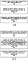
- FIG. 5 illustrates a flowchart for an example method for automatically measuring alignment of at least one of a subject's eyes.
- This embodiment of a method can include Step 502, performing an autorefraction measurement, and capturing an image of the subject's eyes using an image capturing device.
- the autorefraction measurement is a measurement a power of a subject's eye by any known technique, including but not limited to, autorefraction or photorefraction.
- the image can include a reflection of the light from each of the subject's eyes.
- the method can also include Step 504, analyzing the image to identify a position of the reflection of the light within each of the subject's eyes, respectively, and determining an alignment measurement of at least one of the subject's eyes based on the position of the reflection of the light within each of the subject's eyes, respectively.
- the method can optionally include comparing a position of the reflection of the light within one of the subject's eyes (e.g., a left or right eye) and a position of the reflection of the light within another one the subject's eyes (e.g., the right or left eye).
- the phoria measurement can be determined based on a result of the comparison.
- the step of analyzing the image to identify a position of the reflection of the light within each of the subject's eyes, respectively further comprises identifying a position of the reflection of the light relative to a landmark of each of the subject's eyes, respectively.
- the image can include a reflection of the light from at least one of an outer or inner surface of a cornea (e.g., a first or second Purkinje image, respectively) of at least one of the subject's eyes.
- the image can include a reflection of the light from at least one of an outer (anterior) or inner (posterior) surface of a lens (e.g., a third or fourth Purkinje image, respectively) of at least one of the subject's eyes.
- the image can be a first, second, third or fourth Purkinje image.
- the first through fourth Purkinje images are provided as examples, this disclosure contemplates that the image can include a reflection of the light from any surface of a subject's eye.
- the alignment measurement can be a phoria measurement or a tropia measurement.
- FIG. 6 illustrates a flowchart of another example method for measuring alignment of at least one eye.
- This embodiment of a method can include Step 602, performing an autorefraction measurement of at least one of a subject's eyes, Step 604, performing an alignment measurement of at least one of the subject's eyes, and Step 606, compensating the alignment measurement based on the autorefraction measurement.
- the autorefraction measurement is a measurement of the power of a subject's eye by any known technique, including but not limited to, autorefraction or photorefraction.
- the autorefraction measurement can be taken while the subject is focusing on the visual target, for example.
- the step of compensating the alignment measurement based on the autorefraction measurement includes performing the alignment measurement only when the autorefraction measurement is within a predetermined range.
- the step of compensating the alignment measurement based on the autorefraction measurement includes adjusting the alignment measurement based on the autorefraction measurement.
- the alignment measurement can be a phoria measurement or a tropia measurement.
- FIG. 7 illustrates a flowchart of another example method for automatically measuring a subject's phoria while the subject fixates on a visual target.
- This embodiment of a method can include Step 702, capturing an image of at least one of the subject's eyes using an image capturing device.
- the image can include at least two reflections of the light from at least one of the subject's eyes.
- the method can also include Step 704, analyzing the image to identify respective positions of the at least two reflections of the light within at least one of the subject's eyes, and determining a phoria measurement based on the respective positions of the at least two reflections of the light within at least one of the subject's eyes.
- the method can further include comparing respective positions of the at least two reflections of the light within one of the subject's eyes and respective positions of the at least two reflections of the light within another one the subject's eyes.
- the phoria measurement can be determined based on a result of the comparison.
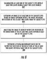
- FIG. 8 illustrates a flowchart of yet another example method for automatically measuring a subject's phoria while the subject fixates on a visual target.
- This embodiment of a method can include Step 802, illuminating at least one of the subject's eyes with at least two lights using at least two light sources, and Step 804, capturing an image of at least one of the subject's eyes using an image capturing device.
- the image can include reflections of the at least two lights from at least one of the subject's eyes.
- the image can include reflections of the at least two lights from at least one of an outer or inner surface of a cornea (e.g., a first or second Purkinje image, respectively) of at least one of the subject's eyes or an outer (anterior) or inner (posterior) surface of a lens (e.g., a third or fourth Purkinje image, respectively) of at least one of the subject's eyes.
- a cornea e.g., a first or second Purkinje image, respectively
- an outer (anterior) or inner (posterior) surface of a lens e.g., a third or fourth Purkinje image, respectively
- the image can include reflections of the at least two lights from any surface of a subject's eyes and should not be limited to the above examples (e.g., the first through fourth Purkinje images).
- the method can also include Step 806, analyzing the image to identify respective positions of the reflections of the at least two lights within at least one of the subject's eyes, and Step 808, determining a phoria measurement based on the respective positions of the reflections of the at least two lights within at least one of the subject's eyes.
- the method can include comparing respective positions of the reflections of the at least two lights within one of the subject's eyes and respective positions of the reflections of the at least two lights within another one the subject's eyes, wherein the phoria measurement is determined based on a result of the comparison.
- FIG. 9 illustrates a flowchart of another example method for automatically measuring a subject's phoria while the subject fixates on a visual target.
- This embodiment of a method can include Step 902, capturing an image of at least one of the subject's eyes using an image capturing device.
- the image can include a landmark within at least one of the subject's eyes.
- the landmark can be a feature within at least one of the subject's eyes such as a blood vessel, for example.
- landmarks other than blood vessels can be used such as a feature of the iris, the visible portion of the iris, the midpoint of the pupil, or the midpoint of the visible iris, and the like.
- the landmark can be any feature captured and identifiable within the captured image.
- the method can also include Step 904, analyzing the image to identify a position of the landmark within at least one of the subject's eyes, and Step 906, determining a phoria measurement based on the position of the landmark within at least one of the subject's eyes.
- At least one of the subject's eyes can be the subject's left eye or right eye.
- the phoria measurement can be made based on the subject's left eye or right eye.
- at least one of the subject's eyes can be the subject's left eye and right eye.
- the phoria measurement can be made based on the subject's left eye and right eye. This disclosure contemplates that the phoria measurement based on the subject's left eye and right eye can be the same or different.
Landscapes
- Health & Medical Sciences (AREA)
- Life Sciences & Earth Sciences (AREA)
- Engineering & Computer Science (AREA)
- Medical Informatics (AREA)
- Surgery (AREA)
- Biophysics (AREA)
- Biomedical Technology (AREA)
- Heart & Thoracic Surgery (AREA)
- Physics & Mathematics (AREA)
- Molecular Biology (AREA)
- Ophthalmology & Optometry (AREA)
- Animal Behavior & Ethology (AREA)
- General Health & Medical Sciences (AREA)
- Public Health (AREA)
- Veterinary Medicine (AREA)
- Signal Processing (AREA)
- Multimedia (AREA)
- Human Computer Interaction (AREA)
- Eye Examination Apparatus (AREA)
Claims (17)
- Procédé pour mesurer automatiquement la phorie d'un sujet pendant que le sujet fixe une cible visuelle, comprenant :la capture d'une image d'au moins un des yeux du sujet à l'aide d'un dispositif de capture d'image, l'image incluant une réflexion de lumière par n'importe quelle surface dudit au moins un des yeux du sujet, dans lequel l'image est capturée en réponse à une puissance de l'un des yeux du sujet se situant dans une plage prédéterminée ;l'analyse de l'image pour identifier une position de la réflexion de la lumière à l'intérieur dudit au moins un des yeux du sujet ;la détermination d'une mesure de phorie sur la base de la position de la réflexion de la lumière à l'intérieur dudit au moins un des yeux du sujet ;l'exécution d'une mesure d'autoréfraction, la mesure d'autoréfraction mesurant la puissance d'un des yeux du sujet tout en focalisant sur la cible visuelle ; etle calcul d'un rapport d'accommodation de convergence accommodative basé sur une position de la réflexion de la lumière dans au moins un des yeux du sujet et la mesure de l'autoréfraction.
- Procédé selon la revendication 1, comprenant en outre la comparaison d'une position de la réflexion de la lumière à l'intérieur d'un des yeux du sujet et une position de la réflexion de la lumière à l'intérieur d'un autre des yeux du sujet, dans lequel la mesure de phorie est déterminée sur la base d'un résultat de la comparaison.
- Procédé selon l'une quelconque des revendications 1 à 2, dans lequel l'analyse de l'image pour identifier une position de la réflexion de la lumière à l'intérieur dudit au moins un des yeux du sujet comprend en outre l'identification d'une position de la réflexion de la lumière par rapport à un repère dudit au moins un des yeux du sujet, dans lequel le repère est de préférence une caractéristique à l'intérieur dudit au moins un des yeux du sujet, en particulier un vaisseau sanguin.
- Procédé selon l'une quelconque des revendications 1 à 3, dans lequel l'image inclut une réflexion de la lumière par au moins l'une parmi une surface externe ou interne d'une cornée ou une surface externe ou interne d'une lentille dudit au moins un des yeux du sujet, dans lequel, de préférence, l'image est une première, deuxième, troisième ou quatrième image de Purkinje.
- Procédé selon l'une quelconque des revendications 1 à 4, comprenant en outre
couvrir ou découvrir ledit au moins un des yeux du sujet, dans lequel l'image est capturée après avoir découvert ledit au moins un des yeux du sujet ; et
le procédé comprenant en outre de préférence la capture d'une séquence d'images des yeux du sujet après avoir découvert ledit au moins un des yeux du sujet, et comparé une position de la réflexion de la lumière à l'intérieur dudit au moins un des yeux du sujet dans une image parmi la séquence d'images, à une position de la réflexion de la lumière à l'intérieur dudit au moins un des yeux du sujet dans une autre image de la séquence d'images pour déterminer l'un quelconque parmi un ordre de grandeur et une direction de n'importe quel mouvement après que ledit au moins un des yeux du sujet est découvert. - Procédé selon l'une quelconque des revendications 1 à 4, comprenant en outre le fait de couvrir au moins un des yeux du sujet avec un filtre, dans lequel l'image est capturée alors qu'au moins un des yeux du sujet est couvert par le filtre.
- Procédé selon la revendication 1, comprenant en outre l'ajustement de la mesure de phorie sur la base de la mesure d'autoréfraction.
- Procédé selon la revendication 1, comprenant en outre l'éclairage d'au moins un des yeux du sujet avec une lumière en utilisant une source lumineuse pour créer la réflexion, dans lequel la lumière est dans une partie visible ou non visible d'un spectre électromagnétique.
- Appareil (100) pour mesurer automatiquement la phorie d'un sujet pendant que le sujet fixe une cible visuelle, comprenant :un dispositif de capture d'image (110) pour capturer une image d'au moins un des yeux du sujet ;un processeur (206) ; etune mémoire (204) en communication opérationnelle avec le processeur, la mémoire ayant des instructions exécutables par un ordinateur stockées dans celle-ci qui, lorsqu'elles sont exécutées par le processeur, entrainent le processeur à :recevoir l'image depuis le dispositif de capture d'image, l'image incluant une réflexion de lumière par n'importe quelle surface dudit au moins un des yeux du sujet, dans lequel l'image est capturée en réponse à une puissance de l'un des yeux du sujet se situant dans une plage prédéterminée ;analyser l'image pour identifier une position de la réflexion de la lumière à l'intérieur dudit au moins un des yeux du sujet ;déterminer une mesure de phorie sur la base de la position de la réflexion de la lumière à l'intérieur dudit au moins un des yeux du sujet ;exécuter une mesure d'autoréfraction qui mesure la puissance d'un des yeux du sujet tout en focalisant sur la cible visuelle ; etcalculer un rapport d'accommodation de convergence accommodative basé sur une position de la réflexion de la lumière dans au moins un des yeux du sujet et la mesure de l'autoréfraction.
- Appareil selon la revendication 9, dans lequel la mémoire a en outre des instructions exécutables par ordinateur stockées dans celle-ci qui lorsqu'elles sont exécutées par le processeur, entrainent le processeur à comparer une position de la réflexion de la lumière à l'intérieur d'un des yeux du sujet et une position de la réflexion de la lumière à l'intérieur d'un autre des yeux du sujet, dans lequel la mesure de phorie est déterminée sur la base d'un résultat de la comparaison.
- Appareil selon l'une quelconque des revendications 9 à 10, dans lequel l'analyse de l'image pour identifier une position de la réflexion de la lumière à l'intérieur dudit au moins un des yeux du sujet comprend en outre l'identification d'une position de la réflexion de la lumière par rapport à un repère dudit au moins un des yeux du sujet, dans lequel le repère est de préférence une caractéristique à l'intérieur dudit au moins un des yeux du sujet, en particulier un vaisseau sanguin.
- Appareil selon l'une quelconque des revendications 9 à 11, dans lequel l'image inclut une réflexion de la lumière par au moins l'une parmi une surface externe ou interne d'une cornée ou une surface externe ou interne d'une lentille d'au moins un des yeux du sujet,
dans lequel l'image est de préférence une première, deuxième, troisième ou quatrième image de Purkinje. - Appareil selon l'une quelconque des revendications 9 à 12, dans lequel l'image est capturée alors qu'au moins un des yeux du sujet est couvert par un filtre, et/ou après avoir couvert et découvert ledit au moins un des yeux du sujet.
- Appareil selon la revendication 13, dans lequel le dispositif de capture d'image est un dispositif de capture vidéo ou une caméra pour capturer une séquence d'images des yeux du sujet après avoir découvert ledit au moins un des yeux du sujet et dans lequel le processeur exécute des instructions lisibles par un ordinateur pour comparer une position de la réflexion de la lumière à l'intérieur dudit au moins un des yeux du sujet dans une image de la séquence d'images, à une position de la réflexion de la lumière à l'intérieur dudit au moins un des yeux du sujet dans une autre image de la séquence d'images pour déterminer un ordre de grandeur et une direction de n'importe quel mouvement après que ledit au moins un des yeux du sujet est découvert, l'appareil comprenant de préférence en outre un dispositif d'affichage, dans lequel l'appareil définit une première surface et une deuxième surface opposée à la première surface, le dispositif d'affichage étant agencé sur la première surface et le dispositif de capture d'image étant agencé sur la deuxième surface.
- Appareil selon l'une quelconque des revendications 9 à 14, comprenant en outre une source lumineuse pour éclairer ledit au moins un des yeux du sujet avec une lumière, dans lequel la lumière est de préférence dans une partie visible ou non visible d'un spectre électromagnétique.
- Appareil selon l'une quelconque des revendications 9 à 15, dans lequel l'appareil fournit la cible visuelle pour le sujet, dans lequel la mémoire possède en outre de préférence des instructions exécutables par ordinateur stockées dans celle-ci qui, lorsqu'elles sont exécutées par le processeur, entrainent le processeur à ajuster la mesure de phorie sur la base de la mesure d'autoréfraction.
- Appareil selon la revendication 9, dans lequel l'appareil comprend un ordinateur portable, une tablette ou un téléphone mobile.
Priority Applications (4)
| Application Number | Priority Date | Filing Date | Title |
|---|---|---|---|
| EP23199823.8A EP4275588A3 (fr) | 2013-11-07 | 2014-11-07 | Détection automatique de l'alignement oculaire |
| DK18188964.3T DK3430977T3 (da) | 2013-11-07 | 2014-11-07 | Automatiseret detektering af øjenindstilling |
| EP18188964.3A EP3430977B1 (fr) | 2013-11-07 | 2014-11-07 | Détection automatique de l'alignement oculaire |
| EP20165310.2A EP3760103B1 (fr) | 2013-11-07 | 2014-11-07 | Détection automatique de l'alignement oculaire |
Applications Claiming Priority (2)
| Application Number | Priority Date | Filing Date | Title |
|---|---|---|---|
| US201361901432P | 2013-11-07 | 2013-11-07 | |
| PCT/US2014/064555 WO2015070023A2 (fr) | 2013-11-07 | 2014-11-07 | Détection automatisée d'un alignement des yeux |
Related Child Applications (3)
| Application Number | Title | Priority Date | Filing Date |
|---|---|---|---|
| EP18188964.3A Division EP3430977B1 (fr) | 2013-11-07 | 2014-11-07 | Détection automatique de l'alignement oculaire |
| EP20165310.2A Division EP3760103B1 (fr) | 2013-11-07 | 2014-11-07 | Détection automatique de l'alignement oculaire |
| EP23199823.8A Division EP4275588A3 (fr) | 2013-11-07 | 2014-11-07 | Détection automatique de l'alignement oculaire |
Publications (3)
| Publication Number | Publication Date |
|---|---|
| EP3065624A2 EP3065624A2 (fr) | 2016-09-14 |
| EP3065624A4 EP3065624A4 (fr) | 2016-12-21 |
| EP3065624B1 true EP3065624B1 (fr) | 2018-08-15 |
Family
ID=53042333
Family Applications (4)
| Application Number | Title | Priority Date | Filing Date |
|---|---|---|---|
| EP18188964.3A Active EP3430977B1 (fr) | 2013-11-07 | 2014-11-07 | Détection automatique de l'alignement oculaire |
| EP20165310.2A Active EP3760103B1 (fr) | 2013-11-07 | 2014-11-07 | Détection automatique de l'alignement oculaire |
| EP23199823.8A Withdrawn EP4275588A3 (fr) | 2013-11-07 | 2014-11-07 | Détection automatique de l'alignement oculaire |
| EP14859789.1A Active EP3065624B1 (fr) | 2013-11-07 | 2014-11-07 | Détection automatisée d'un alignement des yeux |
Family Applications Before (3)
| Application Number | Title | Priority Date | Filing Date |
|---|---|---|---|
| EP18188964.3A Active EP3430977B1 (fr) | 2013-11-07 | 2014-11-07 | Détection automatique de l'alignement oculaire |
| EP20165310.2A Active EP3760103B1 (fr) | 2013-11-07 | 2014-11-07 | Détection automatique de l'alignement oculaire |
| EP23199823.8A Withdrawn EP4275588A3 (fr) | 2013-11-07 | 2014-11-07 | Détection automatique de l'alignement oculaire |
Country Status (8)
| Country | Link |
|---|---|
| US (3) | US9867537B2 (fr) |
| EP (4) | EP3430977B1 (fr) |
| JP (2) | JP2016536105A (fr) |
| CA (2) | CA2931381C (fr) |
| DK (3) | DK3760103T3 (fr) |
| ES (3) | ES2693526T3 (fr) |
| PT (3) | PT3065624T (fr) |
| WO (1) | WO2015070023A2 (fr) |
Families Citing this family (27)
| Publication number | Priority date | Publication date | Assignee | Title |
|---|---|---|---|---|
| US9767358B2 (en) * | 2014-10-22 | 2017-09-19 | Veridium Ip Limited | Systems and methods for performing iris identification and verification using mobile devices |
| EA033741B1 (ru) * | 2015-03-01 | 2019-11-21 | Novasight Ltd | Система и способ для измерения движений глазного яблока |
| JP6529862B2 (ja) * | 2015-08-25 | 2019-06-12 | 株式会社クリュートメディカルシステムズ | 眼検査装置 |
| US9877649B2 (en) * | 2015-10-23 | 2018-01-30 | Gobiquity, Inc. | Photorefraction method and product |
| US11064881B2 (en) * | 2015-11-13 | 2021-07-20 | Hennepin Healthcare System, Inc | Method for predicting convergence disorders caused by concussion or other neuropathology |
| US11534066B2 (en) | 2016-02-11 | 2022-12-27 | Ara KESHISHIAN | System and device for promoting eye alignment |
| EP3416537A4 (fr) | 2016-02-16 | 2019-11-13 | Massachusetts Eye & Ear Infirmary | Application de dispositif mobile pour mesure de désalignement oculaire |
| JP6714402B2 (ja) * | 2016-03-18 | 2020-06-24 | 国立大学法人大阪大学 | 眼疲労検査装置及び眼疲労検査方法 |
| WO2017208227A1 (fr) | 2016-05-29 | 2017-12-07 | Nova-Sight Ltd. | Système et procédé d'affichage |
| CN106060142A (zh) * | 2016-06-17 | 2016-10-26 | 杨斌 | 一种能够检查视力的手机及使用其进行视力检查的方法 |
| JP2020509790A (ja) | 2016-09-23 | 2020-04-02 | ノバサイト リミテッド | スクリーニング装置及び方法 |
| US10338409B2 (en) | 2016-10-09 | 2019-07-02 | eyeBrain Medical, Inc. | Lens with off-axis curvature center |
| EP3542705B1 (fr) * | 2016-12-28 | 2020-07-29 | JVC Kenwood Corporation | Dispositif d'examen de fonction visuelle, procédé d'examen de fonction visuelle et programme informatique |
| US20180196281A1 (en) | 2017-01-06 | 2018-07-12 | eyeBrain Medical, Inc. | Prismatic contact lens |
| CN109288493A (zh) * | 2017-07-25 | 2019-02-01 | 珠海学院有限公司 | 一种数字化斜视诊断方法、装置和系统 |
| US10420467B2 (en) * | 2017-09-05 | 2019-09-24 | eyeBrain Medical, Inc. | Method and system for measuring binocular alignment |
| US11589745B2 (en) | 2017-09-05 | 2023-02-28 | Neurolens, Inc. | Method and system for measuring binocular alignment |
| US12114930B2 (en) | 2017-09-05 | 2024-10-15 | Neurolens, Inc. | System for measuring binocular alignment with adjustable displays and eye trackers |
| US12569135B2 (en) | 2017-09-05 | 2026-03-10 | Newton, Inc. | Headset-based system for measuring binocular alignment |
| US11360329B2 (en) | 2017-12-31 | 2022-06-14 | Neurolens, Inc. | Negative power eye-strain reducing lens |
| US10921614B2 (en) | 2017-12-31 | 2021-02-16 | Neurolens, Inc. | Low-convergence negative power spectacles |
| US10908434B2 (en) | 2018-01-01 | 2021-02-02 | Neurolens, Inc. | Negative power lens with off-axis curvature center |
| US11051689B2 (en) | 2018-11-02 | 2021-07-06 | International Business Machines Corporation | Real-time passive monitoring and assessment of pediatric eye health |
| EP3653191B1 (fr) * | 2018-11-16 | 2024-05-29 | Ara Keshishian | Système et dispositif pour favoriser l'alignement oculaire |
| CN109431450A (zh) * | 2018-11-23 | 2019-03-08 | 长春奥普光电技术股份有限公司 | 一种隐斜视客观检测方法和系统 |
| IL320532A (en) * | 2022-10-31 | 2025-06-01 | Carmel Haifa Univ Economic Corporation Ltd | System and method for diagnosis and treatment of various movement disorders and diseases of the eye |
| JP7553989B1 (ja) * | 2023-09-13 | 2024-09-19 | InnoJin株式会社 | 斜視判定装置、斜視判定方法及びプログラム |
Family Cites Families (43)
| Publication number | Priority date | Publication date | Assignee | Title |
|---|---|---|---|---|
| US2057412A (en) | 1934-12-15 | 1936-10-13 | Arthur L Boyd | Apparatus for eye testing |
| US2238207A (en) | 1937-11-06 | 1941-04-15 | Dartmouth College | Testing eyes |
| US2676588A (en) | 1950-04-25 | 1954-04-27 | Shamsky Burton | Orthoptic training device |
| US2986068A (en) | 1955-07-19 | 1961-05-30 | Gurney L Mandaville | Phoria meter |
| US3891311A (en) | 1971-07-07 | 1975-06-24 | Nasa | Multiparameter vision testing apparatus |
| US3879113A (en) | 1973-05-07 | 1975-04-22 | Howard C Howland | Photo-refractometer and methods for ophthalmic testing of young children |
| FR2463606B1 (fr) | 1979-08-22 | 1986-04-11 | Essilor Int | Dispositif pour le controle de la phorie d'un individu en vision rapprochee |
| JPS6092730A (ja) * | 1983-10-26 | 1985-05-24 | 株式会社トプコン | 眼位検査装置 |
| JPS61293430A (ja) * | 1985-06-22 | 1986-12-24 | 株式会社トプコン | 眼屈折力測定装置 |
| ES1001441Y (es) | 1987-01-14 | 1988-12-01 | Aguirre Vila-Coro Alejandro | Linterna perfeccionada |
| US5088810A (en) | 1989-01-23 | 1992-02-18 | Galanter Stephen M | Vision training method and apparatus |
| US5026151A (en) | 1989-06-23 | 1991-06-25 | Mentor O & O, Inc. | Visual function tester with binocular vision testing |
| JP2942312B2 (ja) * | 1990-06-08 | 1999-08-30 | 株式会社トプコン | 眼屈折力測定装置 |
| US5094521A (en) * | 1990-11-07 | 1992-03-10 | Vision Research Laboratories | Apparatus for evaluating eye alignment |
| JPH04279143A (ja) * | 1990-12-29 | 1992-10-05 | Koonan:Kk | 眼球運動検査装置 |
| US5757460A (en) | 1996-09-16 | 1998-05-26 | Cockley; Thomas D. | Combination ophthalmic device |
| US5838424A (en) * | 1997-02-26 | 1998-11-17 | Welch Allyn, Inc. | View port for eye test apparatus |
| JP3927695B2 (ja) * | 1998-07-16 | 2007-06-13 | キヤノン株式会社 | 眼科装置 |
| US6120461A (en) | 1999-08-09 | 2000-09-19 | The United States Of America As Represented By The Secretary Of The Army | Apparatus for tracking the human eye with a retinal scanning display, and method thereof |
| AUPQ842800A0 (en) | 2000-06-28 | 2000-07-20 | Aivision Pty Ltd | Flat screen vision testing |
| US6663242B1 (en) | 2001-03-12 | 2003-12-16 | Wayne Davenport | Simultaneous, wavelength multiplexed vision screener |
| JP4609697B2 (ja) * | 2003-09-30 | 2011-01-12 | 株式会社ニデック | 眼屈折力測定装置 |
| JP4503354B2 (ja) | 2004-05-31 | 2010-07-14 | 株式会社トプコン | 検眼装置 |
| EP1928295A2 (fr) * | 2005-09-02 | 2008-06-11 | EL-Vision Ltd. | Systeme optometrique ophtalmique multifonctionnel permettant de tester, de diagnostiquer ou de traiter la vue ou les yeux d'un patient et procedes correspondants |
| US7878652B2 (en) * | 2006-01-24 | 2011-02-01 | University Of Tennessee Research Foundation | Adaptive photoscreening system |
| US7475989B2 (en) * | 2006-03-14 | 2009-01-13 | Amo Manufacturing Usa, Llc | Shack-Hartmann based integrated autorefraction and wavefront measurements of the eye |
| US7976162B2 (en) * | 2006-09-29 | 2011-07-12 | Daniel Ian Flitcroft | Eye examining system and method |
| JP2008246143A (ja) * | 2007-03-30 | 2008-10-16 | Hamamatsu Photonics Kk | 瞳孔測定装置 |
| US7771051B2 (en) | 2007-06-13 | 2010-08-10 | Rahim Hirji | Near eye opthalmic device |
| EP2231277B1 (fr) * | 2007-12-23 | 2017-08-30 | Carl Zeiss Meditec, Inc. | Dispositifs permettant de détecter, contrôler et prévoir l'administration d'un rayonnement |
| JP5011144B2 (ja) * | 2008-02-01 | 2012-08-29 | 株式会社トプコン | 眼科測定装置 |
| WO2010009450A1 (fr) * | 2008-07-18 | 2010-01-21 | Doheny Eye Institute | Dispositif d’examen ophtalmique à base de tomographie de cohérence optique, procédé et système |
| JP5654225B2 (ja) * | 2009-09-30 | 2015-01-14 | 株式会社ニデック | 角膜形状測定装置 |
| EP2329761A1 (fr) * | 2009-12-02 | 2011-06-08 | RetCorr AB | Appareil et procédé pour établir et/ou améliorer une vision binoculaire améliorée |
| JP5714949B2 (ja) | 2010-10-05 | 2015-05-07 | パナソニック株式会社 | 眼位ずれ計測装置 |
| JP5727191B2 (ja) | 2010-10-15 | 2015-06-03 | 株式会社ニデック | 検眼装置 |
| US8727534B2 (en) * | 2011-01-24 | 2014-05-20 | Huvitz Co., Ltd. | Automatic refracto-keratometer |
| US9895058B2 (en) * | 2011-03-25 | 2018-02-20 | Carl Zeiss Meditec Ag | Heads-up vision analyzer |
| US8684529B2 (en) | 2011-04-28 | 2014-04-01 | Carl Zeiss Meditec, Inc. | Systems and methods for improved visual field testing |
| JP5818233B2 (ja) * | 2011-05-24 | 2015-11-18 | 国立大学法人神戸大学 | 視線計測装置および方法 |
| KR20140103900A (ko) | 2011-09-08 | 2014-08-27 | 아이체크 헬스 커넥션, 인크. | 유아 및 소아에 있어서 눈의 동공 적색 반사 검사 및 각막 광 반사 선별의 기록 및 문서화를 위한 방법 및 시스템 |
| US8764188B2 (en) | 2011-10-21 | 2014-07-01 | Kent Tabor | Functional vision tester |
| IL215883A0 (en) * | 2011-10-24 | 2012-03-01 | Iriss Medical Technologies Ltd | System and method for indentifying eye conditions |
-
2014
- 2014-11-07 PT PT14859789T patent/PT3065624T/pt unknown
- 2014-11-07 CA CA2931381A patent/CA2931381C/fr active Active
- 2014-11-07 CA CA3188783A patent/CA3188783A1/fr active Pending
- 2014-11-07 ES ES14859789.1T patent/ES2693526T3/es active Active
- 2014-11-07 EP EP18188964.3A patent/EP3430977B1/fr active Active
- 2014-11-07 PT PT201653102T patent/PT3760103T/pt unknown
- 2014-11-07 WO PCT/US2014/064555 patent/WO2015070023A2/fr not_active Ceased
- 2014-11-07 ES ES18188964T patent/ES2793574T3/es active Active
- 2014-11-07 ES ES20165310T patent/ES2964752T3/es active Active
- 2014-11-07 EP EP20165310.2A patent/EP3760103B1/fr active Active
- 2014-11-07 DK DK20165310.2T patent/DK3760103T3/da active
- 2014-11-07 EP EP23199823.8A patent/EP4275588A3/fr not_active Withdrawn
- 2014-11-07 DK DK14859789.1T patent/DK3065624T3/en active
- 2014-11-07 DK DK18188964.3T patent/DK3430977T3/da active
- 2014-11-07 JP JP2016553247A patent/JP2016536105A/ja active Pending
- 2014-11-07 EP EP14859789.1A patent/EP3065624B1/fr active Active
- 2014-11-07 US US14/893,626 patent/US9867537B2/en active Active
- 2014-11-07 PT PT181889643T patent/PT3430977T/pt unknown
-
2015
- 2015-11-24 US US14/950,755 patent/US10575727B2/en active Active
- 2015-11-24 US US14/950,968 patent/US9750405B2/en active Active
-
2019
- 2019-09-30 JP JP2019179312A patent/JP2020018875A/ja active Pending
Non-Patent Citations (1)
| Title |
|---|
| None * |
Also Published As
| Publication number | Publication date |
|---|---|
| EP4275588A3 (fr) | 2024-01-17 |
| CA2931381C (fr) | 2023-04-04 |
| EP3430977B1 (fr) | 2020-03-25 |
| US20160073869A1 (en) | 2016-03-17 |
| EP3430977A1 (fr) | 2019-01-23 |
| US20160270653A1 (en) | 2016-09-22 |
| US9750405B2 (en) | 2017-09-05 |
| WO2015070023A9 (fr) | 2015-12-03 |
| DK3430977T3 (da) | 2020-06-02 |
| EP3065624A2 (fr) | 2016-09-14 |
| EP3065624A4 (fr) | 2016-12-21 |
| PT3760103T (pt) | 2023-11-29 |
| ES2693526T3 (es) | 2018-12-12 |
| JP2016536105A (ja) | 2016-11-24 |
| EP3760103B1 (fr) | 2023-09-27 |
| EP3760103A1 (fr) | 2021-01-06 |
| EP4275588A2 (fr) | 2023-11-15 |
| JP2020018875A (ja) | 2020-02-06 |
| PT3065624T (pt) | 2018-11-16 |
| DK3760103T3 (da) | 2023-12-04 |
| ES2964752T3 (es) | 2024-04-09 |
| WO2015070023A2 (fr) | 2015-05-14 |
| CA2931381A1 (fr) | 2015-05-14 |
| US20160073870A1 (en) | 2016-03-17 |
| US9867537B2 (en) | 2018-01-16 |
| ES2793574T3 (es) | 2020-11-16 |
| WO2015070023A3 (fr) | 2015-08-06 |
| CA3188783A1 (fr) | 2015-05-14 |
| US10575727B2 (en) | 2020-03-03 |
| DK3065624T3 (en) | 2018-10-15 |
| PT3430977T (pt) | 2020-04-16 |
Similar Documents
| Publication | Publication Date | Title |
|---|---|---|
| EP3430977B1 (fr) | Détection automatique de l'alignement oculaire | |
| Mariakakis et al. | PupilScreen: using smartphones to assess traumatic brain injury | |
| US9872615B2 (en) | Systems and methods for improved ease and accuracy of gaze tracking | |
| EP2881891B1 (fr) | Procédé et dispositif de traitement d'images | |
| ES3039315T3 (en) | Reliability of gaze tracking data for left and right eye | |
| EP3214993B1 (fr) | Procédés et appareil permettant d'effectuer une détermination concernant un il dans des conditions d'éclairage ambiant | |
| KR102344493B1 (ko) | 인공 지능 기반의 스마트 안진 검사 시스템, 방법 및 프로그램 | |
| CN115414002A (zh) | 一种基于视频流的眼检测方法及斜视筛查系统 | |
| Luo et al. | Using an automated Hirschberg test app to evaluate ocular alignment | |
| Sadok et al. | Performing the HINTS-exam using a mixed-reality head-mounted display in patients with acute vestibular syndrome: a feasibility study | |
| HK40038784B (en) | Automated detection of eye alignment | |
| HK40003661A (en) | Automated detection of eye alignment | |
| HK40003661B (en) | Automated detection of eye alignment | |
| HK40038784A (en) | Automated detection of eye alignment | |
| HK1225254A1 (en) | Automated detection of eye alignment | |
| HK1225254B (en) | Automated detection of eye alignment | |
| US20240341590A1 (en) | Methods and systems for aligning an eye for making a determination about the eye using remote or telehealth procedures | |
| Phillips | OPEN NYSTAGMUS: AN OPEN-SOURCE SMARTPHONE-BASED FRAMEWORK FOR MARKER-FREE QUANTIFICATION OF OPTOKINETIC NYSTAGMUS | |
| Quang et al. | Mobile traumatic brain injury assessment system |
Legal Events
| Date | Code | Title | Description |
|---|---|---|---|
| PUAI | Public reference made under article 153(3) epc to a published international application that has entered the european phase |
Free format text: ORIGINAL CODE: 0009012 |
|
| 17P | Request for examination filed |
Effective date: 20160602 |
|
| AK | Designated contracting states |
Kind code of ref document: A2 Designated state(s): AL AT BE BG CH CY CZ DE DK EE ES FI FR GB GR HR HU IE IS IT LI LT LU LV MC MK MT NL NO PL PT RO RS SE SI SK SM TR |
|
| AX | Request for extension of the european patent |
Extension state: BA ME |
|
| RIN1 | Information on inventor provided before grant (corrected) |
Inventor name: BAILEY, MELISSA DIANE |
|
| A4 | Supplementary search report drawn up and despatched |
Effective date: 20161122 |
|
| RIC1 | Information provided on ipc code assigned before grant |
Ipc: A61B 3/103 20060101ALI20161116BHEP Ipc: A61B 3/113 20060101ALI20161116BHEP Ipc: A61B 3/14 20060101AFI20161116BHEP |
|
| DAX | Request for extension of the european patent (deleted) | ||
| REG | Reference to a national code |
Ref country code: HK Ref legal event code: DE Ref document number: 1225254 Country of ref document: HK |
|
| GRAP | Despatch of communication of intention to grant a patent |
Free format text: ORIGINAL CODE: EPIDOSNIGR1 |
|
| STAA | Information on the status of an ep patent application or granted ep patent |
Free format text: STATUS: GRANT OF PATENT IS INTENDED |
|
| INTG | Intention to grant announced |
Effective date: 20170918 |
|
| GRAJ | Information related to disapproval of communication of intention to grant by the applicant or resumption of examination proceedings by the epo deleted |
Free format text: ORIGINAL CODE: EPIDOSDIGR1 |
|
| STAA | Information on the status of an ep patent application or granted ep patent |
Free format text: STATUS: REQUEST FOR EXAMINATION WAS MADE |
|
| GRAP | Despatch of communication of intention to grant a patent |
Free format text: ORIGINAL CODE: EPIDOSNIGR1 |
|
| STAA | Information on the status of an ep patent application or granted ep patent |
Free format text: STATUS: GRANT OF PATENT IS INTENDED |
|
| INTC | Intention to grant announced (deleted) | ||
| INTG | Intention to grant announced |
Effective date: 20180227 |
|
| GRAS | Grant fee paid |
Free format text: ORIGINAL CODE: EPIDOSNIGR3 |
|
| GRAA | (expected) grant |
Free format text: ORIGINAL CODE: 0009210 |
|
| STAA | Information on the status of an ep patent application or granted ep patent |
Free format text: STATUS: THE PATENT HAS BEEN GRANTED |
|
| AK | Designated contracting states |
Kind code of ref document: B1 Designated state(s): AL AT BE BG CH CY CZ DE DK EE ES FI FR GB GR HR HU IE IS IT LI LT LU LV MC MK MT NL NO PL PT RO RS SE SI SK SM TR |
|
| REG | Reference to a national code |
Ref country code: CH Ref legal event code: EP Ref country code: GB Ref legal event code: FG4D Ref country code: AT Ref legal event code: REF Ref document number: 1028788 Country of ref document: AT Kind code of ref document: T Effective date: 20180815 |
|
| REG | Reference to a national code |
Ref country code: IE Ref legal event code: FG4D |
|
| REG | Reference to a national code |
Ref country code: DE Ref legal event code: R096 Ref document number: 602014030645 Country of ref document: DE |
|
| REG | Reference to a national code |
Ref country code: DK Ref legal event code: T3 Effective date: 20181008 |
|
| REG | Reference to a national code |
Ref country code: SE Ref legal event code: TRGR |
|
| REG | Reference to a national code |
Ref country code: NL Ref legal event code: FP |
|
| REG | Reference to a national code |
Ref country code: CH Ref legal event code: NV Representative=s name: STOLMAR AND PARTNER INTELLECTUAL PROPERTY S.A., CH |
|
| REG | Reference to a national code |
Ref country code: PT Ref legal event code: SC4A Ref document number: 3065624 Country of ref document: PT Date of ref document: 20181116 Kind code of ref document: T Free format text: AVAILABILITY OF NATIONAL TRANSLATION Effective date: 20181029 |
|
| REG | Reference to a national code |
Ref country code: ES Ref legal event code: FG2A Ref document number: 2693526 Country of ref document: ES Kind code of ref document: T3 Effective date: 20181212 |
|
| REG | Reference to a national code |
Ref country code: LT Ref legal event code: MG4D |
|
| PG25 | Lapsed in a contracting state [announced via postgrant information from national office to epo] |
Ref country code: NO Free format text: LAPSE BECAUSE OF FAILURE TO SUBMIT A TRANSLATION OF THE DESCRIPTION OR TO PAY THE FEE WITHIN THE PRESCRIBED TIME-LIMIT Effective date: 20181115 Ref country code: BG Free format text: LAPSE BECAUSE OF FAILURE TO SUBMIT A TRANSLATION OF THE DESCRIPTION OR TO PAY THE FEE WITHIN THE PRESCRIBED TIME-LIMIT Effective date: 20181115 Ref country code: RS Free format text: LAPSE BECAUSE OF FAILURE TO SUBMIT A TRANSLATION OF THE DESCRIPTION OR TO PAY THE FEE WITHIN THE PRESCRIBED TIME-LIMIT Effective date: 20180815 Ref country code: IS Free format text: LAPSE BECAUSE OF FAILURE TO SUBMIT A TRANSLATION OF THE DESCRIPTION OR TO PAY THE FEE WITHIN THE PRESCRIBED TIME-LIMIT Effective date: 20181215 Ref country code: GR Free format text: LAPSE BECAUSE OF FAILURE TO SUBMIT A TRANSLATION OF THE DESCRIPTION OR TO PAY THE FEE WITHIN THE PRESCRIBED TIME-LIMIT Effective date: 20181116 Ref country code: FI Free format text: LAPSE BECAUSE OF FAILURE TO SUBMIT A TRANSLATION OF THE DESCRIPTION OR TO PAY THE FEE WITHIN THE PRESCRIBED TIME-LIMIT Effective date: 20180815 Ref country code: LT Free format text: LAPSE BECAUSE OF FAILURE TO SUBMIT A TRANSLATION OF THE DESCRIPTION OR TO PAY THE FEE WITHIN THE PRESCRIBED TIME-LIMIT Effective date: 20180815 |
|
| PG25 | Lapsed in a contracting state [announced via postgrant information from national office to epo] |
Ref country code: AL Free format text: LAPSE BECAUSE OF FAILURE TO SUBMIT A TRANSLATION OF THE DESCRIPTION OR TO PAY THE FEE WITHIN THE PRESCRIBED TIME-LIMIT Effective date: 20180815 Ref country code: HR Free format text: LAPSE BECAUSE OF FAILURE TO SUBMIT A TRANSLATION OF THE DESCRIPTION OR TO PAY THE FEE WITHIN THE PRESCRIBED TIME-LIMIT Effective date: 20180815 Ref country code: LV Free format text: LAPSE BECAUSE OF FAILURE TO SUBMIT A TRANSLATION OF THE DESCRIPTION OR TO PAY THE FEE WITHIN THE PRESCRIBED TIME-LIMIT Effective date: 20180815 |
|
| PG25 | Lapsed in a contracting state [announced via postgrant information from national office to epo] |
Ref country code: EE Free format text: LAPSE BECAUSE OF FAILURE TO SUBMIT A TRANSLATION OF THE DESCRIPTION OR TO PAY THE FEE WITHIN THE PRESCRIBED TIME-LIMIT Effective date: 20180815 Ref country code: RO Free format text: LAPSE BECAUSE OF FAILURE TO SUBMIT A TRANSLATION OF THE DESCRIPTION OR TO PAY THE FEE WITHIN THE PRESCRIBED TIME-LIMIT Effective date: 20180815 Ref country code: CZ Free format text: LAPSE BECAUSE OF FAILURE TO SUBMIT A TRANSLATION OF THE DESCRIPTION OR TO PAY THE FEE WITHIN THE PRESCRIBED TIME-LIMIT Effective date: 20180815 Ref country code: PL Free format text: LAPSE BECAUSE OF FAILURE TO SUBMIT A TRANSLATION OF THE DESCRIPTION OR TO PAY THE FEE WITHIN THE PRESCRIBED TIME-LIMIT Effective date: 20180815 |
|
| REG | Reference to a national code |
Ref country code: DE Ref legal event code: R097 Ref document number: 602014030645 Country of ref document: DE |
|
| PG25 | Lapsed in a contracting state [announced via postgrant information from national office to epo] |
Ref country code: SK Free format text: LAPSE BECAUSE OF FAILURE TO SUBMIT A TRANSLATION OF THE DESCRIPTION OR TO PAY THE FEE WITHIN THE PRESCRIBED TIME-LIMIT Effective date: 20180815 Ref country code: SM Free format text: LAPSE BECAUSE OF FAILURE TO SUBMIT A TRANSLATION OF THE DESCRIPTION OR TO PAY THE FEE WITHIN THE PRESCRIBED TIME-LIMIT Effective date: 20180815 |
|
| PLBE | No opposition filed within time limit |
Free format text: ORIGINAL CODE: 0009261 |
|
| STAA | Information on the status of an ep patent application or granted ep patent |
Free format text: STATUS: NO OPPOSITION FILED WITHIN TIME LIMIT |
|
| 26N | No opposition filed |
Effective date: 20190516 |
|
| PG25 | Lapsed in a contracting state [announced via postgrant information from national office to epo] |
Ref country code: LU Free format text: LAPSE BECAUSE OF NON-PAYMENT OF DUE FEES Effective date: 20181107 Ref country code: MC Free format text: LAPSE BECAUSE OF FAILURE TO SUBMIT A TRANSLATION OF THE DESCRIPTION OR TO PAY THE FEE WITHIN THE PRESCRIBED TIME-LIMIT Effective date: 20180815 |
|
| PG25 | Lapsed in a contracting state [announced via postgrant information from national office to epo] |
Ref country code: SI Free format text: LAPSE BECAUSE OF FAILURE TO SUBMIT A TRANSLATION OF THE DESCRIPTION OR TO PAY THE FEE WITHIN THE PRESCRIBED TIME-LIMIT Effective date: 20180815 |
|
| PG25 | Lapsed in a contracting state [announced via postgrant information from national office to epo] |
Ref country code: MT Free format text: LAPSE BECAUSE OF NON-PAYMENT OF DUE FEES Effective date: 20181107 |
|
| PG25 | Lapsed in a contracting state [announced via postgrant information from national office to epo] |
Ref country code: TR Free format text: LAPSE BECAUSE OF FAILURE TO SUBMIT A TRANSLATION OF THE DESCRIPTION OR TO PAY THE FEE WITHIN THE PRESCRIBED TIME-LIMIT Effective date: 20180815 |
|
| PG25 | Lapsed in a contracting state [announced via postgrant information from national office to epo] |
Ref country code: CY Free format text: LAPSE BECAUSE OF FAILURE TO SUBMIT A TRANSLATION OF THE DESCRIPTION OR TO PAY THE FEE WITHIN THE PRESCRIBED TIME-LIMIT Effective date: 20180815 Ref country code: HU Free format text: LAPSE BECAUSE OF FAILURE TO SUBMIT A TRANSLATION OF THE DESCRIPTION OR TO PAY THE FEE WITHIN THE PRESCRIBED TIME-LIMIT; INVALID AB INITIO Effective date: 20141107 Ref country code: MK Free format text: LAPSE BECAUSE OF NON-PAYMENT OF DUE FEES Effective date: 20180815 |
|
| REG | Reference to a national code |
Ref country code: AT Ref legal event code: UEP Ref document number: 1028788 Country of ref document: AT Kind code of ref document: T Effective date: 20180815 |
|
| REG | Reference to a national code |
Ref country code: FR Ref legal event code: PLFP Year of fee payment: 9 |
|
| P01 | Opt-out of the competence of the unified patent court (upc) registered |
Effective date: 20230528 |
|
| REG | Reference to a national code |
Ref country code: CH Ref legal event code: U11 Free format text: ST27 STATUS EVENT CODE: U-0-0-U10-U11 (AS PROVIDED BY THE NATIONAL OFFICE) Effective date: 20251201 |
|
| PGFP | Annual fee paid to national office [announced via postgrant information from national office to epo] |
Ref country code: PT Payment date: 20251024 Year of fee payment: 12 |
|
| PGFP | Annual fee paid to national office [announced via postgrant information from national office to epo] |
Ref country code: NL Payment date: 20251126 Year of fee payment: 12 |
|
| PGFP | Annual fee paid to national office [announced via postgrant information from national office to epo] |
Ref country code: DE Payment date: 20251128 Year of fee payment: 12 |
|
| PGFP | Annual fee paid to national office [announced via postgrant information from national office to epo] |
Ref country code: GB Payment date: 20251127 Year of fee payment: 12 |
|
| PGFP | Annual fee paid to national office [announced via postgrant information from national office to epo] |
Ref country code: AT Payment date: 20251128 Year of fee payment: 12 |
|
| PGFP | Annual fee paid to national office [announced via postgrant information from national office to epo] |
Ref country code: DK Payment date: 20251125 Year of fee payment: 12 Ref country code: IT Payment date: 20251119 Year of fee payment: 12 |
|
| PGFP | Annual fee paid to national office [announced via postgrant information from national office to epo] |
Ref country code: FR Payment date: 20251125 Year of fee payment: 12 |
|
| PGFP | Annual fee paid to national office [announced via postgrant information from national office to epo] |
Ref country code: BE Payment date: 20251127 Year of fee payment: 12 |
|
| PGFP | Annual fee paid to national office [announced via postgrant information from national office to epo] |
Ref country code: CH Payment date: 20251201 Year of fee payment: 12 Ref country code: SE Payment date: 20251127 Year of fee payment: 12 |
|
| PGFP | Annual fee paid to national office [announced via postgrant information from national office to epo] |
Ref country code: IE Payment date: 20251127 Year of fee payment: 12 |
|
| PGFP | Annual fee paid to national office [announced via postgrant information from national office to epo] |
Ref country code: ES Payment date: 20251201 Year of fee payment: 12 |