EP3070162B1 - Procédé de production de cellules épithéliales de pigment rétinien - Google Patents
Procédé de production de cellules épithéliales de pigment rétinien Download PDFInfo
- Publication number
- EP3070162B1 EP3070162B1 EP14860084.4A EP14860084A EP3070162B1 EP 3070162 B1 EP3070162 B1 EP 3070162B1 EP 14860084 A EP14860084 A EP 14860084A EP 3070162 B1 EP3070162 B1 EP 3070162B1
- Authority
- EP
- European Patent Office
- Prior art keywords
- substance
- medium
- serum
- culture
- cells
- Prior art date
- Legal status (The legal status is an assumption and is not a legal conclusion. Google has not performed a legal analysis and makes no representation as to the accuracy of the status listed.)
- Active
Links
Images

Classifications
-
- C—CHEMISTRY; METALLURGY
- C12—BIOCHEMISTRY; BEER; SPIRITS; WINE; VINEGAR; MICROBIOLOGY; ENZYMOLOGY; MUTATION OR GENETIC ENGINEERING
- C12N—MICROORGANISMS OR ENZYMES; COMPOSITIONS THEREOF; PROPAGATING, PRESERVING, OR MAINTAINING MICROORGANISMS; MUTATION OR GENETIC ENGINEERING; CULTURE MEDIA
- C12N5/00—Undifferentiated human, animal or plant cells, e.g. cell lines; Tissues; Cultivation or maintenance thereof; Culture media therefor
- C12N5/06—Animal cells or tissues; Human cells or tissues
- C12N5/0602—Vertebrate cells
- C12N5/0618—Cells of the nervous system
- C12N5/0621—Eye cells, e.g. cornea, iris pigmented cells
-
- C—CHEMISTRY; METALLURGY
- C12—BIOCHEMISTRY; BEER; SPIRITS; WINE; VINEGAR; MICROBIOLOGY; ENZYMOLOGY; MUTATION OR GENETIC ENGINEERING
- C12N—MICROORGANISMS OR ENZYMES; COMPOSITIONS THEREOF; PROPAGATING, PRESERVING, OR MAINTAINING MICROORGANISMS; MUTATION OR GENETIC ENGINEERING; CULTURE MEDIA
- C12N5/00—Undifferentiated human, animal or plant cells, e.g. cell lines; Tissues; Cultivation or maintenance thereof; Culture media therefor
- C12N5/06—Animal cells or tissues; Human cells or tissues
- C12N5/0602—Vertebrate cells
- C12N5/0618—Cells of the nervous system
- C12N5/062—Sensory transducers, e.g. photoreceptors; Sensory neurons, e.g. for hearing, taste, smell, pH, touch, temperature, pain
-
- A—HUMAN NECESSITIES
- A61—MEDICAL OR VETERINARY SCIENCE; HYGIENE
- A61K—PREPARATIONS FOR MEDICAL, DENTAL OR TOILETRY PURPOSES
- A61K35/00—Medicinal preparations containing materials or reaction products thereof with undetermined constitution
- A61K35/12—Materials from mammals; Compositions comprising non-specified tissues or cells; Compositions comprising non-embryonic stem cells; Genetically modified cells
- A61K35/30—Nerves; Brain; Eyes; Corneal cells; Cerebrospinal fluid; Neuronal stem cells; Neuronal precursor cells; Glial cells; Oligodendrocytes; Schwann cells; Astroglia; Astrocytes; Choroid plexus; Spinal cord tissue
-
- A—HUMAN NECESSITIES
- A61—MEDICAL OR VETERINARY SCIENCE; HYGIENE
- A61L—METHODS OR APPARATUS FOR STERILISING MATERIALS OR OBJECTS IN GENERAL; DISINFECTION, STERILISATION OR DEODORISATION OF AIR; CHEMICAL ASPECTS OF BANDAGES, DRESSINGS, ABSORBENT PADS OR SURGICAL ARTICLES; MATERIALS FOR BANDAGES, DRESSINGS, ABSORBENT PADS OR SURGICAL ARTICLES
- A61L27/00—Materials for grafts or prostheses or for coating grafts or prostheses
- A61L27/36—Materials for grafts or prostheses or for coating grafts or prostheses containing ingredients of undetermined constitution or reaction products thereof, e.g. transplant tissue, natural bone, extracellular matrix
- A61L27/38—Materials for grafts or prostheses or for coating grafts or prostheses containing ingredients of undetermined constitution or reaction products thereof, e.g. transplant tissue, natural bone, extracellular matrix containing added animal cells
- A61L27/3804—Materials for grafts or prostheses or for coating grafts or prostheses containing ingredients of undetermined constitution or reaction products thereof, e.g. transplant tissue, natural bone, extracellular matrix containing added animal cells characterised by specific cells or progenitors thereof, e.g. fibroblasts, connective tissue cells, kidney cells
- A61L27/3813—Epithelial cells, e.g. keratinocytes, urothelial cells
-
- A—HUMAN NECESSITIES
- A61—MEDICAL OR VETERINARY SCIENCE; HYGIENE
- A61L—METHODS OR APPARATUS FOR STERILISING MATERIALS OR OBJECTS IN GENERAL; DISINFECTION, STERILISATION OR DEODORISATION OF AIR; CHEMICAL ASPECTS OF BANDAGES, DRESSINGS, ABSORBENT PADS OR SURGICAL ARTICLES; MATERIALS FOR BANDAGES, DRESSINGS, ABSORBENT PADS OR SURGICAL ARTICLES
- A61L27/00—Materials for grafts or prostheses or for coating grafts or prostheses
- A61L27/36—Materials for grafts or prostheses or for coating grafts or prostheses containing ingredients of undetermined constitution or reaction products thereof, e.g. transplant tissue, natural bone, extracellular matrix
- A61L27/38—Materials for grafts or prostheses or for coating grafts or prostheses containing ingredients of undetermined constitution or reaction products thereof, e.g. transplant tissue, natural bone, extracellular matrix containing added animal cells
- A61L27/3804—Materials for grafts or prostheses or for coating grafts or prostheses containing ingredients of undetermined constitution or reaction products thereof, e.g. transplant tissue, natural bone, extracellular matrix containing added animal cells characterised by specific cells or progenitors thereof, e.g. fibroblasts, connective tissue cells, kidney cells
- A61L27/3834—Cells able to produce different cell types, e.g. hematopoietic stem cells, mesenchymal stem cells, marrow stromal cells, embryonic stem cells
-
- A—HUMAN NECESSITIES
- A61—MEDICAL OR VETERINARY SCIENCE; HYGIENE
- A61L—METHODS OR APPARATUS FOR STERILISING MATERIALS OR OBJECTS IN GENERAL; DISINFECTION, STERILISATION OR DEODORISATION OF AIR; CHEMICAL ASPECTS OF BANDAGES, DRESSINGS, ABSORBENT PADS OR SURGICAL ARTICLES; MATERIALS FOR BANDAGES, DRESSINGS, ABSORBENT PADS OR SURGICAL ARTICLES
- A61L27/00—Materials for grafts or prostheses or for coating grafts or prostheses
- A61L27/36—Materials for grafts or prostheses or for coating grafts or prostheses containing ingredients of undetermined constitution or reaction products thereof, e.g. transplant tissue, natural bone, extracellular matrix
- A61L27/38—Materials for grafts or prostheses or for coating grafts or prostheses containing ingredients of undetermined constitution or reaction products thereof, e.g. transplant tissue, natural bone, extracellular matrix containing added animal cells
- A61L27/3895—Materials for grafts or prostheses or for coating grafts or prostheses containing ingredients of undetermined constitution or reaction products thereof, e.g. transplant tissue, natural bone, extracellular matrix containing added animal cells using specific culture conditions, e.g. stimulating differentiation of stem cells, pulsatile flow conditions
-
- A—HUMAN NECESSITIES
- A61—MEDICAL OR VETERINARY SCIENCE; HYGIENE
- A61P—SPECIFIC THERAPEUTIC ACTIVITY OF CHEMICAL COMPOUNDS OR MEDICINAL PREPARATIONS
- A61P27/00—Drugs for disorders of the senses
-
- A—HUMAN NECESSITIES
- A61—MEDICAL OR VETERINARY SCIENCE; HYGIENE
- A61P—SPECIFIC THERAPEUTIC ACTIVITY OF CHEMICAL COMPOUNDS OR MEDICINAL PREPARATIONS
- A61P27/00—Drugs for disorders of the senses
- A61P27/02—Ophthalmic agents
-
- A—HUMAN NECESSITIES
- A61—MEDICAL OR VETERINARY SCIENCE; HYGIENE
- A61K—PREPARATIONS FOR MEDICAL, DENTAL OR TOILETRY PURPOSES
- A61K35/00—Medicinal preparations containing materials or reaction products thereof with undetermined constitution
- A61K35/12—Materials from mammals; Compositions comprising non-specified tissues or cells; Compositions comprising non-embryonic stem cells; Genetically modified cells
- A61K35/48—Reproductive organs
- A61K35/54—Ovaries; Ova; Ovules; Embryos; Foetal cells; Germ cells
- A61K35/545—Embryonic stem cells; Pluripotent stem cells; Induced pluripotent stem cells; Uncharacterised stem cells
-
- A—HUMAN NECESSITIES
- A61—MEDICAL OR VETERINARY SCIENCE; HYGIENE
- A61L—METHODS OR APPARATUS FOR STERILISING MATERIALS OR OBJECTS IN GENERAL; DISINFECTION, STERILISATION OR DEODORISATION OF AIR; CHEMICAL ASPECTS OF BANDAGES, DRESSINGS, ABSORBENT PADS OR SURGICAL ARTICLES; MATERIALS FOR BANDAGES, DRESSINGS, ABSORBENT PADS OR SURGICAL ARTICLES
- A61L2430/00—Materials or treatment for tissue regeneration
- A61L2430/16—Materials or treatment for tissue regeneration for reconstruction of eye parts, e.g. intraocular lens, cornea
-
- C—CHEMISTRY; METALLURGY
- C12—BIOCHEMISTRY; BEER; SPIRITS; WINE; VINEGAR; MICROBIOLOGY; ENZYMOLOGY; MUTATION OR GENETIC ENGINEERING
- C12N—MICROORGANISMS OR ENZYMES; COMPOSITIONS THEREOF; PROPAGATING, PRESERVING, OR MAINTAINING MICROORGANISMS; MUTATION OR GENETIC ENGINEERING; CULTURE MEDIA
- C12N2501/00—Active agents used in cell culture processes, e.g. differentation
- C12N2501/10—Growth factors
- C12N2501/113—Acidic fibroblast growth factor (aFGF, FGF-1)
-
- C—CHEMISTRY; METALLURGY
- C12—BIOCHEMISTRY; BEER; SPIRITS; WINE; VINEGAR; MICROBIOLOGY; ENZYMOLOGY; MUTATION OR GENETIC ENGINEERING
- C12N—MICROORGANISMS OR ENZYMES; COMPOSITIONS THEREOF; PROPAGATING, PRESERVING, OR MAINTAINING MICROORGANISMS; MUTATION OR GENETIC ENGINEERING; CULTURE MEDIA
- C12N2501/00—Active agents used in cell culture processes, e.g. differentation
- C12N2501/10—Growth factors
- C12N2501/155—Bone morphogenic proteins [BMP]; Osteogenins; Osteogenic factor; Bone inducing factor
-
- C—CHEMISTRY; METALLURGY
- C12—BIOCHEMISTRY; BEER; SPIRITS; WINE; VINEGAR; MICROBIOLOGY; ENZYMOLOGY; MUTATION OR GENETIC ENGINEERING
- C12N—MICROORGANISMS OR ENZYMES; COMPOSITIONS THEREOF; PROPAGATING, PRESERVING, OR MAINTAINING MICROORGANISMS; MUTATION OR GENETIC ENGINEERING; CULTURE MEDIA
- C12N2501/00—Active agents used in cell culture processes, e.g. differentation
- C12N2501/40—Regulators of development
- C12N2501/41—Hedgehog proteins; Cyclopamine (inhibitor)
-
- C—CHEMISTRY; METALLURGY
- C12—BIOCHEMISTRY; BEER; SPIRITS; WINE; VINEGAR; MICROBIOLOGY; ENZYMOLOGY; MUTATION OR GENETIC ENGINEERING
- C12N—MICROORGANISMS OR ENZYMES; COMPOSITIONS THEREOF; PROPAGATING, PRESERVING, OR MAINTAINING MICROORGANISMS; MUTATION OR GENETIC ENGINEERING; CULTURE MEDIA
- C12N2501/00—Active agents used in cell culture processes, e.g. differentation
- C12N2501/40—Regulators of development
- C12N2501/415—Wnt; Frizzeled
-
- C—CHEMISTRY; METALLURGY
- C12—BIOCHEMISTRY; BEER; SPIRITS; WINE; VINEGAR; MICROBIOLOGY; ENZYMOLOGY; MUTATION OR GENETIC ENGINEERING
- C12N—MICROORGANISMS OR ENZYMES; COMPOSITIONS THEREOF; PROPAGATING, PRESERVING, OR MAINTAINING MICROORGANISMS; MUTATION OR GENETIC ENGINEERING; CULTURE MEDIA
- C12N2501/00—Active agents used in cell culture processes, e.g. differentation
- C12N2501/70—Enzymes
- C12N2501/72—Transferases [EC 2.]
- C12N2501/727—Kinases (EC 2.7.)
-
- C—CHEMISTRY; METALLURGY
- C12—BIOCHEMISTRY; BEER; SPIRITS; WINE; VINEGAR; MICROBIOLOGY; ENZYMOLOGY; MUTATION OR GENETIC ENGINEERING
- C12N—MICROORGANISMS OR ENZYMES; COMPOSITIONS THEREOF; PROPAGATING, PRESERVING, OR MAINTAINING MICROORGANISMS; MUTATION OR GENETIC ENGINEERING; CULTURE MEDIA
- C12N2506/00—Differentiation of animal cells from one lineage to another; Differentiation of pluripotent cells
- C12N2506/02—Differentiation of animal cells from one lineage to another; Differentiation of pluripotent cells from embryonic cells
-
- C—CHEMISTRY; METALLURGY
- C12—BIOCHEMISTRY; BEER; SPIRITS; WINE; VINEGAR; MICROBIOLOGY; ENZYMOLOGY; MUTATION OR GENETIC ENGINEERING
- C12N—MICROORGANISMS OR ENZYMES; COMPOSITIONS THEREOF; PROPAGATING, PRESERVING, OR MAINTAINING MICROORGANISMS; MUTATION OR GENETIC ENGINEERING; CULTURE MEDIA
- C12N2506/00—Differentiation of animal cells from one lineage to another; Differentiation of pluripotent cells
- C12N2506/45—Differentiation of animal cells from one lineage to another; Differentiation of pluripotent cells from artificially induced pluripotent stem cells
Definitions
- the present invention relates to a method for producing retinal pigment epithelial cells and others.
- Non-patent document 1 A report on the production of retinal pigment epithelial cells using pluripotent stem cells is known (non-patent document 1).
- Certain methods for producing retinal tissue and retina-related cells (patent document 2), together with certain methods for differention of stem cells into retinal cells (patent document 3) are known.
- the present invention provides a method for producing retinal pigment epithelial cells or a retinal pigment epithelial cell sheet from pluripotent stem cells, and so on.
- the present invention provides:
- a retinal pigment epithelial cell or retinal pigment epithelial cell sheet can be produced with high efficiency.
- a retinal pigment epithelial cell or retinal pigment epithelial cell sheet can be obtained by floating culture of an aggregate without adding a basement membrane preparation to a medium, namely, in the absence of a basement membrane preparation, the risk of contamination of the obtained cell or cell sheet with a component derived from a heterologous species is reduced.
- a retinal pigment epithelial cell or retinal pigment epithelial cell sheet can be efficiently provided for the purpose of toxicity or efficacy evaluation of a chemical substance etc., a transplantation treatment and so on.
- the "floating culture” in the present invention means cultivating under conditions prohibiting adhesion of cell or cell mass to a cell culture vessel material etc.
- the culture vessel to be used in floating culture is not particularly limited as long as it enables "floating culture", and those of ordinary skill in the art can appropriately determine same.
- Examples of such culture vessel include flask, tissue culture flask, dish, petri dish, tissue culture dish, multidish, microplate, microwell plate, micropore, multiplate, multiwell plate, chamber slide, schale, tube, tray, culture bag, and roller bottle. Since these culture vessels are used for floating culture, they are preferably cell non-adhesive.
- a cell non-adhesive vessel one having its surface not artificially treated to improve cell adhesiveness (e.g., coating treatment with extracellular matrix, etc.) and so on can be used.
- the medium to be generally used in the present invention can be prepared from a medium used for culture of animal cell as a basal medium.
- the basal medium include those that can be used for culturing animal cells, such as BME medium, BGJb medium, CMRL1066 medium, Glasgow MEM medium, Improved MEM Zinc Option medium, IMDM medium, Medium199 medium, Eagle MEM medium, ⁇ MEM medium, DMEM medium, F-12 medium, Ham's medium, RPMI1640 medium, Fischer's medium, and mixed medium thereof.
- the "serum-free medium” in the present invention means a medium free of unadjusted or unpurified serum.
- a medium containing purified blood-derived components and animal tissue-derived components e.g., growth factor
- animal tissue-derived components e.g., growth factor
- the serum-free medium may contain a serum replacement.
- the serum replacement include those appropriately containing, for example, albumin, transferrin, fatty acid, collagen precursor, trace element, 2-mercaptoethanol or 3' thiolglycerol, an equivalent thereof and so on.
- Such serum replacement can be prepared by, for example, the method described in WO98/30679 .
- the serum replacement can be a commercially available product. Examples of such commercially available serum replacement include KnockoutTM Serum Replacement (manufactured by Invitrogen: hereinafter sometimes referred to as KSR), Chemically defined lipid concentrate (manufactured by Gibco), and GlutamaxTM (manufactured by Gibco).
- the serum-free medium to be used for floating culture may contain fatty acid or lipid, amino acid (e.g., non-essential amino acid), vitamin, growth factor, cytokine, antioxidant, 2-mercaptoethanol, pyruvic acid, buffering agent, inorganic salts and so on.
- amino acid e.g., non-essential amino acid
- vitamin e.g., non-essential amino acid
- growth factor e.g., cytokine
- antioxidant e.g., 2-mercaptoethanol
- pyruvic acid e.g., pyruvic acid
- buffering agent e.g., inorganic salts and so on.
- a serum-free medium supplemented with an appropriate amount (e.g., about 1-about 20%) of commercially available KSR can be used as the serum-free medium (e.g., medium obtained by adding 10% KSR and 450 ⁇ M 1-monothioglycerol to a 1:1 mixture of F-12 medium and IMDM medium).
- the "serum-containing medium” in the present invention means a medium containing unadjusted or unpurified serum.
- the medium may contain fatty acid or lipid, amino acid (e.g., non-essential amino acid), vitamin, growth factor, cytokine, antioxidant, 2-mercaptoethanol, 1-monothioglycerol, pyruvic acid, buffering agent, inorganic salts and so on.
- the “basement membrane preparation” in the present invention refers to one containing basement membrane-constituting components having a function to control cell form, differentiation, growth, motility, expression of function and so on which are similar to those of epithelial cell, when intended cells capable of forming a basement membrane are plated thereon and cultured.
- the “basement membrane constituting component” refers to an extracellular matrix molecule in the form of a thin membrane present between epithelial cell layer and interstitial cell layer and so on in animal tissues.
- a basement membrane preparation can be produced by, for example, removing cells capable of forming a basement membrane, which adhere onto a support via a basement membrane, with a solution capable of dissolving the lipid of the cells, an alkali solution and so on.
- basement membrane preparation examples include products commercially available as basement membrane product ((e.g., MatrigelTM (manufactured by Beckton Dickinson: hereinafter sometimes to be referred to as Matrigel)), and extracellular matrix molecules known as basement membrane components (e.g., laminin, type IV collagen, heparan sulfate proteoglycan, entactin and so on).
- basement membrane product e.g., MatrigelTM (manufactured by Beckton Dickinson: hereinafter sometimes to be referred to as Matrigel)
- extracellular matrix molecules e.g., laminin, type IV collagen, heparan sulfate proteoglycan, entactin and so on.
- MatrigelTM is a basement membrane product extracted from Engelbreth Holm Swarn (EHS) mouse sarcoma.
- the main component of MatrigelTM is type IV collagen, laminin, heparan sulfate proteoglycan, and entactin.
- TGF- ⁇ , fibroblast growth factor (FGF), tissue plasminogen activator, and a growth factor naturally produced by EHS tumor are contained.
- the "growth factor reduced product" of MatrigelTM has a lower growth factor concentration than common MatrigelTM, and the standard concentration thereof is ⁇ 0.5 ng/ml for EGF, ⁇ 0.2 ng/ml for NGF, ⁇ 5 pg/ml for PDGF, 5 ng/ml for IGF-1, and 1.7 ng/ml for TGF- ⁇ .
- the "medium containing substance X” means a medium supplemented with an exogenous substance X or a medium containing an exogenous substance X
- the “medium free of substance X” means a medium not supplemented with an exogenous substance X or a medium not containing an exogenous substance X.
- the "exogenous substance X” means a substance X exogenous to a cell or tissue to be cultured in the medium, and an endogenous substance X produced by the cell or tissue is not included therein.
- a “medium containing a substance acting on the BMP signal transduction pathway” is a medium supplemented with an exogenous substance acting on the BMP signal transduction pathway or a medium containing an exogenous substance acting on the BMP signal transduction pathway.
- a “medium free of a substance acting on the Sonic hedgehog signal transduction pathway” is a medium not supplemented with an exogenous substance acting on the Sonic hedgehog signal transduction pathway or a medium not containing an exogenous substance acting on the Sonic hedgehog signal transduction pathway.
- the "primates” in the present invention mean mammals belonging to primate.
- Examples of the primates include Strepsirrhini such as lemur, loris, and Tsubai, and Haplorhini such as monkey, anthropoid ape, and human.
- the stem cell refers to a cell that maintains the same differentiation capacity even after cell division, which can contribute to the regeneration of a tissue thereof when the tissue is injured.
- the stem cell may be an embryonic stem cell (hereinafter sometimes to be referred to as ES cell) or a tissue stem cell (also called tissular stem cell, tissue-specific stem cell or somatic stem cell), or an artificial pluripotent stem cell (iPS cell: induced pluripotent stem cell).
- ES cell embryonic stem cell
- tissue stem cell also called tissular stem cell, tissue-specific stem cell or somatic stem cell
- iPS cell artificial pluripotent stem cell
- Reference stem cells are available from given organizations, or a commercially available product can also be purchased.
- human embryonic stem cells KhES-1, KhES-2 and KhES-3, are available from Kyoto University's Institute for Frontier Medical Sciences.
- EB5 cell is available from RIKEN, and D3 cell line is available from ATCC, each of which is a mouse embryonic stem cell.
- Stem cells can be maintained by culturing according to a method known per se.
- human stem cells can be maintained by culturing in a medium supplemented with KnockoutTM Serum Replacement (Invitrogen).
- Mouse stem cells can be maintained by adding fetal bovine serum (FCS) and Leukemia Inhibitory Factor (LIF) and culturing without feeder cells.
- FCS fetal bovine serum
- LIF Leukemia Inhibitory Factor
- the "pluripotent stem cell” refers to a stem cell that can be cultured in vitro and has an ability to differentiate into any cell (triploblast (ectoderm, mesoderm, endoderm)-derived tissue) constituting a living body except for placenta (pluripotency), and an embryonic stem cell (ES cell) is included in the pluripotent stem cell.
- the "pluripotent stem cell” is obtained from fertilized egg, clone embryo, reproductive stem cell, and stem cell in a tissue.
- a cell having artificial differentiation pluripotency similar to that of embryonic stem cells, after introducing several kinds of genes into a somatic cell (also called artificial pluripotent stem cell) is also included in the pluripotent stem cell.
- Pluripotent stem cell can be produced by a method known per se. Examples of the production method include the methods described in Cell, 2007, 131(5) pp. 861-872 and Cell, 2006, 126(4) pp. 663-676 .
- the "embryonic stem cell (ES cell)” refers to a stem cell having a self replication ability and multipotency (particularly, "pluripotency”), which is a pluripotent stem cell derived from an early embryo.
- embryonic stem cell was first established in 1981, and has also been applied to the generation of knockout mouse since 1989.
- a human embryonic stem cell was established, which is also being utilized for regenerative medicine.
- an “artificial pluripotent stem cell” refers to a cell induced to have multipotency by directly reprogramming a differentiated cell such as fibroblast etc. by the expression of several kinds of genes such as Oct3/4, Sox2, Klf4, and Myc, which was established by Yamanaka et al. in mouse cell in 2006 ( Cell. 2006, 126(4), pp. 663-676 ).
- 2007, induced pluripotent stem cell was also established in human fibroblast, and has multipotency similar to that of embryonic stem cells ( Cell, 2007, 131(5) pp. 861-872 ; Science, 2007, 318(5858) pp. 1917-1920 ; Nat. Biotechnol., 2008, 26(1) pp. 101-106 ).
- a genetically-modified pluripotent stem cell can be produced, for example, using a homologous recombination technique.
- the gene on the chromosome to be modified include a cell marker gene, a histocompatibility antigen gene, a gene related to a disease due to a disorder of nerve system cell and so on.
- a target gene on the chromosome can be modified by the methods described in Manipulating the Mouse Embryo, A Laboratory Manual, Second Edition, Cold Spring Harbor Laboratory Press (1994 ); Gene Targeting, A Practical Approach, IRL Press at Oxford University Press (1993 ); Bio Manual series 8, gene targeting, Production of mutant mouse using ES cells, YODOSHA CO., LTD. (1995 ) and so on.
- the genomic gene of a target gene to be modified e.g., cell marker gene, histocompatibility antigen gene, disease-related gene and so on
- a target vector used for homologous recombination of the target gene is produced using the isolated genomic gene.
- the produced target vector is introduced into stem cells, and cells showing homologous recombination between the target gene and the target vector are selected, whereby stem cells having modified gene on the chromosome can be produced.
- genomic gene of the target gene can be isolated using genomic DNA library screening system (manufactured by Genome Systems), Universal GenomeWalker Kits (manufactured by CLONTECH) and so on.
- a target vector used for homologous recombination of the target gene can be produced, and a homologous recombinant can be efficiently selected according to the methods described in Gene Targeting, A Practical Approach, IRL Press at Oxford University Press (1993 ); Bio Manual series 8, gene targeting, Production of mutant mouse using ES cells, YODOSHA CO., LTD. (1995 ) and so on.
- the target vector may be any of replacement type and insertion type, and the selection method may be positive selection, promoter selection, negative selection, polyA selection and so on.
- the “aggregate” in the present invention refers to a mass of the cells dispersed in the medium but gathered to form same.
- the “aggregate” in the present invention includes an aggregate formed by the cells dispersed at the start of the floating culture and an aggregate already formed at the start of the floating culture.
- form aggregate means to "rapidly aggregate a given number of dispersed cells” to form qualitatively homogeneous cell aggregates.
- pluripotent stem cells it is preferable to rapidly gather pluripotent stem cells to allow formation of an aggregate of pluripotent stem cells.
- an aggregate of pluripotent stem cells By forming an aggregate of pluripotent stem cells in this manner, an epithelium-like structure can be formed with good reproducibility in the cells induced to differentiate from the formed aggregate.
- Examples of the experimental operation to form an aggregate include a method involving keeping cells in a small space using a plate with small wells (96 well plate), micropore and so on, a method involving aggregating cells by centrifugation for a short time using a small centrifugation tube.
- Whether aggregates of pluripotent stem cells have been formed and whether an epithelial-like structure has been formed in the cells forming the aggregate can be determined based on the size and cell number of aggregates, macroscopic morphology, microscopic morphology by tissue staining analysis and uniformity thereof, expression of differentiation and undifferentiation markers and uniformity thereof, control of expression of differentiation marker and synchronism thereof, reproducibility of differentiation efficiency between aggregates, and so on.
- tissue refers to a structure of a cell population, which has a conformation wherein more than one type of cell different in the shape and property are sterically configured in a given pattern.
- a “retinal tissue” means a retinal tissue wherein at least two or more types of cells such as photoreceptors, horizontal cells, bipolar cells, amacrin cells, retinal ganglion cells, their progenitor cells or retinal progenitor cells thereof, which constitute respective retinal layers in living retina, are sterically arranged in layers.
- each cell which cell constitutes which retinal layer can be confirmed by a known method, for example, the presence or absence or the level of the expression of a cell marker, and so on.
- a “retinal layer” means each layer constituting the retina. Specific examples thereof include retinal pigment epithelial layer, photoreceptor layer, external limiting membrane, outer nuclear layer, outer plexiform layer, inner nuclear layer, inner plexiform layer, ganglion cell layer, nerve fiber layer and inner limiting membrane.
- a "retinal layer-specific neural cell” means a neural cell constituting a retinal layer and specific to the retinal layer.
- Examples of the retinal layer-specific neural cell include bipolar cell, ganglion cell, amacrine cell, horizontal cell, photoreceptor, pigment epithelium cell, rod cell and cone cell.
- the "retinal pigment epithelial cell” in the present invention means epithelial cells present on the outside of the neuroretinal tissue in biological retina. Whether the cell is a retinal pigment epithelial cell can be confirmed by those of ordinary skill in the art based on, for example, expression of cell markers (RPE65 (pigment epithelium cell), Mitf (pigment epithelium cell) and the like), presence of melanin granule, characteristic cell form of polygon and so on.
- RPE65 pigment epithelium cell
- Mitf pigment epithelium cell
- the "retinal progenitor cell” in the present invention refers to a progenitor cell that can be differentiated into any mature retinal cell of a photoreceptor, a horizontal cell, a bipolar cell, an amacrine cell, a retinal ganglion cell and a retinal pigment epithelial cell.
- the photoreceptor progenitor cell, horizontal progenitor cell, bipolar progenitor cell, amacrine progenitor cell, retinal ganglion progenitor cell and retinal pigment epithelial progenitor cell are progenitor cells determined to differentiate into a photoreceptor, a horizontal cell, a bipolar cell, an amacrine cell, a retinal ganglion cell, and a retinal pigment epithelial cell, respectively.
- retinal cell marker examples include Rax and PAX6 expressed in retinal progenitor cells, Nkx2.1 expressed in progenitor cells of hypothalamus neuron but not expressed in retinal progenitor cells, Sox1 expressed in hypothalamus neuroepithelium and not expressed in retina, Crx expressed in progenitor cells of photoreceptor, and so on.
- retinal layer-specific neural cell marker examples include Chx10 and L7 expressed in bipolar cells, Tuj1 and Brn3 expressed in ganglion cells, Calretinin expressed in amacrine cells, Calbindin expressed in horizontal cells, Rhodopsin and Recoverin expressed in photoreceptors, RPE65 and Mitf expressed in pigment epithelium cells, Nrl expressed in rod cells, Rxr-gamma expressed in cone cells, and so on.
- the production method 1 of the present invention is a method for producing retinal pigment epithelial cell, which includes the following steps (1), (2) and (3):
- Step (1) for subjecting pluripotent stem cells to floating culture in a serum-free medium to form an aggregate of pluripotent stem cells is explained.
- the serum-free medium used in step (1) is not particularly limited as long as it is as mentioned above.
- a serum-free medium not supplemented with any of a substance acting on the BMP signal transduction pathway and a substance inhibiting the Wnt signal pathway can be used.
- a serum-free medium supplemented with an appropriate amount of a serum replacement such as commercially available KSR (e.g., a medium obtained by adding 10% KSR, 450 ⁇ M 1-monothioglycerol and 1 x Chemically Defined Lipid Concentrate to a 1:1 mixture of IMDM and F-12) is preferably used.
- KSR e.g., a medium obtained by adding 10% KSR, 450 ⁇ M 1-monothioglycerol and 1 x Chemically Defined Lipid Concentrate to a 1:1 mixture of IMDM and F-12
- the amount of KSR to be added to a serum-free medium is generally about 1% to about 20%, preferably about 2% to about 20%, in the case of, for example
- the culture conditions such as culture temperature, CO 2 concentration in step (1) can be appropriately determined.
- the culture temperature is, for example, about 30°C to about 40°C, preferably about 37°C.
- the CO 2 concentration is, for example, about 1% to about 10%, preferably about 5%.
- the concentration of the pluripotent stem cells in step (1) can be determined as appropriate to form aggregates of pluripotent stem cells more uniformly and efficiently.
- a liquid prepared to about 1 ⁇ 10 3 to about 5 ⁇ 10 5 cells, preferably about 3 ⁇ 10 3 to about 5 ⁇ 10 4 cells, more preferably about 5 ⁇ 10 3 to about 3 ⁇ 10 4 cells, most preferably about 1.2 ⁇ 10 4 cells, per well is added to a well, and the plate is left standing to form aggregates.
- the time of floating culture necessary forming aggregates can be determined as appropriate according to the pluripotent stem cell to be used, to allow for uniform aggregation of the cells. To form uniform aggregates, it is desirably as short as possible. For example, in the case of human ES cells, aggregates are formed preferably within about for 24 hr, more preferably within about for 12 hr.
- the time for aggregate formation can be appropriately adjusted by controlling the tools for aggregating the cells, centrifugation conditions and so on.
- Whether aggregates of pluripotent stem cells have been formed can be determined based on the size and cell number of aggregates, macroscopic morphology, microscopic morphology by tissue staining analysis and uniformity thereof, expression of differentiation and undifferentiation markers and uniformity thereof, control of expression of differentiation marker and synchronism thereof, reproducibility of differentiation efficiency between aggregates, and so on.
- Step (2) including subjecting the aggregate formed in step (1) to floating culture in a serum-free medium or serum-containing medium each being free of a substance acting on the Sonic hedgehog signal transduction pathway and containing a substance acting on the BMP signal transduction pathway, thereby obtaining an aggregate containing retinal progenitor cells is explained.
- a serum-free medium or a serum-containing medium not supplemented with a substance acting on the Sonic hedgehog signal transduction pathway and supplemented with a substance acting on the BMP signal transduction pathway is used, and addition of a basement membrane preparation is not necessary.
- the serum-free medium or serum-containing medium to be used as such medium is not particularly limited as long as it is as mentioned above.
- a serum-free medium supplemented with an appropriate amount of a serum replacement such as commercially available KSR (e.g., a medium obtained by adding 10% KSR, 450 ⁇ M 1-monothioglycerol and 1 x Chemically Defined Lipid Concentrate to a 1:1 mixture of IMDM and F-12) is preferably used.
- KSR e.g., a medium obtained by adding 10% KSR, 450 ⁇ M 1-monothioglycerol and 1 x Chemically Defined Lipid Concentrate to a 1:1 mixture of IMDM and F-12
- the amount of KSR to be added to a serum-free medium is generally about 1% to about 20%, preferably about 2% to about 20%, in the case of, for example, human ES cells.
- the serum-free medium used in step (1) may be used as it is as long as it does not contain a substance acting on the Sonic hedgehog signal transduction pathway, or may be replaced with a fresh serum-free medium.
- a substance acting on the BMP signal transduction pathway only needs to be added to the medium.
- a substance acting on the Sonic hedgehog (hereinafter sometimes referred to as Shh) signal transduction pathway is a substance that can enhance signal transduction mediated by Shh.
- the substance acting on the Shh signal transduction pathway include proteins belonging to the Hedgehog family (e.g., Shh), Shh receptor, Shh receptor agonist, Purmorphamine, SAG and so on.
- a medium "free of a substance acting on the Sonic hedgehog signal transduction pathway” also includes a medium substantially free of a substance acting on the Sonic hedgehog signal transduction pathway, such as a medium free of a substance acting on the Sonic hedgehog signal transduction pathway, at a concentration exerting an adverse influence on the selective differentiation into retinal progenitor cell and retinal tissue.
- a medium "not supplemented with a substance acting on the Sonic hedgehog signal transduction pathway” also includes a medium substantially not supplemented with a substance acting on the Sonic hedgehog signal transduction pathway, such as a medium not supplemented with a substance acting on the Sonic hedgehog signal transduction pathway, at a concentration exerting an adverse influence on the selective differentiation into retinal progenitor cell and retinal tissue.
- a substance acting on the BMP signal transduction pathway is a substance that can enhance signal transduction pathway mediated by BMP.
- the substance acting on the BMP signal transduction pathway include BMP proteins such as BMP2, BMP4 or BMP7, GDF proteins such as GDF7, anti-BMP receptor antibody, BMP partial peptide and so on.
- BMP2 protein, BMP4 protein and BMP7 protein are available from, for example, R&D Systems, and GDF7 protein is available from, for example, Wako Pure Chemical Industries, Ltd.
- the concentration of a substance acting on the BMP signal transduction pathway only needs to be a concentration capable of inducing differentiation of cells forming pluripotent stem cell aggregates into retinal cells.
- BMP4 for example, it is added to a medium at a concentration of about 0.01 nM to about 1 ⁇ M, preferably about 0.1 nM to about 100 nM, more preferably about 1.5 nM.
- a substance acting on the BMP signal transduction pathway only needs to be added after about 24 hours from the start of the floating culture in step (1), and may be added to the medium within several days from the start of the floating culture (e.g., within 15 days).
- a substance acting on the BMP signal transduction pathway is added to the medium between day 1 and day 15, more preferably between day 1 and day 9, most preferably at day 3, from the start of the floating culture.
- the substance acting on the BMP signal transduction pathway After a substance acting on the BMP signal transduction pathway is added to the medium and differentiation induction of cells forming pluripotent stem cell aggregates into retinal cells is started, the substance acting on the BMP signal transduction pathway does not need to be added to the medium, and the medium can be exchanged with a serum-free medium or serum-containing medium each being free of a substance acting on the BMP signal transduction pathway, whereby the cost of the medium can be suppressed.
- a cell in which differentiation induction into a retinal cell has been started can be confirmed, for example, by detecting the expression of Rax gene in the cell.
- step (1) it is also possible to confirm the time when the differentiation induction into retinal cell was started by subjecting the aggregates formed in step (1) using a pluripotent stem cell, in which a fluorescence reporter protein gene such as GFP has been knocked-in in the Rax gene locus, to floating culture in the presence of a substance acting on the BMP signal transduction pathway at a concentration necessary for differentiation induction into retinal cells, and detecting the fluorescence emitted from the expressed fluorescence reporter protein.
- a pluripotent stem cell in which a fluorescence reporter protein gene such as GFP has been knocked-in in the Rax gene locus
- step (2) is a step including subjecting the aggregates formed in step (1) to floating culture in a serum-free medium or serum-containing medium each containing a substance acting on the BMP signal transduction pathway at a concentration necessary for differentiation induction into retinal cells and being free of a substance acting on the Sonic hedgehog signal transduction pathway until a cell expressing the Rax gene appears to obtain an aggregate containing a retinal progenitor cell.
- the culture conditions such as culture temperature, CO 2 concentration in step (2) can be appropriately determined.
- the culture temperature is, for example, about 30°C to about 40°C, preferably about 37°C.
- the CO 2 concentration is, for example, about 1% to about 10%, preferably about 5%.
- That an aggregate containing a retinal progenitor cell has been obtained can be confirmed by, for example, detecting the presence of a cell expressing a retinal progenitor cell marker Rax or PAX6 in the aggregate.
- the step (3) including subjecting the aggregate obtained in step (2) to floating culture in a serum-free medium or serum-containing medium each being free of a substance acting on the Sonic hedgehog signal transduction pathway and a substance acting on the BMP signal transduction pathway and containing a substance acting on the Wnt signal pathway, thereby obtaining an aggregate containing retinal pigment epithelial cell is explained.
- the medium used in step (3) is, for example, a serum-free medium or serum-containing medium not supplemented with any of a substance acting on the Sonic hedgehog signal transduction pathway and a substance acting on the BMP signal transduction pathway, and supplemented with a substance acting on the Wnt signal pathway.
- the medium "free of a substance acting on the Sonic hedgehog signal transduction pathway and a substance acting on the BMP signal transduction pathway” also includes a medium substantially free of a substance acting on the Sonic hedgehog signal transduction pathway and a substance acting on the BMP signal transduction pathway, for example, a medium not containing a substance acting on the Sonic hedgehog signal transduction pathway and a substance acting on the BMP signal transduction pathway, at a concentration exerting an adverse influence on the selective differentiation into retinal tissues.
- the medium "not supplemented with any of a substance acting on the Sonic hedgehog signal transduction pathway and a substance acting on the BMP signal transduction pathway” also includes a medium substantially not supplemented with a substance acting on the Sonic hedgehog signal transduction pathway and a substance acting on the BMP signal transduction pathway, for example, a medium not supplemented with a substance acting on the Sonic hedgehog signal transduction pathway and a substance acting on the BMP signal transduction pathway, at a concentration exerting an adverse influence on the selective differentiation into retinal tissues.
- serum-free medium or serum-containing medium to be used as such medium examples include the media mentioned above.
- a serum-free medium which is a basal medium such as DMEM-F12 medium supplemented with a mixture of neurotrophic factors such as N2 supplement (Invitrogen), B27 supplement (Invitrogen) and not supplemented with KSR.
- the substance acting on the Wnt signal pathway is a substance that can enhance signal transduction mediated by Wnt.
- the substance acting on the Wnt signal pathway include protein belonging to Wnt family (e.g., Wnt1, Wnt3a, Wnt7a), Wnt receptor agonist, GSK3 ⁇ inhibitor (e.g., 6-Bromoindirubin-3'-oxime (BIO), CHIR99021, Kenpaullone) and so on.
- the concentration of the substance acting on the Wnt signal pathway may be any as long as differentiation of the cells forming pluripotent stem cell aggregates into retinal cells can be induced.
- it is added at a concentration of about 0.1 ⁇ M to about 100 ⁇ M, preferably about 1 ⁇ M to about 30 ⁇ M, more preferably about 3 ⁇ M.
- a substance acting on the Wnt signal pathway is added, for example, not earlier than day 12 from the start of the floating culture in step (1), preferably on day 18, when human ES cells are used.
- step (3) it is more preferable to culture aggregates in a serum-free medium or serum-containing medium each being free of a substance acting on the Sonic hedgehog signal transduction pathway and a substance acting on the BMP signal transduction pathway, and containing a substance acting on the Wnt signal pathway and a substance inhibiting the FGF signal pathway.
- a substance inhibiting the FGF signal pathway is a substance capable of inhibiting a signal mediated by FGF.
- the substance inhibiting the FGF signal pathway include FGF receptor, FGF receptor inhibitor (e.g., SU-5402, AZD4547, BGJ398), MAP kinase cascade inhibitory substance (e.g., MEK inhibitor, MAPK inhibitor, ERK inhibitor), PI3 kinase inhibitor, Akt inhibitor and the like.
- the concentration of a substance inhibiting the FGF signal pathway to be used in step (3) may be any as long as differentiation of the cells forming pluripotent stem cell aggregates into retinal cells can be induced.
- a substance inhibiting the FGF signal pathway to be used in step (3) may be any as long as differentiation of the cells forming pluripotent stem cell aggregates into retinal cells can be induced.
- it is added at a concentration of about 0.1 ⁇ M to about 100 ⁇ M, preferably about 1 ⁇ M to about 30 ⁇ M, more preferably about 10 ⁇ M.
- retinal pigment epithelial cells are present on the surface of an aggregate, they can be confirmed by microscopic observation and the like. It is also possible to obtain a highly pure retinal pigment epithelial cell by subjecting the aggregate containing retinal pigment epithelial cells to a dispersion treatment (e.g., trypsin/EDTA treatment), and selecting the resulting cells by using FACS. It is also possible to physically cut out retinal pigment epithelial cells from the aggregate by using tweezers and the like, and culture the cells. The retinal pigment epithelial cells thus dispersed or cut out can be cultured under adhesion conditions.
- a dispersion treatment e.g., trypsin/EDTA treatment
- a cell adhesive culture vessel for example, a culture vessel after a coating treatment with an extracellular matrix etc. (e.g., poly-D-lysine, laminin, fibronectin), is preferably used.
- the culture conditions of the adhesion culture such as culture temperature, CO 2 concentration, and O 2 concentration can be determined as appropriate.
- the cells may be cultured in the presence of a serum, a known growth factor, and additives and chemical substances that promote growth.
- the known growth factor include EGF, FGF and the like.
- the additives that promote growth include N2 supplement (Invitrogen), B27 supplement (Invitrogen) and the like.
- the production method 2 of the present invention is a method for producing a retinal pigment epithelial cell sheet, comprising the following steps (1), (2), (3) and (4):
- step (1), step (2) and step (3) of the production method 2 of the present invention can be performed similarly to step (1), step (2) and step (3) of the production method 1 of the present invention.
- Step (4) for dispersing the aggregate obtained in step (3) and subjecting the resulting cells to adhesion culture is explained.
- Step (4) is performed within 60 days, preferably within 30 days, more preferably 3 days, after the start of step (3).
- the cells derived from the aggregates dispersed in step (4) are seeded in an adhesion culture container to a concentration capable of efficiently forming a uniform sheet of retinal pigment epithelial cells.
- a solution prepared to contain about 1 ⁇ 10 3 to about 1 ⁇ 10 7 cells, preferably about 3 ⁇ 10 3 to about 5 ⁇ 10 6 cells, more preferably about 5 ⁇ 10 3 to about 1 ⁇ 10 6 cells, further preferably about 2 ⁇ 10 5 cells, per well is added to wells, and the plate is stood to allow the cells derived from the aggregates to adhere and cultivate.
- Examples of the serum-free medium or serum-containing medium to be used for the adhesion culture in step (4) include the aforementioned media.
- a serum-free medium supplemented with an appropriate amount of a serum replacement such as commercially available KSR (e.g., medium wherein 1:1 mixture of DMEM/F-12 and Neurobasal is added with 1/2x N2 supplement, 1/2x B27 supplement and 100 ⁇ M 2-mercaptoethanol) is preferably used.
- KSR e.g., medium wherein 1:1 mixture of DMEM/F-12 and Neurobasal is added with 1/2x N2 supplement, 1/2x B27 supplement and 100 ⁇ M 2-mercaptoethanol
- the concentration of KSR to be added to a serum-free medium is generally about 1% to about 20%, preferably about 2% to about 20%, in the case of, for example, cells derived from human ES cells.
- the cells are preferably cultured in a serum-free medium or serum-containing medium containing a ROCK inhibitory substance.
- the ROCK inhibitory substance is a substance capable of inhibiting a signal mediated via a Rho kinase.
- examples of the ROCK inhibitory substance include Y-27632 and Fasudil.
- the concentration of a ROCK inhibitory substance to be used in step (4) may be any as long as a uniform sheet of retinal pigment epithelial cells can be efficiently formed.
- a ROCK inhibitory substance to be used in step (4) is added at a concentration of about 0.01 ⁇ M to about 100 ⁇ M, preferably about 1 ⁇ M to about 30 ⁇ M, more preferably about 20 ⁇ M.
- step (4) it is more preferable to culture the cells in a serum-free medium or serum-containing medium further containing one or more substances selected from the group consisting of a substance acting on the Wnt signal pathway, a substance inhibiting the FGF signal pathway, a substance acting on the Activin signal pathway and a substance acting on the BMP signal transduction pathway.
- Wnt signal pathway examples include protein belonging to Wnt family (e.g., Wnt1, Wnt3a, Wnt7a), Wnt receptor agonist, GSK3 ⁇ inhibitor (e.g., 6-Bromoindirubin-3'-oxime (BIO), CHIR99021, Kenpaullone) and the like.
- Wnt family e.g., Wnt1, Wnt3a, Wnt7a
- GSK3 ⁇ inhibitor e.g., 6-Bromoindirubin-3'-oxime (BIO), CHIR99021, Kenpaullone
- the concentration of a substance acting on the Wnt signal pathway to be used in step (4) may be any as long as a uniform sheet of retinal pigment epithelial cells can be efficiently formed.
- a concentration of a substance acting on the Wnt signal pathway to be used in step (4) may be any as long as a uniform sheet of retinal pigment epithelial cells can be efficiently formed.
- it is added at a concentration of about 0.1 ⁇ M to about 100 ⁇ M, preferably about 1 ⁇ M to about 30 ⁇ M, more preferably about 3 ⁇ M.
- a substance acting on the Wnt signal pathway is added, for example, within 18 days, preferably on day 6, from the start of step (4).
- FGF receptor examples include FGF receptor, FGF receptor inhibitor (e.g., SU-5402, AZD4547, BGJ398), MAP kinase cascade inhibitory substance (e.g., MEK inhibitor, MAPK inhibitor, ERK inhibitor), PI3 kinase inhibitor, and Akt inhibitor.
- FGF receptor inhibitor e.g., SU-5402, AZD4547, BGJ398
- MAP kinase cascade inhibitory substance e.g., MEK inhibitor, MAPK inhibitor, ERK inhibitor
- PI3 kinase inhibitor examples include Akt inhibitor.
- the concentration of a substance inhibiting the FGF signal pathway to be used in step (4) may be any as long as a uniform sheet of retinal pigment epithelial cells can be efficiently formed.
- a concentration of about 0.1 ⁇ M to about 100 ⁇ M preferably about 1 ⁇ M to about 30 ⁇ M, more preferably about 10 ⁇ M.
- a substance inhibiting the FGF signal pathway is added, for example, within 18 days, preferably on day 6, from the start of step (4).
- a substance acting on the Activin signal pathway is a substance capable of enhancing a signal mediated by Activin.
- the substance acting on the Activin signal pathway include proteins belonging to the Activin family (e.g., Activin A, Activin B, Activin C, Activin AB etc.), Activin receptor and Activin receptor agonist.
- the concentration of a substance acting on the Activin signal pathway to be used in step (4) may be any as long as a uniform sheet of retinal pigment epithelial cells can be efficiently formed.
- a substance acting on the Activin signal pathway to be used in step (4) may be any as long as a uniform sheet of retinal pigment epithelial cells can be efficiently formed.
- Recombinant Human/Mouse/Rat Activin A R&D Systems, Inc. #338-AC
- it is added at a concentration of about 1 ng/ml to about 10 ⁇ g/ml, preferably about 10 ng/ml to about 1 ⁇ g/ml, more preferably about 100 ng/ml.
- a substance acting on the Activin signal pathway is added, for example, within 18 days, preferably on 6 day, from the start of step (4).
- BMP proteins such as BMP2, BMP4 or BMP7 and, GDF proteins such as GDF7, and so on, anti-BMP receptor antibody, BMP partial peptide and the like.
- the concentration of a substance acting on the BMP signal transduction pathway to be used in step (4) may be any as long as a uniform sheet of retinal pigment epithelial cells can be efficiently formed.
- BMP4 it is added at a concentration of about 0.01 nM to about 1 ⁇ M, preferably about 0.1 nM to about 100 nM, more preferably about 1.5 nM.
- a substance acting on the BMP signal transduction pathway is added, for example, within 18 days, preferably on day 6, from the start of step (4).
- step (4) cells are cultured in a serum-free medium or serum-containing medium further containing one kind of substance selected from the group consisting of a substance acting on the Wnt signal pathway, a substance inhibiting the FGF signal pathway, a substance acting on the Activin signal pathway and a substance acting on the BMP signal transduction pathway, or a serum-free medium or serum-containing medium further containing a substance acting on the Wnt signal pathway and a substance inhibiting the FGF signal pathway.
- a serum-free medium or serum-containing medium further containing one kind of substance selected from the group consisting of a substance acting on the Wnt signal pathway, a substance inhibiting the FGF signal pathway, a substance acting on the Activin signal pathway and a substance acting on the BMP signal transduction pathway, or a serum-free medium or serum-containing medium further containing a substance acting on the Wnt signal pathway and a substance inhibiting the FGF signal pathway.
- step (4) the adhesion culture is preferably performed on culture vessel material which is surface-treated with a culture substrate.
- a cell culture substrate permitting adhesion culture of cells derived from aggregate, and formation of a retinal pigment epithelial cell sheet can be mentioned.
- a synthetic culture substrate a basement membrane component derived from human, a recombinant human basement membrane protein and so on is preferable.
- culture substrate examples include synthetic culture substrates such as SynthemaxTM (Corning incorporated) and so on, basement membrane components derived from human such as CellstartTM (Invitrogen), human extracellular substrate (BD) and so on, recombinant human laminin such as human recombinant laminin111 (Biolamina), human recombinant laminin511 (Biolamina), human recombinant laminin521 (Biolamina), human recombinant laminin411 (Biolamina), human recombinant laminin421 (Biolamina), human recombinant laminin332 (Biolamina) and human recombinant laminin211 (Biolamina) and so on.
- synthetic culture substrates such as SynthemaxTM (Corning incorporated) and so on, basement membrane components derived from human such as CellstartTM (Invitrogen), human extracellular substrate (BD) and so on
- recombinant human laminin such as human re
- laminin111 is a laminin consisting of ⁇ 1 chain, ⁇ 1 chain and ⁇ 1 chain
- laminin511 is a laminin consisting of ⁇ 5 chain, ⁇ 1 chain and ⁇ 1 chain
- laminin521 is a laminin consisting of ⁇ 5 chain, ⁇ 2 chain and ⁇ 1 chain
- laminin411 is a laminin consisting of ⁇ 4 chain, ⁇ 1 chain and ⁇ 1 chain
- laminin421 is a laminin consisting of ⁇ 4 chain, ⁇ 2 chain and ⁇ 1 chain
- laminin332 is a laminin consisting of ⁇ 3 chain, ⁇ 3 chain and ⁇ 2 chain
- laminin211 is a laminin consisting of ⁇ 2 chain, ⁇ 1 chain and ⁇ 1 chain.
- E8 fragment which is constituted of the C-terminal region of each of the ⁇ chain, ⁇ chain and ⁇ chain of laminin, is also known to show a binding activity to integrin expressed by cells ( J. Biol. Chem., 284, 7820-7831 (2009 )). Therefore, it is expected to be similarly usable as the above-mentioned laminin.
- a retinal pigment epithelial cell or retinal pigment epithelial cell sheet produced by the production method 1 or 2 of the present invention has characteristics extremely similar to those of retinal pigment epithelial cells in the body. Therefore, it can also be used for screening for a therapeutic drug for a disease due to a disorder of retinal cells, or a transplantation material for cell treatment, a material for the study of diseases or a drug discovery material for a therapeutic drug for a cell damage due to other etiology. In addition, they can be utilized for the study of toxicity such as phototoxicity, toxicity test and so on in the toxicity and drug efficacy evaluation of chemical substances and so on.
- Examples of the disease due to a disorder of retinal cells include organic mercury poisoning, chloroquine retinopathy, retinitis pigmentosa, age-related macular degeneration, glaucoma, diabetic retinopathy, neonatal retinopathy, and so on.
- a retinal pigment epithelial cell or retinal pigment epithelial cell sheet produced by the production method 1 or 2 of the present invention can be used as a retinal pigment epithelial cell for transplantation, which is used for supplementing a damaged cell or disordered tissue itself in a cell damage state (e.g., used for transplantation operation) and so on.
- Reference Example 1 Production of retinal pigment epithelial cell using human ES cells
- KhES-1-derived Human ES cells (KhES-1-derived) were cultured according to the methods described in " Ueno, M. et al. PNAS 2006, 103(25), 9554-9559 “ and “ Watanabe, K. et al. Nat Biotech 2007, 25, 681-686 ".
- As the medium a medium obtained by adding 20% KSR (KnockoutTM Serum Replacement; Invitrogen), 0.1 mM 2-mercaptoethanol, 2 mM L-glutamine, 1x non-essential amino acid, 5 ng/ml bFGF to DMEM/F12 medium (Sigma) was used.
- the cultured ES cells were dispersed into single cells using TrypLE Express (Invitrogen), and the singly dispersed ES cells were suspended in 100 ⁇ l of a serum-free medium at 1.2 ⁇ 10 4 cells per one well of a non-cell adhesive 96 well culture plate (sumilon spheroid plates, SUMITOMO BAKELITE Co., Ltd.) to allow for rapid formation of aggregates, and subjected to culture at 37°C, 5% CO 2 .
- serum-free medium therefor, a serum-free medium obtained by adding 10% KSR, 450 ⁇ M 1-monothioglycerol, 1x Chemically defined lipid concentrate, 20 ⁇ M Y27632 to a 1:1 mixture of F-12 medium and IMDM medium was used.
- BMP4 was added at a final concentration 1.5 nM, and the floating culture was continued.
- a half amount of the culture medium in the well was exchanged every 3 days with the above-mentioned medium not supplemented with a substance acting on the BMP signal transduction pathway.
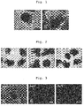
- Fig. 1A At day 18 from the start of the floating culture, formation of neuroepithelium was confirmed ( Fig. 1A ). At day 21 from the start of the floating culture, differentiation of cells into retinal pigment epithelial cells having thin epithelium was confirmed ( Fig. 1B ).
- Reference Example 2 Production of retinal pigment epithelial cell using human ES cells
- KhES-1-derived Human ES cells (KhES-1-derived) were cultured according to the methods described in " Ueno, M. et al. PNAS 2006, 103(25), 9554-9559 “ and “ Watanabe, K. et al. Nat Biotech 2007, 25, 681-686 ".
- As the medium a medium obtained by adding 20% KSR (KnockoutTM Serum Replacement; Invitrogen), 0.1 mM 2-mercaptoethanol, 2 mM L-glutamine, 1x non-essential amino acid, 5 ng/ml bFGF to DMEM/F12 medium (Sigma) was used.
- the cultured ES cells were dispersed into single cells using TrypLE Express (Invitrogen), and the singly dispersed ES cells were suspended in 100 ⁇ l of a serum-free medium at 1.2 ⁇ 10 4 cells per one well of a non-cell adhesive 96 well culture plate (sumilon spheroid plates, SUMITOMO BAKELITE Co., Ltd.) to allow for rapid formation of aggregates, and subjected to culture at 37°C, 5% CO 2 .
- serum-free medium therefor, a serum-free medium obtained by adding 10% KSR, 450 ⁇ M 1-monothioglycerol, 1x Chemically defined lipid concentrate, 20 ⁇ M Y27632 to a 1:1 mixture of F-12 medium and IMDM medium was used.
- BMP4 was added at a final concentration 1.5nM, and the floating culture was continued.
- a half amount of the culture medium in the well was exchanged every 3 days with the above-mentioned medium not supplemented with a substance acting on the BMP signal transduction pathway.
- a part of the aggregates subjected to floating culture in the aforementioned medium containing CHIR99021 underwent floating culture under the same medium conditions until day 27 from the start of the floating culture, and the other part underwent floating culture in a serum-free medium, which was DMEM/F-12 medium containing 3 ⁇ M CHIR99021 (a substance acting on the Wnt signal pathway) and 10 ⁇ M SU-5402 (a substance inhibiting the FGF signal pathway) and added with 1x N2 supplement, from day 21 to day 27 from the start of the floating culture at 37°C, 5% CO 2 .
- DMEM/F-12 medium containing 3 ⁇ M CHIR99021 (a substance acting on the Wnt signal pathway) and 10 ⁇ M SU-5402 (a substance inhibiting the FGF signal pathway) and added with 1x N2 supplement, from day 21 to day 27 from the start of the floating culture at 37°C, 5% CO 2 .
- the aggregates cultured in the presence of a substance acting on the Wnt signal pathway and a substance inhibiting the FGF signal pathway showed an increase in the differentiation induction efficiency, and aggregates having retinal pigment epithelial cells on almost all the surface of the aggregates were formed ( Fig. 2C ).
- Reference Example 3 Production of retinal pigment epithelial cell using human ES cells
- KhES-1-derived Human ES cells (KhES-1-derived) were cultured according to the methods described in " Ueno, M. et al. PNAS 2006, 103(25), 9554-9559 “ and “ Watanabe, K. et al. Nat Biotech 2007, 25, 681-686 ".
- As the medium a medium obtained by adding 20% KSR (KnockoutTM Serum Replacement; Invitrogen), 0.1 mM 2-mercaptoethanol, 2 mM L-glutamine, 1x non-essential amino acid, 5 ng/ml bFGF to DMEM/F12 medium (Sigma) was used.
- the cultured ES cells were dispersed into single cells using TrypLE Express (Invitrogen), and the singly dispersed ES cells were suspended in 100 ⁇ l of a serum-free medium at 1.2 ⁇ 10 4 cells per one well of a non-cell adhesive 96 well culture plate (sumilon spheroid plates, SUMITOMO BAKELITE Co., Ltd.) to allow for rapid formation of aggregates, and subjected to culture at 37°C, 5% CO 2 .
- serum-free medium therefor, a serum-free medium obtained by adding 10% KSR, 450 ⁇ M 1-monothioglycerol, 1x Chemically defined lipid concentrate, 20 ⁇ M Y27632 to a 1:1 mixture of F-12 medium and IMDM medium was used.
- BMP4 was added at a final concentration 1.5 nM, and floating culture was continued. A half amount of the culture medium in the well was exchanged every 3 days with the above-mentioned medium not supplemented with a substance acting on the BMP signal transduction pathway.
- the aggregates were transferred to a serum-free medium, which was DMEM/F-12 medium containing 3 ⁇ M CHIR99021 and added with 1x N2 supplement, and floating culture was continued until day 21 from the start of the floating culture.
- a serum-free medium which was DMEM/F-12 medium containing 3 ⁇ M CHIR99021 (a substance acting on the Wnt signal pathway) and added with 1x N2 supplement, at 37°C, 5% CO 2 .
- Fig. 3A retinal pigment epithelial cells having a characteristic polygonal cell form were observed.
- the expression of protein was confirmed by a cell staining method.
- the cells were positive to the transcription factor Mitf, which is a retinal pigment epithelial cell marker ( Fig. 3B ) and positive to ZO-1, which is a tight junction marker ( Fig. 3C ), from which it was confirmed that the obtained cells were retinal pigment epithelial cells.
- Mitf is a retinal pigment epithelial cell marker
- ZO-1 which is a tight junction marker
- Reference Example 4 Production of retinal pigment epithelial cell sheet using human ES cells
- KhES-1-derived Human ES cells (KhES-1-derived) were cultured according to the methods described in " Ueno, M. et al. PNAS 2006, 103(25), 9554-9559 “ and “ Watanabe, K. et al. Nat Biotech 2007, 25, 681-686 ".
- As the medium a medium obtained by adding 20% KSR (KnockoutTM Serum Replacement; Invitrogen), 0.1 mM 2-mercaptoethanol, 2 mM L-glutamine, 1x non-essential amino acid, 5 ng/ml bFGF to DMEM/F12 medium (Sigma) was used.
- the cultured ES cells were dispersed into single cells using TrypLE Express (Invitrogen), and the singly dispersed ES cells were suspended in 100 ⁇ l of a serum-free medium at 1.2 ⁇ 10 4 cells per one well of a non-cell adhesive 96 well culture plate (sumilon spheroid plates, SUMITOMO BAKELITE Co., Ltd.) to allow for rapid formation of aggregates, and subjected to culture at 37°C, 5% CO 2 .
- serum-free medium therefor, a serum-free medium obtained by adding 10% KSR, 450 ⁇ M 1-monothioglycerol, 1x Chemically defined lipid concentrate, 20 ⁇ M Y27632 to a 1:1 mixture of F-12 medium and IMDM medium was used.
- BMP4 was added at a final concentration 1.5 nM, and floating culture was continued. A half amount of the culture medium in the well was exchanged every 3 days with the above-mentioned medium not supplemented with a substance acting on the BMP signal transduction pathway.
- the aggregates were transferred to a serum-free medium, which was DMEM/F-12 medium containing 3 ⁇ M CHIR99021 and added with 1x N2 supplement, and cultured until day 21 from the start of the floating culture.
- a serum-free medium which was DMEM/F-12 medium containing 3 ⁇ M CHIR99021 and added with 1x N2 supplement.
- the aggregates were dispersed in cell dispersion solution Accutase (ICT), and debris was removed using a 40 ⁇ m cell strainer.
- the resulting cells were suspended in a serum-free medium (200 ⁇ l), which was a 1:1 mixture of DMEM/F-12 medium and Neurobasal medium added with 10% KSR, 1/2 N2 supplement, 1/2 B27 supplement, 20 ⁇ M Y27632, and seeded in a 65 mm Boyden chamber (Transwell, Corning Incorporated) after a coating treatment with SynthemaxTM at 2x10 5 cells per well, a medium (1 ml) having the same composition was also added to a lower well, and adhesion culture was performed at 37°C, 5% CO 2 . The medium was exchanged every 3 days, and the level of epithelial formation was observed under a microscope at day 40 from the start of the floating culture.
- Reference Example 5 Production of retinal pigment epithelial cell sheet using human ES cells
- KhES-1-derived Human ES cells (KhES-1-derived) were cultured according to the methods described in " Ueno, M. et al. PNAS 2006, 103(25), 9554-9559 “ and “ Watanabe, K. et al. Nat Biotech 2007, 25, 681-686 ".
- As the medium a medium obtained by adding 20% KSR (KnockoutTM Serum Replacement; Invitrogen), 0.1 mM 2-mercaptoethanol, 2 mM L-glutamine, 1x non-essential amino acid, 5 ng/ml bFGF to DMEM/F12 medium (Sigma) was used.
- the cultured ES cells were dispersed into single cells using TrypLE Express (Invitrogen), and the above-mentioned cultured ES cells were suspended in 100 ⁇ l of a serum-free medium at 1.2 ⁇ 10 4 cells per one well of a non-cell adhesive 96 well culture plate (sumilon spheroid plates, SUMITOMO BAKELITE Co., Ltd.) to allow for rapid formation of aggregates and subjected to culture at 37°C, 5% CO 2 .
- serum-free medium therefor, a serum-free medium obtained by adding 10% KSR, 450 ⁇ M 1-monothioglycerol, 1x Chemically defined lipid concentrate, 20 ⁇ M Y27632 to a 1:1 mixture of F-12 medium and IMDM medium was used.
- BMP4 was added at a concentration 1.5 nM, and floating culture was continued. A half amount of the culture medium in the well was exchanged every 3 days with the above-mentioned medium not supplemented with a substance acting on the BMP signal transduction pathway.
- the aggregates were transferred to a serum-free medium, which was DMEM/F-12 medium containing 3 ⁇ M CHIR99021 and added with 1x N2 supplement, and cultured until day 21 from the start of the floating culture.
- a serum-free medium which was DMEM/F-12 medium containing 3 ⁇ M CHIR99021 and added with 1x N2 supplement.
- the aggregates were dispersed in cell dispersion solution Accutase (ICT), and debris was removed using a 40 ⁇ m cell strainer.
- the resulting cells were suspended in a serum-free medium (200 ⁇ l), which was a 1:1 mixture of DMEM/F-12 medium and Neurobasal medium added with 10% KSR, 1/2 N2 supplement, 1/2 B27 supplement, 20 ⁇ M Y27632, and seeded in a 65 mm Boyden chamber (Transwell, Corning Incorporated) after a coating treatment with SynthemaxTM at 2x10 5 cells per well, a medium (1 ml) having the same composition was also added to a lower well, and adhesion culture was performed at 37°C, 5% CO 2 .
- Example 1 Production of retinal pigment epithelial cell sheet using human ES cells
- Growth Factor Reduced MatrigelTM (diluted 30-fold), or SynthemaxTM (Corning Incorporated) (diluted 40-fold) was added by 100 ⁇ l each to the wells of a 65 mm Boyden chamber (Transwell, Corning Incorporated).
- the above-mentioned chamber added with Matrigel was incubated at 4°C for 24 hr, and the above-mentioned chamber added with SynthemaxTM was incubated at room temperature for 2 hr, whereby a coating treatment of the culture vessel materials was performed.
- a single cell suspension was prepared from the aggregates at day 21 from the start of the floating culture by the method described in Example 4, the suspended cells were seeded in a serum-free medium (200 ⁇ l), which was a 1:1 mixture of DMEM/F-12 medium and Neurobasal medium added with 10% KSR, 1/2 N2 supplement, 1/2 B27 supplement, 20 ⁇ M Y27632 at 2x10 5 cells per well, a medium (1 ml) having the same composition was also added to a lower well, and adhesion culture was performed at 37°C, 5% CO 2 . Adhesion and epithelium formation of the cells were observed at day 5 from the seeding to a Boyden chamber.
- Example 2 Production Example of retinal pigment epithelial cell using induced pluripotent stem cells (iPS cells)
- Human iPS cell line 201B7 (available from RIKEN BioResource center or iPS Academia Japan Inc.) is cultivated according to the methods described in " Ueno, M. et al. PNAS 2006, 103(25), 9554-9559 “ and “ Watanabe, K. et al. Nat Biotech 2007, 25, 681-686 ".
- As the medium a medium obtained by adding 20% KSR (KnockoutTM Serum Replacement; Invitrogen), 0.1 mM 2-mercaptoethanol, 2 mM L-glutamine, 1x non-essential amino acid, 5 ng/ml bFGF to DMEM/F12 medium (Sigma) is used.
- the cultured iPS cells are dispersed into single cells using TrypLE Express (Invitrogen), suspended in 100 ⁇ l of a serum-free medium at 1.2 ⁇ 10 4 cells per one well of a non-cell adhesive 96 well culture plate (sumilon spheroid plates, SUMITOMO BAKELITE Co., Ltd.) to allow for rapid formation of aggregates and subjected to floating culture at 37°C, 5% CO 2 .
- a serum-free medium obtained by adding 10% KSR, 450 ⁇ M 1-monothioglycerol, 1x Chemically defined lipid concentrate, 20 ⁇ M Y27632 to a 1:1 mixture of F-12 medium and IMDM medium is used.
- BMP4 is added at a final concentration of 1.5 nM, and the floating culture is continued.
- a half amount of the culture medium in the well is exchanged every 3 days with the above-mentioned medium not supplemented with any substance acting on the BMP signal transduction pathway.
- the aggregates are transferred to a serum-free medium, which was DMEM/F-12 medium containing 3 ⁇ M CHIR99021 and added with 1x N2 supplement, and cultured until day 21 from the start of the floating culture.
- the aggregates are disrupted by injecting and ejecting the solution by a micropipette, and the resulting cells are subjected to adhesion culture in a serum-free medium, which is DMEM/F-12 medium containing 3 ⁇ M CHIR99021 (a substance acting on the Wnt signal pathway) and added with 1x N2 supplement, at 37°C, 5% CO 2 .
- a serum-free medium which is DMEM/F-12 medium containing 3 ⁇ M CHIR99021 (a substance acting on the Wnt signal pathway) and added with 1x N2 supplement, at 37°C, 5% CO 2 .
- cell form is observed under a microscope and expression of retinal pigment epithelial marker genes (Mitf, ZO-1) is confirmed by a cell staining method.
- retinal pigment epithelial cells are produced from human iPS cells.
- Example 3 Production Example of retinal pigment epithelial cell sheet using induced pluripotent stem cells (iPS cells)
- Human iPS cell line 201B7 (available from RIKEN BioResource center or iPS Academia Japan Inc.) is cultivated according to the methods described in " Ueno, M. et al. PNAS 2006, 103(25), 9554-9559 “ and “ Watanabe, K. et al. Nat Biotech 2007, 25, 681-686 ".
- As the medium a medium obtained by adding 20% KSR (KnockoutTM Serum Replacement; Invitrogen), 0.1 mM 2-mercaptoethanol, 2 mM L-glutamine, 1x non-essential amino acid, 5 ng/ml bFGF to DMEM/F12 medium (Sigma) is used.
- the cultured iPS cells are dispersed into single cells using TrypLE Express (Invitrogen), suspended in 100 ⁇ l of a serum-free medium at 1.2 ⁇ 10 4 cells per one well of a non-cell adhesive 96 well culture plate (sumilon spheroid plates, SUMITOMO BAKELITE Co., Ltd.) to allow for rapid formation of aggregates and subjected to floating culture at 37°C, 5% CO 2 .
- a serum-free medium obtained by adding 10% KSR, 450 ⁇ M 1-monothioglycerol, 1x Chemically defined lipid concentrate, 20 ⁇ M Y27632 to a 1:1 mixture of F-12 medium and IMDM medium is used.
- BMP4 is added at a final concentration of 1.5 nM and floating culture is continued. A half amount of the culture medium in the well is exchanged every 3 days with the above-mentioned medium not supplemented with a substance acting on the BMP signal transduction pathway.
- the aggregates are transferred to a serum-free medium, which is DMEM/F-12 medium containing 3 ⁇ M CHIR99021 and added with 1x N2 supplement, and cultured until day 21 from the start of the floating culture.
- the aggregates are dispersed in cell dispersion solution Accutase (ICT), and debris was removed using a 40 ⁇ m cell strainer.
- the resulting cells are suspended in a serum-free medium (200 ⁇ l), which is a 1:1 mixture of DMEM/F-12 medium and Neurobasal medium added with 10% KSR, 1/2 N2 supplement, 1/2 B27 supplement, 20 ⁇ M Y27632, and seeded in a 65 mm Boyden chamber (Transwell, Corning Incorporated) after a coating treatment with SynthemaxTM at 2x10 5 cells per well, a medium (1 ml) having the same composition is also added to a lower well, and adhesion culture is performed at 37°C, 5% CO 2 .
- a serum-free medium 200 ⁇ l
- a serum-free medium 200 ⁇ l
- Neurobasal medium a 1:1 mixture of DMEM/F-12 medium and Neurobasal medium added with 10% KSR, 1/2 N2 supplement, 1/2 B27 supplement, 20 ⁇ M Y27632, and seeded in a 65 mm Boyden chamber (Transwell, Corning Incorporated) after a coating treatment with SynthemaxTM at 2x10 5
- any of 3 ⁇ M CHIR99021, 10 ⁇ M SU-5402, 1 nM recombinant human BMP4 protein and 50 ng/ml recombinant human activin, or these in combination may be added.
- the medium is exchanged every 3 days, and the level of epithelial formation is observed under a microscope at day 40 from the start of the floating culture.
- retinal pigment epithelial cell sheet is produced from human iPS cells.
- retinal pigment epithelial cell or retinal pigment epithelial cell sheet can be produced with high efficiency.
Landscapes
- Health & Medical Sciences (AREA)
- Life Sciences & Earth Sciences (AREA)
- Engineering & Computer Science (AREA)
- Biomedical Technology (AREA)
- Chemical & Material Sciences (AREA)
- Cell Biology (AREA)
- Zoology (AREA)
- General Health & Medical Sciences (AREA)
- Medicinal Chemistry (AREA)
- Animal Behavior & Ethology (AREA)
- Public Health (AREA)
- Veterinary Medicine (AREA)
- Developmental Biology & Embryology (AREA)
- Epidemiology (AREA)
- Chemical Kinetics & Catalysis (AREA)
- Biotechnology (AREA)
- Bioinformatics & Cheminformatics (AREA)
- Organic Chemistry (AREA)
- Transplantation (AREA)
- Dermatology (AREA)
- Botany (AREA)
- Oral & Maxillofacial Surgery (AREA)
- Genetics & Genomics (AREA)
- Neurology (AREA)
- Wood Science & Technology (AREA)
- Urology & Nephrology (AREA)
- Neurosurgery (AREA)
- Biochemistry (AREA)
- General Engineering & Computer Science (AREA)
- Microbiology (AREA)
- Pharmacology & Pharmacy (AREA)
- Ophthalmology & Optometry (AREA)
- Hematology (AREA)
- Analytical Chemistry (AREA)
- Acoustics & Sound (AREA)
- Physics & Mathematics (AREA)
- Immunology (AREA)
- Reproductive Health (AREA)
- Virology (AREA)
- General Chemical & Material Sciences (AREA)
Claims (16)
- Procédé pour produire une cellule épithéliale pigmentaire rétinienne, comprenant(1) une première étape de soumission de cellules souches pluripotentes à une culture flottante dans un milieu sans sérum pour former un agrégat de cellules souches pluripotentes,(2) une deuxième étape de soumission de l'agrégat formé à l'étape (1) à une culture flottante dans un milieu sans sérum ou un milieu contenant du sérum, chacun de ces milieux étant exempt d'une substance qui peut amplifier la transduction de signal médiée par Sonic hedgehog et contenant une substance qui peut amplifier la voie de transduction de signal médiée par BMP, obtenant ainsi un agrégat contenant des cellules progénitrices rétiniennes, et(3) une troisième étape de soumission de l'agrégat obtenu dans l'étape (2) à une culture flottante dans un milieu sans sérum ou un milieu contenant du sérum, chacun de ces milieux étant exempt d'une substance qui peut amplifier la transduction de signal médiée par Sonic hedgehog et d'une substance qui peut amplifier la voie de transduction de signal médiée par BMP et contenant une substance qui peut amplifier la transduction de signal médiée par Wnt, obtenant ainsi un agrégat contenant des cellules épithéliales pigmentaires rétiniennes.
- Procédé pour produire un feuillet de cellules épithéliales pigmentaires rétiniennes, comprenant(1) une première étape de soumission de cellules souches pluripotentes à une culture flottante dans un milieu sans sérum pour former un agrégat de cellules souches pluripotentes,(2) une deuxième étape de soumission de l'agrégat formé dans l'étape (1) à une culture flottante dans un milieu sans sérum ou un milieu contenant du sérum, chacun de ces milieux étant exempt d'une substance qui peut amplifier la transduction de signal médiée par Sonic hedgehog et contenant une substance qui peut amplifier la voie de transduction de signal médiée par BMP, obtenant ainsi un agrégat contenant des cellules progénitrices rétiniennes,(3) une troisième étape de soumission de l'agrégat obtenu dans l'étape (2) à une culture flottante dans un milieu sans sérum ou un milieu contenant du sérum, chacun de ces milieux étant exempt d'une substance qui peut amplifier la transduction de signal médiée par Sonic hedgehog et d'une substance qui peut amplifier la voie de transduction de signal médiée par BMP et contenant une substance qui peut amplifier la transduction de signal médiée par Wnt, ce qui donne ainsi un agrégat contenant des cellules épithéliales pigmentaires rétiniennes, et(4) une quatrième étape de dispersion de l'agrégat obtenu dans l'étape (3) et de soumission des cellules résultantes à une culture par adhésion.
- Procédé de production selon la revendication 2, dans lequel(a) la culture par adhésion est effectuée en présence d'un substitut de sérum dans ladite étape (4) ; et/ou(b) la culture par adhésion est effectuée en présence d'une substance inhibitrice de ROCK dans ladite étape (4).
- Procédé de production selon la revendication 2 ou 3, dans lequel la culture par adhésion est effectuée dans un milieu sans sérum ou un milieu contenant du sérum et comprenant en outre une ou plusieurs substances choisies dans le groupe constitué par une substance qui peut amplifier la transduction de signal médiée par Wnt, une substance inhibant la voie de signalisation de FGF, une substance capable d'amplifier un signal médiée par l'activine et une substance qui peut amplifier la voie de transduction de signal médiée par BMP, dans ladite étape (4).
- Procédé de production selon l'une quelconque des revendications 2 à 4, dans lequel la culture par adhésion est effectuée sur un matériau de récipient de culture ayant une surface traitée avec un substrat de culture, dans ladite étape (4).
- Procédé de production selon la revendication 5, dans lequel(a) ledit substrat de culture est un substrat de culture synthétique ; ou(b) ledit substrat de culture est la laminine.
- Procédé de production selon l'une quelconque des revendications 1 à 6, dans lequel(a) les cellules souches pluripotentes sont des cellules souches pluripotentes de primate ; ou(b) les cellules souches pluripotentes sont des cellules souches pluripotentes humaines.
- Procédé de production selon l'une quelconque des revendications 1 à 7, dans lequel l'étape (1) et l'étape (2) sont effectuées en présence d'un substitut de sérum.
- Procédé de production selon l'une quelconque des revendications 1 à 8, dans lequel la culture flottante est effectuée en l'absence d'une préparation de membrane basale.
- Procédé de production selon l'une quelconque des revendications 1 à 9, dans lequel la substance qui peut amplifier la voie de transduction de signal médiée par BMP est une ou plusieurs protéines choisies dans le groupe constitué par BMP2, BMP4, BMP7 et GDF7.
- Procédé de production selon l'une quelconque des revendications 1 à 10, dans lequel le milieu sans sérum ou le milieu contenant du sérum, chacun étant exempt d'une substance qui peut amplifier la transduction de signal médiée par Sonic hedgehog et d'une substance qui peut amplifier la voie de transduction de signal médiée par BMP et contenant une substance qui peut amplifier la transduction de signal médiée par Wnt dans l'étape (3) comprend en outre une substance inhibant la voie de signalisation de FGF.
- Procédé de production selon l'une quelconque des revendications 1 à 11, dans lequel la substance qui peut amplifier la transduction de signal médiée par Wnt est choisie dans le groupe constitué par les protéines appartenant à la famille Wnt, un agoniste de récepteur de Wnt, et un inhibiteur de GSK3β.
- Procédé selon la revendication 12, dans lequel la protéine appartenant à la famille Wnt est Wnt1, Wnt3a ou Wnt7a, et dans lequel l'inhibiteur de GSK3β est la 6-bromoindirubine-3'-oxime (BIO), CHIR99021 ou la kenpaullone.
- Procédé de production selon l'une quelconque des revendications 4 à 13, dans lequel la substance inhibant la voie de signalisation de FGF est choisie dans le groupe constitué par un récepteur de FGF, un inhibiteur de récepteur de FGF, une substance inhibitrice de la cascade de MAP kinase, un inhibiteur de PI3 kinase et un inhibiteur d'Akt.
- Procédé selon la revendication 14, dans lequel l'inhibiteur de récepteur de FGF est SU-5402, AZD4547 ou BGJ398, et dans lequel la substance inhibitrice de la cascade de MAP kinase est un inhibiteur de MEK, un inhibiteur de MAPK ou un inhibiteur d'ERK.
- Procédé selon l'une quelconque des revendications 4 à 15, dans lequel la substance capable d'amplifier un signal médié par l'activine est choisie dans le groupe constitué par les protéines appartenant à la famille de l'activine, un récepteur d'activine, et un agoniste de récepteur d'activine.
Applications Claiming Priority (2)
| Application Number | Priority Date | Filing Date | Title |
|---|---|---|---|
| JP2013232795 | 2013-11-11 | ||
| PCT/JP2014/076471 WO2015068505A1 (fr) | 2013-11-11 | 2014-10-02 | Procédé de production de cellules épithéliales de pigment rétinien |
Publications (3)
| Publication Number | Publication Date |
|---|---|
| EP3070162A1 EP3070162A1 (fr) | 2016-09-21 |
| EP3070162A4 EP3070162A4 (fr) | 2017-05-31 |
| EP3070162B1 true EP3070162B1 (fr) | 2019-09-18 |
Family
ID=53041288
Family Applications (1)
| Application Number | Title | Priority Date | Filing Date |
|---|---|---|---|
| EP14860084.4A Active EP3070162B1 (fr) | 2013-11-11 | 2014-10-02 | Procédé de production de cellules épithéliales de pigment rétinien |
Country Status (9)
| Country | Link |
|---|---|
| US (1) | US10760050B2 (fr) |
| EP (1) | EP3070162B1 (fr) |
| JP (1) | JP6495176B2 (fr) |
| KR (1) | KR102297584B1 (fr) |
| CN (2) | CN120384050A (fr) |
| AU (1) | AU2014345110B2 (fr) |
| CA (1) | CA2930092C (fr) |
| ES (1) | ES2759447T3 (fr) |
| WO (1) | WO2015068505A1 (fr) |
Families Citing this family (31)
| Publication number | Priority date | Publication date | Assignee | Title |
|---|---|---|---|---|
| EP2784152B1 (fr) * | 2011-11-25 | 2019-04-10 | Sumitomo Chemical Company, Limited | Procédés pour la production d'un tissu de la rétine et de cellules associées à la rétine |
| US11241460B2 (en) | 2013-03-15 | 2022-02-08 | Astellas Institute For Regenerative Medicine | Photoreceptors and photoreceptor progenitors produced from pluripotent stem cells |
| EP4292600A3 (fr) | 2013-03-15 | 2024-04-10 | Astellas Institute for Regenerative Medicine | Photorécepteurs et progéniteurs de photorécepteurs produits à partir de cellules souches pluripotentes |
| TWI810142B (zh) | 2014-10-24 | 2023-08-01 | 日商住友製藥股份有限公司 | 神經組織的製造方法 |
| EP3211071B1 (fr) | 2014-10-24 | 2021-05-05 | Sumitomo Dainippon Pharma Co., Ltd. | Procédé de production de tissu rétinien |
| GB201509264D0 (en) | 2015-05-29 | 2015-07-15 | Univ Leuven Kath | Novel ATMP's |
| AU2016318774B2 (en) | 2015-09-08 | 2022-08-18 | FUJIFILM Cellular Dynamics, Inc. | MACS-based purification of stem cell-derived retinal pigment epithelium |
| SG11201801770VA (en) * | 2015-09-08 | 2018-04-27 | Sumitomo Dainippon Pharma Co Ltd | Method for producing retinal pigment epithelial cells |
| DK3347456T3 (da) * | 2015-09-08 | 2024-02-19 | Us Health | Fremgangsmåde til reproducerbar differentiering af retinale pigmentepitelceller af klinisk kvalitet |
| US12385008B2 (en) * | 2015-09-08 | 2025-08-12 | Sumitomo Pharma Co., Ltd. | Method for producing retinal tissue |
| JP7024974B2 (ja) * | 2016-04-20 | 2022-02-24 | 京都府公立大学法人 | 培養上皮シートの製造方法 |
| SG11201809201UA (en) * | 2016-04-22 | 2018-11-29 | Sumitomo Dainippon Pharma Co Ltd | Method for producing retinal tissue |
| EP4733316A3 (fr) * | 2017-01-31 | 2026-05-06 | Osaka University | Procédé de régulation de différenciation pour cellules souches pluripotentes |
| WO2018164240A1 (fr) * | 2017-03-08 | 2018-09-13 | 大日本住友製薬株式会社 | Procédé de production de cellules épithéliales pigmentaires rétiniennes |
| CA3074426A1 (fr) | 2017-09-08 | 2019-03-14 | Sumitomo Dainippon Pharma Co., Ltd. | Agregat cellulaire comprenant du tissu retinien, et methode de production correspondante |
| US12090252B2 (en) | 2017-09-14 | 2024-09-17 | Riken | Method for producing retinal tissues |
| CA3075883A1 (fr) * | 2017-09-14 | 2019-03-21 | Riken | Procede d'amplification de photorecepteurs de cones ou photorecepteurs de batonnets a l'aide d'un transmetteur de signal de dorsalisation ou un transmetteur de signal de ventralis ation |
| JP7553924B2 (ja) | 2019-04-26 | 2024-09-19 | 国立研究開発法人理化学研究所 | 神経網膜と網膜色素上皮細胞とハイドロゲルとを含む複合体及びその製造方法 |
| EP3969570A1 (fr) * | 2019-05-15 | 2022-03-23 | Novo Nordisk A/S | Procédé d'obtention de cellules progénitrices su champ de l'oeil à partir de cellules souches pluripotentes humaines |
| JP7760131B2 (ja) | 2020-09-11 | 2025-10-27 | 株式会社Racthera | 移植用組織のための媒体 |
| JPWO2022191216A1 (fr) | 2021-03-09 | 2022-09-15 | ||
| CN113481158A (zh) * | 2021-06-24 | 2021-10-08 | 暨南大学 | 一种无支架视网膜色素上皮细胞片的制备方法 |
| CN115873797B (zh) * | 2021-09-29 | 2024-08-13 | 北京干细胞与再生医学研究院 | 一种视网膜色素上皮细胞的扩增培养基及培养方法 |
| EP4428227A4 (fr) | 2021-11-19 | 2025-11-12 | Riken | Procédé de production de tissu rétinien en forme de feuille |
| US20260000538A1 (en) | 2022-01-12 | 2026-01-01 | Riken | Tissue transplantation resin tip, tissue transplantation device, retina tissue transplantation method, and retina tissue transplantation kit |
| EP4477744A4 (fr) * | 2022-02-07 | 2025-06-11 | Osaka University | Procédé pour induire la différenciation de cellules souches pluripotentes en cellules épithéliales pigmentaires rétiniennes |
| CN118871141A (zh) | 2022-03-16 | 2024-10-29 | 住友制药株式会社 | 移植用介质 |
| TW202434721A (zh) | 2023-02-14 | 2024-09-01 | 國立大學法人山口大學 | 細胞群、三維細胞培養體、醫藥組成物、胞泌體及製作此細胞群的方法 |
| EP4659755A1 (fr) | 2023-02-14 | 2025-12-10 | RACTHERA Co., Ltd. | Médicament thérapeutique pour des déchirures d'épithélium pigmentaire rétinien |
| JPWO2024204596A1 (fr) | 2023-03-29 | 2024-10-03 | ||
| CN117417891A (zh) * | 2023-12-19 | 2024-01-19 | 南京艾尔普再生医学科技有限公司 | 一种视网膜色素上皮细胞球形制剂 |
Family Cites Families (13)
| Publication number | Priority date | Publication date | Assignee | Title |
|---|---|---|---|---|
| CA2277278A1 (fr) | 1997-01-10 | 1998-07-16 | Life Technologies, Inc. | Substitut de serum pour cellules souches embryonnaires |
| EP2147094B2 (fr) * | 2007-04-18 | 2018-02-21 | Hadasit Medical Research Services & Development Limited | Cellules de l'épithélium pigmentaire de la rétine dérivées de cellules souches |
| EP2470645B1 (fr) | 2009-08-24 | 2019-06-26 | Wisconsin Alumni Research Foundation | Progéniteur rétinien humain sensiblement pur, progéniteur de cerveau antérieur, et cultures de cellules d'épithélium pigmentaire rétinien et leurs procédés de fabrication |
| EP2486126B1 (fr) | 2009-10-06 | 2017-12-06 | SNU R & DB Foundation | Procédé pour la différenciation en cellules rétiniennes à partir de cellules souches |
| JP5787370B2 (ja) | 2009-11-05 | 2015-09-30 | 国立研究開発法人理化学研究所 | 幹細胞の分化誘導方法 |
| US9902933B2 (en) | 2011-02-25 | 2018-02-27 | Riken | Method of producing retinal pigment epithelial cell sheet |
| JPWO2012173207A1 (ja) * | 2011-06-14 | 2015-02-23 | 独立行政法人理化学研究所 | 網膜細胞への分化誘導方法 |
| JP6210881B2 (ja) * | 2011-10-31 | 2017-10-11 | 国立研究開発法人理化学研究所 | 幹細胞の培養方法 |
| JP5985207B2 (ja) * | 2011-11-25 | 2016-09-06 | 住友化学株式会社 | 網膜色素上皮細胞の製造方法 |
| EP2784152B1 (fr) | 2011-11-25 | 2019-04-10 | Sumitomo Chemical Company, Limited | Procédés pour la production d'un tissu de la rétine et de cellules associées à la rétine |
| EP2789152B1 (fr) * | 2012-03-01 | 2018-06-27 | Nec Corporation | Procédé de fourniture d'accès pour un terminal utilisateur à un service fourni par une fonction applicative à l'intérieur d'une structure de réseau, et structure de réseau correspondante |
| US9458428B2 (en) * | 2012-06-05 | 2016-10-04 | The Regents Of The University Of California | Methods and compositions for the rapid production of retinal pigmented epithelial cells from pluripotent cells |
| KR102278978B1 (ko) | 2013-08-23 | 2021-07-19 | 스미또모 가가꾸 가부시끼가이샤 | 망막 조직 및 망막 관련 세포의 제조 방법 |
-
2014
- 2014-10-02 EP EP14860084.4A patent/EP3070162B1/fr active Active
- 2014-10-02 AU AU2014345110A patent/AU2014345110B2/en not_active Ceased
- 2014-10-02 CN CN202510408783.5A patent/CN120384050A/zh active Pending
- 2014-10-02 ES ES14860084T patent/ES2759447T3/es active Active
- 2014-10-02 US US15/035,966 patent/US10760050B2/en active Active
- 2014-10-02 KR KR1020167015215A patent/KR102297584B1/ko active Active
- 2014-10-02 CA CA2930092A patent/CA2930092C/fr active Active
- 2014-10-02 CN CN201480068651.2A patent/CN105829527A/zh active Pending
- 2014-10-02 WO PCT/JP2014/076471 patent/WO2015068505A1/fr not_active Ceased
- 2014-10-02 JP JP2015546349A patent/JP6495176B2/ja active Active
Non-Patent Citations (1)
| Title |
|---|
| None * |
Also Published As
| Publication number | Publication date |
|---|---|
| ES2759447T3 (es) | 2020-05-11 |
| AU2014345110A1 (en) | 2016-06-30 |
| KR20160082248A (ko) | 2016-07-08 |
| US10760050B2 (en) | 2020-09-01 |
| EP3070162A1 (fr) | 2016-09-21 |
| JPWO2015068505A1 (ja) | 2017-03-09 |
| CN105829527A (zh) | 2016-08-03 |
| KR102297584B1 (ko) | 2021-09-03 |
| JP6495176B2 (ja) | 2019-04-03 |
| AU2014345110B2 (en) | 2021-01-14 |
| EP3070162A4 (fr) | 2017-05-31 |
| WO2015068505A1 (fr) | 2015-05-14 |
| CA2930092C (fr) | 2024-03-12 |
| CA2930092A1 (fr) | 2015-05-14 |
| US20160264936A1 (en) | 2016-09-15 |
| CN120384050A (zh) | 2025-07-29 |
Similar Documents
| Publication | Publication Date | Title |
|---|---|---|
| EP3070162B1 (fr) | Procédé de production de cellules épithéliales de pigment rétinien | |
| US20250354112A1 (en) | Method for producing retinal tissue and retina-related cells | |
| AU2017254264C1 (en) | Method for producing retinal tissue | |
| EP3081637B1 (fr) | Procédé pour la fabrication de structure analogue à la zone marginale ciliaire | |
| AU2014378349B2 (en) | Method for manufacturing ciliary margin stem cells | |
| HK1228951B (en) | Method for producing retinal pigment epithelial cells | |
| HK1228951A1 (en) | Method for producing retinal pigment epithelial cells | |
| HK1226441A1 (en) | Method for producing retinal tissue and retina-related cells | |
| HK1226441B (en) | Method for producing retinal tissue and retina-related cells | |
| HK1261232A1 (en) | Method for producing retinal tissue | |
| HK1230238A1 (en) | Method for manufacturing ciliary marginal zone-like structure | |
| HK1230238B (en) | Method for manufacturing ciliary marginal zone-like structure |
Legal Events
| Date | Code | Title | Description |
|---|---|---|---|
| PUAI | Public reference made under article 153(3) epc to a published international application that has entered the european phase |
Free format text: ORIGINAL CODE: 0009012 |
|
| 17P | Request for examination filed |
Effective date: 20160610 |
|
| AK | Designated contracting states |
Kind code of ref document: A1 Designated state(s): AL AT BE BG CH CY CZ DE DK EE ES FI FR GB GR HR HU IE IS IT LI LT LU LV MC MK MT NL NO PL PT RO RS SE SI SK SM TR |
|
| AX | Request for extension of the european patent |
Extension state: BA ME |
|
| DAX | Request for extension of the european patent (deleted) | ||
| A4 | Supplementary search report drawn up and despatched |
Effective date: 20170503 |
|
| RIC1 | Information provided on ipc code assigned before grant |
Ipc: C12N 5/074 20100101ALI20170425BHEP Ipc: C12N 5/0735 20100101AFI20170425BHEP Ipc: A61L 27/00 20060101ALI20170425BHEP Ipc: A61K 35/44 20150101ALI20170425BHEP Ipc: A61K 35/12 20150101ALI20170425BHEP Ipc: C12N 5/10 20060101ALI20170425BHEP Ipc: A61P 27/00 20060101ALI20170425BHEP |
|
| REG | Reference to a national code |
Ref country code: HK Ref legal event code: DE Ref document number: 1228951 Country of ref document: HK |
|
| STAA | Information on the status of an ep patent application or granted ep patent |
Free format text: STATUS: EXAMINATION IS IN PROGRESS |
|
| 17Q | First examination report despatched |
Effective date: 20180118 |
|
| GRAP | Despatch of communication of intention to grant a patent |
Free format text: ORIGINAL CODE: EPIDOSNIGR1 |
|
| STAA | Information on the status of an ep patent application or granted ep patent |
Free format text: STATUS: GRANT OF PATENT IS INTENDED |
|
| INTG | Intention to grant announced |
Effective date: 20181008 |
|
| GRAJ | Information related to disapproval of communication of intention to grant by the applicant or resumption of examination proceedings by the epo deleted |
Free format text: ORIGINAL CODE: EPIDOSDIGR1 |
|
| STAA | Information on the status of an ep patent application or granted ep patent |
Free format text: STATUS: EXAMINATION IS IN PROGRESS |
|
| INTC | Intention to grant announced (deleted) | ||
| GRAP | Despatch of communication of intention to grant a patent |
Free format text: ORIGINAL CODE: EPIDOSNIGR1 |
|
| STAA | Information on the status of an ep patent application or granted ep patent |
Free format text: STATUS: GRANT OF PATENT IS INTENDED |
|
| INTG | Intention to grant announced |
Effective date: 20190329 |
|
| GRAS | Grant fee paid |
Free format text: ORIGINAL CODE: EPIDOSNIGR3 |
|
| GRAA | (expected) grant |
Free format text: ORIGINAL CODE: 0009210 |
|
| STAA | Information on the status of an ep patent application or granted ep patent |
Free format text: STATUS: THE PATENT HAS BEEN GRANTED |
|
| AK | Designated contracting states |
Kind code of ref document: B1 Designated state(s): AL AT BE BG CH CY CZ DE DK EE ES FI FR GB GR HR HU IE IS IT LI LT LU LV MC MK MT NL NO PL PT RO RS SE SI SK SM TR |
|
| RAP1 | Party data changed (applicant data changed or rights of an application transferred) |
Owner name: SUMITOMO CHEMICAL COMPANY, LIMITED Owner name: RIKEN |
|
| REG | Reference to a national code |
Ref country code: GB Ref legal event code: FG4D |
|
| RIN1 | Information on inventor provided before grant (corrected) |
Inventor name: OZONE, CHIKAFUMI Inventor name: NAKANO, TOKUSHIGE Inventor name: SASAI, YOSHIKI |
|
| REG | Reference to a national code |
Ref country code: CH Ref legal event code: EP |
|
| REG | Reference to a national code |
Ref country code: DE Ref legal event code: R096 Ref document number: 602014054019 Country of ref document: DE |
|
| RIN2 | Information on inventor provided after grant (corrected) |
Inventor name: NAKANO, TOKUSHIGE Inventor name: OZONE, CHIKAFUMI Inventor name: SASAI, YOSHIKI |
|
| REG | Reference to a national code |
Ref country code: AT Ref legal event code: REF Ref document number: 1181335 Country of ref document: AT Kind code of ref document: T Effective date: 20191015 |
|
| REG | Reference to a national code |
Ref country code: IE Ref legal event code: FG4D |
|
| REG | Reference to a national code |
Ref country code: CH Ref legal event code: NV Representative=s name: BOHEST AG, CH |
|
| REG | Reference to a national code |
Ref country code: NL Ref legal event code: MP Effective date: 20190918 |
|
| PG25 | Lapsed in a contracting state [announced via postgrant information from national office to epo] |
Ref country code: HR Free format text: LAPSE BECAUSE OF FAILURE TO SUBMIT A TRANSLATION OF THE DESCRIPTION OR TO PAY THE FEE WITHIN THE PRESCRIBED TIME-LIMIT Effective date: 20190918 Ref country code: BG Free format text: LAPSE BECAUSE OF FAILURE TO SUBMIT A TRANSLATION OF THE DESCRIPTION OR TO PAY THE FEE WITHIN THE PRESCRIBED TIME-LIMIT Effective date: 20191218 Ref country code: SE Free format text: LAPSE BECAUSE OF FAILURE TO SUBMIT A TRANSLATION OF THE DESCRIPTION OR TO PAY THE FEE WITHIN THE PRESCRIBED TIME-LIMIT Effective date: 20190918 Ref country code: LT Free format text: LAPSE BECAUSE OF FAILURE TO SUBMIT A TRANSLATION OF THE DESCRIPTION OR TO PAY THE FEE WITHIN THE PRESCRIBED TIME-LIMIT Effective date: 20190918 Ref country code: FI Free format text: LAPSE BECAUSE OF FAILURE TO SUBMIT A TRANSLATION OF THE DESCRIPTION OR TO PAY THE FEE WITHIN THE PRESCRIBED TIME-LIMIT Effective date: 20190918 Ref country code: NO Free format text: LAPSE BECAUSE OF FAILURE TO SUBMIT A TRANSLATION OF THE DESCRIPTION OR TO PAY THE FEE WITHIN THE PRESCRIBED TIME-LIMIT Effective date: 20191218 |
|
| REG | Reference to a national code |
Ref country code: LT Ref legal event code: MG4D |
|
| PG25 | Lapsed in a contracting state [announced via postgrant information from national office to epo] |
Ref country code: RS Free format text: LAPSE BECAUSE OF FAILURE TO SUBMIT A TRANSLATION OF THE DESCRIPTION OR TO PAY THE FEE WITHIN THE PRESCRIBED TIME-LIMIT Effective date: 20190918 Ref country code: GR Free format text: LAPSE BECAUSE OF FAILURE TO SUBMIT A TRANSLATION OF THE DESCRIPTION OR TO PAY THE FEE WITHIN THE PRESCRIBED TIME-LIMIT Effective date: 20191219 Ref country code: AL Free format text: LAPSE BECAUSE OF FAILURE TO SUBMIT A TRANSLATION OF THE DESCRIPTION OR TO PAY THE FEE WITHIN THE PRESCRIBED TIME-LIMIT Effective date: 20190918 Ref country code: LV Free format text: LAPSE BECAUSE OF FAILURE TO SUBMIT A TRANSLATION OF THE DESCRIPTION OR TO PAY THE FEE WITHIN THE PRESCRIBED TIME-LIMIT Effective date: 20190918 |
|
| REG | Reference to a national code |
Ref country code: AT Ref legal event code: MK05 Ref document number: 1181335 Country of ref document: AT Kind code of ref document: T Effective date: 20190918 |
|
| PG25 | Lapsed in a contracting state [announced via postgrant information from national office to epo] |
Ref country code: NL Free format text: LAPSE BECAUSE OF FAILURE TO SUBMIT A TRANSLATION OF THE DESCRIPTION OR TO PAY THE FEE WITHIN THE PRESCRIBED TIME-LIMIT Effective date: 20190918 Ref country code: PT Free format text: LAPSE BECAUSE OF FAILURE TO SUBMIT A TRANSLATION OF THE DESCRIPTION OR TO PAY THE FEE WITHIN THE PRESCRIBED TIME-LIMIT Effective date: 20200120 Ref country code: EE Free format text: LAPSE BECAUSE OF FAILURE TO SUBMIT A TRANSLATION OF THE DESCRIPTION OR TO PAY THE FEE WITHIN THE PRESCRIBED TIME-LIMIT Effective date: 20190918 Ref country code: AT Free format text: LAPSE BECAUSE OF FAILURE TO SUBMIT A TRANSLATION OF THE DESCRIPTION OR TO PAY THE FEE WITHIN THE PRESCRIBED TIME-LIMIT Effective date: 20190918 Ref country code: RO Free format text: LAPSE BECAUSE OF FAILURE TO SUBMIT A TRANSLATION OF THE DESCRIPTION OR TO PAY THE FEE WITHIN THE PRESCRIBED TIME-LIMIT Effective date: 20190918 Ref country code: PL Free format text: LAPSE BECAUSE OF FAILURE TO SUBMIT A TRANSLATION OF THE DESCRIPTION OR TO PAY THE FEE WITHIN THE PRESCRIBED TIME-LIMIT Effective date: 20190918 |
|
| REG | Reference to a national code |
Ref country code: ES Ref legal event code: FG2A Ref document number: 2759447 Country of ref document: ES Kind code of ref document: T3 Effective date: 20200511 |
|
| PG25 | Lapsed in a contracting state [announced via postgrant information from national office to epo] |
Ref country code: SM Free format text: LAPSE BECAUSE OF FAILURE TO SUBMIT A TRANSLATION OF THE DESCRIPTION OR TO PAY THE FEE WITHIN THE PRESCRIBED TIME-LIMIT Effective date: 20190918 Ref country code: IS Free format text: LAPSE BECAUSE OF FAILURE TO SUBMIT A TRANSLATION OF THE DESCRIPTION OR TO PAY THE FEE WITHIN THE PRESCRIBED TIME-LIMIT Effective date: 20200224 Ref country code: SK Free format text: LAPSE BECAUSE OF FAILURE TO SUBMIT A TRANSLATION OF THE DESCRIPTION OR TO PAY THE FEE WITHIN THE PRESCRIBED TIME-LIMIT Effective date: 20190918 Ref country code: CZ Free format text: LAPSE BECAUSE OF FAILURE TO SUBMIT A TRANSLATION OF THE DESCRIPTION OR TO PAY THE FEE WITHIN THE PRESCRIBED TIME-LIMIT Effective date: 20190918 |
|
| REG | Reference to a national code |
Ref country code: DE Ref legal event code: R097 Ref document number: 602014054019 Country of ref document: DE |
|
| PLBE | No opposition filed within time limit |
Free format text: ORIGINAL CODE: 0009261 |
|
| STAA | Information on the status of an ep patent application or granted ep patent |
Free format text: STATUS: NO OPPOSITION FILED WITHIN TIME LIMIT |
|
| PG2D | Information on lapse in contracting state deleted |
Ref country code: IS |
|
| PG25 | Lapsed in a contracting state [announced via postgrant information from national office to epo] |
Ref country code: LU Free format text: LAPSE BECAUSE OF NON-PAYMENT OF DUE FEES Effective date: 20191002 Ref country code: DK Free format text: LAPSE BECAUSE OF FAILURE TO SUBMIT A TRANSLATION OF THE DESCRIPTION OR TO PAY THE FEE WITHIN THE PRESCRIBED TIME-LIMIT Effective date: 20190918 Ref country code: IS Free format text: LAPSE BECAUSE OF FAILURE TO SUBMIT A TRANSLATION OF THE DESCRIPTION OR TO PAY THE FEE WITHIN THE PRESCRIBED TIME-LIMIT Effective date: 20200119 |
|
| REG | Reference to a national code |
Ref country code: BE Ref legal event code: MM Effective date: 20191031 |
|
| 26N | No opposition filed |
Effective date: 20200619 |
|
| PG25 | Lapsed in a contracting state [announced via postgrant information from national office to epo] |
Ref country code: SI Free format text: LAPSE BECAUSE OF FAILURE TO SUBMIT A TRANSLATION OF THE DESCRIPTION OR TO PAY THE FEE WITHIN THE PRESCRIBED TIME-LIMIT Effective date: 20190918 Ref country code: BE Free format text: LAPSE BECAUSE OF NON-PAYMENT OF DUE FEES Effective date: 20191031 Ref country code: MC Free format text: LAPSE BECAUSE OF FAILURE TO SUBMIT A TRANSLATION OF THE DESCRIPTION OR TO PAY THE FEE WITHIN THE PRESCRIBED TIME-LIMIT Effective date: 20190918 |
|
| PG25 | Lapsed in a contracting state [announced via postgrant information from national office to epo] |
Ref country code: IE Free format text: LAPSE BECAUSE OF NON-PAYMENT OF DUE FEES Effective date: 20191002 |
|
| PG25 | Lapsed in a contracting state [announced via postgrant information from national office to epo] |
Ref country code: CY Free format text: LAPSE BECAUSE OF FAILURE TO SUBMIT A TRANSLATION OF THE DESCRIPTION OR TO PAY THE FEE WITHIN THE PRESCRIBED TIME-LIMIT Effective date: 20190918 |
|
| PG25 | Lapsed in a contracting state [announced via postgrant information from national office to epo] |
Ref country code: MT Free format text: LAPSE BECAUSE OF FAILURE TO SUBMIT A TRANSLATION OF THE DESCRIPTION OR TO PAY THE FEE WITHIN THE PRESCRIBED TIME-LIMIT Effective date: 20190918 Ref country code: HU Free format text: LAPSE BECAUSE OF FAILURE TO SUBMIT A TRANSLATION OF THE DESCRIPTION OR TO PAY THE FEE WITHIN THE PRESCRIBED TIME-LIMIT; INVALID AB INITIO Effective date: 20141002 |
|
| PG25 | Lapsed in a contracting state [announced via postgrant information from national office to epo] |
Ref country code: TR Free format text: LAPSE BECAUSE OF FAILURE TO SUBMIT A TRANSLATION OF THE DESCRIPTION OR TO PAY THE FEE WITHIN THE PRESCRIBED TIME-LIMIT Effective date: 20190918 |
|
| PG25 | Lapsed in a contracting state [announced via postgrant information from national office to epo] |
Ref country code: MK Free format text: LAPSE BECAUSE OF FAILURE TO SUBMIT A TRANSLATION OF THE DESCRIPTION OR TO PAY THE FEE WITHIN THE PRESCRIBED TIME-LIMIT Effective date: 20190918 |
|
| P01 | Opt-out of the competence of the unified patent court (upc) registered |
Effective date: 20230505 |
|
| PGFP | Annual fee paid to national office [announced via postgrant information from national office to epo] |
Ref country code: IT Payment date: 20250926 Year of fee payment: 12 |
|
| PGFP | Annual fee paid to national office [announced via postgrant information from national office to epo] |
Ref country code: GB Payment date: 20250925 Year of fee payment: 12 |
|
| PGFP | Annual fee paid to national office [announced via postgrant information from national office to epo] |
Ref country code: FR Payment date: 20250925 Year of fee payment: 12 |
|
| REG | Reference to a national code |
Ref country code: CH Ref legal event code: U11 Free format text: ST27 STATUS EVENT CODE: U-0-0-U10-U11 (AS PROVIDED BY THE NATIONAL OFFICE) Effective date: 20251101 |
|
| PGFP | Annual fee paid to national office [announced via postgrant information from national office to epo] |
Ref country code: DE Payment date: 20250925 Year of fee payment: 12 |
|
| PGFP | Annual fee paid to national office [announced via postgrant information from national office to epo] |
Ref country code: CH Payment date: 20251101 Year of fee payment: 12 |
|
| PGFP | Annual fee paid to national office [announced via postgrant information from national office to epo] |
Ref country code: ES Payment date: 20251113 Year of fee payment: 12 |