EP3079730B1 - Verfahren zum verkleben von gewebeoberflächen und materialien und biomedizinische verwendungen davon - Google Patents
Verfahren zum verkleben von gewebeoberflächen und materialien und biomedizinische verwendungen davon Download PDFInfo
- Publication number
- EP3079730B1 EP3079730B1 EP14809410.5A EP14809410A EP3079730B1 EP 3079730 B1 EP3079730 B1 EP 3079730B1 EP 14809410 A EP14809410 A EP 14809410A EP 3079730 B1 EP3079730 B1 EP 3079730B1
- Authority
- EP
- European Patent Office
- Prior art keywords
- tissue
- nanoparticles
- surgery
- procedure
- gland
- Prior art date
- Legal status (The legal status is an assumption and is not a legal conclusion. Google has not performed a legal analysis and makes no representation as to the accuracy of the status listed.)
- Active
Links
Images






Classifications
-
- A—HUMAN NECESSITIES
- A61—MEDICAL OR VETERINARY SCIENCE; HYGIENE
- A61L—METHODS OR APPARATUS FOR STERILISING MATERIALS OR OBJECTS IN GENERAL; DISINFECTION, STERILISATION OR DEODORISATION OF AIR; CHEMICAL ASPECTS OF BANDAGES, DRESSINGS, ABSORBENT PADS OR SURGICAL ARTICLES; MATERIALS FOR BANDAGES, DRESSINGS, ABSORBENT PADS OR SURGICAL ARTICLES
- A61L24/00—Surgical adhesives or cements; Adhesives for colostomy devices
- A61L24/001—Use of materials characterised by their function or physical properties
- A61L24/0015—Medicaments; Biocides
-
- A—HUMAN NECESSITIES
- A61—MEDICAL OR VETERINARY SCIENCE; HYGIENE
- A61L—METHODS OR APPARATUS FOR STERILISING MATERIALS OR OBJECTS IN GENERAL; DISINFECTION, STERILISATION OR DEODORISATION OF AIR; CHEMICAL ASPECTS OF BANDAGES, DRESSINGS, ABSORBENT PADS OR SURGICAL ARTICLES; MATERIALS FOR BANDAGES, DRESSINGS, ABSORBENT PADS OR SURGICAL ARTICLES
- A61L24/00—Surgical adhesives or cements; Adhesives for colostomy devices
- A61L24/0005—Ingredients of undetermined constitution or reaction products thereof
-
- A—HUMAN NECESSITIES
- A61—MEDICAL OR VETERINARY SCIENCE; HYGIENE
- A61L—METHODS OR APPARATUS FOR STERILISING MATERIALS OR OBJECTS IN GENERAL; DISINFECTION, STERILISATION OR DEODORISATION OF AIR; CHEMICAL ASPECTS OF BANDAGES, DRESSINGS, ABSORBENT PADS OR SURGICAL ARTICLES; MATERIALS FOR BANDAGES, DRESSINGS, ABSORBENT PADS OR SURGICAL ARTICLES
- A61L24/00—Surgical adhesives or cements; Adhesives for colostomy devices
- A61L24/001—Use of materials characterised by their function or physical properties
-
- A—HUMAN NECESSITIES
- A61—MEDICAL OR VETERINARY SCIENCE; HYGIENE
- A61L—METHODS OR APPARATUS FOR STERILISING MATERIALS OR OBJECTS IN GENERAL; DISINFECTION, STERILISATION OR DEODORISATION OF AIR; CHEMICAL ASPECTS OF BANDAGES, DRESSINGS, ABSORBENT PADS OR SURGICAL ARTICLES; MATERIALS FOR BANDAGES, DRESSINGS, ABSORBENT PADS OR SURGICAL ARTICLES
- A61L24/00—Surgical adhesives or cements; Adhesives for colostomy devices
- A61L24/02—Surgical adhesives or cements; Adhesives for colostomy devices containing inorganic materials
-
- A—HUMAN NECESSITIES
- A61—MEDICAL OR VETERINARY SCIENCE; HYGIENE
- A61L—METHODS OR APPARATUS FOR STERILISING MATERIALS OR OBJECTS IN GENERAL; DISINFECTION, STERILISATION OR DEODORISATION OF AIR; CHEMICAL ASPECTS OF BANDAGES, DRESSINGS, ABSORBENT PADS OR SURGICAL ARTICLES; MATERIALS FOR BANDAGES, DRESSINGS, ABSORBENT PADS OR SURGICAL ARTICLES
- A61L24/00—Surgical adhesives or cements; Adhesives for colostomy devices
- A61L24/04—Surgical adhesives or cements; Adhesives for colostomy devices containing macromolecular materials
- A61L24/08—Polysaccharides
-
- A—HUMAN NECESSITIES
- A61—MEDICAL OR VETERINARY SCIENCE; HYGIENE
- A61L—METHODS OR APPARATUS FOR STERILISING MATERIALS OR OBJECTS IN GENERAL; DISINFECTION, STERILISATION OR DEODORISATION OF AIR; CHEMICAL ASPECTS OF BANDAGES, DRESSINGS, ABSORBENT PADS OR SURGICAL ARTICLES; MATERIALS FOR BANDAGES, DRESSINGS, ABSORBENT PADS OR SURGICAL ARTICLES
- A61L26/00—Chemical aspects of, or use of materials for, wound dressings or bandages in liquid, gel or powder form
- A61L26/0004—Chemical aspects of, or use of materials for, wound dressings or bandages in liquid, gel or powder form containing inorganic materials
-
- A—HUMAN NECESSITIES
- A61—MEDICAL OR VETERINARY SCIENCE; HYGIENE
- A61L—METHODS OR APPARATUS FOR STERILISING MATERIALS OR OBJECTS IN GENERAL; DISINFECTION, STERILISATION OR DEODORISATION OF AIR; CHEMICAL ASPECTS OF BANDAGES, DRESSINGS, ABSORBENT PADS OR SURGICAL ARTICLES; MATERIALS FOR BANDAGES, DRESSINGS, ABSORBENT PADS OR SURGICAL ARTICLES
- A61L26/00—Chemical aspects of, or use of materials for, wound dressings or bandages in liquid, gel or powder form
- A61L26/0009—Chemical aspects of, or use of materials for, wound dressings or bandages in liquid, gel or powder form containing macromolecular materials
- A61L26/0023—Polysaccharides
-
- A—HUMAN NECESSITIES
- A61—MEDICAL OR VETERINARY SCIENCE; HYGIENE
- A61L—METHODS OR APPARATUS FOR STERILISING MATERIALS OR OBJECTS IN GENERAL; DISINFECTION, STERILISATION OR DEODORISATION OF AIR; CHEMICAL ASPECTS OF BANDAGES, DRESSINGS, ABSORBENT PADS OR SURGICAL ARTICLES; MATERIALS FOR BANDAGES, DRESSINGS, ABSORBENT PADS OR SURGICAL ARTICLES
- A61L26/00—Chemical aspects of, or use of materials for, wound dressings or bandages in liquid, gel or powder form
- A61L26/0057—Ingredients of undetermined constitution or reaction products thereof
-
- A—HUMAN NECESSITIES
- A61—MEDICAL OR VETERINARY SCIENCE; HYGIENE
- A61L—METHODS OR APPARATUS FOR STERILISING MATERIALS OR OBJECTS IN GENERAL; DISINFECTION, STERILISATION OR DEODORISATION OF AIR; CHEMICAL ASPECTS OF BANDAGES, DRESSINGS, ABSORBENT PADS OR SURGICAL ARTICLES; MATERIALS FOR BANDAGES, DRESSINGS, ABSORBENT PADS OR SURGICAL ARTICLES
- A61L26/00—Chemical aspects of, or use of materials for, wound dressings or bandages in liquid, gel or powder form
- A61L26/0061—Use of materials characterised by their function or physical properties
- A61L26/0066—Medicaments; Biocides
-
- A—HUMAN NECESSITIES
- A61—MEDICAL OR VETERINARY SCIENCE; HYGIENE
- A61F—FILTERS IMPLANTABLE INTO BLOOD VESSELS; PROSTHESES; DEVICES PROVIDING PATENCY TO, OR PREVENTING COLLAPSING OF, TUBULAR STRUCTURES OF THE BODY, e.g. STENTS; ORTHOPAEDIC, NURSING OR CONTRACEPTIVE DEVICES; FOMENTATION; TREATMENT OR PROTECTION OF EYES OR EARS; BANDAGES, DRESSINGS OR ABSORBENT PADS; FIRST-AID KITS
- A61F2210/00—Particular material properties of prostheses classified in groups A61F2/00 - A61F2/26 or A61F2/82 or A61F9/00 or A61F11/00 or subgroups thereof
- A61F2210/0076—Particular material properties of prostheses classified in groups A61F2/00 - A61F2/26 or A61F2/82 or A61F9/00 or A61F11/00 or subgroups thereof multilayered, e.g. laminated structures
-
- A—HUMAN NECESSITIES
- A61—MEDICAL OR VETERINARY SCIENCE; HYGIENE
- A61F—FILTERS IMPLANTABLE INTO BLOOD VESSELS; PROSTHESES; DEVICES PROVIDING PATENCY TO, OR PREVENTING COLLAPSING OF, TUBULAR STRUCTURES OF THE BODY, e.g. STENTS; ORTHOPAEDIC, NURSING OR CONTRACEPTIVE DEVICES; FOMENTATION; TREATMENT OR PROTECTION OF EYES OR EARS; BANDAGES, DRESSINGS OR ABSORBENT PADS; FIRST-AID KITS
- A61F2240/00—Manufacturing or designing of prostheses classified in groups A61F2/00 - A61F2/26 or A61F2/82 or A61F9/00 or A61F11/00 or subgroups thereof
- A61F2240/001—Designing or manufacturing processes
-
- A—HUMAN NECESSITIES
- A61—MEDICAL OR VETERINARY SCIENCE; HYGIENE
- A61F—FILTERS IMPLANTABLE INTO BLOOD VESSELS; PROSTHESES; DEVICES PROVIDING PATENCY TO, OR PREVENTING COLLAPSING OF, TUBULAR STRUCTURES OF THE BODY, e.g. STENTS; ORTHOPAEDIC, NURSING OR CONTRACEPTIVE DEVICES; FOMENTATION; TREATMENT OR PROTECTION OF EYES OR EARS; BANDAGES, DRESSINGS OR ABSORBENT PADS; FIRST-AID KITS
- A61F2250/00—Special features of prostheses classified in groups A61F2/00 - A61F2/26 or A61F2/82 or A61F9/00 or A61F11/00 or subgroups thereof
- A61F2250/0004—Special features of prostheses classified in groups A61F2/00 - A61F2/26 or A61F2/82 or A61F9/00 or A61F11/00 or subgroups thereof adjustable
- A61F2250/0009—Special features of prostheses classified in groups A61F2/00 - A61F2/26 or A61F2/82 or A61F9/00 or A61F11/00 or subgroups thereof adjustable for adjusting thickness
-
- A—HUMAN NECESSITIES
- A61—MEDICAL OR VETERINARY SCIENCE; HYGIENE
- A61L—METHODS OR APPARATUS FOR STERILISING MATERIALS OR OBJECTS IN GENERAL; DISINFECTION, STERILISATION OR DEODORISATION OF AIR; CHEMICAL ASPECTS OF BANDAGES, DRESSINGS, ABSORBENT PADS OR SURGICAL ARTICLES; MATERIALS FOR BANDAGES, DRESSINGS, ABSORBENT PADS OR SURGICAL ARTICLES
- A61L2400/00—Materials characterised by their function or physical properties
- A61L2400/02—Treatment of implants to prevent calcification or mineralisation in vivo
-
- A—HUMAN NECESSITIES
- A61—MEDICAL OR VETERINARY SCIENCE; HYGIENE
- A61L—METHODS OR APPARATUS FOR STERILISING MATERIALS OR OBJECTS IN GENERAL; DISINFECTION, STERILISATION OR DEODORISATION OF AIR; CHEMICAL ASPECTS OF BANDAGES, DRESSINGS, ABSORBENT PADS OR SURGICAL ARTICLES; MATERIALS FOR BANDAGES, DRESSINGS, ABSORBENT PADS OR SURGICAL ARTICLES
- A61L2400/00—Materials characterised by their function or physical properties
- A61L2400/12—Nanosized materials, e.g. nanofibres, nanoparticles, nanowires, nanotubes; Nanostructured surfaces
Definitions
- the present invention relates to a composition of nanoparticles for use in a surgical method for adhering a first tissue surface to a second tissue surface.
- tissue adhesives have been used in various medical procedures and applications, including topical wound closure, supplementing or replacing surgical sutures or staples, adhesion of synthetic materials to biological tissues, and drug delivery. These substances are characterized by the ability to polymerize and, thus, form a solid precipitate from a stable liquid monomeric form, amenable to catheter or needle injection.
- tissue adhesives are generally unfit for use as hemostatic or internal fluid-stasis devices, for reasons generally related to mild toxicity and inability to be easily prepared and applied in the field.
- a good example of this is the cyanoacrylate family of topical skin adhesives, such as Dermabond TM , Indermil TM , Liquiband TM etc.
- Dermabond TM Indermil TM
- Liquiband TM Liquiband TM etc.
- the nature of cyanoacrylate's rapid activation when exposed to air renders cyanoacrylate- based products inappropriate for use in an active hemostatic field dressing and their inability to bind to wet surfaces renders them inappropriate for internal hemostatis or fluid-stasis usage.
- BioGlue TM (Cryolife Inc.) is a strong adhesive and sealant but contains albumin crosslinked by glutaraldehyde, a substance which is toxic and highly neurotoxic.
- Another sealant is CoSeal (Baxter), which is composed of polyethylene glycol (PEG). Though it is non-toxic, it has only weak adhesive strength, greatly limiting its applications.
- Gelatin has been used in a variety of wound dressings. Since gelatin gels have a relatively low melting point, they are not very stable at body temperature. Therefore, it is imperative to stabilize these gels by establishing cross-links between the protein chains.
- cross-linked gelatin may be fabricated into dry sponges which are useful for inducing hemostasis in bleeding wounds.
- Commercially available examples of such sponges include Spongostan (Ferrosan, Denmark), Gelfoam (Upjohn, USA), and Surgifoam (Ethicon. Somerville, NJ).
- a major disadvantage of these sponges is that the cross-linking agent used (formaldehyde or glutaraldehyde) is toxic for cells.
- WO03026481 discloses a method involving localized induction of hyperthermia in tissue or materials by delivering nanoparticles to the tissue or materials and exposing the nanoparticles to an excitation source under conditions wherein they emit heat. The generation of heat effects the joining of the tissue or materials.
- the present invention relates to a composition of nanoparticles for use in a surgical method for adhering a first tissue surface to a second tissue surface.
- the present invention is defined by the claims.
- the references to the methods of treatment by surgery in this description are to be interpreted as references to compositions disclosed for use in those methods.
- Sutures are traumatic to soft connective tissues such as liver or lungs.
- Polymer tissue adhesives require complex in vivo control of polymerization or cross-linking reactions and currently suffer from being toxic, weak or inefficient within the wet conditions of the body.
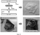
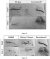
- the inventors demonstrate using Stöber silica or iron oxide nanoparticles that nanobridging, adhesion by aqueous nanoparticle solutions, can be used in vivo in rats to achieve rapid and strong closure and healing of deep wounds in tissues as different as skin and liver. Nanoparticles were also used to fix polymer membranes to tissues even in the presence of blood flow such as occurring after liver resection, yielding permanent hemostasis within a minute.
- medical devices and tissue engineering constructs were secured to organs such as a beating heart. The simplicity, rapidity, and robustness of nanobridging bode well for translation to clinical applications, surgery and regenerative medicine.
- the present invention relates to a composition of nanoparticles for use in a surgical method for adhering a first tissue surface to a second tissue surface wherein said method comprises the steps of adsorbing a layer of nanoparticles on at least one of the tissue surfaces, and approximating the surfaces for a time sufficient for allowing the surfaces to adhere to each other.
- the method herein disclosed may be carried out with any subject.
- the subject is preferably a mammal, more preferably a primate and more preferably still, a human.
- Subjects may be male or female and may be of any age, including prenatal (i.e., in utero), neonatal, infant, juvenile, adolescent, adult, and geriatric subjects.
- the method herein disclosed is applied to at least one tissue surface selected from the group consisting of skin tissue, hair tissue, nail tissue, corneal tissue, tongue tissue, oral cavity tissue, esophageal tissue, anal tissue, urethral tissue, vaginal tissue, urinary epithelial tissue, salivary gland tissue, mammary gland tissue, lacrimal gland tissue, sweat gland tissue, prostate gland tissue, bulbourethral gland tissue, Bartholin's gland tissue, uterine tissue, respiratory and gastrointestinal tract goblet cell tissue, gastric mucosal tissue, gastric gland tissue, pancreatic tissue, spleen tissue, pulmonary tissue, pituitary gland tissue, thyroid gland tissue, parathyroid gland tissue, testicular tissue, ovarian tissue, respiratory gland tissue, gastrointestinal gland tissue, adrenal gland tissue, renal tissue, liver tissue, adipose tissue, duct cell tissue, gall bladder tissue, epidydimal tissue, vas deferens tissue, blood vessel tissue, lymph gland tissue, lymphatic duct tissue, syn
- the present invention also relates to a composition of nanoparticles for use in a surgical -method for adhering a material to a biological tissue in a subject in need thereof, comprising the steps of adsorbing a layer of nanoparticles on the surface of the material and/or the biological tissue and approximating the material and the biological tissue for a time sufficient for allowing the material and the biological tissue to adhere to each other.
- the term "material” denotes any material that can be used to adhere to a tissue, for any purposes, including but not limiting to, research purposes, diagnostic purposes, and therapeutic purposes.
- the material is a natural material or is an artificial material (i.e. a man-made material).
- the material can be less or more solid, less or more flexible, can have more or less ability to swell.
- the material is an artificial material.
- the material is selected form the group consisting of membranes, scaffold materials, films, sheets, tapes, patches, meshes or medical devices.
- the material is biocompatible material.
- biocompatible generally refers having the property or characteristic of not generating injury, toxicity or immunological reaction to living tissues. Accordingly, the material does not substantively provoke injury, toxicity or an immunological reaction, such as a foreign body reaction or inflammatory response (in particular excessive inflammatory response), upon implantation of the material in a subject.
- the material is biodegradable.
- biodegradable as used herein is defined to include both bioabsorbable and bioresorbable materials
- biodegradable it is meant that the materials decompose, or lose structural integrity under body conditions (e.g., enzymatic degradation or hydrolysis) or are broken down (physically or chemically) under physiologic conditions in the body such that the degradation products are excretable or absorbable by the body.
- the outer surface of the material shall have the capability of adsorbing the nanoparticles of the invention.
- the at least one surface of the material, or a part thereof is polar.
- the material is a hydrogel scaffold.
- Tissue engineering is generally defined as the creation of tissue or organ equivalents by seeding of cells onto or into a scaffold suitable for implantation.
- the scaffolds must be biocompatible and cells must be able to attach and proliferate on the scaffolds in order for them to form tissue or organ equivalents. These scaffolds are therefore considered as substrates for cell growth either in vitro or in vivo.
- the attributes of an ideal biocompatible scaffold includes the ability to support cell growth either in vitro or in vivo, the ability to support the growth of a wide variety of cell types or lineages, the ability to be endowed with varying degrees of flexibility or rigidity required, the ability to have varying degrees of biodegradability, the ability to be introduced into the intended site in vivo without provoking secondary damage, and the ability to serve as a vehicle or reservoir for delivery of drugs or bioactive substances to the desired site of action.
- Hydrogels represent an appealing scaffold material because they are structurally similar to the extracellular matrix of many tissues, can often be processed under relatively mild conditions, and may be delivered in a minimally invasive manner. Consequently, hydrogels, a class of highly hydrated polymer materials (water content higher than 30% by weight), are utilized as scaffold materials for drug and growth factor delivery, engineering tissue replacements, and a variety of other applications.
- Synthetic materials include poly(ethylene oxide) (PEO), poly(vinyl alcohol) (PVA), poly(acrylic acid) (PAA), polypropylene furmarate-co-ethylene glycol) (P(PF-co-EG)), polypeptides, poly(hydroxyethyl methacrylate) (PHEMA), poly(2-acrylamido-2-methylpropanesulfonic acid) (PAMPS), poly(acrylamide) (PAAm), poly(ethyleneoxide) (PEG), poly(N-isopropylacrylamide) (PNIPAM), cyclodextrin-based polyrotaxanes gels (CD-PRs), protein-grafted hydrogels, peptide-grafted hydrogels, DNA-grafted polyacrylamide , Terra-amine-terminated PEG (TAPEG), Tetra-NHS-glutarate-terminated PEG (TNPEG), Tetrahydroxyl-terminated PEG (TH)
- Representative naturally derived polymers include agarose, alginate, chitosan, collagen, fibrin, gelatins, and hyaluronic acid (HA), dextran, chitosan, carrageenans.
- HA hyaluronic acid
- suitable polysaccharides include, but are not limited to, dextran, agar, alginic acid, hyaluronic acid, inulin, pullulan, heparin, fucoidan, scleroglucan, curdlan, starch, cellulose and mixtures thereof.
- Monosaccharides that may be used to produce the desired polysaccharide include but are not limited to ribose, glucose, mannose, galactose, fructose, sorbose, sorbitol, mannitol, iditol, dulcitol and mixtures thereof. Many of these compounds are available commercially from companies such as Sigma-Aldrich (St. Louis, Michigan, US).
- the preferred weight-average molecular weight for the polysaccharide is from about 10,000 Daltons to about 2,000,000 Daltons, more preferably from about 10,000 Daltons to about 500,000 Daltons, most preferably from about 10,000 Daltons to about 200,000 Daltons.
- the porous material e.g. hydrogel
- the porous material is prepared according to the method described in WO2009047346A1 or according to the method described in WO2009047347A1 .
- the material e.g. hydrogel
- the material is porous.
- the average pore size of the material is from about 100 nm to about 500 ⁇ m.
- the density of the pores is from about 4% to about 25%.
- biologically active agents may be incorporated in the material (e.g. hydrogel).
- Active agents amenable include growth factors, such as transforming growth factors (TGFs), fibroblast growth factors (FGFs), platelet derived growth factors (PDGFs), epidermal growth factors (EGFs), connective tissue activated peptides (CTAPs), osteogenic factors, and biologically active analogs, fragments, and derivatives of such growth factors.
- TGFs transforming growth factors
- FGFs fibroblast growth factors
- PDGFs platelet derived growth factors
- EGFs epidermal growth factors
- CTAPs connective tissue activated peptides
- osteogenic factors and biologically active analogs, fragments, and derivatives of such growth factors.
- TGF transforming growth factor
- TGF transforming growth factor
- FGFs fibroblast growth factors
- PDGFs platelet derived growth factors
- EGFs epidermal growth factors
- CAPs connective tissue activated peptides
- osteogenic factors
- TGF supergene family include the beta transforming growth factors (for example, TGF- ⁇ 1, TGF- ⁇ 2, TGF- ⁇ 3); bone morphogenetic proteins (for example, BMP-1, BMP-2, BMP-3, BMP-4, BMP-5, BMP-6, BMP-7, BMP-8, BMP-9); heparin-binding growth factors (for example, fibroblast growth factor (FGF), epidermal growth factor (EGF), platelet-derived growth factor (PDGF), insulin-like growth factor (IGF)); Inhibins (for example, Inhibin A, Inhibin B); growth differentiating factors (for example, GDF-1); and Activins (for example, Activin A, Activin B, Activin AB).
- FGF fibroblast growth factor
- EGF epidermal growth factor
- PDGF platelet-derived growth factor
- IGF insulin-like growth factor
- Inhibins for example, Inhibin A, Inhibin B
- growth differentiating factors for example, GDF
- pharmaceutical agent includes without limitation, medicaments; vitamins; mineral supplements; substances used for the treatment, prevention, diagnosis, cure or mitigation of disease or illness; or substances which affect the structure or function of the body; or pro-drugs, which become biologically active or more active after they have been placed in a predetermined physiological environment.
- Non-limiting examples of broad categories of useful pharmaceutical agents include the following therapeutic categories: anabolic agents, antacids, anti-asthmatic agents, anti-cholesterolemic and anti-lipid agents, anti-coagulants, anti-convulsants, anti-diarrheals, antiemetics, anti-infective agents, anti-inflammatory agents, anti-manic agents, anti-nauseants, anti-neoplastic agents, anti-obesity agents, anti-pyretic and analgesic agents, anti-spasmodic agents, anti-thrombotic agents, anti-uricemic agents, anti-anginal agents, antihistamines, anti-tussives, appetite suppressants, biologicals, cerebral dilators, coronary dilators, decongestants, diuretics, diagnostic agents, erythropoietic agents, expectorants, gastrointestinal sedatives, hyperglycemic agents, hypnotics, hypoglycemic agents, ion exchange resins, laxatives, mineral
- non-limiting examples of useful pharmaceutical agents include the following therapeutic categories: analgesics, such as nonsteroidal anti-inflammatory drugs, opiate agonists and salicylates; antihistamines, such as H1-blockers and H2-blockers; anti-infective agents, such as anthelmintics, antianaerobics, antibiotics, aminoglycoside antibiotics, antifungal antibiotics, cephalosporin antibiotics, macrolide antibiotics, miscellaneous beta-lactam antibiotics, penicillin antibiotics, quinolone antibiotics, sulfonamide antibiotics, tetracycline antibiotics, antimycobacterials, antituberculosis antimycobacterials, antiprotozoals, antimalarial antiprotozoals, antiviral agents, anti-retroviral agents, scabicides, and urinary anti-infectives; antineoplastic agents, such as alkylating agents, nitrogen mustard alkylating agents, nitrosourea alkylating
- useful pharmaceutical agents from the above categories include: (a) anti-neoplastics such as androgen inhibitors, antimetabolites, cytotoxic agents, and immunomodulators; (b) anti-tussives such as dextromethorphan, dextromethorphan hydrobromide, noscapine, carbetapentane citrate, and chlorphedianol hydrochloride; (c) antihistamines such as chlorpheniramine maleate, phenindamine tartrate, pyrilamine maleate, doxylamine succinate, and phenyltoloxamine citrate; (d) decongestants such as phenylephrine hydrochloride, phenylpropanolamine hydrochloride, pseudoephedrine hydrochloride, and ephedrine; (e) various alkaloids such as codeine phosphate, codeine sulfate and morphine; (f) mineral supplements such as potassium chloride, zinc chloride, calcium carbonates, magnesium
- TGF-beta fibroblast growth factor
- FGF tumor necrosis factor-alpha & beta
- NGF-alpha & beta nerve growth factor
- GRF growth hormone releasing factor
- EGF epidermal growth factor
- FGFHF fibroblast growth factor homologous factor
- HGF hepatocyte growth factor
- IGF insulin growth factor
- IIF-2 invasion inhibiting factor-2
- BMP 1--7 bone morphogenetic proteins 1-7
- SOD superoxide dismutase
- complement factors hGH, tPA, calcitonin, ANF, EPO and insulin
- anti-infective agents such as antifungals, anti-virals, antiseptics and antibiotics.
- the biologically active substances are used in amounts that are therapeutically effective. While the effective amount of a biologically active substance will depend on the particular material being used, amounts of the biologically active substance from about 1% to about 65% may be desirable. Lesser amounts may be used to achieve efficacious levels of treatment for certain biologically active substances.
- the material e.g. hydrogel
- the material comprises an amount of a radiosensitizer.
- Radiosensitizers are drugs that make cancer cells more sensitive to the effects of radiation therapy.
- Non- limiting examples of metal radiosensitizers that could be used in accordance with the present invention include metals, preferably inert metals such as platinum, gold, iridium, osmium, palladium, radium, zinc, chromium, copper, silver, cobalt, nickel and ruthenium. The greater the atomic number, the better is the interaction with radiation.
- Other useful metals although less preferred because of their small atomic number, include iron.
- radio sensitizers include but are not limited to metoclopramide, sensamide or neusensamide (manufactured by Oxigene); profiromycin (made by Vion); RSR13 (made by Allos); Thymitaq (made by Agouron), etanidazole or lobenguane (manufactured by Nycomed); gadolinium texaphrin (made by Pharmacyclics); BuDR/Broxine (made by NeoPharm); IPdR (made by Sparta); CR2412 (made by Cell Therapeutic); L1X (made by Terrapin); or the like.
- materials comprising amount of radio sensitizers are particularly suitable for the treatment of cancer by radiotherapy.
- radiotherapy can be applied.
- the cancer is selected from the group consisting of breast cancer, prostate cancer, lymphoma, skin cancer, pancreatic cancer, colon cancer, melanoma, malignant melanoma, ovarian cancer, brain cancer, primary brain carcinoma, head-neck cancer, glioma, glioblastoma, liver cancer, bladder cancer, non-small cell lung cancer, head or neck carcinoma, breast carcinoma, ovarian carcinoma, lung carcinoma, small-cell lung carcinoma, Wilms' tumor, cervical carcinoma, testicular carcinoma, bladder carcinoma, pancreatic carcinoma, stomach carcinoma, colon carcinoma, prostatic carcinoma, genitourinary carcinoma, thyroid carcinoma, esophageal carcinoma, myeloma, multiple myeloma, adrenal carcinoma, renal cell carcinoma, endometrial carcinoma, adrenal cortex carcinoma
- the porous materials are loaded with a plurality of cells.
- a difference in porosity may indeed facilitate migration of different cell types to the appropriate regions of the material (e.g. hydrogel).
- a difference in porosity may facilitate development of appropriate cell-to-cell connections among the cell types comprising the material (e.g. hydrogel), required for appropriate structuring of the developing/repairing/regenerating tissue.
- the material e.g. hydrogel
- the material may comprise cells of any tissue.
- the cells are seeded on the material (e.g. hydrogel).
- the materials e.g. hydrogels
- the materials are submerged in a culture solution comprising the desired cells for an amount of time sufficient to enable penetration of the cells throughout the material (e.g. hydrogel).
- the material e.g. hydrogel
- the material (e.g. hydrogel) is capable of supporting the viability and the growth of the seeded cells in culture over long periods of time without inducing differentiation.
- the material (e.g. hydrogel) of the invention can provide an environment for unstimulated cell growth (without activation by growth stimulants)
- the cells are pluripotent stem cells or progenitor cells.
- Pluripotent stem cells have the potential to differentiate into endoderm, mesoderm, and ectoderm.
- pluripotent includes pluripotent stem cells from all sources, including modified adult stem or somatic cells (ASCs), that is, induced pluripotent stem cells (iPSC), and very small embryonic- like stem cells (VSELs).
- Pluripotent stem cells have also been artificially generated (i.e., induced pluripotent stem cells (iPSC)) from other sources, such as placenta or from genetic manipulation of adult stem cells (ASC) or even adult somatic cells.
- ASC are located in tissues throughout the body and function as a reservoir to replace damaged or aging cells. ASC are generally restricted in their differentiation to cell lineages of the organ system from which they originate (i.e., "multipotent” stem cells), although recent research suggests that adult tissues, such as bone marrow, may harbor dormant pluripotent stem cells referred to as “very small embryonic-like stem cells” or "VSELs.”
- adult stem cells can be isolated from mammalian tissue, including from any adult organ, umbilical cord blood, or placenta.
- the adult stem cells are multipotent, but they may be manipulated to provide pluripotent stem cells (iPSC.) using conventional techniques.
- the stem cells can be derived from mammals, such as but not limited to rodents, pigs, cats, dogs, and primates, including humans.
- the pluripotent stem cells useful herein are nonviable.
- nonviable stem cells do not form teratomas.
- the stem cells may be made nonviable with irradiation, phototherapy, chemical treatment, and/ or lyophilization.
- the selection of the method of making pluripotent stem cells nonviable is not particularly limited, but it is preferred that the method used is effective to retain the intracellular contents of the stem cells.
- the material e.g. hydrogel
- the material is seeded with cells selected from the group consisting of chondrocytes; fibrochondrocytes; osteocytes; osteoblasts; osteoclasts; synoviocytes; bone marrow cells; mesenchymal cells; muscle cells; stromal cells; stem cells; embryonic stem cells; precursor cells derived from adipose tissue; peripheral blood progenitor cells; stem cells isolated from adult tissue; genetically transformed cells; a combination of chondrocytes and other cells; a combination of osteocytes and other cells; a combination of synoviocytes and other cells; a combination of bone marrow cells and other cells; a combination of mesenchymal cells and other cells; a combination of stromal cells and other cells; a combination of stem cells and other cells; a combination of embryonic stem cells and other cells; a combination of progenitor cells isolated from adult tissue and other cells; a combination of peripheral blood progenitor cells and other cells
- the cells may be genetically engineered to express a desired molecule, such as for example heparin binding growth factor (HBGF), transforming growth factor alpha or beta (TGF beta.), alpha fibroblastic growth factor (FGF), epidermal growth factor (TGF), vascular endothelium growth factor (VEGF) and SDF-1, some of which are also angiogenic factors.
- HBGF heparin binding growth factor
- TGF beta. transforming growth factor alpha or beta
- FGF alpha fibroblastic growth factor
- TGF epidermal growth factor
- VEGF vascular endothelium growth factor
- SDF-1 vascular endothelium growth factor
- expressed factors include hormones such as insulin, glucagon, and estrogen.
- factors such as nerve growth factor (NGF) or muscle morphogenic factor (MMF), or in another embodiment, TNF alpha/beta are expressed.
- the hydrogels according to the invention are suitable to prepare vascular substitutes to replace compromised arteries as described for example, in Chaouat et al. ( Chaouat M, Le Visage C, Autissier A, Chaubet F, Letourneur D. The evaluation of a small-diameter polysaccharide-based arterial graft in rats. Biomaterials. 2006 Nov;27(32):5546-53 ).
- Such substitutes may be prepared according to the methods of the invention by using a mould.
- Such substitutes may then comprise a population of cells to reconstruct in vitro or in vivo a vessel.
- the cells may include but are not limited to Mesenchymal Stem Cells (MSC), Endothelial Progenitor cells (EPCs), endothelial cells, fibroblastic cells and smooth muscle cells.
- the materials (e.g. hydrogel) of the invention are suitable to prepare cartilage implants.
- the scaffolds of the invention may be loaded with chondrocytes, osteocytes; osteoblasts: osteoclasts: vascular cells or mixtures thereof, and then be fixing to the cartilage to be repaired by the nanoparticles of the present invention.
- the site of implantation of the material is dependent on the diseased/injured tissue that requires treatment.
- the cell-seeded composite material e.g. hydrogel
- the defect site to promote repair of the damaged tissue.
- the material e.g. hydrogel
- the material can be seeded with a combination of adult neuronal stem cells, embryonic stem cells, glial cells and Sertoli cells.
- the composite scaffold can be seeded with Sertoli cells derived from transformed cell lines, xenogeneic or allogeneic sources in combination with neuronal stem cells.
- the Sertoli cells can be cultured with the composite scaffold for a period before addition of stem cells and subsequent implantation at the site of injury. This approach can circumvent one of the major hurdles of cell therapy for CNS applications, namely the survival of the stem cells following transplantation.
- a composite scaffold that entraps a large number of Sertoli cells can provide an environment that is more amenable for the survival of stem cells
- the material e.g. hydrogel
- the material can be effectively used as a raw material for fabricating artificial tissues or organs such as artificial blood vessels, artificial esophagus, artificial nerves, artificial hearts, prostatic heart valves, artificial skins, orthopedic implants, artificial muscles, artificial ligaments, artificial respiratory organs, etc.
- the material e.g. hydrogel
- the material can be prepared in the form of a hybrid tissue by blending or incorporating on or into other types of biomaterials and with functional cells derived from tissues or organs. It may have various biomedical applications, for example, to maintain cell functions, tissue regeneration, etc.
- the material e.g. hydrogel
- the wound healing material e.g. hydrogel
- the wound to be healed may be a result of a variety of acute or chronic internal or external injuries, diseases, or ailments, including, for example, abrasions, cuts, punctures, incisions, lacerations, ulcers, burns, surgical, bullet, bites, knife, or improvised explosive device induced wounds, and the like.
- the wound healing material e.g. hydrogel
- the wound healing material e.g. hydrogel
- the material (e.g. hydrogel) for the treatment of wounds may further comprise one or more additional components or agents, such as antibiotics or other antimicrobial compounds or agents and other agents known to improve wound healing.
- the material e.g. hydrogel
- the affected area of the heart may include, for example, an area of the heart impacting cardiac function. Short and or long term ischemia can result in myocyte death, tissue infarction, and loss of contractile function.
- the area to be treated may include ischemic penumbra or area best characterized as hibernating myocardium. Hibernating myocardium is a condition due to acute or chronic ischemia where certain portions of the myocardium exhibit abnormal or no contractile function but the ceils remain viable. Accordingly the material (e.g.
- hydrogel can be used, for example, in cardiac muscle regeneration for a number of principal indications: (i) acute heart attacks; (ii) therapy for congestive heart failure patients; (iii) prevention of further disease for patients undergoing coronary artery bypass graft; (iv) conductive tissue regeneration; (v) vessel smooth muscle regeneration; (vi) valve regeneration; and (vii) to wean patients from left ventricular assist devices implanted as a bridge to transplant and or destination therapy.
- Cardiac muscle normally does not have or has only limited reparative potential.
- the material e.g. hydrogel
- the material as above described is sealed to the injured cardiac tissue the regenerate cardiac muscle in the subject.
- a method is provided for regenerating or repairing cardiac muscle comprising adhering a material (e.g. hydrogel) as above described to a damaged or aged area of the heart with the nanoparticles of the present invention.
- the method is thus particularly suitable for improving ejection fraction (EF) and/or for decreasing the infarct size.
- EF ejection fraction
- treatment of the heart as described herein may provide significant improvement in cardiac function such that no further treatment is necessary.
- treatment of the heart may prolong survival of the subject prior to more radical therapy, including heart transplant.
- nanoparticles means particles from 2 to 500 nm and even more preferably from 5 to 300 nm in size.
- the size of the nanoparticles is the distance between the two most distant points in the nanoparticle.
- the size of the diameter is the diameter of the smallest cylinder in which the nanoparticle is inscribed.
- Nanoparticle size can be determined by different methods such as Dynamic Light Scattering (DLS), Small Angle X-ray Scattering (SAXS), Scanning Mobility Particle Sizer (SMPS), Scanning Electron Microscopy (SEM), Transmission Electron Microscopy (TEM) ( Orts-Gil, G., K.
- DLS Dynamic Light Scattering
- SAXS Small Angle X-ray Scattering
- SMPS Scanning Mobility Particle Sizer
- SEM Scanning Electron Microscopy
- TEM Transmission Electron Microscopy
- the nanoparticles are selected among solid mineral nanoparticles selected from the group consisting of metal oxides, alumina, silica, kaolin, hydroxyapatite, calcium carbonate, silicates such as micas quartz, zeolites and clays such as hectorite, laponite, montmorillonite, bentonite, smectite.
- the nanoparticles that are adsorbed on the surface may be made of different chemical nature, of different sizes, and/or of different shapes.
- the nanoparticles can be in the form of a sphere, needle, flake, platelet, tube, fiber, cube, prism, whiskers or have an irregular shape.
- Nanoparticles include without limitation the nanofibrils, nanochips, nanolatexes, nanotubes, expandable nanoparticles.
- particles may be particles of metal oxides, such as iron oxides (FeO, Fe 2 O 3 , Fe 3 O 4 ), cerium oxide (CeO), alumina (Al 2 O 3 ), zirconium oxide (ZrO 2 ), titanium oxide (TiO 2 ), titanates (BaTiOs, Ba0.5Sr0.5TiO 3 , SrTiOs), indium oxide (In 2 O 3 ), tin oxides, such as iron oxides (FeO, Fe 2 O 3 , Fe 3 O 4 ), cerium oxide (CeO), alumina (Al 2 O 3 ), zirconium oxide (ZrO 2 ), titanium oxide (TiO 2 ), titanates (BaTiOs, Ba0.5Sr0.5TiO 3 , SrTiOs), indium oxide (In 2 O 3 ), tin oxide
- metal oxides such as iron oxides (FeO, Fe 2 O 3 , Fe 3 O 4 ), cerium oxide (CeO),
- the nanoparticles are selected from: clays, silicates, alumina, silica, kaolin, hydroxyapatite, magnetic nanoparticles like iron oxides, calcium carbonates. Small molecules or polymer chains can be grafted to stabilize nanoparticles in suspensions when necessary.
- At least one part of the nanoparticles are silica nanoparticles.
- the nanoparticles will act as contrast agents that can be directly imaged.
- the embodiment may find various applications.
- the contrast nanoparticles may tag the material and once implanted in the subject it will be possible to image it in vivo.
- the embodiment offers the possibility for a structural or functional imaging procedure, e.g. for implanting the material in the subject with image guidance, for following the implantation of the material so as to verify that the maintenance of the material in the implantation site or to verify that the biodegradability of the material occurs.
- the physician will have the opportunity to image it for analysing the implantation and then choose the best surgical procedure.
- the nanoparticles can be detectable by imaging techniques such as ultrasonography, elastography, Supersonic Shear Wave Imaging, Magnetic Resonance Imaging (MRI), Positron Emission Tomography (PET), Single Photon Emission Computed Tomography (SPECT), fluorescence spectroscopy, Computed Tomography, X-ray radiography, or any combination of these techniques.
- imaging techniques such as ultrasonography, elastography, Supersonic Shear Wave Imaging, Magnetic Resonance Imaging (MRI), Positron Emission Tomography (PET), Single Photon Emission Computed Tomography (SPECT), fluorescence spectroscopy, Computed Tomography, X-ray radiography, or any combination of these techniques.
- the nanoparticles are designed to be detectable by x-ray imaging.
- the nanoparticles can be core and core-shell nanoparticles containing iodine for x-ray imaging as described in WO 2006106513 .
- the nanoparticles are designed to be detectable by Magnetic Resonance Imaging (MRI).
- MRI Magnetic Resonance Imaging
- NMR Nuclear Magnetic Resonance
- nanoparticle comprises at least one paramagnetic metal ion.
- paramagnetic metal ions detectable by MRI are gadolinium III (Gd3+), chromium III (Cr3+), dysprosium III (Dy3+), iron III (Fe3+), manganese II (Mn2+), and ytterbium III (Yb3+).
- the paramagnetic metal ion is gadolinium III (Gd3+).
- Gadolinium is an FDA-approved contrast agent for MRI.
- the nanoparticles consist of ultrasmall superparamagnetic iron oxide (USPIO) particles.
- USPIO particles are currently under investigation as contrast agents for imaging human pathologies ( C. Corot et al., Adv. Drug Deliv. Rev., 2006, 56: 1472-1504 ). They are composed of a crystalline iron oxide core containing thousands of iron atoms which provide a large disturbance of the Magnetic Resonance signal of surrounding water.
- quantum dots currently under investigation as extremely sensitive fluorescent probes
- USPIO particles exhibit a very good biocompatibility. Chemical coating of USPIO particles is required to ensure their dispersion in biological media.
- USPIO particles are known in the art and have been described (see, for example, J. Petersein et al., Magn. Reson. Imaging Clin. Am., 1996, 4: 53-60 ; B. Bonnemain, J. Drug Target, 1998, 6: 167-174 ; E.X. Wu et al., NMR Biomed., 2004, 17: 478-483 ; C. Corot et al., Adv. Drug Deliv. Rev., 2006, 58: 1471-1504 ; M. Di Marco et al., Int. J. Nanomedicine, 2007, 2: 609-622 ). USPIO particles are commercially available, for example, from AMAG Pharmaceuticals, Inc. under the tradenames Sinerem ® and Combidex ® .
- the nanoparticles are designed to be detectable by fluorescence spectroscopy.
- fluorescent moieties to be used in the practice herein disclosed include high molecular absorption coefficient, high fluorescence quantum yield, and photostability.
- Preferred fluorescent moieties exhibit absorption and emission wavelengths in the visible (i.e., between 400 and 700 nm) or the near infra-red (i.e., between 700 and 950 nm). Selection of a particular fluorescent moiety will be governed by the nature and characteristics of the illumination and detection systems used in the diagnostic method.
- In vivo fluorescence imaging uses a sensitive camera to detect fluorescence emission from fluorophores in whole-body living mammals.
- NIR near-infrared
- fluorescent labels include, but are not limited to, quantum dots (i.e., fluorescent inorganic semiconductor nanocrystals) and fluorescent dyes such as Texas red, fluorescein isothiocyanate (FITC), phycoerythrin (PE), rhodamine, fluorescein, carbocyanine, Cy-3TM and Cy-5TM (i.e., 3- and 5-N,N'-diethyltetra-methylindodicarbocyanine, respectively), Cy5.5, Cy7, DY-630, DY-635, DY-680, and Atto 565 dyes, merocyanine, styryl dye, oxonol dye, BODIPY dye (i.e., boron dipyrromethene difluoride fluorophore), and analogues, derivatives or combinations of these molecules.
- quantum dots i.e., fluorescent inorganic semiconductor nanocrystals
- fluorescent dyes such as Texas red, fluorescein isothiocyan
- Nanoparticles which are used in the invention are selected as a function of the tissue nature or material.
- the nanoparticles should be capable of adsorption at the tissue or material surface.
- the selection of the appropriate nanoparticles suspension can be achieved by testing the nanoparticles affinity (adsorption) with the tissue or material. Briefly, a first method rests on Fourier transform infrared spectroscopy coupled with ATR. Attenuated total reflectance (ATR) is a sampling technique used in conjunction with infrared spectroscopy which enables sample surfaces to be examined. The detection and the quantification of adsorbed nanoparticles layer onto the tissue or material surface can be achieved.
- ATR Attenuated total reflectance
- the proposed method consists in immersing the tissue sample or material into the nanoparticle solution or depositing a droplet of nanoparticle solution on the tissue surface, then the tissue sample or material is soaked and washed in a large volume of water during several days. Samples can be dried prior to ATR-FTIR analysis. The presence of an adsorbed nanoparticle layer on the tissue or material surface which persists after soaking enables to select the appropriate nanoparticles to use as adhesive. Conversely, the absence of nanoparticle at the tissue or material surface implies weak adhesive properties of the tested nanoparticles. Alternately, a second method rests on Scanning Electron Microscopy (SEM) in combination with Energy Dispersive X-ray (EDX).
- SEM Scanning Electron Microscopy
- EDX Energy Dispersive X-ray
- tissue sample or material preparation is identical to the one disclosed above for the ATR-FTIR method.
- EDX is an analytical technique used for the elemental analysis or chemical characterization of a sample. The first micrometers of the surface are probed. Finally, with the method of thermal isotherms ( Hourdet, D. and L. Petit (2010). Macromolecular Symposia. C. S. Patrickios. Weinheim, Wiley-V C H Verlag Gmbh.
- the skilled professional can also determine comparatively the nanoparticles best suited for providing adhesion to a tissue surface or can carry out thermal isotherms adjusting pH to optimize adsorption mechanism, for example for anionic polyacrylic acid (PAA) macromolecules on the silica surface ( Wisniewska, M. (2010) Journal of Thermal Analysis and Calorimetry, 101(2), 753-760. doi:10.1007/s10973-010-0888-4 ).
- PAA anionic polyacrylic acid
- the nanoparticles are applied on the surface as an aqueous suspension (or dispersion) of nanoparticles.
- Aqueous suspensions of nanoparticles are commercially available.
- the aqueous suspension of nanoparticles which can be used according to the invention does not contain any other adhesive agent.
- the aqueous suspension of nanoparticles does not contain a compound known as an adhering agent in a concentration that would allow it to play the function of adhesive agent.
- adhesive agents such as monomers, synthetic polymers (other than polymer nanoparticles), notably cyanoacrylates, urethanes, dendrimers; or natural adhesives such as fibrin, collagen, gelatin, and polysaccharides.
- the aqueous suspension of nanoparticles consists essentially of nanoparticles suspended in water. It means that other components can be present in the suspension, but they do not modify the properties of the suspension in a noticeable manner. Especially, other components can be present in the suspension, but they have to be selected so as not to modify the adhesive properties of the suspension (e.g. dispersion property of the nanoparticles).
- the preparation of nanoparticles suitable for dispersing the nanoparticles contains no more than 20%, or better, no more than 10% by weight of other adhering agent as compared to the weight of the dry matter of the composition, respectively aqueous suspension, preferably, less than 5% weight, even more preferably less than 2 % and better less than 1%, even better, less than 0,5%.
- materials distinct from the nanoparticles can be present in the preparation suitable for dispersing the nanoparticles, and notably mineral or organic ions can be present in the preparation,.
- mineral or organic ions such components can be anti infectives, anti bacterians, preservatives, antiobiotics, PEG, polymers of varied nature.
- the preparation of nanoparticles comprises an amount of pharmaceutical agent or a biological active agent as above described for the hydrogel.
- the nanoparticles have the function of adhering agent in the compositions wherein they are present.
- the nanoparticles represent from 10 to 100% by weight of the weight of the dry matter of the preparation.
- the nanoparticles represent from 20 to 100% by weight of the weight of the dry matter of the preparation, (e.g. the aqueous suspension), even more preferably, from 30 to 100%, and advantageously, from 40 to 100%, better from 50 to 100%, even better from 60 to 100%, preferably from 70 to 100%, even better from 80 to 100%, even more preferably from 90 to 100%.
- the nanoparticles represent from 95 to 100% by weight of the weight of the dry matter of the preparation, (e.g. the aqueous suspension), even better from 98 to 100%, and even more preferably from 99 to 100%.
- Concentrations are adjusted to obtain suitable viscosities for application. In general suspensions of viscosity from about 10 Pa.s or less are used, preferably lower viscosities (10 -3 Pa.s). For non-spherical particles, like particles of CNT, or CNC type, the concentration is adjusted so that the viscosity remains fairly low.
- the pH of the suspension of nanoparticles can be of any value from 1 to 14 and is adapted according to the application. pH can be adjusted to optimize adsorption, for example for anionic polyacrylic acid (PAA) macromolecules on the silica surface ( Wisniewska, M. (2010), Journal of Thermal Analysis and Calorimetry, 101(2), 753-760. doi:10.1007/s10973-010-0888-4 ) but also to keep the stability of the nanoparticles composition. For polyelectrolyte or amphoteric gels, the pH of the nanoparticles composition is adjusted to obtain nanoparticles of charges opposed to gel's charges.
- PAA anionic polyacrylic acid
- amphoteric gels the pH of the nanoparticles composition is adjusted to obtain nanoparticles of charges opposed to gel's charges.
- the preparation of nanoparticles of the invention is applied using conventional techniques. Coating, dipping, spraying, spreading and solvent casting are possible approaches. More particularly, said applying is manual applying, applicator applying, instrument applying, manual spray applying, aerosol spray applying, syringe applying, airless tip applying, gas-assist tip applying, percutaneous applying, surface applying, topical applying, internal applying, enteral applying, parenteral applying, protective applying, catheter applying, endoscopic applying, arthroscopic applying, encapsulation scaffold applying, stent applying, wound dressing applying, vascular patch applying, vascular graft applying, image-guided applying, radiologic applying, brush applying, wrap applying, or drip applying.
- the nanoparticles in particular for cutaneous application, can be deposited on the tissue with means typically selected from the group consisting of a patch, a dressing, a elastoplasts or a band-aid having a plurality of capsules (e.g. nanocapsules) having the ability to release the nanoparticles (e.g. in the form of powder or a solution) when they are contacted by the tissue (e.g. because of a variation of temperature, physical pressure, osmotic pressure). Then after a while the means can be pull out, and the material or tissue can be approximated with the tissue where the nanoparticles were adsorbed.
- means typically selected from the group consisting of a patch, a dressing, a elastoplasts or a band-aid having a plurality of capsules (e.g. nanocapsules) having the ability to release the nanoparticles (e.g. in the form of powder or a solution) when they are contacted by the tissue (e.g. because of a
- the quantity of nanoparticles deposited at the surface of the tissue surface (or hydrogel) is from 0.1 mg/m 2 to 10 g/m 2 .
- the coverage of the surface is to be adjusted. These values can be from 1 mg/m 2 , preferably for small particles, and up to 0.2 g/m 2 , preferably for large particles.
- large particles typically of the order of 300 nm
- the coverage is large, of the order of 4 g / m 2 .
- coverage is preferably of the order of 10 mg / m 2 .
- the density of coverage can be evaluated on the assembly by ATR-FTIR or by SEM.
- the volume of nanoparticles that is deposited at the surface ranges from 0.01 to 5 ⁇ l. per mm 2 .
- a volume of 0,1; 0,2; 0,3; 0,4; 0,5; 0,6; 0,7; 0,8; 0,9; 1; 1,1; 1,2; 1,3; 1,4; 1,5; 1,6; 1,7; 1,8; 1,9; 2; 2,1; 2,2; 2,3; 2,4; 2,5; 2,6; 2,7; 2,8; 2,9; 3; 3,1; 3,2; 3,3; 3,4; 3,5; 3,6; 3,7; 3,8; 3,9; 4; 4,1; 4,2; 4,3; 4,4; 4,5; 4,6; 4,7; 4,8; 4,9; or 5 ⁇ l per mm 2 is deposited at the surface.
- the nanoparticles are adsorbed in the two surfaces that shall be adhered (i.e. the two tissue surfaces, the tissue surface and the material surface). However, in a preferred embodiment only one surface is adsorbed with the nanoparticles. For example, when a material shall adhere to a tissue, it is preferable to absorb the nanoparticles on the material surface rather than on the tissue surface. In some embodiments, it may be desirable to get only one layer of nanoparticles.
- the approximating step is a manual approximating, a mechanical approximating, a suture approximating, a staple approximating, a synthetic mesh approximating, a biologic mesh approximating, a transverse approximating, a longitudinal approximating, an end-to-end approximating, or an overlapping approximating.
- the two tissue surfaces are approximated for a time ranging from 5s to 2min, preferably from 10s to 1 min, more preferably from 20s to 50s.
- the 2 tissue surfaces are approximated during 20; 21; 22; 23; 24; 25; 26; 27; 28; 29; 30; 31; 32; 33; 34; 35; 36; 37; 38; 39; 40; 41; 42; 43; 44; 45; 46; 47; 48; 49; 50; 51; 52; 53; 54; 55; 56; 57; 58; 59; 60; 61; 62; 63; 64; 65; 66; 67; 68; 69; 70; 71; 72; 73; 74; 75; 76; 77; 78; 79; 80; 81; 82; 83; 84; 85; 86; 87; 88; 89; 90; 91; 92; 93; 94; 95; 96; 97; 98; 99; 100; 101; 102;
- the nanoparticles are just absorbed on the surface of the material just before being applied to the tissue.
- the physician that would like to adhere a material on a tissue prepares the material as above described by adsorbing the nanoparticles to the surface of the material. Then he approximates the material and the tissue for a time sufficient for allowing the surfaces of the material and the tissue to adhere to each other.
- the nanoparticles are previously adsorbed on the surface of the material. Accordingly, the invention encompasses use of ready-to-use materials that can be prepared in an industrial manner and then be stocked in a proper manner. Once the clinician would like to use the material he has just to release the material and adhere it to the tissue without any preparation. For example, it is not necessary to hydrates the material before applying it to the tissue. The material, such as hydrogel can thus be applied directly to the tissue, and will automatically adhere to the tissue. For example the hydrogel will naturally swell in contact of the biological fluids present in the implantation site (e.g. blood, lymph, exudates).
- the biological fluids present in the implantation site e.g. blood, lymph, exudates.
- a further object of the invention relates to a material as above described wherein an amount of nanoparticles is adsorbed on at least one surface of the material.
- the material is typically prepared as described above.
- the material may be immerged in aqueous suspension of nanoparticles for a sufficient time for allowing the nanoparticles to adsorb to the surface of the material.
- an amount of nanoparticles are deposited on the surface of the material with a brush that was previously dipped in an aqueous suspension of nanoparticles.
- the aqueous suspension of nanoparticles may also be sprayed on the surface of the material.
- the material can be dried, optionally lyophilized (e.g. hydrogel), sterilized, packaged and properly stocked for a subsequent medical use.
- a powder of nanoparticles is dispersed (e.g. by spraying) on the surface of the material and the excess is then washed.
- the material is optionally lyophilized (e.g. hydrogel), sterilized, packaged and properly stocked for a subsequently medical use.
- a variant of the method herein disclosed for adhering a material to a biological tissue in a subject in need thereof comprises the steps of providing the material wherein the nanoparticles were previously adsorbed in at least one surface and approximating the material and the biological tissue for a time sufficient for allowing the material and the biological tissue to adhere to each other.
- the methods herein disclosed may find very various medical applications.
- the methods herein disclosed provide the following advantages.
- the methods herein disclosed may be used directly in vivo even in presence of body fluids such as blood, lymph, exudates, urine, bile, intestine contents...
- the methods herein disclosed can be applied in tissue that are normally perfused or can be applied to tissues that are leaking (e.g. blood).
- the inventors surprisingly demonstrate that nanoparticles can be adsorbed on the tissue surfaces in a sufficient manner for adhering even if a part of them is flowed by the presence of the body fluid, in particular blood.
- the methods herein disclosed offer the advantage to maintain the physical, chemical and biological integrities of the tissue where the nanoparticles are adsorbed.
- no physical barrier is created (as generally observed with glues made of cyanoacrylate) that will prevent the tissue diffusion, e.g. the circulation of the biological molecules, cells (e.g. immune cells) or fluids between the adhering tissues or between the material (e.g. hydrogel) and the tissue.
- the physical properties of the tissue are maintained in particular the elasticity of the tissue.
- the methods herein disclosed are very easy to settle and may be performed very quickly in very different conditions (temperature, presence of body fluids, organ or tissues in motion (e.g. a beating heart)).
- the adhesion offer by the method of the herein disclosed may be a permanent adhesion or a temporary adhesion.
- the methods herein disclosed may be performed during a surgery procedure so as to prevent in urgent manner a leaking of blood vessels till the surgeon stabilizes the haemostatic assembly with sutures, meshes or staples.
- the methods herein disclosed are particularly suitable for sealing a defect between a first and second tissue in the subject.
- the methods herein disclosed are particularly suitable for providing a barrier to, for example, microbial contamination, infection, chemical or drug exposure, inflammation, or metastasis.
- the methods herein disclosed are particularly suitable for stabilizing the physical orientation of a first tissue surface with respect to a second tissue surface.
- the methods herein disclosed are particularly suitable for reinforcing the integrity of a first and second tissue surface achieved by, for example, sutures, staples, mechanical fixators, or mesh.
- the method herein disclosed is particularly suitable providing control of bleeding.
- the methods herein disclosed are particularly suitable for delivery of drugs including, for example, drugs to control bleeding, treat infection or malignancy, or promote tissue regeneration.
- the methods herein disclosed are particularly suitable in bariatric surgery, cardiac surgery, thoracic surgery, colon and rectal surgery, dermatologic surgery, general surgery, gynecologic surgery, maxillofacial surgery, neurosurgery, obstetric surgery, oncologic surgery, ophthalmologic surgery, oral surgery, orthopedic surgery, otolaryngologic surgery, pediatric surgery, plastic surgery, cosmetic and reconstructive surgery, podiatric surgery, spine surgery, transplant surgery, trauma surgery, vascular surgery, urologic surgery, dental surgery, veterinary surgery, endoscopic surgery, anesthesiology, an interventional radiologic procedure, an emergency medicine procedure, a battlefield procedure, a deep or superficial laceration repair, a cardiologic procedure, an internal medicine procedure, an intensive care procedure, an endocrinologic procedure, a gastroenterologic procedure, a hematologic procedure, a hepatologic procedure, a diagnostic radiologic procedure, an infectious disease procedure, a nephrologic procedure, an oncologic procedure, a proc
- the methods herein disclosed are particularly suitable in a dural repair, a nerve anastomosis, an endoscopic procedure, a skull base repair, a discectomy procedure, a fibrosis prevention after lumbar discectomy procedure, a scar formation prevention procedure, a posterior fossa procedure, an aneurysm repair, an arteriovenous malformation repair, a cerebrospinal fluid rhinorrhea prevention or repair procedure, a fusion procedure, a procedure to prevent fracture of weakened vertebral bodies, a procedure to repair disc herniation or to prevent the progression of disc herniation, a procedure to provide growth factors in spine surgery, a procedure to prevent or to manage dead space or seroma in spine surgery, an endoscopic neurosurgery or spine surgery procedure, or a procedure to repair an entrance portal in nucleoplasty.
- the methods herein disclosed are particularly suitable for hernia repair.
- a hernia occurs when an organ, intestine or fatty tissue squeezes through a hole or a weak spot in the surrounding muscle or connective tissue. Hernias often occur at the abdominal wall. Sometimes a hernia can be visible as an external bulge particularly when straining or bearing down.
- hernias are inguinal hernia which occurs in the inner groin, femoral hernia which occurs in the upper thigh/outer groin, incisional hernia which occurs through an incision or scar in the abdomen, ventral hernia which occurs in the general abdominal/ventral wall, umbilical hernia which occurs at the belly button and hiatal hernia which occurs inside the abdomen, along the upper stomach/diaphragm. Most hernias are caused by a combination of pressure and an opening or weakness of muscle or connective tissue. The pressure pushes an organ or tissue through the opening or weak spot. Sometimes the muscle weakness is present at birth but more often it occurs later in life.
- the methods herein disclosed are particularly suitable in a laparoscopic procedure, a hematoma, a subcutaneous flap, a mastectomy, an abdominopasty, a bowel resection, a bowel anastomosis, a thyroidectomy, an anastomotic leak after a gastric bypass procedure, a peritoneal adhesion prevention procedure, a burn injury, a fistula in ano, a pancreatic leak, a seroma after axial dissection, an intralesional support for tumor removal procedure, a spleen injury, an appendectomy, a cholecstectomy, a peptic or gastric ulcer repair procedure, closure of dead space to prevent a seroma in a general surgical procedure, fixation and sealing of the insertion site of a transcutaneous device, or a colostomy or other stoma procedure.
- the methods herein disclosed are particularly suitable in a neck dissection, a tonsillectomy, an adenoidectomy, a tumor removal procedure, a frontal sinus repair, an endoscopic otolaryngologic procedure, or nasal septal surgery.
- the methods herein disclosed are particularly suitable in a vascular graft procedure, an anastomotic bleeding repair procedure, a primary anastomosis, a percutaneous endovascular procedure, a prosthetic vascular graft procedure, a femoral artery repair, a carotid artery repair, attachment of endothelial cells to prosthetic grafts to create new endothelial lining, an endoscopic vascular surgery procedure, or an aortic reconstruction.
- the methods herein disclosed are particularly suitable in amniocentesis, premature rupture of amniotic membranes, an endoscopic obstetric procedure, or a cervical occlusion procedure.
- the methods herein disclosed are particularly suitable in a Fallopian tube occlusion, a contraceptive procedure, a urinary incontinence procedure, a cystocoele repair, a rectocoele repair, a pelvic floor repair, a vulvo-vaginal reconstruction procedure, an amniotic membrane graft procedure, an endoscopic gynecologic procedure, or fixation of embryo transfer with in vitro fertilization.
- the methods herein disclosed are particularly suitable in a pancreatic islet cell implantation, liver transplantation, kidney transplantation, pancreas transplantation, an endoscopic transplant procedure, or a combination thereof.
- the methods herein disclosed are particularly suitable in balloon tracheal occlusion, closure of amniotic membranes, or a fetoscopic procedure.
- the methods herein disclosed are particularly suitable in a pulmonary lobectomy, bi-lobectomy, sleeve lobectomy, bullectomy, segmentectomy, pulmonary wedge resection, an air leak, a tracheoesophageal fistula repair, a neotracheal reconstruction, a pleural leak, a thoracoscopic or bonchoscopic procedure, an endoscopic thoracic surgery procedure, closure of a tracheal or bronchial defect, or repair of a bronchopleural fistula.
- Types of lung surgery include lobectomy, lung biopsy, lung-tissue removal, and pneumonectomy.
- Risks associated with lung surgery include wound infection; post-surgical internal bleeding; air leaks; pain or numbness at the incision site; and infection of the lungs (pneumonia). Further, air leakage is frequently observed after thoracic procedures, such as pulmonary resection and decortication. It is important to create an airtight seal so as to prevent or reduce severe complications, such as bronchopleural fistulas and infection resulting from extended chest tube drainage, extended recovery time, and postoperative morbidity related to pulmonary surgery. The method herein disclosed is thus particularly useful for preventing some of the problematic aspects of lung surgery, such as treatment of pneumothorax and pulmonary leaks.
- the methods herein disclosed are particularly suitable in an ocular procedure, a retinal procedure, a retinal detachment procedure, a corneal repair, a glaucoma procedure, a glaucoma drainage device procedure, a laser procedure, a tissue flap procedure after laser surgery, a conjunctival repair, a pterygium repair, cataract surgery, repair of wet or dry macular degeneration, an endoscopic ophthalmologic procedure, or a sclera flap procedure.
- the methods herein disclosed are particularly suitable in an oral wound closure, a tongue injury, a cheek injury, a tooth bed injury, a wisdom tooth removal, a root canal procedure, a bridge reconstruction procedure, a canker sore, a gum wound or graft procedure, removal of an oral tumor or other lesion, an endoscopic oral surgery procedure, or periodontal flap surgery.
- the methods herein disclosed are particularly suitable in browplasty, a flap seroma repair, aesthetic surgery, a ptosis repair, rhytidectomy, a fasciocutaneous flap, body contouring surgery, a seroma after breast, face and body reconstructive surgery, a rhinoplasty, a skin graft to a wound or burn site, a muscle transfer to a wound site, a musculocutaneous flap, a decubitus injury, an ulcerative condition, a diabetic ulcer, a body contouring procedure, a liposuction procedure, a skin graft donor site repair, an endoscopic plastic surgery procedure, or a muscle transfer donor site repair.
- the methods herein disclosed are particularly suitable in coronary artery anastomotic bleeding, a heart valve placement procedure, placement of a ventricular patch, control of bleeding from adhesions during a re-operative cardiac procedure, bleeding after a congenital heart defect repair, an endoscopic cardiac surgery procedure, or bleeding during and after cardiopulmonary bypass.
- the methods herein disclosed are particularly suitable in an incontinence repair, a hypospadius repair, a fistula after hypospadius repair, a percutaneous nephrostomy, a percutaneous nephrolithotomy, a percutaneous nephrectomy, a vasovasotomy, a urinary fistula, a ureteral reconstruction, a circumcision, prostate surgery, vas deferens surgery, an anastomosis of the urethra, a stoma procedure, an endoscopic urologic procedure, or urologic trauma.
- the methods herein disclosed are particularly suitable in an amputation, a tissue leak, a tissue perforation, a hematoma, a bleeding control procedure, a repair of luminal tissue, a tissue defect, a skin lesion, a topical wound closure, a microbial colonization or infection barrier procedure, a bum, or a mucus membrane lesion
- the methods herein disclosed are particularly suitable for the treatment of skin lacerations.
- Skin lacerations are tears in the skin produced by accidents, trauma, or as a result of a surgical procedure. Lacerations often require treatment in order to close the hole in the skin, stop bleeding, and prevent infection. Accordingly, lacerations in the skin may be treated using the preparation of nanoparticles of the invention.
- the methods herein disclosed are particularly suitable for the treatment of liver lacerations. Lacerations of the liver can occur from trauma or as a result of a surgical procedure.
- the liver is a highly vascularized organ and bleeds profusely when lacerated or traumatized.
- Gastrointestinal anastomosis is the technique of joining two pieces of bowel together.
- methods herein disclosed could be used to supplement the sutures or staples used in intestinal anastomoses, providing a better seal that reduces leakage.
- Compositions and procedures for proper sealing the consequences of a failed anastomosis are severe and frequently life-threatening.
- failures can be caused by myriad factors, including poor surgical technique (e.g., sutures that were not inserted correctly; knots that were tied too tightly rendering the ends ischaemic; or incorrect use of a staple gun), the methods herein disclosed should decrease or eliminate some of the causes of failed gastrointestinal anastomosis procedures.
- Prostatectomy urethral-bladder anastomosis is the technique of joining together a patient's ureter and bladder after surgical removal of his prostate gland. Failures are caused by myriad factors, including poor surgical technique (e.g., sutures that were not inserted correctly; knots that were tied too tightly rendering the ends ischaemic).
- the methods herein disclosed are suitable to decrease or eliminate some of the causes of failed prostatectomy urethral-bladder anastomosis procedures.
- the methods herein disclosed are particularly suitable for prosthetic dentistry. Replacement of teeth that are extracted or lost due to, mostly, periodontal disease, caries or trauma, can be performed with full dentures, partial dentures, bridges or implants. Accordingly the methods herein disclosed allow retention for prosthetic appliances.
- the nanoparticles of the invention can be applied to two planes of tissue and then these two tissues can be sealed together. Over time the layer of nanoparticles degrades as new tissue grows into the area.
- Applications include a number of cosmetic and tissue restoration surgeries.
- the preparation of nanoparticles is used when the procedures involve significant tissue plane separation that may result in formation of seroma with associated complications, such as infection, e.g., general surgery procedures, such as mastectomies and lumpectomies, and plastic surgery procedures, such as abdominoplastys, rhytidectomy or rhinoplastys, mammaplasty and other cosmetic or reconstructive surgeries or procedures, forehead lifts and buttocks lifts, as well as skin grafts, biopsy closure, cleft-palate reconstruction, hernia repair, lymph node resection, groin repair, Caesarean section, laparoscopic trocar repair, vaginal tear repair, and hand surgery.
- the method herein disclosed is useful in sealing injection site wounds.
- the injection can be given and then the nanoparticles to the injection site, or alternatively the nanoparticles can be applied and then the injection can be done through the layer of nanoparticles.
- said wound is in the eye.
- said wound is in the liver.
- said wound is in the lung.
- said wound is in the heart.
- said wound is the pancreas.
- the said wound is in the dura matter.
- said wound is in an artery or vein.
- said wound is in cartilage.
- the said wound is in a sinus cavity.
- said wound is in or around the ear. In some embodiments, said wound is of the type classified as a tissue plane. In some embodiments, said wound is associated with a mastectomy. In some embodiments, said wound is in the dura mater of the nervous system. In some embodiments, the said wound is in a cardiac artery or cardiac vein.
- the preparation of nanoparticles is a "treatment" when it improves the response of at least one biological tissue to which it is applied.
- the improved response is lessening overall inflammation, improving the specific response at the wound site/interface of the tissue, enhancing healing, or a combination thereof.
- the phrase “lessening overall inflammation” refers to an improvement of histology scores that reflect the severity of inflammation.
- the phrase “improving the specific response at the wound site/interface of the tissue” refers to an improvement of histology scores that reflect the severity of serosal neutrophils.
- the phrase “enhancing healing” refers to an improvement of histology scores that reflect the severity of serosal fibrosis.
- the methods herein disclosed provide particularly advantageous applications in the treatment of loss of tissue architecture (including multiple cell types and matrix components precisely organized in three dimensions) caused for example by a trauma or a disease and which leads to loss of tissue function. It has been found that such loss of tissue architecture could be treated through generation of biological tissues involving the use of engineering and material methods to obtain the appropriate combination of cells and the suitable biochemical and physicochemical factors which mimic both the micro-environment of cells and the micro-architecture of tissue in the body. In this context, tissue engineering which aims to provide for biological substitutes which restore, maintain or improve tissue function or a whole organ has been developed.
- Bioprinting consists in an automated, computer-aided layer-by-layer deposition, transfer and patterning of biological materials including cells and cell aggregates (" Organ printing: computer-aided jet-based 3D tissue engineering", V. Mironov, T. Boland, T. Trusk, G. Forgacs, and R. R. Markwald, Trends in Biotechnology, vol. 21, April.
- LGDW laser-guided direct writing
- the methods herein disclosed are particularly suitable for building an assembly made with a multilayer of tissues and materials.
- the methods of the herein disclosed may be indeed suitable for assembling the layer of tissues and material by adhering the tissue layers between them or for adhering the tissue layers to the adequate material(s).
- the methods herein disclosed are also particularly suitable for implanting said assembly in the subject by allowing the assembly to adhere to the targeted tissue.
- FIGURES are a diagrammatic representation of FIGURES.
- Silica AL30 particles were synthesized by hydrolysis and condensation of tetraethylorthosilicate (TEOS, 99+%) following a procedure adapted from Stöber ( Stöber, W., Fink, A. & Bohn, E. Controlled growth of monodisperse silica spheres in the micron size range. J. Colloid Interface Sci. 26, 62-69 (1968 ).). 600 mL of absolute ethanol and 36 mL of ammonium hydroxide solution (35 wt.% in water) were added to a round bottom flask and stirred for 5 min. 18 mL of TEOS were then quickly poured and the resulting solution was stirred overnight at room temperature.
- TEOS tetraethylorthosilicate
- Silica particles were retrieved by centrifugation (7600 rpm, 45 min) and washed with absolute ethanol and followed by four cycles of centrifugation-dispersion. Silica particles were eventually air dried over 6 hrs at 80°C. Particles characterization was performed using dynamic light scattering (DLS) and transmission electron microscopy (TEM). The particles hydrodynamic radius (DLS) was 80 nm and the polydispersity index 15%. The radius determined from TEM images analysis was 50 nm.
- DLS dynamic light scattering
- TEM transmission electron microscopy
- DLS measurements were performed with an ALV/CGS-3 Compact Goniometer System equipped with a 22 mW HeNe Laser on diluted dispersion (2.7 mg / 100 mL) in deionized water. The detection angle was varied from 30° to 150° with a 10° step.
- TEM images were obtained with a Zeiss CEM 902 microscope. The observation was made on a 1 wt.% dispersion in deionized water on a 400 mesh copper grid with a carbon film.
- Fe 2 O 3 magnetic nanoparticles of 20-40 nm diameter and surface area equal to 30-60 m 2 /g were purchased from Alfa Aesar (Iron III oxide, magnetic NanoArc ® ) were processed with citric acid following a slightly modified procedure previously reported in the literature by Pinho et al. (ACS Nano, 2010, 4, 5339-5349 ).
- Nano-hydroxyapatite nanoparticules suitable for tissue engineering, in situ tissue regeneration, as well as for drug delivery such as described in patent WO/2012/028620 , PCT/EP2011/064924 for bone reconstruction and by JC Fricain et al. in Biomaterials 2013 Volume 34, Issue 12, April 2013, Pages 2947-2959 (A nano-hydroxyapatite - Pullulan/dextran polysaccharide composite macroporous material for bone tissue engineering).
- These nanoparticles can be used directly as components of NP-Glue alone, or on the surface of a medical device/biomaterial/tissue engineering product. They can be chemical modified or doped with several elements such as magnesium or strontium as reported by Brook I et al in J Biomater Appl. 2012 Sep;27(3):291-8 , or by Hao Y et al in J Nanosci Nanotechnol. 2012 Jan;12(1):207-12 .
- nHA is synthesized by wet chemical precipitation at room temperature. Fiftynanometer nHA nanoparticles were obtained and characterized by using transmission electron microscopy, Fourier-transformed infrared spectroscopy and X-ray diffraction.
- conventional hemostatic methods have limited efficacy and may cause surrounding tissue damage.
- the side effects include a systemic inflammatory reaction to foreign body, ie, pain and fever, local tissue necrosis and inflammatory reaction to foreign body, thromboembolic complications and septic complications.
- adherence of the needle to wall of varix and occlusion of the sclerotherapy catheter by residual adhesive has been reported.
- fibrin sealants have the advantage of being biocompatible and biodegradable. The fibrin clot is resorbed as a part of the normal wound-healing process. As such, they are generally not associated with inflammation, foreign body reactions, tissue necrosis, or extensive fibrosis.
- Example 1 we use the preparation of nanoparticles of Example 1 to evaluate the ability of this device in hemostasis and liver or spleen tissue regeneration.
- Anastomotic leakage still remains a major complication in general surgery and can result in significant morbidity, mortality.
- Several patient related risk factors for leakage of intestinal anastomoses corticosteroids, perioperative blood transfusion) or technical surgical factors (tight knotting of sutures, staplers, and intraluminal devices) have been identified to influence the outcome of gastrointestinal anastomoses. These considerations lead to the reinforcement of the anastomosis by glue.
- Tissue adhesive offers a reduction of suture material and, therefore, could ameliorate healing of intestinal anastomoses.
- Example 1 In order to evaluate the effect of the preparation of nanoparticles of Example 1 on intestine small intestine, a piece of 10 cm jejunum was harvested from rat and cut in two equal parts. A first part (5cm length) was open and a drop of the preparation of nanoparticles was placed onto the last 2cm of the intestine. Then 2cm from the second intestine sample was applied to the preparation of nanoparticles. The two parts of tissue are stuck together and not separated in during the traction test.
- the rat spleen removed and was cut vertically in two parts, then one section was coated with the preparation of nanoparticles and the two edges were brought together during 1 min. As shown in the figure, the two edges were glued together.
- EXAMPLE 8 SCAFFOLDS FIXATION ONTO PERITONEUM.
- Sutures are natural or synthetic textile biomaterials widely used in wound closure, to ligate blood vessels and to draw tissues together.
- Sutures consist of a fiber or fibrous structure with a metallic needle attached at one of the fiber ends and they can be classified into two broad categories: absorbable and non-absorbable.
- the most crucial requirements of sutures materials are physical and mechanical properties, handling properties, biocompatibility and antimicrobial nature and all these properties are interrelated.
- the choice of suture material also impacts on the wound healing process.
- One additional feature which is esthetically needed in the sutures is the scar prevention. Prevention of scarring is the major challenge to the wound healing process.
- Bioadhesives can be used for soft tissue repair to create a seal preventing leakage of biological fluids or to reinforce anatomic integrity. These products are widely used by many surgeons and some dermatological or plastics surgeons as an attractive alternative to sutures and staples, and a convenient and practical wound sealant.
- the wound was observed after 1, 3 and 4 days after surgery. After 4 days, the rats were euthanized by an intraperitoneal injection of sodium pentobarbital (60 mg/kg) and the scar and the 0.5 cm of surrounding tissue was excised, gently rinsed in saline, fixed in a 4% paraformaldehyde solution, dehydrated, and embedded in paraffin. Seven-micron-thick sections were obtained (Leitz Wetzlar microtome, France), stained with Hemalun-eosin, and photographed using Q Capture Pro Software (Qimaging, Canada).
- NP-Glue powder EXAMPLE 2
- Fe2O3 NP-glue EXAMPLE 3
- Figure 8 shows the NP-glue or Fe2O3 NP-glue macroscopic final results.
- Hepatic resection has been increasing in frequency in the management of metastatic or primary neoplasms of the liver. Although mortality for this procedure has steadily decreased, the associated morbidity remains high. Morbidity is mainly associated with operative time and blood loss, especially in jaundiced and cirrhotic patients. During hepatic resection, control of bleeding from various sources is the most important problem faced by surgeons.
- Tissue constructs are indicated for use in general surgical procedures for the reinforcement and repair of soft tissue where weakness exists, including, but not limited to, defects of the thoracic wall, suture line reinforcement, muscle flap reinforcement, hernia repair, soft tissue reconstructive procedures including plastic and reconstructive surgical applications, and for reinforcement of soft tissues which are repaired by suture or suture anchors.
- NP-glue (Example 2) to fix a 3D system
- a construction composed by 2 layers of SIS matrix, then a 3D-matrix, and a layer of SIS.
- the four layers are then bonded together with the NP-glue (Example 2), and implanted subcutaneously using NP-glue as a fixation system.
- the construct was then used in an ex vivo experiment.
- the device was glued onto the peritoneum with the NP-glue (Example 2). As shown in figure 11 , traction with a forceps did not remove the structure.
- EXAMPLE 13 PREPARATION OF Fe 2 0 3 - NANOPARTICLES IN SKIN WOUND REPAIR
- EXAMPLE 14 COMPARISON OF NP-GLUE AND DERMABOND ® TREATMENT IN SKIN WOUND REPAIR.
- EXAMPLE 15 ORGAN REPAIR, HEMOSTASIS, AND IN VIVO BONDING OF MEDICAL DEVICES BY AQUEOUS SOLUTIONS OF NANOPARTICLES
- Silica SiO2NP nanoparticles were prepared using Stöber et al. method [17] In particular 600 mL of absolute ethanol and 36 mL of ammonium hydroxide solution (35 wt.% in water) were added to a round bottom flask and stirred for 5 min. 18 mL of TEOS were then quickly poured and the resulting solution was stirred overnight at room temperature. Silica particles were retrieved by centrifugation (7600 rpm, 45 min) and washed with absolute ethanol and followed by four cycles of centrifugation- dispersion. Silica particles were eventually air dried over 6 hours at 80°C. Particles characterization was performed using dynamic light scattering (DLS) and transmission electron microscopy (TEM).
- DLS dynamic light scattering
- TEM transmission electron microscopy
- the particles hydrodynamic radius (DLS) was 80 nm and the polydispersity index 15%.
- the radius determined from TEM images analysis was about 50 nm (Supporting Information, Fig. S4).
- the particles were dispersed in milli-Q waters at 30 wt%.
- Silica Ludox ® TM-50 water solutions with concentration of 52 wt% of silica particles at pH 9 with particle radius of about 15 nm was purchased from Aldrich and used as received.
- Iron oxide Fe2O3NP nanoparticle solutions were prepared using commercially available (NanoArc ® purchased from Alfa Aesar) magnetic Fe 2 O 3 nanoparticles of 20-40 nm diameter and surface area equal to 30-60 m 2 /g.
- 0.5067 g of Fe 2 O 3 nanoparticles were dispersed in 8 mL of milli-Q water via ultrasonication for 5 minutes.
- the dispersion was subsequently transferred in a glass reactor equipped with a glass anchor-shaped stirrer that was preloaded with 100 mL of 0.02 M citric acid and was left under mechanical stirring overnight. Following the collection of the particles in a flask, they were decanted using a magnet and washed three times with milli-Q water.
- the citrated particles were re-dispersed in 12 mL milli-Q water via ultrasonication and were peptized with 40 ⁇ L of 35 % w/w NH 4 OH aqueous solution to obtain a 42.2 g/L concentration of the initial Fe2O3 particles.
- the pH of the solution measured using a pH meter paper was found to be between 7 and 7.5.
- Supporting Information Fig. S5 shows TEM image of Fe2O3NP nanoparticles.
- tissue adhesives that rely on in situ polymerization or cross-linking reactions emerge as a complementary technique.
- tissue adhesives currently available in clinical practice present significant inherent limitations such as toxicity, insufficient strength and/or excessive swelling.
- Wound closure is not the only area of applications that could benefit from adhesion brought by nanoparticles.
- hepatic resection has been increasing in frequency in the management of metastatic or primary neoplasms of the liver.
- mortality for this procedure has steadily decreased, the morbidity mainly associated with operative time and blood loss remains high especially in cirrhotic patients.
- control of bleeding is a crucial problem faced by surgeons.
- particle nanobridging can provide means for rapid and permanent hemostasis after rat liver resection.
- a polymer synthetic film was coated by nanoparticles by adsorbing nanoparticles onto its surface and spread to cover the intensely bleeding liver section. Strong adhesion and permanent hemostasis were achieved within a minute.
- closure suturing is the clinical gold standard ( Figure 16 ).
- [1a, 1b, 12] Indeed, cyanoacrylate adhesives cannot be properly used in this situation since they form a rigid joint and in contact with living cells they provoke local tissue reaction (toxicity and/or inflammation).
- nanoparticles should not affect substantially the natural wound healing process and should not lead to formation of a rigid macroscopic barrier.
- Dorsal wounds of 1.5 cm in length and 3 mm in depth were nanobridged by Stöber silica (SiO2NP), commercial silica suspension Ludox TM50, or iron oxide (Fe2O3NP) nanoparticles solutions and compared to a standard suture by non-resorbable clinical thread (4/0, Ethicon) and commercial cyanoacrylate glues (Dermabond ® , Histacryl ® ).
- nanoparticles applied by brush or micropipette does not modify the first stages of healing process namely vascular clot formation and inflammation that prevent bleeding and remove cells and dying tissue.
- the granulation tissue formed a new connective matrix serving as a migration structure for the cells.
- Hematoxyline-Phloxine-Ponceau staining reveals the presence of small amount of aggregates ( Figure 17 ). Controlling particle aggregation is important. Indeed, when powders of silica nanoparticle rather than solutions were spread, the particle agglomerates limit the adequate wound closure and healing.
- sutures or hemostatic sealants can treat surface lacerations of soft and wet tissues deeply penetrated by blood such as liver, spleen or kidney.
- 11a-d, 11g use of these techniques for deep wounds closure is very challenging.
- a 1.5 cm long and 6 mm deep horizontal incision on a right hepatic rat lobe was performed with a scalpel.
- SiO 2 or Fe 2 O 3 nanoparticle solutions were deposited to the bleeding injury area with a pipette. The two edges of the wound were brought manually together and kept in contact. After about 1 min, hemostasis was complete and the injury stayed closed. In control experiments, the mechanical pressure did not lead to any permanent hemostasis in the absence of nanoparticle-solution.
- PVA poly(vinylalcohol)
- SiO2NP powder was spread on a surface of the swollen film.
- the unattached silica particles were removed by gently shaking the film.
- a ventral midline laparotomy (5cm) was performed on a Wistar rat.
- the thorax was opened, and a drop of the silica Ludox ® TM50 was spread on the surface of the heart with a brush.
- a 3D-scaffold of 6mm in diameter made of a porous polysaccharide biodegradable hydrogel optimized for cell therapy [16] was brought into contact with the surface coated by nanoparticles and stayed firmly fixed resisting heart contractions and wet environment. After 3 days, the thorax was re-opened and the 3D-scaffold was still visible onto the heart (Figure 19). The macroscopic evaluation does not evidence any sign of inflammation and as expected the degradation of the polysaccharide scaffold started to occur.
- nanoparticles with intrinsic biological effects such as silver nanoparticles for skin infection or drug delivery systems could provide useful options.
- Translation to clinical practice will require careful safety and toxicity investigations.
- a better understanding of biological mechanisms of the adhesion by nanobridging will guide the design of future-generation tissue adhesives.
Landscapes
- Health & Medical Sciences (AREA)
- Chemical & Material Sciences (AREA)
- Public Health (AREA)
- Epidemiology (AREA)
- Life Sciences & Earth Sciences (AREA)
- Animal Behavior & Ethology (AREA)
- General Health & Medical Sciences (AREA)
- Veterinary Medicine (AREA)
- Materials Engineering (AREA)
- Engineering & Computer Science (AREA)
- Surgery (AREA)
- Chemical Kinetics & Catalysis (AREA)
- Inorganic Chemistry (AREA)
- Materials For Medical Uses (AREA)
- Adhesives Or Adhesive Processes (AREA)
Claims (12)
- Zusammensetzung aus Nanopartikeln zur Verwendung in einem chirurgischen Verfahren zum Verkleben einer ersten Gewebeoberfläche mit einer zweiten Gewebeoberfläche in einem Subjekt, das dies benötigt, umfassend die Schritte des Adsorbierens einer Schicht der Nanopartikel an mindestens einer der Gewebeoberflächen und des Annäherns der Oberflächen über eine ausreichende Zeit, um es zu erlauben, dass sich die Oberflächen aneinander verkleben, wobei:- die Zusammensetzung eine wässrige Suspension aus Nanopartikeln ist,- die Nanopartikel feste Nanopartikel sind, die eine Größe von 1 nm bis 1000 nm aufweisen;- die Nanopartikel von 10 bis 100 Gewichts-% des Gewichts der Trockensubstanz der Zusammensetzung darstellen;- die Menge von auf der Oberfläche der Gewebeoberfläche abgelagerten Nanopartikeln von 0,1 mg/m2 bis 10 g/m2 ist, und- das Gewebe ausgewählt ist aus der Gruppe, bestehend aus Hautgewebe, Haargewebe, Nagelgewebe, Zungengewebe, Mundhöhlengewebe, Speiseröhrengewebe, Analgewebe, Harnröhrengewebe, Vaginalgewebe, Epithelgewebe der Harnwege, Speicheldrüsengewebe, Brustdrüsengewebe, Tränendrüsengewebe, Schweißdrüsengewebe, Prostatadrüsengewebe, Bulbourethraldrüsengewebe, Bartholin-Drüsengewebe, uterines Gewebe, respiratorisches Becherzellgewebe, Magenschleimhautgewebe, Magendrüsengewebe, Bauchspeicheldrüsengewebe, Milzgewebe, Hypophysegewebe, Schilddrüsengewebe, Nebenschilddrüsengewebe, Hodengewebe, Eierstockgewebe, Atmungsdrüsengewebe, Nebennierengewebe, adipöses Gewebe, Kanalzellengewebe, Gallenblasengewebe, Nebenhodengewebe, Samenleitergewebe, Lymphdrüsengewebe, Lymphbahngewebe, Synovialgewebe, Serosagewebe, Schuppengewebe, Kochleärgewebe, Adergeflechtgewebe, Ependymgewebe, Pia-Arachnoidea-Gewebe, Skleragewebe, Netzhautgewebe, Irisgewebe, Wimpergewebe, Zahngewebe, Ligament-Gewebe, elastisches Knorpelgewebe, Faserknorpelgewebe, hyalines Knorpelgewebe, Knochenmarksgewebe, Bandscheibengewebe, Kompakta-Gewebe, Knochenschwammgewebe, Herzklappengewebe, Perikard-Gewebe, Pleura-Gewebe, Peritoneumgewebe, Blutzellengewebe, neuronales Gewebe, Gliagewebe, Reiztransduktorenzellengewebe, schmerzempfindliches Gewebe, autonomes Neurongewebe, Gewebe des peripheren Nervensystems, Kranialnervgewebe, Augenlinsengewebe, Keimzellgewebe, Thymusgewebe, Mutterkuchengewebe, Fötalmembrangewebe, Nabelschnurgewebe, Stammzellgewebe, Mesodermalgewebe, Ektodermalgewebe, Endodermalgewebe, autologes Gewebe, allogenetisches Transplantatgewebe oder eine Kombination daraus.
- Zusammensetzung aus Nanopartikeln zur Verwendung in einem chirurgischen Verfahren zum Verkleben eines Materials mit einem biologischen Gewebe in einem Subjekt, das dies benötigt, umfassend die Schritte des Adsorbierens einer Schicht der Nanopartikel auf der Oberfläche des Materials und/oder des biologischen Gewebes und des Annäherns des Materials und des biologischen Gewebes über eine ausreichende Zeit, um zuzulassen, dass das Material und das biologische Gewebe aneinander verkleben, wobei:- das Material ein Hydrogelgerüst ist,- die Zusammensetzung eine wässrige Suspension aus Nanopartikeln ist,- die Nanopartikel feste Nanopartikel sind, die eine Größe von 1 nm bis 1000 nm aufweisen;- die Nanopartikel von 10 bis 100 Gewichts-% des Gewichts der Trockensubstanz der Zusammensetzung darstellen;- die Menge von auf der Oberfläche der Gewebeoberfläche oder des Hydrogels abgelagerten Nanopartikeln von 0,1 mg/m2 bis 10 g/m2 ist,- das Gewebe ausgewählt ist aus der Gruppe, bestehend aus Hautgewebe, Haargewebe, Nagelgewebe, Zungengewebe, Mundhöhlengewebe, Speiseröhrengewebe, Analgewebe, Harnröhrengewebe, Vaginalgewebe, Epithelgewebe der Harnwege, Speicheldrüsengewebe, Brustdrüsengewebe, Tränendrüsengewebe, Schweißdrüsengewebe, Prostatadrüsengewebe, Bulbourethraldrüsengewebe, Bartholin-Drüsengewebe, uterines Gewebe, respiratorisches Becherzellgewebe, Magenschleimhautgewebe, Magendrüsengewebe, Bauchspeicheldrüsengewebe, Milzgewebe, Hypophysegewebe, Schilddrüsengewebe, Nebenschilddrüsengewebe, Hodengewebe, Eierstockgewebe, Atmungsdrüsengewebe, Nebennierengewebe, adipöses Gewebe, Kanalzellengewebe, Gallenblasengewebe, Nebenhodengewebe, Samenleitergewebe, Lymphdrüsengewebe, Lymphbahngewebe, Synovialgewebe, Serosagewebe, Schuppengewebe, Kochleärgewebe, Adergeflechtgewebe, Ependymgewebe, Pia-Arachnoidea-Gewebe, Skleragewebe, Netzhautgewebe, Irisgewebe, Wimpergewebe, Zahngewebe, Ligament-Gewebe, elastisches Knorpelgewebe, Faserknorpelgewebe, hyalines Knorpelgewebe, Knochenmarksgewebe, Bandscheibengewebe, Kompakta-Gewebe, Knochenschwammgewebe, Herzklappengewebe, Perikard-Gewebe, Pleura-Gewebe, Peritoneumgewebe, Blutzellengewebe, neuronales Gewebe, Gliagewebe, Reiztransduktorenzellengewebe, schmerzempfindliches Gewebe, autonomes Neurongewebe, Gewebe des peripheren Nervensystems, Kranialnervgewebe, Augenlinsengewebe, Keimzellgewebe, Thymusgewebe, Mutterkuchengewebe, Fötalmembrangewebe, Nabelschnurgewebe, Stammzellgewebe, Mesodermalgewebe, Ektodermalgewebe, Endodermalgewebe, autologes Gewebe, und allogenetisches Transplantatgewebe.
- Zusammensetzung aus Nanopartikeln zur Verwendung nach Anspruch 1 oder 2, wobei die Nanopartikel Partikel aus Eisenoxiden sind.
- Zusammensetzung aus Nanopartikeln zur Verwendung nach Anspruch 1 oder 2 wobei die Nanopartikel Partikel aus Kieselerde sind.
- Zusammensetzung aus Nanopartikeln zur Verwendung nach Anspruch 1 oder 2, wobei die Nanopartikeln auf die Oberfläche mit einer Technik aufgetragen werden, ausgewählt aus der Gruppe bestehend aus Beschichten, Baden, Aufsprühen und Ausbreiten.
- Zusammensetzung aus Nanopartikeln zur Verwendung nach Anspruch 2, wobei die Nanopartikel auf der Oberfläche des Materials absorbiert werden, unmittelbar bevor es auf das Gewebe aufgetragen wird.
- Zusammensetzung aus Nanopartikeln zur Verwendung nach Anspruch 1 oder 2 zum Bereitstellen von Blutungskontrolle.
- Zusammensetzung aus Nanopartikeln zur Verwendung nach Anspruch 1 oder 2 in der Behandlung von Wunden.
- Zusammensetzung aus Nanopartikeln zur Verwendung nach Anspruch 1 oder 2 für Hautwundverschluss.
- Zusammensetzung aus Nanopartikeln zur Verwendung nach Anspruch 1 oder 2 zum Verstärken der Unversehrtheit einer ersten und einer zweiten Gewebeoberfläche, erreicht durch Nähte, Klammern, mechanische Fixatoren oder Maschengeflecht.
- Zusammensetzung aus Nanopartikeln zur Verwendung nach Anspruch 2 zur Abgabe von Medikamenten.
- Zusammensetzung aus Nanopartikeln zur Verwendung nach Anspruch 2 in bariatrischer Chirurgie, Herzchirurgie, Thoraxchirurgie, Kolon- und Rektalchirurgie, dermatologischer Chirurgie, allgemeiner Chirurgie, gynäkologischer Chirurgie, Kiefer-Gesichtschirurgie, Neurochirurgie, Entbindungschirurgie, onkologischer Chirurgie, ophthalmologischer Chirurgie, oraler Chirurgie, orthopädischer Chirurgie, otolaryngologischer Chirurgie, pädiatrischer Chirurgie, plastischer Chirurgie, kosmetischer und rekonstruktiver Chirurgie, Fußchirurgie, Wirbelsäulenchirurgie, Transplantationschirurgie, Unfallchirurgie, Gefäßchirurgie, urologischer Chirurgie, Zahnchirurgie, Veterinärchirurgie, endoskopischer Chirurgie, Anästhesiologie, einem interventionellen radiologischen Verfahren, einem Verfahren der Notfallmedizin, einem Verfahren auf einem Schlachtfeld, einer tiefen oder oberflächlichen Lazerationsreparatur, einem kardiologischen Verfahren, einem Verfahren der inneren Medizin, einem Verfahren der Intensivpflege, einem endokrinologischen Verfahren, einem gastroenterologischen Verfahren, einem hämatologischen Verfahren, einem hepatologischen Verfahren, einem radiologischen Diagnoseverfahren, einem Verfahren einer Infektionskrankheit, einem nephrologischen Verfahren, einem onkologischen Verfahren, einem proktologischen Verfahren, einem Verfahren der Pulmonologie, einem rheumatologischen Verfahren, einem pädiatrischen Verfahren, einem Verfahren der Physiatrie oder Rehabilitationsmedizin, einem geriatrischen Verfahren, einem Verfahren der Palliativpflege, einem medizinischen genetischen Verfahren, einem fötalen Verfahren oder eine Kombination daraus.
Priority Applications (2)
| Application Number | Priority Date | Filing Date | Title |
|---|---|---|---|
| EP22207322.3A EP4159248A1 (de) | 2013-12-10 | 2014-12-10 | Verfahren zum verkleben von gewebeoberflächen und materialien und biomedizinische verwendungen davon |
| EP14809410.5A EP3079730B1 (de) | 2013-12-10 | 2014-12-10 | Verfahren zum verkleben von gewebeoberflächen und materialien und biomedizinische verwendungen davon |
Applications Claiming Priority (4)
| Application Number | Priority Date | Filing Date | Title |
|---|---|---|---|
| EP13306692 | 2013-12-10 | ||
| EP14305211 | 2014-02-17 | ||
| EP14809410.5A EP3079730B1 (de) | 2013-12-10 | 2014-12-10 | Verfahren zum verkleben von gewebeoberflächen und materialien und biomedizinische verwendungen davon |
| PCT/EP2014/077122 WO2015086640A1 (en) | 2013-12-10 | 2014-12-10 | Methods for adhering tissue surfaces and materials and biomedical uses thereof |
Related Child Applications (2)
| Application Number | Title | Priority Date | Filing Date |
|---|---|---|---|
| EP22207322.3A Division-Into EP4159248A1 (de) | 2013-12-10 | 2014-12-10 | Verfahren zum verkleben von gewebeoberflächen und materialien und biomedizinische verwendungen davon |
| EP22207322.3A Division EP4159248A1 (de) | 2013-12-10 | 2014-12-10 | Verfahren zum verkleben von gewebeoberflächen und materialien und biomedizinische verwendungen davon |
Publications (2)
| Publication Number | Publication Date |
|---|---|
| EP3079730A1 EP3079730A1 (de) | 2016-10-19 |
| EP3079730B1 true EP3079730B1 (de) | 2023-02-01 |
Family
ID=52016088
Family Applications (2)
| Application Number | Title | Priority Date | Filing Date |
|---|---|---|---|
| EP14809410.5A Active EP3079730B1 (de) | 2013-12-10 | 2014-12-10 | Verfahren zum verkleben von gewebeoberflächen und materialien und biomedizinische verwendungen davon |
| EP22207322.3A Withdrawn EP4159248A1 (de) | 2013-12-10 | 2014-12-10 | Verfahren zum verkleben von gewebeoberflächen und materialien und biomedizinische verwendungen davon |
Family Applications After (1)
| Application Number | Title | Priority Date | Filing Date |
|---|---|---|---|
| EP22207322.3A Withdrawn EP4159248A1 (de) | 2013-12-10 | 2014-12-10 | Verfahren zum verkleben von gewebeoberflächen und materialien und biomedizinische verwendungen davon |
Country Status (9)
| Country | Link |
|---|---|
| US (3) | US10286101B2 (de) |
| EP (2) | EP3079730B1 (de) |
| JP (1) | JP6437552B2 (de) |
| KR (1) | KR20160098343A (de) |
| CN (1) | CN105979976A (de) |
| CA (1) | CA2932645A1 (de) |
| ES (1) | ES2941651T3 (de) |
| IL (1) | IL246141A0 (de) |
| WO (1) | WO2015086640A1 (de) |
Families Citing this family (59)
| Publication number | Priority date | Publication date | Assignee | Title |
|---|---|---|---|---|
| US11931227B2 (en) | 2013-03-15 | 2024-03-19 | Cook Medical Technologies Llc | Bimodal treatment methods and compositions for gastrointestinal lesions with active bleeding |
| EP3079730B1 (de) * | 2013-12-10 | 2023-02-01 | INSERM - Institut National de la Santé et de la Recherche Médicale | Verfahren zum verkleben von gewebeoberflächen und materialien und biomedizinische verwendungen davon |
| WO2016022807A1 (en) * | 2014-08-08 | 2016-02-11 | The Brigham And Women's Hospital, Inc. | Elastic biopolymer and use as a tissue adhesive |
| CA2998170C (en) | 2015-09-15 | 2023-10-03 | Savage Medical, Inc. | Devices and methods for anchoring a sheath in a tissue cavity |
| KR20180075601A (ko) * | 2015-10-28 | 2018-07-04 | 테크놀로지 이노베이션 모멘텀 펀드 (이스라엘) 리미티드 파트너쉽 | 복합 생체 접착성 실란트 |
| CN107913435B (zh) * | 2016-10-10 | 2022-09-09 | 北京邦塞科技有限公司 | 复合型硬脑(脊)膜植入物及其制备方法和用途 |
| KR102387327B1 (ko) * | 2017-04-28 | 2022-04-15 | 쿡 메디컬 테크놀러지스 엘엘씨 | 활발한 출혈을 갖는 위장 병변에 대한 이중양식 치료 방법 및 조성물 |
| RU2649449C1 (ru) * | 2017-05-04 | 2018-04-03 | Антон Алексеевич Тучин | Многослойный глазной протез на основе диоксида кремния |
| CN107213508B (zh) * | 2017-06-09 | 2018-04-06 | 中南大学 | 一种含铁氧化物/纳米高岭土复合止血剂及其制备方法 |
| CN111386132B (zh) * | 2017-10-02 | 2022-11-18 | 阿莱奥生物医学工程有限公司 | 水性组织胶粘剂 |
| CN107583110A (zh) * | 2017-10-24 | 2018-01-16 | 杭州恩格生物医疗科技有限公司 | 一种具有生物学结构和功能的人工食道的制备方法 |
| CN107596442B (zh) * | 2017-10-31 | 2020-10-27 | 上海纳米技术及应用国家工程研究中心有限公司 | Pet材料表面的梯度生物涂层的制备方法及其产品和应用 |
| US11028502B2 (en) * | 2017-11-02 | 2021-06-08 | Wake Forest University Health Sciences | Vascular constructs |
| KR102216008B1 (ko) * | 2017-12-29 | 2021-02-16 | (주) 비이에프메디컬 | 인간 섬유연골 또는 탄성연골 재생용 조성물 |
| FR3078540B1 (fr) * | 2018-03-02 | 2020-12-25 | Centre Nat Rech Scient | Procede de depot de nanoobjets a la surface d'un gel polymerique comprenant des zones de rigidites distinctes |
| FR3078695B1 (fr) | 2018-03-12 | 2023-10-27 | Centre Nat Rech Scient | Procede de depot de nanoobjets a la surface d'un gel polymerique de rigidite uniforme |
| EP3768438A4 (de) * | 2018-03-21 | 2022-03-23 | Bar Ilan University | In-situ-dünnbeschichtung von oxidpartikeln auf oberflächen und deren anwendungen |
| WO2019190944A1 (en) * | 2018-03-24 | 2019-10-03 | Avalon Medical, Inc. | Intestinal tissue enhanced with therapeutic salts |
| CN108676769B (zh) * | 2018-04-24 | 2022-09-23 | 武汉仝干医疗科技股份有限公司 | 一种促进肝细胞生长的无纺布支架材料及其制备方法 |
| CN112807480B (zh) * | 2018-08-20 | 2022-08-09 | 稳得希林(杭州)生物科技有限公司 | 多糖基组织粘合医用胶及其应用 |
| CN108926744A (zh) * | 2018-09-13 | 2018-12-04 | 广州贝奥吉因生物科技有限公司 | 一种用于软骨修复的复合支架及其制备方法 |
| CN109385691B (zh) * | 2018-12-26 | 2021-01-05 | 江南大学 | 一种ZnNPs-TCA-PLA纳米纤维的制备方法及其产品 |
| US20200268537A1 (en) * | 2019-01-22 | 2020-08-27 | Lean Medical Technologies, Inc. | Obesity treatment device and method |
| US12403157B2 (en) * | 2019-03-05 | 2025-09-02 | Medcura, Inc. | System and method to reduce tissue ororgan adhesion |
| FR3093419B1 (fr) * | 2019-03-08 | 2021-06-18 | Univ Grenoble Alpes | Composition et procédé associé de mesure de l’observance thérapeutique |
| WO2020185771A1 (en) * | 2019-03-11 | 2020-09-17 | University Of Maryland, College Park | Solution blow spun polymer constructs, compositions and methods of fabrication and uses relating thereto |
| CN111686307B (zh) * | 2019-03-13 | 2022-05-13 | 南京理工大学 | 生物导电纳米纤维组织工程支架的制备方法 |
| CN109868097B (zh) * | 2019-03-15 | 2020-08-25 | 中国科学技术大学 | 一种用于粘结水凝胶材料与固体材料的粘结剂以及粘结方法 |
| KR102022829B1 (ko) * | 2019-05-07 | 2019-09-18 | 최홍조 | 함침지 기반의 일회용 반창고의 제조 방법 |
| US11904067B2 (en) | 2019-05-10 | 2024-02-20 | Massachusetts Institute Of Technology | Dry double-sided material for adhesion of wet tissues and devices |
| US11399918B2 (en) * | 2019-07-07 | 2022-08-02 | Nicholas Smith | Orthodontic device |
| EP3786618A1 (de) * | 2019-08-26 | 2021-03-03 | Veterinärmedizinische Universität Wien | Verfahren zur analyse einer peritonealdialyseprobe |
| US12246108B2 (en) * | 2019-10-14 | 2025-03-11 | Board Of Regents, The University Of Texas System | Mussel inspired nanocomposite adhesives for biomedical applications |
| CN110772379B (zh) * | 2019-11-01 | 2020-11-13 | 山东汉方制药有限公司 | 一种负载纳米酶的复合纳米纤维膜的制备方法及其创面敷贴 |
| CN111001036B (zh) * | 2019-12-19 | 2021-10-22 | 北京大学人民医院(北京大学第二临床医学院) | 一种单壁碳纳米管复合材料导电神经套管及其制备方法与应用 |
| US12083216B2 (en) * | 2020-02-18 | 2024-09-10 | Boston Scientific Scimed, Inc. | Hemostatic compositions and related methods |
| WO2021183497A1 (en) * | 2020-03-10 | 2021-09-16 | Boston Scientific Scimed, Inc. | Structures for intracorporeal spacing and methods of using same |
| US20210346208A1 (en) * | 2020-05-05 | 2021-11-11 | Meghana Sai Sunkara | AlgaeAid |
| US20230190648A1 (en) * | 2020-05-15 | 2023-06-22 | Mayo Foundation For Medical Education And Research | Bioactive Tissue Derived Nanocomposite Hydrogels for Permanent Arterial Embolization and Enhanced Vascular Healing |
| CN111714699A (zh) * | 2020-06-05 | 2020-09-29 | 大连大学附属中山医院 | 一种双层人工硬脑膜及其制作方法 |
| EP3922282A1 (de) * | 2020-06-09 | 2021-12-15 | Dompe' Farmaceutici S.P.A. | Neues arzneimittelabgabesystem zur ophthalmischen verwendung |
| DE102020124372A1 (de) * | 2020-09-18 | 2022-03-24 | Carl Zeiss Meditec Ag | Gewebekleber zur Anwendung in einem Behandlungsverfahren, bei welchem einem menschlichen oder tierischen Patienten ein ophthalmologisches Implantat implantiert wird, und ophthalmologisches Implantationssystem |
| CN113274412B (zh) * | 2021-04-28 | 2022-12-13 | 山东大学 | 通用型钙制剂在神经干细胞分化调节上的应用 |
| WO2022246175A1 (en) * | 2021-05-21 | 2022-11-24 | The Trustees Of Columbia University In The City Of New York | Compositions and methods for treating depression and anxiety |
| US12599698B2 (en) * | 2021-10-28 | 2026-04-14 | Kismet Technologies Inc. | Therapeutic article of manufacture with nanoparticles to promote wound healing and/or antimicrobial infection control |
| CN114392273B (zh) * | 2021-11-24 | 2023-02-28 | 安徽医科大学第一附属医院 | 氧化铋氧化锰复合纳米球及其在治疗特应性皮炎中的应用 |
| US20230320351A1 (en) * | 2022-04-11 | 2023-10-12 | Nano And Advanced Materials Institute Limited | Antibacterial and antiviral fabric, formulation for soft coating and method of fabricating the same |
| CN115054727B (zh) * | 2022-06-07 | 2023-10-13 | 东华大学 | 一种贴合心脏曲面的导电心肌补片及其制备方法 |
| KR102891033B1 (ko) * | 2022-06-30 | 2025-12-01 | 씨제이제일제당(주) | 의료용 조성물 |
| JP2025525354A (ja) * | 2022-06-30 | 2025-08-05 | シージェイ チェイルジェダン コーポレーション | 医療用組成物 |
| JP2024066199A (ja) * | 2022-11-01 | 2024-05-15 | Toppanホールディングス株式会社 | 生体組織モデル及びその製造方法 |
| CN115944780B (zh) * | 2022-12-22 | 2024-08-06 | 上海交通大学医学院附属第九人民医院 | 基于免疫隔离策略的同种异体软骨组织复合物、其制备方法与应用 |
| KR102806052B1 (ko) * | 2023-01-18 | 2025-05-15 | (주)티엠비 | 의료용 실리콘 겔 조성물 및 그 제조방법 |
| CN116617191A (zh) * | 2023-05-30 | 2023-08-22 | 南通市优玖医疗用品有限公司 | 一种抗菌、抑制疤痕再生的敷贴及其制备工艺 |
| CN118557722B (zh) * | 2024-05-17 | 2024-11-19 | 山东第一医科大学附属省立医院(山东省立医院) | 一种可注射的磁热响应性温敏水凝胶的制备方法及应用 |
| WO2026015741A1 (en) * | 2024-07-10 | 2026-01-15 | The Regents Of The University Of California | Hybrid nanomaterials for assisted coral recruitment and reef rehabilitation |
| WO2026029775A1 (en) * | 2024-08-01 | 2026-02-05 | Techline Technologies, Inc. | Graft simulant for wound care training without biological matter |
| WO2026073274A1 (en) * | 2024-09-30 | 2026-04-02 | Altrazeal Life Sciences Inc. | Compositions and devices for extended efficacy and customization of pharmaceutical agents through composite powder combinations |
| CN120514920B (zh) * | 2025-07-23 | 2025-10-14 | 上海市第六人民医院 | 一种负载镁锰掺杂空位氧化铋的生物墨水及其制备方法、生物打印支架和应用 |
Citations (3)
| Publication number | Priority date | Publication date | Assignee | Title |
|---|---|---|---|---|
| US20110125170A1 (en) * | 2008-01-25 | 2011-05-26 | The Johns Hopkins University | Hydrogel-grafted degradable nerve guides |
| WO2015036410A1 (en) * | 2013-09-10 | 2015-03-19 | Espci Innov | Nanoparticles for use in bioadhesion |
| US20210187160A1 (en) * | 2018-07-02 | 2021-06-24 | Tulavi Therapeutics, Inc. | Methods and devices for in situ formed nerve cap |
Family Cites Families (27)
| Publication number | Priority date | Publication date | Assignee | Title |
|---|---|---|---|---|
| GB1261281A (en) * | 1969-02-14 | 1972-01-26 | Squibb & Sons Inc | Adhesive composition |
| US4477607A (en) * | 1983-08-31 | 1984-10-16 | Loctite Corporation | Thixotropic cyanoacrylate compositions |
| WO1999030629A1 (en) * | 1997-12-17 | 1999-06-24 | Hemodynamics, Inc. | Sealing media for surgery and wound closure |
| US6060461A (en) * | 1999-02-08 | 2000-05-09 | Drake; James Franklin | Topically applied clotting material |
| US6565873B1 (en) * | 2000-10-25 | 2003-05-20 | Salvona Llc | Biodegradable bioadhesive controlled release system of nano-particles for oral care products |
| US6887493B2 (en) * | 2000-10-25 | 2005-05-03 | Adi Shefer | Multi component controlled release system for oral care, food products, nutraceutical, and beverages |
| US6468660B2 (en) * | 2000-12-29 | 2002-10-22 | St. Jude Medical, Inc. | Biocompatible adhesives |
| AU2002343413A1 (en) * | 2001-09-26 | 2003-04-07 | Rice University | Optically-absorbing nanoparticles for enhanced tissue repair |
| US7670623B2 (en) * | 2002-05-31 | 2010-03-02 | Materials Modification, Inc. | Hemostatic composition |
| WO2004045494A2 (en) * | 2002-11-20 | 2004-06-03 | Bar-Ilan University | Biological glue based on thrombin-conjugated nanoparticles |
| US20060034935A1 (en) * | 2004-07-22 | 2006-02-16 | Pronovost Allan D | Compositions and methods for treating excessive bleeding |
| KR100810947B1 (ko) * | 2005-01-28 | 2008-03-10 | 롬 앤드 하아스 컴패니 | 에멀젼 폴리머로부터 제조된 의료용 필름 및 제품 |
| US9326995B2 (en) * | 2005-04-04 | 2016-05-03 | The Regents Of The University Of California | Oxides for wound healing and body repair |
| EP1866155A2 (de) | 2005-04-05 | 2007-12-19 | Bar-Ilan University | Neue kern- und kern-schale-nanopartikel mit iod für die röntgenabbildung |
| US20070092483A1 (en) * | 2005-10-21 | 2007-04-26 | Pollock Polymer Group | Surgical adhesive compostion and process for enhanced tissue closure and healing |
| US20100092573A1 (en) * | 2007-03-12 | 2010-04-15 | Thomas Lally | Hemostatic bio-material composition and method |
| SG185281A1 (en) | 2007-10-11 | 2012-11-29 | Inserm Inst Nat De Sante Et De La Rech Medicale | Method for preparing porous scaffold for tissue engineering |
| JP5579609B2 (ja) | 2007-10-11 | 2014-08-27 | アンセルム(アンスチチュ ナショナル ドゥ ラ サンテ エ ドゥ ラ ルシェルシュ メディカル) | 組織工学、細胞培養、および細胞送達用の多孔質足場の調製方法 |
| JP2010064956A (ja) * | 2008-09-08 | 2010-03-25 | Tokyo Univ Of Agriculture & Technology | 粒子およびその製造方法、ならびにゲル |
| EP2448604B1 (de) * | 2009-07-02 | 2016-03-23 | Actamax Surgical Materials LLC | Hydrogel-gewebekleber zum medizinischen gebrauch |
| US8173154B2 (en) * | 2010-01-06 | 2012-05-08 | The Curators Of The University Of Missouri | Boron trioxide glass-based fibers and particles in dressings, sutures, surgical glue, and other wound care compositions |
| CA2799818A1 (en) * | 2010-05-24 | 2011-12-01 | University Of Utah Research Foundation | Reinforced adhesive complex coacervates and methods of making and using thereof |
| JP6049619B2 (ja) | 2010-08-31 | 2016-12-21 | アンスティチュ ナショナル ドゥ ラ サンテ エ ドゥ ラ ルシェルシュ メディカル | ナノヒドロキシアパタイトを含む多孔質多糖足場、及び骨形成のための使用 |
| EP2637708A2 (de) * | 2010-11-12 | 2013-09-18 | University of Utah Research Foundation | Einfache haftkoazervate sowie verfahren zu ihrer herstellung und verwendung |
| US9642933B2 (en) * | 2012-01-30 | 2017-05-09 | Board Of Regents, The University Of Texas System | Compositions comprising bioadhesives and methods of making the same |
| CA2907220A1 (en) * | 2013-03-15 | 2014-10-16 | Euromed Inc. | Adhesive composition comprising silica |
| EP3079730B1 (de) * | 2013-12-10 | 2023-02-01 | INSERM - Institut National de la Santé et de la Recherche Médicale | Verfahren zum verkleben von gewebeoberflächen und materialien und biomedizinische verwendungen davon |
-
2014
- 2014-12-10 EP EP14809410.5A patent/EP3079730B1/de active Active
- 2014-12-10 CA CA2932645A patent/CA2932645A1/en not_active Abandoned
- 2014-12-10 CN CN201480067372.4A patent/CN105979976A/zh active Pending
- 2014-12-10 KR KR1020167018538A patent/KR20160098343A/ko not_active Withdrawn
- 2014-12-10 WO PCT/EP2014/077122 patent/WO2015086640A1/en not_active Ceased
- 2014-12-10 US US15/102,541 patent/US10286101B2/en active Active
- 2014-12-10 ES ES14809410T patent/ES2941651T3/es active Active
- 2014-12-10 JP JP2016535636A patent/JP6437552B2/ja active Active
- 2014-12-10 EP EP22207322.3A patent/EP4159248A1/de not_active Withdrawn
-
2016
- 2016-06-09 IL IL246141A patent/IL246141A0/en unknown
-
2019
- 2019-04-01 US US16/371,235 patent/US11383004B2/en active Active
-
2022
- 2022-06-06 US US17/832,805 patent/US20230091258A1/en not_active Abandoned
Patent Citations (3)
| Publication number | Priority date | Publication date | Assignee | Title |
|---|---|---|---|---|
| US20110125170A1 (en) * | 2008-01-25 | 2011-05-26 | The Johns Hopkins University | Hydrogel-grafted degradable nerve guides |
| WO2015036410A1 (en) * | 2013-09-10 | 2015-03-19 | Espci Innov | Nanoparticles for use in bioadhesion |
| US20210187160A1 (en) * | 2018-07-02 | 2021-06-24 | Tulavi Therapeutics, Inc. | Methods and devices for in situ formed nerve cap |
Non-Patent Citations (2)
| Title |
|---|
| MEDDAHI-PELLE A ET AL: "Organ repair, hemostasis, and in vivo bonding of medical devices by aqueous solutions of nanoparticles", ANGEWANDTE CHEMIE - INTERNATIONAL EDITION 20140616 WILEY-VCH VERLAG DEU,, vol. 53, no. 25, 16 June 2014 (2014-06-16), pages 6369 - 6373, XP002802036, DOI: 10.1002/ANIE.201401043 * |
| SÉVERINE ROSE ET AL: "Nanoparticle solutions as adhesives for gels and biological tissues", NATURE, vol. 505, no. 7483, 11 December 2013 (2013-12-11), London, pages 382 - 385, XP055539621, ISSN: 0028-0836, DOI: 10.1038/nature12806 * |
Also Published As
| Publication number | Publication date |
|---|---|
| CN105979976A (zh) | 2016-09-28 |
| EP3079730A1 (de) | 2016-10-19 |
| WO2015086640A1 (en) | 2015-06-18 |
| US20230091258A1 (en) | 2023-03-23 |
| JP6437552B2 (ja) | 2018-12-12 |
| CA2932645A1 (en) | 2015-06-18 |
| ES2941651T3 (es) | 2023-05-24 |
| KR20160098343A (ko) | 2016-08-18 |
| US11383004B2 (en) | 2022-07-12 |
| US10286101B2 (en) | 2019-05-14 |
| JP2016539699A (ja) | 2016-12-22 |
| EP4159248A1 (de) | 2023-04-05 |
| US20160325010A1 (en) | 2016-11-10 |
| US20190275195A1 (en) | 2019-09-12 |
| IL246141A0 (en) | 2016-07-31 |
Similar Documents
| Publication | Publication Date | Title |
|---|---|---|
| US20230091258A1 (en) | Methods for adhering tissue surfaces and materials and biomedical uses thereof | |
| CN113278168B (zh) | 一种两场耦合交联的、可注射、可塑形、可打印颗粒水凝胶材料及其制备方法和应用 | |
| US12246108B2 (en) | Mussel inspired nanocomposite adhesives for biomedical applications | |
| Huang et al. | Rapidly in situ forming an injectable Chitosan/PEG hydrogel for intervertebral disc repair | |
| US9861701B2 (en) | Hydrogel compositions | |
| US20170360912A1 (en) | Chitosan-based hydrogel and applications thereof. | |
| Payne et al. | A methylcellulose and collagen based temperature responsive hydrogel promotes encapsulated stem cell viability and proliferation in vitro | |
| US20150297784A1 (en) | Imidated biopolymer adhesive and hydrogel | |
| BR112017022045B1 (pt) | Composição para matrizes de microgel pseudoplástico | |
| US20100069927A1 (en) | Polymeric Masking Materials for Spanning Wound Sites, and Methods of Use Thereof | |
| US9889086B2 (en) | Bioadhesive and injectable hydrogel | |
| Zhong et al. | Mussel-inspired hydrogels as tissue adhesives for hemostasis with fast-forming and self-healing properties | |
| CN119219992A (zh) | 一种可注射性的硫酸软骨素基水凝胶及其制备方法和应用 | |
| JP7863314B2 (ja) | 組成物、及び接着剤 | |
| US20250262349A1 (en) | Hybrid bioink biomaterial | |
| CN101601643A (zh) | 一种骨骼止血凝胶 | |
| JP2023027762A (ja) | 修飾ゼラチン、これを含む、組織接着剤、ハイドロゲル、薬物送達担体、フィルム、多孔体および血管塞栓剤、多孔体の製造方法 | |
| CN118217440A (zh) | 一种pam-aa-nhs基多功能创面修复用仿生高分子粘合剂 | |
| Daristotle | Sprayable, Biodegradable Polymer Blends for Tissue Adhesion | |
| Chai et al. | Nano-Crosslinking Reinforcement and In-Situ Bubble Regulation Strategy for Constructing Injectable Quaternized Chitosan/Sodium Alginate Hydrogel with Robust Hemostatic Performance |
Legal Events
| Date | Code | Title | Description |
|---|---|---|---|
| PUAI | Public reference made under article 153(3) epc to a published international application that has entered the european phase |
Free format text: ORIGINAL CODE: 0009012 |
|
| 17P | Request for examination filed |
Effective date: 20160615 |
|
| AK | Designated contracting states |
Kind code of ref document: A1 Designated state(s): AL AT BE BG CH CY CZ DE DK EE ES FI FR GB GR HR HU IE IS IT LI LT LU LV MC MK MT NL NO PL PT RO RS SE SI SK SM TR |
|
| AX | Request for extension of the european patent |
Extension state: BA ME |
|
| DAX | Request for extension of the european patent (deleted) | ||
| STAA | Information on the status of an ep patent application or granted ep patent |
Free format text: STATUS: EXAMINATION IS IN PROGRESS |
|
| 17Q | First examination report despatched |
Effective date: 20170922 |
|
| RAP1 | Party data changed (applicant data changed or rights of an application transferred) |
Owner name: UNIVERSITE PIERRE ET MARIE CURIE (PARIS 6) Owner name: ECOLE SUPERIEURE DE PHYSIQUE ET DE CHIMIE INDUSTRIELLES DE LA VILLE DE PARIS Owner name: UNIVERSITE PARIS 13 PARIS NORD Owner name: CENTRE NATIONAL DE LA RECHERCHE SCIENTIFIQUE (C.N.R.S.) Owner name: UNIVERSITE DE PARIS Owner name: INSERM - INSTITUT NATIONAL DE LA SANTE ET DE LA RECHERCHE MEDICALE |
|
| RAP3 | Party data changed (applicant data changed or rights of an application transferred) |
Owner name: UNIVERSITE PIERRE ET MARIE CURIE (PARIS 6) Owner name: ECOLE SUPERIEURE DE PHYSIQUE ET DE CHIMIE INDUSTRIELLES DE LA VILLE DE PARIS Owner name: UNIVERSITE PARIS 13 PARIS NORD Owner name: CENTRE NATIONAL DE LA RECHERCHE SCIENTIFIQUE (C.N.R.S.) Owner name: UNIVERSITE PARIS CITE Owner name: INSERM - INSTITUT NATIONAL DE LA SANTE ET DE LA RECHERCHE MEDICALE |
|
| GRAP | Despatch of communication of intention to grant a patent |
Free format text: ORIGINAL CODE: EPIDOSNIGR1 |
|
| STAA | Information on the status of an ep patent application or granted ep patent |
Free format text: STATUS: GRANT OF PATENT IS INTENDED |
|
| INTG | Intention to grant announced |
Effective date: 20220715 |
|
| RAP3 | Party data changed (applicant data changed or rights of an application transferred) |
Owner name: UNIVERSITE PIERRE ET MARIE CURIE (PARIS 6) Owner name: ECOLE SUPERIEURE DE PHYSIQUE ET DE CHIMIE INDUSTRIELLES DE PARIS Owner name: UNIVERSITE PARIS XIII PARIS-NORD Owner name: CENTRE NATIONAL DE LA RECHERCHE SCIENTIFIQUE (C.N.R.S.) Owner name: UNIVERSITE PARIS CITE Owner name: INSERM - INSTITUT NATIONAL DE LA SANTE ET DE LA RECHERCHE MEDICALE |
|
| GRAS | Grant fee paid |
Free format text: ORIGINAL CODE: EPIDOSNIGR3 |
|
| GRAA | (expected) grant |
Free format text: ORIGINAL CODE: 0009210 |
|
| STAA | Information on the status of an ep patent application or granted ep patent |
Free format text: STATUS: THE PATENT HAS BEEN GRANTED |
|
| RAP1 | Party data changed (applicant data changed or rights of an application transferred) |
Owner name: SORBONNE UNIVERSITE Owner name: ECOLE SUPERIEURE DE PHYSIQUE ET DE CHIMIE INDUSTRIELLES DE PARIS Owner name: UNIVERSITE PARIS XIII PARIS-NORD Owner name: CENTRE NATIONAL DE LA RECHERCHE SCIENTIFIQUE (C.N.R.S.) Owner name: UNIVERSITE PARIS CITE Owner name: INSERM - INSTITUT NATIONAL DE LA SANTE ET DE LA RECHERCHE MEDICALE |
|
| AK | Designated contracting states |
Kind code of ref document: B1 Designated state(s): AL AT BE BG CH CY CZ DE DK EE ES FI FR GB GR HR HU IE IS IT LI LT LU LV MC MK MT NL NO PL PT RO RS SE SI SK SM TR |
|
| REG | Reference to a national code |
Ref country code: GB Ref legal event code: FG4D |
|
| REG | Reference to a national code |
Ref country code: CH Ref legal event code: EP Ref country code: AT Ref legal event code: REF Ref document number: 1546759 Country of ref document: AT Kind code of ref document: T Effective date: 20230215 |
|
| REG | Reference to a national code |
Ref country code: DE Ref legal event code: R096 Ref document number: 602014086184 Country of ref document: DE |
|
| REG | Reference to a national code |
Ref country code: IE Ref legal event code: FG4D |
|
| REG | Reference to a national code |
Ref country code: NL Ref legal event code: FP |
|
| REG | Reference to a national code |
Ref country code: ES Ref legal event code: FG2A Ref document number: 2941651 Country of ref document: ES Kind code of ref document: T3 Effective date: 20230524 |
|
| REG | Reference to a national code |
Ref country code: LT Ref legal event code: MG9D |
|
| REG | Reference to a national code |
Ref country code: AT Ref legal event code: MK05 Ref document number: 1546759 Country of ref document: AT Kind code of ref document: T Effective date: 20230201 |
|
| PG25 | Lapsed in a contracting state [announced via postgrant information from national office to epo] |
Ref country code: RS Free format text: LAPSE BECAUSE OF FAILURE TO SUBMIT A TRANSLATION OF THE DESCRIPTION OR TO PAY THE FEE WITHIN THE PRESCRIBED TIME-LIMIT Effective date: 20230201 Ref country code: PT Free format text: LAPSE BECAUSE OF FAILURE TO SUBMIT A TRANSLATION OF THE DESCRIPTION OR TO PAY THE FEE WITHIN THE PRESCRIBED TIME-LIMIT Effective date: 20230601 Ref country code: NO Free format text: LAPSE BECAUSE OF FAILURE TO SUBMIT A TRANSLATION OF THE DESCRIPTION OR TO PAY THE FEE WITHIN THE PRESCRIBED TIME-LIMIT Effective date: 20230501 Ref country code: LV Free format text: LAPSE BECAUSE OF FAILURE TO SUBMIT A TRANSLATION OF THE DESCRIPTION OR TO PAY THE FEE WITHIN THE PRESCRIBED TIME-LIMIT Effective date: 20230201 Ref country code: LT Free format text: LAPSE BECAUSE OF FAILURE TO SUBMIT A TRANSLATION OF THE DESCRIPTION OR TO PAY THE FEE WITHIN THE PRESCRIBED TIME-LIMIT Effective date: 20230201 Ref country code: HR Free format text: LAPSE BECAUSE OF FAILURE TO SUBMIT A TRANSLATION OF THE DESCRIPTION OR TO PAY THE FEE WITHIN THE PRESCRIBED TIME-LIMIT Effective date: 20230201 Ref country code: AT Free format text: LAPSE BECAUSE OF FAILURE TO SUBMIT A TRANSLATION OF THE DESCRIPTION OR TO PAY THE FEE WITHIN THE PRESCRIBED TIME-LIMIT Effective date: 20230201 |
|
| PG25 | Lapsed in a contracting state [announced via postgrant information from national office to epo] |
Ref country code: SE Free format text: LAPSE BECAUSE OF FAILURE TO SUBMIT A TRANSLATION OF THE DESCRIPTION OR TO PAY THE FEE WITHIN THE PRESCRIBED TIME-LIMIT Effective date: 20230201 Ref country code: PL Free format text: LAPSE BECAUSE OF FAILURE TO SUBMIT A TRANSLATION OF THE DESCRIPTION OR TO PAY THE FEE WITHIN THE PRESCRIBED TIME-LIMIT Effective date: 20230201 Ref country code: IS Free format text: LAPSE BECAUSE OF FAILURE TO SUBMIT A TRANSLATION OF THE DESCRIPTION OR TO PAY THE FEE WITHIN THE PRESCRIBED TIME-LIMIT Effective date: 20230601 Ref country code: GR Free format text: LAPSE BECAUSE OF FAILURE TO SUBMIT A TRANSLATION OF THE DESCRIPTION OR TO PAY THE FEE WITHIN THE PRESCRIBED TIME-LIMIT Effective date: 20230502 Ref country code: FI Free format text: LAPSE BECAUSE OF FAILURE TO SUBMIT A TRANSLATION OF THE DESCRIPTION OR TO PAY THE FEE WITHIN THE PRESCRIBED TIME-LIMIT Effective date: 20230201 |
|
| PG25 | Lapsed in a contracting state [announced via postgrant information from national office to epo] |
Ref country code: SM Free format text: LAPSE BECAUSE OF FAILURE TO SUBMIT A TRANSLATION OF THE DESCRIPTION OR TO PAY THE FEE WITHIN THE PRESCRIBED TIME-LIMIT Effective date: 20230201 Ref country code: RO Free format text: LAPSE BECAUSE OF FAILURE TO SUBMIT A TRANSLATION OF THE DESCRIPTION OR TO PAY THE FEE WITHIN THE PRESCRIBED TIME-LIMIT Effective date: 20230201 Ref country code: EE Free format text: LAPSE BECAUSE OF FAILURE TO SUBMIT A TRANSLATION OF THE DESCRIPTION OR TO PAY THE FEE WITHIN THE PRESCRIBED TIME-LIMIT Effective date: 20230201 Ref country code: DK Free format text: LAPSE BECAUSE OF FAILURE TO SUBMIT A TRANSLATION OF THE DESCRIPTION OR TO PAY THE FEE WITHIN THE PRESCRIBED TIME-LIMIT Effective date: 20230201 Ref country code: CZ Free format text: LAPSE BECAUSE OF FAILURE TO SUBMIT A TRANSLATION OF THE DESCRIPTION OR TO PAY THE FEE WITHIN THE PRESCRIBED TIME-LIMIT Effective date: 20230201 |
|
| REG | Reference to a national code |
Ref country code: DE Ref legal event code: R097 Ref document number: 602014086184 Country of ref document: DE |
|
| PG25 | Lapsed in a contracting state [announced via postgrant information from national office to epo] |
Ref country code: SK Free format text: LAPSE BECAUSE OF FAILURE TO SUBMIT A TRANSLATION OF THE DESCRIPTION OR TO PAY THE FEE WITHIN THE PRESCRIBED TIME-LIMIT Effective date: 20230201 |
|
| PLBE | No opposition filed within time limit |
Free format text: ORIGINAL CODE: 0009261 |
|
| STAA | Information on the status of an ep patent application or granted ep patent |
Free format text: STATUS: NO OPPOSITION FILED WITHIN TIME LIMIT |
|
| P01 | Opt-out of the competence of the unified patent court (upc) registered |
Effective date: 20231114 |
|
| 26N | No opposition filed |
Effective date: 20231103 |
|
| PG25 | Lapsed in a contracting state [announced via postgrant information from national office to epo] |
Ref country code: SI Free format text: LAPSE BECAUSE OF FAILURE TO SUBMIT A TRANSLATION OF THE DESCRIPTION OR TO PAY THE FEE WITHIN THE PRESCRIBED TIME-LIMIT Effective date: 20230201 |
|
| PG25 | Lapsed in a contracting state [announced via postgrant information from national office to epo] |
Ref country code: MC Free format text: LAPSE BECAUSE OF FAILURE TO SUBMIT A TRANSLATION OF THE DESCRIPTION OR TO PAY THE FEE WITHIN THE PRESCRIBED TIME-LIMIT Effective date: 20230201 |
|
| PG25 | Lapsed in a contracting state [announced via postgrant information from national office to epo] |
Ref country code: MC Free format text: LAPSE BECAUSE OF FAILURE TO SUBMIT A TRANSLATION OF THE DESCRIPTION OR TO PAY THE FEE WITHIN THE PRESCRIBED TIME-LIMIT Effective date: 20230201 |
|
| PG25 | Lapsed in a contracting state [announced via postgrant information from national office to epo] |
Ref country code: BG Free format text: LAPSE BECAUSE OF FAILURE TO SUBMIT A TRANSLATION OF THE DESCRIPTION OR TO PAY THE FEE WITHIN THE PRESCRIBED TIME-LIMIT Effective date: 20230201 |
|
| PG25 | Lapsed in a contracting state [announced via postgrant information from national office to epo] |
Ref country code: BG Free format text: LAPSE BECAUSE OF FAILURE TO SUBMIT A TRANSLATION OF THE DESCRIPTION OR TO PAY THE FEE WITHIN THE PRESCRIBED TIME-LIMIT Effective date: 20230201 |
|
| PGFP | Annual fee paid to national office [announced via postgrant information from national office to epo] |
Ref country code: LU Payment date: 20241121 Year of fee payment: 11 Ref country code: NL Payment date: 20241121 Year of fee payment: 11 |
|
| PGFP | Annual fee paid to national office [announced via postgrant information from national office to epo] |
Ref country code: BE Payment date: 20241121 Year of fee payment: 11 |
|
| PGFP | Annual fee paid to national office [announced via postgrant information from national office to epo] |
Ref country code: IE Payment date: 20241122 Year of fee payment: 11 |
|
| PGFP | Annual fee paid to national office [announced via postgrant information from national office to epo] |
Ref country code: CH Payment date: 20250101 Year of fee payment: 11 |
|
| PG25 | Lapsed in a contracting state [announced via postgrant information from national office to epo] |
Ref country code: CY Free format text: LAPSE BECAUSE OF FAILURE TO SUBMIT A TRANSLATION OF THE DESCRIPTION OR TO PAY THE FEE WITHIN THE PRESCRIBED TIME-LIMIT; INVALID AB INITIO Effective date: 20141210 |
|
| PG25 | Lapsed in a contracting state [announced via postgrant information from national office to epo] |
Ref country code: HU Free format text: LAPSE BECAUSE OF FAILURE TO SUBMIT A TRANSLATION OF THE DESCRIPTION OR TO PAY THE FEE WITHIN THE PRESCRIBED TIME-LIMIT; INVALID AB INITIO Effective date: 20141210 |
|
| PG25 | Lapsed in a contracting state [announced via postgrant information from national office to epo] |
Ref country code: TR Free format text: LAPSE BECAUSE OF FAILURE TO SUBMIT A TRANSLATION OF THE DESCRIPTION OR TO PAY THE FEE WITHIN THE PRESCRIBED TIME-LIMIT Effective date: 20230201 |
|
| PGFP | Annual fee paid to national office [announced via postgrant information from national office to epo] |
Ref country code: DE Payment date: 20251126 Year of fee payment: 12 |
|
| PGFP | Annual fee paid to national office [announced via postgrant information from national office to epo] |
Ref country code: GB Payment date: 20251119 Year of fee payment: 12 |
|
| PGFP | Annual fee paid to national office [announced via postgrant information from national office to epo] |
Ref country code: IT Payment date: 20251119 Year of fee payment: 12 |
|
| PGFP | Annual fee paid to national office [announced via postgrant information from national office to epo] |
Ref country code: FR Payment date: 20251120 Year of fee payment: 12 |
|
| PGFP | Annual fee paid to national office [announced via postgrant information from national office to epo] |
Ref country code: ES Payment date: 20260102 Year of fee payment: 12 |
|
| REG | Reference to a national code |
Ref country code: CH Ref legal event code: H13 Free format text: ST27 STATUS EVENT CODE: U-0-0-H10-H13 (AS PROVIDED BY THE NATIONAL OFFICE) Effective date: 20260723 |
|
| REG | Reference to a national code |
Ref country code: NL Ref legal event code: MM Effective date: 20260101 |