EP3090040B1 - Appareil et procédé de traitement d'informations diagnostiques associées à des échantillons de matériel microbiologique - Google Patents
Appareil et procédé de traitement d'informations diagnostiques associées à des échantillons de matériel microbiologique Download PDFInfo
- Publication number
- EP3090040B1 EP3090040B1 EP14830705.1A EP14830705A EP3090040B1 EP 3090040 B1 EP3090040 B1 EP 3090040B1 EP 14830705 A EP14830705 A EP 14830705A EP 3090040 B1 EP3090040 B1 EP 3090040B1
- Authority
- EP
- European Patent Office
- Prior art keywords
- image
- images
- support
- user interface
- microbiological
- Prior art date
- Legal status (The legal status is an assumption and is not a legal conclusion. Google has not performed a legal analysis and makes no representation as to the accuracy of the status listed.)
- Active
Links
Images


Classifications
-
- G—PHYSICS
- G16—INFORMATION AND COMMUNICATION TECHNOLOGY [ICT] SPECIALLY ADAPTED FOR SPECIFIC APPLICATION FIELDS
- G16H—HEALTHCARE INFORMATICS, i.e. INFORMATION AND COMMUNICATION TECHNOLOGY [ICT] SPECIALLY ADAPTED FOR THE HANDLING OR PROCESSING OF MEDICAL OR HEALTHCARE DATA
- G16H30/00—ICT specially adapted for the handling or processing of medical images
- G16H30/40—ICT specially adapted for the handling or processing of medical images for processing medical images, e.g. editing
-
- C—CHEMISTRY; METALLURGY
- C12—BIOCHEMISTRY; BEER; SPIRITS; WINE; VINEGAR; MICROBIOLOGY; ENZYMOLOGY; MUTATION OR GENETIC ENGINEERING
- C12M—APPARATUS FOR ENZYMOLOGY OR MICROBIOLOGY; APPARATUS FOR CULTURING MICROORGANISMS FOR PRODUCING BIOMASS, FOR GROWING CELLS OR FOR OBTAINING FERMENTATION OR METABOLIC PRODUCTS, i.e. BIOREACTORS OR FERMENTERS
- C12M41/00—Means for regulation, monitoring, measurement or control, e.g. flow regulation
- C12M41/30—Means for regulation, monitoring, measurement or control, e.g. flow regulation of concentration
- C12M41/36—Means for regulation, monitoring, measurement or control, e.g. flow regulation of concentration of biomass, e.g. colony counters or by turbidity measurements
-
- C—CHEMISTRY; METALLURGY
- C12—BIOCHEMISTRY; BEER; SPIRITS; WINE; VINEGAR; MICROBIOLOGY; ENZYMOLOGY; MUTATION OR GENETIC ENGINEERING
- C12M—APPARATUS FOR ENZYMOLOGY OR MICROBIOLOGY; APPARATUS FOR CULTURING MICROORGANISMS FOR PRODUCING BIOMASS, FOR GROWING CELLS OR FOR OBTAINING FERMENTATION OR METABOLIC PRODUCTS, i.e. BIOREACTORS OR FERMENTERS
- C12M41/00—Means for regulation, monitoring, measurement or control, e.g. flow regulation
- C12M41/48—Automatic or computerized control
-
- G—PHYSICS
- G01—MEASURING; TESTING
- G01N—INVESTIGATING OR ANALYSING MATERIALS BY DETERMINING THEIR CHEMICAL OR PHYSICAL PROPERTIES
- G01N1/00—Sampling; Preparing specimens for investigation
- G01N1/28—Preparing specimens for investigation including physical details of (bio-)chemical methods covered elsewhere, e.g. G01N33/50, C12Q
- G01N1/30—Staining; Impregnating ; Fixation; Dehydration; Multistep processes for preparing samples of tissue, cell or nucleic acid material and the like for analysis
-
- G—PHYSICS
- G01—MEASURING; TESTING
- G01N—INVESTIGATING OR ANALYSING MATERIALS BY DETERMINING THEIR CHEMICAL OR PHYSICAL PROPERTIES
- G01N1/00—Sampling; Preparing specimens for investigation
- G01N1/28—Preparing specimens for investigation including physical details of (bio-)chemical methods covered elsewhere, e.g. G01N33/50, C12Q
- G01N1/30—Staining; Impregnating ; Fixation; Dehydration; Multistep processes for preparing samples of tissue, cell or nucleic acid material and the like for analysis
- G01N1/31—Apparatus therefor
-
- G—PHYSICS
- G06—COMPUTING OR CALCULATING; COUNTING
- G06F—ELECTRIC DIGITAL DATA PROCESSING
- G06F3/00—Input arrangements for transferring data to be processed into a form capable of being handled by the computer; Output arrangements for transferring data from processing unit to output unit, e.g. interface arrangements
- G06F3/01—Input arrangements or combined input and output arrangements for interaction between user and computer
- G06F3/048—Interaction techniques based on graphical user interfaces [GUI]
-
- G—PHYSICS
- G06—COMPUTING OR CALCULATING; COUNTING
- G06T—IMAGE DATA PROCESSING OR GENERATION, IN GENERAL
- G06T7/00—Image analysis
- G06T7/0002—Inspection of images, e.g. flaw detection
- G06T7/0012—Biomedical image inspection
-
- G—PHYSICS
- G16—INFORMATION AND COMMUNICATION TECHNOLOGY [ICT] SPECIALLY ADAPTED FOR SPECIFIC APPLICATION FIELDS
- G16H—HEALTHCARE INFORMATICS, i.e. INFORMATION AND COMMUNICATION TECHNOLOGY [ICT] SPECIALLY ADAPTED FOR THE HANDLING OR PROCESSING OF MEDICAL OR HEALTHCARE DATA
- G16H10/00—ICT specially adapted for the handling or processing of patient-related medical or healthcare data
- G16H10/40—ICT specially adapted for the handling or processing of patient-related medical or healthcare data for data related to laboratory analysis, e.g. patient specimen analysis
-
- G—PHYSICS
- G16—INFORMATION AND COMMUNICATION TECHNOLOGY [ICT] SPECIALLY ADAPTED FOR SPECIFIC APPLICATION FIELDS
- G16H—HEALTHCARE INFORMATICS, i.e. INFORMATION AND COMMUNICATION TECHNOLOGY [ICT] SPECIALLY ADAPTED FOR THE HANDLING OR PROCESSING OF MEDICAL OR HEALTHCARE DATA
- G16H30/00—ICT specially adapted for the handling or processing of medical images
- G16H30/20—ICT specially adapted for the handling or processing of medical images for handling medical images, e.g. DICOM, HL7 or PACS
-
- G—PHYSICS
- G01—MEASURING; TESTING
- G01N—INVESTIGATING OR ANALYSING MATERIALS BY DETERMINING THEIR CHEMICAL OR PHYSICAL PROPERTIES
- G01N35/00—Automatic analysis not limited to methods or materials provided for in any single one of groups G01N1/00 - G01N33/00; Handling materials therefor
- G01N35/00584—Control arrangements for automatic analysers
- G01N35/00722—Communications; Identification
- G01N2035/00891—Displaying information to the operator
-
- G—PHYSICS
- G01—MEASURING; TESTING
- G01N—INVESTIGATING OR ANALYSING MATERIALS BY DETERMINING THEIR CHEMICAL OR PHYSICAL PROPERTIES
- G01N35/00—Automatic analysis not limited to methods or materials provided for in any single one of groups G01N1/00 - G01N33/00; Handling materials therefor
- G01N35/00584—Control arrangements for automatic analysers
- G01N35/00722—Communications; Identification
- G01N2035/00891—Displaying information to the operator
- G01N2035/0091—GUI [graphical user interfaces]
Definitions
- the present invention relates to an apparatus and a method for treatment of diagnostic information relating to samples of microbiological material.
- the invention in particular is applicable as a support for carrying out diagnoses in the health, clinical and environmental fields, in all cases in which samples of microbiological materials are to be analysed, and in particular bacteriological material.
- testing is carried out which, in combination with other evidence obtained from further tests which can be carried out in other laboratories too, combine to convince a doctor of the presence of any infective agent afflicting a patient or which is in a different examination site such as a surface of an environment to be analysed; the final aim of the diagnosis is to lead to prescription, where required, of an adequate therapy, especially of an antibiotic nature, with the aim of neutralizing the pathogen agent identified by means of the analyses carried out.
- the microbiologist and the laboratory team in practice, starting from one or more samples collected from a same examination site, carry out a series of tests, in general following the laboratory guidelines, which often originate from guidelines set down by the Health Ministry or the corporations defining the norms and standards of Good Laboratory Practice.
- a microbiologist's work method requires an "investigative" approach, characterised by a non-linear process during the course of which outcomes of a series of examinations are obtained in times that can be variable from test to test, the outcomes providing various evidence; and all the preceding is done in a diachronic timescale: for one or more samples collected from a same investigative site various methods made the results available over a timescale that can go from a few minutes to a few days.
- bacterial cultures can be long in incubation before enabling conclusions to be drawn in relation to the presence or not of bacterial strains in the collected sample.
- the microbiologist therefore has an iterative approach, as it is often necessary to "re-read” the elements already evaluated previously in the light of new elements to be considered, and vice versa.
- the main method used in bacteriology is the bacterial culture, carried out on media that enable growth, contained in Petri dishes or capsules, following a period of incubation, of bacteria which might afflict a patient and which are present in a biological sample originating from the patient him or herself, such as for example urine, swabs (carried out in one or more anatomical sites), faeces, CSF liquid, respiratory material, etc.
- the sample can come from a patient, an animal, or a surface, in cases of applications respectively in the veterinary sector or the environmental sector.
- the culture dishes themselves are a perishable entity since after some days of incubation the flora normally present in each sample tends to overgrow and "cover" the colonies of the pathogen agent for which identification is sought.
- patent document US2012231517 a method of introducing exogenous molecules into cells including cell lines and primary cells are provided, in which additionally miniaturized electroporation-ready microwell arrays are provided which provide a miniaturized high-throughput functional genomics screening platform to carry out genome-size screens in a variety of cell types.
- patent document US2010208960 a culture apparatus and a culture information management method, in which an imaging section generates data of a plurality of observing images and an image analyzing section performs an image analyzing process on such data obtained by imaging the incubation container at different times.
- patent document WO2013004239 a system and a method for determining quality criteria in order to select the most viable embryos after in vitro fertilization.
- Patent document US2013165745 further discloses methods, compositions and kits for determining the developmental potential of one or more embryos or pluripotent cells and the presence of chromosomal abnormalities in one or more embryos or pluripotent cells.
- a main aim of the present invention is to obviate one or more of the problems encountered in the prior art.
- An aim of the present invention is to disclose an apparatus and a method for treatment of diagnostic information relating to samples of microbiological material which enable eliminating or at least significantly reducing risk of human error in identifying samples to be analysed, in carrying out analytical procedures and/or in determining results thereof.
- a further aim of the present invention is to provide an apparatus and a method for treatment of diagnostic information relating to samples of microbiological material which enable significantly increasing the reliability and the quality of the diagnostic process, and therefore also the safety of the patients.
- a further aim of the present invention is to provide an apparatus and a method for treatment of diagnostic information relating to samples of microbiological material which enable a more rapid, accurate and traced decision on the part of the laboratory doctor concerning the diagnosis.
- a further aim of the present invention is to provide an apparatus and a method for treatment of diagnostic information relating to samples of microbiological material which exhibit a high degree of reliability.
- a further aim of the present invention is to provide an apparatus and a method for treatment of diagnostic information relating to samples of microbiological material which are very flexible and adaptable to various operative requirements.
- a further aim of the present invention is to provide an apparatus and a method for treatment of diagnostic information relating to samples of microbiological material which enable simplification and acceleration of treatment and retrieving processes of the data relating the sample analysis.
- a further aim of the present invention is to provide an apparatus and a method for treatment of diagnostic information relating to samples of microbiological material which provide a high traceability of the historical data relating to the analyses carried out and the devices and subjects involved, so as to enable further studies and statistics to be made on the results themselves.
- a further aim of the present invention is to provide an apparatus and a method for treatment of diagnostic information relating to samples of microbiological material which are simple to realise and which involve sufficiently low costs.
- the apparatus 1 comprises at least a first processor 2 and a first viewing device 3, for example a display, operatively connected to the first processor 2.
- the apparatus can further comprise a second processor and other processors besides, operatively directly or indirectly connected to the first processor 2 such as to carry out additional functions or part of the functions performed by the first processor 2.
- the apparatus 1 further comprises at least a first software program operating on the first processor 2 and configured for defining at least a first user interface 4 on the first viewing device 3.
- the first software program can be memorized on a local memory 5, associated to the processor, or on an external memory or in a remote location.
- the first processor 2, the first viewing device 3, as well as the first user interface 4 can also be integrated in a single device, such as for example a tablet or smartphone.
- the term "memory” comprises any type of technological system for storing and making accessible data in a digital format, such as for example hard disks, flash memories, rams, roms and so on, locally or remotely available (for example using cloud technology).
- software is taken to comprise both an autonomous program stored and operating locally on the processor, and a web-based program comprising the remote software program accessible via a telematic web or the internet by means of a browser operating on the processor.
- the apparatus 1 preferably comprises a data entry device 6 usable by a user, such as a keyboard, touch-screen, mouse, bar code reader, or another device.
- the first user interface 4 provides data and image-viewing functions, and further enables entry of data by an operator or user.
- the software program can further comprise a data entry function relative to an examination site, an operator or a user by receiving an identifying datum coming from a reading device operatively connected to the apparatus 1 directly or indirectly, such as for example a bar code reader or another reading device suitable for the purpose.
- a reading device operatively connected to the apparatus 1 directly or indirectly, such as for example a bar code reader or another reading device suitable for the purpose.
- image is taken to mean an image in digital format obtained by an appropriate high-defining photographic or television camera, possibly associated to an optical or electronic microscope, as well as by means of other known systems of image-acquisition, for example by scanning.
- the image acquisition can also be done by progressive reading and subsequent assembly of partial portions of the support, for example by progressive reading of lines (for example using a linear tv camera) or areas of the support.
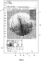
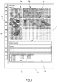
- the first user interface 4 can comprise a plurality of interface screen pages 4a, 4b, 4c representable on the viewing device 3 alternatively and/or contextually.
- the screen pages are reciprocally connected to one another and can be reachable one following another, directly, thus by a direct link between two screen pages, or indirectly, i.e. via intermediate screen pages.
- the first software program is further configured at least to retrieve and visualize, in the first user interface 4, at least a first datum 7, or a first plurality of data 7, relative to a first examination site.
- the term "examination site” is taken to mean a collecting site in a patient, for example a patient's orifice or another surface at which a biological sample is taken, or a different collecting site with respect to which an analysis is to be performed, and therefore for example also a surface to be analysed in a room or environment.
- the expression "information relative to an examination site” are taken to mean data of various nature such as for example a patient's personal data, identifying data of a surface to be analysed, identifying data of an examination site, data relating to a bar code, results in the form of text or in numerical form relating to diagnostic examinations carried out, textual data supplied by doctors or laboratory experts, protocols used and the like.
- the term "information" does not comprise images.
- the first software is further configured such as to retrieve at least a first image 8a of a first support 8, of a first type, for a microbiological culture, which is a Petri dish provided with Agar medium for bacterial cultures, relative to a first microbiological sample originating from the first examination site and for enabling selective viewing at least of the first image 8a in the first user interface 4.
- the first software is further configured such as to retrieve at least a second image 9a of a second support 9 of a second and different type, for microbiological samples, which is a slide for Gram-staining of micro-organisms and/or a slide for morphological analysis of micro-organisms, relative to a second microbiological sample originating from the first examination site and for enabling selective viewing at least of the second image 9a in the first user interface 4.
- the possibility of retrieving and viewing the images 8a of the first support 8 and the images 9a of the second support 9 in a single context enables the user or the specialized operator to carry out a combined analysis of the data relating to the first examination site and of the images relating to the first and/or the second support 9.
- the combined analysis can be carried out in particular by a simultaneous viewing of the data and images, or by a viewing thereof later on but quite close in terms of time, sufficiently to enable the operator or user to consider all the information that can be drawn from the data and images with the aim of formulating her or his diagnosis.
- the operator can at any time reprocess the information already obtained previously in the light of the more recent information received, not being limited to the reading only of a report written on the basis of the previously-analysed images, but with the possibility of viewing the original images (in a digitized form) at the same time or in rapid succession, thus enabling a complete traceability of all the work previously carried out, a significant level of verification, and a reliability and quality in the definition of the diagnosis that is impossible with the traditional systems and approaches.
- the first software program is further configured such as to retrieve the first image 8a and/or the second image 9a from at least a first memory 5 operatively connected to the processor.
- the first software program can be configured so as to retrieve the first image 8a and/or the second image 9a directly from at least a first and/or a second device 10, 11 for acquiring images connected to the processor, directly or indirectly.
- the first biological sample and the second biological sample can be different though coming from a same examination site, for example a site belonging to a patients, but preferably correspond to one another and originate from a same original sample taken.
- the first software program can further be configured for retrieving and viewing, selectively or contextually, in the first user interface 4, a first plurality of images 8a, 8b, 8c, 8d etc., of the first support 8 corresponding at least to a corresponding plurality of various time instants correlated to different incubation periods of the first microbiological sample on the first support 9 which is a Petri dish.
- the first software program can further be configured to retrieve and visualise, selectively or contextually, in the first user interface 4, a second plurality of images 9a, 9b, 9c, 9d etc., of the second support 9 corresponding to a corresponding plurality of different time instants or different levels of enlargement, thus at different levels of imaging zoom and images taken by immersion of the lens in oil, of the second sample arranged on the second support 9.
- the first software program can further be configured for enabling visualizing at least selectively and alternatively, in the first user interface 4, the first image 8a or the first plurality of images 8a, 8b, 8c, and the second image 9a, or the second plurality of images 9a, 9b, 9c.
- the first software program can be further configured for further enabling simultaneous viewing in the first user interface 4 of the first image 8a, or the first plurality of images 8a, 8b, 8c, of the first support 8 and the second image 9a, or the second plurality of images 9a, 9b, 9c, of the second support 9.
- the first software program can further be configured for enabling viewing, in the first user interface 4, of further metadata 12 associated to the first image 8a, or to the first plurality of images 8a, 8b, 8c, of the first support 8 and/or associated to the second image 9a, or to the second plurality of images 9a, 9b, 9c, of the second support 9.
- the metadata 12 can comprise, for example, data and/or time of taking the photographic image, the incubation time interval of the support at the moment of taking the image, the place the image was taken at, mode of capture of the image, the device that took the image, etc.
- the first software program is further configured for enabling the user to enter and store further data, in particular diagnostic data, deriving from a combined use and evaluation of the first image 8a, or of the first plurality of images 8a, 8b, 8c, of the first support 8, and/or of the second image 9a, or the second plurality of images 9a, 9b, 9c, of the second support 9.
- the further data 13 can comprise data relating to the examined micro-organisms, such as the morphology thereof (whether they are coccus, diplococcus, bacillus, streptococcus or yeasts), the Gram-test (i.e. whether they are Gram-positive, Gram-negative or Gram-variable), the quantity or numerousness of the micro-organisms, and so on.
- the first software program is further configured so as to retrieve, process and visualise the data and relative images of the first and second supports relative to a plurality of patients, for example with the aim of enabling the operator to carry out a mass screening of the samples in order to identify the macroscopically positive cases, those in which growths or presence of pathogens are evidenced, to which to give priority in the successive laboratory processes.
- the first software program is configured so as to enable visualizing the first image 8a, or the first plurality of images 8a, 8b, 8c and the second image 9a, or the second plurality of images 9a, 9b, 9c in a same interface screen page, or in interface screen pages 4a, 4b, 4c visualized alternatively and successively on the viewing device 3 and reciprocally mutually connected and reachable directly or indirectly.
- the apparatus 1 further comprises a connecting port 14 configured for, and destined to, enabling operating connection of the apparatus 1 with a laboratory information system 15 or a laboratory computer system and an exchange of data, monodirectional or preferably bidirectional, between the apparatus 1 and the laboratory information system 15.
- the information exchanged between the systems comprise at least the data relative to the first examination site and/or the images relative to samples of micro-biological material and/or diagnostic data determined by the user and entered in the first user interface 4.
- the first software program can be provided with numerous functionalities able to increase the practicality of use and the effectiveness of the apparatus 1 as a support in the diagnostic process, such as for example:
- the invention further relates to a process for treatment of diagnostic information relating to samples of microbiological material, as defined in claim 13.
- the method can further comprise steps of retrieving and visualising, selectively or contextually, in the first user interface 4, a first plurality of images 8a, 8b, 8c of the first support 8 corresponding at least to a corresponding plurality of different time instants correlated to different incubation periods of the first microbiological sample on the first type of support which is a Petri dish.
- the method can further comprise steps of retrieving and visualizing, selectively or contextually, in the first user interface 4, a second plurality of images 9a, 9b, 9c of the second support 9 corresponding to a corresponding plurality of different time instants or different levels of enlargement or to a different mode of image-detecting of the second sample arranged on the second support 9.
- the method can further comprise steps of visualizing at least selectively and alternatingly, in the user interface, the first image 8a or the first plurality of images 8a, 8b, 8c, and the second image 9a or the second plurality of images 9a, 9b, 9c.
- the process can further comprise steps of simultaneously visualizing, in the user interface, the first image 8a, or the first plurality of images 8a, 8b, 8c, of the first support 8 and the second image 9a, or the second plurality of images 9a, 9b, 9c, of the second support 9.
- the first image 8a and/or the second image 9a can be retrieved for example from at least a first memory and/or directly from at least a device 10, 11 for acquiring images.
- the method can further comprise steps of displaying the first image 8a, or the first plurality of images 8a, 8b, 8c and the second image 9a, or the second plurality of images 9a, 9b, 9c in a same interface screen page, or in interface screen pages 4a, 4b, 4c viewed alternatively or successively on the viewing device 3 and reciprocally mutually connected to one another and reachable directly or indirectly.
- the method can comprise steps of further visualizing, in the user interface, further metadata 12 associated to the first image 8a, or to the first plurality of images 8a, 8b, 8c of the first support 8 and/or associated to the second image 9a, or to the second plurality of images 9a, 9b, 9c of the second support 9.
- the method can further comprise steps of inserting and memorising further diagnostic data 13 deriving from a combined analysis of the first image 8a, or the first plurality of images 8a, 8b, 8c of the first support 8 and/or the second image 9a, or the second plurality of images 9a, 9b, 9c of the second support 9.
- the method can further comprise steps of retrieving, from a laboratory information system 15, processing and visualizing the data and/or the relative images of the first and second supports relatively to a plurality of patients.
- the method can further comprise the steps of exchanging information, comprising at least data relative to the first examination site and/or the images, relative to samples of microbiological material, and/or diagnostic data deriving from the combined fruition and evaluation of the images, between an apparatus 1 for treatment of diagnostic information relative sample of microbiological material and a laboratory information system 15.
- the present invention enables obtaining one or more of the following advantages. Primarily, the invention enables obviating the problems encountered in the prior art.
- the invention further enables a rapid, accurate and traced decision on the part of the bacteriologist and the laboratory doctor regarding the diagnosis, based not only on other evidence (relative to bacterial growths on culture dish) by indeed having access to the digitized original images of the dishes and supports.
- the invention further enables a simplification and acceleration of the treatment methods and a retrieval of the information relative to the analyses of the samples.
- the invention further enables eliminating or at least significantly reducing the risk of human error in identifying the samples to be analysed, in carrying out the analytic procedures and/or in determining the results thereof.
- the invention further offers a high degree of reliability and repeatability of the results, and makes available an apparatus and a method which are extremely flexible and adaptable to various operational needs and various types of analysis.
- the invention further enables simplification of the treatment processes of the information relating to the analyses of the analysed samples.
- the invention further enables simplifying the treatment processes of the information relative to the analyses of the analysed samples.
- the invention further enables complete traceability of the historical information relative to the analyses carried out and the devices and subjects involved, and further enables summings-up, studies and statistics on the results themselves.
- the invention further enables significantly increasing the reliability and quality of the diagnostic process, the safety of the analytical processes of the samples, and therefore also the safety of the patients.
- the invention is simple and convenient to actuate.
Landscapes
- Engineering & Computer Science (AREA)
- Health & Medical Sciences (AREA)
- Chemical & Material Sciences (AREA)
- Life Sciences & Earth Sciences (AREA)
- General Health & Medical Sciences (AREA)
- Analytical Chemistry (AREA)
- Wood Science & Technology (AREA)
- Bioinformatics & Cheminformatics (AREA)
- Organic Chemistry (AREA)
- Zoology (AREA)
- Biochemistry (AREA)
- Biomedical Technology (AREA)
- General Engineering & Computer Science (AREA)
- Medical Informatics (AREA)
- General Physics & Mathematics (AREA)
- Physics & Mathematics (AREA)
- Primary Health Care (AREA)
- Public Health (AREA)
- Epidemiology (AREA)
- Sustainable Development (AREA)
- Biotechnology (AREA)
- Genetics & Genomics (AREA)
- Microbiology (AREA)
- Nuclear Medicine, Radiotherapy & Molecular Imaging (AREA)
- Radiology & Medical Imaging (AREA)
- Theoretical Computer Science (AREA)
- Molecular Biology (AREA)
- Immunology (AREA)
- Pathology (AREA)
- Computer Hardware Design (AREA)
- Human Computer Interaction (AREA)
- Quality & Reliability (AREA)
- Computer Vision & Pattern Recognition (AREA)
- Apparatus Associated With Microorganisms And Enzymes (AREA)
- Measuring Or Testing Involving Enzymes Or Micro-Organisms (AREA)
- Investigating Or Analysing Materials By Optical Means (AREA)
Claims (15)
- Appareil de traitement d'informations de diagnostic concernant des prélèvements de matériau microbiologique, l'appareil (1) comprenant au moins :un premier processeur (2) ;un premier dispositif de visualisation (3) fonctionnellement relié au premier processeur (2) ;un premier programme logiciel fonctionnant sur le premier processeur (2) et configuré pour définir au moins une première interface utilisateur (4) sur le premier dispositif de visualisation (3) et pour récupérer et visualiser, dans la première interface utilisateur (4), au moins une première donnée (7), ou une première pluralité de données (7), concernant un même premier site d'examen ;caractérisé en ce que le premier programme logiciel est en outre configuré pour récupérer au moins une première image (8a) d'un premier support (8), d'un premier type, pour la culture microbiologique, qui est une boîte de Pétri avec un terrain pour la culture bactérienne, concernant un premier prélèvement microbiologique provenant du premier site d'examen et pour permettre la visualisation sélective d'au moins la première image (8a) dans la première interface utilisateur (4), le premier programme logiciel étant en outre configuré pour récupérer au moins une seconde image (9a) d'un second support (9) d'un second et différent type, de prélèvements microbiologiques, qui est une lamelle pour coloration Gram de micro-organismes, et/ou une lamelle pour l'analyse morphologique de micro-organismes, concernant un second prélèvement microbiologique provenant du premier site d'examen et pour permettre la visualisation sélective d'au moins la seconde image (9a) dans la première interface utilisateur (4), afin de permettre à l'utilisateur, à l'aide de la première interface utilisateur (4), d'effectuer l'utilisation et l'évaluation combinées, à l'aide d'une visualisation simultanée et contextuelle ou ultérieure, des données concernant le premier site d'examen et des images concernant le premier et le second support (9) corrélées au premier site d'examen.
- Appareil selon la revendication 1, dans lequel le premier programme logiciel est configuré pour récupérer la première image (8a) et/ou la seconde image (9a) à partir d'au moins une première mémoire (5) fonctionnellement reliée au processeur.
- Appareil selon l'une quelconque des revendications précédentes, dans lequel le premier programme logiciel est configuré pour récupérer la première image (8a) et/ou la seconde image (9a) directement à partir d'au moins un premier dispositif (10, 11) pour acquérir des images fonctionnellement reliées au processeur.
- Appareil selon l'une quelconque des revendications précédentes, dans lequel le premier prélèvement biologique et le second prélèvement biologique correspondent l'un à l'autre et proviennent d'un même prélèvement d'origine recueilli à partir du premier site d'examen.
- Appareil selon l'une quelconque des revendications précédentes, dans lequel le premier programme logiciel est configuré pour récupérer et visualiser, sélectivement ou contextuellement, dans la première interface utilisateur (4), une première pluralité d'images (8a, 8b, 8c) du premier support (8) correspondant à au moins une pluralité correspondante de différents instants de temps corrélés à différentes périodes d'incubation du premier prélèvement microbiologique sur la boîte de Pétri.
- Appareil selon l'une quelconque des revendications précédentes, dans lequel le premier programme logiciel est configuré pour récupérer et visualiser, sélectivement ou contextuellement, dans la première interface utilisateur (4) une seconde pluralité d'images (9a, 9b, 9c) du second support (9) correspondant à une pluralité correspondante de différents instants de temps ou de différents niveaux d'agrandissement ou de différents modes d'image de détection du second prélèvement disposé sur le second support (9).
- Appareil selon la revendication 6, dans lequel le premier programme logiciel est configuré pour permettre la visualisation au moins sélectivement et alternativement, dans la première interface utilisateur (4), de la première image (8a) ou de la première pluralité d'images (8a, 8b, 8c), et de la seconde image (9a), ou de la seconde pluralité d'images (9a, 9b, 9c).
- Appareil selon l'une quelconque des revendications précédentes, dans lequel le premier programme logiciel est configuré pour permettre en outre la visualisation simultanée dans la première interface utilisateur (4) de la première image (8a), ou de la première pluralité d'images (8a, 8b, 8c), du premier support (8) et la seconde image (9a), ou la seconde pluralité d'images (9a, 9b, 9c), du second support (9).
- Appareil selon l'une quelconque des revendications précédentes, dans lequel la première interface utilisateur (4) comprend une pluralité de pages d'écran d'interface (4a, 4b, 4c) pouvant être représentées alternativement sur le dispositif de visualisation (3) et mutuellement réciproquement reliées et pouvant être atteintes, directement ou indirectement, et dans lequel le premier logiciel est configuré pour permettre la visualisation de la première image (8a), ou de la première pluralité d'images (8a, 8b, 8c), et de la seconde image (9a), ou de la seconde pluralité d'images (9a, 9b, 9c), dans une même page d'écran d'interface, ou dans des pages d'écran d'interface (4a, 4b, 4c) visualisées alternativement et en succession sur le dispositif de visualisation (3), et mutuellement réciproquement reliées et pouvant être atteintes, directement ou indirectement.
- Appareil selon l'une quelconque des revendications précédentes, dans lequel le premier programme logiciel est configuré pour permettre en outre la visualisation, dans la première interface utilisateur (4), de métadonnées supplémentaires (12) associées à la première image (8a), ou à la première pluralité d'images (8a, 8b, 8c), du premier support (8) et/ou associées à la seconde image (9a), ou à la seconde pluralité d'images (9a, 9b, 9c), du second support (9).
- Appareil selon l'une quelconque des revendications précédentes, dans lequel le premier programme logiciel est en outre configuré pour permettre à l'utilisateur de saisir et de stocker des données supplémentaires, en particulier des données de diagnostic, provenant d'une utilisation et d'une évaluation combinées de la première image (8a), ou de la première pluralité d'images (8a, 8b, 8c), du premier support (8), et/ou de la seconde image (9a), ou de la seconde pluralité d'images (9a, 9b, 9c), du second support (9).
- Appareil selon l'une quelconque des revendications précédentes, comprenant en outre un port de connexion (14) configuré pour, et destiné à, établir la connexion de fonctionnement de l'appareil (1) avec un système d'informations de laboratoire (15) ou un système informatique de laboratoire et un échange de données, comprenant au moins les données concernant le premier site d'examen et/ou les images concernant les prélèvements de matériau microbiologique et/ou des données de diagnostic déterminées par l'utilisateur et saisies dans la première interface utilisateur (4), entre l'appareil (1) et le système d'informations de laboratoire (15).
- Procédé de traitement d'informations de diagnostic concernant des prélèvements de matériau microbiologique, comprenant au moins les étapes de :définition d'au moins une première interface utilisateur (4) sur un premier dispositif de visualisation (3) ;récupération et visualisation, dans la première interface utilisateur (4), d'au moins une première donnée (7), ou d'une première pluralité de données (7), concernant un premier site d'examen ;caractérisé en ce qu'il comprend en outre les étapes de :récupération d'au moins une première image (8a) d'un premier support (8), d'un premier type, pour la culture microbiologique, qui est une boîte de Pétri avec du terrain pour des cultures bactériennes, par rapport à un premier prélèvement microbiologique provenant du premier site d'examen ;visualisation d'au moins la première image (8a) dans la première interface utilisateur (4) ;récupération en outre d'au moins une seconde image (9a) d'un second support (9) d'un second et différent type, pour les prélèvements microbiologiques, qui est une lamelle pour coloration Gram de micro-organismes et/ou une lamelle pour l'analyse morphologique de micro-organismes, par rapport à un second prélèvement microbiologique provenant du premier site d'examen ; etvisualisation d'au moins la seconde image (9a) dans la première interface utilisateur (4), afin de permettre à l'utilisateur d'effectuer une utilisation et une évaluation combinées, par visualisation simultanée ou ultérieure, des données concernant le premier site d'examen et des images concernant le premier et le second support (9) liées au premier site d'examen.
- Procédé selon la revendication 13, comprenant les étapes de récupération et de visualisation, sélectivement ou contextuellement, dans la première interface utilisateur (4), d'une première pluralité d'images (8a, 8b, 8c) du premier support (8) correspondant à au moins une pluralité correspondante de différents instants de temps corrélés à différentes périodes d'incubation du premier prélèvement microbiologique du premier type de support ou boîte de Pétri et/ou de récupération et de visualisation, sélectivement ou contextuellement, dans la première interface utilisateur (4), d'une seconde pluralité d'images (9a, 9b, 9c) du second support (9) correspondant à une pluralité correspondante de différents instants de temps ou de différents niveaux d'agrandissement ou d'un mode différent d'image de détection du second prélèvement disposé sur le second support (9).
- Procédé selon l'une quelconque des revendications 13 ou 14, comprenant en outre les étapes de visualisation au moins sélectivement et alternativement, dans l'interface utilisateur, de la première image (8a) ou de la première pluralité d'images (8a, 8b, 8c), et de la seconde image (9a) ou de la seconde pluralité d'images (9a, 9b, 9c) ; et/ou les étapes de visualisation simultanément, dans l'interface utilisateur, de la première image (8a), ou de la première pluralité d'images (8a, 8b, 8c), du premier support (8) et de la seconde image (9a), ou de la seconde pluralité d'images (9a, 9b, 9c), du second support (9).
Applications Claiming Priority (2)
| Application Number | Priority Date | Filing Date | Title |
|---|---|---|---|
| ITMI20140004 | 2014-01-03 | ||
| PCT/IB2014/067059 WO2015101886A1 (fr) | 2014-01-03 | 2014-12-18 | Appareil et procédé de traitement d'informations diagnostiques associées à des échantillons de matériel microbiologique |
Publications (2)
| Publication Number | Publication Date |
|---|---|
| EP3090040A1 EP3090040A1 (fr) | 2016-11-09 |
| EP3090040B1 true EP3090040B1 (fr) | 2019-11-27 |
Family
ID=50116016
Family Applications (1)
| Application Number | Title | Priority Date | Filing Date |
|---|---|---|---|
| EP14830705.1A Active EP3090040B1 (fr) | 2014-01-03 | 2014-12-18 | Appareil et procédé de traitement d'informations diagnostiques associées à des échantillons de matériel microbiologique |
Country Status (9)
| Country | Link |
|---|---|
| US (1) | US9892508B2 (fr) |
| EP (1) | EP3090040B1 (fr) |
| JP (1) | JP2017502673A (fr) |
| CN (1) | CN106062172B (fr) |
| AU (1) | AU2014374989B2 (fr) |
| CA (1) | CA2935480C (fr) |
| DK (1) | DK3090040T3 (fr) |
| ES (1) | ES2768681T3 (fr) |
| WO (1) | WO2015101886A1 (fr) |
Families Citing this family (4)
| Publication number | Priority date | Publication date | Assignee | Title |
|---|---|---|---|---|
| BR112016017321B1 (pt) | 2014-01-30 | 2021-03-16 | Bd Kiestra B.V. | sistema e método para imagiologia de amostras biológicas dispostas em meio de cultura |
| EP3286732B1 (fr) | 2015-04-23 | 2025-09-10 | BD Kiestra B.V. | Procédé et système pour compter automatiquement de colonies microbienne |
| EP3286731B1 (fr) | 2015-04-23 | 2020-09-16 | BD Kiestra B.V. | Collecte de colonies par contraste |
| TWI755755B (zh) * | 2019-06-17 | 2022-02-21 | 邦睿生技股份有限公司 | 用於測試生物樣本的裝置 |
Family Cites Families (10)
| Publication number | Priority date | Publication date | Assignee | Title |
|---|---|---|---|---|
| US6271022B1 (en) * | 1999-03-12 | 2001-08-07 | Biolog, Inc. | Device for incubating and monitoring multiwell assays |
| US7657070B2 (en) | 2006-01-20 | 2010-02-02 | Sakura Finetek U.S.A., Inc. | Automated system of processing biological specimens and method |
| EP2031051B1 (fr) | 2006-06-16 | 2021-05-19 | PHC Holdings Corporation | Système de surveillance de culture |
| KR20090114349A (ko) | 2007-02-28 | 2009-11-03 | 가부시키가이샤 니콘 | 인큐베이터, 스케줄 관리 방법 및 프로그램 |
| JPWO2009031283A1 (ja) * | 2007-09-03 | 2010-12-09 | 株式会社ニコン | 培養装置、培養情報管理方法およびプログラム |
| US20100082365A1 (en) * | 2008-10-01 | 2010-04-01 | Mckesson Financial Holdings Limited | Navigation and Visualization of Multi-Dimensional Image Data |
| PL2827150T3 (pl) | 2009-08-22 | 2021-05-31 | The Board Of Trustees Of The Leland Stanford Junior University | Obrazowanie i ocena zarodków, oocytów i komórek macierzystych |
| US20120231517A1 (en) * | 2009-10-13 | 2012-09-13 | The Scripps Research Institute | Miniaturized electroporation-ready microwell aray for high-throughput genomic screening |
| AU2012280743A1 (en) * | 2011-07-02 | 2014-01-16 | Unisense Fertilitech A/S | Adaptive embryo selection criteria optimized through iterative customization and collaboration |
| MX344792B (es) * | 2011-09-25 | 2017-01-06 | Theranos Inc | Sistemas y métodos para múltiples análisis. |
-
2014
- 2014-12-18 EP EP14830705.1A patent/EP3090040B1/fr active Active
- 2014-12-18 DK DK14830705.1T patent/DK3090040T3/da active
- 2014-12-18 ES ES14830705T patent/ES2768681T3/es active Active
- 2014-12-18 US US15/109,017 patent/US9892508B2/en active Active
- 2014-12-18 AU AU2014374989A patent/AU2014374989B2/en active Active
- 2014-12-18 WO PCT/IB2014/067059 patent/WO2015101886A1/fr not_active Ceased
- 2014-12-18 JP JP2016544432A patent/JP2017502673A/ja active Pending
- 2014-12-18 CA CA2935480A patent/CA2935480C/fr active Active
- 2014-12-18 CN CN201480076580.0A patent/CN106062172B/zh active Active
Non-Patent Citations (1)
| Title |
|---|
| None * |
Also Published As
| Publication number | Publication date |
|---|---|
| ES2768681T3 (es) | 2020-06-23 |
| US9892508B2 (en) | 2018-02-13 |
| AU2014374989A1 (en) | 2016-07-21 |
| US20160328844A1 (en) | 2016-11-10 |
| CA2935480A1 (fr) | 2015-07-09 |
| CA2935480C (fr) | 2021-06-29 |
| JP2017502673A (ja) | 2017-01-26 |
| AU2014374989B2 (en) | 2019-09-12 |
| CN106062172A (zh) | 2016-10-26 |
| DK3090040T3 (da) | 2020-01-27 |
| EP3090040A1 (fr) | 2016-11-09 |
| CN106062172B (zh) | 2018-06-12 |
| WO2015101886A1 (fr) | 2015-07-09 |
Similar Documents
| Publication | Publication Date | Title |
|---|---|---|
| Bailey et al. | Clinical microbiology is growing up: the total laboratory automation revolution | |
| Dauwalder et al. | Does bacteriology laboratory automation reduce time to results and increase quality management? | |
| Egli et al. | Digital microbiology | |
| Novak et al. | Automation in the clinical microbiology laboratory | |
| Sautter et al. | Point-counterpoint: consolidated clinical microbiology laboratories | |
| EP3090040B1 (fr) | Appareil et procédé de traitement d'informations diagnostiques associées à des échantillons de matériel microbiologique | |
| EP2958497A1 (fr) | Système, procédé et appareil permettant de constituer un dossier et de gérer des spécimens de biopsie et des informations spécifiques d'un patient sur site | |
| Rhoads et al. | A review of the current state of digital plate reading of cultures in clinical microbiology | |
| Trevisan et al. | Visualization and application of disease diagnosis codes for population health management using porcine diseases as a model | |
| Chaturvedi et al. | Applications of artificial intelligence and machine learning in microbial diagnostics and identification | |
| López-Córdova et al. | Construction of a New Data Set of Pleural Fluid Cytological Images for Research. | |
| Zhang et al. | Introduction of an integrated pathology image management, artificial intelligence, and reporting system | |
| Drummond | Antifungal Immunity | |
| US20140100811A1 (en) | System and Method for Guided Laboratory Data Collection, Analysis, and Reporting | |
| Helman | The role of the veterinary diagnostic lab in the management of BRD | |
| US6934637B2 (en) | Method for comparing and correlating analyses of changes in cells and tissues | |
| Kidney et al. | Guidelines for resident training in veterinary clinical pathology. III: cytopathology and surgical pathology | |
| Loy et al. | Ruminant Diagnostics and Interpretation, An Issue of Veterinary Clinics of North America: Food Animal Practice, E-Book: Ruminant Diagnostics and Interpretation, An Issue of Veterinary Clinics of North America: Food Animal Practice, E-Book | |
| CN102508885A (zh) | 一种用于医学图象阅读行为的数据分析方法和系统 | |
| US11348350B2 (en) | Observation system and information management method | |
| Sandle et al. | Digital advances in modern pathology | |
| JP7450180B2 (ja) | 情報処理装置 | |
| Ramalingam | Innovations in Oral Pathology Laboratory-A Mini Review | |
| Pławińska-Czarnak et al. | Design and implementation of a database enhancing the collection, management and analysis of data in an animal sciences project | |
| Wellman et al. | Clinical Pathology, An Issue of Veterinary Clinics of North America: Small Animal Practice, E-Book: Clinical Pathology, An Issue of Veterinary Clinics of North America: Small Animal Practice, E-Book |
Legal Events
| Date | Code | Title | Description |
|---|---|---|---|
| PUAI | Public reference made under article 153(3) epc to a published international application that has entered the european phase |
Free format text: ORIGINAL CODE: 0009012 |
|
| 17P | Request for examination filed |
Effective date: 20160630 |
|
| AK | Designated contracting states |
Kind code of ref document: A1 Designated state(s): AL AT BE BG CH CY CZ DE DK EE ES FI FR GB GR HR HU IE IS IT LI LT LU LV MC MK MT NL NO PL PT RO RS SE SI SK SM TR |
|
| AX | Request for extension of the european patent |
Extension state: BA ME |
|
| DAX | Request for extension of the european patent (deleted) | ||
| STAA | Information on the status of an ep patent application or granted ep patent |
Free format text: STATUS: EXAMINATION IS IN PROGRESS |
|
| 17Q | First examination report despatched |
Effective date: 20181205 |
|
| GRAP | Despatch of communication of intention to grant a patent |
Free format text: ORIGINAL CODE: EPIDOSNIGR1 |
|
| STAA | Information on the status of an ep patent application or granted ep patent |
Free format text: STATUS: GRANT OF PATENT IS INTENDED |
|
| INTG | Intention to grant announced |
Effective date: 20190808 |
|
| GRAS | Grant fee paid |
Free format text: ORIGINAL CODE: EPIDOSNIGR3 |
|
| GRAA | (expected) grant |
Free format text: ORIGINAL CODE: 0009210 |
|
| STAA | Information on the status of an ep patent application or granted ep patent |
Free format text: STATUS: THE PATENT HAS BEEN GRANTED |
|
| AK | Designated contracting states |
Kind code of ref document: B1 Designated state(s): AL AT BE BG CH CY CZ DE DK EE ES FI FR GB GR HR HU IE IS IT LI LT LU LV MC MK MT NL NO PL PT RO RS SE SI SK SM TR |
|
| REG | Reference to a national code |
Ref country code: GB Ref legal event code: FG4D |
|
| REG | Reference to a national code |
Ref country code: CH Ref legal event code: EP |
|
| REG | Reference to a national code |
Ref country code: CH Ref legal event code: NV Representative=s name: ING. ALESSANDRO GALASSI C/O PGA S.P.A., MILANO, CH |
|
| REG | Reference to a national code |
Ref country code: AT Ref legal event code: REF Ref document number: 1206680 Country of ref document: AT Kind code of ref document: T Effective date: 20191215 |
|
| REG | Reference to a national code |
Ref country code: DE Ref legal event code: R096 Ref document number: 602014057599 Country of ref document: DE |
|
| REG | Reference to a national code |
Ref country code: IE Ref legal event code: FG4D |
|
| REG | Reference to a national code |
Ref country code: DK Ref legal event code: T3 Effective date: 20200122 |
|
| REG | Reference to a national code |
Ref country code: NO Ref legal event code: T2 Effective date: 20191127 |
|
| REG | Reference to a national code |
Ref country code: SE Ref legal event code: TRGR |
|
| REG | Reference to a national code |
Ref country code: NL Ref legal event code: FP |
|
| REG | Reference to a national code |
Ref country code: LT Ref legal event code: MG4D |
|
| PG25 | Lapsed in a contracting state [announced via postgrant information from national office to epo] |
Ref country code: FI Free format text: LAPSE BECAUSE OF FAILURE TO SUBMIT A TRANSLATION OF THE DESCRIPTION OR TO PAY THE FEE WITHIN THE PRESCRIBED TIME-LIMIT Effective date: 20191127 Ref country code: BG Free format text: LAPSE BECAUSE OF FAILURE TO SUBMIT A TRANSLATION OF THE DESCRIPTION OR TO PAY THE FEE WITHIN THE PRESCRIBED TIME-LIMIT Effective date: 20200227 Ref country code: GR Free format text: LAPSE BECAUSE OF FAILURE TO SUBMIT A TRANSLATION OF THE DESCRIPTION OR TO PAY THE FEE WITHIN THE PRESCRIBED TIME-LIMIT Effective date: 20200228 Ref country code: LV Free format text: LAPSE BECAUSE OF FAILURE TO SUBMIT A TRANSLATION OF THE DESCRIPTION OR TO PAY THE FEE WITHIN THE PRESCRIBED TIME-LIMIT Effective date: 20191127 Ref country code: LT Free format text: LAPSE BECAUSE OF FAILURE TO SUBMIT A TRANSLATION OF THE DESCRIPTION OR TO PAY THE FEE WITHIN THE PRESCRIBED TIME-LIMIT Effective date: 20191127 |
|
| PG25 | Lapsed in a contracting state [announced via postgrant information from national office to epo] |
Ref country code: IS Free format text: LAPSE BECAUSE OF FAILURE TO SUBMIT A TRANSLATION OF THE DESCRIPTION OR TO PAY THE FEE WITHIN THE PRESCRIBED TIME-LIMIT Effective date: 20200327 Ref country code: HR Free format text: LAPSE BECAUSE OF FAILURE TO SUBMIT A TRANSLATION OF THE DESCRIPTION OR TO PAY THE FEE WITHIN THE PRESCRIBED TIME-LIMIT Effective date: 20191127 Ref country code: RS Free format text: LAPSE BECAUSE OF FAILURE TO SUBMIT A TRANSLATION OF THE DESCRIPTION OR TO PAY THE FEE WITHIN THE PRESCRIBED TIME-LIMIT Effective date: 20191127 |
|
| REG | Reference to a national code |
Ref country code: ES Ref legal event code: FG2A Ref document number: 2768681 Country of ref document: ES Kind code of ref document: T3 Effective date: 20200623 |
|
| PG25 | Lapsed in a contracting state [announced via postgrant information from national office to epo] |
Ref country code: AL Free format text: LAPSE BECAUSE OF FAILURE TO SUBMIT A TRANSLATION OF THE DESCRIPTION OR TO PAY THE FEE WITHIN THE PRESCRIBED TIME-LIMIT Effective date: 20191127 |
|
| PG25 | Lapsed in a contracting state [announced via postgrant information from national office to epo] |
Ref country code: CZ Free format text: LAPSE BECAUSE OF FAILURE TO SUBMIT A TRANSLATION OF THE DESCRIPTION OR TO PAY THE FEE WITHIN THE PRESCRIBED TIME-LIMIT Effective date: 20191127 Ref country code: EE Free format text: LAPSE BECAUSE OF FAILURE TO SUBMIT A TRANSLATION OF THE DESCRIPTION OR TO PAY THE FEE WITHIN THE PRESCRIBED TIME-LIMIT Effective date: 20191127 Ref country code: PT Free format text: LAPSE BECAUSE OF FAILURE TO SUBMIT A TRANSLATION OF THE DESCRIPTION OR TO PAY THE FEE WITHIN THE PRESCRIBED TIME-LIMIT Effective date: 20200419 Ref country code: RO Free format text: LAPSE BECAUSE OF FAILURE TO SUBMIT A TRANSLATION OF THE DESCRIPTION OR TO PAY THE FEE WITHIN THE PRESCRIBED TIME-LIMIT Effective date: 20191127 |
|
| REG | Reference to a national code |
Ref country code: DE Ref legal event code: R097 Ref document number: 602014057599 Country of ref document: DE |
|
| PG25 | Lapsed in a contracting state [announced via postgrant information from national office to epo] |
Ref country code: SM Free format text: LAPSE BECAUSE OF FAILURE TO SUBMIT A TRANSLATION OF THE DESCRIPTION OR TO PAY THE FEE WITHIN THE PRESCRIBED TIME-LIMIT Effective date: 20191127 Ref country code: SK Free format text: LAPSE BECAUSE OF FAILURE TO SUBMIT A TRANSLATION OF THE DESCRIPTION OR TO PAY THE FEE WITHIN THE PRESCRIBED TIME-LIMIT Effective date: 20191127 Ref country code: MC Free format text: LAPSE BECAUSE OF FAILURE TO SUBMIT A TRANSLATION OF THE DESCRIPTION OR TO PAY THE FEE WITHIN THE PRESCRIBED TIME-LIMIT Effective date: 20191127 |
|
| REG | Reference to a national code |
Ref country code: AT Ref legal event code: MK05 Ref document number: 1206680 Country of ref document: AT Kind code of ref document: T Effective date: 20191127 |
|
| PLBE | No opposition filed within time limit |
Free format text: ORIGINAL CODE: 0009261 |
|
| STAA | Information on the status of an ep patent application or granted ep patent |
Free format text: STATUS: NO OPPOSITION FILED WITHIN TIME LIMIT |
|
| PG25 | Lapsed in a contracting state [announced via postgrant information from national office to epo] |
Ref country code: LU Free format text: LAPSE BECAUSE OF NON-PAYMENT OF DUE FEES Effective date: 20191218 Ref country code: IE Free format text: LAPSE BECAUSE OF NON-PAYMENT OF DUE FEES Effective date: 20191218 |
|
| 26N | No opposition filed |
Effective date: 20200828 |
|
| PG25 | Lapsed in a contracting state [announced via postgrant information from national office to epo] |
Ref country code: PL Free format text: LAPSE BECAUSE OF FAILURE TO SUBMIT A TRANSLATION OF THE DESCRIPTION OR TO PAY THE FEE WITHIN THE PRESCRIBED TIME-LIMIT Effective date: 20191127 Ref country code: AT Free format text: LAPSE BECAUSE OF FAILURE TO SUBMIT A TRANSLATION OF THE DESCRIPTION OR TO PAY THE FEE WITHIN THE PRESCRIBED TIME-LIMIT Effective date: 20191127 Ref country code: SI Free format text: LAPSE BECAUSE OF FAILURE TO SUBMIT A TRANSLATION OF THE DESCRIPTION OR TO PAY THE FEE WITHIN THE PRESCRIBED TIME-LIMIT Effective date: 20191127 |
|
| PG25 | Lapsed in a contracting state [announced via postgrant information from national office to epo] |
Ref country code: CY Free format text: LAPSE BECAUSE OF FAILURE TO SUBMIT A TRANSLATION OF THE DESCRIPTION OR TO PAY THE FEE WITHIN THE PRESCRIBED TIME-LIMIT Effective date: 20191127 |
|
| PG25 | Lapsed in a contracting state [announced via postgrant information from national office to epo] |
Ref country code: MT Free format text: LAPSE BECAUSE OF FAILURE TO SUBMIT A TRANSLATION OF THE DESCRIPTION OR TO PAY THE FEE WITHIN THE PRESCRIBED TIME-LIMIT Effective date: 20191127 Ref country code: HU Free format text: LAPSE BECAUSE OF FAILURE TO SUBMIT A TRANSLATION OF THE DESCRIPTION OR TO PAY THE FEE WITHIN THE PRESCRIBED TIME-LIMIT; INVALID AB INITIO Effective date: 20141218 |
|
| PG25 | Lapsed in a contracting state [announced via postgrant information from national office to epo] |
Ref country code: TR Free format text: LAPSE BECAUSE OF FAILURE TO SUBMIT A TRANSLATION OF THE DESCRIPTION OR TO PAY THE FEE WITHIN THE PRESCRIBED TIME-LIMIT Effective date: 20191127 |
|
| PG25 | Lapsed in a contracting state [announced via postgrant information from national office to epo] |
Ref country code: MK Free format text: LAPSE BECAUSE OF FAILURE TO SUBMIT A TRANSLATION OF THE DESCRIPTION OR TO PAY THE FEE WITHIN THE PRESCRIBED TIME-LIMIT Effective date: 20191127 |
|
| P01 | Opt-out of the competence of the unified patent court (upc) registered |
Effective date: 20230420 |
|
| REG | Reference to a national code |
Ref country code: CH Ref legal event code: U11 Free format text: ST27 STATUS EVENT CODE: U-0-0-U10-U11 (AS PROVIDED BY THE NATIONAL OFFICE) Effective date: 20260101 |
|
| PGFP | Annual fee paid to national office [announced via postgrant information from national office to epo] |
Ref country code: GB Payment date: 20251223 Year of fee payment: 12 |
|
| PGFP | Annual fee paid to national office [announced via postgrant information from national office to epo] |
Ref country code: NO Payment date: 20251218 Year of fee payment: 12 |
|
| PGFP | Annual fee paid to national office [announced via postgrant information from national office to epo] |
Ref country code: IT Payment date: 20251218 Year of fee payment: 12 Ref country code: DK Payment date: 20251222 Year of fee payment: 12 |
|
| PGFP | Annual fee paid to national office [announced via postgrant information from national office to epo] |
Ref country code: FR Payment date: 20251223 Year of fee payment: 12 Ref country code: NL Payment date: 20251222 Year of fee payment: 12 |
|
| PGFP | Annual fee paid to national office [announced via postgrant information from national office to epo] |
Ref country code: SE Payment date: 20251222 Year of fee payment: 12 |
|
| PGFP | Annual fee paid to national office [announced via postgrant information from national office to epo] |
Ref country code: ES Payment date: 20260122 Year of fee payment: 12 |
|
| PGFP | Annual fee paid to national office [announced via postgrant information from national office to epo] |
Ref country code: DE Payment date: 20251229 Year of fee payment: 12 |
|
| PGFP | Annual fee paid to national office [announced via postgrant information from national office to epo] |
Ref country code: BE Payment date: 20260109 Year of fee payment: 12 |
|
| PGFP | Annual fee paid to national office [announced via postgrant information from national office to epo] |
Ref country code: CH Payment date: 20260101 Year of fee payment: 12 |