EP3093678A1 - Procédé pour optimiser la prédiction de l'heure d'arrivée du bolus en utilisant l'irm pour l'imagerie diagnostique - Google Patents
Procédé pour optimiser la prédiction de l'heure d'arrivée du bolus en utilisant l'irm pour l'imagerie diagnostique Download PDFInfo
- Publication number
- EP3093678A1 EP3093678A1 EP15167568.3A EP15167568A EP3093678A1 EP 3093678 A1 EP3093678 A1 EP 3093678A1 EP 15167568 A EP15167568 A EP 15167568A EP 3093678 A1 EP3093678 A1 EP 3093678A1
- Authority
- EP
- European Patent Office
- Prior art keywords
- vessel position
- vessel
- contrast agent
- bolus
- blood flow
- Prior art date
- Legal status (The legal status is an assumption and is not a legal conclusion. Google has not performed a legal analysis and makes no representation as to the accuracy of the status listed.)
- Withdrawn
Links
- 238000000034 method Methods 0.000 title claims abstract description 59
- 238000002059 diagnostic imaging Methods 0.000 title description 3
- 239000002872 contrast media Substances 0.000 claims abstract description 136
- 230000017531 blood circulation Effects 0.000 claims abstract description 66
- 230000001419 dependent effect Effects 0.000 claims abstract description 8
- 230000002596 correlated effect Effects 0.000 claims abstract description 7
- 238000005259 measurement Methods 0.000 claims description 43
- 210000002376 aorta thoracic Anatomy 0.000 claims description 15
- 238000002347 injection Methods 0.000 claims description 12
- 239000007924 injection Substances 0.000 claims description 12
- 238000012546 transfer Methods 0.000 claims description 12
- 230000002093 peripheral effect Effects 0.000 claims description 10
- 210000001367 artery Anatomy 0.000 claims description 9
- 230000006870 function Effects 0.000 claims description 9
- 230000002123 temporal effect Effects 0.000 claims description 8
- 238000002583 angiography Methods 0.000 claims description 6
- 210000003462 vein Anatomy 0.000 claims description 6
- 230000000694 effects Effects 0.000 claims description 5
- 210000002414 leg Anatomy 0.000 claims description 5
- 238000011144 upstream manufacturing Methods 0.000 claims description 3
- 210000000702 aorta abdominal Anatomy 0.000 claims description 2
- 210000003414 extremity Anatomy 0.000 claims description 2
- 210000003127 knee Anatomy 0.000 claims description 2
- 210000003752 saphenous vein Anatomy 0.000 claims description 2
- 238000012360 testing method Methods 0.000 description 18
- 230000002792 vascular Effects 0.000 description 16
- 238000002595 magnetic resonance imaging Methods 0.000 description 11
- 238000003384 imaging method Methods 0.000 description 8
- 210000000709 aorta Anatomy 0.000 description 7
- 238000002591 computed tomography Methods 0.000 description 6
- 210000004369 blood Anatomy 0.000 description 5
- 239000008280 blood Substances 0.000 description 5
- 230000002526 effect on cardiovascular system Effects 0.000 description 5
- 210000004204 blood vessel Anatomy 0.000 description 4
- 239000006185 dispersion Substances 0.000 description 4
- 230000004087 circulation Effects 0.000 description 3
- 230000000875 corresponding effect Effects 0.000 description 3
- 238000001514 detection method Methods 0.000 description 3
- 230000002349 favourable effect Effects 0.000 description 3
- 244000309715 mini pig Species 0.000 description 3
- 238000010171 animal model Methods 0.000 description 2
- QVGXLLKOCUKJST-UHFFFAOYSA-N atomic oxygen Chemical compound [O] QVGXLLKOCUKJST-UHFFFAOYSA-N 0.000 description 2
- 238000012937 correction Methods 0.000 description 2
- 238000003745 diagnosis Methods 0.000 description 2
- 238000002405 diagnostic procedure Methods 0.000 description 2
- 238000010790 dilution Methods 0.000 description 2
- 239000012895 dilution Substances 0.000 description 2
- 238000011156 evaluation Methods 0.000 description 2
- 230000005284 excitation Effects 0.000 description 2
- 230000006872 improvement Effects 0.000 description 2
- 210000003141 lower extremity Anatomy 0.000 description 2
- 210000004072 lung Anatomy 0.000 description 2
- 239000001301 oxygen Substances 0.000 description 2
- 229910052760 oxygen Inorganic materials 0.000 description 2
- 238000012545 processing Methods 0.000 description 2
- 238000012800 visualization Methods 0.000 description 2
- 208000024172 Cardiovascular disease Diseases 0.000 description 1
- UFHFLCQGNIYNRP-UHFFFAOYSA-N Hydrogen Chemical compound [H][H] UFHFLCQGNIYNRP-UHFFFAOYSA-N 0.000 description 1
- 241001465754 Metazoa Species 0.000 description 1
- 208000005764 Peripheral Arterial Disease Diseases 0.000 description 1
- 208000030831 Peripheral arterial occlusive disease Diseases 0.000 description 1
- FAPWRFPIFSIZLT-UHFFFAOYSA-M Sodium chloride Chemical compound [Na+].[Cl-] FAPWRFPIFSIZLT-UHFFFAOYSA-M 0.000 description 1
- 230000009471 action Effects 0.000 description 1
- 230000001154 acute effect Effects 0.000 description 1
- 238000013459 approach Methods 0.000 description 1
- 210000005249 arterial vasculature Anatomy 0.000 description 1
- 210000002048 axillary vein Anatomy 0.000 description 1
- 230000005540 biological transmission Effects 0.000 description 1
- 210000000746 body region Anatomy 0.000 description 1
- 238000004364 calculation method Methods 0.000 description 1
- 210000005242 cardiac chamber Anatomy 0.000 description 1
- 230000002612 cardiopulmonary effect Effects 0.000 description 1
- 230000008859 change Effects 0.000 description 1
- 238000010968 computed tomography angiography Methods 0.000 description 1
- 238000004590 computer program Methods 0.000 description 1
- 229940039231 contrast media Drugs 0.000 description 1
- 230000006866 deterioration Effects 0.000 description 1
- 238000011161 development Methods 0.000 description 1
- 230000018109 developmental process Effects 0.000 description 1
- 201000009101 diabetic angiopathy Diseases 0.000 description 1
- 238000002474 experimental method Methods 0.000 description 1
- ZPDFIIGFYAHNSK-UHFFFAOYSA-K gadobutrol Chemical compound [Gd+3].OCC(O)C(CO)N1CCN(CC([O-])=O)CCN(CC([O-])=O)CCN(CC([O-])=O)CC1 ZPDFIIGFYAHNSK-UHFFFAOYSA-K 0.000 description 1
- LGMLJQFQKXPRGA-VPVMAENOSA-K gadopentetate dimeglumine Chemical compound [Gd+3].CNC[C@H](O)[C@@H](O)[C@H](O)[C@H](O)CO.CNC[C@H](O)[C@@H](O)[C@H](O)[C@H](O)CO.OC(=O)CN(CC([O-])=O)CCN(CC([O-])=O)CCN(CC(O)=O)CC([O-])=O LGMLJQFQKXPRGA-VPVMAENOSA-K 0.000 description 1
- 239000001257 hydrogen Substances 0.000 description 1
- 229910052739 hydrogen Inorganic materials 0.000 description 1
- 230000008595 infiltration Effects 0.000 description 1
- 238000001764 infiltration Methods 0.000 description 1
- 230000004941 influx Effects 0.000 description 1
- 230000003993 interaction Effects 0.000 description 1
- 238000010253 intravenous injection Methods 0.000 description 1
- 238000011835 investigation Methods 0.000 description 1
- 210000005248 left atrial appendage Anatomy 0.000 description 1
- 210000005240 left ventricle Anatomy 0.000 description 1
- 238000005457 optimization Methods 0.000 description 1
- 230000007170 pathology Effects 0.000 description 1
- 210000005259 peripheral blood Anatomy 0.000 description 1
- 239000011886 peripheral blood Substances 0.000 description 1
- 230000035479 physiological effects, processes and functions Effects 0.000 description 1
- 230000037452 priming Effects 0.000 description 1
- 230000004088 pulmonary circulation Effects 0.000 description 1
- 238000005086 pumping Methods 0.000 description 1
- 238000011160 research Methods 0.000 description 1
- 239000011780 sodium chloride Substances 0.000 description 1
- 210000003813 thumb Anatomy 0.000 description 1
- 230000009466 transformation Effects 0.000 description 1
Images


Classifications
-
- G—PHYSICS
- G01—MEASURING; TESTING
- G01R—MEASURING ELECTRIC VARIABLES; MEASURING MAGNETIC VARIABLES
- G01R33/00—Arrangements or instruments for measuring magnetic variables
- G01R33/20—Arrangements or instruments for measuring magnetic variables involving magnetic resonance
- G01R33/44—Arrangements or instruments for measuring magnetic variables involving magnetic resonance using nuclear magnetic resonance [NMR]
- G01R33/48—NMR imaging systems
- G01R33/54—Signal processing systems, e.g. using pulse sequences ; Generation or control of pulse sequences; Operator console
- G01R33/56—Image enhancement or correction, e.g. subtraction or averaging techniques, e.g. improvement of signal-to-noise ratio and resolution
- G01R33/5601—Image enhancement or correction, e.g. subtraction or averaging techniques, e.g. improvement of signal-to-noise ratio and resolution involving use of a contrast agent for contrast manipulation, e.g. a paramagnetic, super-paramagnetic, ferromagnetic or hyperpolarised contrast agent
-
- A—HUMAN NECESSITIES
- A61—MEDICAL OR VETERINARY SCIENCE; HYGIENE
- A61B—DIAGNOSIS; SURGERY; IDENTIFICATION
- A61B5/00—Measuring for diagnostic purposes; Identification of persons
- A61B5/02—Detecting, measuring or recording for evaluating the cardiovascular system, e.g. pulse, heart rate, blood pressure or blood flow
- A61B5/026—Measuring blood flow
- A61B5/0263—Measuring blood flow using NMR
-
- G—PHYSICS
- G01—MEASURING; TESTING
- G01R—MEASURING ELECTRIC VARIABLES; MEASURING MAGNETIC VARIABLES
- G01R33/00—Arrangements or instruments for measuring magnetic variables
- G01R33/20—Arrangements or instruments for measuring magnetic variables involving magnetic resonance
- G01R33/44—Arrangements or instruments for measuring magnetic variables involving magnetic resonance using nuclear magnetic resonance [NMR]
- G01R33/48—NMR imaging systems
- G01R33/54—Signal processing systems, e.g. using pulse sequences ; Generation or control of pulse sequences; Operator console
- G01R33/56—Image enhancement or correction, e.g. subtraction or averaging techniques, e.g. improvement of signal-to-noise ratio and resolution
- G01R33/563—Image enhancement or correction, e.g. subtraction or averaging techniques, e.g. improvement of signal-to-noise ratio and resolution of moving material, e.g. flow contrast angiography
- G01R33/5635—Angiography, e.g. contrast-enhanced angiography [CE-MRA] or time-of-flight angiography [TOF-MRA]
-
- G—PHYSICS
- G16—INFORMATION AND COMMUNICATION TECHNOLOGY [ICT] SPECIALLY ADAPTED FOR SPECIFIC APPLICATION FIELDS
- G16H—HEALTHCARE INFORMATICS, i.e. INFORMATION AND COMMUNICATION TECHNOLOGY [ICT] SPECIALLY ADAPTED FOR THE HANDLING OR PROCESSING OF MEDICAL OR HEALTHCARE DATA
- G16H10/00—ICT specially adapted for the handling or processing of patient-related medical or healthcare data
-
- G—PHYSICS
- G16—INFORMATION AND COMMUNICATION TECHNOLOGY [ICT] SPECIALLY ADAPTED FOR SPECIFIC APPLICATION FIELDS
- G16H—HEALTHCARE INFORMATICS, i.e. INFORMATION AND COMMUNICATION TECHNOLOGY [ICT] SPECIALLY ADAPTED FOR THE HANDLING OR PROCESSING OF MEDICAL OR HEALTHCARE DATA
- G16H20/00—ICT specially adapted for therapies or health-improving plans, e.g. for handling prescriptions, for steering therapy or for monitoring patient compliance
- G16H20/10—ICT specially adapted for therapies or health-improving plans, e.g. for handling prescriptions, for steering therapy or for monitoring patient compliance relating to drugs or medications, e.g. for ensuring correct administration to patients
- G16H20/17—ICT specially adapted for therapies or health-improving plans, e.g. for handling prescriptions, for steering therapy or for monitoring patient compliance relating to drugs or medications, e.g. for ensuring correct administration to patients delivered via infusion or injection
-
- G—PHYSICS
- G16—INFORMATION AND COMMUNICATION TECHNOLOGY [ICT] SPECIALLY ADAPTED FOR SPECIFIC APPLICATION FIELDS
- G16H—HEALTHCARE INFORMATICS, i.e. INFORMATION AND COMMUNICATION TECHNOLOGY [ICT] SPECIALLY ADAPTED FOR THE HANDLING OR PROCESSING OF MEDICAL OR HEALTHCARE DATA
- G16H30/00—ICT specially adapted for the handling or processing of medical images
- G16H30/20—ICT specially adapted for the handling or processing of medical images for handling medical images, e.g. DICOM, HL7 or PACS
-
- G—PHYSICS
- G16—INFORMATION AND COMMUNICATION TECHNOLOGY [ICT] SPECIALLY ADAPTED FOR SPECIFIC APPLICATION FIELDS
- G16H—HEALTHCARE INFORMATICS, i.e. INFORMATION AND COMMUNICATION TECHNOLOGY [ICT] SPECIALLY ADAPTED FOR THE HANDLING OR PROCESSING OF MEDICAL OR HEALTHCARE DATA
- G16H40/00—ICT specially adapted for the management or administration of healthcare resources or facilities; ICT specially adapted for the management or operation of medical equipment or devices
- G16H40/60—ICT specially adapted for the management or administration of healthcare resources or facilities; ICT specially adapted for the management or operation of medical equipment or devices for the operation of medical equipment or devices
-
- A—HUMAN NECESSITIES
- A61—MEDICAL OR VETERINARY SCIENCE; HYGIENE
- A61B—DIAGNOSIS; SURGERY; IDENTIFICATION
- A61B5/00—Measuring for diagnostic purposes; Identification of persons
- A61B5/0033—Features or image-related aspects of imaging apparatus, e.g. for MRI, optical tomography or impedance tomography apparatus; Arrangements of imaging apparatus in a room
-
- G—PHYSICS
- G01—MEASURING; TESTING
- G01R—MEASURING ELECTRIC VARIABLES; MEASURING MAGNETIC VARIABLES
- G01R33/00—Arrangements or instruments for measuring magnetic variables
- G01R33/20—Arrangements or instruments for measuring magnetic variables involving magnetic resonance
- G01R33/44—Arrangements or instruments for measuring magnetic variables involving magnetic resonance using nuclear magnetic resonance [NMR]
- G01R33/48—NMR imaging systems
- G01R33/54—Signal processing systems, e.g. using pulse sequences ; Generation or control of pulse sequences; Operator console
- G01R33/56—Image enhancement or correction, e.g. subtraction or averaging techniques, e.g. improvement of signal-to-noise ratio and resolution
- G01R33/563—Image enhancement or correction, e.g. subtraction or averaging techniques, e.g. improvement of signal-to-noise ratio and resolution of moving material, e.g. flow contrast angiography
- G01R33/56308—Characterization of motion or flow; Dynamic imaging
- G01R33/56316—Characterization of motion or flow; Dynamic imaging involving phase contrast techniques
Definitions
- the invention relates to a method for optimizing the predetermination of the time course of a contrast agent concentration at a vessel position in an examination environment in a contrast-agent-based MR image of a patient exclusively in the first flooding phase of the contrast agent.
- CE-MRA Contrast Enhanced MR Angiography
- the CE-MRA is the preferred method of radiation-free, non-invasive vascular diagnosis in many clinical cases compared to other, non-contrast enhanced imaging techniques. It was first introduced approximately 20 years ago and has been undergoing continuous technical improvement is an advanced standard diagnostic procedure in most countries.
- the contrast medium flowing in the vascular system of the target region as closely as possible with the beginning of the data acquisition, both in order to be able to fully exploit the available contrast enhancement by the contrast agent and to precede the subsequent re-flooding of the contrast agent in the venous vascular system.
- CT computed tomography
- CT angiography in which the blood vessels of a region to be examined are flooded with contrast medium in the typical range of about 25-50 seconds, the contrast medium is substantially shorter Time span in the typical range of about 3 to 15 seconds applied.
- contrast-enhanced MR imaging in contrast to contrast-enhanced fluoroscopic X-ray or CT examination, smaller volumes are considered, a fundamentally different acquisition technique with special features of K-space acquisition and Fourier transformation, different measurement times and requirements Contrast medium concentration during the measurement and a non-linear signal intensity as a function of the local contrast agent concentration in the examination area.
- a direct transfer of the conditions in the CT in the contrast agent application to those in the MRI is therefore usually not possible.
- a compromise between spatial and temporal resolution of the image data obtained is generally accepted, in that the contrast agent flooding is achieved with a dynamically repeated acquisition of the three-dimensional target region, for example in each case within 3 to 5 seconds.
- the spatial resolution is limited at these short measurement times and is typically at least 2mm - 3mm.
- the measuring time is usually about 15 seconds. Due to the dynamically repeated measurements, the contrast agent bolus is always recorded, in most cases even with several 3D data sets. A bolus synchronization is therefore superfluous.
- peripheral angiography which is important in vascular diagnosis for the detection of diabetic vascular diseases, such as peripheral arterial disease, cardiovascular disease, or "smoker's leg," the lower limbs, is still associated with an undesirably high rate of artifact-related and non-diagnostic test results.
- the inventors have recognized that, in contrast to the temporally relatively long contrast agent inputs in a CT examination, a slight widening (dispersion) of the bolus in relation to the duration of the flooding hardly plays a role plays in the contrast-enhanced MR imaging, in particular arterial, blood vessels is necessary to know and take into account the effect of widening a short contrast agent, especially in the range of a few seconds in order to predict the contrast agent as accurately as possible.
- the inventors have recognized that there is an evaluable correlation between the size of the broadening and the blood flow properties, such as the flow velocity or the volume flow, of the blood in the vascular system at the selected location or the velocity difference between two selected locations, which predicts an incoming broadening of a Contrast agent bolus allowed.
- the correlation values which are as meaningful as possible, it is particularly favorable if, when determining the correlation values, patient comparisons which are as comparable as possible are used, that is to say suitable parameters such as gender, height, weight, etc. are used as far as possible for the patient to be examined.
- the correlation between the broadening of an applied contrast agent bolus and blood flow properties measured in the vascular system or parameters derived therefrom can be determined.
- the contrast-enhanced examination of a patient can then first in the absence of contrast agent the corresponding blood flow characteristic or dependent parameters are determined and from this, the expected widening of the bolus, for example, via the application of a look up table (LUT), be concluded.
- LUT look up table
- the transit time of a bolus depends on the present flow velocities or volume flows, such a LUT can also be created for the determination of the transit time of the bolus (BTT), or another known method for predicting a BTT can be used here. From both information, the actual expected start and end time of an applied bolus can then be predicted and used for temporal synchronization of an applied bolus with a contrast-enhanced MRI measurement.
- the method described above is particularly suitable for presentation in the periphery of the lower extremities (CE-pMRA), because the current limitations are based in the current state of the art regularly on an inaccurate or incomplete knowledge of the user of the individual cardiovascular conditions of the patient, as they directly in front the contrast agent examination are present.
- the flow velocities determined immediately prior to the imaging of the contrast agent result in a particularly up-to-date, more accurate and comprehensive knowledge of the individual cardiovascular conditions of the patient than was hitherto customary, since the patient's current stress conditions in the examination environment are automatically taken into account.
- blood flow measurements can thus be carried out at different positions of the arterial vascular system in order to gain knowledge of the acute and patient-specific cardiovascular conditions based on a postulated and for the first time proven correlation of the bolus dispersion and various blood flow parameters, so that a necessary contrast agent bolus for achieving a desired contrast agent concentration profile and timing is relatively can be correctly selected to perform the MRI with improved safety.
- the inventors propose a method for optimizing the predetermination of the time course of a contrast agent concentration at a vessel position in an examination environment in a contrast agent-assisted MR image of a patient exclusively in the first priming phase of the contrast agent, in which a broadening of a Contrast agent bolus course having a first width at a predetermined first vessel position of a patient to a contrast agent concentration course having a second width at a predetermined second vessel position, which is arranged in the examination environment, by determining at least one, at least one blood flow characteristic at a third vessel position dependent and with the broadening correlated, flow parameters is determined.
- contrast agent concentration course not only the course of the actual concentration of the contrast agent in the blood, but also the course of the contrast agent-induced increase of the MR signal is to be understood in an MR examination.
- the method is specifically intended to determine the contrast agent bolus course for vessel positions, which are flow-related behind the cardiopulmonary passage, ie arterially behind the heart, arranged, in particular, this peripheral vessel positions and / or examination environments are to be understood, which in a peripheral Area of the patient, especially in one of the limbs.
- the determined expected broadening for the predetermination of the temporal contrast agent concentration profile at a predetermined vessel position can thus be made available for further use, wherein the storage is preferably carried out in an electronic memory for further use by a computer.
- the at least one blood flow property can be determined in the absence of contrast agent by means of a phase-contrast magnetic resonance measurement.
- a measurement can be made immediately before the actual MR examination, so that advantageously such a measurement is the actually existing constitution and constitution considered by the examined patients and thus optimally adapted to the actual MR examination.
- the determination according to the invention of widening a bolus with a determination, known in the prior art, of a bolus transfer time BTT or bolus arrival time BAT, which optionally also uses a test bolus, in order to predict an overall contrast agent concentration course.
- a bolus transfer time BTT or bolus arrival time BAT which optionally also uses a test bolus
- the additionally determined bolus transfer time between the first vessel position and the second vessel position or the bolus arrival time is determined on the basis of a predetermined correlation to a blood flow characteristic measured without contrast.
- the determination of the blood flow property be carried out at least at the third vessel position of the patient by a phase-contrast magnetic resonance examination.
- the phase-contrast magnetic resonance examination for determining the blood flow characteristic can be carried out in the absence of contrast medium in the bloodstream of the patient.
- the group of patients from which the data from the preliminary investigation relating to the broadening should be as similar as possible to that currently being investigated Be patient. This can be ensured by the fact that the current patient and the relevant patient collective from which the data from the preliminary examination for the widening originate agree with regard to as many patient parameters as possible.
- patient parameters should be taken into account, which have an anatomical, physiological and medical relation to the circulation and the vascular structure.
- at least one of the following parameters can be used as at least one patient parameter: gender, weight, height, age, heart rate and / or BMI.
- the contrast agent itself can also be introduced as an additional parameter characterizing the widening. Accordingly, the proposed LUT can be typed around this contrast agent parameter of the contrast agent used, without departing from the scope of the invention.
- the blood flow through a vessel can generally not be defined by a single variable which is constant over the vessel cross-section and over the pulse phase and for a longer time and can be measured directly.
- the blood flow through a vessel at a vessel position is characterized not only by a velocity in cm / s or a blood flow in mL / s but temporally and spatially changing profiles. Nevertheless or just because of such measurements can be characteristic characteristics of blood flow, such as minimum, maximum or average values of speed and flow, express that are in correlation with the widening of a bolus in good correlation.
- the blood flow characteristic itself can be used as a flow parameter.
- the method according to the invention relates exclusively to contrast agent-based MR examinations and no CT examinations.
- the width of the contrast agent concentration course in the vascular system taking into account the broadening present in each case in a similar framework. While the width of the contrast agent bolus course is uniquely determined due to its steep edges, the definition of the width of the contrast agent concentration profile is subject to some blurring.
- the person skilled in the art will set the width as the half-width of the contrast agent concentration or of a signal generated by the contrast agent or use the achievement of a predetermined concentration value or the achievement of a width-defined signal level for width definition.
- the same width definition should be used in each case.
- a previously determined correlation between the expected broadening and the respectively considered flow parameter can be represented in an arbitrary manner, for example also by the use of a mathematically described functional relationship.
- the application of a "look up" table is particularly favorable and simple, so that the correlation of the widening with the flow parameter is taken from a table, preferably stored electronically.
- the table predetermined from reference measurements may also contain at least one of the aforementioned patient parameters.
- the correlation of the broadening with the flow parameter can be calculated by a function predetermined from reference measurements, whereby the function predetermined from reference measurements can also contain at least one patient parameter as a variable.
- the scope of the invention also includes a computer, at least having a memory for programs which are executed during operation, programs being stored therein which execute the method according to the invention in accordance with a feature combination disclosed herein.
- the invention also includes a magnetic resonance system for generating a contrast-enhanced MR image or an MR vessel representation with a computer-controlled contrast agent injector, the computer is equipped with a memory for programs that are executed in operation, wherein programs are stored, which carry out the method according to the invention.
- FIG. 1 schematically a magnetic resonance imaging (MRI) system 1 is shown.
- MRT system 1 In this MRT system 1 are located in a housing 6 magnetic coils 2 for generating a strong magnetic field, whereby the hydrogen nuclei in the body of the patient 7 align themselves according to their spin parallel or anti-parallel to the magnetic field lines.
- Additional magnetic coils 4 generate a weak magnetic field with a defined field gradient, as a result of which the signals emitted by the cores receive location information by means of which the position of the emitted signal can be defined.
- the control of this system 1 and the evaluation of the measurement signals is carried out by the control and computing unit 8, which has in its memory programs 9, which also perform the inventive method in addition to the control and image calculation.
- contrast agent which is usually a contrast agent injector 5 is used
- the electronically controlled - either by the arithmetic unit 8 or by another separate processing unit - the volume flow to a measurement produced applying contrast agent.
- FIG. 2 a very schematically shown blood circulation of a patient shown.
- This closed circuit divides into a venous circuit (dashed lines) and an arterial circuit (solid lines) and is operated essentially by the pumping action of the heart.
- the arterial blood - in contrast to the other blood circulation - is poor in oxygen and the venous oxygen rich.
- contrast media are usually applied by such approaches, usually with the aid of automatically controlled contrast agent injectors.
- the essential vessel positions P1 to P4 and the region of interest ROI to be imaged are marked at exemplary and typical positions.
- the application takes place in the region of the venous circulation corresponding to the registered vessel position P1 on a peripheral vessel, for example an arm or hand vein.
- the area of interest ROI which is to be examined by imaging, can, for example, be located in the area of a leg artery, in which the vessel position P2 is located, as shown here.
- the other measuring positions at which blood flow properties are then determined are then usually located in the arterial vascular system between the left atrial appendage of the heart and the ROI. It is pointed out, however, that also a positioning of the measuring points Vessel positions is possible, which are arranged in the flow direction behind the ROI or P2.
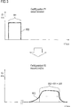
- a contrast agent bolus which is applied at a first vessel position P1, usually has a bolus course B (t), as it is in the FIG. 3 is drawn in the upper part. There the volume flow of a bolus B is plotted over the time t.
- a bolus course can - as shown here assume a rectangular course, but can also emulate by appropriate adjustment to a contrast agent injector any function B (t).
- Such a volume flow B is selected depending on the contrast agent used (eg Gadovist® or Magnevist®) and specific patient properties in order to achieve a desired contrast agent concentration at the site of the examination in the vessel to be displayed.
- the contrast agent used eg Gadovist® or Magnevist®
- the width of the applied bolus W1 has widened considerably over the distance traveled between the positions P1 to P2, so that, taking into account a half-width, a width of W2 is present.
- the size of such broadening can be measured directly with contrast-free measurable blood flow properties on the transport path of the contrast agent or on flow parameters derived therefrom is in clear correlation, so that by determining these flow parameters, in particular using contrast-free measurements of blood flow properties, determine the flow-related widening of an applied bolus and therefore can predict.
- the time profile of the contrast agent concentration on a vessel, in particular a peripheral vessel can then be determined and thus predicted on the basis of the knowledge of the applied bolus.
- the necessary course of a bolus application can also be determined retroactively, which leads to the achievement of a desired contrast agent concentration course and thus also to a desired signal course in an MR measurement at an ROI.
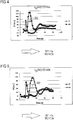
- AD mean volume flow v
- the measured width based on the half-width of the signal waveform is 4.5 seconds.
- the shows FIG. 5 with the same bolus application width of W1 1s also a measured signal course an MR measurement immediately after the application site (ear vein) with diamond-shaped measuring points and a signal course of an MR measurement at the descending aorta (AD).
- the mean volume flow v (AD) in the descending aorta is 23 ml / s in this measurement.
- the measured width based on the half width of the signal waveform is 7.5 seconds.
- such a relationship can be provided in the form of a look-up table (LUT), which is stored in a computer memory and can serve for predetermining the widening of a bolus.
- LUT look-up table
- the superscript indices denote the different value ranges of the parameters in the row or column title. Intermediate values may optionally be obtained by interpolation.
- a corresponding LUT can therefore be designed as follows: Table 2: BTT from P 1 to P2 d -2 (ROI) / cm ... ... d -1 (ROI) / cm ... ... ... d avg (ROI) / cm d +1 (ROI) / cm ... ... ... d + 2 (ROI) / cm ... ...
- the superscript indices each denote the different value ranges of the respective parameter in the row or column title. Intermediate values may optionally be obtained by interpolation.
- a patient parameter is given in the form of a distance d i (ROI), here the distance in cm between a Vessel position P3, where the flow parameter r f1 is measured, and represents a vessel position P2 in the region of the target region ROI.
- ROI distance in cm between a Vessel position P3, where the flow parameter r f1 is measured, and represents a vessel position P2 in the region of the target region ROI.
- the current flow parameters blood flow velocities V G (AD), V G (AB) and V G (AP) are shown as different input parameters.
- the blood flow velocity V G (AD) again exemplifies the blood flow velocity measured in the region of the descending aorta.
- LUT 1 and LUT 2 can thus be used to determine the timing of a contrast medium bolus delivery with the determination of different blood flow and patient parameters without contrast.
- the invention thus proposes a method in which for optimizing the predetermination of the time course of a contrast agent concentration at a vessel position in an examination environment (ROI) in a contrast agent-based MR image of a patient (P) exclusively in the first flooding phase of the contrast agent, the Correlation of a broadening and / or a bolus transfer time of a contrast agent bolus between two vessel positions and at least one flow parameter - which is a function of at least one flow characteristic - is predetermined in the bloodstream of a patient collective, so that then before an MR examination of a patient to be performed these flow parameters by a contrast agent Phase contrast MR examination can be determined and on the previously known correlations to broadening and / or bolus transfer time of the expected contrast agent concentration course at a Gefäßpositi is determined in the examination area.
Landscapes
- Health & Medical Sciences (AREA)
- Engineering & Computer Science (AREA)
- Physics & Mathematics (AREA)
- General Health & Medical Sciences (AREA)
- Nuclear Medicine, Radiotherapy & Molecular Imaging (AREA)
- Medical Informatics (AREA)
- Public Health (AREA)
- Life Sciences & Earth Sciences (AREA)
- Radiology & Medical Imaging (AREA)
- Biomedical Technology (AREA)
- Primary Health Care (AREA)
- Epidemiology (AREA)
- Signal Processing (AREA)
- General Physics & Mathematics (AREA)
- Condensed Matter Physics & Semiconductors (AREA)
- High Energy & Nuclear Physics (AREA)
- Pathology (AREA)
- Surgery (AREA)
- Veterinary Medicine (AREA)
- Animal Behavior & Ethology (AREA)
- Molecular Biology (AREA)
- Heart & Thoracic Surgery (AREA)
- Biophysics (AREA)
- Physiology (AREA)
- Cardiology (AREA)
- Hematology (AREA)
- Vascular Medicine (AREA)
- Business, Economics & Management (AREA)
- Chemical & Material Sciences (AREA)
- Bioinformatics & Cheminformatics (AREA)
- General Business, Economics & Management (AREA)
- Medicinal Chemistry (AREA)
- Magnetic Resonance Imaging Apparatus (AREA)
- Medicines Containing Antibodies Or Antigens For Use As Internal Diagnostic Agents (AREA)
Priority Applications (20)
| Application Number | Priority Date | Filing Date | Title |
|---|---|---|---|
| EP15167568.3A EP3093678A1 (fr) | 2015-05-13 | 2015-05-13 | Procédé pour optimiser la prédiction de l'heure d'arrivée du bolus en utilisant l'irm pour l'imagerie diagnostique |
| KR1020177035445A KR102538423B1 (ko) | 2015-05-13 | 2016-05-10 | 자기 공명 시스템을 사용한 진단 영상화에서의 조영제 농도의 시간 프로파일의 사전결정을 최적화하는 방법 |
| CA2985688A CA2985688C (fr) | 2015-05-13 | 2016-05-10 | Procede pour optimiser la predetermination du profil temporel de concentration en agent de contraste en imagerie diagnostique en utilisant un systeme de resonance magnetique |
| EP16722164.7A EP3295197B1 (fr) | 2015-05-13 | 2016-05-10 | Procédé pour optimiser la prédiction de l'heure d'arrivée du bolus en utilisant l'irm pour l'imagerie diagnostique |
| CN201680039633.0A CN108283017B (zh) | 2015-05-13 | 2016-05-10 | 在使用磁共振系统的诊断成像中优化造影剂浓度的时间分布的预先确定的方法和磁共振系统 |
| US15/571,353 US10852378B2 (en) | 2015-05-13 | 2016-05-10 | Method for optimizing the predetermination of the time profile of a contrast agent concentration in diagnostic imaging using a magnetic resonance system |
| TR2019/05880T TR201905880T4 (tr) | 2015-05-13 | 2016-05-10 | Bir manyetik rezonans sistemi kullanılarak tanısal görüntülemedeki bir kontrast ajan konsantrasyonunun zaman profilinin önceden belirlenmesinin optimize edilmesine yönelik yöntem. |
| PL16722164T PL3295197T3 (pl) | 2015-05-13 | 2016-05-10 | Sposób optymalizacji wstępnego określania profilu czasowego stężenia środka kontrastowego w obrazowaniu diagnostycznym przy użyciu systemu rezonansu magnetycznego |
| ES16722164T ES2726073T3 (es) | 2015-05-13 | 2016-05-10 | Procedimiento para optimizar la predeterminación del perfil de tiempo de la concentración de un agente de contraste en imágenes de diagnóstico mediante la utilización de un sistema de resonancia magnética |
| AU2016259747A AU2016259747B2 (en) | 2015-05-13 | 2016-05-10 | Method for optimizing the predetermination of the time profile of a contrast agent concentration in diagnostic imaging using a magnetic resonance system |
| JP2017559018A JP6763882B2 (ja) | 2015-05-13 | 2016-05-10 | 磁気共鳴システムを用いた画像診断の造影剤濃度の時間プロファイルの事前決定を最適化する方法及び磁気共鳴システム |
| PCT/EP2016/060371 WO2016180799A1 (fr) | 2015-05-13 | 2016-05-10 | Procédé pour optimiser la prédétermination du profil temporel de concentration en agent de contraste en imagerie diagnostique en utilisant un système de résonance magnétique |
| MX2017014517A MX2017014517A (es) | 2015-05-13 | 2016-05-10 | Metodo para optimizar la predeterminacion del perfil de tiempo de la concentracion de un agente de contraste en imagenes de diagnostico mediante la utilizacion de un sistema de resonancia magnetica. |
| TNP/2017/000481A TN2017000481A1 (en) | 2015-05-13 | 2016-05-10 | Method for optimizing the predetermination of the time profile of a contrast agent concentration in diagnostic imaging using a magnetic resonance system |
| BR112017026282A BR112017026282A8 (pt) | 2015-05-13 | 2016-05-10 | Método para otimizar a predeterminação do perfil temporal de uma concentração do agente de contraste na imagem diagnóstica com o uso de um sistema de ressonância magnética |
| HK18114782.3A HK1255646B (en) | 2015-05-13 | 2016-05-10 | Method for optimizing the predetermination of the time profile of a contrast agent concentration in diagnostic imaging using a magnetic resonance system and magnetic resonance system |
| EA201792495A EA037889B1 (ru) | 2015-05-13 | 2016-05-10 | Способ прогнозирования временной зависимости концентрации контрастного агента при диагностической визуализации с использованием магнитно-резонансной системы |
| IL255432A IL255432B (en) | 2015-05-13 | 2017-11-05 | Method for optimizing the predetermination of the time profile of a contrast agent concentration in diagnostic imaging using a magnetic resonance system |
| PH12017502068A PH12017502068A1 (en) | 2015-05-13 | 2017-11-10 | Method for optimizing the predetermination of the time profile of a contrast agent concentration in diagnostic imaging using a magnetic resonance system |
| CL2017002873A CL2017002873A1 (es) | 2015-05-13 | 2017-11-13 | Método para optimizar la predeterminación del perfil de tiempo de la concentración de un agente de contraste en imágenes de diagnóstico mediante la utilización de un sistema de resonancia magnética. |
Applications Claiming Priority (1)
| Application Number | Priority Date | Filing Date | Title |
|---|---|---|---|
| EP15167568.3A EP3093678A1 (fr) | 2015-05-13 | 2015-05-13 | Procédé pour optimiser la prédiction de l'heure d'arrivée du bolus en utilisant l'irm pour l'imagerie diagnostique |
Publications (1)
| Publication Number | Publication Date |
|---|---|
| EP3093678A1 true EP3093678A1 (fr) | 2016-11-16 |
Family
ID=53175349
Family Applications (2)
| Application Number | Title | Priority Date | Filing Date |
|---|---|---|---|
| EP15167568.3A Withdrawn EP3093678A1 (fr) | 2015-05-13 | 2015-05-13 | Procédé pour optimiser la prédiction de l'heure d'arrivée du bolus en utilisant l'irm pour l'imagerie diagnostique |
| EP16722164.7A Active EP3295197B1 (fr) | 2015-05-13 | 2016-05-10 | Procédé pour optimiser la prédiction de l'heure d'arrivée du bolus en utilisant l'irm pour l'imagerie diagnostique |
Family Applications After (1)
| Application Number | Title | Priority Date | Filing Date |
|---|---|---|---|
| EP16722164.7A Active EP3295197B1 (fr) | 2015-05-13 | 2016-05-10 | Procédé pour optimiser la prédiction de l'heure d'arrivée du bolus en utilisant l'irm pour l'imagerie diagnostique |
Country Status (18)
| Country | Link |
|---|---|
| US (1) | US10852378B2 (fr) |
| EP (2) | EP3093678A1 (fr) |
| JP (1) | JP6763882B2 (fr) |
| KR (1) | KR102538423B1 (fr) |
| CN (1) | CN108283017B (fr) |
| AU (1) | AU2016259747B2 (fr) |
| BR (1) | BR112017026282A8 (fr) |
| CA (1) | CA2985688C (fr) |
| CL (1) | CL2017002873A1 (fr) |
| EA (1) | EA037889B1 (fr) |
| ES (1) | ES2726073T3 (fr) |
| IL (1) | IL255432B (fr) |
| MX (1) | MX2017014517A (fr) |
| PH (1) | PH12017502068A1 (fr) |
| PL (1) | PL3295197T3 (fr) |
| TN (1) | TN2017000481A1 (fr) |
| TR (1) | TR201905880T4 (fr) |
| WO (1) | WO2016180799A1 (fr) |
Cited By (3)
| Publication number | Priority date | Publication date | Assignee | Title |
|---|---|---|---|---|
| EP3203255A1 (fr) * | 2016-02-02 | 2017-08-09 | Bayer Pharma Aktiengesellschaft | Determination de la duree minimum d'un bolus de contraste et limitation de debits d'injection |
| EP3413073A1 (fr) * | 2017-06-07 | 2018-12-12 | Siemens Healthcare GmbH | Angiographie par irm à contraste amélioré avec estimation de la vitesse d'écoulement de l'agent de contraste |
| US12406372B2 (en) | 2022-05-10 | 2025-09-02 | Canon Medical Systems Corporation | Image processing apparatus, a method of processing image data, and a computer program product |
Families Citing this family (3)
| Publication number | Priority date | Publication date | Assignee | Title |
|---|---|---|---|---|
| WO2020047153A1 (fr) | 2018-08-31 | 2020-03-05 | A123 Systems Llc | Additifs de liant polymère pour électrodes |
| CN111544019B (zh) * | 2020-04-10 | 2023-07-14 | 北京东软医疗设备有限公司 | 确定造影剂注入时间的方法、装置及系统 |
| CN119548174B (zh) * | 2024-11-29 | 2025-11-28 | 无忧跳动医疗科技(深圳)有限公司 | 自动发泡控制方法和系统 |
Citations (4)
| Publication number | Priority date | Publication date | Assignee | Title |
|---|---|---|---|---|
| US5590654A (en) * | 1993-06-07 | 1997-01-07 | Prince; Martin R. | Method and apparatus for magnetic resonance imaging of arteries using a magnetic resonance contrast agent |
| US6425864B1 (en) * | 1999-04-15 | 2002-07-30 | General Electric Company | Method and apparatus for optimal imaging of the peripheral vasculature |
| US20030036694A1 (en) * | 2001-08-16 | 2003-02-20 | Marconi Medical Systems, Inc. | System for assistance of parameter determination and diagnosis in mri dynamic uptake studies |
| DE102013204994A1 (de) | 2013-03-21 | 2014-09-25 | Siemens Aktiengesellschaft | Zeitaufgelöste Phasenkontrast-MR-Bildgebung mit Geschwindigkeitskodierung |
Family Cites Families (9)
| Publication number | Priority date | Publication date | Assignee | Title |
|---|---|---|---|---|
| US5583902A (en) * | 1995-10-06 | 1996-12-10 | Bhb General Partnership | Method of and apparatus for predicting computed tomography contrast enhancement |
| US6597937B2 (en) * | 2001-02-05 | 2003-07-22 | Koninklijke Philips Electronics N.V. | Self-adaptive tracking and phase encoding during data collection for contrast-enhanced MRA and dynamic agent uptake studies |
| DE102005006657A1 (de) * | 2005-02-14 | 2006-08-31 | Siemens Ag | Verfahren zur Vorhersage des Kontrastmittelflusses in einem lebenden Körper |
| CN101277648B (zh) * | 2005-10-05 | 2010-12-15 | 皇家飞利浦电子股份有限公司 | 用于在血管造影中预测增强的方法和设备 |
| US8315449B2 (en) | 2008-06-24 | 2012-11-20 | Medrad, Inc. | Identification of regions of interest and extraction of time value curves in imaging procedures |
| CN103126673B (zh) | 2011-11-25 | 2016-08-03 | 东芝医疗系统株式会社 | 一种用于确定ce-mra扫描的触发时机的装置和方法 |
| US9014781B2 (en) * | 2012-04-19 | 2015-04-21 | General Electric Company | Systems and methods for magnetic resonance angiography |
| DE102013201136B4 (de) | 2013-01-24 | 2023-01-19 | Siemens Healthcare Gmbh | Vorhersage eines voraussichtlichen Kontrastmittelverlaufs |
| CA3068739A1 (fr) | 2017-08-31 | 2019-03-07 | Bayer Healthcare Llc | Evaluation d'impedance de trajet de fluide permettant d'ameliorer les performances de distribution de fluide |
-
2015
- 2015-05-13 EP EP15167568.3A patent/EP3093678A1/fr not_active Withdrawn
-
2016
- 2016-05-10 EA EA201792495A patent/EA037889B1/ru unknown
- 2016-05-10 TR TR2019/05880T patent/TR201905880T4/tr unknown
- 2016-05-10 EP EP16722164.7A patent/EP3295197B1/fr active Active
- 2016-05-10 PL PL16722164T patent/PL3295197T3/pl unknown
- 2016-05-10 CA CA2985688A patent/CA2985688C/fr active Active
- 2016-05-10 US US15/571,353 patent/US10852378B2/en active Active
- 2016-05-10 JP JP2017559018A patent/JP6763882B2/ja active Active
- 2016-05-10 BR BR112017026282A patent/BR112017026282A8/pt not_active Application Discontinuation
- 2016-05-10 CN CN201680039633.0A patent/CN108283017B/zh active Active
- 2016-05-10 KR KR1020177035445A patent/KR102538423B1/ko active Active
- 2016-05-10 WO PCT/EP2016/060371 patent/WO2016180799A1/fr not_active Ceased
- 2016-05-10 ES ES16722164T patent/ES2726073T3/es active Active
- 2016-05-10 MX MX2017014517A patent/MX2017014517A/es unknown
- 2016-05-10 AU AU2016259747A patent/AU2016259747B2/en active Active
- 2016-05-10 TN TNP/2017/000481A patent/TN2017000481A1/en unknown
-
2017
- 2017-11-05 IL IL255432A patent/IL255432B/en unknown
- 2017-11-10 PH PH12017502068A patent/PH12017502068A1/en unknown
- 2017-11-13 CL CL2017002873A patent/CL2017002873A1/es unknown
Patent Citations (4)
| Publication number | Priority date | Publication date | Assignee | Title |
|---|---|---|---|---|
| US5590654A (en) * | 1993-06-07 | 1997-01-07 | Prince; Martin R. | Method and apparatus for magnetic resonance imaging of arteries using a magnetic resonance contrast agent |
| US6425864B1 (en) * | 1999-04-15 | 2002-07-30 | General Electric Company | Method and apparatus for optimal imaging of the peripheral vasculature |
| US20030036694A1 (en) * | 2001-08-16 | 2003-02-20 | Marconi Medical Systems, Inc. | System for assistance of parameter determination and diagnosis in mri dynamic uptake studies |
| DE102013204994A1 (de) | 2013-03-21 | 2014-09-25 | Siemens Aktiengesellschaft | Zeitaufgelöste Phasenkontrast-MR-Bildgebung mit Geschwindigkeitskodierung |
Non-Patent Citations (2)
| Title |
|---|
| FOO T K F ET AL: "AUTOMATED DETECTION OF BOLUS AND INITIATION OF DATA ACQUISITION IN FAST, THREE-DIMENSIONAL, GADOLINIUM-ENHANCED MR ANGIOGRAPHY", RADIOLOGY, RADIOLOGICAL SOCIETY OF NORTH AMERICA, INC, US, vol. 203, 1 January 1997 (1997-01-01), pages 275 - 280, XP000921005, ISSN: 0033-8419 * |
| L H CHEONG ET AL: "An automatic approach for estimating bolus arrival time in dynamic contrast MRI using piecewise continuous regression models Home Search Collections Journals About Contact us My IOPscience An automatic approach for estimating bolus arrival time in dynamic contrast MRI using piecewise continuous regr", PHYSICS IN MEDICINE AND BIOLOGY PHYS. MED. BIOL, 1 January 2003 (2003-01-01), pages 83 - 88, XP055199846, Retrieved from the Internet <URL:http://iopscience.iop.org/0031-9155/48/5/403/pdf/0031-9155_48_5_403.pdf> [retrieved on 20150702] * |
Cited By (7)
| Publication number | Priority date | Publication date | Assignee | Title |
|---|---|---|---|---|
| EP3203255A1 (fr) * | 2016-02-02 | 2017-08-09 | Bayer Pharma Aktiengesellschaft | Determination de la duree minimum d'un bolus de contraste et limitation de debits d'injection |
| WO2017144239A3 (fr) * | 2016-02-02 | 2017-10-19 | Bayer Pharma Aktiengesellschaft | Détermination de la durée minimale d'un bolus d'agent de contraste et limitation de vitesses d'injection |
| EP3413073A1 (fr) * | 2017-06-07 | 2018-12-12 | Siemens Healthcare GmbH | Angiographie par irm à contraste amélioré avec estimation de la vitesse d'écoulement de l'agent de contraste |
| CN109001655A (zh) * | 2017-06-07 | 2018-12-14 | 西门子医疗有限公司 | 用于执行血管造影术测量的方法 |
| CN109001655B (zh) * | 2017-06-07 | 2021-03-26 | 西门子医疗有限公司 | 用于执行血管造影术测量的方法 |
| US11243285B2 (en) | 2017-06-07 | 2022-02-08 | Siemens Healthcare Gmbh | Method for performing an angiographic measurement |
| US12406372B2 (en) | 2022-05-10 | 2025-09-02 | Canon Medical Systems Corporation | Image processing apparatus, a method of processing image data, and a computer program product |
Also Published As
| Publication number | Publication date |
|---|---|
| CN108283017B (zh) | 2021-01-12 |
| HK1255646A1 (zh) | 2019-08-23 |
| MX2017014517A (es) | 2018-03-15 |
| KR20180006410A (ko) | 2018-01-17 |
| US20180284207A1 (en) | 2018-10-04 |
| WO2016180799A1 (fr) | 2016-11-17 |
| JP2018514337A (ja) | 2018-06-07 |
| CN108283017A (zh) | 2018-07-13 |
| EA201792495A1 (ru) | 2018-05-31 |
| IL255432A0 (en) | 2017-12-31 |
| CL2017002873A1 (es) | 2018-06-01 |
| AU2016259747B2 (en) | 2021-10-28 |
| EA037889B1 (ru) | 2021-06-01 |
| ES2726073T3 (es) | 2019-10-01 |
| CA2985688A1 (fr) | 2016-11-17 |
| AU2016259747A1 (en) | 2017-11-30 |
| IL255432B (en) | 2021-10-31 |
| EP3295197A1 (fr) | 2018-03-21 |
| TN2017000481A1 (en) | 2019-04-12 |
| BR112017026282A2 (pt) | 2023-02-07 |
| EP3295197B1 (fr) | 2019-03-20 |
| BR112017026282A8 (pt) | 2023-04-18 |
| CA2985688C (fr) | 2023-07-25 |
| JP6763882B2 (ja) | 2020-09-30 |
| PH12017502068A1 (en) | 2018-04-23 |
| PL3295197T3 (pl) | 2019-08-30 |
| TR201905880T4 (tr) | 2019-05-21 |
| US10852378B2 (en) | 2020-12-01 |
| KR102538423B1 (ko) | 2023-06-01 |
Similar Documents
| Publication | Publication Date | Title |
|---|---|---|
| DE69630245T2 (de) | System zur Abbildung von Arterien mittels magnetischer Resonanz unter Verwendung eines Kontrastmittels für die magnetische Resonanz | |
| DE102007057553B4 (de) | Verfahren zur Untersuchung eines menschlichen oder tierischen Körpers sowie medizinische Bildgebungsvorrichtung hierfür | |
| DE69631607T2 (de) | Voreinstellung der kontrastastanhebung bei computer-gestützter tomographie mittels rückkopplung | |
| DE102007041826B4 (de) | Verfahren zur Optimierung von angiographischen MR-Bildern | |
| DE102014210591B4 (de) | Fluiddynamische Analyse eines Gefäßbaums mittels Angiographie | |
| DE10117752C1 (de) | Kernspintomographievorrichtung mit einer Einrichtung zur Bewegungskorrektur | |
| DE60024073T2 (de) | System zur bestimmung hämodynamischer indizes mittels tomographischer daten | |
| EP4231037A1 (fr) | Accélération d'examens irm | |
| DE102010062030A1 (de) | Verfahren zur Berechnung von Perfusionsdaten aus 2-D-Angiographiedaten oder DSA-Sequenzen | |
| DE102007028901B4 (de) | Verfahren und Vorrichtung zur automatischen Bestimmung von Perfusion mittels einer Magnetresonanzanlage | |
| DE102006045174A1 (de) | Verfahren sowie Bildverarbeitungseinheit und medizinisches Bildaufnahmegerät zum Herstellen eines kontrastverbesserten Bilddatensatzes eines Untersuchungsbereichs eines Patienten | |
| EP3875979A1 (fr) | Procédé d'imagerie à résonance magnétique améliorée à contraste dynamique optimisé | |
| DE102009030110A1 (de) | Verfahren zur Ermittlung der Ventilation einer Lunge | |
| CN108283017B (zh) | 在使用磁共振系统的诊断成像中优化造影剂浓度的时间分布的预先确定的方法和磁共振系统 | |
| DE102008058740A1 (de) | Korrektur von Artefakten in der Time of Flight MR-Angiographie | |
| DE102014210218B4 (de) | Verfahren zum Generieren von medizinischen Bilddatensätzen, sowie medizinisches Bildgebungsgerät | |
| DE102009031164B4 (de) | Automatische Positionierung einer Schichtebene bei MR-Angiographiemessungen | |
| EP4143784B1 (fr) | Apprentissage automatique dans le domaine de la radiologie assistée par contraste | |
| DE102008020814A1 (de) | Verfahren zur Ermittlung und Darstellung von Perfusionsparametern in der Tomographie | |
| DE102014201499A1 (de) | Verfahren und medizinische Bildgebungseinrichtung zur Bestimmung der Perfusion | |
| DE102007049409C5 (de) | Verfahren und Vorrichtung für Indikator-Verdünnungsmessungen | |
| DE102011003606A1 (de) | Verfahren zum Entfernen unerwünschter perfusionsbedingter Signale aus MR-Bildern, Magnetresonanzanlage, Computerprogrammprodukt und elektronisch lesbarer Datenträger | |
| DE102016215976A1 (de) | Ermittelung einer klinischen Kenngröße mit einer Kombination unterschiedlicher Aufnahmemodalitäten | |
| EP3203255A1 (fr) | Determination de la duree minimum d'un bolus de contraste et limitation de debits d'injection | |
| DE102007045527A1 (de) | Verfahren und Vorrichtung zum Bestimmen eines Durchblutungsparameters von einem Gewebe sowie dessen Verwendung |
Legal Events
| Date | Code | Title | Description |
|---|---|---|---|
| PUAI | Public reference made under article 153(3) epc to a published international application that has entered the european phase |
Free format text: ORIGINAL CODE: 0009012 |
|
| AK | Designated contracting states |
Kind code of ref document: A1 Designated state(s): AL AT BE BG CH CY CZ DE DK EE ES FI FR GB GR HR HU IE IS IT LI LT LU LV MC MK MT NL NO PL PT RO RS SE SI SK SM TR |
|
| AX | Request for extension of the european patent |
Extension state: BA ME |
|
| STAA | Information on the status of an ep patent application or granted ep patent |
Free format text: STATUS: REQUEST FOR EXAMINATION WAS MADE |
|
| 17P | Request for examination filed |
Effective date: 20170516 |
|
| RBV | Designated contracting states (corrected) |
Designated state(s): AL AT BE BG CH CY CZ DE DK EE ES FI FR GB GR HR HU IE IS IT LI LT LU LV MC MK MT NL NO PL PT RO RS SE SI SK SM TR |
|
| STAA | Information on the status of an ep patent application or granted ep patent |
Free format text: STATUS: EXAMINATION IS IN PROGRESS |
|
| 17Q | First examination report despatched |
Effective date: 20180918 |
|
| STAA | Information on the status of an ep patent application or granted ep patent |
Free format text: STATUS: THE APPLICATION HAS BEEN WITHDRAWN |
|
| 18W | Application withdrawn |
Effective date: 20190212 |