EP3094238B1 - Systèmes permettant de déterminer une respiration efficace chez des individus - Google Patents
Systèmes permettant de déterminer une respiration efficace chez des individus Download PDFInfo
- Publication number
- EP3094238B1 EP3094238B1 EP15735041.4A EP15735041A EP3094238B1 EP 3094238 B1 EP3094238 B1 EP 3094238B1 EP 15735041 A EP15735041 A EP 15735041A EP 3094238 B1 EP3094238 B1 EP 3094238B1
- Authority
- EP
- European Patent Office
- Prior art keywords
- respiratory
- individual
- ppg
- microprocessor
- respiration
- Prior art date
- Legal status (The legal status is an assumption and is not a legal conclusion. Google has not performed a legal analysis and makes no representation as to the accuracy of the status listed.)
- Active
Links
Images



Classifications
-
- A—HUMAN NECESSITIES
- A61—MEDICAL OR VETERINARY SCIENCE; HYGIENE
- A61B—DIAGNOSIS; SURGERY; IDENTIFICATION
- A61B5/00—Measuring for diagnostic purposes; Identification of persons
- A61B5/08—Measuring devices for evaluating the respiratory organs
- A61B5/087—Measuring breath flow
- A61B5/0878—Measuring breath flow using temperature sensing means
-
- A—HUMAN NECESSITIES
- A61—MEDICAL OR VETERINARY SCIENCE; HYGIENE
- A61B—DIAGNOSIS; SURGERY; IDENTIFICATION
- A61B5/00—Measuring for diagnostic purposes; Identification of persons
- A61B5/02—Detecting, measuring or recording for evaluating the cardiovascular system, e.g. pulse, heart rate, blood pressure or blood flow
- A61B5/0205—Simultaneously evaluating both cardiovascular conditions and different types of body conditions, e.g. heart and respiratory condition
-
- A—HUMAN NECESSITIES
- A61—MEDICAL OR VETERINARY SCIENCE; HYGIENE
- A61B—DIAGNOSIS; SURGERY; IDENTIFICATION
- A61B5/00—Measuring for diagnostic purposes; Identification of persons
- A61B5/08—Measuring devices for evaluating the respiratory organs
- A61B5/0816—Measuring devices for examining respiratory frequency
-
- A—HUMAN NECESSITIES
- A61—MEDICAL OR VETERINARY SCIENCE; HYGIENE
- A61B—DIAGNOSIS; SURGERY; IDENTIFICATION
- A61B5/00—Measuring for diagnostic purposes; Identification of persons
- A61B5/08—Measuring devices for evaluating the respiratory organs
- A61B5/083—Measuring rate of metabolism by using breath test, e.g. measuring rate of oxygen consumption
- A61B5/0836—Measuring rate of CO2 production
-
- A—HUMAN NECESSITIES
- A61—MEDICAL OR VETERINARY SCIENCE; HYGIENE
- A61B—DIAGNOSIS; SURGERY; IDENTIFICATION
- A61B5/00—Measuring for diagnostic purposes; Identification of persons
- A61B5/72—Signal processing specially adapted for physiological signals or for diagnostic purposes
- A61B5/7221—Determining signal validity, reliability or quality
-
- A—HUMAN NECESSITIES
- A61—MEDICAL OR VETERINARY SCIENCE; HYGIENE
- A61B—DIAGNOSIS; SURGERY; IDENTIFICATION
- A61B2505/00—Evaluating, monitoring or diagnosing in the context of a particular type of medical care
- A61B2505/03—Intensive care
-
- A—HUMAN NECESSITIES
- A61—MEDICAL OR VETERINARY SCIENCE; HYGIENE
- A61B—DIAGNOSIS; SURGERY; IDENTIFICATION
- A61B5/00—Measuring for diagnostic purposes; Identification of persons
- A61B5/08—Measuring devices for evaluating the respiratory organs
- A61B5/0826—Detecting or evaluating apnoea events
-
- A—HUMAN NECESSITIES
- A61—MEDICAL OR VETERINARY SCIENCE; HYGIENE
- A61B—DIAGNOSIS; SURGERY; IDENTIFICATION
- A61B5/00—Measuring for diagnostic purposes; Identification of persons
- A61B5/08—Measuring devices for evaluating the respiratory organs
- A61B5/087—Measuring breath flow
-
- A—HUMAN NECESSITIES
- A61—MEDICAL OR VETERINARY SCIENCE; HYGIENE
- A61B—DIAGNOSIS; SURGERY; IDENTIFICATION
- A61B5/00—Measuring for diagnostic purposes; Identification of persons
- A61B5/08—Measuring devices for evaluating the respiratory organs
- A61B5/087—Measuring breath flow
- A61B5/0871—Peak expiratory flowmeters
-
- A—HUMAN NECESSITIES
- A61—MEDICAL OR VETERINARY SCIENCE; HYGIENE
- A61B—DIAGNOSIS; SURGERY; IDENTIFICATION
- A61B5/00—Measuring for diagnostic purposes; Identification of persons
- A61B5/145—Measuring characteristics of blood in vivo, e.g. gas concentration or pH-value ; Measuring characteristics of body fluids or tissues, e.g. interstitial fluid or cerebral tissue
- A61B5/1455—Measuring characteristics of blood in vivo, e.g. gas concentration or pH-value ; Measuring characteristics of body fluids or tissues, e.g. interstitial fluid or cerebral tissue using optical sensors, e.g. spectral photometrical oximeters
- A61B5/14551—Measuring characteristics of blood in vivo, e.g. gas concentration or pH-value ; Measuring characteristics of body fluids or tissues, e.g. interstitial fluid or cerebral tissue using optical sensors, e.g. spectral photometrical oximeters for measuring blood gases
-
- A—HUMAN NECESSITIES
- A61—MEDICAL OR VETERINARY SCIENCE; HYGIENE
- A61B—DIAGNOSIS; SURGERY; IDENTIFICATION
- A61B5/00—Measuring for diagnostic purposes; Identification of persons
- A61B5/68—Arrangements of detecting, measuring or recording means, e.g. sensors, in relation to patient
- A61B5/6801—Arrangements of detecting, measuring or recording means, e.g. sensors, in relation to patient specially adapted to be attached to or worn on the body surface
- A61B5/6813—Specially adapted to be attached to a specific body part
- A61B5/6814—Head
- A61B5/6819—Nose
-
- A—HUMAN NECESSITIES
- A61—MEDICAL OR VETERINARY SCIENCE; HYGIENE
- A61B—DIAGNOSIS; SURGERY; IDENTIFICATION
- A61B5/00—Measuring for diagnostic purposes; Identification of persons
- A61B5/74—Details of notification to user or communication with user or patient; User input means
- A61B5/746—Alarms related to a physiological condition, e.g. details of setting alarm thresholds or avoiding false alarms
Definitions
- the present invention relates to biological sensors, and in particular, to photoplethysmography sensors.
- the present invention also relates to mammalian respiration and ventilation and devices for monitoring the same.
- US 2010/016694 A1 discloses an air delivery system including a controllable flow generator operable to generate a supply of pressurized breathable gas to be provided to a patient for treatment and a pulse oximeter.
- the pulse oximeter may be configured to determine a measure of patient effort during a treatment period and provide a patient effort signal for input to control operation of the flow generator.
- US 2013/133655 A1 discloses a method of providing high frequency ventilation to a patient, comprises delivering a flow of breathing gas to the patient, the flow of breathing gas having a first positive pressure level and a second positive pressure level, the first and second positive pressure levels alternating with one another in a plurality of cycles in the flow of breathing gas to have a frequency and an amplitude, the flow of breathing gas to the patient generating a mean airway pressure; determining whether the patient is breathing spontaneously or is trying to breath spontaneously; and, in response to the determination that the patient is breathing spontaneously or trying to breath spontaneously or according to the user settings for HFV and user intervention for non-spontaneous breathing patient, adjusting the mean airway pressure, modulating the frequency and duty cycle of the flow of breathing gas, or modulating the level of flow and pressure amplitude of the breathing gas.
- US 2014/005557 A1 discloses photoplethysmography sensors including a clip body that includes a first end portion and a second end portion, a flex circuit attached or adjacent to the clip body, and an elastomeric sleeve that envelops at least part of the first end portion and at least part of the flex circuit attached or adjacent thereto, or at least part of the second end portion and at least part of the flex circuit attached or adjacent thereto.
- US 2008/302364 A1 discloses a therapy system adapted to treat a patient's ventilatory instability using a ventilatory therapy, a gas modulation therapy, or both.
- the algorithm implemented by the therapy system monitors the ventilatory instability, such as Cheyne Stokes Respiration, mixed apneas, CPAP emergent apneas, and complex sleep disordered breathing and treats the ventilatory instability.
- the algorithm also determine a reference point with respect to the ventilatory instability.
- the therapy delivery system initiates the treatment based on the reference point.
- the invention is a system according to claim 1.
- Methods and systems described herein compare the data output from at least one photoplethysmography (PPG) sensor and at least one secondary respiration sensor to determine whether one or more respiratory attempts occurred and whether the attempt(s) resulted in effective ventilation (exchange of oxygen and carbon dioxide).
- PPG photoplethysmography
- the comparison of the data from the PPG sensor(s) and the secondary respiration sensor(s) may be used to assess whether the individual is suffering from respiratory distress, for example, is apneic (central or obstructive), hypopneic or hyperpneic.
- the data from multiple sensors may also be used to better correlate the generated signals with the actual physiological processes occurring in the individual.
- an individual also referred to as a patient, includes any mammal, including humans of any age.
- the individual may be monitored in any care setting including, but not limited to, hospitals (e.g., operating room (OR), intensive care unit (1CU),general care floors, or during transport therein); nursing homes, medical offices, medical transport and homes.
- hospitals e.g., operating room (OR), intensive care unit (1CU),general care floors, or during transport therein
- nursing homes e.g., medical offices, medical transport and homes.
- a "raw PPG signal” includes both completely unprocessed signals and those that have been conditioned.
- the raw PPG signals are "conditioned” or filtered before the signal processing methods described herein.
- such conditioning is achieved by band pass filters, which may filter out undesirably high or low frequency noise in the signal.
- the term "respiratory attempt” is meant to refer to an attempt by the individual to take a breath, whether or not ventilation occurs. Respiratory attempts imply that the muscles of respiration are contracting in response to signals from the brainstem. The degree of contraction of the respiratory muscles determines the tidal volume (V T ) when the airway is patent. If airway obstruction occurs, muscle contraction may result in decreased V T or in the case of complete obstruction, no airway movement despite muscle contraction. Numerous brainstem inputs including the arterial oxygen saturation (P a O 2 ), arterial CO 2 (P a CO 2 ) and inputs from various receptors in the respiratory muscles determine the degree of contraction of the respiratory muscles. Disease states (CNS and non-CNS), medications (e.g. opioids, benzodiazepines, etc) and other inputs may alter the "gain" of the brainstem and may decrease or prevent contraction of the respiratory muscles.
- P a O 2 arterial oxygen saturation
- P a CO 2 arterial CO 2
- medications e.g. opioids,
- the term "respiratory air flow/volume” refers to the gas flow/volume in and out of the airways and lungs of an individual, and includes both air flow in the conducting airways (dead space) in the individual's respiratory system and to actual ventilation.
- ventilation is meant to refer to air movement that results in exchange of CO 2 and oxygen in the individual.
- a “ventilation” may also be referred to herein as a "breath”.
- PPG is commonly used for the determination of blood oxygen saturation (S p O 2 ). This is termed pulse oximetry and is based on the absorption characteristics of red and IR light at different hemoglobin saturations. Because the path length of the light is not fixed, the "ratio of ratios" of the AC and DC components of the two wavelengths is used to calculate S p O 2 . In “classic” oxygen saturation measurements, it is assumed that the important information is contained in the "AC" component of the PPG signal and that the DC is only used to determine the "offset" of the signal from the baseline.
- the inventors have determined that both the AC and DC components of the PPG signals obtained at or on the head (including at the nose or ears), and in particular, at the nasal alae or other sites at or near the nose (e.g., columella, nasal septum, over the ophthalmic artery) contain valuable respiratory information that is unavailable from conventional digit based PPG measurements.
- the raw signals are used to determine the respiratory parameters described herein
- the PPG signals are separated into AC and/or DC component signal streams, and the isolated AC and/or DC signal stream may be used to monitor respiration in the individual.
- the DC component stream is used to monitor respiration.
- the separation of the AC and DC component signal streams may be achieved by a number of different methods, but in some embodiments, the components are separated as discussed in U.S. Patent No. 8,529,459 , which is herein incorporated by reference in its entirety.
- the DC component signal stream is determined by interpolating the peaks of the raw signal stream and interpolating the troughs of the combined signal stream and then averaging the two interpolated lines (interpolated peak line and interpolated trough line) to form the DC component signal stream.
- Other methods of separating AC and DC components of PPG signals that are known in the art may also be used in some embodiments.
- One reason for monitoring both the AC and DC component signal streams is that the AC and DC components both may provide information regarding respiration (including respiratory rate, effort, obstruction, and the like) and the strength of each signal may vary based on the position or physiological condition of the individual.
- the amplitude of the PPG waveforms may vary with changes in blood volume reflecting the effects of intrathoracic pressure changes throughout the respiratory cycle on differential volume in the right and left ventricle and therefore the carotid arteries.
- the changes in the DC (and to a lesser degree that AC) component show the inspiration and expiration of a respiratory effort and thus, a presumed respiration rate (RR) can be calculated based on the respiratory efforts.
- Body position and the degree of airway obstructions (AO) also affect the amplitude of the AC and DC component signals. For instance, because more blood is present in the head when patients are reclining (prone or supine) or in a head down position, the PPG signal (amplitude) is generally larger and the respiratory efforts are easier to identify. Increased tidal volume and airway obstruction may also lead to larger amplitude of the PPG signals due to the increase in intrathoracic pressure.
- the PPG signals may vary with the intrathoracic pressure from an attempted breath, and thus, these PPG signals can be used to measure the frequency of respiratory attempts, and the relative effort associated with each attempt.
- the rate of respiratory attempts may correlate with the individual's respiration rate.
- the raw, AC and/or DC component signal streams may be evaluated to determine whether the individual has made a respiratory attempt.
- the DC component signal stream is evaluated to determine if a respiratory effort has occurred.
- a computer may evaluate the amplitude of the PPG signal stream over time. As an increase in intrathoracic pressure will cause the amplitude of the PPG signal to increase, upon inhalation the amplitude of the PPG signal should increase. Likewise, upon exhalation, the intrathoracic pressure decreases and the PPG signal decreases. Thus, peaks are generated in the PPG signal when effective or ineffective respiratory efforts occur.
- Criteria can be set for determining whether an increase and decrease in amplitude is sufficiently large to indicate that a respiratory attempt has occurred.
- the area under the curve or area above the curve, or other similar waveform parameters for a plethysmography waveform may be used, alone or in combination with the amplitude, to determine whether a respiratory attempt has occurred.
- a PPG signal stream (e.g., a DC component signal stream) may be evaluated over time.
- a zero cross method may be used to determine whether a respiratory attempt has occurred.
- a zero point baseline
- the PPG signal stream amplitude crosses the zero point twice once for inspiration and once for expiration
- the zero point is the average amplitude over some predetermined time range, such as over the previous 1, 2, 5 or 10 minutes.
- the computer may determine whether the signal crossing is statistically valid (e.g., via a t-test) and if not, the process may then be iterated.
- a "band cross" method may be used such that an amplitude range centered around a zero crossing point may be assigned to the signal, and the size of the band may be determined, for example as fixed amplitude band (e.g., average amplitude +/- a predetermined range) or as a percentage of the total signal amplitude, and in some cases may be determined over a predetermined time range, such as over the previous 1, 2, 5 or 10 minutes.
- a respiratory effort is determined to have occurred when the signal crosses the amplitude band twice (inspiratory effort and expiratory effort).
- the computer may determine whether the signal crossing is statistically valid (e.g., via a t-test) and if not, the process may then be iterated.
- Peak "A” is deemed to be a respiratory effort
- Peaks "B” and “C” would not be sufficiently large to be deemed respiratory efforts by the band cross method.
- the magnitude of the increase in signal over the baseline may be used to assess the degree of respiratory effort, which may provide information regarding the presence and degree of obstruction (i.e., a large and/or increasing amplitude may suggest obstruction).
- the methods described herein relate to PPG signals from a central site on or at the head, if a respiratory attempt could be determined from other PPG sensors at the digits or elsewhere on the body, then the methods and systems described herein may be used with PPG signals obtained at any location on the body.
- Secondary respiration sensors are used to compare with the respiratory information obtained from the PPG sensor(s).
- sensors include, but are not limited to, nasal air flow sensors, nasal pressure sensors, capnometers, thermistors, acoustic sensors, differential pressure transducers, chest or abdominal bands, and the like.
- both the PPG sensor(s) and the secondary respiration sensor(s) are situated at the nose, and in some cases, a single device or system (e.g., an array) may include both the PPG sensor(s) and the secondary respiration sensor(s).
- the secondary respiration sensor may detect respiratory airflow or temperature changes at the nostril, such as with a thermistor. For example, during inspiration, a thermistor placed at the nostril detects a relative decrease in temperature compared to exhalation since, in most situations, body temperature, and therefore exhaled breath temperature, is higher than ambient temperature. Thus, detection of changes in temperature may be a suitable means to determine respiratory air flow and therefore, respiratory rate. Air flow from one or both nostrils may be monitored and compared with the PPG information.
- capnometry may provide a number of respiratory parameters. Such parameters may generally be reliably used for monitoring adequacy of ventilation if the patient is intubated. Unfortunately both hyper- and hypoventilation in patients may cause the results to be unreliable. However, in some cases, capnometry may be useful as a secondary respiration sensor to detect the respiratory airflow and thus, may be helpful to determine whether respiratory attempts lead to effective ventilation.
- the respiratory data from the secondary respiration sensor may be handled analogously to the PPG data.
- a zero cross method may be used to measure whether respiratory airflow is sufficient to be deemed ventilation.
- a zero point baseline
- the secondary respiration sensor e.g., thermistor
- the zero point is the average amplitude over some predetermined time range, such as over the previous 1, 2, 5 or 10 minutes.
- the computer may determine whether the signal crossing is statistically valid (e.g., via a t-test) and if not, the process may then be iterated.
- a "band cross" method may be used such that an amplitude range centered around a zero crossing point may be assigned to the signal, and the size of the band may be determined, for example as fixed amplitude band (e.g., average amplitude +/- a predetermined range) or as a percentage of the total signal amplitude, and in some cases may be determined over a predetermined time range, such as over the previous 1, 2, 5 or 10 minutes. In such cases, ventilation is determined to have occurred when the signal crosses the amplitude band twice (inspiratory effort and expiratory effort).
- the computer may determine whether the signal crossing is statistically valid (e.g., via a t-test) and if not, the process may then be iterated.
- the waveform in Figure 1B was a thermistor waveform instead of a PPG waveform, Peak "A” would be deemed to indicate ventilation, while Peaks "B” and “C” would not be sufficiently large to be deemed ventilation by the band cross method.
- the magnitude of the increase or decrease in the signal relative to a baseline value may be used to assess the depth of breathing, which may provide information regarding whether hypopnea, apnea or hyperpnea is occurring. For example, if the amplitude of the thermistor or other secondary respiration sensor becomes "high” (e.g., twice a baseline amplitude or greater), then hyperpnea may be indicated. If the amplitude of the thermistor or other secondary respiration sensor becomes "low” (e.g., 25% or less of a baseline amplitude), then hypopnea and or lack of ventilation may be deemed to have occurred.
- a computer may evaluate the PPG data and the secondary respiration sensor(s) data to identify whether there has been an attempted breath (PPG) and whether the respiratory attempt resulted in ventilation (secondary respiration sensor).
- PPG attempted breath
- secondary respiration sensor the respiratory attempt resulted in ventilation
- Figure 3 provides a list of respiratory processes or problems (and their possible causes), along with how they might be diagnosed by analysis of the PPG and secondary respiration sensor (e.g., a thermistor) signals.
- a predetermined reaction may be effected based on which physiological process or problem is indicated by the two signals.
- medication e.g., anti-anxiety agent, narcotic reversal agent, anti-asthma medication, and the like
- oxygen may be administered, the patient may be alerted, or medical staff may be alerted.
- the ability to distinguish respiratory attempts from effective ventilation may be extremely useful in a clinical setting, both to identify patients in respiratory distress and to decrease false alarms that may be present with other sensors such as thermistors or capnomters (when used by themselves). While this analysis may be on a breath-to-breath basis, analysis of the trend or pattern of breathing over a particular time period may be clinically useful. For example, the total number of respiratory attempts vs. the number of effective breaths for a given time period may be useful to assess the ventilation status of the patient. The percentage of ventilations vs. the total respiratory attempts may be measured at particular time intervals. The trend in this parameter over time may indicate an increase or decrease in the individual's respiratory function.
- a predetermined reaction e.g., alarm, medication or oxygen administration, alerting of individual or staff, etc.
- a predetermined reaction e.g., alarm, medication or oxygen administration, alerting of individual or staff, etc.
- a predetermined reaction e.g., increase or decrease in medication or oxygen
- a predetermined reaction may be effected if the percentage of effective ventilations increases above a certain value.
- a patient may be breathing spontaneously with adequate tidal volume to have effective gas exchange.
- medication may be administered and/or the patient may fall asleep.
- the PPG amplitude and the area under the curve (AUC) may fall while a thermistor shows smaller breaths.
- the combination of a decrease in the PPG amplitude and the thermistor amplitude are indicative of hypopnea and hypoventilation.
- the use of PPG data plus the secondary respiration data can allow for the characterization of obstructive apnea from central apnea.
- the PPG demonstrates continued and/or increasing respiratory efforts, but the secondary respiration sensor shows less or now air flow indicating the lack of effective ventilation.
- the PPG signal does not show respiratory attempts and the secondary respiration sensor shows no ventilation in the patient.
- Blood oxygen saturation measurements may be used to confirm this determination and may be particulary helpful in defining when an alarm is effected.
- Research by the inventors has shown that sensors at the nose respond more quickly to decreases in oxygen saturation than sensors placed on digits Thus, sensors on the nose provide an "early warning" of oxygen desaturation (from hypoventilation, atalectasis, or airway obstruction, for example).
- changes detected by the PPG and thermistor can be trended, thus reducing the number of false alarms, but still providing feedback to healthcare providers that the patient's condition is deteriorating, albeit, at a slower rate.
- Trend analysis can be particularly valuable in pinpointing the cause of changes in respiration, and in reducing false alarms while providing pertinent information presently unavailable to the healthcare team.
- respiration is monitored by comparing the respiration rate from the PPG (e. g , respiratory efforts/min) to respiration rate (e.g., ventilations/min) derived from the secondary respiration sensor. When these rates agree (within a predetermined tolerance), the respiratory attempts are considered successful (ventilation occurs). When these rates diverge such as when the secondary respiration sensor RR decreases by a certain percentage (e.g., 10, 20, 30% or more), then the patient may be considered apneic or hypopneic to the point of hypoventilation or apnea.
- respiration rate e.g., respiratory efforts/min
- respiration rate e.g., ventilations/min
- this divergence if this divergence lasts longer than a predetermined time period, e.g., 20-30 seconds (the length of time for defined apnea), and in some cases, the saturation has declined (for instance greater than 3%), an alarm is generated (or other predetermined reaction discussed above is effected).
- a second predetermined reaction may be generated (e.g., an error message may be generated, additional data is obtained, etc.).
- One or more algorithms may be used to combine or fuse the PPG data with the data from the secondary respiration sensor. This algorithm may use multivariate analysis to filter and demodulate physiological signals from the sensors to provide information on the desired parameters. The current quality and trending quality of the input signals may also be measured.
- One measure of signal quality is the signal to noise ratio (SNR). By monitoring the SNR and establishing a trend of "normal" signal amplitude, an alarm can be set if the SNR ratio decreases below a threshold determined to reflect the point of adequate ventilation.
- SNR signal to noise ratio
- the PPG sensors may be calibrated to obtain more quantitative information regarding the respiratory effort in the individual.
- a semi-quantitative respiratory effort (work of breathing) can be calculated by calibrating the PPG sensor(s) at the time of placement and periodically thereafter.
- a surrogate for respiratory effort can be calibrated by having the patient breath through a number of tubes of known resistance (for instance at resistances of 2cm H 2 O/L/sec, 10 cm H 2 O/L/sec and 25 cm H 2 O/L/sec) at a fixed flow rate (e.g., 15 L/minute) to simulate normal effort, moderately increased effort and markedly increased effort.
- the patient would inhale through the tubes at a fixed flow rate which, for example, could be easily presented to the patient by placing a reed inside the tube which would provide auditory feedback when the appropriate flow rate is reached. While the patient breaths through the tubes, the PPG amplitude, AUC and AC and DC derived parameters would be measured.
- Tidal volume could be estimated from a thermistor (or similar nasal air flow, pressure or volume measuring devices) by having the patient inhale a fixed volume of gas using a face mask connected to a bag containing various volumes of air (e.g. 0.25L, 0.5L, 1.0L and 2.0L). The amplitude and more accurately the AUC of the thermistor signal would be proportional to the tidal volume. Once the system is calibrated, only the PPG and thermistor signals would be needed to continuously monitor the respiratory status of the patient. Calibration could be repeated at regular intervals thereafter.
- a general-purpose microprocessor which may include one, two or more individual microprocessors.
- a microprocessor may be adapted to execute software, which may include an operating system and one or more applications, as part of performing the functions described herein.
- a computer memory such as a read-only memory (ROM), random access memory (RAM), and the like.
- ROM read-only memory
- RAM random access memory
- Any suitable computer-readable media may be used in the system for data storage.
- Computer-readable media are capable of storing information that can be interpreted by microprocessor. This information may be data or may take the form of computer-executable instructions, such as software applications, that cause the microprocessor to perform certain functions and/or computer-implemented methods.
- such computer-readable media may include computer storage media and communication media.
- Computer storage media may include volatile and non-volatile, removable and non-removable media implemented in any method or technology for storage of information such as computer-readable instructions, data structures, program modules or other data.
- Computer storage media may include, but is not limited to, RAM, ROM, EPROM, EEPROM, flash memory or other solid state memory technology, CD-ROM, DVD, or other optical storage, magnetic cassettes, magnetic tape, magnetic disk storage or other magnetic storage devices, or any other medium which can be used to store the desired information and which can be accessed by components of the system.
- a microprocessor may determine whether a respiratory attempt has occurred based on the signals from the PPG sensor and whether this attempt is successful based on the signals from the secondary respiration sensor.
- the microprocessor may isolate the AC and DC components of the PPG signal ( e.g., as discussed supra and by algorithms that may be stored in the computer storage media in electrical communication with the microprocessor). Such AC and DC components may be analyzed (e.g., as discussed supra ) to assess whether a respiratory attempt has occurred.
- the microprocessor may also receive the signal from the secondary respiration detector and process and analyze such signals (e.g., as discussed supra and by algorithms that may be stored in the computer storage media in electrical communication with the microprocessor) to identify whether respiratory air flow occurred, and whether it was sufficiently large to be deemed ventilation.
- signals e.g., as discussed supra and by algorithms that may be stored in the computer storage media in electrical communication with the microprocessor
- the microprocessor may also then compare the determination of whether the respiratory attempt has occurred (optionally in combination with the confidence level of the determination). If a respiratory attempt is deemed to have occurred by the PPG sensor and the respiratory airflow detected by the secondary respiration detector is sufficient to be deemed ventilation, in some embodiments, a breath will be deemed to have occurred and the microprocessor will include this data in any monitor function or physiological parameter calculation (e.g., respiration rate). If no respiratory attempt and no ventilation occurs, then no breath is counted.
- any monitor function or physiological parameter calculation e.g., respiration rate
- this may indicate central apnea and if no breathing occurs for a predetermined amount of time (e.g., for a time in a range of 10 to 20, 30 or 40 seconds), an alarm may sound, oxygen administered or other action may be taken to promote ventilation and/or oxygenation of the individual.
- the patient may be alerted or stimulated, such as via a wisp of air on the cheek, vibration of a monitor or other devices and methods of rousing the individual from sleep or a respiratory depressed state.
- the respiration rate may also be calculated for each sensor separately and the respiration rates compared, as discussed supra, whereby a predefined divergence between the two respiration rates may result in a predetermined reaction, such as an alarm, oxygen administration (or as discussed above).
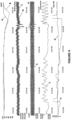
- Figures 4 and 5 show the PPG and thermistor signal streams for a patient in the operating room who is having periods of obstructive apnea typical of obstructive sleep apnea.
- the combination of the PPG data with the thermistor gives a reliable picture of respiratory status of the patient.
- the PPG provides oxygen saturation over time (“SpO2"), as well as IR DC and Raw and AC waveforms.
- the thermistor indicates the air flow from the nostrils over time.
- the thermistor provides the most reliable data during periods of a patent airway and adequate tidal volume, and the PPG data is more reliable when the patient is partially or completely obstructed (although the signal may work well most of the time and is not affected by obstruction or preferential nasal flow as may be the case with the thermistor) and amplitude is a indicates "effort".
- waveform "A” denotes the oxygen saturation obtained at the nasal alar
- the “B” waveform denotes oxygen saturation obtained at the finger. It is noted that the desaturation is detected at the nasal alar several seconds before it is detected at the finger.
- the “C” waveform is the raw PPG signal
- the “D” waveform is the IR DC component signal.
- the “E” waveform is the AC component signal stream.
- the “F” waveform is the thermistor waveform. It is noted that when the thermistor loses signal, the IR DC signal becomes more pronounced, the individual increases respiratory efforts against an apparent obstruction.
Landscapes
- Health & Medical Sciences (AREA)
- Life Sciences & Earth Sciences (AREA)
- Physiology (AREA)
- Engineering & Computer Science (AREA)
- Public Health (AREA)
- Surgery (AREA)
- Pulmonology (AREA)
- Physics & Mathematics (AREA)
- Veterinary Medicine (AREA)
- Biophysics (AREA)
- Pathology (AREA)
- Biomedical Technology (AREA)
- Heart & Thoracic Surgery (AREA)
- Medical Informatics (AREA)
- Molecular Biology (AREA)
- General Health & Medical Sciences (AREA)
- Animal Behavior & Ethology (AREA)
- Cardiology (AREA)
- Psychiatry (AREA)
- Artificial Intelligence (AREA)
- Computer Vision & Pattern Recognition (AREA)
- Signal Processing (AREA)
- Emergency Medicine (AREA)
- Obesity (AREA)
- Measurement Of The Respiration, Hearing Ability, Form, And Blood Characteristics Of Living Organisms (AREA)
Claims (13)
- Système pour surveiller l'efficacité de la respiration chez un individu, le système comprenant un microprocesseur,dans lequel le microprocesseur est conçu pour recevoir des signaux de photopléthysmographie (PPG) obtenus à partir d'un capteur PPG fixé à l'individu et des signaux provenant d'un capteur respiratoire secondaire,dans lequel le microprocesseur est configuré pour :déterminer si une tentative respiratoire chez l'individu s'est produite au cours d'une période de temps prédéfinie sur la base des signaux PPG ;déterminer si une ventilation s'est produite dans la période de temps prédéfinie à l'aide des signaux obtenus à partir du capteur de respiration secondaire ; etcomparer la détermination des tentatives respiratoires basée sur les signaux PPG avec la détermination de la ventilation basée sur le capteur de respiration secondaire pour évaluer si les tentatives respiratoires de l'individu sont efficaces,dans lequel le système est caractérisé en ce que le microprocesseur est en outre configuré pour calculer une première fréquence respiratoire sur la base des tentatives respiratoires déterminées à partir des signaux PPG, calculer une seconde fréquence respiratoire sur la base des respirations déterminées à partir du capteur de respiration secondaire, et comparer les première et seconde fréquences respiratoires pour déterminer si l'individu souffre d'hyperpnée, d'apnée ou d'hypopnée.
- Système selon la revendication 1, dans lequel le système comprend un capteur de respiration secondaire comprenant au moins l'un parmi une thermistance, un capnomètre, un capteur de débit nasal, un transducteur de pression différentielle et un capteur de pression nasale.
- Système selon la revendication 1, dans lequel s'il est déterminé qu'une tentative respiratoire s'est produite sur la base des signaux PPG et s'il est déterminé qu'une ventilation s'est produite sur la base des signaux du capteur de respiration secondaire, alors la tentative respiratoire efficace est utilisée pour calculer la fréquence respiratoire de l'individu.
- Système selon la revendication 1, dans lequel s'il est déterminé qu'une tentative respiratoire s'est produite mais s'il est déterminé qu'une ventilation ne s'est pas produite, alors une action prédéterminée est effectuée par le microprocesseur.
- Système selon la revendication 4, dans lequel la réaction prédéterminée comprend le déclenchement d'un signal d'alarme.
- Système selon la revendication 4, dans lequel le microprocesseur est configuré pour calculer la saturation en oxygène du sang de l'individu et pour déterminer si la saturation en oxygène du sang de l'individu a diminué jusqu'à un niveau prédéfini, et si tel est le cas, pour effectuer une réaction prédéterminée.
- Système selon la revendication 1, dans lequel s'il est déterminé que l'individu souffre d'hyperpnée, d'apnée ou d'hypopnée, une réaction prédéterminée est effectuée par le microprocesseur.
- Système selon la revendication 7, dans lequel la réaction prédéterminée comprend le déclenchement d'un signal d'alarme.
- Système selon l'une quelconque des revendications 1-8, dans lequel le microprocesseur est configuré pour déterminer au moins l'un parmi (a) si une ventilation s'est produite, (b) si le patient souffre d'apnée centrale et (c) si le patient souffre d'apnée obstructive.
- Système selon la revendication 9, dans lequel le microprocesseur est en outre configuré pour isoler un signal de composante alternative et continue des signaux PPG, utiliser le signal de composante alternative et/ou continue isolé pour déterminer si une tentative respiratoire chez l'individu s'est produite.
- Système selon la revendication 1, dans lequel une réaction prédéterminée est effectuée par le microprocesseur s'il est déterminé que la tentative respiratoire n'a pas été efficace.
- Système selon la revendication 11, dans lequel la réaction prédéterminée comprend le déclenchement d'un signal d'alarme.
- Système selon la revendication 11, dans lequel le microprocesseur est configuré pour calculer la saturation en oxygène du sang de l'individu et pour déterminer si la saturation en oxygène du sang de l'individu a diminué d'un pourcentage prédéfini, et la réaction prédéterminée est effectuée s'il est déterminé que la tentative respiratoire n'a pas été efficace et si la saturation en oxygène du sang a diminué d'un pourcentage prédéfini.
Applications Claiming Priority (3)
| Application Number | Priority Date | Filing Date | Title |
|---|---|---|---|
| US201461926434P | 2014-01-13 | 2014-01-13 | |
| US14/512,425 US10368807B2 (en) | 2013-10-11 | 2014-10-11 | Fusion of data from multiple sources for non-invasive detection of respiratory parameters |
| PCT/US2015/011235 WO2015106280A1 (fr) | 2014-01-13 | 2015-01-13 | Procédés et systèmes permettant de déterminer une respiration efficace chez des individus |
Publications (3)
| Publication Number | Publication Date |
|---|---|
| EP3094238A1 EP3094238A1 (fr) | 2016-11-23 |
| EP3094238A4 EP3094238A4 (fr) | 2017-10-11 |
| EP3094238B1 true EP3094238B1 (fr) | 2024-10-02 |
Family
ID=53524424
Family Applications (1)
| Application Number | Title | Priority Date | Filing Date |
|---|---|---|---|
| EP15735041.4A Active EP3094238B1 (fr) | 2014-01-13 | 2015-01-13 | Systèmes permettant de déterminer une respiration efficace chez des individus |
Country Status (3)
| Country | Link |
|---|---|
| EP (1) | EP3094238B1 (fr) |
| CA (1) | CA2936566A1 (fr) |
| WO (1) | WO2015106280A1 (fr) |
Families Citing this family (1)
| Publication number | Priority date | Publication date | Assignee | Title |
|---|---|---|---|---|
| AU2016304594A1 (en) * | 2015-08-06 | 2018-02-22 | Xhale Assurance, Inc. | Methods and devices for monitoring respiration using photoplethysmography sensors |
Family Cites Families (8)
| Publication number | Priority date | Publication date | Assignee | Title |
|---|---|---|---|---|
| US7206636B1 (en) * | 1999-11-10 | 2007-04-17 | Pacesetter, Inc. | Pacing optimization based on changes in pulse amplitude and pulse amplitude variability |
| US6909912B2 (en) * | 2002-06-20 | 2005-06-21 | University Of Florida | Non-invasive perfusion monitor and system, specially configured oximeter probes, methods of using same, and covers for probes |
| WO2008058328A1 (fr) | 2006-11-13 | 2008-05-22 | Resmed Ltd | Systèmes, procédés et/ou appareils pour la surveillance non invasive de paramètres respiratoires lors de troubles respiratoires du sommeil |
| US8794235B2 (en) | 2007-06-08 | 2014-08-05 | Ric Investments, Llc | System and method for treating ventilatory instability |
| WO2011073839A2 (fr) * | 2009-12-15 | 2011-06-23 | Koninklijke Philips Electronics N.V. | Système et procédé d'assistance à des volumes respiratoires sous-physiologiques et physiologiques dans une respiration spontanée ou non spontanée durant une ventilation à haute fréquence |
| US8801619B2 (en) * | 2011-06-30 | 2014-08-12 | Covidien Lp | Photoplethysmography for determining ventilation weaning readiness |
| WO2014003806A1 (fr) * | 2012-06-29 | 2014-01-03 | Xhale, Inc. | Capteurs de photopléthysmographie |
| US8740806B2 (en) * | 2012-11-07 | 2014-06-03 | Somnarus Inc. | Methods for detection of respiratory effort and sleep apnea monitoring devices |
-
2015
- 2015-01-13 EP EP15735041.4A patent/EP3094238B1/fr active Active
- 2015-01-13 WO PCT/US2015/011235 patent/WO2015106280A1/fr not_active Ceased
- 2015-01-13 CA CA2936566A patent/CA2936566A1/fr not_active Abandoned
Also Published As
| Publication number | Publication date |
|---|---|
| EP3094238A1 (fr) | 2016-11-23 |
| WO2015106280A1 (fr) | 2015-07-16 |
| EP3094238A4 (fr) | 2017-10-11 |
| CA2936566A1 (fr) | 2015-07-16 |
Similar Documents
| Publication | Publication Date | Title |
|---|---|---|
| US10368807B2 (en) | Fusion of data from multiple sources for non-invasive detection of respiratory parameters | |
| RU2338457C2 (ru) | Способ и аппарат для поддержания и текущего контроля качества сна во время терапии | |
| US8069852B2 (en) | Method and apparatus for maintaining and monitoring sleep quality during therapeutic treatments | |
| US8221319B2 (en) | Medical device for assessing intravascular blood volume and technique for using the same | |
| JP6794425B2 (ja) | フォトプレチスモグラフィセンサを用いた呼吸を監視するための方法および装置 | |
| US20140158124A1 (en) | Method and system for the delivery of carbon dioxide to a patient | |
| US20160045154A1 (en) | Sleep apnea | |
| EP3148426B1 (fr) | Procédé, système et logiciel pour l'évaluation d'un échec d'extubation | |
| ZA200502982B (en) | Method and apparatus for maintaining and monitoring sleep quality during therapeutic treatments. | |
| US10729334B2 (en) | Methods and systems for determining effectiveness of respiration in individuals | |
| EP2988660B1 (fr) | Dispositifs de surveillance de la respiration par photopléthysmographie | |
| EP3094238A1 (fr) | Procédés et systèmes permettant de déterminer une respiration efficace chez des individus | |
| Fiala et al. | Tailoring the sleep laboratory for chronic respiratory failure | |
| Ermer et al. | Identification of Respiratory Distress Through Combining Modern Sensors in Patients Receiving Opioids and Anesthetics | |
| Ermer et al. | Detecting Adverse Respiratory Effects of Anesthetics and Opiod Analgesics in the Postoperative Period |
Legal Events
| Date | Code | Title | Description |
|---|---|---|---|
| PUAI | Public reference made under article 153(3) epc to a published international application that has entered the european phase |
Free format text: ORIGINAL CODE: 0009012 |
|
| 17P | Request for examination filed |
Effective date: 20160812 |
|
| AK | Designated contracting states |
Kind code of ref document: A1 Designated state(s): AL AT BE BG CH CY CZ DE DK EE ES FI FR GB GR HR HU IE IS IT LI LT LU LV MC MK MT NL NO PL PT RO RS SE SI SK SM TR |
|
| AX | Request for extension of the european patent |
Extension state: BA ME |
|
| RIN1 | Information on inventor provided before grant (corrected) |
Inventor name: MELKER, RICHARD Inventor name: COHEN, SEAN Inventor name: TAN, HUWEI |
|
| DAX | Request for extension of the european patent (deleted) | ||
| A4 | Supplementary search report drawn up and despatched |
Effective date: 20170912 |
|
| RIC1 | Information provided on ipc code assigned before grant |
Ipc: A61B 5/087 20060101ALN20170906BHEP Ipc: A61M 16/00 20060101AFI20170906BHEP Ipc: A61B 5/08 20060101ALI20170906BHEP |
|
| RAP1 | Party data changed (applicant data changed or rights of an application transferred) |
Owner name: XHALE ASSURANCE, INC. |
|
| STAA | Information on the status of an ep patent application or granted ep patent |
Free format text: STATUS: EXAMINATION IS IN PROGRESS |
|
| 17Q | First examination report despatched |
Effective date: 20211115 |
|
| REG | Reference to a national code |
Ref country code: DE Ref legal event code: R079 Free format text: PREVIOUS MAIN CLASS: A61B0005000000 Ipc: A61M0016000000 Ref country code: DE Ref legal event code: R079 Ref document number: 602015090030 Country of ref document: DE Free format text: PREVIOUS MAIN CLASS: A61B0005000000 Ipc: A61M0016000000 |
|
| GRAP | Despatch of communication of intention to grant a patent |
Free format text: ORIGINAL CODE: EPIDOSNIGR1 |
|
| STAA | Information on the status of an ep patent application or granted ep patent |
Free format text: STATUS: GRANT OF PATENT IS INTENDED |
|
| RIC1 | Information provided on ipc code assigned before grant |
Ipc: A61B 5/087 20060101ALN20240411BHEP Ipc: A61B 5/08 20060101ALI20240411BHEP Ipc: A61M 16/00 20060101AFI20240411BHEP |
|
| RIC1 | Information provided on ipc code assigned before grant |
Ipc: A61B 5/087 20060101ALN20240423BHEP Ipc: A61B 5/08 20060101ALI20240423BHEP Ipc: A61M 16/00 20060101AFI20240423BHEP |
|
| INTG | Intention to grant announced |
Effective date: 20240507 |
|
| GRAS | Grant fee paid |
Free format text: ORIGINAL CODE: EPIDOSNIGR3 |
|
| GRAA | (expected) grant |
Free format text: ORIGINAL CODE: 0009210 |
|
| STAA | Information on the status of an ep patent application or granted ep patent |
Free format text: STATUS: THE PATENT HAS BEEN GRANTED |
|
| AK | Designated contracting states |
Kind code of ref document: B1 Designated state(s): AL AT BE BG CH CY CZ DE DK EE ES FI FR GB GR HR HU IE IS IT LI LT LU LV MC MK MT NL NO PL PT RO RS SE SI SK SM TR |
|
| REG | Reference to a national code |
Ref country code: GB Ref legal event code: FG4D |
|
| REG | Reference to a national code |
Ref country code: CH Ref legal event code: EP |
|
| REG | Reference to a national code |
Ref country code: DE Ref legal event code: R096 Ref document number: 602015090030 Country of ref document: DE |
|
| REG | Reference to a national code |
Ref country code: IE Ref legal event code: FG4D |
|
| REG | Reference to a national code |
Ref country code: DE Ref legal event code: R084 Ref document number: 602015090030 Country of ref document: DE |
|
| REG | Reference to a national code |
Ref country code: LT Ref legal event code: MG9D |
|
| REG | Reference to a national code |
Ref country code: NL Ref legal event code: MP Effective date: 20241002 |
|
| REG | Reference to a national code |
Ref country code: AT Ref legal event code: MK05 Ref document number: 1728016 Country of ref document: AT Kind code of ref document: T Effective date: 20241002 |
|
| PG25 | Lapsed in a contracting state [announced via postgrant information from national office to epo] |
Ref country code: NL Free format text: LAPSE BECAUSE OF FAILURE TO SUBMIT A TRANSLATION OF THE DESCRIPTION OR TO PAY THE FEE WITHIN THE PRESCRIBED TIME-LIMIT Effective date: 20241002 |
|
| PG25 | Lapsed in a contracting state [announced via postgrant information from national office to epo] |
Ref country code: NL Free format text: LAPSE BECAUSE OF FAILURE TO SUBMIT A TRANSLATION OF THE DESCRIPTION OR TO PAY THE FEE WITHIN THE PRESCRIBED TIME-LIMIT Effective date: 20241002 |
|
| PG25 | Lapsed in a contracting state [announced via postgrant information from national office to epo] |
Ref country code: HR Free format text: LAPSE BECAUSE OF FAILURE TO SUBMIT A TRANSLATION OF THE DESCRIPTION OR TO PAY THE FEE WITHIN THE PRESCRIBED TIME-LIMIT Effective date: 20241002 Ref country code: IS Free format text: LAPSE BECAUSE OF FAILURE TO SUBMIT A TRANSLATION OF THE DESCRIPTION OR TO PAY THE FEE WITHIN THE PRESCRIBED TIME-LIMIT Effective date: 20250202 Ref country code: PT Free format text: LAPSE BECAUSE OF FAILURE TO SUBMIT A TRANSLATION OF THE DESCRIPTION OR TO PAY THE FEE WITHIN THE PRESCRIBED TIME-LIMIT Effective date: 20250203 |
|
| PG25 | Lapsed in a contracting state [announced via postgrant information from national office to epo] |
Ref country code: FI Free format text: LAPSE BECAUSE OF FAILURE TO SUBMIT A TRANSLATION OF THE DESCRIPTION OR TO PAY THE FEE WITHIN THE PRESCRIBED TIME-LIMIT Effective date: 20241002 |
|
| PG25 | Lapsed in a contracting state [announced via postgrant information from national office to epo] |
Ref country code: BG Free format text: LAPSE BECAUSE OF FAILURE TO SUBMIT A TRANSLATION OF THE DESCRIPTION OR TO PAY THE FEE WITHIN THE PRESCRIBED TIME-LIMIT Effective date: 20241002 |
|
| PG25 | Lapsed in a contracting state [announced via postgrant information from national office to epo] |
Ref country code: ES Free format text: LAPSE BECAUSE OF FAILURE TO SUBMIT A TRANSLATION OF THE DESCRIPTION OR TO PAY THE FEE WITHIN THE PRESCRIBED TIME-LIMIT Effective date: 20241002 |
|
| PG25 | Lapsed in a contracting state [announced via postgrant information from national office to epo] |
Ref country code: NO Free format text: LAPSE BECAUSE OF FAILURE TO SUBMIT A TRANSLATION OF THE DESCRIPTION OR TO PAY THE FEE WITHIN THE PRESCRIBED TIME-LIMIT Effective date: 20250102 |
|
| PG25 | Lapsed in a contracting state [announced via postgrant information from national office to epo] |
Ref country code: GR Free format text: LAPSE BECAUSE OF FAILURE TO SUBMIT A TRANSLATION OF THE DESCRIPTION OR TO PAY THE FEE WITHIN THE PRESCRIBED TIME-LIMIT Effective date: 20250103 Ref country code: LV Free format text: LAPSE BECAUSE OF FAILURE TO SUBMIT A TRANSLATION OF THE DESCRIPTION OR TO PAY THE FEE WITHIN THE PRESCRIBED TIME-LIMIT Effective date: 20241002 Ref country code: AT Free format text: LAPSE BECAUSE OF FAILURE TO SUBMIT A TRANSLATION OF THE DESCRIPTION OR TO PAY THE FEE WITHIN THE PRESCRIBED TIME-LIMIT Effective date: 20241002 |
|
| PG25 | Lapsed in a contracting state [announced via postgrant information from national office to epo] |
Ref country code: PL Free format text: LAPSE BECAUSE OF FAILURE TO SUBMIT A TRANSLATION OF THE DESCRIPTION OR TO PAY THE FEE WITHIN THE PRESCRIBED TIME-LIMIT Effective date: 20241002 Ref country code: CZ Free format text: LAPSE BECAUSE OF FAILURE TO SUBMIT A TRANSLATION OF THE DESCRIPTION OR TO PAY THE FEE WITHIN THE PRESCRIBED TIME-LIMIT Effective date: 20241002 |
|
| PG25 | Lapsed in a contracting state [announced via postgrant information from national office to epo] |
Ref country code: RS Free format text: LAPSE BECAUSE OF FAILURE TO SUBMIT A TRANSLATION OF THE DESCRIPTION OR TO PAY THE FEE WITHIN THE PRESCRIBED TIME-LIMIT Effective date: 20250102 |
|
| PG25 | Lapsed in a contracting state [announced via postgrant information from national office to epo] |
Ref country code: SM Free format text: LAPSE BECAUSE OF FAILURE TO SUBMIT A TRANSLATION OF THE DESCRIPTION OR TO PAY THE FEE WITHIN THE PRESCRIBED TIME-LIMIT Effective date: 20241002 |
|
| REG | Reference to a national code |
Ref country code: DE Ref legal event code: R097 Ref document number: 602015090030 Country of ref document: DE |
|
| PG25 | Lapsed in a contracting state [announced via postgrant information from national office to epo] |
Ref country code: DK Free format text: LAPSE BECAUSE OF FAILURE TO SUBMIT A TRANSLATION OF THE DESCRIPTION OR TO PAY THE FEE WITHIN THE PRESCRIBED TIME-LIMIT Effective date: 20241002 |
|
| PG25 | Lapsed in a contracting state [announced via postgrant information from national office to epo] |
Ref country code: EE Free format text: LAPSE BECAUSE OF FAILURE TO SUBMIT A TRANSLATION OF THE DESCRIPTION OR TO PAY THE FEE WITHIN THE PRESCRIBED TIME-LIMIT Effective date: 20241002 |
|
| PG25 | Lapsed in a contracting state [announced via postgrant information from national office to epo] |
Ref country code: RO Free format text: LAPSE BECAUSE OF FAILURE TO SUBMIT A TRANSLATION OF THE DESCRIPTION OR TO PAY THE FEE WITHIN THE PRESCRIBED TIME-LIMIT Effective date: 20241002 |
|
| PG25 | Lapsed in a contracting state [announced via postgrant information from national office to epo] |
Ref country code: SK Free format text: LAPSE BECAUSE OF FAILURE TO SUBMIT A TRANSLATION OF THE DESCRIPTION OR TO PAY THE FEE WITHIN THE PRESCRIBED TIME-LIMIT Effective date: 20241002 |
|
| PG25 | Lapsed in a contracting state [announced via postgrant information from national office to epo] |
Ref country code: IT Free format text: LAPSE BECAUSE OF FAILURE TO SUBMIT A TRANSLATION OF THE DESCRIPTION OR TO PAY THE FEE WITHIN THE PRESCRIBED TIME-LIMIT Effective date: 20241002 |
|
| PLBE | No opposition filed within time limit |
Free format text: ORIGINAL CODE: 0009261 |
|
| STAA | Information on the status of an ep patent application or granted ep patent |
Free format text: STATUS: NO OPPOSITION FILED WITHIN TIME LIMIT |
|
| REG | Reference to a national code |
Ref country code: CH Ref legal event code: PL |
|
| PG25 | Lapsed in a contracting state [announced via postgrant information from national office to epo] |
Ref country code: SE Free format text: LAPSE BECAUSE OF FAILURE TO SUBMIT A TRANSLATION OF THE DESCRIPTION OR TO PAY THE FEE WITHIN THE PRESCRIBED TIME-LIMIT Effective date: 20241002 |
|
| 26N | No opposition filed |
Effective date: 20250703 |
|
| PG25 | Lapsed in a contracting state [announced via postgrant information from national office to epo] |
Ref country code: LU Free format text: LAPSE BECAUSE OF NON-PAYMENT OF DUE FEES Effective date: 20250113 Ref country code: MC Free format text: LAPSE BECAUSE OF FAILURE TO SUBMIT A TRANSLATION OF THE DESCRIPTION OR TO PAY THE FEE WITHIN THE PRESCRIBED TIME-LIMIT Effective date: 20241002 |
|
| GBPC | Gb: european patent ceased through non-payment of renewal fee |
Effective date: 20250113 |
|
| PG25 | Lapsed in a contracting state [announced via postgrant information from national office to epo] |
Ref country code: BE Free format text: LAPSE BECAUSE OF NON-PAYMENT OF DUE FEES Effective date: 20250131 Ref country code: GB Free format text: LAPSE BECAUSE OF NON-PAYMENT OF DUE FEES Effective date: 20250113 |
|
| PG25 | Lapsed in a contracting state [announced via postgrant information from national office to epo] |
Ref country code: FR Free format text: LAPSE BECAUSE OF NON-PAYMENT OF DUE FEES Effective date: 20250131 |
|
| PG25 | Lapsed in a contracting state [announced via postgrant information from national office to epo] |
Ref country code: CH Free format text: LAPSE BECAUSE OF NON-PAYMENT OF DUE FEES Effective date: 20250131 |
|
| REG | Reference to a national code |
Ref country code: BE Ref legal event code: MM Effective date: 20250131 |
|
| PG25 | Lapsed in a contracting state [announced via postgrant information from national office to epo] |
Ref country code: IE Free format text: LAPSE BECAUSE OF NON-PAYMENT OF DUE FEES Effective date: 20250113 |
|
| PGFP | Annual fee paid to national office [announced via postgrant information from national office to epo] |
Ref country code: DE Payment date: 20260127 Year of fee payment: 12 |