EP3095376A1 - Endoskopisches bilddiagnosehilfssystem - Google Patents
Endoskopisches bilddiagnosehilfssystem Download PDFInfo
- Publication number
- EP3095376A1 EP3095376A1 EP15746291.2A EP15746291A EP3095376A1 EP 3095376 A1 EP3095376 A1 EP 3095376A1 EP 15746291 A EP15746291 A EP 15746291A EP 3095376 A1 EP3095376 A1 EP 3095376A1
- Authority
- EP
- European Patent Office
- Prior art keywords
- identification
- target region
- endoscopic image
- identification target
- pathological types
- Prior art date
- Legal status (The legal status is an assumption and is not a legal conclusion. Google has not performed a legal analysis and makes no representation as to the accuracy of the status listed.)
- Granted
Links
Images




Classifications
-
- G—PHYSICS
- G06—COMPUTING OR CALCULATING; COUNTING
- G06T—IMAGE DATA PROCESSING OR GENERATION, IN GENERAL
- G06T7/00—Image analysis
- G06T7/0002—Inspection of images, e.g. flaw detection
- G06T7/0012—Biomedical image inspection
-
- G—PHYSICS
- G02—OPTICS
- G02B—OPTICAL ELEMENTS, SYSTEMS OR APPARATUS
- G02B23/00—Telescopes, e.g. binoculars; Periscopes; Instruments for viewing the inside of hollow bodies; Viewfinders; Optical aiming or sighting devices
- G02B23/24—Instruments or systems for viewing the inside of hollow bodies, e.g. fibrescopes
- G02B23/2476—Non-optical details, e.g. housings, mountings, supports
- G02B23/2484—Arrangements in relation to a camera or imaging device
-
- G—PHYSICS
- G06—COMPUTING OR CALCULATING; COUNTING
- G06T—IMAGE DATA PROCESSING OR GENERATION, IN GENERAL
- G06T2207/00—Indexing scheme for image analysis or image enhancement
- G06T2207/10—Image acquisition modality
- G06T2207/10068—Endoscopic image
Definitions
- the present disclosure relates to an endoscopic image diagnosis support system that identifies a pathological type in an identification target region in an endoscopic image, and more particularly to a technology for improving the identification precision.
- NBI images spectral images
- image recognition system i.e., endoscopic image diagnosis support system
- This system uses a technique called bag-of-features (BoF) or bag-of-keypoints, transforms a local feature value extracted from an image of a recognition target region (hereinafter also referred to as a scan window (SW)) in an endoscopic image to a histogram of visual words (VW), and performs image matching (i.e., feature value matching) with learning images pre-classified into pathological types (i.e., each of learning images is represented as a feature value of a histogram of visual words), thereby computing the identification probabilities of the pathological types in the identification target region.
- a technique called bag-of-features (BoF) or bag-of-keypoints transforms a local feature value extracted from an image of a recognition target region (hereinafter also referred to as a scan window (SW)) in an endoscopic image to a histogram of visual words (VW), and performs image matching (i.e., feature value matching) with learning images pre-classified into pathological types (
- the inventors have succeeded in hardware implementation of the feature value extraction processing from an image of an identification target region and the pathologic determination (i.e., identification) processing, which are otherwise especially high in computation cost in the system, thereby permitting processing of a full HD (i.e., 1920 ⁇ 1080 pixels) or higher quality endoscopic image in real time with high identification precision (see Non-Patent Documents 1 and 2, for example).
- the entire full HD endoscopic image has been scanned with scan windows (SW) (e.g., regions having a size of 120 ⁇ 120 pixels) to determine pathological types in the scan windows.
- SW scan windows
- the actual affected area projected in the endoscopic image is not rectangular in many cases.
- the identification probabilities of these pathological types may compete with each other, making it difficult to determine the pathological type in the scan window.
- the size of the scan windows may be reduced. In this case, however, since the feature value extracted from the image of one scan window will decrease, the identification precision may possibly be degraded.
- the number of times of scanning of the entire full HD (i.e., 1920 ⁇ 1080 pixels) endoscopic image will increase, reducing the processing speed and increasing the time required until the processed results are displayed (latency), resulting in difficulty in real-time display.
- an objective of the present disclosure is improving the identification precision in an endoscopic image diagnosis support system that identifies the pathological type in an identification target region in an endoscopic image.
- the endoscopic image diagnosis support system includes: a memory that stores learning images pre-classified into pathological types; and a processor that, given an endoscopic image, performs feature value matching between an image of an identification target region in the endoscopic image and the learning images, to identify the pathological types in the identification target region, wherein the processor performs feature value matching between the image of the identification target region and the learning images to compute identification probabilities of the pathological types in the identification target region, and, when a maximum value of the identification probabilities is smaller than a threshold, subdivides the identification target region, to perform feature value matching between images of subdivided regions and the learning images to compute identification probabilities of the pathological types in the subdivided regions, and computes average values of the identification probabilities of the pathological types in the identification target region and the subdivided regions, the average values corresponding to identification probabilities of the pathological types in hierarchical overlap regions of the identification target region and the subdivided regions.
- the endoscopic image diagnosis support system includes: a memory that stores learning images pre-classified into pathological types; and a processor that, given an endoscopic image, performs feature value matching between an image of an identification target region in the endoscopic image and the learning images, to identify the pathological types in the identification target region, wherein the processor performs feature value matching between images of the identification target region and subdivided regions of the identification target region and the learning images to compute identification probabilities of the pathological types in the identification target region and the subdivided regions, and computes average values of the identification probabilities of the pathological types in the identification target region and the subdivided regions, the average values corresponding to identification probabilities of the pathological types in hierarchical overlap regions of the identification target region and the subdivided regions.
- the identification probabilities of the pathological types are computed in hierarchical overlap regions having various sizes in an endoscopic image, and are averaged, permitting identification of a pathological type of an affected area having a complicated shape.
- the processor may compute the average values by performing weighting for the identification probabilities of the pathological types in the identification target region and the subdivided regions according to region size-specific identification precision. According to this, the identification precision can be further improved.
- the feature values obtained from the images of the identification target region and the subdivided regions may be represented by histograms of visual words based on a bag-of-features technique, and the processor may compute a histogram representing a feature value obtained from an image of a high-level region by addition of a histogram representing a feature value obtained from an image of a low-level region hierarchically overlapping the high-level region. According to this, the processing of producing a histogram representing the feature value obtained from an image of a high-level region becomes easy, and thus, with reduction in computation amount, the processing speed can be improved.
- the processor may perform one-versus-multi two-class identification for the pathological types, and compute the identification probabilities of the pathological types in the identification target region and the subdivided regions based on the identification results of the two-class identification. According to this, the identification precision can be improved.
- the identification precision can be improved in an endoscopic image diagnosis support system that identifies the pathological type in an identification target region in an endoscopic image.
- FIG. 1 shows a schematic configuration of an endoscopic image diagnosis support system according to an embodiment of the disclosure (hereinafter simply referred to as "the system").
- the system 100 supports endoscopic diagnosis by a physician, and also assists the education of less-experienced physicians, by identifying the pathological type of an affected area projected in an NBI magnified colorectal endoscopic image on the basis of objective indicators.
- the system 100 includes a memory 10, a processor 20, and a display 30.
- the memory 10 is constituted by a nonvolatile memory such as a flash memory, a hard disk drive, etc.
- the processor 20 is constituted by various types of hardware such as a central processing unit (CPU), a field programmable gate array (FPGA), and a graphics processing unit (GPU).
- the display 30 is constituted by a cathode ray tube (CRT), a liquid crystal display (LCD), etc.
- the processor 20 receives an endoscopic image and determines the pathological type in an identification target region (i.e., scan window SW) in the endoscopic image.
- the determination results are represented as the probability (i.e., identification probability) for each pathological type, for example, and shown on the display 30 in real time together with the endoscopic image, or on another screen.
- the endoscopic image input into the processor 20 may be a real-time video image from an endoscopic camera during the examination or a visual image shot and stored in a past examination.
- real-time determination of the pathological type of the affected area can be performed during the examination.
- education and training of the endoscopic examination can be performed with the help of an enormous amount of past archive visual images.
- the processor 20 includes a feature value extraction processor 21, an identification feature vector transform processor 22, an identification determinator 23, and an identification probability computer 24. Out of these modules, the feature value extraction processor 21 and the identification determinator 23 have been successfully implemented as hardware (see Non-Patent Documents 1 and 2). The remaining identification feature vector transform processor 22 and identification probability computer 24 can also be implemented as hardware, or may be processed by software by a CPU, etc. not shown in the processor 20.
- the feature value extraction processor 21 extracts a local feature value from an image of a scan window in an endoscopic image.
- a local feature descriptor dense scale-invariant feature transform (D-SIFT), for example, can be used.
- feature points i.e., keypoints
- gradient information of brightness values in a local region around each feature point is transformed to a parameter (i.e., feature value) of 128 dimensions.
- the feature value extraction processor 21 performs smoothing with a Gaussian filter and determines brightness gradients in the x and y directions for each pixel.
- the feature value extraction processor 21 then classifies the gradient directions of the pixels into eight directions using the determined brightness gradients, and computes the gradient strengths.
- Non-Patent Document 1 by omitting the weighting for the blocks according to the distance from the feature point, it is possible to share the values of all blocks in overlapping feature value description regions. This can largely reduce the required memory amount. Further, as disclosed in the aforementioned document, by achieving computation of the brightness gradient directions of the pixels without use of arctan function, by reducing the brightness gradient directions to four directions, and by changing the normalization processing to threshold processing, software and hardware implementation of the D-SIFT algorithm will be facilitated. This will largely increase the speed of the feature value extraction processing.
- the identification feature vector transform processor 22 transforms the feature vectors extracted by the feature value extraction processor 21 to a histogram of visual words (VW) based on the bag-of-features (BoF) technique.
- the visual words are obtained by performing clustering of feature vectors extracted from learning images. More specifically, feature vectors are extracted from learning images pre-specified as Type A, and performs clustering of the feature vectors into a plurality of (e.g., 256) clusters by a k-means method, etc. The centers of the clusters are visual words. In other words, a plurality of (e.g., 256) visual words featuring Type A are obtained from the learning images pre-classified as Type A. Similarly, a plurality of (e.g., 256) visual words featuring Type B are obtained from learning images pre-classified as Type B, and a plurality of (e.g., 256) visual words featuring Type C3 are obtained from learning images pre-classified as Type C3.
- feature vectors are extracted from learning images pre-specified as Type A, and performs clustering of the feature vectors into a plurality of (e.g., 256) clusters by a k-means method, etc.
- Another method may be used to produce visual words using feature vectors previously extracted from the learning images. Also, the number of dimensions of the visual words can be changed.
- the memory 10 stores learning images pre-classified into pathological types.
- the learning images stored in the memory 10 are also represented by a histogram of 768 visual words, i.e., feature vectors in the 768 dimensional identification space.
- the identification determinator 23 identifies which pathological type, Type A, Type B, or Type C3, the histogram of visual words produced by the identification feature vector transform processor 22 belongs to, using the learning images stored in the memory 10 as teacher data.
- a support vector machine (SVM) for example, can be used as an identifier.
- Non-Patent Document 2 there are one-versus-one approach and two-stage determination as multi-class identification based on the SVM that is a two-class identifier.
- an identifier "A vs B" that identifies Type A from Type B an identifier "B vs C3" that identifies Type B from Type C3
- an identifier "C3 vs A” that identifies Type C3 from Type A are placed in parallel, whereby the final identification result can be obtained based on the identification results of the identifiers.
- FIG. 2 is a conceptual view of the cascade identification.
- an identifier "A vs ⁇ A” that identifies Type A from the other
- an identifier "B vs ⁇ B” that identifies Type B from the other
- an identifier "C3 vs ⁇ C3" that identifies Type C3 from the other are cascaded, whereby the final identification result can be obtained based on the identification results of the identifiers.
- An identifier "A vs B" that identifies Type A from Type B may further be connected at the stage following the identifier "C3 vs ⁇ C3.” This is for giving unknown data x that should be classified as Type A a change of being classified as Type A again when this data has been classified as ⁇ A by the first-stage identifier "A vs ⁇ A.” This is effective when the precision of the identifier "A vs ⁇ A" is not 100%. With this connection of the identifier "A vs B" at the final stage, the identification precision of the identification determinator 23 can be improved.
- the endoscopic image input into the processor 20 may have blown out highlights of the image due to lighting, disturbance of the image associated with cleaning with water, pulsation caused by blood vessels on the large intestine wall, blurring of the image during the operation of the endoscope, etc. Since the pathological type of such a blurring image may not be correctly identified with high probability, a two-class identifier that distinguishes a blurring image from the other may be connected at the initial stage of the above-described cascade identification.
- FIG. 3 shows an architecture example of hardware implementation of the cascade identification.
- the identifiers are placed in parallel, whereby the final identification result can be obtained based on the identification results of the identifiers.
- the identification probability computer 24 receives the identification results of the identifiers in the identification determinator 23, and computes the identification probability of each pathological type by a steepest descent method, for example.
- the processor 20 receives an endoscopic image and performs image matching (i.e., feature value matching) between images of scan windows in the endoscopic image and the learning images, to identify the pathological types in the scan windows.
- image matching i.e., feature value matching
- the processor 20 hierarchically subdivides the scan windows, to permit identification of the pathological type of an affected area having a complicated shape, not a rectangular shape.
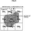
- FIG. 4 is a view for explaining the hierarchical subdivision of scan windows.
- scan windows SW 1 to SW 9 in an endoscopic image are subjected to processing to compute the identification probabilities of the pathological types.
- the identification probability of Type A is very high in these scan windows.
- Type B occupies roughly the entire region in the scan window SW 5 , the identification probability of Type B is very high in this scan window.
- the determination whether the pathological type is Type A or Type B is difficult.
- Such scan windows are hierarchically subdivided to produce smaller-sized scan windows at a lower hierarchical level (e.g., Level 2) and further smaller-sized scan windows at a further lower hierarchical level (e.g., Level 3), to identify the pathological types in such small-sized scan windows (hereinafter referred to as hierarchical identification).
- Level 2 refers to the hierarchical level of the scan windows SW.
- FIG. 5 shows an example of top-down hierarchical identification.
- the endoscopic image is scanned for large-sized scan windows, i.e., the highest-level scan windows, to perform identification processing in each scan window.
- the left side of FIG. 5 shows the results of the identification processing performed for scan windows SW 1,1 to SW 9,1 into which the endoscopic image has been divided.
- the right-side number of the subscript of each SW represents the level of the scan window (assuming that the highest level is Level 1)
- the left-side number represents the serial number of the scan window at this level. This also applies to the following description.
- FIG. 1 shows an example of top-down hierarchical identification.
- the identification probability of Type A is 90% in the scan windows SW 1,1 to SW 4,1 and SW 6,1 to SW 9,1 . It can therefore be determined that the pathological type in these scan windows is Type A. In the scan window SW 5,1 , however, the identification probabilities of three pathological types compete with one another. It is therefore difficult to determine the pathological type in the scan window SW 5,1 .
- the processor 20 When it is difficult to determine which pathological type a scan window belongs to, like the scan window SW 5,1 , more specifically when the maximum value of the identification probabilities is smaller than a threshold (e.g., 80 to 90%), the processor 20 subdivides the scan window in question to perform identification processing for low-level scan windows, i.e., smaller-sized scan windows.
- a threshold e.g. 80 to 90%
- the scan window SW 5,1 where three pathological types compete with one another is subdivided into four, to perform the identification processing for smaller scan windows SW 1,2 to SW 4,2 .
- the pathological type of an affected area having a complicated shape, not a rectangular shape can be identified more precisely.
- scan window SW 5,1 is subdivided into scan windows SW 1,2 to SW 4,2 the regions of which do not overlap one another in the example of FIG. 5
- the regions of the subdivided scan windows may overlap one another.
- the above-described top-down identification processing is suitable for software processing, in particular, in its sequential processing of subdividing a scan window when the maximum value of the identification probabilities is smaller than a threshold. Also, since the identification processing is performed, not for small-sized scan windows over the entire endoscopic image, but for smaller-sized scan windows only when necessary (when the maximum value of the identification probabilities is smaller than a threshold), the entire full HD (i.e., 1920 ⁇ 1080 pixels) endoscopic image can be processed at high speed.
- FIG. 6 shows an example of bottom-up hierarchical identification.
- the identification processing is performed for various-sized scan windows.
- large (Level 1), middle (Level 2), and small (Level 3) sized scan windows are used to perform the identification processing.
- the regions of the scan windows may overlap partially or entirely one another, not only between different levels but also within the same level.
- the histogram of visual words obtained from the feature value of an image of a high-level scan window can be computed by weighted addition of histograms obtained from the feature values of images of low-level scan windows.
- the histogram representing the feature value obtained from an image of a high-level scan window can be produced by simply adding histograms representing the feature values obtained from images of low-level scan windows and normalizing the results. In other words, it is only necessary to produce histograms of visual words for scan windows at the lowest level (Level 3 in the example of FIG.
- histograms of higher-level scan windows can be easily computed from the histograms of the lowest-level scan windows. Also, the feature value computation for the lowest-level scan windows can be made by parallel processing. Considering these aspects, the bottom-up identification processing is suitable for hardware processing where parallel processing is especially easy.
- the identification probabilities of the pathological types in a hierarchical overlap region of scan windows are computed as average values of the identification probabilities of the pathological types in the scan windows in the overlap region.
- the processor 20 computes the average values of the identification probabilities of the pathological types in the scan window SW 1,2 and its higher-level scan window SW 5,1 .
- the processor 20 computes the average values of the identification probabilities of the pathological types in the scan window SW 6,3 , its higher-level scan windows SW 1,2 , SW 5,2 , SW 6,2 , and SW 9,2 , and its further higher-level scan windows SW 1,1 to SW 4,1 .
- FIG. 7 is a graph showing examples of identification precision (i.e., true positive) given by pathological type (e.g., Type A, Type B, Type C3, and the average of all types (All)) and by scan window size (e.g., 60 ⁇ 60 pixels, 120 ⁇ 120 pixels, 200 ⁇ 200 pixels, and 500 ⁇ 500 pixels).
- pathological type e.g., Type A, Type B, Type C3, and the average of all types (All)
- scan window size e.g., 60 ⁇ 60 pixels, 120 ⁇ 120 pixels, 200 ⁇ 200 pixels, and 500 ⁇ 500 pixels.
- the identification precision i.e., true positive
- the processor 20 may provide weighting according to the scan window size-dependent identification precision. This can improve the identification precision.
- the pathological type of an affected area having a complicated shape, not a rectangular shape can be identified at high speed with high precision.
- the scan windows are not necessarily rectangular but may have various shapes. Also, the sizes of the scan windows are arbitrary.
- FIG. 8A shows an endoscopic image according to one example.
- This endoscopic image has an affected area of Type B having a contour made of a curved line and a slant line in a center portion.
- FIG. 9A shows an endoscopic image according to another example. This endoscopic image has an affected area of Type C3 having a curved contour extending from a center portion to a lower right portion.
- FIG. 8B is a view showing the results of analysis of the endoscopic image shown in FIG. 8A by the bottom-up hierarchical identification.
- FIG. 9B is a view showing the results of analysis of the endoscopic image shown in FIG. 9A by the bottom-up hierarchical identification. Note that the squares of the grid in both figures indicate the lowest-level scan windows in the bottom-up hierarchical identification, i.e., the finest identification target regions, each having a size of 60 ⁇ 60 pixels.
- the identification probability of Type B was computed to be high in scan windows corresponding to the affected area of Type B, indicating that the affected area of Type B in the endoscopic image shown in FIG. 8A could be correctively identified.
- the identification probability of Type C3 was computed to be high in scan windows corresponding to the affected area of Type C3, indicating that the affected area of Type C3 in the endoscopic image shown in FIG. 9A could be correctively identified.
Landscapes
- Engineering & Computer Science (AREA)
- Physics & Mathematics (AREA)
- General Physics & Mathematics (AREA)
- Nuclear Medicine, Radiotherapy & Molecular Imaging (AREA)
- Theoretical Computer Science (AREA)
- Radiology & Medical Imaging (AREA)
- Quality & Reliability (AREA)
- Computer Vision & Pattern Recognition (AREA)
- Medical Informatics (AREA)
- General Health & Medical Sciences (AREA)
- Health & Medical Sciences (AREA)
- Multimedia (AREA)
- Astronomy & Astrophysics (AREA)
- Optics & Photonics (AREA)
- Endoscopes (AREA)
- Image Analysis (AREA)
- Image Processing (AREA)
- Instruments For Viewing The Inside Of Hollow Bodies (AREA)
Applications Claiming Priority (2)
| Application Number | Priority Date | Filing Date | Title |
|---|---|---|---|
| JP2014022425A JP6235921B2 (ja) | 2014-02-07 | 2014-02-07 | 内視鏡画像診断支援システム |
| PCT/JP2015/000448 WO2015118850A1 (ja) | 2014-02-07 | 2015-02-02 | 内視鏡画像診断支援システム |
Publications (3)
| Publication Number | Publication Date |
|---|---|
| EP3095376A1 true EP3095376A1 (de) | 2016-11-23 |
| EP3095376A4 EP3095376A4 (de) | 2017-05-10 |
| EP3095376B1 EP3095376B1 (de) | 2018-06-27 |
Family
ID=53777665
Family Applications (1)
| Application Number | Title | Priority Date | Filing Date |
|---|---|---|---|
| EP15746291.2A Not-in-force EP3095376B1 (de) | 2014-02-07 | 2015-02-02 | Endoskopisches bilddiagnosehilfssystem |
Country Status (5)
| Country | Link |
|---|---|
| US (1) | US10062161B2 (de) |
| EP (1) | EP3095376B1 (de) |
| JP (1) | JP6235921B2 (de) |
| CN (1) | CN105979847B (de) |
| WO (1) | WO2015118850A1 (de) |
Cited By (2)
| Publication number | Priority date | Publication date | Assignee | Title |
|---|---|---|---|---|
| CN108280826A (zh) * | 2018-01-23 | 2018-07-13 | 北京易智能科技有限公司 | 一种ct肺亚实性结节特征提取及检测方法 |
| EP3925514A4 (de) * | 2019-02-14 | 2022-04-06 | NEC Corporation | Läsionsbereichteilungsvorrichtung, diagnosesystem für medizinische bilder, läsionsbereichteilungsverfahren und nichttransitorisches computerlesbares medium zur speicherung eines programms |
Families Citing this family (16)
| Publication number | Priority date | Publication date | Assignee | Title |
|---|---|---|---|---|
| JP6235921B2 (ja) * | 2014-02-07 | 2017-11-22 | 国立大学法人広島大学 | 内視鏡画像診断支援システム |
| KR101847587B1 (ko) * | 2016-07-28 | 2018-04-10 | 한림대학교 산학협력단 | 유사 세포 이미지 탐색 장치 및 방법 |
| KR102707594B1 (ko) * | 2016-11-11 | 2024-09-19 | 삼성전자주식회사 | 홍채 영역 추출 방법 및 장치 |
| CN106934799B (zh) * | 2017-02-24 | 2019-09-03 | 安翰科技(武汉)股份有限公司 | 胶囊内窥镜图像辅助阅片系统及方法 |
| CN108364025A (zh) * | 2018-02-11 | 2018-08-03 | 广州市碳码科技有限责任公司 | 基于深度学习的胃镜图像识别方法、装置、设备及介质 |
| WO2019230302A1 (ja) | 2018-05-28 | 2019-12-05 | 富士フイルム株式会社 | 学習データ収集装置、学習データ収集方法及びプログラム、学習システム、学習済みモデル、並びに内視鏡画像処理装置 |
| WO2020012530A1 (ja) | 2018-07-09 | 2020-01-16 | 日本電気株式会社 | 施術支援装置、施術支援方法、及びコンピュータ読み取り可能な記録媒体 |
| WO2020054543A1 (ja) * | 2018-09-11 | 2020-03-19 | 富士フイルム株式会社 | 医療画像処理装置及び方法、内視鏡システム、プロセッサ装置、診断支援装置並びにプログラム |
| WO2020066257A1 (ja) * | 2018-09-26 | 2020-04-02 | 国立研究開発法人理化学研究所 | 分類装置、分類方法、プログラム、ならびに、情報記録媒体 |
| KR102210806B1 (ko) * | 2018-10-02 | 2021-02-01 | 한림대학교 산학협력단 | 위 내시경 이미지의 딥러닝을 이용하여 위 병변을 진단하는 장치 및 방법 |
| CN109801207B (zh) * | 2019-01-08 | 2023-05-30 | 桂林电子科技大学 | Cpu-fpga协同的影像特征高速检测与匹配系统 |
| US11195060B2 (en) * | 2019-07-05 | 2021-12-07 | Art Eye-D Associates Llc | Visualization of subimage classifications |
| EP4345733B1 (de) * | 2019-09-23 | 2025-07-16 | Boston Scientific Scimed, Inc. | System zur endoskopischen videoverstärkung |
| CN111275041B (zh) * | 2020-01-20 | 2022-12-13 | 腾讯科技(深圳)有限公司 | 内窥镜图像展示方法、装置、计算机设备及存储介质 |
| US12154268B2 (en) | 2020-06-18 | 2024-11-26 | Steven Frank | Digital tissue segmentation |
| US20240331847A1 (en) * | 2023-03-28 | 2024-10-03 | Olympus Corporation | Learning support device, endoscope system, method for supporting learning, and recording medium |
Family Cites Families (9)
| Publication number | Priority date | Publication date | Assignee | Title |
|---|---|---|---|---|
| JP4450973B2 (ja) * | 2000-11-30 | 2010-04-14 | オリンパス株式会社 | 診断支援装置 |
| US7194134B2 (en) * | 2001-01-02 | 2007-03-20 | Microsoft Corporation | Hierarchical, probabilistic, localized, semantic image classifier |
| JP5321145B2 (ja) * | 2009-03-04 | 2013-10-23 | 日本電気株式会社 | 画像診断支援装置、画像診断支援方法、画像診断支援プログラム、及びその記憶媒体 |
| US8812431B2 (en) * | 2010-02-03 | 2014-08-19 | Siemens Aktiengesellschaft | Method and system for medical decision support using organ models and learning based discriminative distance functions |
| US8712505B2 (en) * | 2010-11-11 | 2014-04-29 | University Of Pittsburgh-Of The Commonwealth System Of Higher Education | Automated macular pathology diagnosis in three-dimensional (3D) spectral domain optical coherence tomography (SD-OCT) images |
| WO2012111236A1 (ja) * | 2011-02-17 | 2012-08-23 | 三洋電機株式会社 | 画像識別装置およびプログラム |
| WO2013028762A1 (en) * | 2011-08-22 | 2013-02-28 | Siemens Corporation | Method and system for integrated radiological and pathological information for diagnosis, therapy selection, and monitoring |
| JP6235921B2 (ja) * | 2014-02-07 | 2017-11-22 | 国立大学法人広島大学 | 内視鏡画像診断支援システム |
| US10176408B2 (en) * | 2015-08-14 | 2019-01-08 | Elucid Bioimaging Inc. | Systems and methods for analyzing pathologies utilizing quantitative imaging |
-
2014
- 2014-02-07 JP JP2014022425A patent/JP6235921B2/ja active Active
-
2015
- 2015-02-02 WO PCT/JP2015/000448 patent/WO2015118850A1/ja not_active Ceased
- 2015-02-02 US US15/117,194 patent/US10062161B2/en not_active Expired - Fee Related
- 2015-02-02 EP EP15746291.2A patent/EP3095376B1/de not_active Not-in-force
- 2015-02-02 CN CN201580007579.7A patent/CN105979847B/zh not_active Expired - Fee Related
Cited By (2)
| Publication number | Priority date | Publication date | Assignee | Title |
|---|---|---|---|---|
| CN108280826A (zh) * | 2018-01-23 | 2018-07-13 | 北京易智能科技有限公司 | 一种ct肺亚实性结节特征提取及检测方法 |
| EP3925514A4 (de) * | 2019-02-14 | 2022-04-06 | NEC Corporation | Läsionsbereichteilungsvorrichtung, diagnosesystem für medizinische bilder, läsionsbereichteilungsverfahren und nichttransitorisches computerlesbares medium zur speicherung eines programms |
Also Published As
| Publication number | Publication date |
|---|---|
| US20160350912A1 (en) | 2016-12-01 |
| JP2015146970A (ja) | 2015-08-20 |
| EP3095376B1 (de) | 2018-06-27 |
| CN105979847B (zh) | 2018-03-30 |
| WO2015118850A1 (ja) | 2015-08-13 |
| US10062161B2 (en) | 2018-08-28 |
| EP3095376A4 (de) | 2017-05-10 |
| CN105979847A (zh) | 2016-09-28 |
| JP6235921B2 (ja) | 2017-11-22 |
Similar Documents
| Publication | Publication Date | Title |
|---|---|---|
| EP3095376B1 (de) | Endoskopisches bilddiagnosehilfssystem | |
| US10482313B2 (en) | Method and system for classification of endoscopic images using deep decision networks | |
| US10049450B2 (en) | High-throughput adaptive sampling for whole-slide histopathology image analysis | |
| US9177105B2 (en) | Quantitatively characterizing disease morphology with co-occurring gland tensors in localized subgraphs | |
| CN112041912A (zh) | 用于诊断肠胃肿瘤的系统和方法 | |
| US20170076448A1 (en) | Identification of inflammation in tissue images | |
| Alawad et al. | Machine learning and deep learning techniques for optic disc and cup segmentation–a review | |
| Noor et al. | GastroNet: A robust attention‐based deep learning and cosine similarity feature selection framework for gastrointestinal disease classification from endoscopic images | |
| Sumon et al. | Using deep learning systems for imaging methods for recognising brain tumors | |
| CN116034398A (zh) | 用于病变分割的具有注意力的多臂机器学习模型 | |
| CN117392468B (zh) | 基于多示例学习的癌症病理图像分类系统、介质及设备 | |
| Chang et al. | Multi-task contexture learning network for automated vertebrae segmentation and tumor diagnosis from MRI | |
| Tahghighi et al. | Automatic classification of symmetry of hemithoraces in canine and feline radiographs | |
| David et al. | Automatic colon polyp detection in endoscopic capsule images | |
| CN108765399B (zh) | 病变部位识别装置、计算机装置及可读存储介质 | |
| CN119762417B (zh) | 基于边界感知的三维mri肿瘤分割方法及肿瘤诊断设备 | |
| JP2016146132A (ja) | 形状特徴抽出方法、形状特徴抽出処理装置、形状記述方法及び形状分類方法 | |
| Kim et al. | Predictive Analysis of Breast Cancer from Full-Field Digital Mammography Images using Residual Network | |
| Srivastava et al. | Atrous spatial pyramid pooling assisted automatic segmentation model and ellipse fitting approach based fetal head segmentation and head circumference measurement | |
| Anitha et al. | Segmentation and classification of gastric cancer from endoscopic image dataset with the aid of artificial intelligence | |
| Long et al. | A Deep Learning Method for Brain Tumor Classification Based on Image Gradient | |
| EP4546255A1 (de) | Vorrichtung und verfahren zur detektion und segmentierung mindestens eines interessenbereichs in einem 3d-bild | |
| Deepa et al. | New controlled and unmonitored learning methods pulmonary and cancer progression characterisation | |
| Abbas et al. | A Semantic Adversarial Network for Detection and Classification of Myopic Maculopathy | |
| Madhurja et al. | Tracing 3D Anatomy in 2D Strokes: A Multi-Stage Projection Driven Approach to Cervical Spine Fracture Identification |
Legal Events
| Date | Code | Title | Description |
|---|---|---|---|
| PUAI | Public reference made under article 153(3) epc to a published international application that has entered the european phase |
Free format text: ORIGINAL CODE: 0009012 |
|
| 17P | Request for examination filed |
Effective date: 20160812 |
|
| AK | Designated contracting states |
Kind code of ref document: A1 Designated state(s): AL AT BE BG CH CY CZ DE DK EE ES FI FR GB GR HR HU IE IS IT LI LT LU LV MC MK MT NL NO PL PT RO RS SE SI SK SM TR |
|
| AX | Request for extension of the european patent |
Extension state: BA ME |
|
| RIN1 | Information on inventor provided before grant (corrected) |
Inventor name: HIRAKAWA, TSUBASA Inventor name: SUGI, KOUKI Inventor name: TAMAKI, TORU Inventor name: YOSHIDA, SHIGETO Inventor name: SHIGEMI, SATOSHI Inventor name: TUAN, HOANG ANH Inventor name: KOIDE, TETSUSHI Inventor name: MIYAKI, RIE Inventor name: MISHIMA, TSUBASA |
|
| A4 | Supplementary search report drawn up and despatched |
Effective date: 20170411 |
|
| RIC1 | Information provided on ipc code assigned before grant |
Ipc: G06T 7/00 20170101ALI20170405BHEP Ipc: A61B 1/00 20060101AFI20170405BHEP Ipc: G02B 23/24 20060101ALI20170405BHEP Ipc: A61B 1/04 20060101ALI20170405BHEP |
|
| DAX | Request for extension of the european patent (deleted) | ||
| GRAP | Despatch of communication of intention to grant a patent |
Free format text: ORIGINAL CODE: EPIDOSNIGR1 |
|
| STAA | Information on the status of an ep patent application or granted ep patent |
Free format text: STATUS: GRANT OF PATENT IS INTENDED |
|
| INTG | Intention to grant announced |
Effective date: 20180122 |
|
| GRAS | Grant fee paid |
Free format text: ORIGINAL CODE: EPIDOSNIGR3 |
|
| GRAA | (expected) grant |
Free format text: ORIGINAL CODE: 0009210 |
|
| STAA | Information on the status of an ep patent application or granted ep patent |
Free format text: STATUS: THE PATENT HAS BEEN GRANTED |
|
| AK | Designated contracting states |
Kind code of ref document: B1 Designated state(s): AL AT BE BG CH CY CZ DE DK EE ES FI FR GB GR HR HU IE IS IT LI LT LU LV MC MK MT NL NO PL PT RO RS SE SI SK SM TR |
|
| REG | Reference to a national code |
Ref country code: GB Ref legal event code: FG4D |
|
| REG | Reference to a national code |
Ref country code: AT Ref legal event code: REF Ref document number: 1011583 Country of ref document: AT Kind code of ref document: T Effective date: 20180715 |
|
| REG | Reference to a national code |
Ref country code: IE Ref legal event code: FG4D |
|
| REG | Reference to a national code |
Ref country code: DE Ref legal event code: R096 Ref document number: 602015012786 Country of ref document: DE |
|
| PG25 | Lapsed in a contracting state [announced via postgrant information from national office to epo] |
Ref country code: FI Free format text: LAPSE BECAUSE OF FAILURE TO SUBMIT A TRANSLATION OF THE DESCRIPTION OR TO PAY THE FEE WITHIN THE PRESCRIBED TIME-LIMIT Effective date: 20180627 Ref country code: NO Free format text: LAPSE BECAUSE OF FAILURE TO SUBMIT A TRANSLATION OF THE DESCRIPTION OR TO PAY THE FEE WITHIN THE PRESCRIBED TIME-LIMIT Effective date: 20180927 Ref country code: BG Free format text: LAPSE BECAUSE OF FAILURE TO SUBMIT A TRANSLATION OF THE DESCRIPTION OR TO PAY THE FEE WITHIN THE PRESCRIBED TIME-LIMIT Effective date: 20180927 Ref country code: SE Free format text: LAPSE BECAUSE OF FAILURE TO SUBMIT A TRANSLATION OF THE DESCRIPTION OR TO PAY THE FEE WITHIN THE PRESCRIBED TIME-LIMIT Effective date: 20180627 Ref country code: LT Free format text: LAPSE BECAUSE OF FAILURE TO SUBMIT A TRANSLATION OF THE DESCRIPTION OR TO PAY THE FEE WITHIN THE PRESCRIBED TIME-LIMIT Effective date: 20180627 |
|
| REG | Reference to a national code |
Ref country code: NL Ref legal event code: MP Effective date: 20180627 |
|
| REG | Reference to a national code |
Ref country code: LT Ref legal event code: MG4D |
|
| PG25 | Lapsed in a contracting state [announced via postgrant information from national office to epo] |
Ref country code: LV Free format text: LAPSE BECAUSE OF FAILURE TO SUBMIT A TRANSLATION OF THE DESCRIPTION OR TO PAY THE FEE WITHIN THE PRESCRIBED TIME-LIMIT Effective date: 20180627 Ref country code: HR Free format text: LAPSE BECAUSE OF FAILURE TO SUBMIT A TRANSLATION OF THE DESCRIPTION OR TO PAY THE FEE WITHIN THE PRESCRIBED TIME-LIMIT Effective date: 20180627 Ref country code: GR Free format text: LAPSE BECAUSE OF FAILURE TO SUBMIT A TRANSLATION OF THE DESCRIPTION OR TO PAY THE FEE WITHIN THE PRESCRIBED TIME-LIMIT Effective date: 20180928 Ref country code: RS Free format text: LAPSE BECAUSE OF FAILURE TO SUBMIT A TRANSLATION OF THE DESCRIPTION OR TO PAY THE FEE WITHIN THE PRESCRIBED TIME-LIMIT Effective date: 20180627 |
|
| REG | Reference to a national code |
Ref country code: AT Ref legal event code: MK05 Ref document number: 1011583 Country of ref document: AT Kind code of ref document: T Effective date: 20180627 |
|
| PG25 | Lapsed in a contracting state [announced via postgrant information from national office to epo] |
Ref country code: NL Free format text: LAPSE BECAUSE OF FAILURE TO SUBMIT A TRANSLATION OF THE DESCRIPTION OR TO PAY THE FEE WITHIN THE PRESCRIBED TIME-LIMIT Effective date: 20180627 |
|
| PG25 | Lapsed in a contracting state [announced via postgrant information from national office to epo] |
Ref country code: IS Free format text: LAPSE BECAUSE OF FAILURE TO SUBMIT A TRANSLATION OF THE DESCRIPTION OR TO PAY THE FEE WITHIN THE PRESCRIBED TIME-LIMIT Effective date: 20181027 Ref country code: PL Free format text: LAPSE BECAUSE OF FAILURE TO SUBMIT A TRANSLATION OF THE DESCRIPTION OR TO PAY THE FEE WITHIN THE PRESCRIBED TIME-LIMIT Effective date: 20180627 Ref country code: AT Free format text: LAPSE BECAUSE OF FAILURE TO SUBMIT A TRANSLATION OF THE DESCRIPTION OR TO PAY THE FEE WITHIN THE PRESCRIBED TIME-LIMIT Effective date: 20180627 Ref country code: EE Free format text: LAPSE BECAUSE OF FAILURE TO SUBMIT A TRANSLATION OF THE DESCRIPTION OR TO PAY THE FEE WITHIN THE PRESCRIBED TIME-LIMIT Effective date: 20180627 Ref country code: RO Free format text: LAPSE BECAUSE OF FAILURE TO SUBMIT A TRANSLATION OF THE DESCRIPTION OR TO PAY THE FEE WITHIN THE PRESCRIBED TIME-LIMIT Effective date: 20180627 Ref country code: CZ Free format text: LAPSE BECAUSE OF FAILURE TO SUBMIT A TRANSLATION OF THE DESCRIPTION OR TO PAY THE FEE WITHIN THE PRESCRIBED TIME-LIMIT Effective date: 20180627 Ref country code: SK Free format text: LAPSE BECAUSE OF FAILURE TO SUBMIT A TRANSLATION OF THE DESCRIPTION OR TO PAY THE FEE WITHIN THE PRESCRIBED TIME-LIMIT Effective date: 20180627 |
|
| PG25 | Lapsed in a contracting state [announced via postgrant information from national office to epo] |
Ref country code: SM Free format text: LAPSE BECAUSE OF FAILURE TO SUBMIT A TRANSLATION OF THE DESCRIPTION OR TO PAY THE FEE WITHIN THE PRESCRIBED TIME-LIMIT Effective date: 20180627 Ref country code: IT Free format text: LAPSE BECAUSE OF FAILURE TO SUBMIT A TRANSLATION OF THE DESCRIPTION OR TO PAY THE FEE WITHIN THE PRESCRIBED TIME-LIMIT Effective date: 20180627 Ref country code: ES Free format text: LAPSE BECAUSE OF FAILURE TO SUBMIT A TRANSLATION OF THE DESCRIPTION OR TO PAY THE FEE WITHIN THE PRESCRIBED TIME-LIMIT Effective date: 20180627 |
|
| REG | Reference to a national code |
Ref country code: DE Ref legal event code: R097 Ref document number: 602015012786 Country of ref document: DE |
|
| PLBE | No opposition filed within time limit |
Free format text: ORIGINAL CODE: 0009261 |
|
| STAA | Information on the status of an ep patent application or granted ep patent |
Free format text: STATUS: NO OPPOSITION FILED WITHIN TIME LIMIT |
|
| PG25 | Lapsed in a contracting state [announced via postgrant information from national office to epo] |
Ref country code: DK Free format text: LAPSE BECAUSE OF FAILURE TO SUBMIT A TRANSLATION OF THE DESCRIPTION OR TO PAY THE FEE WITHIN THE PRESCRIBED TIME-LIMIT Effective date: 20180627 |
|
| 26N | No opposition filed |
Effective date: 20190328 |
|
| PG25 | Lapsed in a contracting state [announced via postgrant information from national office to epo] |
Ref country code: SI Free format text: LAPSE BECAUSE OF FAILURE TO SUBMIT A TRANSLATION OF THE DESCRIPTION OR TO PAY THE FEE WITHIN THE PRESCRIBED TIME-LIMIT Effective date: 20180627 |
|
| REG | Reference to a national code |
Ref country code: CH Ref legal event code: PL |
|
| PG25 | Lapsed in a contracting state [announced via postgrant information from national office to epo] |
Ref country code: MC Free format text: LAPSE BECAUSE OF FAILURE TO SUBMIT A TRANSLATION OF THE DESCRIPTION OR TO PAY THE FEE WITHIN THE PRESCRIBED TIME-LIMIT Effective date: 20180627 Ref country code: LU Free format text: LAPSE BECAUSE OF NON-PAYMENT OF DUE FEES Effective date: 20190202 |
|
| REG | Reference to a national code |
Ref country code: BE Ref legal event code: MM Effective date: 20190228 |
|
| REG | Reference to a national code |
Ref country code: IE Ref legal event code: MM4A |
|
| PG25 | Lapsed in a contracting state [announced via postgrant information from national office to epo] |
Ref country code: AL Free format text: LAPSE BECAUSE OF FAILURE TO SUBMIT A TRANSLATION OF THE DESCRIPTION OR TO PAY THE FEE WITHIN THE PRESCRIBED TIME-LIMIT Effective date: 20180627 |
|
| PG25 | Lapsed in a contracting state [announced via postgrant information from national office to epo] |
Ref country code: CH Free format text: LAPSE BECAUSE OF NON-PAYMENT OF DUE FEES Effective date: 20190228 Ref country code: LI Free format text: LAPSE BECAUSE OF NON-PAYMENT OF DUE FEES Effective date: 20190228 |
|
| PG25 | Lapsed in a contracting state [announced via postgrant information from national office to epo] |
Ref country code: IE Free format text: LAPSE BECAUSE OF NON-PAYMENT OF DUE FEES Effective date: 20190202 |
|
| PG25 | Lapsed in a contracting state [announced via postgrant information from national office to epo] |
Ref country code: BE Free format text: LAPSE BECAUSE OF NON-PAYMENT OF DUE FEES Effective date: 20190228 |
|
| PG25 | Lapsed in a contracting state [announced via postgrant information from national office to epo] |
Ref country code: TR Free format text: LAPSE BECAUSE OF FAILURE TO SUBMIT A TRANSLATION OF THE DESCRIPTION OR TO PAY THE FEE WITHIN THE PRESCRIBED TIME-LIMIT Effective date: 20180627 |
|
| PGFP | Annual fee paid to national office [announced via postgrant information from national office to epo] |
Ref country code: GB Payment date: 20200219 Year of fee payment: 6 Ref country code: DE Payment date: 20200219 Year of fee payment: 6 |
|
| PG25 | Lapsed in a contracting state [announced via postgrant information from national office to epo] |
Ref country code: MT Free format text: LAPSE BECAUSE OF NON-PAYMENT OF DUE FEES Effective date: 20190202 Ref country code: PT Free format text: LAPSE BECAUSE OF FAILURE TO SUBMIT A TRANSLATION OF THE DESCRIPTION OR TO PAY THE FEE WITHIN THE PRESCRIBED TIME-LIMIT Effective date: 20181029 |
|
| PGFP | Annual fee paid to national office [announced via postgrant information from national office to epo] |
Ref country code: FR Payment date: 20200219 Year of fee payment: 6 |
|
| PG25 | Lapsed in a contracting state [announced via postgrant information from national office to epo] |
Ref country code: CY Free format text: LAPSE BECAUSE OF FAILURE TO SUBMIT A TRANSLATION OF THE DESCRIPTION OR TO PAY THE FEE WITHIN THE PRESCRIBED TIME-LIMIT Effective date: 20180627 |
|
| PG25 | Lapsed in a contracting state [announced via postgrant information from national office to epo] |
Ref country code: HU Free format text: LAPSE BECAUSE OF FAILURE TO SUBMIT A TRANSLATION OF THE DESCRIPTION OR TO PAY THE FEE WITHIN THE PRESCRIBED TIME-LIMIT; INVALID AB INITIO Effective date: 20150202 |
|
| REG | Reference to a national code |
Ref country code: DE Ref legal event code: R119 Ref document number: 602015012786 Country of ref document: DE |
|
| GBPC | Gb: european patent ceased through non-payment of renewal fee |
Effective date: 20210202 |
|
| PG25 | Lapsed in a contracting state [announced via postgrant information from national office to epo] |
Ref country code: FR Free format text: LAPSE BECAUSE OF NON-PAYMENT OF DUE FEES Effective date: 20210228 Ref country code: GB Free format text: LAPSE BECAUSE OF NON-PAYMENT OF DUE FEES Effective date: 20210202 Ref country code: DE Free format text: LAPSE BECAUSE OF NON-PAYMENT OF DUE FEES Effective date: 20210901 |
|
| PG25 | Lapsed in a contracting state [announced via postgrant information from national office to epo] |
Ref country code: MK Free format text: LAPSE BECAUSE OF FAILURE TO SUBMIT A TRANSLATION OF THE DESCRIPTION OR TO PAY THE FEE WITHIN THE PRESCRIBED TIME-LIMIT Effective date: 20180627 |