EP3105246B1 - Targeted tgf beta inhibition - Google Patents
Targeted tgf beta inhibition Download PDFInfo
- Publication number
- EP3105246B1 EP3105246B1 EP15717097.8A EP15717097A EP3105246B1 EP 3105246 B1 EP3105246 B1 EP 3105246B1 EP 15717097 A EP15717097 A EP 15717097A EP 3105246 B1 EP3105246 B1 EP 3105246B1
- Authority
- EP
- European Patent Office
- Prior art keywords
- antibody
- group
- tgfβ
- protein
- tumor
- Prior art date
- Legal status (The legal status is an assumption and is not a legal conclusion. Google has not performed a legal analysis and makes no representation as to the accuracy of the status listed.)
- Active
Links
Images


Classifications
-
- C—CHEMISTRY; METALLURGY
- C07—ORGANIC CHEMISTRY
- C07K—PEPTIDES
- C07K16/00—Immunoglobulins [IG], e.g. monoclonal or polyclonal antibodies
- C07K16/18—Immunoglobulins [IG], e.g. monoclonal or polyclonal antibodies against material from animals or humans
- C07K16/28—Immunoglobulins [IG], e.g. monoclonal or polyclonal antibodies against material from animals or humans against receptors, cell surface antigens or cell surface determinants
- C07K16/2896—Immunoglobulins [IG], e.g. monoclonal or polyclonal antibodies against material from animals or humans against receptors, cell surface antigens or cell surface determinants against molecules with a "CD"-designation, not provided for elsewhere
-
- C—CHEMISTRY; METALLURGY
- C07—ORGANIC CHEMISTRY
- C07K—PEPTIDES
- C07K14/00—Peptides having more than 20 amino acids; Gastrins; Somatostatins; Melanotropins; Derivatives thereof
- C07K14/435—Peptides having more than 20 amino acids; Gastrins; Somatostatins; Melanotropins; Derivatives thereof from animals; from humans
- C07K14/475—Growth factors; Growth regulators
- C07K14/495—Transforming growth factor [TGF]
-
- A—HUMAN NECESSITIES
- A61—MEDICAL OR VETERINARY SCIENCE; HYGIENE
- A61K—PREPARATIONS FOR MEDICAL, DENTAL OR TOILETRY PURPOSES
- A61K38/00—Medicinal preparations containing peptides
- A61K38/16—Peptides having more than 20 amino acids; Gastrins; Somatostatins; Melanotropins; Derivatives thereof
- A61K38/17—Peptides having more than 20 amino acids; Gastrins; Somatostatins; Melanotropins; Derivatives thereof from animals; from humans
- A61K38/177—Receptors; Cell surface antigens; Cell surface determinants
-
- A—HUMAN NECESSITIES
- A61—MEDICAL OR VETERINARY SCIENCE; HYGIENE
- A61K—PREPARATIONS FOR MEDICAL, DENTAL OR TOILETRY PURPOSES
- A61K39/00—Medicinal preparations containing antigens or antibodies
- A61K39/395—Antibodies; Immunoglobulins; Immune serum, e.g. antilymphocytic serum
-
- A—HUMAN NECESSITIES
- A61—MEDICAL OR VETERINARY SCIENCE; HYGIENE
- A61P—SPECIFIC THERAPEUTIC ACTIVITY OF CHEMICAL COMPOUNDS OR MEDICINAL PREPARATIONS
- A61P1/00—Drugs for disorders of the alimentary tract or the digestive system
- A61P1/04—Drugs for disorders of the alimentary tract or the digestive system for ulcers, gastritis or reflux esophagitis, e.g. antacids, inhibitors of acid secretion, mucosal protectants
-
- A—HUMAN NECESSITIES
- A61—MEDICAL OR VETERINARY SCIENCE; HYGIENE
- A61P—SPECIFIC THERAPEUTIC ACTIVITY OF CHEMICAL COMPOUNDS OR MEDICINAL PREPARATIONS
- A61P1/00—Drugs for disorders of the alimentary tract or the digestive system
- A61P1/18—Drugs for disorders of the alimentary tract or the digestive system for pancreatic disorders, e.g. pancreatic enzymes
-
- A—HUMAN NECESSITIES
- A61—MEDICAL OR VETERINARY SCIENCE; HYGIENE
- A61P—SPECIFIC THERAPEUTIC ACTIVITY OF CHEMICAL COMPOUNDS OR MEDICINAL PREPARATIONS
- A61P11/00—Drugs for disorders of the respiratory system
-
- A—HUMAN NECESSITIES
- A61—MEDICAL OR VETERINARY SCIENCE; HYGIENE
- A61P—SPECIFIC THERAPEUTIC ACTIVITY OF CHEMICAL COMPOUNDS OR MEDICINAL PREPARATIONS
- A61P11/00—Drugs for disorders of the respiratory system
- A61P11/04—Drugs for disorders of the respiratory system for throat disorders
-
- A—HUMAN NECESSITIES
- A61—MEDICAL OR VETERINARY SCIENCE; HYGIENE
- A61P—SPECIFIC THERAPEUTIC ACTIVITY OF CHEMICAL COMPOUNDS OR MEDICINAL PREPARATIONS
- A61P13/00—Drugs for disorders of the urinary system
- A61P13/08—Drugs for disorders of the urinary system of the prostate
-
- A—HUMAN NECESSITIES
- A61—MEDICAL OR VETERINARY SCIENCE; HYGIENE
- A61P—SPECIFIC THERAPEUTIC ACTIVITY OF CHEMICAL COMPOUNDS OR MEDICINAL PREPARATIONS
- A61P13/00—Drugs for disorders of the urinary system
- A61P13/10—Drugs for disorders of the urinary system of the bladder
-
- A—HUMAN NECESSITIES
- A61—MEDICAL OR VETERINARY SCIENCE; HYGIENE
- A61P—SPECIFIC THERAPEUTIC ACTIVITY OF CHEMICAL COMPOUNDS OR MEDICINAL PREPARATIONS
- A61P13/00—Drugs for disorders of the urinary system
- A61P13/12—Drugs for disorders of the urinary system of the kidneys
-
- A—HUMAN NECESSITIES
- A61—MEDICAL OR VETERINARY SCIENCE; HYGIENE
- A61P—SPECIFIC THERAPEUTIC ACTIVITY OF CHEMICAL COMPOUNDS OR MEDICINAL PREPARATIONS
- A61P15/00—Drugs for genital or sexual disorders; Contraceptives
-
- A—HUMAN NECESSITIES
- A61—MEDICAL OR VETERINARY SCIENCE; HYGIENE
- A61P—SPECIFIC THERAPEUTIC ACTIVITY OF CHEMICAL COMPOUNDS OR MEDICINAL PREPARATIONS
- A61P17/00—Drugs for dermatological disorders
-
- A—HUMAN NECESSITIES
- A61—MEDICAL OR VETERINARY SCIENCE; HYGIENE
- A61P—SPECIFIC THERAPEUTIC ACTIVITY OF CHEMICAL COMPOUNDS OR MEDICINAL PREPARATIONS
- A61P21/00—Drugs for disorders of the muscular or neuromuscular system
-
- A—HUMAN NECESSITIES
- A61—MEDICAL OR VETERINARY SCIENCE; HYGIENE
- A61P—SPECIFIC THERAPEUTIC ACTIVITY OF CHEMICAL COMPOUNDS OR MEDICINAL PREPARATIONS
- A61P25/00—Drugs for disorders of the nervous system
-
- A—HUMAN NECESSITIES
- A61—MEDICAL OR VETERINARY SCIENCE; HYGIENE
- A61P—SPECIFIC THERAPEUTIC ACTIVITY OF CHEMICAL COMPOUNDS OR MEDICINAL PREPARATIONS
- A61P35/00—Antineoplastic agents
-
- A—HUMAN NECESSITIES
- A61—MEDICAL OR VETERINARY SCIENCE; HYGIENE
- A61P—SPECIFIC THERAPEUTIC ACTIVITY OF CHEMICAL COMPOUNDS OR MEDICINAL PREPARATIONS
- A61P35/00—Antineoplastic agents
- A61P35/02—Antineoplastic agents specific for leukemia
-
- A—HUMAN NECESSITIES
- A61—MEDICAL OR VETERINARY SCIENCE; HYGIENE
- A61P—SPECIFIC THERAPEUTIC ACTIVITY OF CHEMICAL COMPOUNDS OR MEDICINAL PREPARATIONS
- A61P43/00—Drugs for specific purposes, not provided for in groups A61P1/00-A61P41/00
-
- A—HUMAN NECESSITIES
- A61—MEDICAL OR VETERINARY SCIENCE; HYGIENE
- A61P—SPECIFIC THERAPEUTIC ACTIVITY OF CHEMICAL COMPOUNDS OR MEDICINAL PREPARATIONS
- A61P5/00—Drugs for disorders of the endocrine system
-
- A—HUMAN NECESSITIES
- A61—MEDICAL OR VETERINARY SCIENCE; HYGIENE
- A61P—SPECIFIC THERAPEUTIC ACTIVITY OF CHEMICAL COMPOUNDS OR MEDICINAL PREPARATIONS
- A61P5/00—Drugs for disorders of the endocrine system
- A61P5/14—Drugs for disorders of the endocrine system of the thyroid hormones, e.g. T3, T4
-
- A—HUMAN NECESSITIES
- A61—MEDICAL OR VETERINARY SCIENCE; HYGIENE
- A61P—SPECIFIC THERAPEUTIC ACTIVITY OF CHEMICAL COMPOUNDS OR MEDICINAL PREPARATIONS
- A61P7/00—Drugs for disorders of the blood or the extracellular fluid
-
- C—CHEMISTRY; METALLURGY
- C07—ORGANIC CHEMISTRY
- C07K—PEPTIDES
- C07K16/00—Immunoglobulins [IG], e.g. monoclonal or polyclonal antibodies
- C07K16/18—Immunoglobulins [IG], e.g. monoclonal or polyclonal antibodies against material from animals or humans
- C07K16/28—Immunoglobulins [IG], e.g. monoclonal or polyclonal antibodies against material from animals or humans against receptors, cell surface antigens or cell surface determinants
- C07K16/2803—Immunoglobulins [IG], e.g. monoclonal or polyclonal antibodies against material from animals or humans against receptors, cell surface antigens or cell surface determinants against the immunoglobulin superfamily
- C07K16/2827—Immunoglobulins [IG], e.g. monoclonal or polyclonal antibodies against material from animals or humans against receptors, cell surface antigens or cell surface determinants against the immunoglobulin superfamily against B7 molecules, e.g. CD80, CD86
-
- C—CHEMISTRY; METALLURGY
- C07—ORGANIC CHEMISTRY
- C07K—PEPTIDES
- C07K16/00—Immunoglobulins [IG], e.g. monoclonal or polyclonal antibodies
- C07K16/18—Immunoglobulins [IG], e.g. monoclonal or polyclonal antibodies against material from animals or humans
- C07K16/28—Immunoglobulins [IG], e.g. monoclonal or polyclonal antibodies against material from animals or humans against receptors, cell surface antigens or cell surface determinants
- C07K16/2866—Immunoglobulins [IG], e.g. monoclonal or polyclonal antibodies against material from animals or humans against receptors, cell surface antigens or cell surface determinants against receptors for cytokines, lymphokines, interferons
-
- C—CHEMISTRY; METALLURGY
- C07—ORGANIC CHEMISTRY
- C07K—PEPTIDES
- C07K19/00—Hybrid peptides, i.e. peptides covalently bound to nucleic acids, or non-covalently bound protein-protein complexes
-
- A—HUMAN NECESSITIES
- A61—MEDICAL OR VETERINARY SCIENCE; HYGIENE
- A61K—PREPARATIONS FOR MEDICAL, DENTAL OR TOILETRY PURPOSES
- A61K39/00—Medicinal preparations containing antigens or antibodies
- A61K2039/505—Medicinal preparations containing antigens or antibodies comprising antibodies
-
- A—HUMAN NECESSITIES
- A61—MEDICAL OR VETERINARY SCIENCE; HYGIENE
- A61K—PREPARATIONS FOR MEDICAL, DENTAL OR TOILETRY PURPOSES
- A61K39/00—Medicinal preparations containing antigens or antibodies
- A61K39/395—Antibodies; Immunoglobulins; Immune serum, e.g. antilymphocytic serum
- A61K39/39533—Antibodies; Immunoglobulins; Immune serum, e.g. antilymphocytic serum against materials from animals
- A61K39/3955—Antibodies; Immunoglobulins; Immune serum, e.g. antilymphocytic serum against materials from animals against proteinaceous materials, e.g. enzymes, hormones, lymphokines
-
- C—CHEMISTRY; METALLURGY
- C07—ORGANIC CHEMISTRY
- C07K—PEPTIDES
- C07K14/00—Peptides having more than 20 amino acids; Gastrins; Somatostatins; Melanotropins; Derivatives thereof
- C07K14/435—Peptides having more than 20 amino acids; Gastrins; Somatostatins; Melanotropins; Derivatives thereof from animals; from humans
- C07K14/705—Receptors; Cell surface antigens; Cell surface determinants
- C07K14/70503—Immunoglobulin superfamily
- C07K14/70532—B7 molecules, e.g. CD80, CD86
-
- C—CHEMISTRY; METALLURGY
- C07—ORGANIC CHEMISTRY
- C07K—PEPTIDES
- C07K14/00—Peptides having more than 20 amino acids; Gastrins; Somatostatins; Melanotropins; Derivatives thereof
- C07K14/435—Peptides having more than 20 amino acids; Gastrins; Somatostatins; Melanotropins; Derivatives thereof from animals; from humans
- C07K14/705—Receptors; Cell surface antigens; Cell surface determinants
- C07K14/71—Receptors; Cell surface antigens; Cell surface determinants for growth factors; for growth regulators
-
- C—CHEMISTRY; METALLURGY
- C07—ORGANIC CHEMISTRY
- C07K—PEPTIDES
- C07K16/00—Immunoglobulins [IG], e.g. monoclonal or polyclonal antibodies
-
- C—CHEMISTRY; METALLURGY
- C07—ORGANIC CHEMISTRY
- C07K—PEPTIDES
- C07K16/00—Immunoglobulins [IG], e.g. monoclonal or polyclonal antibodies
- C07K16/18—Immunoglobulins [IG], e.g. monoclonal or polyclonal antibodies against material from animals or humans
- C07K16/28—Immunoglobulins [IG], e.g. monoclonal or polyclonal antibodies against material from animals or humans against receptors, cell surface antigens or cell surface determinants
- C07K16/2863—Immunoglobulins [IG], e.g. monoclonal or polyclonal antibodies against material from animals or humans against receptors, cell surface antigens or cell surface determinants against receptors for growth factors, growth regulators
-
- C—CHEMISTRY; METALLURGY
- C07—ORGANIC CHEMISTRY
- C07K—PEPTIDES
- C07K2319/00—Fusion polypeptide
-
- C—CHEMISTRY; METALLURGY
- C07—ORGANIC CHEMISTRY
- C07K—PEPTIDES
- C07K2319/00—Fusion polypeptide
- C07K2319/33—Fusion polypeptide fusions for targeting to specific cell types, e.g. tissue specific targeting, targeting of a bacterial subspecies
Definitions
- This invention relates generally to bifunctional molecules including (a) a TGF ⁇ RII or fragment thereof capable of binding TGF ⁇ and (b) an antibody, or antigen binding fragment thereof, that binds to an immune checkpoint protein, such as Programmed Death Ligand 1 (PD-L1), uses of such molecules (e.g., for treating cancer), and methods of making such molecules.
- a TGF ⁇ RII or fragment thereof capable of binding TGF ⁇ and (b) an antibody, or antigen binding fragment thereof, that binds to an immune checkpoint protein, such as Programmed Death Ligand 1 (PD-L1)
- P-L1 Programmed Death Ligand 1
- cancer immunotherapy is a new paradigm in cancer treatment that instead of targeting cancer cells, focuses on the activation of the immune system. Its principle is to rearm the host's immune response, especially the adaptive T cell response, to provide immune surveillance to kill the cancer cells, in particular, the minimal residual disease that has escaped other forms of treatment, hence achieving long-lasting protective immunity.
- PD-L1/PD-1 While inhibiting the PD-L1/PD-1 interaction results in dis-inhibiting existing chronic immune responses in exhausted T cells that are mostly antiviral or anticancer in nature ( Wherry, E.J., Nat Immunol. 2011; 12:492-9 ), anti-PD-1 therapy can nevertheless sometimes result in potentially fatal lung-related autoimmune adverse events.
- WO2011/109789 reports fusion proteins comprising a targeting moiety fused with an immunomodulatory moiety.
- the targeting moiety may be an anti-PD-L1 antibody.
- the immunomodulatory moiety may be an extracellular ligand binding domain of TGF ⁇ R.
- WO2013/164694 reports chimeric fusion proteins comprising at least one targeting moiety and at least one immunomodulatory moiety that counteracts the immune tolerance of cancer cells.
- the targeting moiety may bind to PD-L1.
- the immunomodulatory moiety may be an extracellular ligand binding domain of TGF ⁇ R.
- the present invention is based on the discovery that a bifunctional protein containing at least portion of TGF ⁇ Receptor II (TGF ⁇ RII) that is capable of binding TGF ⁇ and antibody or antigen-binding fragment that binds to an immune checkpoint protein such as human protein Programmed Death Ligand 1 (PD-L1) can be an effective anti-tumor and anti-cancer therapeutic.
- TGF ⁇ RII TGF ⁇ Receptor II
- PD-L1 human protein Programmed Death Ligand 1
- the protein can exhibit a synergistic effect in cancer treatment, as compared to the effect of administering the two agents separately. Any disclosed matter or teaching is not part of the invention.
- the invention is defined by the claims only.
- the disclosure features a protein including (a) human TGF ⁇ RII, or a fragment thereof capable of binding TGF ⁇ (e.g., a soluble fragment); and (b) an antibody, or an antigen-binding fragment thereof, that binds human PD-L1 ( e.g., any of the antibodies or antibody fragments described herein).
- An aspect of the invention provides a protein as set out in claim 1.
- the disclosure also features a polypeptide including (a) at least a variable domain of a heavy chain of an antibody that binds PD-L1 ( e.g ., amino acids 1-120 of SEQ ID NO: 2); and (b) human TGF ⁇ RII, or a soluble fragment thereof capable of binding TGF ⁇ (e.g., a human TGF ⁇ RII extra-cellular domain (ECD), amino acids 24-159 of SEQ ID NO: 9, or any of those described herein).
- the polypeptide may further include an amino acid linker connecting the C-terminus of the variable domain to the N-terminus of the human TGF ⁇ RII or soluble fragment thereof capable of binding TGF ⁇ .
- the polypeptide may include the amino acid sequence of SEQ ID NO: 3 or an amino acid sequence substantially identical to SEQ ID NO: 3.
- the antibody fragment may be an scFv, Fab, F(ab') 2 , or Fv fragment.
- the protein or polypeptide includes an antibody or antigen-binding fragment thereof that includes SEQ ID NO: 2 and human TGF ⁇ RII.
- the antibody may optionally include a modified constant region (e.g., any described herein, a C-terminal Lys ⁇ Ala substitution, a mutation of the Leu-Ser-Leu-Ser (SEQ ID NO: 19) sequence to Ala-Thr-Ala-Thr (SEQ ID NO: 20), or a hybrid constant region including an IgG1 hinge region and an IgG2 CH2 domain).
- a modified constant region e.g., any described herein, a C-terminal Lys ⁇ Ala substitution, a mutation of the Leu-Ser-Leu-Ser (SEQ ID NO: 19) sequence to Ala-Thr-Ala-Thr (SEQ ID NO: 20), or a hybrid constant region including an IgG1 hinge region and an IgG2 CH2 domain.
- the protein or polypeptide includes an antibody or antigen-binding fragment thereof that includes SEQ ID NO: 2 and a fragment of human TGF ⁇ RII capable of binding TGF ⁇ (e.g., a soluble fragment).
- the antibody may optionally include a modified constant region (e.g., any described herein, including a C-terminal Lys ⁇ Ala substitution, a mutation of the Leu-Ser-Leu-Ser (SEQ ID NO: 19) sequence to Ala-Thr-Ala-Thr (SEQ ID NO: 20), or a hybrid constant region including an IgG1 hinge region and an IgG2 CH2 domain).
- the protein or polypeptide includes an antibody or antigen-binding fragment thereof that includes SEQ ID NO: 2 and a human TGF ⁇ RII ECD.
- the antibody may include a modified constant region (e.g., any described herein, including a C-terminal Lys ⁇ Ala substitution, a mutation of the Leu-Ser-Leu-Ser (SEQ ID NO: 19) sequence to Ala-Thr-Ala-Thr (SEQ ID NO: 20), or a hybrid constant region including an IgG1 hinge region and an IgG2 CH2 domain).
- the protein or polypeptide includes an antibody or antigen-binding fragment thereof that includes amino acids 1-120 of SEQ ID NO: 2 and human TGF ⁇ RII.
- the antibody may include a modified constant region (e.g., any described herein, including a C-terminal Lys ⁇ Ala substitution, a mutation of the Leu-Ser-Leu-Ser (SEQ ID NO: 19) sequence to Ala-Thr-Ala-Thr (SEQ ID NO: 20), or a hybrid constant region including an IgG1 hinge region and an IgG2 CH2 domain).
- the protein or polypeptide includes an antibody or antigen-binding fragment thereof that includes amino acids 1-120 of SEQ ID NO: 2 and a fragment of human TGF ⁇ RII capable of binding TGF ⁇ (e.g., a soluble fragment).
- the antibody may include a modified constant region (e.g., any described herein, including a C-terminal Lys ⁇ Ala substitution, a mutation of the Leu-Ser-Leu-Ser (SEQ ID NO: 19) sequence to Ala-Thr-Ala-Thr (SEQ ID NO: 20), or a hybrid constant region including an IgG1 hinge region and an IgG2 CH2 domain).
- the protein or polypeptide includes an antibody or antigen-binding fragment thereof that includes amino acids 1-120 of SEQ ID NO: 2 and a human TGF ⁇ RII ECD.
- the antibody may include a modified constant region (e.g., any described herein, including a C-terminal Lys ⁇ Ala substitution, a mutation of the Leu-Ser-Leu-Ser (SEQ ID NO: 19) sequence to Ala-Thr-Ala-Thr (SEQ ID NO: 20), or a hybrid constant region including an IgG1 hinge region and an IgG2 CH2 domain).
- the protein or polypeptide includes an antibody or antigen-binding fragment thereof that includes the hypervariable regions present in SEQ ID NO: 2 and human TGF ⁇ RII.
- the antibody may include a modified constant region (e.g., any described herein, including a C-terminal Lys ⁇ Ala substitution, a mutation of the Leu-Ser-Leu-Ser (SEQ ID NO: 19) sequence to Ala-Thr-Ala-Thr (SEQ ID NO: 20), or a hybrid constant region including an IgG1 hinge region and an IgG2 CH2 domain).
- the protein or polypeptide includes an antibody or antigen-binding fragment thereof that includes the hypervariable regions present in SEQ ID NO: 2 and a fragment of human TGF ⁇ RII capable of binding TGF ⁇ (e.g., a soluble fragment).
- the antibody may include a modified constant region (e.g., any described herein, including a C-terminal Lys ⁇ Ala substitution, a mutation of the Leu-Ser-Leu-Ser (SEQ ID NO: 19) sequence to Ala-Thr-Ala-Thr (SEQ ID NO: 20), or a hybrid constant region including an IgG1 hinge region and an IgG2 CH2 domain).
- the protein or polypeptide includes an antibody or antigen-binding fragment thereof that includes the hypervariable regions present in SEQ ID NO: 2 and a human TGF ⁇ RII ECD.
- the antibody may include a modified constant region (e.g., any described herein, including a C-terminal Lys ⁇ Ala substitution, a mutation of the Leu-Ser-Leu-Ser (SEQ ID NO: 19) sequence to Ala-Thr-Ala-Thr (SEQ ID NO: 20), or a hybrid constant region including an IgG1 hinge region and an IgG2 CH2 domain).
- the protein or polypeptide includes an antibody or antigen-binding fragment thereof that includes SEQ ID NO: 12 and human TGF ⁇ RII.
- the antibody may include a modified constant region (e.g., any described herein, including a C-terminal Lys ⁇ Ala substitution, a mutation of the Leu-Ser-Leu-Ser (SEQ ID NO: 19) sequence to Ala-Thr-Ala-Thr (SEQ ID NO: 20), or a hybrid constant region including an IgG1 hinge region and an IgG2 CH2 domain).
- the protein or polypeptide includes an antibody or antigen-binding fragment thereof that includes SEQ ID NO: 12 and a fragment of human TGF ⁇ RII capable of binding TGF ⁇ (e.g., a soluble fragment).
- the antibody may include a modified constant region (e.g., any described herein, including a C-terminal Lys ⁇ Ala substitution, a mutation of the Leu-Ser-Leu-Ser (SEQ ID NO: 19) sequence to Ala-Thr-Ala-Thr (SEQ ID NO: 20), or a hybrid constant region including an IgG1 hinge region and an IgG2 CH2 domain).
- the protein or polypeptide includes an antibody or antigen-binding fragment thereof that includes SEQ ID NO: 12 and a human TGF ⁇ RII ECD.
- the antibody may include a modified constant region (e.g., any described herein, including a C-terminal Lys ⁇ Ala substitution, a mutation of the Leu-Ser-Leu-Ser (SEQ ID NO: 19) sequence to Ala-Thr-Ala-Thr (SEQ ID NO: 20), or a hybrid constant region including an IgG1 hinge region and an IgG2 CH2 domain).
- the protein or polypeptide includes an antibody or antigen-binding fragment thereof that includes the hypervariable regions present in SEQ ID NO: 12 and human TGF ⁇ RII.
- the antibody may include a modified constant region (e.g., any described herein, including a C-terminal Lys ⁇ Ala substitution, a mutation of the Leu-Ser-Leu-Ser (SEQ ID NO: 19) sequence to Ala-Thr-Ala-Thr (SEQ ID NO: 20), or a hybrid constant region including an IgG1 hinge region and an IgG2 CH2 domain).
- the protein or polypeptide includes an antibody or antigen-binding fragment thereof that includes the hypervariable regions present in SEQ ID NO: 12 and a fragment of human TGF ⁇ RII capable of binding TGF ⁇ (e.g., a soluble fragment).
- the antibody may include a modified constant region (e.g., any described herein, including a C-terminal Lys ⁇ Ala substitution, a mutation of the Leu-Ser-Leu-Ser (SEQ ID NO: 19) sequence to Ala-Thr-Ala-Thr (SEQ ID NO: 20), or a hybrid constant region including an IgG1 hinge region and an IgG2 CH2 domain).
- the protein or polypeptide includes an antibody or antigen-binding fragment thereof that includes the hypervariable regions present in SEQ ID NO: 12 and a human TGF ⁇ RII ECD.
- the antibody may include a modified constant region (e.g., any described herein, including a C-terminal Lys ⁇ Ala substitution, a mutation of the Leu-Ser-Leu-Ser (SEQ ID NO: 19) sequence to Ala-Thr-Ala-Thr (SEQ ID NO: 20), or a hybrid constant region including an IgG1 hinge region and an IgG2 CH2 domain).
- the protein or polypeptide includes an antibody or antigen-binding fragment thereof that includes SEQ ID NO: 14 and human TGF ⁇ RII.
- the antibody may include a modified constant region (e.g., any described herein, including a C-terminal Lys ⁇ Ala substitution, a mutation of the Leu-Ser-Leu-Ser (SEQ ID NO: 19) sequence to Ala-Thr-Ala-Thr (SEQ ID NO: 20), or a hybrid constant region including an IgG1 hinge region and an IgG2 CH2 domain).
- the protein or polypeptide includes an antibody or antigen-binding fragment thereof that includes SEQ ID NO: 14 and a fragment of human TGF ⁇ RII capable of binding TGF ⁇ (e.g., a soluble fragment).
- the antibody may include a modified constant region (e.g., any described herein, including a C-terminal Lys ⁇ Ala substitution, a mutation of the Leu-Ser-Leu-Ser (SEQ ID NO: 19) sequence to Ala-Thr-Ala-Thr (SEQ ID NO: 20), or a hybrid constant region including an IgG1 hinge region and an IgG2 CH2 domain).
- the protein or polypeptide includes an antibody or antigen-binding fragment thereof that includes SEQ ID NO: 14 and a human TGF ⁇ RII ECD.
- the antibody may include a modified constant region (e.g., any described herein, including a C-terminal Lys ⁇ Ala substitution, a mutation of the Leu-Ser-Leu-Ser (SEQ ID NO: 19) sequence to Ala-Thr-Ala-Thr (SEQ ID NO: 20), or a hybrid constant region including an IgG1 hinge region and an IgG2 CH2 domain).
- the disclosure also features a nucleic acid that includes a nucleotide sequence that encodes a polypeptide described above.
- the nucleic acid further includes a second nucleotide sequence encoding at least a variable domain of a light chain of an antibody which, when combined with the polypeptide, forms an antigen-binding site that binds PD-L1 (e.g., including amino acids 1-110 of SEQ ID NO: 1).
- the second nucleotide sequence may encode the amino acid sequence of SEQ ID NO: 1 (secreted anti-PD-Ll lambda light chain) or an amino acid sequence substantially identical to SEQ ID NO: 1.
- the disclosure also features a cell including any of the nucleic acids described above.
- the disclosure also features a method of producing a protein including (a) the extracellular domain of the human TGF ⁇ RII, or a fragment thereof capable of binding TGF ⁇ ( e.g., a soluble fragment), and (b) an antibody, or an antigen-binding fragment thereof, that binds human PD-L1.
- the method includes maintaining a cell described above under conditions that permit expression of the protein.
- the method may further include harvesting the protein.
- the disclosure also features a protein including the polypeptide described above and at least a variable domain of a light chain of an antibody which, when combined with the polypeptide, forms an antigen-binding site that binds PD-L1.
- the protein may include (a) two polypeptides, each having an amino acid sequence consisting of the amino acid sequence of SEQ ID NO: 3, and (b) two additional polypeptides each having an amino acid sequence consisting of the amino acid sequence of SEQ ID NO: 1.
- the disclosure also features a protein described above for use in therapy.
- the therapy may include administration of radiation or administration of a chemotherapeutic, a biologic, or a vaccine.
- the disclosure also features a protein described above for use in promoting local depletion of TGF ⁇ at a tumor.
- the disclosure also features a protein described above for use in inhibiting SMAD3 phosphorylation in a cell (e.g., a tumor cell or an immune cell).
- a cell e.g., a tumor cell or an immune cell.
- the disclosure also features a protein described above for use in treating cancer or for use in inhibiting tumor growth.
- the cancer or tumor may be selected from the group consisting of colorectal, breast, ovarian, pancreatic, gastric, prostate, renal, cervical, myeloma, lymphoma, leukemia, thyroid, endometrial, uterine, bladder, neuroendocrine, head and neck, liver, nasopharyngeal, testicular, small cell lung cancer, non-small cell lung cancer, melanoma, basal cell skin cancer, squamous cell skin cancer, dermatofibrosarcoma protuberans, Merkel cell carcinoma, glioblastoma, glioma, sarcoma, mesothelioma, and myelodisplastic syndromes.
- the use may further include administration of radiation or administration of a chemotherapeutic, a biologic, or a vaccine.
- the disclosure also features a method of promoting local depletion of TGF ⁇ .
- the method includes administering a protein described above, where the protein binds TGF ⁇ in solution, binds PD-L1 on a cell surface, and carries the bound TGF ⁇ into the cell (e.g., a cancer cell).
- the disclosure also features a method of inhibiting SMAD3 phosphorylation in a cell (e.g., a cancer cell or an immune cell), the method including exposing the cell in the tumor microenvironment to a protein described above.
- a cell e.g., a cancer cell or an immune cell
- the disclosure also features a method of inhibiting tumor growth or treating cancer.
- the method includes exposing the tumor to a protein described above.
- the method may further include exposing the tumor to radiation or to a chemotherapeutic, a biologic, or a vaccine.
- the tumor or cancer is selected from the group consisting of colorectal, breast, ovarian, pancreatic, gastric, prostate, renal, cervical, myeloma, lymphoma, leukemia, thyroid, endometrial, uterine, bladder, neuroendocrine, head and neck, liver, nasopharyngeal, testicular, small cell lung cancer, non-small cell lung cancer, melanoma, basal cell skin cancer, squamous cell skin cancer, dermatofibrosarcoma protuberans, Merkel cell carcinoma, glioblastoma, glioma, sarcoma, mesothelioma, and myelodisplastic syndromes.
- TGF ⁇ RII or "TGF ⁇ Receptor II” is meant a polypeptide having the wild-type human TGF ⁇ Receptor Type 2 Isoform A sequence (e.g ., the amino acid sequence of NCBI Reference Sequence (RefSeq) Accession No. NP_001020018 (SEQ ID NO: 8)), or a polypeptide having the wild-type human TGF ⁇ Receptor Type 2 Isoform B sequence (e.g ., the amino acid sequence of NCBI RefSeq Accession No. NP_003233 (SEQ ID NO: 9)) or having a sequence substantially identical the amino acid sequence of SEQ ID NO: 8 or of SEQ ID NO: 9.
- the TGF ⁇ RII may retain at least 0.1%, 0.5%, 1%, 5%, 10%, 25%, 35%, 50%, 75%, 90%, 95%, or 99% of the TGF ⁇ -binding activity of the wild-type sequence.
- the polypeptide of expressed TGF ⁇ RII lacks the signal sequence.
- a fragment of TGF ⁇ RII capable of binding TGF ⁇ is meant any portion of NCBI RefSeq Accession No. NP_001020018 (SEQ ID NO: 8) or of NCBI RefSeq Accession No. NP_003233 (SEQ ID NO: 9), or a sequence substantially identical to SEQ ID NO: 8 or SEQ ID NO: 9 that is at least 20 ( e.g., at least 30, 40, 50, 60, 70, 80, 90, 100, 110, 120, 130, 140, 150, 160, 175, or 200) amino acids in length that retains at least some of the TGF ⁇ -binding activity (e.g., at least 0.1%, 0.5%, 1%, 5%, 10%, 25%, 35%, 50%, 75%, 90%, 95%, or 99%) of the wild-type receptor or of the corresponding wild-type fragment.
- a soluble fragment is an exemplary such fragment.
- An exemplary such fragment is a TGF ⁇ RII extra-cellular domain having the sequence of SEQ
- substantially identical is meant a polypeptide exhibiting at least 50%, desirably 60%, 70%, 75%, or 80%, more desirably 85%, 90%, or 95%, and most desirably 99% amino acid sequence identity to a reference amino acid sequence.
- the length of comparison sequences will generally be at least 10 amino acids, desirably at least 15 contiguous amino acids, more desirably at least 20, 25, 50, 75, 90, 100, 150, 200, 250, 300, or 350 contiguous amino acids, and most desirably the full-length amino acid sequence.
- patient is meant either a human or non-human animal (e.g., a mammal).
- treating a disease, disorder, or condition (e.g., a cancer) in a patient is meant reducing at least one symptom of the disease, disorder, or condition by administrating a therapeutic agent to the patient.
- cancer is meant a collection of cells multiplying in an abnormal manner.
- the current disclosure permits localized reduction in TGF ⁇ in a tumor microenvironment by capturing the TGF ⁇ using a soluble cytokine receptor (TGF ⁇ RII) tethered to an antibody moiety targeting a cellular immune checkpoint receptor found on the exterior surface of certain tumor cells or immune cells.
- TGF ⁇ RII soluble cytokine receptor
- An example of an antibody moiety of the disclosure to an immune checkpoint protein is anti-PD-Ll.
- This bifunctional molecule sometimes referred to in this document as an "antibody-cytokine trap," is effective precisely because the anti-receptor antibody and cytokine trap are physically linked.
- the resulting advantage (over, for example, administration of the antibody and the receptor as separate molecules) is partly because cytokines function predominantly in the local environment through autocrine and paracrine functions.
- the antibody moiety directs the cytokine trap to the tumor microenvironment where it can be most effective, by neutralizing the local immunosuppressive autocrine or paracrine effects. Furthermore, in cases where the target of the antibody is internalized upon antibody binding, an effective mechanism for clearance of the cytokine/cytokine receptor complex is provided. Antibody-mediated target internalization has been shown for PD-L1.
- an anti-TGF ⁇ antibody might not be completely neutralizing; and second, the antibody can act as a carrier extending the half-life of the cytokine, and antibody/cytokine complexes often act as a circulating sink that builds up and ultimately dissociates to release the cytokine back in circulation ( Montero-Julian et al., Blood. 1995; 85:917-24 ).
- the use of a cytokine trap to neutralize the ligand can also be a better strategy than blockading the receptor with an antibody, as in the case of CSF-1. Because CSF-1 is cleared from the circulation by receptor-mediated endocytosis, an anti-CSF-1 receptor antibody blockade caused a significant increase in circulating CSF-1 concentration ( Hume et al, Blood. 2012;119:1810-20 )
- anti-PDL1/TGF ⁇ Trap elicits a synergistic anti-tumor effect due to the simultaneous blockade of the interaction between PD-L1 on tumor cells and PD-1 on immune cells, and the neutralization of TGF ⁇ in the tumor microenvironment.
- anti-PDL1/TGF ⁇ Trap has efficacy superior to that of the single agent anti-PD-Ll or TGF ⁇ Trap control. Without being bound by theory, this presumably is due to a synergistic effect obtained from simultaneous blocking the two major immune escape mechanisms, and in addition, the targeted depletion of the TGF ⁇ in the tumor microenvironment by a single molecular entity.
- TGF ⁇ RII does not trap TGF ⁇ 2.
- the TGF ⁇ in the tumor microenvironment is predominantly secreted by myeloid-derived suppressor cells, which secrete TGF ⁇ 1.
- treatment with soluble TGF ⁇ RII can potentially reduce the cardiotoxicity concerns of TGF ⁇ targeting therapies, especially the TGF ⁇ RI kinase inhibitors. This is because of the important roles TGF ⁇ 2 plays in embryonic development of the heart as well as in repair of myocardial damage after ischemia and reperfusion injury ( Roberts et al, J Clin Invest. 1992; 90:2056-62 ).
- TGF ⁇ as a cancer target
- TGF ⁇ had been a somewhat questionable target in cancer immunotherapy because of its paradoxical roles as the molecular Jekyll and Hyde of cancer ( Bierie et al., Nat Rev Cancer. 2006; 6:506-20 ).
- TGF ⁇ activity is developmental stage and context dependent. Indeed TGF ⁇ can act as either a tumor promoter or a tumor suppressor, affecting tumor initiation, progression and metastasis. The mechanisms underlying this dual role of TGF ⁇ remain unclear ( Yang et al., Trends Immunol. 2010; 31:220-227 ).
- TGF ⁇ RI is the signaling chain and cannot bind ligand.
- TGF ⁇ RII binds the ligand TGF ⁇ 1 and 3, but not TGF ⁇ 2, with high affinity.
- the TGF ⁇ RII/TGF ⁇ complex recruits TGF ⁇ RI to form the signaling complex ( Won et al., Cancer Res.
- TGF ⁇ RIII is a positive regulator of TGF ⁇ binding to its signaling receptors and binds all 3 TGF ⁇ isoforms with high affinity. On the cell surface, the TGF ⁇ /TGF ⁇ RIII complex binds TGF ⁇ RII and then recruits TGF ⁇ RI, which displaces TGF ⁇ RIII to form the signaling complex.
- TGF ⁇ isoforms all signal through the same receptor, they are known to have differential expression patterns and non-overlapping functions in vivo.
- the three different TGF- ⁇ isoform knockout mice have distinct phenotypes, indicating numerous non-compensated functions ( Bujak et al., Cardiovasc Res. 2007; 74:184-95 ). While TGF ⁇ 1 null mice have hematopoiesis and vasculogenesis defects and TGF ⁇ 3 null mice display pulmonary development and defective palatogenesis, TGF ⁇ 2 null mice show various developmental abnormalities, the most prominent being multiple cardiac deformities ( Bartram et al., Circulation.
- TGF ⁇ is implicated to play a major role in the repair of myocardial damage after ischemia and reperfusion injury.
- cardiomyocytes secrete TGF ⁇ , which acts as an autocrine to maintain the spontaneous beating rate.
- 70-85% of the TGF ⁇ secreted by cardiomyocytes is TGF ⁇ 2 ( Roberts et al., J Clin Invest. 1992; 90:2056-62 ).
- a therapeutic agent that neutralizes TGF ⁇ 1 but not TGF ⁇ 2 could provide an optimal therapeutic index by minimizing the cardiotoxicity without compromising the anti-tumor activity. This is consistent with the findings by the present inventors, who observed a lack of toxicity, including cardiotoxicity, for anti-PD-L1/TGF ⁇ Trap in monkeys.
- TGF ⁇ receptors include using the extracellular domains of TGF ⁇ receptors as soluble receptor traps and neutralizing antibodies.
- soluble TGF ⁇ RIII may seem the obvious choice since it binds all the three TGF ⁇ ligands.
- TGF ⁇ RIII which occurs naturally as a 280-330 kD glucosaminoglycan (GAG)-glycoprotein, with extracellular domain of 762 amino acid residues, is a very complex protein for biotherapeutic development.
- GAG glucosaminoglycan
- the soluble TGF ⁇ RIII devoid of GAG could be produced in insect cells and shown to be a potent TGF ⁇ neutralizing agent ( Vilchis-Landeros et al, Biochem J 355:215, 2001 ).
- TGF ⁇ RIII The two separate binding domains (the endoglin-related and the uromodulin-related) of TGF ⁇ RIII could be independently expressed, but they were shown to have affinities 20 to 100 times lower than that of the soluble TGF ⁇ RIII, and much diminished neutralizing activity ( Mendoza et al., Biochemistry. 2009; 48:11755-65 ).
- the extracellular domain of TGF ⁇ RII is only 136 amino acid residues in length and can be produced as a glycosylated protein of 25-35 kD.
- the recombinant soluble TGF ⁇ RII was further shown to bind TGF ⁇ 1 with a K D of 200 pM, which is fairly similar to the K D of 50 pM for the full length TGF ⁇ RII on cells ( Lin et al., J Biol Chem. 1995; 270:2747-54 ). Soluble TGF ⁇ RII-Fc was tested as an anti-cancer agent and was shown to inhibit established murine malignant mesothelioma growth in a tumor model ( Suzuki et al., Clin Cancer Res. 2004; 10:5907-18 ).
- TGF ⁇ RII does not bind TGF ⁇ 2
- TGF ⁇ RIII binds TGF ⁇ 1 and 3 with lower affinity than TGF ⁇ RII
- a fusion protein of the endoglin domain of TGF ⁇ RIII and extracellular domain of TGF ⁇ RII was produced in bacteria and was shown to inhibit the signaling of TGF ⁇ 1 and 2 in cell based assays more effectively than either TGF ⁇ RII or RIII ( Verona et al., Protein Eng Des Sel. 2008; 21:463-73 ).
- TGF ⁇ receptor trap recombinant proteins have been tested in the clinic.
- Still another approach to neutralize all three isoforms of the TGF ⁇ ligands is to screen for a pan-neutralizing anti-TGF ⁇ antibody, or an anti-receptor antibody that blocks the receptor from binding to TGF ⁇ 1, 2 and 3.
- GC1008 a human antibody specific for all isoforms of TGF ⁇ , was in a Phase I/II study in patients with advanced malignant melanoma or renal cell carcinoma ( Morris et al., J Clin Oncol 2008; 26:9028 (Meeting abstract)).
- Metelimumab an antibody specific for TGF ⁇ 1 was tested in Phase 2 clinical trial as a treatment to prevent excessive post-operative scarring for glaucoma surgery; and Lerdelimumab, an antibody specific for TGF ⁇ 2, was found to be safe but ineffective at improving scarring after eye surgery in a Phase 3 study ( Khaw et al., Ophthalmology 2007; 114:1822-1830 ).
- Anti-TGF ⁇ RII antibodies that block the receptor from binding to all three TGF ⁇ isoforms such as the anti-human TGF ⁇ RII antibody TR1 and antimouse TGF ⁇ RII antibody MT1, have also shown some therapeutic efficacy against primary tumor growth and metastasis in mouse models ( Zhong et al., Clin Cancer Res.
- the antibody-TGF ⁇ trap of the disclosure is a bifunctional protein containing at least portion of a human TGF ⁇ Receptor II (TGF ⁇ RII) that is capable of binding TGF ⁇ .
- TGF ⁇ trap polypeptide is a soluble portion of the human TGF ⁇ Receptor Type 2 Isoform A (SEQ ID NO: 8) that is capable of binding TGF ⁇ .
- the TGF ⁇ trap polypeptide contains at least amino acids 73-184 of SEQ ID NO: 8.
- the TGF ⁇ trap polypeptide contains amino acids 24-184 of SEQ ID NO: 8.
- the TGF ⁇ trap polypeptide is a soluble portion of the human TGF ⁇ Receptor Type 2 Isoform B (SEQ ID NO: 9) that is capable of binding TGF ⁇ .
- the TGF ⁇ trap polypeptide contains at least amino acids 48-159 of SEQ ID NO: 9.
- the TGF ⁇ trap polypeptide contains amino acids 24-159 of SEQ ID NO: 9.
- the TGF ⁇ trap polypeptide contains amino acids 24-105 of SEQ ID NO: 9.
- the antibody moiety or antigen binding fragment thereof targets T cell inhibition checkpoint receptor proteins on the T cell, such as, for example: CTLA-4, PD-1, BTLA, LAG-3, TIM-3, and LAIR1.
- the antibody moiety targets the counter-receptors on antigen presenting cells and tumor cells (which co-opt some of these counter-receptors for their own immune evasion), such as, for example: PD-L1 (B7-H1), B7-DC, HVEM, TIM-4, B7-H3, or B7-H4.
- the disclosure contemplates antibody TGF ⁇ traps that target, through their antibody moiety or antigen binding fragment thereof, T cell inhibition checkpoints for dis-inhibition.
- T cell inhibition checkpoints for dis-inhibition.
- the present inventors have tested the anti-tumor efficacy of combining a TGF ⁇ trap with antibodies targeting various T cell inhibition checkpoint receptor proteins, such as anti-PD-1, anti-PD-Ll, anti-TIM-3 and anti-LAG3.
- the experimental results are further detailed in Examples 7-18.
- the present inventors found that combining a TGF ⁇ trap with an anti-PD-Ll antibody exhibited remarkable anti-tumor activity beyond what was observed with the monotherapies. In contrast, none of the other combinations with antibodies to the targets listed above showed any superior efficacy.
- a combination treatment of a TGF ⁇ trap with an anti-PD-1 antibody would demonstrate similar activity to the one observed with anti-PD-Ll, as PD-1 /PD-L1 are cognate receptors that bind to each other to effect the immune checkpoint inhibition.
- this is not what the present inventors have found.
- the disclosure can include any anti-PD-Ll antibody, or antigen-binding fragment thereof, described in the art.
- Anti-PD-Ll antibodies are commercially available, for example, the 29E2A3 antibody (Biolegend, Cat. No. 329701).
- Antibodies can be monoclonal, chimeric, humanized, or human.
- Antibody fragments include Fab, F(ab')2, scFv and Fv fragments, which are described in further detail below.
- antibodies are described in PCT Publication WO 2013/079174 . These antibodies can include a heavy chain variable region polypeptide including an HVR-H1, HVR-H2, and HVR-H3 sequence, where:
- X 1 is M, I, or S
- X 2 is R, K, L, M, or I
- X 3 is F or M
- X 4 is F or I
- X 5 is S or T
- X 6 is E or D.
- X 1 is M, I, or S
- X 2 is L, M, or I
- X 3 is F or M
- X 4 is I
- X 5 is S or T
- X 6 is D.
- polypeptide further includes variable region heavy chain framework sequences juxtaposed between the HVRs according to the formula: (HC-FR1)-(HVR-H1)-(HC-FR2)-(HVR-H2)-(HC-FR3)-(HVR-H3)-(HC-FR4).
- the framework sequences are derived from human consensus framework sequences or human germline framework sequences.
- At least one of the framework sequences is the following:
- the heavy chain polypeptide is further combined with a variable region light chain including an HVR-L1, HVR-L2, and HVR-L3, where:
- the light chain further includes variable region light chain framework sequences juxtaposed between the HVRs according to the formula: (LC-FR1MHVR-L1)-(LC-FR2)-(HVR-L2)-(LC-FR3)-(HVR-L3)-(LC-FR4).
- the light chain framework sequences are derived from human consensus framework sequences or human germline framework sequences.
- the light chain framework sequences are lambda light chain sequences.
- At least one of the framework sequence is the following:
- anti-PD-Ll antibody or antigen binding fragment including a heavy chain and a light chain variable region sequence, where:
- X 1 is M, I, or S;
- X 2 is R, K, L, M, or I;
- X 3 is F or M;
- X 4 is F or I;
- X 5 is S or T;
- X 6 is E or D;
- X 7 is N or S;
- X 8 is T, R, or S;
- X 9 is A or G;
- X 10 is E or D;
- X 11 is N or S;
- X 12 is N;
- X 13 is F or Y;
- X 14 is S;
- X 15 is S;
- X 16 is G or S;
- X 17 is T.
- X 1 is M, I, or S;
- X 2 is L, M, or I;
- X 3 is F or M;
- X 4 is I;
- X 5 is S or T;
- X 6 is D;
- X 7 is N or S;
- X 8 is T, R, or S;
- X 9 is A or G;
- X 10 is E or D;
- X 11 is N or S;
- X 12 is N;
- X 13 is F or Y;
- X 14 is S;
- X 15 is S;
- X 16 is G or S;
- X 17 is T.
- the heavy chain variable region includes one or more framework sequences juxtaposed between the HVRs as: (HC-FR1)-(HVR-H1)-(HC-FR2)-(HVR-H2)-(HC-FR3)-(HVR-H3)-(HC-FR4), and the light chain variable regions include one or more framework sequences juxtaposed between the HVRs as: (LC-FR1 MHVR-L1)-(LC-FR2)-(HVR-L2)-(LC-FR3)-(HVR-L3)-(LC-FR4).
- framework sequences are derived from human consensus framework sequences or human germline sequences.
- one or more of the heavy chain framework sequences is the following:
- the light chain framework sequences are lambda light chain sequences.
- one or more of the light chain framework sequences is the following:
- the heavy chain variable region polypeptide, antibody, or antibody fragment further includes at least a C H 1 domain.
- the heavy chain variable region polypeptide, antibody, or antibody fragment further includes a C H 1, a C H 2, and a C H 3 domain.
- variable region light chain, antibody, or antibody fragment further includes a C L domain.
- the antibody further includes a C H 1, a C H 2, a C H 3, and a C L domain.
- the antibody further includes a human or murine constant region.
- the human constant region is selected from the group consisting of IgG1, IgG2, IgG2, IgG3, IgG4.
- the human or murine constant region is IgG1.
- anti-PD-Ll antibody including a heavy chain and a light chain variable region sequence, where:
- sequence identity may be 81%, 82%, 83%, 84%, 85%, 86%, 87%, 88%, 89%, 90%, 91%, 92%, 93%, 94%, 95%, 96%, 97%, 98%, 99%, or 100%.
- anti-PD-Ll antibody including a heavy chain and a light chain variable region sequence, where:
- sequence identity may be 81%, 82%, 83%, 84%, 85%, 86%, 87%, 88%, 89%, 90%, 91%, 92%, 93%, 94%, 95%, 96%, 97%, 98%, 99%, or 100%.
- the heavy chain variable region includes one or more framework sequences juxtaposed between the HVRs as: (HC-FR1)-(HVR-H1)-(HC-FR2)-(HVR-H2)-(HC-FR3)-(HVR-H3)-(HC-FR4), and the light chain variable regions include one or more framework sequences juxtaposed between the HVRs as: (LC-FR1)-(HVR-L1)-(LC-FR2)-(HVR-L2)-(LC-FR3)-(HVR-L3)-(LC-FR4).
- the framework sequences are derived from human germline sequences.
- one or more of the heavy chain framework sequences is the following:
- the light chain framework sequences are derived from a lambda light chain sequence.
- one or more of the light chain framework sequences is the following:
- the antibody further includes a human or murine constant region.
- the human constant region is selected from the group consisting of IgG1, IgG2, IgG2, IgG3, IgG4.
- anti-PD-Ll antibody including a heavy chain and a light chain variable region sequence, where:
- sequence identity may be 86%, 87%, 88%, 89%, 90%, 91%, 92%, 93%, 94%, 95%, 96%, 97%, 98%, 99%, or 100%.
- an anti-PD-Ll antibody including a heavy chain and a light chain variable region sequence, where:
- sequence identity may be 86%, 87%, 88%, 89%, 90%, 91%, 92%, 93%, 94%, 95%, 96%, 97%, 98%, 99% or 100%.
- the antibody binds to human, mouse, or cynomolgus monkey PD-L1.
- the antibody is capable of blocking the interaction between human, mouse, or cynomolgus monkey PD-L1 and the respective human, mouse, or cynomolgus monkey PD-1 receptors.
- the antibody binds to human PD-L1 with a K D of 5x10 -9 M or less, preferably with a K D of 2x10 -9 M or less, and even more preferred with a K D of 1x10 -9 M or less.
- Yet other disclosure relates to an anti-PD-L1 antibody or antigen binding fragment thereof which binds to a functional epitope including residues Y56 and D61 of human PD-L1.
- the functional epitope further includes E58, E60, Q66, R113, and M115 of human PD-L1.
- the antibody binds to a conformational epitope, including residues 54-66 and 112-122 of human PD-L1.
- Further disclosure is related to an anti-PD-L1 antibody, or antigen binding fragment thereof, which cross-competes for binding to PD-L1 with an antibody according to the disclosure.
- proteins and polypeptides including any of the above described anti-PD-L1 antibodies in combination with at least one pharmaceutically acceptable carrier.
- nucleic acid encoding a polypeptide, or light chain or a heavy chain variable region sequence of an anti-PD-L1 antibody, or antigen binding fragment thereof, as described herein. Still further disclosure provides for an isolated nucleic acid encoding a light chain or a heavy chain variable region sequence of an anti-PD-L1 antibody, wherein:
- sequence identity may be 81%, 82%, 83%, 84%, 85%, 86%, 87%, 88%, 89%, 90%, 91%, 92%, 93%, 94%, 95%, 96%, 97%, 98%, 99%, or 100%.
- nucleic acid sequence for the heavy chain is: and the nucleic acid sequence for the light chain is:
- anti-PD-L1 antibodies that can be used in an anti-PD-L1/TGF ⁇ Trap are described in US patent application publication US 2010/0203056 .
- the antibody moiety is YW243.55S70.
- the antibody moiety is MPDL3280A.
- anti-PD-L1 antibody moiety including a heavy chain and a light chain variable region sequence, where:
- sequence identity may be 86%, 87%, 88%, 89%, 90%, 91%, 92%, 93%, 94%, 95%, 96%, 97%, 98%, or 99%.
- anti-PD-L1 antibody moiety including a heavy chain and a light chain variable region sequence, where:
- anti-PD-Ll antibody moiety including a heavy chain and a light chain variable region sequence, where:
- the anti-PD-Ll antibody is MDX-1105.
- the anti-PD-L1 antibody is MEDI-4736.
- the proteins and peptides of the disclosure can include a constant region of an immunoglobulin or a fragment, analog, variant, mutant, or derivative of the constant region.
- the constant region is derived from a human immunoglobulin heavy chain, for example, IgG1, IgG2, IgG3, IgG4, or other classes.
- the constant region includes a CH2 domain.
- the constant region includes CH2 and CH3 domains or includes hinge-CH2-CH3.
- the constant region can include all or a portion of the hinge region, the CH2 domain and/or the CH3 domain.
- the constant region contains a mutation that reduces affinity for an Fc receptor or reduces Fc effector function.
- the constant region can contain a mutation that eliminates the glycosylation site within the constant region of an IgG heavy chain.
- the constant region contains mutations, deletions, or insertions at an amino acid position corresponding to Leu234, Leu235, Gly236, Gly237, Asn297, or Pro331 of IgG1 (amino acids are numbered according to EU nomenclature).
- the constant region contains a mutation at an amino acid position corresponding to Asn297 of IgG1.
- the constant region contains mutations, deletions, or insertions at an amino acid position corresponding to Leu281, Leu282, Gly283, Gly284, Asn344, or Pro378 of IgG1.
- the constant region contains a CH2 domain derived from a human IgG2 or IgG4 heavy chain.
- the CH2 domain contains a mutation that eliminates the glycosylation site within the CH2 domain.
- the mutation alters the asparagine within the Gln-Phe-Asn-Ser (SEQ ID NO: 15) amino acid sequence within the CH2 domain of the IgG2 or IgG4 heavy chain.
- the mutation changes the asparagine to a glutamine.
- the mutation alters both the phenylalanine and the asparagine within the Gln-Phe-Asn-Ser (SEQ ID NO: 15) amino acid sequence.
- the Gln-Phe-Asn-Ser (SEQ ID NO: 15) amino acid sequence is replaced with a Gln-Ala-Gln-Ser (SEQ ID NO: 16) amino acid sequence.
- the asparagine within the Gln-Phe-Asn-Ser (SEQ ID NO: 15) amino acid sequence corresponds to Asn297 of IgG1.
- the constant region includes a CH2 domain and at least a portion of a hinge region.
- the hinge region can be derived from an immunoglobulin heavy chain, e.g., IgG1, IgG2, IgG3, IgG4, or other classes.
- the hinge region is derived from human IgG1, IgG2, IgG3, IgG4, or other suitable classes. More preferably the hinge region is derived from a human IgG1 heavy chain.
- the cysteine in the Pro-Lys-Ser-Cys-Asp-Lys (SEQ ID NO: 17) amino acid sequence of the IgG1 hinge region is altered.
- the Pro-Lys-Ser-Cys-Asp-Lys (SEQ ID NO: 17) amino acid sequence is replaced with a Pro-Lys-Ser-Ser-Asp-Lys (SEQ ID NO: 18) amino acid sequence.
- the constant region includes a CH2 domain derived from a first antibody isotype and a hinge region derived from a second antibody isotype.
- the CH2 domain is derived from a human IgG2 or IgG4 heavy chain, while the hinge region is derived from an altered human IgG1 heavy chain.
- the junction region of a protein or polypeptide of the present disclosure can contain alterations that, relative to the naturally-occurring sequences of an immunoglobulin heavy chain and erythropoietin, preferably lie within about 10 amino acids of the junction point. These amino acid changes can cause an increase in hydrophobicity.
- the constant region is derived from an IgG sequence in which the C-terminal lysine residue is replaced.
- the C-terminal lysine of an IgG sequence is replaced with a non-lysine amino acid, such as alanine or leucine, to further increase serum half-life.
- the constant region is derived from an IgG sequence in which the Leu-Ser-Leu-Ser (SEQ ID NO: 19) amino acid sequence near the C-terminus of the constant region is altered to eliminate potential junctional T-cell epitopes.
- the Leu-Ser-Leu-Ser amino acid sequence is replaced with an Ala-Thr-Ala-Thr (SEQ ID NO: 20) amino acid sequence.
- the amino acids within the Leu-Ser-Leu-Ser (SEQ ID NO: 19) segment are replaced with other amino acids such as glycine or proline.
- Detailed methods of generating amino acid substitutions of the Leu-Ser-Leu-Ser (SEQ ID NO: 19) segment near the C-terminus of an IgG1, IgG2, IgG3, IgG4, or other immunoglobulin class molecule have been described in U.S. Patent Publication No. 2003/0166877 .
- Suitable hinge regions for the present disclosure can be derived from IgG1, IgG2, IgG3, IgG4, and other immunoglobulin classes.
- the IgG1 hinge region has three cysteines, two of which are involved in disulfide bonds between the two heavy chains of the immunoglobulin. These same cysteines permit efficient and consistent disulfide bonding formation between Fc portions. Therefore, a preferred hinge region of the present disclosure is derived from IgG1, more preferably from human IgG1.
- the first cysteine within the human IgG1 hinge region is mutated to another amino acid, preferably serine.
- the IgG2 isotype hinge region has four disulfide bonds that tend to promote oligomerization and possibly incorrect disulfide bonding during secretion in recombinant systems.
- a suitable hinge region can be derived from an IgG2 hinge; the first two cysteines are each preferably mutated to another amino acid.
- the hinge region of IgG4 is known to form interchain disulfide bonds inefficiently.
- a suitable hinge region for the present disclosure can be derived from the IgG4 hinge region, preferably containing a mutation that enhances correct formation of disulfide bonds between heavy chain-derived moieties ( Angal S, et al. (1993) Mol. Immunol., 30:105-8 ).
- the constant region can contain CH2 and/or CH3 domains and a hinge region that are derived from different antibody isotypes, i.e., a hybrid constant region.
- the constant region contains CH2 and/or CH3 domains derived from IgG2 or IgG4 and a mutant hinge region derived from IgG1.
- a mutant hinge region from another IgG subclass is used in a hybrid constant region.
- a mutant form of the IgG4 hinge that allows efficient disulfide bonding between the two heavy chains can be used.
- a mutant hinge can also be derived from an IgG2 hinge in which the first two cysteines are each mutated to another amino acid. Assembly of such hybrid constant regions has been described in U.S. Patent Publication No. 2003/0044423 .
- the constant region can contain one or more mutations described herein.
- the combinations of mutations in the Fc portion can have additive or synergistic effects on the prolonged serum half-life and increased in vivo potency of the bifunctional molecule.
- the constant region can contain (i) a region derived from an IgG sequence in which the Leu-Ser-Leu-Ser (SEQ ID NO: 19) amino acid sequence is replaced with an Ala-Thr-Ala-Thr (SEQ ID NO: 20) amino acid sequence; (ii) a C-terminal alanine residue instead of lysine; (iii) a CH2 domain and a hinge region that are derived from different antibody isotypes, for example, an IgG2 CH2 domain and an altered IgG1 hinge region; and (iv) a mutation that eliminates the glycosylation site within the IgG2-derived CH2 domain, for example, a Gln-Ala-Gln-Ser (SEQ ID NO: 19) amino acid
- the proteins and polypeptides can also include antigen-binding fragments of antibodies.
- exemplary antibody fragments include scFv, Fv, Fab, F(ab') 2 , and single domain VHH fragments such as those of camelid origin.
- Single-chain antibody fragments also known as single-chain antibodies (scFvs) are recombinant polypeptides which typically bind antigens or receptors; these fragments contain at least one fragment of an antibody variable heavy-chain amino acid sequence (V H ) tethered to at least one fragment of an antibody variable light-chain sequence (V L ) with or without one or more interconnecting linkers.
- V H antibody variable heavy-chain amino acid sequence
- V L antibody variable light-chain sequence
- Such a linker may be a short, flexible peptide selected to assure that the proper three-dimensional folding of the V L and V H domains occurs once they are linked so as to maintain the target molecule binding-specificity of the whole antibody from which the single-chain antibody fragment is derived.
- V L or V H sequence is covalently linked by such a peptide linker to the amino acid terminus of a complementary V L and V H sequence.
- Single-chain antibody fragments can be generated by molecular cloning, antibody phage display library or similar techniques. These proteins can be produced either in eukaryotic cells or prokaryotic cells, including bacteria.
- Single-chain antibody fragments contain amino acid sequences having at least one of the variable regions or CDRs of the whole antibodies described in this specification, but are lacking some or all of the constant domains of those antibodies. These constant domains are not necessary for antigen binding, but constitute a major portion of the structure of whole antibodies. Single-chain antibody fragments may therefore overcome some of the problems associated with the use of antibodies containing part or all of a constant domain. For example, single-chain antibody fragments tend to be free of undesired interactions between biological molecules and the heavy-chain constant region, or other unwanted biological activity. Additionally, single-chain antibody fragments are considerably smaller than whole antibodies and may therefore have greater capillary permeability than whole antibodies, allowing single-chain antibody fragments to localize and bind to target antigen-binding sites more efficiently. Also, antibody fragments can be produced on a relatively large scale in prokaryotic cells, thus facilitating their production. Furthermore, the relatively small size of single-chain antibody fragments makes them less likely than whole antibodies to provoke an immune response in a recipient.
- Fragments of antibodies that have the same or comparable binding characteristics to those of the whole antibody may also be present. Such fragments may contain one or both Fab fragments or the F(ab') 2 fragment.
- the antibody fragments may contain all six CDRs of the whole antibody, although fragments containing fewer than all of such regions, such as three, four or five CDRs, are also functional.
- the antibody-cytokine trap proteins are generally produced recombinantly, using mammalian cells containing a nucleic acid engineered to express the protein. Although one example of a suitable cell line and protein production method is described in Examples 1 and 2, a wide variety of suitable vectors, cell lines and protein production methods have been used to produce antibody-based biopharmaceuticals and could be used in the synthesis of these antibody-cytokine trap proteins..
- the anti-PD-L1/TGF ⁇ Trap proteins described in the application can be used to treat cancer or reduce tumor growth in a patient.
- Exemplary cancers include colorectal, breast, ovarian, pancreatic, gastric, prostate, renal, cervical, myeloma, lymphoma, leukemia, thyroid, endometrial, uterine, bladder, neuroendocrine, head and neck, liver, nasopharyngeal, testicular, small cell lung cancer, non-small cell lung cancer, melanoma, basal cell skin cancer, squamous cell skin cancer, dermatofibrosarcoma protuberans, Merkel cell carcinoma, glioblastoma, glioma, sarcoma, mesothelioma, and myelodisplastic syndromes.
- the cancer or tumor to be treated with an anti-PD-L1/TGF ⁇ Trap may be selected based on the expression or elevated expression of PD-L1 and TGF ⁇ in the tumor, the correlation of their expression levels with prognosis or disease progression, and preclinical and clinical experience on the sensitivity of the tumor to treatments targeting PD-L1 and TGF ⁇ .
- Such cancers or tumors include but are not limited to colorectal, breast, ovarian, pancreatic, gastric, prostate, renal, cervical, bladder, head and neck, liver, non-small cell lung cancer, melanoma, Merkel cell carcinoma, and mesothelioma.
- compositions that contain a therapeutically effective amount of a protein described herein.
- the composition can be formulated for use in a variety of drug delivery systems.
- One or more physiologically acceptable excipients or carriers can also be included in the composition for proper formulation.
- Suitable formulations for use in the present disclosure are found in Remington's Pharmaceutical Sciences, Mack Publishing Company, Philadelphia, Pa., 17th ed., 1985 .
- Langer Science 249:1527-1533, 1990 ).
- the pharmaceutical compositions are intended for parenteral, intranasal, topical, oral, or local administration, such as by a transdermal means, for therapeutic treatment.
- the pharmaceutical compositions can be administered parenterally (e.g., by intravenous, intramuscular, or subcutaneous injection), or by oral ingestion, or by topical application or intraarticular injection at areas affected by the vascular or cancer condition. Additional routes of administration include intravascular, intra-arterial, intratumor, intraperitoneal, intraventricular, intraepidural, as well as nasal, ophthalmic, intrascleral, intraorbital, rectal, topical, or aerosol inhalation administration.
- compositions for parenteral administration that comprise the above mention agents dissolved or suspended in an acceptable carrier, preferably an aqueous carrier, e.g., water, buffered water, saline, PBS, and the like.
- an acceptable carrier preferably an aqueous carrier
- the compositions may contain pharmaceutically acceptable auxiliary substances as required to approximate physiological conditions, such as pH adjusting and buffering agents, tonicity adjusting agents, wetting agents, detergents and the like.
- compositions for oral delivery which may contain inert ingredients such as binders or fillers for the formulation of a tablet, a capsule, and the like.
- compositions for local administration which may contain inert ingredients such as solvents or emulsifiers for the formulation of a cream, an ointment, and the like.
- compositions may be sterilized by conventional sterilization techniques, or may be sterile filtered.
- the resulting aqueous solutions may be packaged for use as-is, or lyophilized, the lyophilized preparation being combined with a sterile aqueous carrier prior to administration.
- the pH of the preparations typically will be between 3 and 11, more preferably between 5 and 9 or between 6 and 8, and most preferably between 7 and 8, such as 7 to 7.5.
- the resulting compositions in solid form may be packaged in multiple single dose units, each containing a fixed amount of the above-mentioned agent or agents, such as in a sealed package of tablets or capsules.
- the composition in solid form can also be packaged in a container for a flexible quantity, such as in a squeezable tube designed for a topically applicable cream or ointment.
- the optimal dose of the antibody-TGF ⁇ trap is based on the percent receptor occupancy by the antibody moiety to achieve maximal therapeutic effect because the cytokine trap is used in a large excess.
- the therapeutic dose for a monoclonal antibody targeting a cellular receptor is determined such that the trough level is around 10 to 100 ⁇ g/ml, i.e., 60 to 600 nM (for antibody with a dissociation constant (K D ) of 6 nM, this trough level would ensure that between 90 to 99% of the target receptors on the cells are occupied by the antibody).
- K D dissociation constant
- the optimal dose of antibody-TGF ⁇ trap polypeptide of the disclosure will depend on the disease being treated, the severity of the disease, and the existence of side effects.
- the optimal dose can be determined by routine experimentation.
- a dose between 0.1 mg/kg and 100 mg/kg, alternatively between 0.5 mg/kg and 50 mg/kg, alternatively, between 1 mg/kg and 25 mg/kg, alternatively between 2 mg/kg and 10 mg/kg, alternatively between 5 mg/kg and 10 mg/kg is administered and may be given, for example, once weekly, once every other week, once every third week, or once monthly per treatment cycle.
- Anti-PD-L1/TGF ⁇ Trap is an anti-PD-Ll antibody-TGF ⁇ Receptor II fusion protein.
- the light chain of the molecule is identical to the light chain of the anti-PD-Ll antibody (SEQ ID NO: 1).
- the heavy chain of the molecule (SEQ ID NO:3) is a fusion protein comprising the heavy chain of the anti-PD-Ll antibody (SEQ ID NO:2) genetically fused to via a flexible (Gly 4 Ser) 4 Gly linker (SEQ ID NO:11) to the N-terminus of the soluble TGF ⁇ Receptor II (SEQ ID NO: 10).
- the C-terminal lysine residue of the antibody heavy chain was mutated to alanine to reduce proteolytic cleavage.
- the DNA encoding the anti-PD-L1 light chain (SEQ ID NO:4) and the DNA encoding the anti-PD-L1/TGF ⁇ Receptor II (SEQ ID NO:5) in either the same expression vector or separate expression vectors were used to transfect mammalian cells using standard protocols for transient or stable transfection. Conditioned culture media were harvested and the anti-PD-L1/TGF ⁇ Trap fusion protein was purified by standard Protein A Sepharose chromatography.
- the purified protein comprising one anti-PD-Ll antibody and two soluble TGF ⁇ Receptor II molecules has an estimated molecular weight (MW) of about 190 kilodaltons on size exclusion chromatography and SDS-polyacrylamide electrophoresis under non-reducing conditions. Under reducing conditions, the light and heavy chains have apparent MW of 28 and 75 kilodaltons, respectively ( FIG. 1B ).
- the anti-PD-L1(mut)/TGF ⁇ Trap fusion protein which contains an analogous heavy chain fusion polypeptide (SEQ ID NO:7) and a light chain with the mutations A31G, D52E, R99Y in the variable region that abrogate the binding to PD-L1 (SEQ ID NO:6), was similarly prepared. It was used in subsequent experiments as a TGF ⁇ Trap control.
- the anti-PD-L1/TGF ⁇ Trap produced by transient transfection of human embryonic kidney 293 (HEK) cells was found to contain varying degrees of a clipped species, which appeared as a faint band with an apparent MW of about 60 kD on SDS-PAGE under reducing conditions ( FIG. 1B ). This band was confirmed to be the heavy chain of the anti-PD-L1/TGF ⁇ Trap cleaved at a site in the N-terminal portion of TGF ⁇ RII close to the fusion junction.
- Stable clones expressing anti-PD-L1/TGF ⁇ Trap were generated in the CHO-S host cell line, which was pre-adapted for growth in serum-free media in suspension culture.
- Cells were transfected with an expression vector containing a gene encoding the anti-PD-L1-TGF ⁇ RII protein and a glutamine synthetase selection marker. Subsequent selection of stable integrants was made with L-methionine sulfoximine (MSX).
- MSX L-methionine sulfoximine
- Anti-PD-L1/TGF ⁇ Trap expressing cell lines were generated using a minipool approach, followed by the deposition of single cells into 384-well plates, using a Beckton-Dickinson fluorescence activated cell sorter (FACS Aria II).
- EXAMPLE 3 Fluorescence-activated cell sorting (FACS) analysis of binding of anti-PD-L1/TGF ⁇ Trap and controls to human PD-L1 on cells
- Fig. 3 FACS analysis showed that the anti-PD-L1/TGF ⁇ Trap fusion protein retains similar binding affinity as the positive control anti-PD-Ll antibody on HEK cells stably transfected to express human PD-L1 (HEK/PD-L1 cells).
- the EC50's for anti-PD-L1/TGF ⁇ Trap and anti-PD-Ll are 0.116 ⁇ g/ml (0.64 nM) and 0.061 ⁇ g/ml (0.41 nM), respectively.
- the observed MFI (mean fluorescent intensity) was specific to binding to human PD-L1 since no MFI was observed on the parental HEK cells that were not transfected.
- the anti-PD-L1(mut)/TGF ⁇ Trap negative control did not show any binding to the HEK cells stably transfected to express human PD-L1.
- FIG. 4 shows that anti-PD-L1/TGF ⁇ Trap inhibits TGF ⁇ -induced pSMAD3 reporter activity in a dose dependent manner.
- anti-PD-L1/TGF ⁇ Trap was several-fold more potent than TGF ⁇ RII-Fc (R&D Systems), which places the TGF ⁇ RII at the N-terminus instead of the C-terminus of the fusion protein.
- anti-PD-L1/TGF ⁇ Trap is significantly more potent than 1D11 (GC1008), the anti-TGF ⁇ antibody that was tested in patients with advanced malignant melanoma or renal cell carcinoma ( Morris et al., J Clin Oncol 2008; 26:9028 (Meeting abstract)).
- 1D11 and TGF ⁇ RII-Fc showed similar activity.
- Mouse body weight was recorded before dosing. After a brief warm-up under a heating lamp, each mouse received 120 ⁇ g of protein in 200 ⁇ l intravenously (IV) via the tail vein regardless of its body weight.
- IV intravenously
- one subgroup was withdrawn for blood samples at 1h, 24h, 72h, and 168h, whereas another subgroup was for blood samples at 7h, 48h, 120h, and 240h.
- approximate 50 ⁇ l of blood samples were collected from each mouse via tail vein using a heparinized micro glass capillary (100 ⁇ l in capacity).
- the blood sample was then transferred to a tube pre-coated with Li-Heparin and kept at 4 °C.
- the blood samples were spun at 14,000 rpm for 10 min.
- At least 20 ⁇ l of plasma sample was transferred into a new set of pre-labeled tubes and stored at -20 °C until the day of analysis.
- the ELISA to measure total human IgG used goat anti-Human IgG (H+L) (heavy and light chains) (Jackson ImmunoResearch Laboratories) coated wells for capture and peroxidase-AffiniPure mouse anti-Human IgG, F(ab')2 (Jackson ImmunoResearch Laboratories) for detection.
- the ELISA to measure fully functional anti-PD-L1 antibody and/or fusion protein used PD-L1-Fc (extracellular domain of human PD-L1 fused to Fc) coated wells (coated at 1.25 ⁇ g/ml) for capture and peroxidase-AffiniPure mouse anti-Human IgG, F(ab')2 for detection.
- the ELISA to measure fully functional anti-PD-L1 and intact TGF ⁇ RII used PD-L1-Fc coated wells for capture and biotinylated anti-human TGF ⁇ RII (R&D Systems) for detection.
- FIG. 5A shows that the anti-PD-L1/TGF ⁇ Trap fusion protein had a PK profile very similar to that of the anti-PD-Ll antibody.
- the serum concentrations at the 168 hr time point of anti-PD-L1/TGF ⁇ Trap and anti-PD-L1 were 16.8 and 16.2 ⁇ g/ml, respectively, and the respective area under the curve (AUC) from 0 to 168 hr were 4102 and 3841 hr- ⁇ g/ml.
- the serum concentrations at the 168 hr time point of anti-PD-L1/TGF ⁇ Trap and anti-PD-Ll were 9.5 and 11.1 ⁇ g/ml, respectively, and the respective AUC from 0 to 168 hr were 3562 and 3086 hr- ⁇ g/ml.
- the serum concentration of intact anti-PD-L1/TGF ⁇ Trap fusion protein was determined by the ELISA, which detects fully functional anti-PD-Ll and the fused TGF ⁇ RII.
- the serum concentration of anti-PD-L1/TGF ⁇ Trap was 5.9 ⁇ g/ml at the 168 hr time point and the AUC (0 to 168 hr) was 2656 hr- ⁇ g/ml, which were somewhat lower than those from the fully functional anti-PD-Ll ELISA, presumably due to degradation of the TGF ⁇ RII moiety after receptor-mediated endocytosis.
- Antibody binding to PD-L1 has been shown to result in PD-L1-mediated endocytosis, and an antibody-X fusion protein is known to undergo degradation of the X moiety after receptor-mediated endocytosis ( Gillies et al, Clin Cancer Res. 2002; 8:210-6 ). This is supported by the finding in FIG.
- the exposure is about 3 times higher, with a serum concentration of 53 ⁇ g/ml at the 168 hr time point and AUC(0 to 168 hr) of 9585 hr- ⁇ g/ml, suggesting that at least part of the clearance is receptor-mediated.
- mice (B6.129S2 female mice, 8 wks old, Jackson Lab) were injected with anti-PD-L1/TGF ⁇ Trap or anti-PD-L1(mut)/TGF ⁇ Trap (164 ⁇ g/mouse).
- the serum concentrations of the two fusion proteins were measured by an ELISA using anti-human IgG Fab (Jackson Immunoresearch, West Grove, PA) for capture and biotinylated anti-human TGF ⁇ RII (R&D Systems, Minneapolis, MN) and peroxidase-conjugated streptavidin (Zymed/ThermoFisher Scientific, Grand Island, NY) to detect intact anti-PD-L1/TGF ⁇ Trap proteins.
- the serum concentrations of the intact fusion proteins at various time points were shown in the Table below and plotted in FIG. 5B .
- the total area under the curve (AUC) up to 336 hr is 11781 hr- ⁇ g/ml for anti-PD-L1/TGF ⁇ Trap and 35575 hr- ⁇ g/ml for anti-PD-L1(mut)/TGF ⁇ Trap (Table 1), therefore confirming the three-fold higher exposure of the Trap control molecule.
- Table 1 Exposures of anti-PD-L1/TGF ⁇ Trap and the anti-PD-L1(mut)/TGF ⁇ Trap control as determined by the area under the curve (AUC) in the pharmacokinetics graph in Fig. 5B.
- AUC (h* ⁇ g/ml) Time (h) Anti-PD-L1/TGF ⁇ Trap Anti-PD-L1(mut)/TGF ⁇ Trap 7 72 173 24 1161 2789 48 1306 3511 72 1113 2968 120 2327 5192 168 2014 5225 240 2159 7530 336 1629 8188 total 11781 35575
- Receptor-mediated endocytosis was studied using the Alexa Fluor 488 quenching techniques according to manufacturer's protocol (Life Technologies, Carlsbad, CA). Briefly, HEK cells expressing PD-L1 (HEK/PD-L1 cells) were incubated with 10 ⁇ g/ml Alexa Fluor 488-conjugated anti-PD-L1/TGF ⁇ Trap on ice for about 1 hr and washed 4 times with cold media. Washed cells were then pulsed at 37 °C for 0.25, 0.5, 0.75, 1, 1.5, 2, 3 and 4 hr to allow internalization. Cell samples at each time point were then divided into two portions.
- the receptor-mediated internalization kinetics is very similar to that of the anti-PD-Ll antibody, which is shown in FIG. 6B .
- Group 1 received 400 ⁇ g of isotype antibody control three times weekly (or "eod" (every other day);
- Group 2 received 400 ⁇ g of anti-PD-Ll antibody three times weekly;
- Group 3 received 164 ⁇ g of anti-PD-L1(mut)/TGF ⁇ Trap three times weekly;
- Group 4 received 492 ⁇ g of anti-PD-L1/TGF ⁇ Trap three times weekly;
- Group 5 received 492 ⁇ g of anti-PD-L1/TGF ⁇ Trap twice weekly (equimolar to 400 ⁇ g of anti-PD-Ll antibody);
- Group 6 received 164 ⁇ g of anti-PD-L1/TGF ⁇ Trap three times weekly; and
- Group 7 received 55 ⁇ g of anti-PD-L1/TGF ⁇ Trap three times weekly.
- FIG. 7A which showed the average tumor volumes of the surviving mice
- FIG. 7B which showed the individual tumor volume of the surviving mice, noting that mice with tumors over 2500 mm 3 had to be euthanized.
- Anti-PD-L1/TGF ⁇ Trap demonstrated potent anti-tumor efficacy, achieving T/C ratios of 0.30, 0.40, and 0.44 for the high (492 ⁇ g, Group 4), medium (164 ⁇ g, Group 6), and low (55 ⁇ g, Group 7) dose groups, respectively on Day 28.).
- mice with tumors in complete regression were challenged with 25,000 viable EMT6 cells injected subcutaneously. While all ten naive mice in a control group developed tumors to an average tumor volume of 726 mm 3 by Day 18 post challenge, none of the eleven mice previously treated with PD-L1/TGF ⁇ Trap (three from Group 4, six from Group 5, and one each from Groups 6 and 7) showed any sign of tumor growth.
- EXAMPLE 8 Anti-PD-L1/TGF- ⁇ Trap showed profound synergistic anti-tumor activity in the MC38 (colorectal carcinoma) subcutaneous tumor model.
- Group 1 received 400 ⁇ g of isotype antibody control;
- Group 2 received 400 ⁇ g of anti-PD-Ll antibody;
- Group 3 received 133 ⁇ g of anti-PD-Ll antibody;
- Group 4 received 492 ⁇ g of anti-PD-L1(mut)/TGF ⁇ Trap;
- Group 5 received 164 ⁇ g of anti-PD-L1(mut)/TGF ⁇ Trap;
- Group 6 received 492 ⁇ g of anti-PD-L1/TGF ⁇ Trap;
- Group 7 received 164 ⁇ g of anti-PD-L1/TGF ⁇ Trap.
- the treatment was administered three times weekly for two weeks. Body weights were measured twice weekly to monitor toxicity.
- Group 1 received 133 ⁇ g of isotype antibody control;
- Group 2 received 133 ⁇ g of anti-PD-Ll antibody;
- Group 3 received 164 ⁇ g of anti-PD-L1(mut)/TGF ⁇ Trap;
- Group 4 received 164 ⁇ g of anti-PD-L1/TGF ⁇ Trap;
- Group 5 received a combination of 133 ⁇ g of anti-PD-L1 and 164 ⁇ g of anti-PD-L1(mut)/TGF ⁇ Trap.
- Treatment was repeated on Days 0, 2, 4, 7, 9, 11 ( i.e. 3 times weekly for two weeks). Body weights were measured twice weekly to monitor toxicity.
- T/C ratio the average tumor volumes of the group treated with antibody or fusion protein, and the group treated with the isotype control, respectively.
- the combination therapy of equimolar doses of anti-PD-Ll and anti-PD-L1(mut)/TGF ⁇ Trap achieved almost identical anti-tumor efficacy as the fusion protein, although the exposure of the TGF ⁇ Trap of the fusion protein (Group 4) was estimated to be about 3-fold lower than that of the anti-PD-L1(mut)/TGF ⁇ Trap in the combination (Group 5) based on pharmacokinetics analysis (see Example 5).
- the tumors in Groups 4 and 5 continued to regress after the last day of dosing, e.g ., average tumor size decreased from 212 mm 3 on Day 11, the last day of dosing, to 26 mm 3 on Day 24 for anti-PD-L1/TGF ⁇ Trap treatment, demonstrating the long-lasting immunologic anti-tumor effect of targeting the two immunosuppressive mechanisms simultaneously.
- EXAMPLE 10 Anti-PD-L1/TGF ⁇ Trap has better anti-tumor efficacy than the combination of anti-PD-Ll and TGF ⁇ Trap in an intramuscular MC38 colorectal carcinoma model.
- Group 1 received 400 ⁇ g of isotype antibody control;
- Group 2 received 400 ⁇ g of anti-PD-L1 antibody;
- Group 3 received 133 ⁇ g of anti-PD-L1 antibody;
- Group 4 received 164 ⁇ g of anti-PD-L1(mut)/TGF ⁇ Trap;
- Group 5 received 492 ⁇ g of anti-PD-L1/TGF ⁇ Trap;
- Group 6 received 164 ⁇ g of anti-PD-LI/TGF ⁇ Trap;
- Group 7 received a combination of 133 ⁇ g of anti-PD-Ll and 164 ⁇ g of anti-PD-L1 (mut)/TGF ⁇ Trap.
- Body weights were measured twice weekly to monitor toxicity.
- Anti-PD-L1/TGF ⁇ Trap demonstrated very potent anti-tumor efficacy, achieving T/C ratios of 0.024 (p ⁇ 0.001) and 0.052 (p ⁇ 0.001) for the high (492 ⁇ g, Group 5) and low (164 ⁇ g, Group 6) dose groups, respectively, on Day 15.
- equimolar doses of anti-PD-L1 were less efficacious, giving T/C ratios of 0.59 (p ⁇ 0.001) and 0.45 (p ⁇ 0.001) for the high (400 ⁇ g, Group 2) and low (133 ⁇ g, Group 3) dose groups, respectively.
- Anti-PD-L1(mut)/TGF ⁇ Trap at 164 ⁇ g (Group 4) was completely ineffective, and it should be pointed out that although this dose is equimolar with the low dose anti-PD-L1/TGF ⁇ Trap group (Group 6), the exposure of the TGF ⁇ Trap should be fairly similar to that of the high dose anti-PD-L1/TGF ⁇ Trap group (Group 5) because of the differences in pharmacokinetics (see Example 5). Therefore, the data demonstrated that anti-PD-L1/TGF ⁇ Trap had potent synergistic anti-tumor activity in this model.
- anti-PD-L1/TGF ⁇ Trap was more efficacious than the combination therapy of equimolar doses of anti-PD-Ll and anti-PD-L1(mut)/TGF ⁇ Trap, which had a T/C ratio of 0.16 (p ⁇ 0.001 vs. Group 1 and p>0.05 vs. Group 6) despite a higher TGF ⁇ Trap exposure of about threefold (see Example 5).
- anti-PD-L1/TGF ⁇ Trap treatment resulted in 4 out of 10 mice with complete tumor regression, while the combination of anti-PD-Ll and the Trap control induced complete regression in only 2 out of 10 mice (data not shown).
- the tumors in the mice treated with anti-PD-L1/TGF ⁇ Trap continued to regress after the last day of dosing on day 2, and stayed completely regressed thereafter (until at least Day 102), demonstrating the profound and long-lasting immunologic anti-tumor effect of this fusion protein.
- the data supports a mechanism in which the anti-PD-L1/TGF ⁇ Trap fusion protein not only exploits the synergistic effect of blocking the two major immune escape pathways, but is superior to the combination therapy due to the targeting of the tumor microenvironment by a single molecular entity.
- Many immunosuppressive cytokines secreted by tumor cells or subverted immune cells e.g.
- anti-PD-L1/TGF ⁇ Trap has the capability to deliver the TGF ⁇ Trap to the tumor microenvironment via binding to PD-L1+ tumor cells, where the Trap neutralizes the locally secreted TGF ⁇ .
- anti-PD-L1/TGF ⁇ Trap bound TGF ⁇ could be effectively destroyed through the PD-L1 receptor-mediated endocytosis (Examples 5 and 6).
- EXAMPLE 11 Treatment with anti-PDL1/TGF ⁇ Trap or the combination of anti-PD-L1 and TGF ⁇ Trap control at equivalent exposure in the EMT-6 orthotopic model of metastatic breast cancer.
- anti-PDL1/TGF ⁇ Trap had similar efficacy as the combination of anti-PD-Ll and TGF ⁇ Trap control in the orthotopic EMT-6 breast cancer model (Example 9).
- efficacy of anti-PDL1/TGF ⁇ Trap or the combination of anti-PD-Ll and TGF ⁇ Trap control administered for equivalent exposure was tested.
- Group 1 received 133 ⁇ g of isotype antibody control;
- Group 2 received 164 ⁇ g of anti-PD-L1/TGF ⁇ Trap;
- Group 3 received 55 ⁇ g of anti-PD-L1/TGF ⁇ Trap;
- Group 4 received a combination of 133 ⁇ g of anti-PD-L1 and 55 ⁇ g of anti-PD-L1(mut)/TGF ⁇ Trap;
- Group 5 received a combination of 44.3 ⁇ g of anti-PD-Ll and 18.3 ⁇ g of anti-PD-L1(mut)/TGF ⁇ Trap.
- T/C ratio the average tumor volumes of the group treated with antibody or fusion protein, and the group treated with the isotype control, respectively.
- EXAMPLE 12 Anti-PD-L1/TGF- ⁇ Trap has better antitumor efficacy than the combination of anti-PD-Ll and TGF ⁇ Trap administered to give equivalent exposure in an intramuscular MC38 colorectal carcinoma model.
- Example 10 suggested that at equimolar doses the anti-PD-L1/TGF- ⁇ Trap has better antitumor efficacy than the combination of anti-PD-L1 and TGF ⁇ Trap control even though the in vivo exposure of anti-PD-L1(mut)/TGF ⁇ Trap control is about 3 times that of anti-PD-L1/TGF ⁇ Trap (Example 5).
- Example 5 the anti-tumor efficacy of anti-PD-L1/TGF ⁇ Trap and the combination of anti-PD-L1 and anti-PD-L1 (mut)/TGF ⁇ Trap based on equal exposure was compared. Lower doses than in Example 10 were administered to avoid dosing near saturating levels.
- Group 1 received 133 ⁇ g of isotype antibody control;
- Group 2 received 164 ⁇ g of anti-PD-L1/TGF ⁇ Trap;
- Group 3 received 55 ⁇ g of anti-PD-L1/TGF ⁇ Trap;
- Group 4 received a combination of 133 ⁇ g of anti-PD-L1 and 55 ⁇ g of anti-PD-L1(mut)/TGF ⁇ Trap;
- Group 5 received a combination of 44.3 ⁇ g of anti-PD-Ll and 18.3 ⁇ g of anti-PD-L1(mut)/TGF ⁇ Trap.
- T/C ratio the average tumor volumes of the group treated with antibody or fusion protein, and the group treated with the isotype control, respectively.
- Anti-PD-L1/TGF ⁇ Trap demonstrated very potent anti-tumor efficacy, achieving T/C ratios of 0.13 (p ⁇ 0.001) and 0.19 (p ⁇ 0.001) for the intermediate (164 ⁇ g, Group 2, called intermediate dose relative to the high dose of 492 ⁇ g that seemed to be saturating in Example 10) and low (55 ⁇ g, Group 3) dose groups, respectively, on Day 9.
- the combination of anti-PD-Ll and anti-PD-L1(mut)/TGF ⁇ Trap were less efficacious, giving T/C ratios of 0.34 (p ⁇ 0.001) and 0.37 (p ⁇ 0.001) for the intermediate (Group 4) and low (Group 5) dose groups, respectively ( Fig.
- anti-PD-L1/TGF ⁇ Trap was significantly more efficacious than the combination therapy of anti-PD-Ll and anti-PD-L1(mut)/TGF ⁇ Trap at both dose levels (at the intermediate dose, T/C of 0.13 for anti-PD-L1/TGF ⁇ Trap vs. 0.34 for the combination p ⁇ 0.0001 ( Fig. 12B ); at the low dose, T/C of 0.19 for anti-PD-L1/TGF ⁇ Trap vs. 0.37 for the combination p ⁇ 0.0001 ( Fig. 12C )).
- YW243.55S70 is a human antibody that recognizes both human and murine PD-L1 (US Patent Application Publication No. US2010/0203056 A1 ). Its variable region sequence of the heavy chain (VH) and variable region sequence of the light chain (VL) (provided as SEQ ID NO: 14 and SEQ ID NO: 13, respectively) were used to replace the corresponding variable region sequences of the anti-PD-L1/TGF ⁇ Trap described in Example 1 to give anti-PD-L1(YW)/TGF ⁇ Trap by standard molecular biology techniques. After construction of the DNA coding for anti-PD-Ll (YW)/TGF ⁇ Trap, the antibody fusion protein was expressed as described in Example 1. The anti-PD-L1 antibody YW243.55S70 is similarly expressed for comparison of efficacy in murine tumor models.
- Group 1 received 133 ⁇ g of isotype antibody control;
- Group 2 received 133 ⁇ g of anti-PD-Ll (YW) antibody;
- Group 3 received 164 ⁇ g of anti-PD-L1(mut)/TGF ⁇ Trap;
- Group 4 received 164 ⁇ g of anti-PD-L1(YW)/TGF ⁇ Trap;
- Group 5 received a combination of 133 ⁇ g of anti-PD-L1(YW) and 164 ⁇ g of anti-PD-L1(mut)/TGF ⁇ Trap.
- T/C ratio the average tumor volumes of the group treated with antibody or fusion protein, and the group treated with the isotype control, respectively.
- FIG. 13A The inhibition of tumor growth by the various treatments is shown in FIG. 13A , which showed the average tumor volumes of the mice on Day 17, the last day for which the average tumor volume of all the mice were available, i.e., before mice with tumors over 2500 mm 3 were euthanized.
- the anti-PD-L1/TGF ⁇ Trap treated group had a median survival time of 65 days, which was significantly better than that of the anti-PD-Ll(YW) antibody treated group (24 days) or the TGF ⁇ Trap control treated group (21 days). It also compares favorably with the median survival time of 53.5 days for the combination treatment group. Despite dosing stopped after day 7, the continual tumor growth inhibition and the prolonged survival of the anti-PD-L1(YW)/TGF ⁇ Trap treated mice demonstrate the long-lasting immunologic anti-tumor effect resulting from dual blockade of the two major immunosuppressive pathways.
- Group 1 received 133 ⁇ g of isotype antibody control;
- Group 2 received 133 ⁇ g of anti-PD-L1(YW) antibody;
- Group 3 received 164 ⁇ g of anti-PD-L1(mut)/TGF ⁇ Trap;
- Group 4 received 164 ⁇ g of anti-PD-L1(YW)/TGF ⁇ Trap;
- Group 5 received a combination of 133 ⁇ g of anti-PD-L1(YW) and 164 ⁇ g of anti-PD-L1(mut)/TGF ⁇ Trap.
- T/C ratio the average tumor volumes of the group treated with antibody or fusion protein, and the group treated with the isotype control, respectively.
- FIG. 14A shows the average tumor volumes of the mice on Day 10, the last day for which the average tumor volume of all the mice were available.
- CT-011 also known as pidiluzumab, is a humanized anti-human PD1 antibody that was tested in the clinic for treatment of hematological malignancies ( Berger et al, Clin Cancer Res. 2008; 14:3044-3051 ). It also recognizes murine PD-1 and has shown anti-tumor activity that synergizes with cyclophosphamide and vaccine treatment in syngeneic tumor models ( Mkrtichyan et al., Eur J Immunol. 2011; 41:2977-86 ). The VH and VL sequences of CT-011 were used to produce a recombinant antibody with human IgG1/kappa constant regions by standard molecular biology techniques.
- Group 1 received 364 ⁇ g of isotype antibody control;
- Group 2 received 164 ⁇ g of anti-PD-L1(mut)/TGF ⁇ Trap, which served as the TGF ⁇ Trap control;
- Group 3 received 200 ⁇ g of anti-PD-1(CT-011);
- Group 4 received a combination of 200 ⁇ g of anti-PD-1(CT-011) and 164 ⁇ g of anti-PD-L1(mut)/TGF ⁇ Trap control.
- Treatment was repeated on Days 2, 4, 7, 9, and 11, i.e. 3 times weekly for two weeks. Body weights were measured twice weekly to monitor toxicity.
- T/C ratio the average tumor volumes of the group treated with antibody or fusion protein, and the group treated with the isotype control, respectively.
- Group 1 received 364 ⁇ g of isotype antibody control on Days 0, 2, 4, and 7;
- Group 2 received 164 ⁇ g of the anti-PD-L1(mut)/TGF ⁇ Trap control on Days 0 and 2;
- Group 3 received 200 ⁇ g of anti-PD-1(CT-011) on Days 0, 2, 4, and 7;
- Group 4 received a combination of 200 ⁇ g of anti-PD-1(CT-011) on Days 0, 2, 4, and 7, and 164 ⁇ g of anti-PD-L1(mut)/TGF ⁇ Trap control on Days 0 and 2.
- T/C ratio the average tumor volumes of the group treated with antibody or fusion protein, and the group treated with the isotype control, respectively.
- the anti-LAG3 antibody used is a rat IgG1 monoclonal anti-murine LAG3 antibody C9B7W (BioXcell, Beverly, MA), which was shown to synergize with anti-murine PD-1 treatment in syngeneic tumor models ( Woo et al, Cancer Res, 2011; 72:917-27 ).
- the anti-TIM-3 antibody used is a rat IgG2a monoclonal anti-murine TIM3 antibody RMT3-23 (BioXcell, Beverly, MA), which also was shown to synergize with anti-murine PD-1 treatment in syngeneic tumor models, although its efficacy as a single agent was relatively modest ( Ngiow et al, Cancer Res, 2011; 71:3540-51 ).
- Group 1 received 133 ⁇ g of isotype antibody control;
- Group 2 received 164 ⁇ g of the anti-PD-L1(mut)/TGF ⁇ Trap control;
- Group 3 received 200 ⁇ g of anti-LAG3;
- Group 4 received 250 ⁇ g of anti-TIM3;
- Group 5 received a combination of 200 ⁇ g of anti-LAG3 and 164 ⁇ g of anti-PD-L1(mut)/TGF ⁇ Trap control;
- Group 6 received a combination of 250 ⁇ g of anti-TIM3 and 164 ⁇ g of anti-PD-L1(mut)/TGF ⁇ Trap control.
- Treatment was repeated on Days 2, 4, 7, 9, and 11, i.e. 3 times weekly for two weeks. Body weights were measured twice weekly to monitor toxicity.
- Anti-PD-L1(mut)/TGF ⁇ Trap control (Group 2) showed very modest efficacy in this EMT-6 model.
- Anti-TIM3 (Group 4) as a single agent showed a similarly modest efficacy as the Trap control, and in combination therapy with the Trap control (Group 6) showed no additive effect.
- Anti-LAG3 either as a single agent (Group 3) or in combination therapy with the Trap control (Group 5) did not show any efficacy.
- Group 1 received 133 ⁇ g of isotype antibody control;
- Group 2 received 164 ⁇ g of the anti-PD-L1(mut)/TGF ⁇ Trap control;
- Group 3 received 200 ⁇ g of anti-LAG3;
- Group 4 received 250 ⁇ g of anti-TIM3;
- Group 5 received a combination of 200 ⁇ g of anti-LAG3 and 164 ⁇ g of anti-PD-L1(mut)/TGF ⁇ Trap control;
- Group 6 received a combination of 250 ⁇ g of anti-TIM3 and 164 ⁇ g of anti-PD-L1(mut)/TGF ⁇ Trap control.
- Treatment was repeated on Days 2, 4, 7, 9, 11, 15 and 18. Body weights were measured twice weekly to monitor toxicity.
- Anti-PD-L1(mut)/TGF ⁇ Trap control (Group 2) did not have any efficacy in this MC38 model.
- Anti-LAG3 as a single agent (Group 3) showed a moderate efficacy, achieving a T/C of 0.66 (p ⁇ 0.05).
- combination with the Trap control (Group 5) did not improve its efficacy.
- Anti-TIM3 either as a single agent (Group 4) or in combination therapy with the Trap control (Group 6) did not show any efficacy.
Landscapes
- Health & Medical Sciences (AREA)
- Chemical & Material Sciences (AREA)
- Life Sciences & Earth Sciences (AREA)
- Organic Chemistry (AREA)
- Medicinal Chemistry (AREA)
- General Health & Medical Sciences (AREA)
- Immunology (AREA)
- Veterinary Medicine (AREA)
- Pharmacology & Pharmacy (AREA)
- Animal Behavior & Ethology (AREA)
- Public Health (AREA)
- Bioinformatics & Cheminformatics (AREA)
- Engineering & Computer Science (AREA)
- Chemical Kinetics & Catalysis (AREA)
- General Chemical & Material Sciences (AREA)
- Nuclear Medicine, Radiotherapy & Molecular Imaging (AREA)
- Proteomics, Peptides & Aminoacids (AREA)
- Biochemistry (AREA)
- Biophysics (AREA)
- Genetics & Genomics (AREA)
- Molecular Biology (AREA)
- Gastroenterology & Hepatology (AREA)
- Zoology (AREA)
- Toxicology (AREA)
- Cell Biology (AREA)
- Endocrinology (AREA)
- Epidemiology (AREA)
- Urology & Nephrology (AREA)
- Pulmonology (AREA)
- Diabetes (AREA)
- Hematology (AREA)
- Neurology (AREA)
- Mycology (AREA)
- Microbiology (AREA)
- Oncology (AREA)
- Reproductive Health (AREA)
- Neurosurgery (AREA)
- Otolaryngology (AREA)
- Orthopedic Medicine & Surgery (AREA)
- Dermatology (AREA)
Description
- This invention relates generally to bifunctional molecules including (a) a TGFβRII or fragment thereof capable of binding TGFβ and (b) an antibody, or antigen binding fragment thereof, that binds to an immune checkpoint protein, such as Programmed Death Ligand 1 (PD-L1), uses of such molecules (e.g., for treating cancer), and methods of making such molecules.
- In cancer treatment, it has long been recognized that chemotherapy is associated with high toxicity and can lead to emergence of resistant cancer cell variants. Even with targeted therapy against overexpressed or activated oncoproteins important for tumor survival and growth, cancer cells invariably mutate and adapt to reduce dependency on the targeted pathway, such as by utilizing a redundant pathway. Cancer immunotherapy is a new paradigm in cancer treatment that instead of targeting cancer cells, focuses on the activation of the immune system. Its principle is to rearm the host's immune response, especially the adaptive T cell response, to provide immune surveillance to kill the cancer cells, in particular, the minimal residual disease that has escaped other forms of treatment, hence achieving long-lasting protective immunity.
- FDA approval of the anti-CTLA-4 antibody ipilimumab for the treatment of melanoma in 2011 ushered in a new era of cancer immunotherapy. The demonstration that anti-PD-1 or anti-PD-L1 therapy induced durable responses in melanoma, kidney, and lung cancer in clinical trials further signify its coming of age (Pardoll, D.M., Nat Immunol. 2012; 13:1129-32). However, ipilimumab therapy is limited by its toxicity profile, presumably because anti-CTLA-4 treatment, by interfering with the primary T cell inhibitory checkpoint, can lead to the generation of new autoreactive T cells. While inhibiting the PD-L1/PD-1 interaction results in dis-inhibiting existing chronic immune responses in exhausted T cells that are mostly antiviral or anticancer in nature (Wherry, E.J., Nat Immunol. 2011; 12:492-9), anti-PD-1 therapy can nevertheless sometimes result in potentially fatal lung-related autoimmune adverse events. Despite the promising clinical activities of anti-PD1 and anti-PD-L1 so far, increasing the therapeutic index, either by increasing therapeutic activity or decreasing toxicity, or both, remains a central goal in the development of immunotherapeutics.
WO2011/109789 reports fusion proteins comprising a targeting moiety fused with an immunomodulatory moiety. The targeting moiety may be an anti-PD-L1 antibody. The immunomodulatory moiety may be an extracellular ligand binding domain of TGFβR.WO2013/164694 reports chimeric fusion proteins comprising at least one targeting moiety and at least one immunomodulatory moiety that counteracts the immune tolerance of cancer cells. The targeting moiety may bind to PD-L1. The immunomodulatory moiety may be an extracellular ligand binding domain of TGFβR. - The present invention is based on the discovery that a bifunctional protein containing at least portion of TGFβ Receptor II (TGFβRII) that is capable of binding TGFβ and antibody or antigen-binding fragment that binds to an immune checkpoint protein such as human protein Programmed Death Ligand 1 (PD-L1) can be an effective anti-tumor and anti-cancer therapeutic. The protein can exhibit a synergistic effect in cancer treatment, as compared to the effect of administering the two agents separately. Any disclosed matter or teaching is not part of the invention. The invention is defined by the claims only.
- Accordingly, the disclosure features a protein including (a) human TGFβRII, or a fragment thereof capable of binding TGFβ (e.g., a soluble fragment); and (b) an antibody, or an antigen-binding fragment thereof, that binds human PD-L1 (e.g., any of the antibodies or antibody fragments described herein). An aspect of the invention provides a protein as set out in
claim 1. - The disclosure also features a polypeptide including (a) at least a variable domain of a heavy chain of an antibody that binds PD-L1 (e.g., amino acids 1-120 of SEQ ID NO: 2); and (b) human TGFβRII, or a soluble fragment thereof capable of binding TGFβ (e.g., a human TGFβRII extra-cellular domain (ECD), amino acids 24-159 of SEQ ID NO: 9, or any of those described herein). The polypeptide may further include an amino acid linker connecting the C-terminus of the variable domain to the N-terminus of the human TGFβRII or soluble fragment thereof capable of binding TGFβ. The polypeptide may include the amino acid sequence of SEQ ID NO: 3 or an amino acid sequence substantially identical to SEQ ID NO: 3. The antibody fragment may be an scFv, Fab, F(ab')2, or Fv fragment.
- In certain disclosures herein, the protein or polypeptide includes an antibody or antigen-binding fragment thereof that includes SEQ ID NO: 2 and human TGFβRII. The antibody may optionally include a modified constant region (e.g., any described herein, a C-terminal Lys→Ala substitution, a mutation of the Leu-Ser-Leu-Ser (SEQ ID NO: 19) sequence to Ala-Thr-Ala-Thr (SEQ ID NO: 20), or a hybrid constant region including an IgG1 hinge region and an IgG2 CH2 domain).
- In certain disclosure herein, the protein or polypeptide includes an antibody or antigen-binding fragment thereof that includes SEQ ID NO: 2 and a fragment of human TGFβRII capable of binding TGFβ (e.g., a soluble fragment). The antibody may optionally include a modified constant region (e.g., any described herein, including a C-terminal Lys→Ala substitution, a mutation of the Leu-Ser-Leu-Ser (SEQ ID NO: 19) sequence to Ala-Thr-Ala-Thr (SEQ ID NO: 20), or a hybrid constant region including an IgG1 hinge region and an IgG2 CH2 domain).
- In certain disclosure herein, the protein or polypeptide includes an antibody or antigen-binding fragment thereof that includes SEQ ID NO: 2 and a human TGFβRII ECD. The antibody may include a modified constant region (e.g., any described herein, including a C-terminal Lys→Ala substitution, a mutation of the Leu-Ser-Leu-Ser (SEQ ID NO: 19) sequence to Ala-Thr-Ala-Thr (SEQ ID NO: 20), or a hybrid constant region including an IgG1 hinge region and an IgG2 CH2 domain).
- In certain disclosure herein, the protein or polypeptide includes an antibody or antigen-binding fragment thereof that includes amino acids 1-120 of SEQ ID NO: 2 and human TGFβRII. The antibody may include a modified constant region (e.g., any described herein, including a C-terminal Lys→Ala substitution, a mutation of the Leu-Ser-Leu-Ser (SEQ ID NO: 19) sequence to Ala-Thr-Ala-Thr (SEQ ID NO: 20), or a hybrid constant region including an IgG1 hinge region and an IgG2 CH2 domain).
- In certain disclosure herein, the protein or polypeptide includes an antibody or antigen-binding fragment thereof that includes amino acids 1-120 of SEQ ID NO: 2 and a fragment of human TGFβRII capable of binding TGFβ (e.g., a soluble fragment). The antibody may include a modified constant region (e.g., any described herein, including a C-terminal Lys→Ala substitution, a mutation of the Leu-Ser-Leu-Ser (SEQ ID NO: 19) sequence to Ala-Thr-Ala-Thr (SEQ ID NO: 20), or a hybrid constant region including an IgG1 hinge region and an IgG2 CH2 domain).
- In certain disclosure herein, the protein or polypeptide includes an antibody or antigen-binding fragment thereof that includes amino acids 1-120 of SEQ ID NO: 2 and a human TGFβRII ECD. The antibody may include a modified constant region (e.g., any described herein, including a C-terminal Lys→Ala substitution, a mutation of the Leu-Ser-Leu-Ser (SEQ ID NO: 19) sequence to Ala-Thr-Ala-Thr (SEQ ID NO: 20), or a hybrid constant region including an IgG1 hinge region and an IgG2 CH2 domain).
- In certain disclosure herein, the protein or polypeptide includes an antibody or antigen-binding fragment thereof that includes the hypervariable regions present in SEQ ID NO: 2 and human TGFβRII. The antibody may include a modified constant region (e.g., any described herein, including a C-terminal Lys→Ala substitution, a mutation of the Leu-Ser-Leu-Ser (SEQ ID NO: 19) sequence to Ala-Thr-Ala-Thr (SEQ ID NO: 20), or a hybrid constant region including an IgG1 hinge region and an IgG2 CH2 domain).
- In certain disclosure herein, the protein or polypeptide includes an antibody or antigen-binding fragment thereof that includes the hypervariable regions present in SEQ ID NO: 2 and a fragment of human TGFβRII capable of binding TGFβ (e.g., a soluble fragment). The antibody may include a modified constant region (e.g., any described herein, including a C-terminal Lys→Ala substitution, a mutation of the Leu-Ser-Leu-Ser (SEQ ID NO: 19) sequence to Ala-Thr-Ala-Thr (SEQ ID NO: 20), or a hybrid constant region including an IgG1 hinge region and an IgG2 CH2 domain).
- In certain disclosure herein, the protein or polypeptide includes an antibody or antigen-binding fragment thereof that includes the hypervariable regions present in SEQ ID NO: 2 and a human TGFβRII ECD. The antibody may include a modified constant region (e.g., any described herein, including a C-terminal Lys→Ala substitution, a mutation of the Leu-Ser-Leu-Ser (SEQ ID NO: 19) sequence to Ala-Thr-Ala-Thr (SEQ ID NO: 20), or a hybrid constant region including an IgG1 hinge region and an IgG2 CH2 domain).
- In certain disclosure herein, the protein or polypeptide includes an antibody or antigen-binding fragment thereof that includes SEQ ID NO: 12 and human TGFβRII. The antibody may include a modified constant region (e.g., any described herein, including a C-terminal Lys→Ala substitution, a mutation of the Leu-Ser-Leu-Ser (SEQ ID NO: 19) sequence to Ala-Thr-Ala-Thr (SEQ ID NO: 20), or a hybrid constant region including an IgG1 hinge region and an IgG2 CH2 domain).
- In certain disclosure herein, the protein or polypeptide includes an antibody or antigen-binding fragment thereof that includes SEQ ID NO: 12 and a fragment of human TGFβRII capable of binding TGFβ (e.g., a soluble fragment). The antibody may include a modified constant region (e.g., any described herein, including a C-terminal Lys→Ala substitution, a mutation of the Leu-Ser-Leu-Ser (SEQ ID NO: 19) sequence to Ala-Thr-Ala-Thr (SEQ ID NO: 20), or a hybrid constant region including an IgG1 hinge region and an IgG2 CH2 domain).
- In certain disclosure herein, the protein or polypeptide includes an antibody or antigen-binding fragment thereof that includes SEQ ID NO: 12 and a human TGFβRII ECD. The antibody may include a modified constant region (e.g., any described herein, including a C-terminal Lys→Ala substitution, a mutation of the Leu-Ser-Leu-Ser (SEQ ID NO: 19) sequence to Ala-Thr-Ala-Thr (SEQ ID NO: 20), or a hybrid constant region including an IgG1 hinge region and an IgG2 CH2 domain).
- In certain disclosure herein, the protein or polypeptide includes an antibody or antigen-binding fragment thereof that includes the hypervariable regions present in SEQ ID NO: 12 and human TGFβRII. The antibody may include a modified constant region (e.g., any described herein, including a C-terminal Lys→Ala substitution, a mutation of the Leu-Ser-Leu-Ser (SEQ ID NO: 19) sequence to Ala-Thr-Ala-Thr (SEQ ID NO: 20), or a hybrid constant region including an IgG1 hinge region and an IgG2 CH2 domain).
- In certain disclosure herein, the protein or polypeptide includes an antibody or antigen-binding fragment thereof that includes the hypervariable regions present in SEQ ID NO: 12 and a fragment of human TGFβRII capable of binding TGFβ (e.g., a soluble fragment). The antibody may include a modified constant region (e.g., any described herein, including a C-terminal Lys→Ala substitution, a mutation of the Leu-Ser-Leu-Ser (SEQ ID NO: 19) sequence to Ala-Thr-Ala-Thr (SEQ ID NO: 20), or a hybrid constant region including an IgG1 hinge region and an IgG2 CH2 domain).
- In certain disclosure herein, the protein or polypeptide includes an antibody or antigen-binding fragment thereof that includes the hypervariable regions present in SEQ ID NO: 12 and a human TGFβRII ECD. The antibody may include a modified constant region (e.g., any described herein, including a C-terminal Lys→Ala substitution, a mutation of the Leu-Ser-Leu-Ser (SEQ ID NO: 19) sequence to Ala-Thr-Ala-Thr (SEQ ID NO: 20), or a hybrid constant region including an IgG1 hinge region and an IgG2 CH2 domain).
- In certain disclosure herein, the protein or polypeptide includes an antibody or antigen-binding fragment thereof that includes SEQ ID NO: 14 and human TGFβRII. The antibody may include a modified constant region (e.g., any described herein, including a C-terminal Lys→Ala substitution, a mutation of the Leu-Ser-Leu-Ser (SEQ ID NO: 19) sequence to Ala-Thr-Ala-Thr (SEQ ID NO: 20), or a hybrid constant region including an IgG1 hinge region and an IgG2 CH2 domain).
- In certain disclosure herein, the protein or polypeptide includes an antibody or antigen-binding fragment thereof that includes SEQ ID NO: 14 and a fragment of human TGFβRII capable of binding TGFβ (e.g., a soluble fragment). The antibody may include a modified constant region (e.g., any described herein, including a C-terminal Lys→Ala substitution, a mutation of the Leu-Ser-Leu-Ser (SEQ ID NO: 19) sequence to Ala-Thr-Ala-Thr (SEQ ID NO: 20), or a hybrid constant region including an IgG1 hinge region and an IgG2 CH2 domain).
- In certain disclosure herein, the protein or polypeptide includes an antibody or antigen-binding fragment thereof that includes SEQ ID NO: 14 and a human TGFβRII ECD. The antibody may include a modified constant region (e.g., any described herein, including a C-terminal Lys→Ala substitution, a mutation of the Leu-Ser-Leu-Ser (SEQ ID NO: 19) sequence to Ala-Thr-Ala-Thr (SEQ ID NO: 20), or a hybrid constant region including an IgG1 hinge region and an IgG2 CH2 domain).
- The disclosure also features a nucleic acid that includes a nucleotide sequence that encodes a polypeptide described above. In certain disclosure herein, the nucleic acid further includes a second nucleotide sequence encoding at least a variable domain of a light chain of an antibody which, when combined with the polypeptide, forms an antigen-binding site that binds PD-L1 (e.g., including amino acids 1-110 of SEQ ID NO: 1). The second nucleotide sequence may encode the amino acid sequence of SEQ ID NO: 1 (secreted anti-PD-Ll lambda light chain) or an amino acid sequence substantially identical to SEQ ID NO: 1. The disclosure also features a cell including any of the nucleic acids described above.
- The disclosure also features a method of producing a protein including (a) the extracellular domain of the human TGFβRII, or a fragment thereof capable of binding TGFβ (e.g., a soluble fragment), and (b) an antibody, or an antigen-binding fragment thereof, that binds human PD-L1. The method includes maintaining a cell described above under conditions that permit expression of the protein. The method may further include harvesting the protein.
- The disclosure also features a protein including the polypeptide described above and at least a variable domain of a light chain of an antibody which, when combined with the polypeptide, forms an antigen-binding site that binds PD-L1. The protein may include (a) two polypeptides, each having an amino acid sequence consisting of the amino acid sequence of SEQ ID NO: 3, and (b) two additional polypeptides each having an amino acid sequence consisting of the amino acid sequence of SEQ ID NO: 1.
- The disclosure also features a protein described above for use in therapy. The therapy may include administration of radiation or administration of a chemotherapeutic, a biologic, or a vaccine.
- The disclosure also features a protein described above for use in promoting local depletion of TGFβ at a tumor.
- The disclosure also features a protein described above for use in inhibiting SMAD3 phosphorylation in a cell (e.g., a tumor cell or an immune cell).
- The disclosure also features a protein described above for use in treating cancer or for use in inhibiting tumor growth. The cancer or tumor may be selected from the group consisting of colorectal, breast, ovarian, pancreatic, gastric, prostate, renal, cervical, myeloma, lymphoma, leukemia, thyroid, endometrial, uterine, bladder, neuroendocrine, head and neck, liver, nasopharyngeal, testicular, small cell lung cancer, non-small cell lung cancer, melanoma, basal cell skin cancer, squamous cell skin cancer, dermatofibrosarcoma protuberans, Merkel cell carcinoma, glioblastoma, glioma, sarcoma, mesothelioma, and myelodisplastic syndromes. The use may further include administration of radiation or administration of a chemotherapeutic, a biologic, or a vaccine.
- The disclosure also features a method of promoting local depletion of TGFβ. The method includes administering a protein described above, where the protein binds TGFβ in solution, binds PD-L1 on a cell surface, and carries the bound TGFβ into the cell (e.g., a cancer cell).
- The disclosure also features a method of inhibiting SMAD3 phosphorylation in a cell (e.g., a cancer cell or an immune cell), the method including exposing the cell in the tumor microenvironment to a protein described above.
- The disclosure also features a method of inhibiting tumor growth or treating cancer. The method includes exposing the tumor to a protein described above. The method may further include exposing the tumor to radiation or to a chemotherapeutic, a biologic, or a vaccine. In certain disclosure herein, the tumor or cancer is selected from the group consisting of colorectal, breast, ovarian, pancreatic, gastric, prostate, renal, cervical, myeloma, lymphoma, leukemia, thyroid, endometrial, uterine, bladder, neuroendocrine, head and neck, liver, nasopharyngeal, testicular, small cell lung cancer, non-small cell lung cancer, melanoma, basal cell skin cancer, squamous cell skin cancer, dermatofibrosarcoma protuberans, Merkel cell carcinoma, glioblastoma, glioma, sarcoma, mesothelioma, and myelodisplastic syndromes.
- By "TGFβRII" or "TGFβ Receptor II" is meant a polypeptide having the wild-type human
TGFβ Receptor Type 2 Isoform A sequence (e.g., the amino acid sequence of NCBI Reference Sequence (RefSeq) Accession No. NP_001020018 (SEQ ID NO: 8)), or a polypeptide having the wild-type humanTGFβ Receptor Type 2 Isoform B sequence (e.g., the amino acid sequence of NCBI RefSeq Accession No. NP_003233 (SEQ ID NO: 9)) or having a sequence substantially identical the amino acid sequence of SEQ ID NO: 8 or of SEQ ID NO: 9. The TGFβRII may retain at least 0.1%, 0.5%, 1%, 5%, 10%, 25%, 35%, 50%, 75%, 90%, 95%, or 99% of the TGFβ-binding activity of the wild-type sequence. The polypeptide of expressed TGFβRII lacks the signal sequence. - By a "fragment of TGFβRII capable of binding TGFβ" is meant any portion of NCBI RefSeq Accession No. NP_001020018 (SEQ ID NO: 8) or of NCBI RefSeq Accession No. NP_003233 (SEQ ID NO: 9), or a sequence substantially identical to SEQ ID NO: 8 or SEQ ID NO: 9 that is at least 20 (e.g., at least 30, 40, 50, 60, 70, 80, 90, 100, 110, 120, 130, 140, 150, 160, 175, or 200) amino acids in length that retains at least some of the TGFβ-binding activity (e.g., at least 0.1%, 0.5%, 1%, 5%, 10%, 25%, 35%, 50%, 75%, 90%, 95%, or 99%) of the wild-type receptor or of the corresponding wild-type fragment. Typically such fragment is a soluble fragment. An exemplary such fragment is a TGFβRII extra-cellular domain having the sequence of SEQ ID NO: 10.
- By "substantially identical" is meant a polypeptide exhibiting at least 50%, desirably 60%, 70%, 75%, or 80%, more desirably 85%, 90%, or 95%, and most desirably 99% amino acid sequence identity to a reference amino acid sequence. The length of comparison sequences will generally be at least 10 amino acids, desirably at least 15 contiguous amino acids, more desirably at least 20, 25, 50, 75, 90, 100, 150, 200, 250, 300, or 350 contiguous amino acids, and most desirably the full-length amino acid sequence.
- By "patient" is meant either a human or non-human animal (e.g., a mammal).
- By "treating" a disease, disorder, or condition (e.g., a cancer) in a patient is meant reducing at least one symptom of the disease, disorder, or condition by administrating a therapeutic agent to the patient.
- By "cancer" is meant a collection of cells multiplying in an abnormal manner.
- Other embodiments and details of the invention are presented herein below.
-
-
FIG. 1A is a schematic drawing of an anti-PD-L1/TGFβ Trap molecule comprising one anti-PD-Ll antibody fused to two extracellular domain (ECD) of TGFβ Receptor II via a (Gly4Ser)4Gly linker.FIG. 1B is a photograph of a sodium dodecyl sulfate-polyacrylamide gel electrophoresis (SDS-PAGE) analysis of anti-PD-L1/TGFβ Trap under non-reducing and reducing conditions. -
FIG. 2 is photograph of an SDS-PAGE gel showing analysis of extent of clipping of anti-PD-L1/TGFβ Trap expressed by clone 02B15 at various population doubling levels. Anti-PD-L1/TGFβ Trap from clone 02B15 after a single protein A chromatography step was analyzed by SDS-PAGE under reducing conditions.Lanes See Blue Plus 2 MW Standard;lane 2, purified anti-PD-L1/TGFβ Trap reference;lane 3, clone 02B15 at PDL0;lane 4, clone 02B15 at PDL30;lane 5, clone 02B15 at PDL60; andlane 6, clone 02B15 at PDL90. (PDL, population doubling level). -
FIG. 3 is a graph showing FACS analysis of anti-PD-LI/TGFβ Trap binding to HEK cells transfected to express human PD-L1. -
FIG. 4 is a graph showing the ability of anti-PD-LI/TGFβ Trap to inhibit TGFβ-induced phosphorylation of SMAD3 using a pSMAD3-luciferase reporter cell line (filled circle: anti-PD-Ll; X: anti-PD-Ll(mut); filled square: anti-PD-L1/TGFβ Trap; filled triangle: anti-PD-L1(mut)/TGFβ Trap; +: anti-TGFβ antibody 1D11; star: TGFβ RII-Fc). -
FIGS. 5A and 5B are graphs showing pharmacokinetics of intravenously administered anti-PD-L1/TGFβ Trap and related proteins in mice. -
FIG. 6A is a graph showing PD-L1 target-mediated endocytosis of anti-PD-Ll/ TGFβ Trap.FIG. 6B is a graph showing PD-L1 target-mediated endocytosis of anti-PD-L1.FIG. 6C is a graph showing percent internalization of anti-PD-Ll/ TGFβ Trap and anti-PD-Ll bound on HEK/PD-L1 cells. -
FIGS. 7A-7C are graphs showing anti-tumor efficacy of anti-PD-LI/TGFβ Trap and related proteins in the EMT-6 breast carcinoma subcutaneous model (Example 7).FIG. 7A shows tumor growth curves of average tumor volumes of surviving mice in different treatment groups (star: Group 1: filled circle:Group 2; filled triangle:Group 3; filled square:Group 4; open square:Group 5; filled square/dashed line:Group 6; filled square/stippled line: Group 7).FIG. 7B shows tumor growth curves of individual tumor volumes in different treatment groups.FIG. 7C is a Kaplan-Meier plot of percent survival in different treatment groups (symbols as in 7A). -
FIG. 8 is a graph showing anti-tumor efficacy of anti-PD-L1/TGFβ Trap and related proteins in the MC38 colorectal carcinoma subcutaneous tumor model (Example 8; star:Group 1; filled circle:Group 2; filled circle/dashed line:Group 3; filled triangle:Group 4; filled triangle/dashed line:Group 5; filled square:Group 6; filled square/dashed line: Group 7). -
FIG. 9 is a graph showing anti-tumor efficacy of anti-PDL1/TGFβ Trap and related proteins in an orthotopic EMT-6 breast cancer model (Example 9; star:Group 1; filled circle/dashed line:Group 2; filled triangle:Group 3; filled triangle/dashed line:Group 4; filled diamond: Group 5). -
FIG. 10 is a graph showing anti-tumor efficacy of anti-PDL1/TGFβ Trap and related proteins in an intramuscular MC38 colorectal carcinoma model (Example 10; star:Group 1; filled circle:Group 2; filled circle/dashed line: Group 3: filled diamond/dashed line:Group 4; filled square:Group 5; filled square/dashed line:Group 6; filled diamond: Group 7). -
Fig. 11 is a graph showing anti-tumor efficacy of anti-PD-L1/TGF-β Trap and the combination of anti-PD-Ll and TGFβ Trap control administered to give equivalent in vivo exposure in an orthotopic EMT-6 breast tumor model (Example 11; star:Group 1; filled square:Group 2; open square:Group 3; filled diamond:Group 4; open diamond: Group 5). -
FIGS. 12A-12C are graphs showing anti-tumor efficacy of anti-PD-L1/TGF-β Trap and the combination of anti-PD-L1 and TGFβ Trap control administered to give equivalent in vivo exposure in an intramuscular MC38 colorectal carcinoma model (Example 12).FIG. 12A shows tumor growth curves of mice treated with both intermediate and low doses of the proteins (star:Group 1; filled squares:Group 2; open squares:Group 3; filled diamonds:Group 4; open diamonds Group 5).FIG. 12B (star:Group 1; filled square:Group 2; filled diamond:Group 4; *: p < 0.0001 compared toGroup 1; **: p < 0.0001 compared to Group 2) and 12C (star:Group 1; filled square: Group3; filled diamond:Group 5; *: p < 0.0001 compared toGroup 1; **: p < 0.0001 compared to Group 3) show statistical analysis of tumor growth curves of mice treated with intermediate and low doses of the proteins, respectively -
FIGS. 13A-13B are graphs showing anti-tumor efficacy of anti-PD-L1(YW)/TGF-β Trap and related proteins in an orthotopic EMT-6 breast tumor model (Example 13; star:Group 1; filled circle:Group 2; filled triangle:Group 3; filled square:Group 4; filled diamond: Group 5).FIG. 13A shows tumor growth curves of mice in different treatment groups.FIG. 13B is a Kaplan-Meier plot of percent survival in different treatment groups. -
FIGS. 14A-14B are graphs showing anti-tumor efficacy of anti-PD-L1(YW)/TGF-β Trap and related proteins based on (A) tumor volumes and (B) tumor weights, in an intramuscular MC38 colorectal carcinoma model (Example 14; star:Group 1; filled circle:Group 2; filled triangle:Group 3; filled square:Group 4; filled diamond: Group 5). -
FIG. 15 is a graph comparing the anti-tumor efficacy of an anti-PD-1 antibody treatment with and without TGFβ Trap control in an orthotopic EMT-6 breast tumor model (Example 15; star:Group 1; filled square:Group 2; filled inverted triangle:Group 3; open inverted triangle: Group 4). -
FIG. 16 is a graph comparing the anti-tumor efficacy of an anti-PD-1 antibody treatment with and without TGFβ Trap control in an intramuscular MC38 colorectal tumor model (Example 16; star:Group 1; filled square:Group 2; filled inverted triangle:Group 3; open inverted triangle: Group 4). -
FIG. 17 is a graph comparing the anti-tumor efficacy of an anti-LAG3 or anti-TIM3 antibody treatment with and without TGFβ Trap control in an orthotopic EMT-6 breast tumor model (Example 17; star:Group 1; filled square:Group 2; filled triangle:Group 3; filled inverted triangle:Group 4; open triangle:Group 5; open inverted triangle: Group 6). -
FIG. 18 is a graph comparing the anti-tumor efficacy of an anti-LAG3 or anti-TIM3 antibody treatment with and without TGFβ Trap control in an intramuscular MC38 colorectal tumor model (Example 18; star:Group 1; filled square:Group 2; filled triangle:Group 3; filled inverted triangle:Group 4; open triangle:Group 5; open inverted triangle: Group 6). - The current disclosure permits localized reduction in TGFβ in a tumor microenvironment by capturing the TGFβ using a soluble cytokine receptor (TGFβRII) tethered to an antibody moiety targeting a cellular immune checkpoint receptor found on the exterior surface of certain tumor cells or immune cells. An example of an antibody moiety of the disclosure to an immune checkpoint protein is anti-PD-Ll. This bifunctional molecule, sometimes referred to in this document as an "antibody-cytokine trap," is effective precisely because the anti-receptor antibody and cytokine trap are physically linked. The resulting advantage (over, for example, administration of the antibody and the receptor as separate molecules) is partly because cytokines function predominantly in the local environment through autocrine and paracrine functions. The antibody moiety directs the cytokine trap to the tumor microenvironment where it can be most effective, by neutralizing the local immunosuppressive autocrine or paracrine effects. Furthermore, in cases where the target of the antibody is internalized upon antibody binding, an effective mechanism for clearance of the cytokine/cytokine receptor complex is provided. Antibody-mediated target internalization has been shown for PD-L1. This is a distinct advantage over using an anti-TGFβ antibody because first, an anti-TGFβ antibody might not be completely neutralizing; and second, the antibody can act as a carrier extending the half-life of the cytokine, and antibody/cytokine complexes often act as a circulating sink that builds up and ultimately dissociates to release the cytokine back in circulation (Montero-Julian et al., Blood. 1995; 85:917-24). The use of a cytokine trap to neutralize the ligand can also be a better strategy than blockading the receptor with an antibody, as in the case of CSF-1. Because CSF-1 is cleared from the circulation by receptor-mediated endocytosis, an anti-CSF-1 receptor antibody blockade caused a significant increase in circulating CSF-1 concentration (Hume et al, Blood. 2012;119:1810-20)
- Indeed, as described below, treatment with the anti-PD-L1/TGFβ Trap elicits a synergistic anti-tumor effect due to the simultaneous blockade of the interaction between PD-L1 on tumor cells and PD-1 on immune cells, and the neutralization of TGFβ in the tumor microenvironment. As demonstrated in the following examples, anti-PDL1/TGFβ Trap has efficacy superior to that of the single agent anti-PD-Ll or TGFβ Trap control. Without being bound by theory, this presumably is due to a synergistic effect obtained from simultaneous blocking the two major immune escape mechanisms, and in addition, the targeted depletion of the TGFβ in the tumor microenvironment by a single molecular entity. This depletion is achieved by (1) anti-PD-Ll targeting of tumor cells; (2) binding of the TGFβ autocrine/paracrine in the tumor microenvironment by the TGFβ Trap; and (3) destruction of the bound TGFβ through the PD-L1 receptor-mediated endocytosis. The aforementioned mechanisms of action cannot be achieved by the combination therapy of the two single agents anti-PD-Ll and TGFβ Trap. Furthermore, the TGFβRII fused to the C-terminus of Fc (fragment of crystallization of IgG) was several-fold more potent than the TGFβRII-Fc that places the TGFβRII at the N-terminus of Fc (see Example 3). The superb efficacy obtained with anti-PDL1/TGFβ Trap also allays some concerns that the TGFβRII does not trap TGFβ2. As pointed out by Yang et al, Trends Immunol. 2010; 31:220-227, although some tumor types do secrete TGFβ2 initially, as the tumor progresses, the TGFβ in the tumor microenvironment is predominantly secreted by myeloid-derived suppressor cells, which secrete TGFβ1. In addition to showing great promise as an effective immuno-oncology therapeutic, treatment with soluble TGFβRII can potentially reduce the cardiotoxicity concerns of TGFβ targeting therapies, especially the TGFβRI kinase inhibitors. This is because of the important roles TGFβ2 plays in embryonic development of the heart as well as in repair of myocardial damage after ischemia and reperfusion injury (Roberts et al, J Clin Invest. 1992; 90:2056-62).
- TGFβ had been a somewhat questionable target in cancer immunotherapy because of its paradoxical roles as the molecular Jekyll and Hyde of cancer (Bierie et al., Nat Rev Cancer. 2006; 6:506-20). Like some other cytokines, TGFβ activity is developmental stage and context dependent. Indeed TGFβ can act as either a tumor promoter or a tumor suppressor, affecting tumor initiation, progression and metastasis. The mechanisms underlying this dual role of TGFβ remain unclear (Yang et al., Trends Immunol. 2010; 31:220-227). Although it has been postulated that Smad-dependent signaling mediates the growth inhibition of TGFβ signaling, while the Smad independent pathways contribute to its tumor-promoting effect, there are also data showing that the Smad-dependent pathways are involved in tumor progression (Yang et al, Cancer Res. 2008; 68:9107-11).
- Both the TGFβ ligand and the receptor have been studied intensively as therapeutic targets. There are three ligand isoforms, TGFβ1, 2 and 3, all of which exist as homodimers. There are also three TGFβ receptors (TGFβR), which are called TGFβR type I, II and III (López-Casillas et al, J Cell Biol. 1994; 124:557-68). TGFβRI is the signaling chain and cannot bind ligand. TGFβRII binds the ligand TGFβ1 and 3, but not TGFβ2, with high affinity. The TGFβRII/TGFβ complex recruits TGFβRI to form the signaling complex (Won et al., Cancer Res. 1999; 59:1273-7). TGFβRIII is a positive regulator of TGFβ binding to its signaling receptors and binds all 3 TGFβ isoforms with high affinity. On the cell surface, the TGFβ/TGFβRIII complex binds TGFβRII and then recruits TGFβRI, which displaces TGFβRIII to form the signaling complex.
- Although the three different TGFβ isoforms all signal through the same receptor, they are known to have differential expression patterns and non-overlapping functions in vivo. The three different TGF-β isoform knockout mice have distinct phenotypes, indicating numerous non-compensated functions (Bujak et al., Cardiovasc Res. 2007; 74:184-95). While TGFβ1 null mice have hematopoiesis and vasculogenesis defects and TGFβ3 null mice display pulmonary development and defective palatogenesis, TGFβ2 null mice show various developmental abnormalities, the most prominent being multiple cardiac deformities (Bartram et al., Circulation. 2001; 103:2745-52; Yamagishi et al, Anat Rec. 2012; 295:257-67). Furthermore, TGFβ is implicated to play a major role in the repair of myocardial damage after ischemia and reperfusion injury. In an adult heart, cardiomyocytes secrete TGFβ, which acts as an autocrine to maintain the spontaneous beating rate. Importantly, 70-85% of the TGFβ secreted by cardiomyocytes is TGFβ2 (Roberts et al., J Clin Invest. 1992; 90:2056-62). In summary, given the predominant roles of TGFβ1 and TGFβ2 in the tumor microenvironment and cardiac physiology, respectively, a therapeutic agent that neutralizes TGFβ1 but not TGFβ2 could provide an optimal therapeutic index by minimizing the cardiotoxicity without compromising the anti-tumor activity. This is consistent with the findings by the present inventors, who observed a lack of toxicity, including cardiotoxicity, for anti-PD-L1/TGFβ Trap in monkeys.
- Therapeutic approaches to neutralize TGFβ include using the extracellular domains of TGFβ receptors as soluble receptor traps and neutralizing antibodies. Of the receptor trap approach, soluble TGFβRIII may seem the obvious choice since it binds all the three TGFβ ligands. However, TGFβRIII, which occurs naturally as a 280-330 kD glucosaminoglycan (GAG)-glycoprotein, with extracellular domain of 762 amino acid residues, is a very complex protein for biotherapeutic development. The soluble TGFβRIII devoid of GAG could be produced in insect cells and shown to be a potent TGFβ neutralizing agent (Vilchis-Landeros et al, Biochem J 355:215, 2001). The two separate binding domains (the endoglin-related and the uromodulin-related) of TGFβRIII could be independently expressed, but they were shown to have
affinities 20 to 100 times lower than that of the soluble TGFβRIII, and much diminished neutralizing activity (Mendoza et al., Biochemistry. 2009; 48:11755-65). On the other hand, the extracellular domain of TGFβRII is only 136 amino acid residues in length and can be produced as a glycosylated protein of 25-35 kD. The recombinant soluble TGFβRII was further shown to bind TGFβ1 with a KD of 200 pM, which is fairly similar to the KD of 50 pM for the full length TGFβRII on cells (Lin et al., J Biol Chem. 1995; 270:2747-54). Soluble TGFβRII-Fc was tested as an anti-cancer agent and was shown to inhibit established murine malignant mesothelioma growth in a tumor model (Suzuki et al., Clin Cancer Res. 2004; 10:5907-18). Since TGFβRII does not bind TGFβ2, and TGFβRIII binds TGFβ1 and 3 with lower affinity than TGFβRII, a fusion protein of the endoglin domain of TGFβRIII and extracellular domain of TGFβRII was produced in bacteria and was shown to inhibit the signaling of TGFβ1 and 2 in cell based assays more effectively than either TGFβRII or RIII (Verona et al., Protein Eng Des Sel. 2008; 21:463-73). Despite some encouraging anti-tumor activities in tumor models, to our knowledge no TGFβ receptor trap recombinant proteins have been tested in the clinic. - Still another approach to neutralize all three isoforms of the TGFβ ligands is to screen for a pan-neutralizing anti-TGFβ antibody, or an anti-receptor antibody that blocks the receptor from binding to TGFβ1, 2 and 3. GC1008, a human antibody specific for all isoforms of TGFβ, was in a Phase I/II study in patients with advanced malignant melanoma or renal cell carcinoma (Morris et al., J Clin Oncol 2008; 26:9028 (Meeting abstract)). Although the treatment was found to be safe and well tolerated, only limited clinical efficacy was observed, and hence it was difficult to interpret the importance of anti-TGFβ therapy without further characterization of the immunological effects (Flavell et al., Nat Rev Immunol. 2010; 10:554-67). There were also TGFβ-isoform-specific antibodies tested in the clinic. Metelimumab, an antibody specific for TGFβ1 was tested in
Phase 2 clinical trial as a treatment to prevent excessive post-operative scarring for glaucoma surgery; and Lerdelimumab, an antibody specific for TGFβ2, was found to be safe but ineffective at improving scarring after eye surgery in aPhase 3 study (Khaw et al., Ophthalmology 2007; 114:1822-1830). Anti-TGFβRII antibodies that block the receptor from binding to all three TGFβ isoforms, such as the anti-human TGFβRII antibody TR1 and antimouse TGFβRII antibody MT1, have also shown some therapeutic efficacy against primary tumor growth and metastasis in mouse models (Zhong et al., Clin Cancer Res. 2010; 16:1191-205). To date, the vast majority of the studies on TGFβ targeted anticancer treatment, including small molecule inhibitors of TGFβ signaling that often are quite toxic, are mostly in the preclinical stage and the anti-tumor efficacy obtained has been limited (Calone et al., Exp Oncol. 2012; 34:9-16; Connolly et al, Int J Biol Sci. 2012; 8:964-78). - The antibody-TGFβ trap of the disclosure is a bifunctional protein containing at least portion of a human TGFβ Receptor II (TGFβRII) that is capable of binding TGFβ. In some disclosure, the TGFβ trap polypeptide is a soluble portion of the human
TGFβ Receptor Type 2 Isoform A (SEQ ID NO: 8) that is capable of binding TGFβ. In further disclosure, the TGFβ trap polypeptide contains at least amino acids 73-184 of SEQ ID NO: 8. In yet further disclosure, the TGFβ trap polypeptide contains amino acids 24-184 of SEQ ID NO: 8. In other disclosure, the TGFβ trap polypeptide is a soluble portion of the humanTGFβ Receptor Type 2 Isoform B (SEQ ID NO: 9) that is capable of binding TGFβ. In further disclosure, the TGFβ trap polypeptide contains at least amino acids 48-159 of SEQ ID NO: 9. In yet further disclosure, the TGFβ trap polypeptide contains amino acids 24-159 of SEQ ID NO: 9. In yet further disclosure, the TGFβ trap polypeptide contains amino acids 24-105 of SEQ ID NO: 9. - The approach of targeting T cell inhibition checkpoints for dis-inhibition with therapeutic antibodies is an area of intense investigation (for a review, see Pardoll, Nat Rev Cancer. 2012; 12:253-264). In one approach, the antibody moiety or antigen binding fragment thereof targets T cell inhibition checkpoint receptor proteins on the T cell, such as, for example: CTLA-4, PD-1, BTLA, LAG-3, TIM-3, and LAIR1. In another approach, the antibody moiety targets the counter-receptors on antigen presenting cells and tumor cells (which co-opt some of these counter-receptors for their own immune evasion), such as, for example: PD-L1 (B7-H1), B7-DC, HVEM, TIM-4, B7-H3, or B7-H4.
- The disclosure contemplates antibody TGFβ traps that target, through their antibody moiety or antigen binding fragment thereof, T cell inhibition checkpoints for dis-inhibition. To that end the present inventors have tested the anti-tumor efficacy of combining a TGFβ trap with antibodies targeting various T cell inhibition checkpoint receptor proteins, such as anti-PD-1, anti-PD-Ll, anti-TIM-3 and anti-LAG3. The experimental results are further detailed in Examples 7-18. The present inventors found that combining a TGFβ trap with an anti-PD-Ll antibody exhibited remarkable anti-tumor activity beyond what was observed with the monotherapies. In contrast, none of the other combinations with antibodies to the targets listed above showed any superior efficacy. In particular, one may have expected that a combination treatment of a TGFβ trap with an anti-PD-1 antibody would demonstrate similar activity to the one observed with anti-PD-Ll, as PD-1 /PD-L1 are cognate receptors that bind to each other to effect the immune checkpoint inhibition. However, this is not what the present inventors have found.
- The disclosure can include any anti-PD-Ll antibody, or antigen-binding fragment thereof, described in the art. Anti-PD-Ll antibodies are commercially available, for example, the 29E2A3 antibody (Biolegend, Cat. No. 329701). Antibodies can be monoclonal, chimeric, humanized, or human. Antibody fragments include Fab, F(ab')2, scFv and Fv fragments, which are described in further detail below.
- Exemplary antibodies are described in
PCT Publication WO 2013/079174 . These antibodies can include a heavy chain variable region polypeptide including an HVR-H1, HVR-H2, and HVR-H3 sequence, where: - (a) the HVR-H1 sequence is X1YX2MX3;
- (b) the HVR-H2 sequence is SIYPSGGX4TFYADX5VKG;
- (c) the HVR-H3 sequence is IKLGTVTTVX6Y;
- In some disclosure, X1 is M, I, or S; X2 is R, K, L, M, or I; X3 is F or M; X4 is F or I; X5 is S or T; X6 is E or D.
- In other disclosure X1 is M, I, or S; X2 is L, M, or I; X3 is F or M; X4 is I; X5 is S or T; X6 is D.
- In still other disclosure, X1 is S; X2 is I; X3 is M; X4 is I; X5 is T; X6 is D.
- In other disclosure, the polypeptide further includes variable region heavy chain framework sequences juxtaposed between the HVRs according to the formula: (HC-FR1)-(HVR-H1)-(HC-FR2)-(HVR-H2)-(HC-FR3)-(HVR-H3)-(HC-FR4).
- In yet other disclosure, the framework sequences are derived from human consensus framework sequences or human germline framework sequences.
- In still further disclosure, at least one of the framework sequences is the following:
- HC-FR1 is EVQLLESGGGLVQPGGSLRLSCAASGFTFS;
- HC-FR2 is WVRQAPGKGLEWVS;
- HC-FR3 is RFTISRDNSKNTLYLQMNSLRAEDTAVYYCAR;
- HC-FR4 is WGQGTLVTVSS.
- In still further disclosure, the heavy chain polypeptide is further combined with a variable region light chain including an HVR-L1, HVR-L2, and HVR-L3, where:
- (a) the HVR-L1 sequence is TGTX7X8DVGX9YNYVS;
- (b) the HVR-L2 sequence is X10VX11X12RPS;
- (c) the HVR-L3 sequence is SSX13TX14X15X16X17RV;
- In other disclosure, X7 is N or S; X8 is T, R, or S; X9 is A or G; X10 is E or D; X11 is N or S; X12 is N; X13 is F or Y; X14 is S; X15 is S; X16 is G or S; X17 is T.
- In still other disclosure, X7 is S; X8 is S; X9 is G; X10 is D; X11 is S; X12 is N; X13 is Y; X14 is S; X15 is S; X16 is S; X17 is T.
- In still further disclosure, the light chain further includes variable region light chain framework sequences juxtaposed between the HVRs according to the formula: (LC-FR1MHVR-L1)-(LC-FR2)-(HVR-L2)-(LC-FR3)-(HVR-L3)-(LC-FR4).
- In still further disclosure, the light chain framework sequences are derived from human consensus framework sequences or human germline framework sequences.
- In still further disclosure, the light chain framework sequences are lambda light chain sequences.
- In still further disclosure, at least one of the framework sequence is the following:
- LC-FR1 is QSALTQPASVSGSPGQSITISC;
- LC-FR2 is WYQQHPGKAPKLMIY;
- LC-FR3 is GVSNRFSGSKSGNTASLTISGLQAEDEADYYC;
- LC-FR4 is FGTGTKVTVL.
- Other disclosure provides an anti-PD-Ll antibody or antigen binding fragment including a heavy chain and a light chain variable region sequence, where:
- (a) the heavy chain includes an HVR-H1, HVR-H2, and HVR-H3, wherein further: (i) the HVR-H1 sequence is X1YX2MX3; (ii) the HVR-H2 sequence is SIYPSGGX4TFYADX5VKG; (iii) the HVR-H3 sequence is IKLGTVTTVX6Y, and;
- (b) the light chain includes an HVR-L1, HVR-L2, and HVR-L3, wherein further: (iv) the HVR-L1 sequence is TGTX7X8DVGX9YNYVS; (v) the HVR-L2 sequence is X10VX11X12RPS; (vi) the HVR-L3 sequence is SSX13TX14X15X16X17RV; wherein: X1 is K, R, T, Q, G, A, W, M, I, or S; X2 is V, R, K, L, M, or I; X3 is H, T, N, Q, A, V, Y, W, F, or M; X4 is F or I; X5 is S or T; X6 is E or D; X7 is N or S; X8 is T, R, or S; X9 is A or G; X10 is E or D; X11 is I, N, or S; X12 is D, H, or N; X13 is F or Y; X14 is N or S; X15 is R, T, or S; X16 is G or S; X17 is I or T.
- In some disclosure, X1 is M, I, or S; X2 is R, K, L, M, or I; X3 is F or M; X4 is F or I; X5 is S or T; X6 is E or D; X7 is N or S; X8 is T, R, or S; X9 is A or G; X10 is E or D; X11 is N or S; X12 is N; X13 is F or Y; X14 is S; X15 is S; X16 is G or S; X17 is T.
- In other disclosure, X1 is M, I, or S; X2 is L, M, or I; X3 is F or M; X4 is I; X5 is S or T; X6 is D; X7 is N or S; X8 is T, R, or S; X9 is A or G; X10 is E or D; X11 is N or S; X12 is N; X13 is F or Y; X14 is S; X15 is S; X16 is G or S; X17 is T.
- In still other disclosure, X1 is S; X2 is I; X3 is M; X4 is I; X5 is T; X6 is D; X7 is S; X8 is S; X9 is G; X10 is D; X11 is S; X12 is N; X13 is Y; X14 is S; X15 is S; X16 is S; X17 is T.
- In further disclosure, the heavy chain variable region includes one or more framework sequences juxtaposed between the HVRs as: (HC-FR1)-(HVR-H1)-(HC-FR2)-(HVR-H2)-(HC-FR3)-(HVR-H3)-(HC-FR4), and the light chain variable regions include one or more framework sequences juxtaposed between the HVRs as: (LC-FR1 MHVR-L1)-(LC-FR2)-(HVR-L2)-(LC-FR3)-(HVR-L3)-(LC-FR4).
- In still further disclosure, the framework sequences are derived from human consensus framework sequences or human germline sequences.
- In still further disclosure, one or more of the heavy chain framework sequences is the following:
- HC-FR1 is EVQLLESGGGLVQPGGSLRLSCAASGFTFS;
- HC-FR2 is WVRQAPGKGLEWVS;
- HC-FR3 is RFTISRDNSKNTLYLQMNSLRAEDTAVYYCAR;
- HC-FR4 is WGQGTLVTVSS.
- In still further disclosure, the light chain framework sequences are lambda light chain sequences.
- In still further disclosure, one or more of the light chain framework sequences is the following:
- LC-FR1 is QSALTQPASVSGSPGQSITISC;
- LC-FR2 is WYQQHPGKAPKLMIY;
- LC-FR3 is GVSNRFSGSKSGNTASLTISGLQAEDEADYYC;
- LC-FR4 is FGTGTKVTVL.
- In still further disclosure, the heavy chain variable region polypeptide, antibody, or antibody fragment further includes at least a
C H1 domain. - In more specific disclosure, the heavy chain variable region polypeptide, antibody, or antibody fragment further includes a
C H1, aC H2, and aC H3 domain. - In still further disclosure, the variable region light chain, antibody, or antibody fragment further includes a CL domain.
- In still further disclosure, the antibody further includes a
C H1, aC H2, aC H3, and a CL domain. - In still further specific disclosure, the antibody further includes a human or murine constant region.
- In still further disclosure, the human constant region is selected from the group consisting of IgG1, IgG2, IgG2, IgG3, IgG4.
- In still further specific disclosure, the human or murine constant region is IgG1.
- Other disclosure features an anti-PD-Ll antibody including a heavy chain and a light chain variable region sequence, where:
- (a) the heavy chain includes an HVR-H1, an HVR-H2, and an HVR-H3, having at least 80% overall sequence identity to SYIMM, SIYPSGGITFYADTVKG, and IKLGTVTTVDY, respectively, and
- (b) the light chain includes an HVR-L1, an HVR-L2, and an HVR-L3, having at least 80% overall sequence identity to TGTSSDVGGYNYVS, DVSNRPS, and SSYTSSSTRV, respectively.
- The sequence identity may be 81%, 82%, 83%, 84%, 85%, 86%, 87%, 88%, 89%, 90%, 91%, 92%, 93%, 94%, 95%, 96%, 97%, 98%, 99%, or 100%.
- Other disclosure features an anti-PD-Ll antibody including a heavy chain and a light chain variable region sequence, where:
- (a) the heavy chain includes an HVR-H1, an HVR-H2, and an HVR-H3, having at least 80% overall sequence identity to MYMMM, SIYPSGGITFYADSVKG, and IKLGTVTTVDY, respectively, and
- (b) the light chain includes an HVR-L1, an HVR-L2, and an HVR-L3, having at least 80% overall sequence identity to TGTSSDVGAYNYVS, DVSNRPS, and SSYTSSSTRV, respectively.
- The sequence identity may be 81%, 82%, 83%, 84%, 85%, 86%, 87%, 88%, 89%, 90%, 91%, 92%, 93%, 94%, 95%, 96%, 97%, 98%, 99%, or 100%.
- In still further disclosure, in the antibody or antibody fragment according to the invention, as compared to the sequences of HVR-H1, HVR-H2, and HVR-H3, at least those amino acids remain unchanged that are highlighted by underlining as follows:
- (a) in HVR-H1 SYIMM,
- (b) in HVR-H2 SIYPSGGITFYAPTVKG,
- (c) in HVR-H3 IKLGTVTTVDY;
- (a) HVR-L1 TGTSSDVGGYNYVS
- (b) HVR-L2 DVSNRPS
- (c) HVR-L3 SSYTSSSTRV.
- In other disclosure, the heavy chain variable region includes one or more framework sequences juxtaposed between the HVRs as: (HC-FR1)-(HVR-H1)-(HC-FR2)-(HVR-H2)-(HC-FR3)-(HVR-H3)-(HC-FR4), and the light chain variable regions include one or more framework sequences juxtaposed between the HVRs as: (LC-FR1)-(HVR-L1)-(LC-FR2)-(HVR-L2)-(LC-FR3)-(HVR-L3)-(LC-FR4).
- In yet other disclosure, the framework sequences are derived from human germline sequences.
- In still further disclosure, one or more of the heavy chain framework sequences is the following:
- HC-FR1 is EVQLLESGGGLVQPGGSLRLSCAASGFTFS;
- HC-FR2 is WVRQAPGKGLEWVS;
- HC-FR3 is RFTISRDNSKNTLYLQMNSLRAEDTAVYYCAR;
- HC-FR4 is WGQGTLVTVSS.
- In still further disclosure, the light chain framework sequences are derived from a lambda light chain sequence.
- In still further disclosure, one or more of the light chain framework sequences is the following:
- LC-FR1 is QSALTQPASVSGSPGQSITISC;
- LC-FR2 is WYQQHPGKAPKLMIY;
- LC-FR3 is GVSNRFSGSKSGNTASLTISGLQAEDEADYYC;
- LC-FR4 is FGTGTKVTVL.
- In still further disclosure, the antibody further includes a human or murine constant region.
- In still further disclosure, the human constant region is selected from the group consisting of IgG1, IgG2, IgG2, IgG3, IgG4.
- Still further disclosure features an anti-PD-Ll antibody including a heavy chain and a light chain variable region sequence, where:
- (a) the heavy chain sequence has at least 85% sequence identity to the heavy chain sequence:

- (b) the light chain sequence has at least 85% sequence identity to the light chain sequence:

- disclosure The sequence identity may be 86%, 87%, 88%, 89%, 90%, 91%, 92%, 93%, 94%, 95%, 96%, 97%, 98%, 99%, or 100%.
- Still further disclosure provides for an anti-PD-Ll antibody including a heavy chain and a light chain variable region sequence, where:
- (a) the heavy chain sequence has at least 85% sequence identity to the heavy chain sequence:

- (b) the light chain sequence has at least 85% sequence identity to the light chain sequence:

- The sequence identity may be 86%, 87%, 88%, 89%, 90%, 91%, 92%, 93%, 94%, 95%, 96%, 97%, 98%, 99% or 100%.
- In other disclosure, the antibody binds to human, mouse, or cynomolgus monkey PD-L1. In a specific disclosure the antibody is capable of blocking the interaction between human, mouse, or cynomolgus monkey PD-L1 and the respective human, mouse, or cynomolgus monkey PD-1 receptors.
- In other disclosure, the antibody binds to human PD-L1 with a KD of 5x10⁻⁹ M or less, preferably with a KD of 2x10⁻⁹ M or less, and even more preferred with a KD of 1x10⁻⁹ M or less.
- Yet other disclosure relates to an anti-PD-L1 antibody or antigen binding fragment thereof which binds to a functional epitope including residues Y56 and D61 of human PD-L1.
- In specific disclosure, the functional epitope further includes E58, E60, Q66, R113, and M115 of human PD-L1.
- In a more specific disclosure, the antibody binds to a conformational epitope, including residues 54-66 and 112-122 of human PD-L1.
- Further disclosure is related to an anti-PD-L1 antibody, or antigen binding fragment thereof, which cross-competes for binding to PD-L1 with an antibody according to the disclosure.
- Further disclosure features proteins and polypeptides including any of the above described anti-PD-L1 antibodies in combination with at least one pharmaceutically acceptable carrier.
- Further disclosure features an isolated nucleic acid encoding a polypeptide, or light chain or a heavy chain variable region sequence of an anti-PD-L1 antibody, or antigen binding fragment thereof, as described herein. Still further disclosure provides for an isolated nucleic acid encoding a light chain or a heavy chain variable region sequence of an anti-PD-L1 antibody, wherein:
- (a) the heavy chain includes an HVR-H1, an HVR-H2, and an HVR-H3 sequence having at least 80% sequence identity to SYIMM, SIYPSGGITFYADTVKG, and IKLGTVTTVDY, respectively, or
- (b) the light chain includes an HVR-L1, an HVR-L2, and an HVR-L3 sequence having at least 80% sequence identity to TGTSSDVGGYNYVS, DVSNRPS, and SSYTSSSTRV, respectively.
- The sequence identity may be 81%, 82%, 83%, 84%, 85%, 86%, 87%, 88%, 89%, 90%, 91%, 92%, 93%, 94%, 95%, 96%, 97%, 98%, 99%, or 100%.
- In further disclosure, the nucleic acid sequence for the heavy chain is:and the nucleic acid sequence for the light chain is:


- Further exemplary anti-PD-L1 antibodies that can be used in an anti-PD-L1/TGFβ Trap are described in US patent application publication
US 2010/0203056 . In some disclosure herein, the antibody moiety is YW243.55S70. In other disclosure, the antibody moiety is MPDL3280A. - Further disclosure features an anti-PD-L1 antibody moiety including a heavy chain and a light chain variable region sequence, where:
- (a) the heavy chain sequence has at least 85% sequence identity to the heavy chain sequence:

- (b) the light chain sequence has at least 85% sequence identity to the light chain sequence:

- The sequence identity may be 86%, 87%, 88%, 89%, 90%, 91%, 92%, 93%, 94%, 95%, 96%, 97%, 98%, or 99%.
- Further disclosure features an anti-PD-L1 antibody moiety including a heavy chain and a light chain variable region sequence, where:
- (a) the heavy chain variable region sequence is:

- (b) the light chain variable region sequence is:

- Further disclosure features an anti-PD-Ll antibody moiety including a heavy chain and a light chain variable region sequence, where:
- (a) the heavy chain variable region sequence is:

- (b) the light chain variable region sequence is:

- Yet further exemplary anti-PD-L1 antibodies that can be used in an anti-PD-L1/TGFβ Trap are described in US patent publication
US 7,943,743 . - In some disclosure, the anti-PD-Ll antibody is MDX-1105.
- In yet further disclosure, the anti-PD-L1 antibody is MEDI-4736.
- The proteins and peptides of the disclosure can include a constant region of an immunoglobulin or a fragment, analog, variant, mutant, or derivative of the constant region. In preferred disclosure herein, the constant region is derived from a human immunoglobulin heavy chain, for example, IgG1, IgG2, IgG3, IgG4, or other classes. In some disclosure, the constant region includes a CH2 domain. In other disclosure, the constant region includes CH2 and CH3 domains or includes hinge-CH2-CH3. Alternatively, the constant region can include all or a portion of the hinge region, the CH2 domain and/or the CH3 domain.
- In some disclosure, the constant region contains a mutation that reduces affinity for an Fc receptor or reduces Fc effector function. For example, the constant region can contain a mutation that eliminates the glycosylation site within the constant region of an IgG heavy chain. In some disclosure herein, the constant region contains mutations, deletions, or insertions at an amino acid position corresponding to Leu234, Leu235, Gly236, Gly237, Asn297, or Pro331 of IgG1 (amino acids are numbered according to EU nomenclature). In particular disclosure, the constant region contains a mutation at an amino acid position corresponding to Asn297 of IgG1. In alternative disclosure herein, the constant region contains mutations, deletions, or insertions at an amino acid position corresponding to Leu281, Leu282, Gly283, Gly284, Asn344, or Pro378 of IgG1.
- In some disclosure herein, the constant region contains a CH2 domain derived from a human IgG2 or IgG4 heavy chain. Preferably, the CH2 domain contains a mutation that eliminates the glycosylation site within the CH2 domain. In some disclosure, the mutation alters the asparagine within the Gln-Phe-Asn-Ser (SEQ ID NO: 15) amino acid sequence within the CH2 domain of the IgG2 or IgG4 heavy chain. Preferably, the mutation changes the asparagine to a glutamine. Alternatively, the mutation alters both the phenylalanine and the asparagine within the Gln-Phe-Asn-Ser (SEQ ID NO: 15) amino acid sequence. In some disclosure, the Gln-Phe-Asn-Ser (SEQ ID NO: 15) amino acid sequence is replaced with a Gln-Ala-Gln-Ser (SEQ ID NO: 16) amino acid sequence. The asparagine within the Gln-Phe-Asn-Ser (SEQ ID NO: 15) amino acid sequence corresponds to Asn297 of IgG1.
- In other disclosure, the constant region includes a CH2 domain and at least a portion of a hinge region. The hinge region can be derived from an immunoglobulin heavy chain, e.g., IgG1, IgG2, IgG3, IgG4, or other classes. Preferably, the hinge region is derived from human IgG1, IgG2, IgG3, IgG4, or other suitable classes. More preferably the hinge region is derived from a human IgG1 heavy chain. In some disclosure the cysteine in the Pro-Lys-Ser-Cys-Asp-Lys (SEQ ID NO: 17) amino acid sequence of the IgG1 hinge region is altered. In preferred disclosure the Pro-Lys-Ser-Cys-Asp-Lys (SEQ ID NO: 17) amino acid sequence is replaced with a Pro-Lys-Ser-Ser-Asp-Lys (SEQ ID NO: 18) amino acid sequence. In some disclosure, the constant region includes a CH2 domain derived from a first antibody isotype and a hinge region derived from a second antibody isotype. In specific disclosure, the CH2 domain is derived from a human IgG2 or IgG4 heavy chain, while the hinge region is derived from an altered human IgG1 heavy chain.
- The alteration of amino acids near the junction of the Fc portion and the non-Fc portion can dramatically increase the serum half-life of the Fc fusion protein (
PCT publication WO 01/58957 U.S. Patent Publication No. 2003/0166877 . - Suitable hinge regions for the present disclosure can be derived from IgG1, IgG2, IgG3, IgG4, and other immunoglobulin classes. The IgG1 hinge region has three cysteines, two of which are involved in disulfide bonds between the two heavy chains of the immunoglobulin. These same cysteines permit efficient and consistent disulfide bonding formation between Fc portions. Therefore, a preferred hinge region of the present disclosure is derived from IgG1, more preferably from human IgG1. In some disclosure herein, the first cysteine within the human IgG1 hinge region is mutated to another amino acid, preferably serine. The IgG2 isotype hinge region has four disulfide bonds that tend to promote oligomerization and possibly incorrect disulfide bonding during secretion in recombinant systems. A suitable hinge region can be derived from an IgG2 hinge; the first two cysteines are each preferably mutated to another amino acid. The hinge region of IgG4 is known to form interchain disulfide bonds inefficiently. However, a suitable hinge region for the present disclosure can be derived from the IgG4 hinge region, preferably containing a mutation that enhances correct formation of disulfide bonds between heavy chain-derived moieties (Angal S, et al. (1993) Mol. Immunol., 30:105-8).
- In accordance with the present disclosure, the constant region can contain CH2 and/or CH3 domains and a hinge region that are derived from different antibody isotypes, i.e., a hybrid constant region. For example, in some disclosure, the constant region contains CH2 and/or CH3 domains derived from IgG2 or IgG4 and a mutant hinge region derived from IgG1. Alternatively, a mutant hinge region from another IgG subclass is used in a hybrid constant region. For example, a mutant form of the IgG4 hinge that allows efficient disulfide bonding between the two heavy chains can be used. A mutant hinge can also be derived from an IgG2 hinge in which the first two cysteines are each mutated to another amino acid. Assembly of such hybrid constant regions has been described in
U.S. Patent Publication No. 2003/0044423 . - In accordance with the disclosure, the constant region can contain one or more mutations described herein. The combinations of mutations in the Fc portion can have additive or synergistic effects on the prolonged serum half-life and increased in vivo potency of the bifunctional molecule. Thus, in exemplary disclosure, the constant region can contain (i) a region derived from an IgG sequence in which the Leu-Ser-Leu-Ser (SEQ ID NO: 19) amino acid sequence is replaced with an Ala-Thr-Ala-Thr (SEQ ID NO: 20) amino acid sequence; (ii) a C-terminal alanine residue instead of lysine; (iii) a CH2 domain and a hinge region that are derived from different antibody isotypes, for example, an IgG2 CH2 domain and an altered IgG1 hinge region; and (iv) a mutation that eliminates the glycosylation site within the IgG2-derived CH2 domain, for example, a Gln-Ala-Gln-Ser (SEQ ID NO: 16) amino acid sequence instead of the Gln-Phe-Asn-Ser (SEQ ID NO: 15) amino acid sequence within the IgG2-derived CH2 domain.
- The proteins and polypeptides can also include antigen-binding fragments of antibodies. Exemplary antibody fragments include scFv, Fv, Fab, F(ab')2, and single domain VHH fragments such as those of camelid origin.
- Single-chain antibody fragments, also known as single-chain antibodies (scFvs), are recombinant polypeptides which typically bind antigens or receptors; these fragments contain at least one fragment of an antibody variable heavy-chain amino acid sequence (VH) tethered to at least one fragment of an antibody variable light-chain sequence (VL) with or without one or more interconnecting linkers. Such a linker may be a short, flexible peptide selected to assure that the proper three-dimensional folding of the VL and VH domains occurs once they are linked so as to maintain the target molecule binding-specificity of the whole antibody from which the single-chain antibody fragment is derived. Generally, the carboxyl terminus of the VL or VH sequence is covalently linked by such a peptide linker to the amino acid terminus of a complementary VL and VH sequence. Single-chain antibody fragments can be generated by molecular cloning, antibody phage display library or similar techniques. These proteins can be produced either in eukaryotic cells or prokaryotic cells, including bacteria.
- Single-chain antibody fragments contain amino acid sequences having at least one of the variable regions or CDRs of the whole antibodies described in this specification, but are lacking some or all of the constant domains of those antibodies. These constant domains are not necessary for antigen binding, but constitute a major portion of the structure of whole antibodies. Single-chain antibody fragments may therefore overcome some of the problems associated with the use of antibodies containing part or all of a constant domain. For example, single-chain antibody fragments tend to be free of undesired interactions between biological molecules and the heavy-chain constant region, or other unwanted biological activity. Additionally, single-chain antibody fragments are considerably smaller than whole antibodies and may therefore have greater capillary permeability than whole antibodies, allowing single-chain antibody fragments to localize and bind to target antigen-binding sites more efficiently. Also, antibody fragments can be produced on a relatively large scale in prokaryotic cells, thus facilitating their production. Furthermore, the relatively small size of single-chain antibody fragments makes them less likely than whole antibodies to provoke an immune response in a recipient.
- Fragments of antibodies that have the same or comparable binding characteristics to those of the whole antibody may also be present. Such fragments may contain one or both Fab fragments or the F(ab')2 fragment. The antibody fragments may contain all six CDRs of the whole antibody, although fragments containing fewer than all of such regions, such as three, four or five CDRs, are also functional.
- The antibody-cytokine trap proteins are generally produced recombinantly, using mammalian cells containing a nucleic acid engineered to express the protein. Although one example of a suitable cell line and protein production method is described in Examples 1 and 2, a wide variety of suitable vectors, cell lines and protein production methods have been used to produce antibody-based biopharmaceuticals and could be used in the synthesis of these antibody-cytokine trap proteins..
- The anti-PD-L1/TGFβ Trap proteins described in the application can be used to treat cancer or reduce tumor growth in a patient. Exemplary cancers include colorectal, breast, ovarian, pancreatic, gastric, prostate, renal, cervical, myeloma, lymphoma, leukemia, thyroid, endometrial, uterine, bladder, neuroendocrine, head and neck, liver, nasopharyngeal, testicular, small cell lung cancer, non-small cell lung cancer, melanoma, basal cell skin cancer, squamous cell skin cancer, dermatofibrosarcoma protuberans, Merkel cell carcinoma, glioblastoma, glioma, sarcoma, mesothelioma, and myelodisplastic syndromes.
- The cancer or tumor to be treated with an anti-PD-L1/TGFβ Trap may be selected based on the expression or elevated expression of PD-L1 and TGFβ in the tumor, the correlation of their expression levels with prognosis or disease progression, and preclinical and clinical experience on the sensitivity of the tumor to treatments targeting PD-L1 and TGFβ. Such cancers or tumors include but are not limited to colorectal, breast, ovarian, pancreatic, gastric, prostate, renal, cervical, bladder, head and neck, liver, non-small cell lung cancer, melanoma, Merkel cell carcinoma, and mesothelioma.
- The present disclosure also features pharmaceutical compositions that contain a therapeutically effective amount of a protein described herein. The composition can be formulated for use in a variety of drug delivery systems. One or more physiologically acceptable excipients or carriers can also be included in the composition for proper formulation. Suitable formulations for use in the present disclosure are found in Remington's Pharmaceutical Sciences, Mack Publishing Company, Philadelphia, Pa., 17th ed., 1985. For a brief review of methods for drug delivery, see, e.g., Langer (Science 249:1527-1533, 1990).
- The pharmaceutical compositions are intended for parenteral, intranasal, topical, oral, or local administration, such as by a transdermal means, for therapeutic treatment. The pharmaceutical compositions can be administered parenterally (e.g., by intravenous, intramuscular, or subcutaneous injection), or by oral ingestion, or by topical application or intraarticular injection at areas affected by the vascular or cancer condition. Additional routes of administration include intravascular, intra-arterial, intratumor, intraperitoneal, intraventricular, intraepidural, as well as nasal, ophthalmic, intrascleral, intraorbital, rectal, topical, or aerosol inhalation administration. Thus, the disclosure provides compositions for parenteral administration that comprise the above mention agents dissolved or suspended in an acceptable carrier, preferably an aqueous carrier, e.g., water, buffered water, saline, PBS, and the like. The compositions may contain pharmaceutically acceptable auxiliary substances as required to approximate physiological conditions, such as pH adjusting and buffering agents, tonicity adjusting agents, wetting agents, detergents and the like. The disclosure also provides compositions for oral delivery, which may contain inert ingredients such as binders or fillers for the formulation of a tablet, a capsule, and the like. Furthermore, this disclosure provides compositions for local administration, which may contain inert ingredients such as solvents or emulsifiers for the formulation of a cream, an ointment, and the like.
- These compositions may be sterilized by conventional sterilization techniques, or may be sterile filtered. The resulting aqueous solutions may be packaged for use as-is, or lyophilized, the lyophilized preparation being combined with a sterile aqueous carrier prior to administration. The pH of the preparations typically will be between 3 and 11, more preferably between 5 and 9 or between 6 and 8, and most preferably between 7 and 8, such as 7 to 7.5. The resulting compositions in solid form may be packaged in multiple single dose units, each containing a fixed amount of the above-mentioned agent or agents, such as in a sealed package of tablets or capsules. The composition in solid form can also be packaged in a container for a flexible quantity, such as in a squeezable tube designed for a topically applicable cream or ointment.
- The optimal dose of the antibody-TGFβ trap is based on the percent receptor occupancy by the antibody moiety to achieve maximal therapeutic effect because the cytokine trap is used in a large excess. For example, the therapeutic dose for a monoclonal antibody targeting a cellular receptor is determined such that the trough level is around 10 to 100 µg/ml, i.e., 60 to 600 nM (for antibody with a dissociation constant (KD) of 6 nM, this trough level would ensure that between 90 to 99% of the target receptors on the cells are occupied by the antibody). This is in large excess of cytokines, which are typically present in pg to ng/ml in circulation.
- The optimal dose of antibody-TGFβ trap polypeptide of the disclosure will depend on the disease being treated, the severity of the disease, and the existence of side effects. The optimal dose can be determined by routine experimentation. For parenteral administration a dose between 0.1 mg/kg and 100 mg/kg, alternatively between 0.5 mg/kg and 50 mg/kg, alternatively, between 1 mg/kg and 25 mg/kg, alternatively between 2 mg/kg and 10 mg/kg, alternatively between 5 mg/kg and 10 mg/kg is administered and may be given, for example, once weekly, once every other week, once every third week, or once monthly per treatment cycle.
- The invention now being generally described, will be more readily understood by reference to the following examples, which are included merely for purposes of illustration of certain aspects and embodiments of the present invention, and are not intended to limit the scope of the invention in any way.
- Anti-PD-L1/TGFβ Trap is an anti-PD-Ll antibody-TGFβ Receptor II fusion protein. The light chain of the molecule is identical to the light chain of the anti-PD-Ll antibody (SEQ ID NO: 1). The heavy chain of the molecule (SEQ ID NO:3) is a fusion protein comprising the heavy chain of the anti-PD-Ll antibody (SEQ ID NO:2) genetically fused to via a flexible (Gly4Ser)4Gly linker (SEQ ID NO:11) to the N-terminus of the soluble TGFβ Receptor II (SEQ ID NO: 10). At the fusion junction, the C-terminal lysine residue of the antibody heavy chain was mutated to alanine to reduce proteolytic cleavage. For expression of anti-PD-L1/TGFβ Trap, the DNA encoding the anti-PD-L1 light chain (SEQ ID NO:4) and the DNA encoding the anti-PD-L1/TGFβ Receptor II (SEQ ID NO:5) in either the same expression vector or separate expression vectors were used to transfect mammalian cells using standard protocols for transient or stable transfection. Conditioned culture media were harvested and the anti-PD-L1/TGFβ Trap fusion protein was purified by standard Protein A Sepharose chromatography. The purified protein comprising one anti-PD-Ll antibody and two soluble TGFβ Receptor II molecules (
FIG. 1A ) has an estimated molecular weight (MW) of about 190 kilodaltons on size exclusion chromatography and SDS-polyacrylamide electrophoresis under non-reducing conditions. Under reducing conditions, the light and heavy chains have apparent MW of 28 and 75 kilodaltons, respectively (FIG. 1B ). - The anti-PD-L1(mut)/TGFβ Trap fusion protein, which contains an analogous heavy chain fusion polypeptide (SEQ ID NO:7) and a light chain with the mutations A31G, D52E, R99Y in the variable region that abrogate the binding to PD-L1 (SEQ ID NO:6), was similarly prepared. It was used in subsequent experiments as a TGFβ Trap control.
- The anti-PD-L1/TGFβ Trap produced by transient transfection of human embryonic kidney 293 (HEK) cells was found to contain varying degrees of a clipped species, which appeared as a faint band with an apparent MW of about 60 kD on SDS-PAGE under reducing conditions (
FIG. 1B ). This band was confirmed to be the heavy chain of the anti-PD-L1/TGFβ Trap cleaved at a site in the N-terminal portion of TGFβRII close to the fusion junction. - Stable clones expressing anti-PD-L1/TGFβ Trap were generated in the CHO-S host cell line, which was pre-adapted for growth in serum-free media in suspension culture. Cells were transfected with an expression vector containing a gene encoding the anti-PD-L1-TGFβRII protein and a glutamine synthetase selection marker. Subsequent selection of stable integrants was made with L-methionine sulfoximine (MSX). Anti-PD-L1/TGFβ Trap expressing cell lines were generated using a minipool approach, followed by the deposition of single cells into 384-well plates, using a Beckton-Dickinson fluorescence activated cell sorter (FACS Aria II). Growth, productivity, and protein quality were evaluated in a generic platform fed-batch assay. Based on these analyses, 14 clones were selected as lead candidates for further studies. A stability study with the clones was carried out to ∼90 PDL (population doubling level) from research cell banks established during scale up of clones. At the conclusion of mini-pool development it was discovered that the heavy chain-linker-TGFβRII subunit underwent clipping, as was seen in transient expression. All clones in the stability study produced the clipped species, although it was shown in the protein A-purified material that the percent clipped species relative to the intact subunit varied with each clone. In addition, an improved purification process consisting a protein A chromatography followed by strong cation exchange was developed to reduce co-purification of the clipped species. Even with the improved process, purified material with the required final levels of clipped species of <5% could only be achieved using clones producing low levels of clipping. Based on these combined analyses, clone 02B15 was selected as the final candidate clone. Analysis of anti-PD-L1/TGFβ Trap expressed by this clone at zero PDL, thirty PDL, sixty PDL and ninety PDL shows that the percentage of clipping did not increase with population doubling levels (
FIG. 2 ). - The binding of anti-PD-L1 antibody and fusion proteins on HEK cells stably transfected to express human PD-L1 was studied using the following procedure.
- The following exemplary procedure was used determine PD-L1 binding by FACS:
- a. 50 µl serial dilutions of test samples were set up in FACS buffer.
- b. 50 µl of HEK cells stably transfected to express human PD-L1 at 5 x10⁶ cells/ml were dispensed to the wells with test samples and mixed.
- c. Plate(s) were incubated in the dark on ice for 1 hour.
- d. Cells were pelleted at 300x g for 5 minutes.
- e. Supernatant was decanted and cells were resuspended in 300 µl FACS buffer and re-pelleted at 300x g for 5.
- f. Sample rinse was repeated.
- g. Cells were resuspended in 100 µl FACS buffer containing DyLight 488 conjugated whole IgG Goat Anti-Human IgG, Fcγ (1:300 diluted).
- h. Plate(s) was incubated in the dark on ice for 45 minutes.
- i. Cells were pelleted at 300x g for 5.
- j. Supernatant was decanted and cells were resuspended in 300 µl FACS buffer and re-pelleted at 300x g for 5 minutes
- k. Sample rinse was repeated and cells were finally resuspended in 200 µl FACS buffer.
- l. Data was acquired on FACS Caliber and was analyzed using Microsoft Excel. EC50 was calculated using non-linear regression (Sigmoidal dose-response) with Graphpad Prism5.
- As shown in
Fig. 3 , FACS analysis showed that the anti-PD-L1/TGFβ Trap fusion protein retains similar binding affinity as the positive control anti-PD-Ll antibody on HEK cells stably transfected to express human PD-L1 (HEK/PD-L1 cells). The EC50's for anti-PD-L1/TGFβ Trap and anti-PD-Ll are 0.116 µg/ml (0.64 nM) and 0.061 µg/ml (0.41 nM), respectively. The observed MFI (mean fluorescent intensity) was specific to binding to human PD-L1 since no MFI was observed on the parental HEK cells that were not transfected. The anti-PD-L1(mut)/TGFβ Trap negative control did not show any binding to the HEK cells stably transfected to express human PD-L1. - The ability of anti-PD-LI/TGFβ Trap to neutralize TGFβ was determined using 4T1 cells carrying a SMAD3-luciferase reporter. In the assay detailed below, inhibition of TGFβ-induced phosphorylation of SMAD3 was measured using a luciferase reporter under the control of the SMAD3 promoter.
- An exemplary assay to evaluate potency to inhibit TGFβ-induced reporter activity was performed as follows.
- 1. One day prior to the study, 4T1 cells carrying SMAD3-luciferase reporter were fed.
- 2. On
day 0, cells were plated in a Biocoat 96-well plate at a concentration of 5x10⁴ cells/well in 100 µl of fresh media and incubated overnight at 37°C and 5% CO2. - 3. On day 1:
- i. 50 µl of fresh complete media containing indicated concentration of anti-PD-L1/TGFβ trap samples to be tested or its controls was added to the wells and incubated for one hour. All samples were tested in triplicates.
- ii. 50 µl of fresh complete media containing 20 ng/ml human TGFβ was added to each well and samples were incubated overnight (final concentration in the well is 5 ng/ml).
- 4. On day 2:
- i. 100 µl culture supernatant was removed and 100 µl fresh complete media, containing 150 µg/ml D-Luciferin was added, and samples were incubated for at least five minutes.
- ii. Luminescence was measured using Envision 2104 plate reader by recording CPM.
- 5. Data was analyzed using MS Excel or
Graphpad prism 5. Luciferase activity was recorded as CPM. Inhibitory Activity of (%) was calculated using the following equation:
- 6. Nonlinear regression fit was carried out using Sigmoidal dose-response (variable slope) of
Graphpad prism 5. IC50 values were calculated. -
FIG. 4 shows that anti-PD-L1/TGFβ Trap inhibits TGFβ-induced pSMAD3 reporter activity in a dose dependent manner. The fact that the anti-PD-L1(mut)/TGFβ Trap control had comparable potency and IC50 (concentration required to inhibit 50% of the maximal activity) plus the fact that the anti-PD-Ll antibody had no effect showed that this inhibition of signaling is independent of anti-PD-Ll activity. Surprisingly, anti-PD-L1/TGFβ Trap was several-fold more potent than TGFβRII-Fc (R&D Systems), which places the TGFβRII at the N-terminus instead of the C-terminus of the fusion protein. It is also noteworthy that anti-PD-L1/TGFβ Trap is significantly more potent than 1D11 (GC1008), the anti-TGFβ antibody that was tested in patients with advanced malignant melanoma or renal cell carcinoma (Morris et al., J Clin Oncol 2008; 26:9028 (Meeting abstract)). In this assay, 1D11 and TGFβRII-Fc showed similar activity. - Eighteen male C57BL/6 mice, 5-6 weeks old, were randomly assigned to 3 groups (N=6/group), and each group received one of the three proteins (anti-PD-L1/TGFβ Trap, anti-PD-L1(mut)/TGFβ Trap, and anti-PD-Ll). Mouse body weight was recorded before dosing. After a brief warm-up under a heating lamp, each mouse received 120 µg of protein in 200 µl intravenously (IV) via the tail vein regardless of its body weight. Each group dosed with the same protein was further divided into 2 subgroups (n=3). Blood samples were alternately taken from each of two subgroups, i.e. one subgroup was withdrawn for blood samples at 1h, 24h, 72h, and 168h, whereas another subgroup was for blood samples at 7h, 48h, 120h, and 240h. At each time point, approximate 50 µl of blood samples were collected from each mouse via tail vein using a heparinized micro glass capillary (100 µl in capacity). The blood sample was then transferred to a tube pre-coated with Li-Heparin and kept at 4 °C. Within 10 min of collection, the blood samples were spun at 14,000 rpm for 10 min. At least 20 µl of plasma sample was transferred into a new set of pre-labeled tubes and stored at -20 °C until the day of analysis.
- The ELISA to measure total human IgG used goat anti-Human IgG (H+L) (heavy and light chains) (Jackson ImmunoResearch Laboratories) coated wells for capture and peroxidase-AffiniPure mouse anti-Human IgG, F(ab')2 (Jackson ImmunoResearch Laboratories) for detection. The ELISA to measure fully functional anti-PD-L1 antibody and/or fusion protein used PD-L1-Fc (extracellular domain of human PD-L1 fused to Fc) coated wells (coated at 1.25 µg/ml) for capture and peroxidase-AffiniPure mouse anti-Human IgG, F(ab')2 for detection. The ELISA to measure fully functional anti-PD-L1 and intact TGFβRII used PD-L1-Fc coated wells for capture and biotinylated anti-human TGFβRII (R&D Systems) for detection.
-
FIG. 5A shows that the anti-PD-L1/TGFβ Trap fusion protein had a PK profile very similar to that of the anti-PD-Ll antibody. For example, as measured by the total human IgG ELISA, the serum concentrations at the 168 hr time point of anti-PD-L1/TGFβ Trap and anti-PD-L1 were 16.8 and 16.2 µg/ml, respectively, and the respective area under the curve (AUC) from 0 to 168 hr were 4102 and 3841 hr-µg/ml. Similarly, when the serum concentrations were measured by the total functional anti-PD-Ll ELISA, the serum concentrations at the 168 hr time point of anti-PD-L1/TGFβ Trap and anti-PD-Ll were 9.5 and 11.1 µg/ml, respectively, and the respective AUC from 0 to 168 hr were 3562 and 3086 hr-µg/ml. The serum concentration of intact anti-PD-L1/TGFβ Trap fusion protein was determined by the ELISA, which detects fully functional anti-PD-Ll and the fused TGFβRII. In this case, the serum concentration of anti-PD-L1/TGFβ Trap was 5.9 µg/ml at the 168 hr time point and the AUC (0 to 168 hr) was 2656 hr-µg/ml, which were somewhat lower than those from the fully functional anti-PD-Ll ELISA, presumably due to degradation of the TGFβRII moiety after receptor-mediated endocytosis. Antibody binding to PD-L1 has been shown to result in PD-L1-mediated endocytosis, and an antibody-X fusion protein is known to undergo degradation of the X moiety after receptor-mediated endocytosis (Gillies et al, Clin Cancer Res. 2002; 8:210-6). This is supported by the finding inFIG. 5 that when the antibody moiety does not bind PD-L1, as in the anti-PD-L1(mut)/TGFβ Trap control, the exposure is about 3 times higher, with a serum concentration of 53 µg/ml at the 168 hr time point and AUC(0 to 168 hr) of 9585 hr-µg/ml, suggesting that at least part of the clearance is receptor-mediated. - In order to confirm the ∼3-fold difference in exposure between anti-PD-L1/TGFβ Trap and anti-PD-Ll (mut)/TGFβ Trap, the pharmacokinetics experiment was repeated and the concentrations of the intact fusion proteins in the serum samples were determined. Mice (B6.129S2 female mice, 8 wks old, Jackson Lab) were injected with anti-PD-L1/TGFβ Trap or anti-PD-L1(mut)/TGFβ Trap (164 µg/mouse). The serum concentrations of the two fusion proteins were measured by an ELISA using anti-human IgG Fab (Jackson Immunoresearch, West Grove, PA) for capture and biotinylated anti-human TGFβRII (R&D Systems, Minneapolis, MN) and peroxidase-conjugated streptavidin (Zymed/ThermoFisher Scientific, Grand Island, NY) to detect intact anti-PD-L1/TGFβ Trap proteins. The serum concentrations of the intact fusion proteins at various time points were shown in the Table below and plotted in
FIG. 5B . The total area under the curve (AUC) up to 336 hr is 11781 hr-µg/ml for anti-PD-L1/TGFβ Trap and 35575 hr-µg/ml for anti-PD-L1(mut)/TGFβ Trap (Table 1), therefore confirming the three-fold higher exposure of the Trap control molecule.Table 1. Exposures of anti-PD-L1/TGFβ Trap and the anti-PD-L1(mut)/TGFβ Trap control as determined by the area under the curve (AUC) in the pharmacokinetics graph in Fig. 5B. AUC (h*µg/ml) Time (h) Anti-PD-L1/TGFβ Trap Anti-PD-L1(mut)/ TGFβ Trap 7 72 173 24 1161 2789 48 1306 3511 72 1113 2968 120 2327 5192 168 2014 5225 240 2159 7530 336 1629 8188 total 11781 35575 - Receptor-mediated endocytosis was studied using the Alexa Fluor 488 quenching techniques according to manufacturer's protocol (Life Technologies, Carlsbad, CA). Briefly, HEK cells expressing PD-L1 (HEK/PD-L1 cells) were incubated with 10 µg/ml Alexa Fluor 488-conjugated anti-PD-L1/TGFβ Trap on ice for about 1 hr and washed 4 times with cold media. Washed cells were then pulsed at 37 °C for 0.25, 0.5, 0.75, 1, 1.5, 2, 3 and 4 hr to allow internalization. Cell samples at each time point were then divided into two portions. One portion was incubated on ice and total fluorescence from the Alexa Fluor 488-conjugated anti-PD-L1/TGFβ Trap bound on the cell surface and internalized was measured; the other portion was incubated with anti-Alexa Fluor 488 at 4 °C for about an hour and the non-quenchable fluorescence from the internalized Alexa Fluor 488-conjugated anti-PD-L1/TGFβ Trap was measured. A graph showing a time course of the non-quenchable and total mean fluorescence intensity (MFI) of anti-PD-L1/TGFβ Trap at 37 °C is shown in
FIG. 6A . The receptor-mediated internalization kinetics is very similar to that of the anti-PD-Ll antibody, which is shown inFIG. 6B . The percentage of receptor-mediated internalization of anti-PD-L1/TGFβ Trap and anti-PD-L1 on HEK/PD-L1 cells at various time points at 37 °C is shown inFIG. 6C , using the following formula to account for the fact that the quenching by the anti-Alexa Fluor 488 is not 100%:
- 8-12 week old female Jh (Igh-Jtm1Dhu) Balb/C mice (Taconic Farms, Hudson, NY) were inoculated with 0.5 x10⁶ viable EMT6 cells in 0.1 ml PBS on the right flanks subcutaneously. About five days later, when tumors reached an average size of 20-30 mm3, mice were sorted into groups (N=10) so that the average tumor sizes of all groups were similar, and treatment by intravenous injections was initiated (Day 0).
Group 1 received 400 µg of isotype antibody control three times weekly (or "eod" (every other day);Group 2 received 400 µg of anti-PD-Ll antibody three times weekly;Group 3 received 164 µg of anti-PD-L1(mut)/TGFβ Trap three times weekly;Group 4 received 492 µg of anti-PD-L1/TGFβ Trap three times weekly;Group 5 received 492 µg of anti-PD-L1/TGFβ Trap twice weekly (equimolar to 400 µg of anti-PD-Ll antibody);Group 6 received 164 µg of anti-PD-L1/TGFβ Trap three times weekly; andGroup 7 received 55 µg of anti-PD-L1/TGFβ Trap three times weekly. Body weights were measured twice weekly to monitor toxicity. Tumor volumes were determined at different time points using the formula: tumor volume (mm3) = length x width x height x 0.5236. Any mice with tumors over 2500 mm3 were sacrificed following the institute's animal health protocol. Anti-tumor efficacy was reported as a T/C ratio, where T and C are the average tumor volumes of the group treated with antibody or fusion protein, and the group treated with the isotype control, respectively. - All the treatments were well tolerated. The inhibition of tumor growth by the various treatments is shown in
FIG. 7A , which showed the average tumor volumes of the surviving mice, andFIG. 7B , which showed the individual tumor volume of the surviving mice, noting that mice with tumors over 2500 mm3 had to be euthanized. Anti-PD-L1/TGFβ Trap demonstrated potent anti-tumor efficacy, achieving T/C ratios of 0.30, 0.40, and 0.44 for the high (492 µg, Group 4), medium (164 µg, Group 6), and low (55 µg, Group 7) dose groups, respectively onDay 28.). While the anti-PD-L1 antibody (Group 2, T/C =0.87, p>0.05, onDay 16, the last day for which the average tumor volume of all the mice were available, i.e., before mice with tumors over 2500 mm3 were euthanized) or the TGFβ Trap control (Group 2, T/C =0.97 onDay 16, p>0.05) alone had marginal efficacy in this model, combining the two agents in a single molecule resulted in profound synergistic anti-tumor effect. This is evident in the median survival times observed for the 492 µg dose (58 and greater than 80 days, respectively, for three times weekly dosing and twice weekly dosing) and 164 µg dose (35 days) of the fusion protein (log rank test: p<0.0001) (FIG. 7C ). Importantly, anti-PD-L1/TGFβ Trap at the medium dose of 164 µg (Group 6), with a median survival of 35 days, was far more efficacious than the same dose of anti-PD-L1(mut)/TGFβ Trap (Group 3) or three times the equivalent dose of anti-PD-Ll (Group 2), both of which yielded a median survival of 22 days, respectively (log rank test: p<0.0001). This synergistic anti-tumor activity is especially striking because the exposure of the TGFβ Trap moiety of the 164 µg dose of PD-L1(mut)/TGFβ Trap should be about 3 times higher than that of the 164 µg dose of PD-L1/TGFβ Trap due to receptor-mediated clearance of the latter (see Examples 5 and 6). It is remarkable that tumors in mice which received the high dose of anti-PD-L1/TGFβ Trap continued to regress after dosing was stopped on Day 18 (3 of 10 fromGroup Group 5 with complete regressions at day 78), demonstrating the long-lasting immunologic anti-tumor effect of targeting the two immunosuppressive mechanisms simultaneously (FIG. 7C ). It is also noteworthy that the efficacy forGroup 4 is not any better than that ofGroup 5, suggesting that the dose of 492 µg administered twice weekly was near the saturating dose, or was a more optimal dosing regimen than the 492 µg administered three times weekly. - The protective effect of the anti-tumor immunity elicited by the anti-PD-L1/TGFβ Trap treatment was evident when the mice with tumors in complete regression were challenged with 25,000 viable EMT6 cells injected subcutaneously. While all ten naive mice in a control group developed tumors to an average tumor volume of 726 mm3 by Day 18 post challenge, none of the eleven mice previously treated with PD-L1/TGFβ Trap (three from
Group 4, six fromGroup 5, and one each fromGroups 6 and 7) showed any sign of tumor growth. - 8-12 week old female B6.129S2-Ighmtm1Cgn/J mice (Jackson Laboratory, Bar Harbor, ME) were injected with 0.5x10⁶ viable MC38 tumor cells in 0.1 ml PBS subcutaneously into the right flank. About eight days later, when average tumor size reached about 80-100 mm3, mice were sorted into groups (N=10) so that the average tumor sizes of all groups were similar, and treatment by intravenous injections was initiated (Day 0).
Group 1 received 400 µg of isotype antibody control;Group 2 received 400 µg of anti-PD-Ll antibody;Group 3 received 133 µg of anti-PD-Ll antibody;Group 4 received 492 µg of anti-PD-L1(mut)/TGFβ Trap;Group 5 received 164 µg of anti-PD-L1(mut)/TGFβ Trap;Group 6 received 492 µg of anti-PD-L1/TGFβ Trap; andGroup 7 received 164 µg of anti-PD-L1/TGFβ Trap. The treatment was administered three times weekly for two weeks. Body weights were measured twice weekly to monitor toxicity. Tumor volumes were determined at different time points using the formula: tumor volume (mm3) = length x width x height x 0.5236. Any mice with tumors over 2500 mm3 were sacrificed following the institute's animal health protocol. Anti-tumor efficacy was reported as a T/C ratio, where T and C are the average tumor volumes of the group treated with antibody or fusion protein, and the group treated with the isotype control, respectively. - All the treatments were well tolerated. The inhibition of tumor growth by the various treatments is shown in
FIG. 8 . Onday 19 of the study, anti-PD-L1/TGFβ Trap demonstrated potent dose-dependent anti-tumor efficacy, achieving T/C ratios of 0.18 (p<0.001) and 0.38 (p<0.001) for the high (492 µg, Group 6) and low (164 µg, Group 7) dose groups, respectively. On the other hand, neither anti-PD-Ll or anti-PD-Ll (mut)/TGFβ Trap showed any anti-tumor activity at all. Therefore, a profound syngergistic anti-tumor activity was obtained when the anti-PD-L1 antibody and the TGFβ Trap moiety were combined into one molecule to target these two immunosuppressive mechanisms simultaneously. - 8-12 week old female Jh (Igh-Jtm1Dhu) Balb/C mice (Taconic Farms, Hudson, NY) were inoculated with 0.25x10⁶ viable EMT6 cells in 0.1 ml PBS into the right mammary pad. About a week later, when average tumor size reached about 50 mm3, mice were sorted into groups (N=10) so that the average tumor sizes of all groups were similar, and treatment by intravenous injections was initiated (Day 0).
Group 1 received 133 µg of isotype antibody control;Group 2 received 133 µg of anti-PD-Ll antibody;Group 3 received 164 µg of anti-PD-L1(mut)/TGFβ Trap;Group 4 received 164 µg of anti-PD-L1/TGFβ Trap; andGroup 5 received a combination of 133 µg of anti-PD-L1 and 164 µg of anti-PD-L1(mut)/TGFβ Trap. Treatment was repeated onDays - All treatments were well tolerated. The inhibition of tumor growth by the various treatments is shown in
FIG. 9 . Anti-PD-L1/TGFβ Trap demonstrated potent anti-tumor efficacy, achieving T/C ratio of 0.03 (p<0.001) on Day 21. On the other hand, equimolar doses of anti-PD-L1 or anti-PD-L1(mut)/TGFβ Trap were less efficacious, giving T/C ratios of 0.31 (p<0.001 vs.Group 1; p<0.001 vs. Group 4) and 0.68 (p<0.001 vs.Group 1; p<0.001 vs. Group 4), respectively. The combination therapy of equimolar doses of anti-PD-Ll and anti-PD-L1(mut)/TGFβ Trap achieved almost identical anti-tumor efficacy as the fusion protein, although the exposure of the TGFβ Trap of the fusion protein (Group 4) was estimated to be about 3-fold lower than that of the anti-PD-L1(mut)/TGFβ Trap in the combination (Group 5) based on pharmacokinetics analysis (see Example 5). It is also remarkable that the tumors inGroups Day 24 for anti-PD-L1/TGFβ Trap treatment, demonstrating the long-lasting immunologic anti-tumor effect of targeting the two immunosuppressive mechanisms simultaneously. - 8-12 week old female B6.129S2-Ighmtm1Cgn/J mice (Jackson Laboratory, Bar Harbor, ME) were injected with 0.5x10⁶ viable MC38 tumor cells in 0.1 ml PBS intramuscularly in the right thigh. About a week later, when average tumor size reaches about 50 mm3, mice were sorted into groups (N=8) so that the average tumor sizes of all groups were similar, and treatment by intravenous injections was initiated (Day 0) and repeated again two days later (Day 2).
Group 1 received 400 µg of isotype antibody control;Group 2 received 400 µg of anti-PD-L1 antibody;Group 3 received 133 µg of anti-PD-L1 antibody;Group 4 received 164 µg of anti-PD-L1(mut)/TGFβ Trap;Group 5 received 492 µg of anti-PD-L1/TGFβ Trap;Group 6 received 164 µg of anti-PD-LI/TGFβ Trap; andGroup 7 received a combination of 133 µg of anti-PD-Ll and 164 µg of anti-PD-L1 (mut)/TGFβ Trap. Body weights were measured twice weekly to monitor toxicity. Tumor volumes were determined at different time points using the formula: tumor volume (mm3) = length x width x height x 0.5236. Any mice with tumors over 2500 mm3 were sacrificed following the institute's animal health protocol. Anti-tumor efficacy was reported as a T/C ratio, where T and C are the average tumor volumes of the group treated with antibody or fusion protein, and the group treated with the isotype control, respectively. - All the treatments were well tolerated. The inhibition of tumor growth by the various treatments is shown in
FIG. 10 . Anti-PD-L1/TGFβ Trap demonstrated very potent anti-tumor efficacy, achieving T/C ratios of 0.024 (p<0.001) and 0.052 (p<0.001) for the high (492 µg, Group 5) and low (164 µg, Group 6) dose groups, respectively, onDay 15. On the other hand, equimolar doses of anti-PD-L1 were less efficacious, giving T/C ratios of 0.59 (p<0.001) and 0.45 (p<0.001) for the high (400 µg, Group 2) and low (133 µg, Group 3) dose groups, respectively. Anti-PD-L1(mut)/TGFβ Trap at 164 µg (Group 4) was completely ineffective, and it should be pointed out that although this dose is equimolar with the low dose anti-PD-L1/TGFβ Trap group (Group 6), the exposure of the TGFβ Trap should be fairly similar to that of the high dose anti-PD-L1/TGFβ Trap group (Group 5) because of the differences in pharmacokinetics (see Example 5). Therefore, the data demonstrated that anti-PD-L1/TGFβ Trap had potent synergistic anti-tumor activity in this model. It is especially noteworthy that, anti-PD-L1/TGFβ Trap was more efficacious than the combination therapy of equimolar doses of anti-PD-Ll and anti-PD-L1(mut)/TGFβ Trap, which had a T/C ratio of 0.16 (p<0.001 vs.Group 1 and p>0.05 vs. Group 6) despite a higher TGFβ Trap exposure of about threefold (see Example 5). In addition, anti-PD-L1/TGFβ Trap treatment resulted in 4 out of 10 mice with complete tumor regression, while the combination of anti-PD-Ll and the Trap control induced complete regression in only 2 out of 10 mice (data not shown). It is also remarkable that the tumors in the mice treated with anti-PD-L1/TGFβ Trap continued to regress after the last day of dosing onday 2, and stayed completely regressed thereafter (until at least Day 102), demonstrating the profound and long-lasting immunologic anti-tumor effect of this fusion protein. Without being bound by theory, the data supports a mechanism in which the anti-PD-L1/TGFβ Trap fusion protein not only exploits the synergistic effect of blocking the two major immune escape pathways, but is superior to the combination therapy due to the targeting of the tumor microenvironment by a single molecular entity. Many immunosuppressive cytokines secreted by tumor cells or subverted immune cells (e.g. tumor associated macrophages, myeloid-derived suppressor cells) have autocrines or paracrine functions. Therefore, anti-PD-L1/TGFβ Trap has the capability to deliver the TGFβ Trap to the tumor microenvironment via binding to PD-L1+ tumor cells, where the Trap neutralizes the locally secreted TGFβ. In addition, instead of acting just like a sink for bound TGFβ that accumulates in circulation, anti-PD-L1/TGFβ Trap bound TGFβ could be effectively destroyed through the PD-L1 receptor-mediated endocytosis (Examples 5 and 6). - At equimolar doses, anti-PDL1/TGFβ Trap had similar efficacy as the combination of anti-PD-Ll and TGFβ Trap control in the orthotopic EMT-6 breast cancer model (Example 9). In the following study the efficacy of anti-PDL1/TGFβ Trap or the combination of anti-PD-Ll and TGFβ Trap control administered for equivalent exposure was tested.
- 8-12 week old female Jh (Igh-Jtm1Dhu) Balb/C mice (Taconic Farms, Hudson, NY) were inoculated with 0.25x10⁶ viable EMT6 cells in 0.1 ml PBS into the right mammary pad. About a week later, when average tumor size reached about 80 mm3, mice were sorted into groups (N=12) so that the average tumor sizes of all groups were similar, and treatment by intravenous injections was initiated on
Day 0 and repeated 7 days later.Group 1 received 133 µg of isotype antibody control;Group 2 received 164 µg of anti-PD-L1/TGFβ Trap;Group 3 received 55 µg of anti-PD-L1/TGFβ Trap;Group 4 received a combination of 133 µg of anti-PD-L1 and 55 µg of anti-PD-L1(mut)/TGFβ Trap; andGroup 5 received a combination of 44.3 µg of anti-PD-Ll and 18.3 µg of anti-PD-L1(mut)/TGFβ Trap. Body weights were measured twice weekly to monitor toxicity. Tumor volumes were determined at different time points using the formula: tumor volume (mm3) = length x width x height x 0.5236. Any mice with tumors over 2500 mm3 were sacrificed following the institute's animal health protocol. Anti-tumor efficacy is reported as a T/C ratio, where T and C are the average tumor volumes of the group treated with antibody or fusion protein, and the group treated with the isotype control, respectively. - All the treatments were well tolerated. Anti-PD-L1/TGFβ Trap and the combination therapy demonstrated potent anti-tumor efficacy at both dose levels tested.
- The results in Example 10 suggested that at equimolar doses the anti-PD-L1/TGF-β Trap has better antitumor efficacy than the combination of anti-PD-L1 and TGFβ Trap control even though the in vivo exposure of anti-PD-L1(mut)/TGFβ Trap control is about 3 times that of anti-PD-L1/TGFβ Trap (Example 5). In a follow-up study the anti-tumor efficacy of anti-PD-L1/TGFβ Trap and the combination of anti-PD-L1 and anti-PD-L1 (mut)/TGFβ Trap based on equal exposure was compared. Lower doses than in Example 10 were administered to avoid dosing near saturating levels.
- 8-12 week old female B6.129S2-Ighmtm1Cgn/J mice (Jackson Laboratory, Bar Harbor, ME) were injected with 0.5x10⁶ viable MC38 tumor cells in 0.1 ml PBS intramuscularly in the right thigh. A week later, when average tumor size reached about 200 mm3, mice were sorted into groups (N=12) so that the average tumor sizes of all groups were similar. Treatment by intravenous injections was initiated (Day 0) and repeated again on
Day 4.Group 1 received 133 µg of isotype antibody control;Group 2 received 164 µg of anti-PD-L1/TGFβ Trap;Group 3 received 55 µg of anti-PD-L1/TGFβ Trap;Group 4 received a combination of 133 µg of anti-PD-L1 and 55 µg of anti-PD-L1(mut)/TGFβ Trap; andGroup 5 received a combination of 44.3 µg of anti-PD-Ll and 18.3 µg of anti-PD-L1(mut)/TGFβ Trap. Body weights were measured twice weekly to monitor toxicity. Tumor volumes were determined at different time points using the formula: tumor volume (mm3) = length x width x height x 0.5236. Any mice with tumors over 2500 mm3 were sacrificed following the institute's animal health protocol. Anti-tumor efficacy is reported as a T/C ratio, where T and C are the average tumor volumes of the group treated with antibody or fusion protein, and the group treated with the isotype control, respectively. - All the treatments were well tolerated. Anti-PD-L1/TGFβ Trap demonstrated very potent anti-tumor efficacy, achieving T/C ratios of 0.13 (p<0.001) and 0.19 (p<0.001) for the intermediate (164 µg,
Group 2, called intermediate dose relative to the high dose of 492 µg that seemed to be saturating in Example 10) and low (55 µg, Group 3) dose groups, respectively, onDay 9. On the other hand, the combination of anti-PD-Ll and anti-PD-L1(mut)/TGFβ Trap were less efficacious, giving T/C ratios of 0.34 (p<0.001) and 0.37 (p<0.001) for the intermediate (Group 4) and low (Group 5) dose groups, respectively (Fig. 12A or Table). It is especially noteworthy that when administered to give equivalent in vivo exposure of the anti-PD-Ll antibody and the TGFβ Trap component, anti-PD-L1/TGFβ Trap was significantly more efficacious than the combination therapy of anti-PD-Ll and anti-PD-L1(mut)/TGFβ Trap at both dose levels (at the intermediate dose, T/C of 0.13 for anti-PD-L1/TGFβ Trap vs. 0.34 for the combination p<0.0001 (Fig. 12B ); at the low dose, T/C of 0.19 for anti-PD-L1/TGFβ Trap vs. 0.37 for the combination p<0.0001 (Fig. 12C )). - YW243.55S70 is a human antibody that recognizes both human and murine PD-L1 (US Patent Application Publication No.
US2010/0203056 A1 ). Its variable region sequence of the heavy chain (VH) and variable region sequence of the light chain (VL) (provided as SEQ ID NO: 14 and SEQ ID NO: 13, respectively) were used to replace the corresponding variable region sequences of the anti-PD-L1/TGFβ Trap described in Example 1 to give anti-PD-L1(YW)/TGFβ Trap by standard molecular biology techniques. After construction of the DNA coding for anti-PD-Ll (YW)/TGFβ Trap, the antibody fusion protein was expressed as described in Example 1. The anti-PD-L1 antibody YW243.55S70 is similarly expressed for comparison of efficacy in murine tumor models. - 8-12 week old female Jh (Igh-Jtm1Dhu) Balb/C mice (Taconic Farms, Hudson, NY) were inoculated with 0.25x10⁶ viable EMT6 cells in 0.1 ml PBS into the right mammary pad. About a week later, when average tumor size reached about 50-100 mm3, mice were sorted into groups (N=10) so that the average tumor sizes of all groups were similar, and treatment by intravenous injections was initiated (Day 0).
Group 1 received 133 µg of isotype antibody control;Group 2 received 133 µg of anti-PD-Ll (YW) antibody;Group 3 received 164 µg of anti-PD-L1(mut)/TGFβ Trap;Group 4 received 164 µg of anti-PD-L1(YW)/TGFβ Trap; andGroup 5 received a combination of 133 µg of anti-PD-L1(YW) and 164 µg of anti-PD-L1(mut)/TGFβ Trap. Treatment was repeated onDays - All the treatments were well tolerated. The inhibition of tumor growth by the various treatments is shown in
FIG. 13A , which showed the average tumor volumes of the mice onDay 17, the last day for which the average tumor volume of all the mice were available, i.e., before mice with tumors over 2500 mm3 were euthanized. Anti-PD-Ll (YW)/TGFβ Trap demonstrated potent anti-tumor efficacy, achieving a T/C ratio of 0.25 (p<0.0001) that is slightly better than that of the combination treatment in Group 5 (T/C=0.31, p<0.0001), but superior to that of the anti-PD-Ll(YW) antibody in Group 2 (T/C=0.57, p<0.0001) and the TGFβ Trap control in Group 3 (T/C=0.66, p<0.0001). The synergistic anti-tumor effect of the antibody fusion protein also resulted in prolonged survival of the treated mice, as shown inFIG. 13B . The anti-PD-L1/TGFβ Trap treated group had a median survival time of 65 days, which was significantly better than that of the anti-PD-Ll(YW) antibody treated group (24 days) or the TGFβ Trap control treated group (21 days). It also compares favorably with the median survival time of 53.5 days for the combination treatment group. Despite dosing stopped afterday 7, the continual tumor growth inhibition and the prolonged survival of the anti-PD-L1(YW)/TGFβ Trap treated mice demonstrate the long-lasting immunologic anti-tumor effect resulting from dual blockade of the two major immunosuppressive pathways. - 8-12 week old female B6.129S2-Ighmtm1Cgn/J mice (Jackson Laboratory, Bar Harbor, ME) were injected with 0.5x10⁶ viable MC38 tumor cells in 0.1 ml PBS intramuscularly in the right thigh. About a week later, when average tumor size reaches about 150-200 mm3, mice were sorted into groups (N=10) so that the average tumor sizes of all groups were similar, and treatment by intravenous injections was initiated (Day 0) and repeated again four days later (Day 4).
Group 1 received 133 µg of isotype antibody control;Group 2 received 133 µg of anti-PD-L1(YW) antibody;Group 3 received 164 µg of anti-PD-L1(mut)/TGFβ Trap;Group 4 received 164 µg of anti-PD-L1(YW)/TGFβ Trap; andGroup 5 received a combination of 133 µg of anti-PD-L1(YW) and 164 µg of anti-PD-L1(mut)/TGFβ Trap. Body weights were measured twice weekly to monitor toxicity. Tumor volumes were determined at different time points using the formula tumor volume (mm3) = length x width x height x 0.5236. Any mice with tumors over 2500 mm3 were sacrificed following the institute's animal health protocol. Anti-tumor efficacy was reported as a T/C ratio, where T and C are the average tumor volumes of the group treated with antibody or fusion protein, and the group treated with the isotype control, respectively. - All the treatments were well tolerated. The inhibition of tumor growth by the various treatments is shown in
FIG. 14A , which showed the average tumor volumes of the mice onDay 10, the last day for which the average tumor volume of all the mice were available. Anti-PD-L1(YW)/TGFβ Trap demonstrated very potent anti-tumor efficacy, achieving a T/C ratio of 0.14 (p<0.0001) that is slightly better than that of the combination treatment in Group 5 (T/C=0.19, p<0.0001), but superior to that of the anti-PD-Ll(YW) antibody in Group 2 (T/C=0.34, p<0.0001) and the TGFβ Trap control in Group 3 (T/C=0.99, p<0.0001), which had no activity in this model. The anti-tumor efficacy of anti-PD-L1(YW)/TGFβ Trap was further confirmed by tumor weight measurements taken on Day 11. By this time, the isotype control group had to be euthanized because the tumors had grown beyond 2500 mm3. Therefore, the experiment was terminated and all the groups were euthanized and the tumor weights determined. The individual tumor weights are shown inFIG. 14B . The analysis of tumor weights confirmed that anti-PD-L1(YW)/TGFβ Trap therapy significantly inhibited MC38 tumor growth (T/C=0.13; p<0.0001). The efficacy of anti-PD-L1(YW)/TGFβ Trap was significantly better than that observed with anti-PD-Ll (T/C=0.37; p=0.003) or the TGFβ Trap control (T/C=1.0, p<0.0001). The anti-tumor efficacy of anti-PD-Ll (YW)/TGFβ Trap, based on the tumor weight analysis, was not statistically better than the mice treated with the combination of anti-PD-Ll and the TGFβ Trap control (T/C=0.17; p=0.96). - In this study we tested if the combination treatment of anti-PD-1 and TGFβ Trap provides any additive anti-tumor effect in the EMT-6 orthotopic model. CT-011, also known as pidiluzumab, is a humanized anti-human PD1 antibody that was tested in the clinic for treatment of hematological malignancies (Berger et al, Clin Cancer Res. 2008; 14:3044-3051). It also recognizes murine PD-1 and has shown anti-tumor activity that synergizes with cyclophosphamide and vaccine treatment in syngeneic tumor models (Mkrtichyan et al., Eur J Immunol. 2011; 41:2977-86). The VH and VL sequences of CT-011 were used to produce a recombinant antibody with human IgG1/kappa constant regions by standard molecular biology techniques.
- 8-12 week old female Jh (Igh-Jtm1Dhu) Balb/C mice (Taconic Farms, Hudson, NY) were inoculated with 0.25x10⁶ viable EMT6 cells in 0.1 ml PBS into the right mammary pad. About a week later, when average tumor size reached about 100 mm3, mice were sorted into groups (N=10) so that the average tumor sizes of all groups were similar, and treatment by intravenous injections was initiated (Day 0).
Group 1 received 364 µg of isotype antibody control;Group 2 received 164 µg of anti-PD-L1(mut)/TGFβ Trap, which served as the TGFβ Trap control;Group 3 received 200 µg of anti-PD-1(CT-011); andGroup 4 received a combination of 200 µg of anti-PD-1(CT-011) and 164 µg of anti-PD-L1(mut)/TGFβ Trap control. Treatment was repeated onDays - All the treatments were well tolerated. Anti-PD-1(CT-011) showed very modest anti-tumor efficacy (T/C = 0.87, p>0.05) in this model, while its combination with the TGFβ Trap control had the same efficacy as the TGFβ Trap control alone (
FIG. 15 ). - In this study we tested if the combination treatment of anti-PD-1 and TGFβ Trap provides any additive anti-tumor effect in the intramuscular MC38 colorectal tumor model. 8-12 week old female B6.129S2-Ighmtm1Cgn/J mice (Jackson Laboratory, Bar Harbor, ME) were injected with 0.5x10⁶ viable MC38 tumor cells in 0.1 mL PBS intramuscularly in the right thigh. About a week later, when average tumor size reaches about 190 mm3, mice are sorted into groups (N=10) so that the average tumor sizes of all groups are similar, and treatment by intravenous injections is initiated (Day 0).
Group 1 received 364 µg of isotype antibody control onDays Group 2 received 164 µg of the anti-PD-L1(mut)/TGFβ Trap control onDays Group 3 received 200 µg of anti-PD-1(CT-011) onDays Group 4 received a combination of 200 µg of anti-PD-1(CT-011) onDays Days - All the treatments were well tolerated. Anti-PD-1(CT-011) showed very modest anti-tumor efficacy (T/C = 0.87, p>0.05), while the anti-PD-L1(mut)/TGFβ Trap control had no efficacy in this model, as seen in previous examples. The combination of anti-PD-1(CT-011) with the TGFβ Trap control had no efficacy at all (
FIG. 15 ). - In this study we tested if the combination treatment of TGFβ Trap with either anti-LAG3 or anti-TIM3 provides any additive anti-tumor effect in the orthotopic EMT-6 breast tumor model. The anti-LAG3 antibody used is a rat IgG1 monoclonal anti-murine LAG3 antibody C9B7W (BioXcell, Beverly, MA), which was shown to synergize with anti-murine PD-1 treatment in syngeneic tumor models (Woo et al, Cancer Res, 2011; 72:917-27). The anti-TIM-3 antibody used is a rat IgG2a monoclonal anti-murine TIM3 antibody RMT3-23 (BioXcell, Beverly, MA), which also was shown to synergize with anti-murine PD-1 treatment in syngeneic tumor models, although its efficacy as a single agent was relatively modest (Ngiow et al, Cancer Res, 2011; 71:3540-51).
- 8-12 week old female Jh (Igh-Jtm1Dhu) Balb/C mice (Taconic Farms, Hudson, NY) were inoculated with 0.25x10⁶ viable EMT6 cells in 0.1 ml PBS into the right mammary pad. About a week later, when average tumor size reached about 110 mm3, mice were sorted into groups (N=9) so that the average tumor sizes of all groups were similar, and treatment by intravenous injections was initiated (Day 0).
Group 1 received 133 µg of isotype antibody control;Group 2 received 164 µg of the anti-PD-L1(mut)/TGFβ Trap control;Group 3 received 200 µg of anti-LAG3;Group 4 received 250 µg of anti-TIM3;Group 5 received a combination of 200 µg of anti-LAG3 and 164 µg of anti-PD-L1(mut)/TGFβ Trap control; andGroup 6 received a combination of 250 µg of anti-TIM3 and 164 µg of anti-PD-L1(mut)/TGFβ Trap control. Treatment was repeated onDays - As observed previously, the anti-PD-L1(mut)/TGFβ Trap control (Group 2) showed very modest efficacy in this EMT-6 model. Anti-TIM3 (Group 4) as a single agent showed a similarly modest efficacy as the Trap control, and in combination therapy with the Trap control (Group 6) showed no additive effect. Anti-LAG3 either as a single agent (Group 3) or in combination therapy with the Trap control (Group 5) did not show any efficacy.
- In this study we tested if the combination treatment of TGFβ Trap with either anti-LAG3 (C9B7W) or anti-TIM3 (RMT3-23) provides any additive anti-tumor effect in the intramuscular MC38 colorectal tumor model.
- 8-12 week old female B6.129S2-Ighmtm1Cgn/J mice (Jackson Laboratory, Bar Harbor, ME) were injected with 0.5x10⁶ viable MC38 tumor cells in 0.1 mL PBS intramuscularly in the right thigh. About a week later, when average tumor size reaches about 50 mm3, mice were sorted into groups (N=8) so that the average tumor sizes of all groups were similar, and treatment by intravenous injections is initiated (Day 0).
Group 1 received 133 µg of isotype antibody control;Group 2 received 164 µg of the anti-PD-L1(mut)/TGFβ Trap control;Group 3 received 200 µg of anti-LAG3;Group 4 received 250 µg of anti-TIM3;Group 5 received a combination of 200 µg of anti-LAG3 and 164 µg of anti-PD-L1(mut)/TGFβ Trap control; andGroup 6 received a combination of 250 µg of anti-TIM3 and 164 µg of anti-PD-L1(mut)/TGFβ Trap control. Treatment was repeated onDays - As observed previously, the anti-PD-L1(mut)/TGFβ Trap control (Group 2) did not have any efficacy in this MC38 model. Anti-LAG3 as a single agent (Group 3) showed a moderate efficacy, achieving a T/C of 0.66 (p<0.05). However, combination with the Trap control (Group 5) did not improve its efficacy. Anti-TIM3 either as a single agent (Group 4) or in combination therapy with the Trap control (Group 6) did not show any efficacy.
-


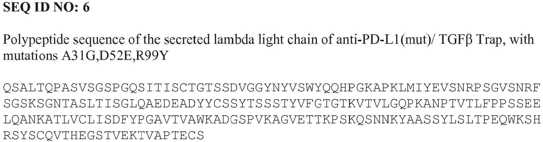


 SEQ ID NO: 11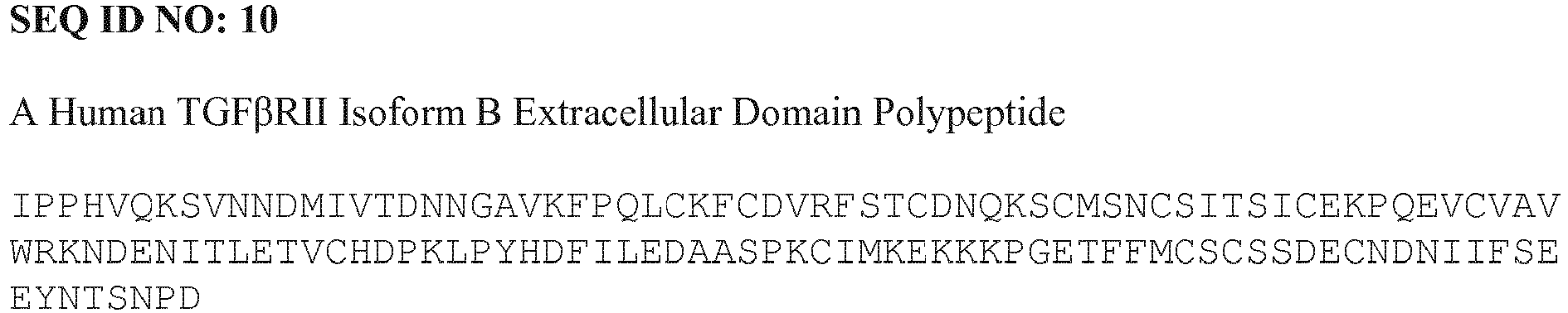
(Gly4Ser)4Gly linker
GGGGSGGGGSGGGGSGGGGSG


Claims (12)
- A protein comprising:a first polypeptide comprisinga) human TGFβRII, or a fragment thereof capable of binding TGFβ; andb) at least a variable region of the heavy chain of an antibody or antigen-binding fragment thereof binds human protein Programmed Death Ligand 1 (PD-L1); anda second polypeptide comprising at least a variable region of the light chain of an antibody or antigen-binding fragment thereof binds human protein PD-L1,wherein the first polypeptide comprises the amino acid sequence of SEQ ID NO:3,wherein the second polypeptide comprises the amino acid sequence of SEQ ID NO: 1, andwherein the heavy chain of the first polypeptide and the light chain of the second polypeptide when combined, form an antigen binding site that binds PD-L1.
- The protein of claim 1 comprising two polypeptides each having an amino acid sequence consisting of the amino acid sequence of SEQ ID NO: 3, and two polypeptides each having an amino acid sequence consisting of the amino acid sequence of SEQ ID NO: 1.
- A nucleic acid comprising a first nucleotide sequence encoding the first polypeptide according to claim 1 and a second nucleotide sequence encoding the second polypeptide according to claim 1.
- A cell comprising the nucleic acid of claim 3
- A method of producing a protein according to claim 1 comprising a) TGFβRII, or a fragment thereof capable of binding TGFβ, and b) an antibody, or an antigen-binding fragment thereof, that binds human protein Programmed Death Ligand 1 (PD-L1), the method comprising maintaining a cell according to claim 4 under conditions permitting expression of the protein.
- A method according to claim 5 further comprising harvesting the protein.
- A protein according to claim 1 for use in therapy.
- A pharmaceutical composition comprising a therapeutically effective amount of a protein according to claim 1.
- A protein according to claim 1 for use in;(i) a method of inhibiting tumor growth in a patient, the method comprising exposing the tumor to the protein; or.(ii) a method of treating cancer in a patient, the method comprising administering the protein to the cancer patient.
- A protein for use according to claim 9, wherein the method further comprises exposing the cancer patient to radiation and/or administering a chemotherapeutic.
- A protein for use according to claim 9 or claim 10, wherein the tumor or cancer is selected from the group consisting of colorectal, breast, ovarian, pancreatic, gastric, prostate, renal, cervical, myeloma, lymphoma, leukemia, thyroid, endometrial, uterine, bladder, neuroendocrine, head and neck, liver, nasopharyngeal, testicular, small cell lung cancer, non-small cell lung cancer, melanoma, basal cell skin cancer, squamous cell skin cancer, dermatofibrosarcoma protuberans, Merkel cell carcinoma, glioblastoma, glioma, sarcoma, mesothelioma, and myelodysplastic syndromes.
- A protein according to claim 1, wherein the protein blocks the interaction between human PD-L1 and the human PD-1 receptor.
Priority Applications (7)
| Application Number | Priority Date | Filing Date | Title |
|---|---|---|---|
| HRP20210977TT HRP20210977T1 (en) | 2014-02-10 | 2015-02-10 | TARGET INHIBITION OF TGF BETA |
| SI201531639T SI3105246T1 (en) | 2014-02-10 | 2015-02-10 | Targeted tgf beta inhibition |
| RS20210783A RS62038B1 (en) | 2014-02-10 | 2015-02-10 | Targeted tgf beta inhibition |
| DK21165709.3T DK3889172T3 (en) | 2014-02-10 | 2015-02-10 | Targeted TGF-Beta Inhibition |
| EP21165709.3A EP3889172B1 (en) | 2014-02-10 | 2015-02-10 | Targeted tgf beta inhibition |
| PL15717097T PL3105246T3 (en) | 2014-02-10 | 2015-02-10 | Targeted tgf beta inhibition |
| SM20210542T SMT202100542T1 (en) | 2014-02-10 | 2015-02-10 | Targeted tgf beta inhibition |
Applications Claiming Priority (2)
| Application Number | Priority Date | Filing Date | Title |
|---|---|---|---|
| US201461938048P | 2014-02-10 | 2014-02-10 | |
| PCT/EP2015/052781 WO2015118175A2 (en) | 2014-02-10 | 2015-02-10 | TARGETED TGFβ INHIBITION |
Related Child Applications (1)
| Application Number | Title | Priority Date | Filing Date |
|---|---|---|---|
| EP21165709.3A Division EP3889172B1 (en) | 2014-02-10 | 2015-02-10 | Targeted tgf beta inhibition |
Publications (2)
| Publication Number | Publication Date |
|---|---|
| EP3105246A2 EP3105246A2 (en) | 2016-12-21 |
| EP3105246B1 true EP3105246B1 (en) | 2021-03-31 |
Family
ID=52988007
Family Applications (2)
| Application Number | Title | Priority Date | Filing Date |
|---|---|---|---|
| EP15717097.8A Active EP3105246B1 (en) | 2014-02-10 | 2015-02-10 | Targeted tgf beta inhibition |
| EP21165709.3A Active EP3889172B1 (en) | 2014-02-10 | 2015-02-10 | Targeted tgf beta inhibition |
Family Applications After (1)
| Application Number | Title | Priority Date | Filing Date |
|---|---|---|---|
| EP21165709.3A Active EP3889172B1 (en) | 2014-02-10 | 2015-02-10 | Targeted tgf beta inhibition |
Country Status (28)
| Country | Link |
|---|---|
| US (4) | US9676863B2 (en) |
| EP (2) | EP3105246B1 (en) |
| JP (2) | JP6731346B2 (en) |
| KR (1) | KR102363008B1 (en) |
| CN (2) | CN113549159A (en) |
| AU (3) | AU2015213988B2 (en) |
| BR (1) | BR112016014952A2 (en) |
| CL (1) | CL2016002017A1 (en) |
| CY (1) | CY1124408T1 (en) |
| DK (2) | DK3105246T3 (en) |
| ES (2) | ES2992668T3 (en) |
| HR (1) | HRP20210977T1 (en) |
| HU (1) | HUE054873T2 (en) |
| IL (2) | IL246968B (en) |
| LT (1) | LT3105246T (en) |
| MX (2) | MX381141B (en) |
| NZ (1) | NZ721364A (en) |
| PE (2) | PE20210168A1 (en) |
| PH (1) | PH12016501549A1 (en) |
| PL (1) | PL3105246T3 (en) |
| PT (1) | PT3105246T (en) |
| RS (1) | RS62038B1 (en) |
| RU (2) | RU2021114500A (en) |
| SG (2) | SG11201606577YA (en) |
| SI (1) | SI3105246T1 (en) |
| SM (1) | SMT202100542T1 (en) |
| UA (1) | UA130563C2 (en) |
| WO (1) | WO2015118175A2 (en) |
Families Citing this family (159)
| Publication number | Priority date | Publication date | Assignee | Title |
|---|---|---|---|---|
| AU2014308751B2 (en) | 2013-08-22 | 2020-03-05 | Acceleron Pharma, Inc. | TGF-beta receptor type II variants and uses thereof |
| EP3757130A1 (en) | 2013-09-26 | 2020-12-30 | Costim Pharmaceuticals Inc. | Methods for treating hematologic cancers |
| CA2935423C (en) | 2014-01-24 | 2023-11-07 | Dana-Farber Cancer Institute, Inc. | Antibody molecules to pd-1 and uses thereof |
| AU2015210750B2 (en) | 2014-01-31 | 2020-08-20 | Children's Medical Center Corporation | Antibody molecules to TIM-3 and uses thereof |
| EP3105246B1 (en) | 2014-02-10 | 2021-03-31 | Merck Patent GmbH | Targeted tgf beta inhibition |
| WO2015138920A1 (en) | 2014-03-14 | 2015-09-17 | Novartis Ag | Antibody molecules to lag-3 and uses thereof |
| WO2016040882A1 (en) | 2014-09-13 | 2016-03-17 | Novartis Ag | Combination therapies of egfr inhibitors |
| WO2016061142A1 (en) | 2014-10-14 | 2016-04-21 | Novartis Ag | Antibody molecules to pd-l1 and uses thereof |
| CN108112254B (en) | 2015-03-13 | 2022-01-28 | 西托姆克斯治疗公司 | anti-PDL 1 antibodies, activatable anti-PDL 1 antibodies, and methods of use thereof |
| CN116327915A (en) | 2015-04-03 | 2023-06-27 | 佐马技术有限公司 | Treatment of cancer using TGF-beta inhibitors and PD-1 inhibitors |
| CA2986765A1 (en) | 2015-05-29 | 2016-12-08 | Agenus Inc. | Anti-ctla-4 antibodies and methods of use thereof |
| ES2878188T3 (en) | 2015-07-29 | 2021-11-18 | Novartis Ag | Combination therapies comprising antibody molecules against LAG-3 |
| CN116327952A (en) | 2015-08-04 | 2023-06-27 | 阿塞勒隆制药公司 | Methods for treating myeloproliferative disorders |
| WO2017020291A1 (en) * | 2015-08-06 | 2017-02-09 | Wuxi Biologics (Shanghai) Co. Ltd. | Novel anti-pd-l1 antibodies |
| CA2996996A1 (en) | 2015-08-31 | 2017-03-09 | National Research Council Of Canada | Tgf-.beta.-receptor ectodomain fusion molecules and uses thereof |
| ES2907486T3 (en) * | 2015-09-24 | 2022-04-25 | Univ North Carolina Chapel Hill | Methods and compositions for reducing metastases |
| US11583593B2 (en) | 2016-01-14 | 2023-02-21 | Synthis Therapeutics, Inc. | Antibody-ALK5 inhibitor conjugates and their uses |
| CN109069638B (en) | 2016-03-24 | 2022-03-29 | 璟尚生物制药公司 | Trispecific inhibitors for cancer therapy |
| AU2017268234A1 (en) * | 2016-05-17 | 2018-12-13 | Genentech, Inc. | Stromal gene signatures for diagnosis and use in immunotherapy |
| MX389710B (en) | 2016-05-27 | 2025-03-20 | Agenus Inc | Antibodies to T-cell immunoglobulin protein and mucin domain 3 (TIM-3) and methods of using them. |
| KR20190039200A (en) * | 2016-08-12 | 2019-04-10 | 메르크 파텐트 게엠베하 | Combination therapy for cancer |
| KR102617453B1 (en) | 2016-09-19 | 2023-12-26 | 현대모비스 주식회사 | Apparatus and method for recognizing supplementarty traffic signs |
| AU2017336546C1 (en) | 2016-09-27 | 2025-05-29 | Epicentrx, Inc. | Immunomodulatory fusion proteins |
| KR102576042B1 (en) | 2016-10-11 | 2023-09-07 | 아게누스 인코포레이티드 | Anti-LAG-3 Antibodies and Methods of Using The Same |
| CN118634323A (en) | 2016-12-07 | 2024-09-13 | 艾吉纳斯公司 | Antibodies and methods of use thereof |
| FI3551660T3 (en) | 2016-12-07 | 2023-12-11 | Agenus Inc | Anti-ctla-4 antibodies and methods of use thereof |
| CN109071656B (en) | 2017-01-05 | 2021-05-18 | 璟尚生物制药公司 | Checkpoint modulator antagonists |
| EP3565599A4 (en) * | 2017-01-07 | 2020-07-01 | Merck Patent GmbH | Dosing regimens and dosage forms for targeted tgf-b inhibition |
| CA3049652A1 (en) * | 2017-01-10 | 2018-07-19 | Intrexon Corporation | Modulating expression of polypeptides via new gene switch expression systems |
| GB201700567D0 (en) | 2017-01-12 | 2017-03-01 | Genagon Therapeutics Ab | Therapeutic agents |
| AU2018207172B2 (en) | 2017-01-13 | 2023-10-12 | Mink Therapeutics, Inc. | T cell receptors that bind to NY-ESO-1 and methods of use thereof |
| JP7231553B2 (en) | 2017-03-02 | 2023-03-01 | ナショナル リサーチ カウンシル オブ カナダ | TGF-B-receptor ectodomain fusion molecule and uses thereof |
| RU2765997C2 (en) * | 2017-03-30 | 2022-02-07 | Мерк Патент Гмбх | Combination of anti-pd-l1 antibody and dna-pk inhibitor for treating malignant neoplasm |
| JP7297672B2 (en) | 2017-04-13 | 2023-06-26 | アジェナス インコーポレイテッド | ANTI-CD137 ANTIBODY AND METHODS OF USE THEREOF |
| JP2020517256A (en) | 2017-04-19 | 2020-06-18 | エルスター セラピューティクス, インコーポレイテッド | Multispecific molecules and uses thereof |
| RS64576B1 (en) | 2017-05-01 | 2023-10-31 | Agenus Inc | Anti-tigit antibodies and methods of use thereof |
| BR112019023071A2 (en) * | 2017-05-04 | 2020-06-09 | Acceleron Pharma Inc | type ii tgf-beta receptor fusion proteins and use thereof |
| JP7340458B2 (en) * | 2017-05-09 | 2023-09-07 | アメリカ合衆国 | Combination of PDL1 and TGFbeta blockade in patients with HPV+ malignancies |
| AU2018264455B2 (en) * | 2017-05-12 | 2024-12-12 | Jiangsu Hengrui Medicine Co., Ltd. | Fusion protein containing TGF-beta receptor and medicinal uses thereof |
| WO2018222901A1 (en) | 2017-05-31 | 2018-12-06 | Elstar Therapeutics, Inc. | Multispecific molecules that bind to myeloproliferative leukemia (mpl) protein and uses thereof |
| BR112019025188A2 (en) | 2017-06-01 | 2020-06-23 | Cytomx Therapeutics, Inc. | ACTIVABLE ANTI-PDL1 ANTIBODIES AND METHODS OF USE OF THE SAME |
| JP7387585B2 (en) | 2017-09-04 | 2023-11-28 | アジェナス インコーポレイテッド | T-cell receptor that binds mixed lineage leukemia (MLL)-specific phosphopeptide and methods of use thereof |
| KR102757384B1 (en) | 2017-09-27 | 2025-01-20 | 에피센트알엑스, 인코포레이티드 | immunomodulatory fusion protein |
| CN111213059B (en) * | 2017-11-06 | 2024-01-09 | 豪夫迈·罗氏有限公司 | Diagnostic and therapeutic methods for cancer |
| EP3720881A1 (en) | 2017-12-08 | 2020-10-14 | Elstar Therapeutics, Inc. | Multispecific molecules and uses thereof |
| WO2019139987A1 (en) | 2018-01-09 | 2019-07-18 | Elstar Therapeutics, Inc. | Calreticulin binding constructs and engineered t cells for the treatment of diseases |
| TWI823906B (en) | 2018-03-09 | 2023-12-01 | 美商艾吉納斯公司 | Anti-cd73 antibodies and methods of use thereof |
| US12152073B2 (en) | 2018-03-14 | 2024-11-26 | Marengo Therapeutics, Inc. | Multifunctional molecules that bind to calreticulin and uses thereof |
| JP2021517145A (en) | 2018-03-14 | 2021-07-15 | メルク パテント ゲゼルシャフト ミット ベシュレンクテル ハフツングMerck Patent Gesellschaft mit beschraenkter Haftung | Compounds and their use to treat tumors of interest |
| EP3765516A2 (en) | 2018-03-14 | 2021-01-20 | Elstar Therapeutics, Inc. | Multifunctional molecules and uses thereof |
| KR20210005646A (en) | 2018-04-26 | 2021-01-14 | 아게누스 인코포레이티드 | Heat shock protein-binding peptide composition and method for use thereof |
| WO2019213442A1 (en) | 2018-05-03 | 2019-11-07 | Acceleron Pharma Inc. | MULTISPECIFIC BINDERS OF TGFβ-SUPERFAMILY LIGANDS AND USES THEREOF |
| WO2019213446A1 (en) | 2018-05-03 | 2019-11-07 | Acceleron Pharma Inc. | NOVEL BINDERS OF TGFβ-SUPERFAMILY LIGANDS AND USES THEREOF |
| KR20210006405A (en) | 2018-05-04 | 2021-01-18 | 메르크 파텐트 게엠베하 | Inhibition of the combination of PD-1/PD-L1, TGFβ and DNA-PK for the treatment of cancer |
| CA3096844A1 (en) * | 2018-05-15 | 2019-11-21 | Merck Patent Gmbh | Dosing regimens for targeted tgf-b inhibition for use in treating cancer in treatment naive subjects |
| AU2019270178A1 (en) | 2018-05-17 | 2021-01-07 | Bolt Biotherapeutics, Inc. | Immunoconjugates |
| SG11202012426WA (en) * | 2018-06-13 | 2021-01-28 | Merck Patent Gmbh | Treatment of stage iii nsclc and mitigation of pathological conditions associated with the treatment |
| CN111867630B (en) | 2018-06-17 | 2023-10-13 | 上海健信生物医药科技有限公司 | Antibodies, bispecific antibodies, ADCs and CARs targeting CLDN18.2 and their applications |
| SG11202012688UA (en) * | 2018-06-22 | 2021-01-28 | Merck Patent Gmbh | Dosing regimens for targeted tgf-b inhibition for use in treating biliary tract cancer |
| WO2020006516A1 (en) | 2018-06-29 | 2020-01-02 | Gensun Biopharma, Inc. | Antitumor immune checkpoint regulator antagonists |
| TW202019405A (en) * | 2018-07-02 | 2020-06-01 | 德商馬克專利公司 | Combination therapy with targeted tgf-β inhibition for treatment of advanced non-small cell lung cancer |
| WO2020010250A2 (en) | 2018-07-03 | 2020-01-09 | Elstar Therapeutics, Inc. | Anti-tcr antibody molecules and uses thereof |
| AU2019301070B2 (en) * | 2018-07-09 | 2025-09-11 | Precigen, Inc. | Fusion constructs and methods of using thereof |
| AU2019325593A1 (en) * | 2018-08-22 | 2021-03-18 | Merck Patent Gmbh | Treatment of triple negative breast cancer with targeted TGF-B inhibition |
| CN113164777B (en) | 2018-09-27 | 2024-12-13 | 马伦戈治疗公司 | CSF1R/CCR2 multispecific antibody |
| WO2020093024A2 (en) | 2018-11-01 | 2020-05-07 | Merck Patent Gmbh | Methods of administering anti-tim-3 antibodies |
| JP2022505925A (en) | 2018-11-01 | 2022-01-14 | メルク パテント ゲーエムベーハー | Anti-TIM-3 antibody |
| TWI829799B (en) * | 2018-11-09 | 2024-01-21 | 大陸商江蘇恆瑞醫藥股份有限公司 | A PHARMACEUTICAL COMPOSITION COMPRISING TGF-β RECEPTOR FUSION PROTEIN AND THE USE THEREOF |
| US20210369776A1 (en) * | 2018-12-06 | 2021-12-02 | Guangdong Tcrcure Biopharma Technology Co., Ltd. | Combinational tcr-t cell therapy targeting tumor antigens, tgf-beta, and immune checkpoints |
| AU2020226893B2 (en) | 2019-02-21 | 2025-02-27 | Marengo Therapeutics, Inc. | Multifunctional molecules that bind to T cell related cancer cells and uses thereof |
| GB2597851B (en) | 2019-02-21 | 2024-05-29 | Marengo Therapeutics Inc | Antibody molecules that bind to NKP30 and uses thereof |
| EP3927745A1 (en) | 2019-02-21 | 2021-12-29 | Marengo Therapeutics, Inc. | Multifunctional molecules that bind to t cells and uses thereof to treat autoimmune disorders |
| US20220226491A1 (en) | 2019-03-15 | 2022-07-21 | Bolt Biotherapeutics, Inc. | Immunoconjugates Targeting PD-L1 |
| EP3929215A4 (en) * | 2019-06-10 | 2022-06-22 | Shandong Boan Biotechnology Co., Ltd. | Bifunctional fusion protein against pdl1 and tgf? and use thereof |
| AU2020291014B2 (en) | 2019-06-13 | 2025-06-05 | Bolt Biotherapeutics, Inc. | Aminobenzazepine compounds, immunoconjugates, and uses thereof |
| CN110218260A (en) * | 2019-06-24 | 2019-09-10 | 王跃驹 | Application of the plant as host in expression M7842 |
| EP3990116A1 (en) | 2019-06-28 | 2022-05-04 | Gensun Biopharma Inc. | ANTITUMOR ANTAGONIST CONSISTING OF A MUTATED TGFß1 - RII EXTRACELLULAR DOMAIN AND AN IMMUNOGLOBULIN SCAFFOLD |
| WO2021007428A2 (en) * | 2019-07-09 | 2021-01-14 | The Johns Hopkins University | Molecules, compositions and methods for treatment of cancer |
| CN112239507A (en) * | 2019-07-17 | 2021-01-19 | 鸿运华宁(杭州)生物医药有限公司 | Fusion protein of ETA antibody and TGF-β Trap, and its pharmaceutical composition and application |
| WO2021030662A1 (en) * | 2019-08-15 | 2021-02-18 | Nantbio, Inc. | Tgf-beta trap |
| CN118126195A (en) * | 2019-08-30 | 2024-06-04 | 正大天晴药业集团南京顺欣制药有限公司 | A fusion protein targeting PD-L1 and TGF-β and its use |
| KR20220053007A (en) | 2019-08-30 | 2022-04-28 | 아게누스 인코포레이티드 | Anti-CD96 antibodies and methods of use thereof |
| CA3148694A1 (en) | 2019-09-03 | 2021-03-11 | Romas Kudirka | Aminoquinoline compounds, immunoconjugates, and uses thereof |
| EP4025255A1 (en) | 2019-09-04 | 2022-07-13 | Bolt Biotherapeutics, Inc. | Immunoconjugate synthesis method |
| CN112442132A (en) * | 2019-09-05 | 2021-03-05 | 复旦大学 | Recombinant difunctional fusion protein targeting tumor and application thereof |
| CN115947846A (en) | 2019-09-12 | 2023-04-11 | 普米斯生物技术(珠海)有限公司 | An anti-PD-L1 nanobody and its derivatives and uses |
| JP2022550420A (en) * | 2019-09-30 | 2022-12-01 | ハーバー・バイオメド・(シャンハイ)・カンパニー・リミテッド | ANTI-PD-L1 ANTIGEN-BINDING PROTEIN AND APPLICATION THEREOF |
| WO2021067242A1 (en) | 2019-09-30 | 2021-04-08 | Bolt Biotherapeutics, Inc. | Amide-linked, aminobenzazepine immunoconjugates, and uses thereof |
| EP4048704A4 (en) | 2019-10-21 | 2023-11-22 | Nanjing Leads Biolabs Co., Ltd. | RECOMBINANT PROTEIN TARGETING PD-1 AND TGFß |
| KR20220088901A (en) | 2019-10-25 | 2022-06-28 | 볼트 바이오테라퓨틱스 인코퍼레이티드 | Thienoazepine immunoconjugates, and uses thereof |
| CA3154413A1 (en) * | 2019-11-01 | 2021-05-06 | Yan Lan | Combined inhibition of pd-1, tgf.beta. and atm together with radiotherapy for the treatment of cancer |
| JP2023502585A (en) * | 2019-11-05 | 2023-01-25 | メルク パテント ゲゼルシャフト ミット ベシュレンクテル ハフツング | Combined inhibition of PD-1, TGFβ, and TIGIT for the treatment of cancer |
| TW202128763A (en) * | 2019-11-12 | 2021-08-01 | 大陸商江蘇恆瑞醫藥股份有限公司 | Fusion protein containing tgf-β receptor and the pharmaceutical use thereof |
| CN114746451A (en) * | 2019-11-27 | 2022-07-12 | 上海岸迈生物科技有限公司 | TGF-beta/PD-L1 bispecific binding proteins |
| WO2021115456A1 (en) * | 2019-12-11 | 2021-06-17 | Wuxi Biologics (Shanghai) Co., Ltd. | BI-FUNCTIONAL ANTIBODY AGAINST PD-L1 AND TGFβ |
| CN113004414B (en) * | 2019-12-20 | 2022-09-06 | 广东菲鹏制药股份有限公司 | Bifunctional antibody for resisting PD1 and TGF beta, preparation method thereof and pharmaceutical composition containing same |
| EP3838260A1 (en) | 2019-12-20 | 2021-06-23 | Ares Trading S.A. | Igg:tgf rii fusion protein composition |
| US20230043290A1 (en) * | 2019-12-20 | 2023-02-09 | Ares Trading S.A. | Igg:tgfbetarii fusion protein composition |
| CN113121699A (en) * | 2019-12-30 | 2021-07-16 | 国典(北京)医药科技有限公司 | PTTRAP1 bispecific fusion protein and application thereof |
| WO2021138407A2 (en) | 2020-01-03 | 2021-07-08 | Marengo Therapeutics, Inc. | Multifunctional molecules that bind to cd33 and uses thereof |
| AU2020416273A1 (en) | 2020-01-03 | 2022-07-28 | Marengo Therapeutics, Inc. | Anti-TCR antibody molecules and uses thereof |
| CN114929734A (en) * | 2020-01-09 | 2022-08-19 | 豪夫迈·罗氏有限公司 | Novel antigen binding molecules comprising 4-1BBL trimers |
| EP4093772A1 (en) * | 2020-01-21 | 2022-11-30 | Bolt Biotherapeutics, Inc. | Anti-pd-l1 antibodies |
| WO2021150701A1 (en) | 2020-01-21 | 2021-07-29 | Bolt Biotherapeutics, Inc. | Anti-pd-l1 antibodies |
| WO2021164701A1 (en) * | 2020-02-18 | 2021-08-26 | Nanjing GenScript Biotech Co., Ltd. | Fusion proteins and uses thereof |
| CN115175942B (en) * | 2020-02-25 | 2025-05-30 | 上海药明生物技术有限公司 | Dual-function fusion protein and application thereof |
| JP7489468B2 (en) * | 2020-03-20 | 2024-05-23 | レメゲン シーオー.,エルティーディー. | Bispecific fusion proteins and their applications |
| WO2021209458A1 (en) | 2020-04-14 | 2021-10-21 | Ares Trading S.A. | Combination treatment of cancer |
| CN115461362A (en) * | 2020-04-14 | 2022-12-09 | 葛兰素史密斯克莱知识产权发展有限公司 | Cancer combination therapy based on ICOS antibody and PD-L1 antibody TGF-beta receptor fusion protein |
| KR20230028242A (en) | 2020-04-24 | 2023-02-28 | 마렝고 테라퓨틱스, 인크. | Multifunctional molecules that bind to T cell-associated cancer cells and their uses |
| KR20230002261A (en) | 2020-04-28 | 2023-01-05 | 더 락커펠러 유니버시티 | Anti-SARS-COV-2 Neutralizing Antibodies and Methods of Using The Same |
| KR20230003178A (en) * | 2020-04-29 | 2023-01-05 | 치아타이 티안큉 파마수티컬 그룹 주식회사 | Bifunctional proteins for PD-1 and TGF-β |
| WO2021222894A1 (en) | 2020-05-01 | 2021-11-04 | Bolt Biotherapeutics, Inc. | Anti-dectin-2 antibodies |
| CA3176626A1 (en) | 2020-05-08 | 2021-11-11 | David Dornan | Elastase-substrate, peptide linker immunoconjugates, and uses thereof |
| JP2023532279A (en) * | 2020-06-26 | 2023-07-27 | ソレント・セラピューティクス・インコーポレイテッド | Oncolytic Herpes Simplex Virus (HSV) Expressing Immunomodulatory Fusion Proteins |
| IL299764A (en) | 2020-07-10 | 2023-03-01 | Precigen Inc | Fusion constructs and methods of using thereof |
| JP7766672B2 (en) * | 2020-07-24 | 2025-11-10 | ▲邁▼威(上海)生物科技股▲フン▼有限公司 | TGF-β RII mutants and fusion proteins thereof |
| CN119409832B (en) * | 2020-07-28 | 2025-08-01 | 乐普生物科技股份有限公司 | Bifunctional molecules targeting PD-L1 and TGF-beta |
| US11028174B1 (en) | 2020-07-28 | 2021-06-08 | Lepu Biopharma Co., Ltd. | Bifunctional molecules targeting PD-L1 and TGF-β |
| WO2022042537A1 (en) * | 2020-08-24 | 2022-03-03 | 江苏恒瑞医药股份有限公司 | APPLICATION OF COMBINATION OF FUSION PROTEIN OF TGF-β RECEPTOR AND MULTI-TARGETED TYROSINE KINASE INHIBITOR IN PREPARING ANTI-TUMOR DRUG |
| CN116249718A (en) | 2020-08-26 | 2023-06-09 | 马伦戈治疗公司 | Multifunctional molecules binding to calreticulin and uses thereof |
| GB2616354A (en) | 2020-08-26 | 2023-09-06 | Marengo Therapeutics Inc | Methods of detecting TRBC1 or TRBC2 |
| KR20230074144A (en) | 2020-08-26 | 2023-05-26 | 마렝고 테라퓨틱스, 인크. | Antibody molecules that bind to NKp30 and uses thereof |
| CN114106190B (en) | 2020-08-31 | 2025-04-08 | 普米斯生物技术(珠海)有限公司 | An anti-VEGF/PD-L1 bispecific antibody and its use |
| WO2022042715A1 (en) * | 2020-08-31 | 2022-03-03 | 杭州九源基因工程有限公司 | BIFUNCTIONAL FUSION PROTEIN TARGETING PD-L1 AND TGFβ, PREPARATION METHOD THEREFOR, AND APPLICATION THEREOF |
| WO2022057061A1 (en) * | 2020-09-16 | 2022-03-24 | Suzhou Neologics Bioscience Co., Ltd. | Pd-l1 antibodies, fusion proteins, and uses thereof |
| WO2022057821A1 (en) * | 2020-09-16 | 2022-03-24 | Suzhou Neologics Bioscience Co. , Ltd. | Pd-l1 antibodies, fusion proteins, and uses thereof |
| WO2022063114A1 (en) * | 2020-09-23 | 2022-03-31 | 海正生物制药有限公司 | Tgfbr2-ecd mutant, and fusion protein comprising same and use thereof |
| WO2022063193A1 (en) * | 2020-09-24 | 2022-03-31 | 上海齐鲁制药研究中心有限公司 | BIFUNCTIONAL MOLECULE SIMULTANEOUSLY TARGETING PD-L1 AND TGFβ AND MEDICAL USE THEREOF |
| CN114349867B (en) * | 2020-10-14 | 2024-05-28 | 广东菲鹏制药股份有限公司 | Fusion proteins and uses thereof |
| WO2022090529A1 (en) | 2020-11-02 | 2022-05-05 | Ares Trading S.A. | Combination treatment of cancer |
| CN116801906A (en) | 2020-11-02 | 2023-09-22 | 阿雷斯贸易股份有限公司 | Combination therapy for cancer |
| CN114573701A (en) * | 2020-12-02 | 2022-06-03 | 上海华奥泰生物药业股份有限公司 | anti-PD-L1/TGF-beta bifunctional antibody and application thereof |
| US20220195066A1 (en) | 2020-12-11 | 2022-06-23 | Bolt Biotherapeutics, Inc. | Anti-cea immunoconjugates, and uses thereof |
| JP2023552790A (en) | 2020-12-11 | 2023-12-19 | ボルト バイオセラピューティクス、インコーポレーテッド | Anti-PD-L1 immunoconjugates and their uses |
| JP2024501453A (en) | 2020-12-11 | 2024-01-12 | ボルト バイオセラピューティクス、インコーポレーテッド | Anti-CEA immunoconjugates and their uses |
| CN116917334B (en) | 2021-02-22 | 2026-02-06 | 浙江道尔生物科技有限公司 | Multi-domain fusion protein with anticancer activity |
| CA3212345A1 (en) | 2021-03-02 | 2022-09-09 | Glaxosmithkline Intellectual Property Development Limited | Substituted pyridines as dnmt1 inhibitors |
| CN117940168A (en) | 2021-03-26 | 2024-04-26 | 博尔特生物治疗药物有限公司 | 2-Amino-4-carboxamide-benzazepine immunoconjugates and uses thereof |
| TW202304520A (en) | 2021-03-26 | 2023-02-01 | 美商博特生物治療公司 | 2-amino-4-carboxamide-benzazepine immunoconjugates, and uses thereof |
| AU2022270880A1 (en) | 2021-05-03 | 2023-09-28 | Merck Patent Gmbh | Her2 targeting fc antigen binding fragment-drug conjugates |
| CN114773485B (en) * | 2021-05-11 | 2024-05-17 | 江苏先声药业有限公司 | Bifunctional fusion protein molecules of anti-human PD-L1 antibodies and TGF-beta RII |
| CN117999101A (en) | 2021-05-25 | 2024-05-07 | 默克专利股份公司 | Fc antigen-binding fragment-drug conjugate targeting EGFR |
| JP2024520764A (en) | 2021-06-07 | 2024-05-24 | アレス トレーディング ソシエテ アノニム | Combination Treatment of Cancer |
| JP2024534263A (en) * | 2021-08-27 | 2024-09-18 | セラノティックス カンパニー リミテッド | Bispecific molecules that specifically bind B7-H3 and TGFβ, and uses thereof |
| WO2023141724A1 (en) | 2022-01-28 | 2023-08-03 | 35Pharma Inc. | Activin receptor type iib variants and uses thereof |
| CN114990129B (en) * | 2022-05-11 | 2023-02-03 | 北京贝来生物科技有限公司 | Preparation and application of mesenchymal stem cells expressing alpha PDL1: fc fusion protein |
| TWI866210B (en) | 2022-05-17 | 2024-12-11 | 大陸商蘇州創勝醫藥集團有限公司 | Bifunctional protein and its preparation and use |
| WO2024137619A1 (en) | 2022-12-20 | 2024-06-27 | Bolt Biotherapeutics, Inc. | Anti-claudin, bis-benzimid azole sting agonist immunoconjugates, and uses thereof |
| CN120981252A (en) | 2023-02-14 | 2025-11-18 | 博尔特生物治疗药物有限公司 | Aza-benzoza immunoconjugates and their uses |
| WO2024186626A1 (en) | 2023-03-03 | 2024-09-12 | Bolt Biotherapeutics, Inc. | Aza-bicyclic sting agonist immunoconjugates, and uses thereof |
| WO2024216028A1 (en) | 2023-04-12 | 2024-10-17 | Agenus Inc. | Methods of treating cancer using an anti-ctla4 antibody and an enpp1 inhibitor |
| WO2024216089A1 (en) | 2023-04-13 | 2024-10-17 | Bolt Biotherapeutics, Inc. | Anti-dectin-2 antibody combination therapy |
| AU2024372189A1 (en) * | 2023-10-31 | 2026-03-05 | Immunitybio, Inc. | Multi-chain chimeric polypeptides and uses thereof |
| WO2025110822A1 (en) * | 2023-11-24 | 2025-05-30 | 주식회사 메드팩토 | Fusion protein specifically binding to extradomain b of fibronectin (edb-fn) and transforming growth factor β (tgfβ), and use thereof |
| WO2025140212A1 (en) * | 2023-12-26 | 2025-07-03 | Laekna Therapeutics Shanghai Co., Ltd. | Multispecific binding agents comprising anti-fibroblast activation protein (fap) antibodies and one or more monovalent ligand traps and uses thereof |
| US20260092096A1 (en) | 2024-05-14 | 2026-04-02 | 35Pharma Inc. | Activin receptor type iib variants and uses thereof |
| WO2026050213A1 (en) | 2024-08-27 | 2026-03-05 | Usbolt Biotherapeutics, Inc. | Tlr agonist immunoconjugates and uses thereof |
| WO2026055185A1 (en) | 2024-09-04 | 2026-03-12 | Bolt Biotherapeutics, Inc. | Sting agonist immunoconjugates and uses thereof |
Family Cites Families (34)
| Publication number | Priority date | Publication date | Assignee | Title |
|---|---|---|---|---|
| US5785682A (en) | 1995-03-22 | 1998-07-28 | Abbott Laboratories | Pre-filled syringe drug delivery system |
| ES2289778T3 (en) * | 1997-04-18 | 2008-02-01 | Biogen Idec Ma Inc. | FUSION PROTEINS OF THE CONSTANT REGION OF THE IMMUNOGLOBULIN / RECEIVER TGF-BETA TYPE II. |
| JP5179689B2 (en) | 2000-02-11 | 2013-04-10 | メルク パテント ゲゼルシャフト ミット ベシュレンクテル ハフツング | Enhancing the half-life of antibody-based fusion proteins in the circulation |
| HUP0303428A2 (en) | 2001-03-07 | 2004-01-28 | Merck Patent Gmbh. | Expression technology for proteins containing a hybrid isotype antibody moiety |
| WO2002079415A2 (en) | 2001-03-30 | 2002-10-10 | Lexigen Pharmaceuticals Corp. | Reducing the immunogenicity of fusion proteins |
| JP2005112812A (en) * | 2003-10-09 | 2005-04-28 | Zeria Pharmaceut Co Ltd | Phosphorylated site-specific antibody and screening method using the same |
| BRPI0417213A (en) | 2003-12-24 | 2007-02-06 | Scios Inc | treatment of malignant gliomas with tgf-beta inhibitors |
| US7670595B2 (en) | 2004-06-28 | 2010-03-02 | Merck Patent Gmbh | Fc-interferon-beta fusion proteins |
| US7795389B2 (en) | 2004-09-28 | 2010-09-14 | The Board Of Regents Of The University Of Texas System | Antagonizing TGF-beta activity with various ectodomains TGF-beta receptors used in combination or as fusion proteins |
| CA2612241C (en) | 2005-07-01 | 2018-11-06 | Medarex, Inc. | Human monoclonal antibodies to programmed death ligand 1 (pd-l1) |
| CN101541832B (en) * | 2006-09-07 | 2014-11-12 | 克鲁塞尔荷兰公司 | Human binding molecules capable of neutralizing influenza virus H5N1 and uses thereof |
| EP2171062A1 (en) | 2007-06-15 | 2010-04-07 | Genzyme Corporation | Fusion proteins containing two tgf-beta binding domains of tgf-beta type ii receptor |
| US20140127200A1 (en) | 2008-01-03 | 2014-05-08 | The Scripps Research Institute | Multispecific Antibody Targeting and Multivalency Through Modular Recognition Domains |
| WO2009152610A1 (en) | 2008-06-20 | 2009-12-23 | The Royal Institution For The Advancement Of Learning/Mcgill University | Interleukin-2/soluble tgf-beta type ii receptor b conjugates and methods and uses thereof |
| KR20110044992A (en) | 2008-07-02 | 2011-05-03 | 이머전트 프로덕트 디벨롭먼트 시애틀, 엘엘씨 | TVF-β antagonist multi-target binding protein |
| EP4209510B1 (en) * | 2008-12-09 | 2024-01-31 | F. Hoffmann-La Roche AG | Anti-pd-l1 antibodies and their use to enhance t-cell function |
| FR2944448B1 (en) * | 2008-12-23 | 2012-01-13 | Adocia | STABLE PHARMACEUTICAL COMPOSITION COMPRISING AT LEAST ONE MONODONAL ANTIBODY AND AT LEAST ONE AMPHIPHILIC POLYSACHARIDE COMPRISING SUBSTITUENTS DERIVED FROM HYDROFOB ALCOHOLS OR HYDROPHOBIC AMINES. |
| US9533055B2 (en) | 2009-03-18 | 2017-01-03 | Armagen Technologies, Inc. | Compositions and methods for blood-brain barrier delivery of IgG-decoy receptor fusion proteins |
| EP2493929B1 (en) | 2009-10-28 | 2017-08-16 | AbbVie Biotherapeutics Inc. | Anti-egfr antibodies and their uses |
| NO2504364T3 (en) | 2009-11-24 | 2018-01-06 | ||
| SI2519543T1 (en) * | 2009-12-29 | 2016-08-31 | Emergent Product Development Seattle, Llc | Heterodimer binding proteins and uses thereof |
| US20130129723A1 (en) * | 2009-12-29 | 2013-05-23 | Emergent Product Development Seattle, Llc | Heterodimer Binding Proteins and Uses Thereof |
| TWI619509B (en) | 2010-02-23 | 2018-04-01 | 建南德克公司 | Combination of carboplatin, paclitaxel and anti-VEGF antibodies in previously untreated stage III or IV ovarian, fallopian tube or primary peritoneal cancer |
| PT2542590T (en) | 2010-03-05 | 2017-08-31 | Univ Johns Hopkins | COMPOSITIONS AND METHODS FOR DIRECTED IMMUNOMODULATOR ANTIBODIES AND FUSION PROTEINS |
| EP2739358B1 (en) * | 2011-08-01 | 2018-11-28 | F.Hoffmann-La Roche Ag | Methods of treating cancer using pd-1 axis binding antagonists and mek inhibitors |
| US9624298B2 (en) | 2011-11-28 | 2017-04-18 | Merck Patent Gmbh | Anti-PD-L1 antibodies and uses thereof |
| WO2013164694A1 (en) | 2012-04-30 | 2013-11-07 | Biocon Limited | Targeted/immunomodulatory fusion proteins and methods for making same |
| JP6312659B2 (en) | 2012-05-31 | 2018-04-18 | ジェネンテック, インコーポレイテッド | Cancer treatment method using PD-1 binding antagonist and VEGF antagonist |
| WO2014193898A1 (en) | 2013-05-31 | 2014-12-04 | Merck Sharp & Dohme Corp. | Combination therapies for cancer |
| WO2015095423A2 (en) | 2013-12-17 | 2015-06-25 | Genentech, Inc. | Combination therapy comprising ox40 binding agonists and pd-1 axis binding antagonists |
| EP3105246B1 (en) | 2014-02-10 | 2021-03-31 | Merck Patent GmbH | Targeted tgf beta inhibition |
| GB201403875D0 (en) | 2014-03-05 | 2014-04-16 | Cantargia Ab | Novel antibodies and uses thereof |
| US9840553B2 (en) | 2014-06-28 | 2017-12-12 | Kodiak Sciences Inc. | Dual PDGF/VEGF antagonists |
| DK3180363T3 (en) | 2014-08-15 | 2019-11-04 | Merck Patent Gmbh | SIRP-ALPHA-IMMUNOGLOBULIN FUSION PROTEINS |
-
2015
- 2015-02-10 EP EP15717097.8A patent/EP3105246B1/en active Active
- 2015-02-10 RU RU2021114500A patent/RU2021114500A/en unknown
- 2015-02-10 SI SI201531639T patent/SI3105246T1/en unknown
- 2015-02-10 ES ES21165709T patent/ES2992668T3/en active Active
- 2015-02-10 NZ NZ721364A patent/NZ721364A/en unknown
- 2015-02-10 DK DK15717097.8T patent/DK3105246T3/en active
- 2015-02-10 HU HUE15717097A patent/HUE054873T2/en unknown
- 2015-02-10 US US14/618,454 patent/US9676863B2/en active Active
- 2015-02-10 KR KR1020167024679A patent/KR102363008B1/en active Active
- 2015-02-10 PL PL15717097T patent/PL3105246T3/en unknown
- 2015-02-10 EP EP21165709.3A patent/EP3889172B1/en active Active
- 2015-02-10 LT LTEP15717097.8T patent/LT3105246T/en unknown
- 2015-02-10 SM SM20210542T patent/SMT202100542T1/en unknown
- 2015-02-10 BR BR112016014952A patent/BR112016014952A2/en not_active Application Discontinuation
- 2015-02-10 UA UAA201609356A patent/UA130563C2/en unknown
- 2015-02-10 SG SG11201606577YA patent/SG11201606577YA/en unknown
- 2015-02-10 JP JP2016546949A patent/JP6731346B2/en active Active
- 2015-02-10 CN CN202110830081.8A patent/CN113549159A/en active Pending
- 2015-02-10 ES ES15717097T patent/ES2876430T3/en active Active
- 2015-02-10 SG SG10202108879SA patent/SG10202108879SA/en unknown
- 2015-02-10 AU AU2015213988A patent/AU2015213988B2/en active Active
- 2015-02-10 HR HRP20210977TT patent/HRP20210977T1/en unknown
- 2015-02-10 RS RS20210783A patent/RS62038B1/en unknown
- 2015-02-10 PE PE2020002093A patent/PE20210168A1/en unknown
- 2015-02-10 PT PT157170978T patent/PT3105246T/en unknown
- 2015-02-10 WO PCT/EP2015/052781 patent/WO2015118175A2/en not_active Ceased
- 2015-02-10 PE PE2016001267A patent/PE20161096A1/en unknown
- 2015-02-10 CN CN201580007865.3A patent/CN106103488B/en active Active
- 2015-02-10 RU RU2016135062A patent/RU2752424C2/en active
- 2015-02-10 DK DK21165709.3T patent/DK3889172T3/en active
- 2015-02-10 MX MX2016010067A patent/MX381141B/en unknown
-
2016
- 2016-07-27 IL IL246968A patent/IL246968B/en active IP Right Grant
- 2016-08-03 MX MX2021003685A patent/MX2021003685A/en unknown
- 2016-08-04 PH PH12016501549A patent/PH12016501549A1/en unknown
- 2016-08-10 CL CL2016002017A patent/CL2016002017A1/en unknown
-
2017
- 2017-06-08 US US15/617,876 patent/US20180002436A1/en not_active Abandoned
-
2019
- 2019-07-25 US US16/522,304 patent/US11370819B2/en active Active
- 2019-10-11 AU AU2019246876A patent/AU2019246876B2/en not_active Ceased
-
2020
- 2020-07-06 JP JP2020116566A patent/JP2020174674A/en active Pending
-
2021
- 2021-03-16 IL IL281565A patent/IL281565B/en unknown
- 2021-06-23 CY CY20211100564T patent/CY1124408T1/en unknown
-
2022
- 2022-01-20 AU AU2022200365A patent/AU2022200365A1/en not_active Abandoned
- 2022-06-07 US US17/834,458 patent/US20230056881A1/en not_active Abandoned
Non-Patent Citations (1)
| Title |
|---|
| None * |
Also Published As
Similar Documents
| Publication | Publication Date | Title |
|---|---|---|
| EP3889172B1 (en) | Targeted tgf beta inhibition | |
| JP7389833B2 (en) | Humanized or chimeric CD3 antibodies | |
| AU2019264965B2 (en) | Antibodies specific to human Nectin4 | |
| EP3497130B1 (en) | Combination therapy for cancer | |
| CA3230426A1 (en) | Bispecific binding proteins that bind cd137 and a tumor associated antigen | |
| KR102634374B1 (en) | Anti-OX40 antibodies and uses thereof | |
| US20220372161A1 (en) | Antibodies against the poliovirus receptor (pvr) and uses thereof | |
| TW202041537A (en) | Engineered antibodies to lag-3 and bispecific pd-1/lag-3 binding proteins made therefrom | |
| CN117355331A (en) | Anti-Siglec compositions and uses thereof | |
| CN116333125A (en) | Antibodies that bind death receptor 4 and death receptor 5 | |
| HK1231904A1 (en) | Targeted tgf beta inhibition | |
| HK1231904B (en) | Targeted tgf beta inhibition | |
| EP4491631A1 (en) | Anti-pd-1 antibody binding domain and immunoconjugate | |
| CA2934979C (en) | Targeted tgf.beta. inhibition | |
| HK40109079A (en) | Methods of cancer treatment using anti-ox40 antibodies in combination with anti-tim3 antibodies | |
| EP4185388A1 (en) | A combination of anti-dr5 antibodies and an immunomodulatory imide drug for use in treating multiple myeloma | |
| HK40074321B (en) | Methods of cancer treatment using anti-ox40 antibodies in combination with anti-tim3 antibodies | |
| HK40045878B (en) | Igf-1r antibody and its use as addressing vehicle for the treatment of cancer |
Legal Events
| Date | Code | Title | Description |
|---|---|---|---|
| PUAI | Public reference made under article 153(3) epc to a published international application that has entered the european phase |
Free format text: ORIGINAL CODE: 0009012 |
|
| STAA | Information on the status of an ep patent application or granted ep patent |
Free format text: STATUS: REQUEST FOR EXAMINATION WAS MADE |
|
| 17P | Request for examination filed |
Effective date: 20160713 |
|
| AK | Designated contracting states |
Kind code of ref document: A2 Designated state(s): AL AT BE BG CH CY CZ DE DK EE ES FI FR GB GR HR HU IE IS IT LI LT LU LV MC MK MT NL NO PL PT RO RS SE SI SK SM TR |
|
| AX | Request for extension of the european patent |
Extension state: BA ME |
|
| DAX | Request for extension of the european patent (deleted) | ||
| REG | Reference to a national code |
Ref country code: HK Ref legal event code: DE Ref document number: 1231904 Country of ref document: HK |
|
| STAA | Information on the status of an ep patent application or granted ep patent |
Free format text: STATUS: EXAMINATION IS IN PROGRESS |
|
| 17Q | First examination report despatched |
Effective date: 20180213 |
|
| TPAC | Observations filed by third parties |
Free format text: ORIGINAL CODE: EPIDOSNTIPA |
|
| GRAP | Despatch of communication of intention to grant a patent |
Free format text: ORIGINAL CODE: EPIDOSNIGR1 |
|
| STAA | Information on the status of an ep patent application or granted ep patent |
Free format text: STATUS: GRANT OF PATENT IS INTENDED |
|
| INTG | Intention to grant announced |
Effective date: 20201014 |
|
| GRAS | Grant fee paid |
Free format text: ORIGINAL CODE: EPIDOSNIGR3 |
|
| GRAA | (expected) grant |
Free format text: ORIGINAL CODE: 0009210 |
|
| STAA | Information on the status of an ep patent application or granted ep patent |
Free format text: STATUS: THE PATENT HAS BEEN GRANTED |
|
| AK | Designated contracting states |
Kind code of ref document: B1 Designated state(s): AL AT BE BG CH CY CZ DE DK EE ES FI FR GB GR HR HU IE IS IT LI LT LU LV MC MK MT NL NO PL PT RO RS SE SI SK SM TR |
|
| REG | Reference to a national code |
Ref country code: GB Ref legal event code: FG4D Ref country code: CH Ref legal event code: EP |
|
| REG | Reference to a national code |
Ref country code: AT Ref legal event code: REF Ref document number: 1376842 Country of ref document: AT Kind code of ref document: T Effective date: 20210415 |
|
| REG | Reference to a national code |
Ref country code: DE Ref legal event code: R096 Ref document number: 602015067441 Country of ref document: DE |
|
| REG | Reference to a national code |
Ref country code: IE Ref legal event code: FG4D |
|
| REG | Reference to a national code |
Ref country code: DK Ref legal event code: T3 Effective date: 20210608 |
|
| REG | Reference to a national code |
Ref country code: FI Ref legal event code: FGE |
|
| REG | Reference to a national code |
Ref country code: HR Ref legal event code: TUEP Ref document number: P20210977 Country of ref document: HR |
|
| REG | Reference to a national code |
Ref country code: PT Ref legal event code: SC4A Ref document number: 3105246 Country of ref document: PT Date of ref document: 20210628 Kind code of ref document: T Free format text: AVAILABILITY OF NATIONAL TRANSLATION Effective date: 20210622 |
|
| REG | Reference to a national code |
Ref country code: SE Ref legal event code: TRGR |
|
| REG | Reference to a national code |
Ref country code: NL Ref legal event code: FP |
|
| REG | Reference to a national code |
Ref country code: EE Ref legal event code: FG4A Ref document number: E021006 Country of ref document: EE Effective date: 20210616 |
|
| REG | Reference to a national code |
Ref country code: SK Ref legal event code: T3 Ref document number: E 37576 Country of ref document: SK |
|
| REG | Reference to a national code |
Ref country code: NO Ref legal event code: T2 Effective date: 20210331 |
|
| REG | Reference to a national code |
Ref country code: GR Ref legal event code: EP Ref document number: 20210401658 Country of ref document: GR Effective date: 20210813 |
|
| REG | Reference to a national code |
Ref country code: HR Ref legal event code: T1PR Ref document number: P20210977 Country of ref document: HR |
|
| REG | Reference to a national code |
Ref country code: HU Ref legal event code: AG4A Ref document number: E054873 Country of ref document: HU |
|
| REG | Reference to a national code |
Ref country code: ES Ref legal event code: FG2A Ref document number: 2876430 Country of ref document: ES Kind code of ref document: T3 Effective date: 20211112 |
|
| REG | Reference to a national code |
Ref country code: AT Ref legal event code: UEP Ref document number: 1376842 Country of ref document: AT Kind code of ref document: T Effective date: 20210331 |
|
| REG | Reference to a national code |
Ref country code: DE Ref legal event code: R097 Ref document number: 602015067441 Country of ref document: DE |
|
| PLBE | No opposition filed within time limit |
Free format text: ORIGINAL CODE: 0009261 |
|
| STAA | Information on the status of an ep patent application or granted ep patent |
Free format text: STATUS: NO OPPOSITION FILED WITHIN TIME LIMIT |
|
| REG | Reference to a national code |
Ref country code: HR Ref legal event code: ODRP Ref document number: P20210977 Country of ref document: HR Payment date: 20220208 Year of fee payment: 8 |
|
| 26N | No opposition filed |
Effective date: 20220104 |
|
| REG | Reference to a national code |
Ref country code: HR Ref legal event code: ODRP Ref document number: P20210977 Country of ref document: HR Payment date: 20230117 Year of fee payment: 9 |
|
| P01 | Opt-out of the competence of the unified patent court (upc) registered |
Effective date: 20230518 |
|
| REG | Reference to a national code |
Ref country code: HR Ref legal event code: ODRP Ref document number: P20210977 Country of ref document: HR Payment date: 20231228 Year of fee payment: 10 |
|
| REG | Reference to a national code |
Ref country code: HR Ref legal event code: ODRP Ref document number: P20210977 Country of ref document: HR Payment date: 20241224 Year of fee payment: 11 |
|
| PGFP | Annual fee paid to national office [announced via postgrant information from national office to epo] |
Ref country code: AL Payment date: 20250207 Year of fee payment: 11 |
|
| PGFP | Annual fee paid to national office [announced via postgrant information from national office to epo] |
Ref country code: LU Payment date: 20260121 Year of fee payment: 12 Ref country code: NL Payment date: 20260121 Year of fee payment: 12 |
|
| REG | Reference to a national code |
Ref country code: HR Ref legal event code: ODRP Ref document number: P20210977 Country of ref document: HR Payment date: 20260130 Year of fee payment: 12 |
|
| REG | Reference to a national code |
Ref country code: CH Ref legal event code: U11 Free format text: ST27 STATUS EVENT CODE: U-0-0-U10-U11 (AS PROVIDED BY THE NATIONAL OFFICE) Effective date: 20260301 |
|
| PGFP | Annual fee paid to national office [announced via postgrant information from national office to epo] |
Ref country code: HU Payment date: 20260223 Year of fee payment: 12 |
|
| PGFP | Annual fee paid to national office [announced via postgrant information from national office to epo] |
Ref country code: SM Payment date: 20260122 Year of fee payment: 12 |
|
| PGFP | Annual fee paid to national office [announced via postgrant information from national office to epo] |
Ref country code: SE Payment date: 20260121 Year of fee payment: 12 |
|
| PGFP | Annual fee paid to national office [announced via postgrant information from national office to epo] |
Ref country code: GB Payment date: 20260121 Year of fee payment: 12 Ref country code: LT Payment date: 20260121 Year of fee payment: 12 |
|
| PGFP | Annual fee paid to national office [announced via postgrant information from national office to epo] |
Ref country code: ES Payment date: 20260302 Year of fee payment: 12 Ref country code: MC Payment date: 20260128 Year of fee payment: 12 |
|
| PGFP | Annual fee paid to national office [announced via postgrant information from national office to epo] |
Ref country code: DE Payment date: 20260121 Year of fee payment: 12 Ref country code: NO Payment date: 20260123 Year of fee payment: 12 Ref country code: DK Payment date: 20260121 Year of fee payment: 12 Ref country code: IE Payment date: 20260123 Year of fee payment: 12 |
|
| PGFP | Annual fee paid to national office [announced via postgrant information from national office to epo] |
Ref country code: HR Payment date: 20260130 Year of fee payment: 12 Ref country code: AT Payment date: 20260123 Year of fee payment: 12 |
|
| PGFP | Annual fee paid to national office [announced via postgrant information from national office to epo] |
Ref country code: FI Payment date: 20260121 Year of fee payment: 12 Ref country code: RO Payment date: 20260209 Year of fee payment: 12 Ref country code: IT Payment date: 20260121 Year of fee payment: 12 Ref country code: BE Payment date: 20260121 Year of fee payment: 12 |
|
| PGFP | Annual fee paid to national office [announced via postgrant information from national office to epo] |
Ref country code: RS Payment date: 20260206 Year of fee payment: 12 |
|
| PGFP | Annual fee paid to national office [announced via postgrant information from national office to epo] |
Ref country code: IS Payment date: 20260213 Year of fee payment: 12 Ref country code: FR Payment date: 20260121 Year of fee payment: 12 |
|
| PGFP | Annual fee paid to national office [announced via postgrant information from national office to epo] |
Ref country code: TR Payment date: 20260127 Year of fee payment: 12 |
|
| PGFP | Annual fee paid to national office [announced via postgrant information from national office to epo] |
Ref country code: PT Payment date: 20260122 Year of fee payment: 12 Ref country code: CZ Payment date: 20260130 Year of fee payment: 12 Ref country code: CH Payment date: 20260301 Year of fee payment: 12 |
|
| PGFP | Annual fee paid to national office [announced via postgrant information from national office to epo] |
Ref country code: GR Payment date: 20260123 Year of fee payment: 12 Ref country code: MT Payment date: 20260204 Year of fee payment: 12 Ref country code: PL Payment date: 20260202 Year of fee payment: 12 Ref country code: CY Payment date: 20260122 Year of fee payment: 12 |
|
| PGFP | Annual fee paid to national office [announced via postgrant information from national office to epo] |
Ref country code: EE Payment date: 20260210 Year of fee payment: 12 Ref country code: SK Payment date: 20260129 Year of fee payment: 12 Ref country code: LV Payment date: 20260121 Year of fee payment: 12 |
|
| PGFP | Annual fee paid to national office [announced via postgrant information from national office to epo] |
Ref country code: SI Payment date: 20260128 Year of fee payment: 12 |
|
| PGFP | Annual fee paid to national office [announced via postgrant information from national office to epo] |
Ref country code: BG Payment date: 20260129 Year of fee payment: 12 |
|
| PGFP | Annual fee paid to national office [announced via postgrant information from national office to epo] |
Ref country code: MK Payment date: 20260126 Year of fee payment: 12 |