EP3106094A2 - Systeme radiographique multimode integrant mammographie/tomosynthese - Google Patents
Systeme radiographique multimode integrant mammographie/tomosynthese Download PDFInfo
- Publication number
- EP3106094A2 EP3106094A2 EP16176648.0A EP16176648A EP3106094A2 EP 3106094 A2 EP3106094 A2 EP 3106094A2 EP 16176648 A EP16176648 A EP 16176648A EP 3106094 A2 EP3106094 A2 EP 3106094A2
- Authority
- EP
- European Patent Office
- Prior art keywords
- ray
- imaging
- mode
- tomosynthesis
- breast
- Prior art date
- Legal status (The legal status is an assumption and is not a legal conclusion. Google has not performed a legal analysis and makes no representation as to the accuracy of the status listed.)
- Granted
Links
Images


Classifications
-
- A—HUMAN NECESSITIES
- A61—MEDICAL OR VETERINARY SCIENCE; HYGIENE
- A61B—DIAGNOSIS; SURGERY; IDENTIFICATION
- A61B6/00—Apparatus or devices for radiation diagnosis; Apparatus or devices for radiation diagnosis combined with radiation therapy equipment
- A61B6/10—Safety means specially adapted therefor
- A61B6/107—Protection against radiation, e.g. shielding
-
- A—HUMAN NECESSITIES
- A61—MEDICAL OR VETERINARY SCIENCE; HYGIENE
- A61B—DIAGNOSIS; SURGERY; IDENTIFICATION
- A61B6/00—Apparatus or devices for radiation diagnosis; Apparatus or devices for radiation diagnosis combined with radiation therapy equipment
- A61B6/02—Arrangements for diagnosis sequentially in different planes; Stereoscopic radiation diagnosis
- A61B6/025—Tomosynthesis
-
- A—HUMAN NECESSITIES
- A61—MEDICAL OR VETERINARY SCIENCE; HYGIENE
- A61B—DIAGNOSIS; SURGERY; IDENTIFICATION
- A61B6/00—Apparatus or devices for radiation diagnosis; Apparatus or devices for radiation diagnosis combined with radiation therapy equipment
- A61B6/44—Constructional features of apparatus for radiation diagnosis
- A61B6/4417—Constructional features of apparatus for radiation diagnosis related to combined acquisition of different diagnostic modalities
-
- A—HUMAN NECESSITIES
- A61—MEDICAL OR VETERINARY SCIENCE; HYGIENE
- A61B—DIAGNOSIS; SURGERY; IDENTIFICATION
- A61B6/00—Apparatus or devices for radiation diagnosis; Apparatus or devices for radiation diagnosis combined with radiation therapy equipment
- A61B6/44—Constructional features of apparatus for radiation diagnosis
- A61B6/4429—Constructional features of apparatus for radiation diagnosis related to the mounting of source units and detector units
- A61B6/4452—Constructional features of apparatus for radiation diagnosis related to the mounting of source units and detector units the source unit and the detector unit being able to move relative to each other
-
- A—HUMAN NECESSITIES
- A61—MEDICAL OR VETERINARY SCIENCE; HYGIENE
- A61B—DIAGNOSIS; SURGERY; IDENTIFICATION
- A61B6/00—Apparatus or devices for radiation diagnosis; Apparatus or devices for radiation diagnosis combined with radiation therapy equipment
- A61B6/44—Constructional features of apparatus for radiation diagnosis
- A61B6/4476—Constructional features of apparatus for radiation diagnosis related to motor-assisted motion of the source unit
-
- A—HUMAN NECESSITIES
- A61—MEDICAL OR VETERINARY SCIENCE; HYGIENE
- A61B—DIAGNOSIS; SURGERY; IDENTIFICATION
- A61B6/00—Apparatus or devices for radiation diagnosis; Apparatus or devices for radiation diagnosis combined with radiation therapy equipment
- A61B6/50—Apparatus or devices for radiation diagnosis; Apparatus or devices for radiation diagnosis combined with radiation therapy equipment specially adapted for specific body parts; specially adapted for specific clinical applications
- A61B6/502—Apparatus or devices for radiation diagnosis; Apparatus or devices for radiation diagnosis combined with radiation therapy equipment specially adapted for specific body parts; specially adapted for specific clinical applications for diagnosis of breast, i.e. mammography
-
- A—HUMAN NECESSITIES
- A61—MEDICAL OR VETERINARY SCIENCE; HYGIENE
- A61B—DIAGNOSIS; SURGERY; IDENTIFICATION
- A61B6/00—Apparatus or devices for radiation diagnosis; Apparatus or devices for radiation diagnosis combined with radiation therapy equipment
- A61B6/42—Arrangements for detecting radiation specially adapted for radiation diagnosis
- A61B6/4291—Arrangements for detecting radiation specially adapted for radiation diagnosis the detector being combined with a grid or grating
-
- A—HUMAN NECESSITIES
- A61—MEDICAL OR VETERINARY SCIENCE; HYGIENE
- A61B—DIAGNOSIS; SURGERY; IDENTIFICATION
- A61B6/00—Apparatus or devices for radiation diagnosis; Apparatus or devices for radiation diagnosis combined with radiation therapy equipment
- A61B6/48—Diagnostic techniques
- A61B6/482—Diagnostic techniques involving multiple energy imaging
-
- G—PHYSICS
- G06—COMPUTING OR CALCULATING; COUNTING
- G06T—IMAGE DATA PROCESSING OR GENERATION, IN GENERAL
- G06T2207/00—Indexing scheme for image analysis or image enhancement
- G06T2207/30—Subject of image; Context of image processing
- G06T2207/30004—Biomedical image processing
- G06T2207/30068—Mammography; Breast
Definitions
- This patent specification pertains to x-ray mammography and, more specifically, to an integrated system for selectively carrying out x-ray mammography and/or tomosynthesis imaging and a method of using such a system.
- X-ray mammography has long been a screening modality for breast cancer and other lesions, and also has been relied on for diagnostic and other purposes.
- the breast image was recorded on x-ray film but more recently digital x-ray image receptors have come into use, as in the SeleniaTM mammography system available from Hologic Inc. of Bedford, MA and its division Lorad Corporation of Danbury, CT.
- a cone-shaped or pyramid-shaped x-ray beam passes through the compressed breast and forms a two-dimensional projection image. Any one of a number of orientations can be used, such as cranial-caudal (CC) or MLO (mediolateral-oblique) orientation. More recently, breast x-ray tomosynthesis has been proposed.
- CC cranial-caudal
- MLO mediumolateral-oblique
- the technology typically involves taking two-dimensional (2D) projection images of the immobilized breast at each of a number of angles of the x-ray beam relative to the breast and processing the resulting x-ray measurements to reconstruct images of breast slices that typically are in planes transverse to the x-ray beam axis, such as parallel to the image plane of a mammogram of the same breast.
- the range of angles is substantially less than in computerized tomography, i.e. substantially less than 180°, e.g. ⁇ 15°.
- Tomosynthesis technology is described in U.S. Patent Application Ser. No. 10/723,486 filed November 26, 2003 ; a prototype of a unit with at least some of the described features was shown at the 2003 Radiological Society of North America meeting in Chicago, I11.
- Mammography systems can also be used in interventional procedures, such as biopsy, by adding a biopsy station (for example, the StereoLoc IITM Upright Stereotactic Breast Biopsy System, which is available from Hologic, Inc.).
- a biopsy station for example, the StereoLoc IITM Upright Stereotactic Breast Biopsy System, which is available from Hologic, Inc.
- Mammograms may offer good visualization of microcalcifications, and can offer higher spatial resolution compared with tomosynthesis.
- Tomosynthesis images may have different desirable characteristics - e.g., they may offer better visualization of structures that can be obscured by overlying or underlying tissue in a conventional mammogram.
- a single system carries out breast imaging in modes that include standard mammography, diagnostic mammography, dynamic imaging such as with a contrast agent and at different x-ray energies, tomosynthesis imaging, combined standard and tomosynthesis imaging during a single breast compression, needle localization, and stereotactic imaging with a biopsy station mounted to the system.
- a compression arm assembly for compressing and immobilizing the breast for x-ray imaging, an x-ray tube assembly, and an x-ray image receptor can be angled relative to each other for different imaging protocols and modes. They can be independently rotated and synchronized as needed, or can be mechanically linked for appropriate synchronized rotation.
- a patient shield can be mounted to the compression arm assembly to provide a mechanical interlock against patient contact with the rotating x-ray tube assembly.
- a fully retractable anti-scatter grid can be used that can cover the imaging area of the x-ray receptor in some modes but be retracted completely outside the imaging area for other modes.
- the exemplary system further includes a breast compression paddle that is laterally movable, under manual control or when motorized and operating under software control.
- the compression paddle can shift automatically depending on the view to be acquired.
- the paddle can be centered on the x-ray receptor for a CC view, shifted to one lateral side of the receptor for an MLO view of one breast and to the other lateral side of the receptor for an MLO view of the other breast.
- the paddle can be automatically recognized by the system when mounted so that the shifts can be adjusted to the type of paddle.
- the compression paddle can be easily removable from a support that has a mechanism for laterally moving the paddle and for allowing the paddle to tilt for better conformance with the breast for selected image modes but locking the paddle against tilt for other modes.
- the paddle With the movement mechanism in the support and not integral with the paddle, the paddle can be simple and inexpensive, and easy to mount to and remove from the support.
- a number of relatively inexpensive paddles of different sizes and shapes can be provided and conveniently interchanged to suit different procedures and patients.
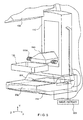
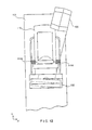
- Figs. 1-6 illustrate a non-limiting example of a multi-mode mammography/ tomosynthesis system comprising a gantry 100 and a data acquisition work-station 102.
- Gantry 100 includes a housing 104 supporting a tube arm assembly 106 rotatably mounted thereon to pivot about a horizontal axis 402 ( Fig. 4 ) and carrying an x-ray tube assembly 108.
- X-ray tube assembly 108 includes (1) an x-ray tube generating x-ray energy in a selected range, such as 20-50 kV, at mAs such as in the range 3-400 mAs, with focal spots such as a nominal size 0.3 mm large spot and nominal size 0.1 mm small spot (2) supports for multiple filters such as molybdenum, rhodium, aluminum, copper, and tin filters, and (3) an adjustable collimation assembly selectively collimating the x-ray beam from the focal spot in a range such as from 7x8 cm to 24x29 when measured at the image plane of an x-ray image receptor included in the system, at a maximum source-image distance such as 75 cm.
- a selected range such as 20-50 kV
- focal spots such as a nominal size 0.3 mm large spot and nominal size 0.1 mm small spot
- supports for multiple filters such as molybdenum, rhodium, aluminum, copper, and tin filters
- an adjustable collimation assembly selectively
- a compression arm assembly 110 that comprises a compression plate 122 and a receptor housing 114 having an upper surface 116 serving as a breast plate and enclosing a detector subsystem system 117 comprising a flat panel x-ray receptor 502 ( Fig. 5 ), a retractable anti-scatter grid 504 and a mechanism 506 for driving and retracting anti-scatter grid 504.
- Housing 104 also encloses the following components schematically illustrated in Fig.
- housing 104 also encloses suitable motors and electrical and mechanical components and connections to implement the functions discussed here.
- a patient shield 200 schematically illustrated in Fig.
- Work-station 102 comprises components similar to those in the SeleniaTM mammography system, including a display screen (typically a flat panel display that may include touch-screen functionality), user interface devices such as a keyboard, possibly a touch-screen, and a mouse or trackball, and various switches and indicator lights and/or displays. Work-station 102 also includes computer facilities similar to those of the SeleniaTM system (but adapted through hardware, firmware and software differences) for controlling gantry 100 and for processing, storing and displaying data received from gantry 100.
- a power generation facility for x-ray tube assembly 108 may be included in housing 104 or in work-station 102.
- a power source 118 powers work-station 102.
- Gantry 100 and work-station 102 exchange data and controls over a schematically illustrated connection 120.
- additional storage facilities 602 can be connected to work-station 102, such as one or more optical disc drives for storing information such as images and/or for providing information to work-station 102 such as previously obtained images and software, or a local printer (not shown).
- work-station 102 such as one or more optical disc drives for storing information such as images and/or for providing information to work-station 102 such as previously obtained images and software, or a local printer (not shown).
- the disclosed system can be connected to a hospital or local area or other network 604, and through the network to other systems such as a soft copy workstation 606, a CAD (Computer Aided Detection) station 608 for computer- processing mammography and/or tomosynthesis images to identify likely abnormalities, an image printer 610 for printing images, a technologist workstation 612, other imaging systems 614 such as other mammography systems or systems for other modalities for exchange of images and/or other information, and to a PACS (Picture Archiving) systems 616 for archiving images and other information and/or retrieving images and other information.
- CAD Computer Aided Detection
- PACS Picture Archiving
- the illustrated system has several modes of operation.
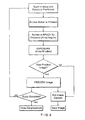
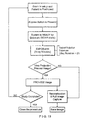
- An example of typical workflow generally applicable for each mode is illustrated in Fig. 7 , and several examples of operational modes are discussed below. Of course, this is only one example and workflow steps may be arranged differently.
- the operator can perform x-ray exposure using manual setting of technic factors such as mA and mSec, or can use an automatic exposure control as known in the art to set the exposure time, kV and filter modes for an image, for example by using a short, low-x-ray dose pre-exposure.
- Work-station 102 is set up to record the exposure technic information and associate it with the breast image for later review.
- tube arm assembly 106 and compression arm assembly 110 are coupled and locked together by 410 in a relative position such as seen in Fig. 1 , such that an x-ray beam from x-ray tube assembly 108 illuminates x-ray receptor 502 when the patient's breast is compressed by compression device 112.
- the system operates in a manner similar to said SeleniaTM system to take a mammogram.
- Vertical travel assembly 404 and tube arm rotation mechanism 406 can make vertical adjustments to accommodate a patient, and can rotate tube arm assembly 106 and compression arm assembly 110 together as a unit about axis 402 for different image orientations such as for CC and for MLO images.
- tube arm assembly 106 and compression arm assembly 110 can rotate between (-195°) and (+150°) about axis 402.
- compression device 112 includes a compression paddle 122 that can move laterally, in a direction along the chest wall of a patient, to adjust for different imaging orientations.
- the mechanism for supporting and moving compression paddle 122 is different.
- anti-scatter grid 504 is over x-ray receptor 502 in the standard mammography mode to reduce the effect of x-ray scatter.
- Fig. 8 illustrates a typical workflow for an exposure in standard mammography mode
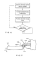
- Fig. 10 illustrates an example of the operation of detector subsystem 117 in standard mammography. Of course, these are only examples; other workflow steps or orders of steps can be used instead.
- the patient's breast can be spaced from upper surface 116, for example by an x-ray translucent spacer gantry 1002 ( Fig. 10 ), with the system otherwise similar to Fig. 1 , for a magnification of up to 1.8, for example.
- tube arm assembly 106 and compression arm assembly 110 are locked to each other and can move up or down and rotate about axis 402 for different image orientation.
- a different spacer 1002 can be used for a different degree of magnification.
- differently shaped or dimensioned compression paddles 122 can be used for different breast compression effects.
- the x-ray tube in x-ray tube assembly 108 can be set to a smaller focal spot size to improve a diagnostic image.
- anti-scatter grid 504 typically is retracted when magnification is used such that grid 504 is completely out of the image.
- the user can elect not to use a spacer 1002 in diagnostic imaging, in which case anti-scatter grid 504 can be used over the entire image.
- a number of breast images are taken while the patient's breast remains compressed.
- an agent such as iodine is injected into the patient and after a suitable waiting time such as about one minute for a maximum uptake, two images breast are taken in rapid succession, for example one at an x-ray energy just above the K-edge of iodine and one at an energy just below the K-edge.
- a succession of breast images can be taken at a single x-ray energy band or bands just above and below the K-edge, or at another x-ray energy range, to track the uptake of agent over time.
- Still another dynamic imaging mode technique comprises injecting a contrast agent and taking a succession of images over a period such as 5-7 minutes, for example one image every minute, and processing the image data to generate for each pixel, or at least for each pixel of interest, a histogram of the change in the pixel value, to thereby use the manner in which pixel values change to differential abnormal tissue.
- work-station 102 can store preset data that commands gantry 100 and work-station 102 to take a desired sequence of images for the dynamic mode technique selected by the operator, such that the command data sets the appropriate parameters such as x-ray energy, dose, timing of images, etc.
- processing to assess changes in pixel values can be done for a region of interest rather than over individual pixels, to produce information such as a measure of changes in the average pixel values in the region of interest.
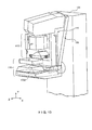
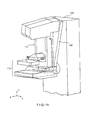
- tube arm assembly 106 and compression arm assembly 110 are decoupled by unit 410 such that compression arm assembly 110 stays in one position, compressing the patient's breast, while tube arm assembly 106 rotates about axis 402, for example between the position illustrated in Fig. 2 to that illustrated in Fig. 11 , or ⁇ 15° relative to compression arm assembly 110.
- Tomosynthesis can be carried out for different image orientations, so that compression arm assembly 110 can be rotated about axis 402 (alone or together with assembly 106) for a desired image orientation and locked in place, and then tube arm assembly 106 can be rotated relative to that position of compression arm assembly 110 for tomosynthesis imaging over ⁇ 15° or some other desired angular range.
- 11 images are taken during an angular sweep of tube arm assembly 106, one every approximately 3°.
- a different number of images can be taken, for example up to 21 during a single sweep.
- the x-ray tube in x-ray tube assembly 108 continuously rotates and the x-ray tube is pulsed for each image, for example, for x-ray energy pulses each lasting approximately 100 mSec, although pulses of different duration can be selected.
- the rotational motion can stop for taking each image, or continuous motion without pulsing can be used (and the timing of data measurements relied to define pixel values). As seen in Figs.
- mechanism 506 fully retracts anti-scatter grid 504 away from x-ray receptor 502 so grid 504 is out of the image.
- x-ray receptor 502 rocks within receptor housing 114. In this rocking motion, controlled by unit 408 ( Fig. 4 ), a line normal to the image face of x-ray receptor 502 may keep pointing to the focal spot of the x-ray tube in x-ray tube assembly 108.
- the rotation of tube arm assembly 106 and rocking of x-ray receptor 502 can be through different angles; for example, tube arm assembly 106 can rotate through 15° while x-ray receptor 502 rocks through 5°, i.e. the rocking angle can be an amount one-third that of assembly 108.
- Synchronous rotation of tube arm assembly 106 and rocking of x-ray receptor 502 can be achieved by controlling separate motors for each or, alternatively, through using a motor to drive tube arm assembly 106 and a mechanical coupling between the rotation of tube arm assembly 106 and rocking of x-ray receptor 502.
- Image data can be obtained and processed into tomosynthesis images for display and/or storage as described in the material incorporated by reference, for example in co-pending patent application Ser. No.
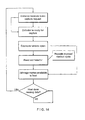
- Fig. 13 illustrates a typical workflow for tomosynthesis mode operation
- Fig. 14 illustrates an example of the operation of detector subsystem 117 in that mode. Again, these are only examples, and other steps or orders of steps can be used instead.
- a combination mode during a single compression of the patient's breast the system takes a conventional mammogram and tomosynthesis images.
- tube arm assembly 106 sweeps and x-ray receptor 502 rocks, each through an appropriate angle, and exposures are taken for tomosynthesis images, and (2) a standard mammogram is taken.
- the standard mammogram can be taken at a 0° relative angle between tube arm assembly 106 and a normal to the imaging plane of x-ray receptor 502, and can be taken before or after the tomosynthesis images are taken or between the taking of two successive tomosynthesis images.
- each tomosynthesis image utilizes substantially lower x-ray dose than the standard mammogram.
- the total x-ray dosage for tomosynthesis imaging in one sweep of tube arm assembly 106 can be approximately the same as that for a single standard mammogram, or up to approximately three times that dosage.
- the relationship between the two dosages can be user-selected.
- Figure 15 illustrates an example of workflow for the combination mode
- Fig. 16 illustrates an example of the operation of detector subsystem 117 in that mode. Again, these are examples, and different steps or orders of steps can be used instead.
- a preferred approach may be to take the standard mammogram first, then move arm 106 to one end of its rotational range for tomosynthesis and take the tomosynthesis images.
- the order in which the two types of images are taken may be optimized such that the overall imaging time is minimized, and an order that achieves such minimization can be the preferred order.
- the exposure (tube current mA, tube voltage kVp, and exposure length msec) techniques for the standard mammogram and the tomosynthesis exposures can be set manually, or by using automatic methods. If the standard mammogram is taken first, its exposure techniques can be used to set an optimal technique for the subsequent tomosynthesis images, and vice versa.
- the exposure technique can be modified dynamically, if the software senses that the signal reaching the image receptor is either too low or too high and adjust subsequent exposures as needed.
- X-ray receptor 502 can remain in place for this procedure, or can be rocked through a selected angle, for example through an angle sufficient to maintain the same orientation of the imaging surface of receptor 502 relative to tube arm assembly 106.
- a spacer 1002 can be used for magnification.
- the two or more images can be used to identify the location of a lesion, so that needle biopsy can be used, for example with an upright needle biopsy station 412 ( Fig. 4 ) in a manner similar to that used with the commercially available SeleniaTM system and StereoLoc IITM.
- a compression paddle 122 appropriate for needle biopsy typically is used when taking the stereotactic images.
- some or all of the images taken in the tomosynthesis mode and/or in the combined mode can be used to identify the location of a lesion for biopsy, in which case a compression paddle 122 appropriate for the purpose typically is used when taking the images.
- x-ray images can be taken after a biopsy or other needle is inserted into the compressed breast.
- imaging such as in the stereotactic mode, the tomosynthesis mode, or the combined mode can be used.
- compression paddle 122 is movable laterally, as generally described in U.S. Patent Application Publication No. 2005/0063509 A1 , hereby incorporated by reference herein.
- compression paddle 122 can pivot about an axis along the patient's chest wall to conform the breast shape in certain procedures, as discussed in said U.S. Patent 5,706,327 .
- compression paddle 122 is mounted differently and moves in a different manner.
- compression paddle 122 is removably mounted to a support 510 that moves up and down compression arm assembly 110 as needed for breast compression.
- a projection compression paddle 122a of the paddle engages a projection 510a of the support, and a projection 122b of the paddle latches onto projection 510b of the support.
- Projection 510a is spring-loaded, such as by a spring schematically illustrates at 510c to allow for pivoting compression paddle 122 about an axis where it latches onto 510, as illustrated by arrow A, for better conformance with the compressed breast in some imaging protocols.
- Imaging protocols may require compression paddle 122 not to pivot, in which case projection 510a is locked in place by a locking mechanism in 510 (not shown) to keep compression paddle 122 in place relative to support 510.
- the locking mechanism can be manually set to a lock position, and manually unlocked by the operator. Alternatively, the locking mechanism can be controlled through an operator input at gantry 100 or work-station 102.
- a sensing mechanism can be included to sense whether compression paddle 122 is locked against pivoting, to provide information that work-station 102 can use for setting imaging protocols such as for automated breast compression and automated exposure methods.
- Two knobs 510d can be manually rotated to move projection 510b and thus compression paddle 122 laterally such that it compress a breast that is not centered laterally on upper surface 116, for example for MLO imaging.
- Each knob 510d can operate a mechanism such as an endless screw rotating in a nut secured to projection 510b.
- projection 510b and thus compression paddle 122 can be driven laterally by a motor, under control of operator switches or other interface at gantry 100 or at work-station 102, or automatically positioned laterally under computer control.
- compression paddle 122 is driven for lateral movement by components that are a part of support 510.
- compression paddle 122 can be simple structure, and can even be disposable, with a new one used for each patient or for only a few patients. This can simplify and reduce the cost of using the system, because an imaging facility usually stocks a number of different paddles for different purposes. If the lateral movement mechanism is integral with a compression paddle, the paddle assembly is considerably larger, heavier and more expensive.
- Compression paddle 122 can include a bar code that is automatically read by a bar code reader in support 510, to keep work-station 102 informed of the paddle currently mounted to support 510, for use in automating imaging protocols.
- the bar code information can be checked to ensure through computer processing that the type of paddle that is currently mounted on support 510 matches the imaging that will be commanded, and the information from the sensor for whether compression paddle 122 is locked in non-tilting mode can be used to automatically make adjustments for compression height to ensure accurate automatic x-ray exposure operation. Further, the bar code information identifying the paddle can be used to automatically set collimation in x-ray tube assembly 108 so that the x-ray beam matches the size and shape of the currently installed compression paddle 122.
Landscapes
- Health & Medical Sciences (AREA)
- Life Sciences & Earth Sciences (AREA)
- Medical Informatics (AREA)
- Engineering & Computer Science (AREA)
- Radiology & Medical Imaging (AREA)
- Molecular Biology (AREA)
- Biophysics (AREA)
- Nuclear Medicine, Radiotherapy & Molecular Imaging (AREA)
- Optics & Photonics (AREA)
- Pathology (AREA)
- Physics & Mathematics (AREA)
- Biomedical Technology (AREA)
- Heart & Thoracic Surgery (AREA)
- High Energy & Nuclear Physics (AREA)
- Surgery (AREA)
- Animal Behavior & Ethology (AREA)
- General Health & Medical Sciences (AREA)
- Public Health (AREA)
- Veterinary Medicine (AREA)
- Dentistry (AREA)
- Oral & Maxillofacial Surgery (AREA)
- Apparatus For Radiation Diagnosis (AREA)
Applications Claiming Priority (3)
| Application Number | Priority Date | Filing Date | Title |
|---|---|---|---|
| US63129604P | 2004-11-26 | 2004-11-26 | |
| PCT/US2005/042613 WO2006058160A2 (fr) | 2004-11-26 | 2005-11-23 | Systeme et procede radiographiques multimode integrant mammographie/tomosynthese |
| EP05852126.1A EP1816965B1 (fr) | 2004-11-26 | 2005-11-23 | Systeme radiographique multimode integrant mammographie/tomosynthese |
Related Parent Applications (1)
| Application Number | Title | Priority Date | Filing Date |
|---|---|---|---|
| EP05852126.1A Division EP1816965B1 (fr) | 2004-11-26 | 2005-11-23 | Systeme radiographique multimode integrant mammographie/tomosynthese |
Publications (3)
| Publication Number | Publication Date |
|---|---|
| EP3106094A2 true EP3106094A2 (fr) | 2016-12-21 |
| EP3106094A3 EP3106094A3 (fr) | 2017-01-04 |
| EP3106094B1 EP3106094B1 (fr) | 2021-09-08 |
Family
ID=36498527
Family Applications (2)
| Application Number | Title | Priority Date | Filing Date |
|---|---|---|---|
| EP16176648.0A Expired - Lifetime EP3106094B1 (fr) | 2004-11-26 | 2005-11-23 | Systeme radiographique multimode integrant mammographie/tomosynthese |
| EP05852126.1A Expired - Lifetime EP1816965B1 (fr) | 2004-11-26 | 2005-11-23 | Systeme radiographique multimode integrant mammographie/tomosynthese |
Family Applications After (1)
| Application Number | Title | Priority Date | Filing Date |
|---|---|---|---|
| EP05852126.1A Expired - Lifetime EP1816965B1 (fr) | 2004-11-26 | 2005-11-23 | Systeme radiographique multimode integrant mammographie/tomosynthese |
Country Status (3)
| Country | Link |
|---|---|
| US (8) | US7869563B2 (fr) |
| EP (2) | EP3106094B1 (fr) |
| WO (1) | WO2006058160A2 (fr) |
Families Citing this family (140)
| Publication number | Priority date | Publication date | Assignee | Title |
|---|---|---|---|---|
| US8565372B2 (en) * | 2003-11-26 | 2013-10-22 | Hologic, Inc | System and method for low dose tomosynthesis |
| US8571289B2 (en) | 2002-11-27 | 2013-10-29 | Hologic, Inc. | System and method for generating a 2D image from a tomosynthesis data set |
| US7577282B2 (en) | 2002-11-27 | 2009-08-18 | Hologic, Inc. | Image handling and display in X-ray mammography and tomosynthesis |
| US10638994B2 (en) | 2002-11-27 | 2020-05-05 | Hologic, Inc. | X-ray mammography with tomosynthesis |
| US7616801B2 (en) * | 2002-11-27 | 2009-11-10 | Hologic, Inc. | Image handling and display in x-ray mammography and tomosynthesis |
| US7123684B2 (en) * | 2002-11-27 | 2006-10-17 | Hologic, Inc. | Full field mammography with tissue exposure control, tomosynthesis, and dynamic field of view processing |
| US8768026B2 (en) | 2003-11-26 | 2014-07-01 | Hologic, Inc. | X-ray imaging with x-ray markers that provide adjunct information but preserve image quality |
| US7662082B2 (en) | 2004-11-05 | 2010-02-16 | Theragenics Corporation | Expandable brachytherapy device |
| EP2602743B1 (fr) | 2004-11-15 | 2014-11-05 | Hologic, Inc. | Affichage et génération de géométrie de mise en correspondance de mammographies et d'images de tomosynthèse |
| EP3106094B1 (fr) * | 2004-11-26 | 2021-09-08 | Hologic, Inc. | Systeme radiographique multimode integrant mammographie/tomosynthese |
| US8079946B2 (en) | 2005-11-18 | 2011-12-20 | Senorx, Inc. | Asymmetrical irradiation of a body cavity |
| WO2007095330A2 (fr) | 2006-02-15 | 2007-08-23 | Hologic Inc | Biopsie mammaire et localisation a l'aiguille a l'aide de systemes de tomosynthese |
| US10682107B2 (en) * | 2007-01-31 | 2020-06-16 | Philips Digital Mammography Sweden Ab | Method and arrangement relating to x-ray imaging |
| US7630533B2 (en) | 2007-09-20 | 2009-12-08 | Hologic, Inc. | Breast tomosynthesis with display of highlighted suspected calcifications |
| DE102008004473A1 (de) * | 2008-01-15 | 2009-07-23 | Siemens Aktiengesellschaft | Verfahren und Vorrichtung zur Erzeugung eines tomosynthetischen 3D-Röntgenbildes |
| US7792245B2 (en) * | 2008-06-24 | 2010-09-07 | Hologic, Inc. | Breast tomosynthesis system with shifting face shield |
| US7991106B2 (en) | 2008-08-29 | 2011-08-02 | Hologic, Inc. | Multi-mode tomosynthesis/mammography gain calibration and image correction using gain map information from selected projection angles |
| AU2015224382B2 (en) * | 2008-09-04 | 2017-03-30 | Hologic, Inc. | Integrated multi-mode mammography/tomosynthesis x-ray system and method |
| KR20110063659A (ko) * | 2008-09-04 | 2011-06-13 | 홀로직, 인크. | 통합된 다중 모드 마모그래피/토모신테시스 x선 시스템 및 방법 |
| US7801267B2 (en) * | 2008-10-23 | 2010-09-21 | General Electric Co. | Method and system for auto positioning compression mechanism in a mammography system |
| CN102176866B (zh) | 2008-11-24 | 2013-10-16 | 霍罗吉克公司 | 控制用于体层合成和乳房x射线照相术成像的x射线焦斑特性的方法和系统 |
| FI123261B (fi) | 2008-11-28 | 2013-01-15 | Planmed Oy | 3D mammografia |
| US9579524B2 (en) | 2009-02-11 | 2017-02-28 | Hologic, Inc. | Flexible multi-lumen brachytherapy device |
| US9248311B2 (en) | 2009-02-11 | 2016-02-02 | Hologic, Inc. | System and method for modifying a flexibility of a brachythereapy catheter |
| US8170320B2 (en) | 2009-03-03 | 2012-05-01 | Hologic, Inc. | Mammography/tomosynthesis systems and methods automatically deriving breast characteristics from breast x-ray images and automatically adjusting image processing parameters accordingly |
| EP2408375B1 (fr) | 2009-03-20 | 2017-12-06 | Orthoscan Incorporated | Appareil mobile d'imagerie |
| JP5373450B2 (ja) * | 2009-03-31 | 2013-12-18 | 富士フイルム株式会社 | 生検装置及び生検装置の動作方法 |
| US10207126B2 (en) | 2009-05-11 | 2019-02-19 | Cytyc Corporation | Lumen visualization and identification system for multi-lumen balloon catheter |
| JP5355271B2 (ja) * | 2009-07-24 | 2013-11-27 | 富士フイルム株式会社 | 放射線画像撮影装置 |
| JP5572040B2 (ja) * | 2009-09-28 | 2014-08-13 | 富士フイルム株式会社 | 放射線撮影装置 |
| EP2485651B1 (fr) | 2009-10-08 | 2020-12-23 | Hologic, Inc. | Système de ponction-biopsie du sein |
| US8744041B2 (en) | 2010-09-09 | 2014-06-03 | Hologic, Inc. | Methods and systems for dynamically modifying acquisition parameter during image acquisition |
| US9352172B2 (en) | 2010-09-30 | 2016-05-31 | Hologic, Inc. | Using a guide member to facilitate brachytherapy device swap |
| EP2624761B1 (fr) | 2010-10-05 | 2021-07-14 | Hologic, Inc. | Imagerie du sein par rayon x en position debout avec un mode de tomodensitométrie, de multiples modes de tomosynthèse et un mode de mammographie |
| WO2015054518A1 (fr) | 2013-10-09 | 2015-04-16 | Hologic, Inc | Tomosynthèse du sein à rayons x améliorant la résolution spatiale y compris dans le sens de l'épaisseur du sein aplati |
| WO2012074885A1 (fr) | 2010-11-24 | 2012-06-07 | Hologic, Inc. | Système pour une manipulation améliorée de tissu et analyse du tissu en ligne |
| US20120133600A1 (en) | 2010-11-26 | 2012-05-31 | Hologic, Inc. | User interface for medical image review workstation |
| WO2012082799A1 (fr) | 2010-12-13 | 2012-06-21 | Orthoscan, Inc. | Système d'imagerie fluoroscopique mobile |
| WO2012082861A2 (fr) * | 2010-12-14 | 2012-06-21 | Hologic, Inc. | Système et procédé de fusion de données d'image tridimensionnelle provenant d'une pluralité de systèmes d'imagerie différents destinés à être utilisés en imagerie diagnostique |
| FR2969918B1 (fr) * | 2010-12-29 | 2013-12-13 | Gen Electric | Procede et dispositif de mise en oeuvre d'une grille anti-diffusante |
| US10342992B2 (en) | 2011-01-06 | 2019-07-09 | Hologic, Inc. | Orienting a brachytherapy applicator |
| ITBO20110086A1 (it) * | 2011-02-25 | 2012-08-26 | I M S Internaz Medicoscienti Fica S R L | Apparecchiatura per la mammografia e/o la tomosintesi con dispositivo di rimozione della radiazione diffusa. |
| CA2829349C (fr) | 2011-03-08 | 2021-02-09 | Hologic, Inc. | Systeme et procede pour une imagerie de seins a double energie et/ou a injection d'un agent de contraste pour un depistage, un diagnostic et une biopsie |
| JP5355619B2 (ja) * | 2011-04-27 | 2013-11-27 | 富士フイルム株式会社 | 放射線画像撮影装置 |
| CN103841893A (zh) * | 2011-07-01 | 2014-06-04 | 韩国睿恩斯有限公司 | 具备多传感器的乳房成像用探测器及可获得三维影像的乳房成像装置 |
| CN103648388B (zh) * | 2011-07-04 | 2017-05-03 | 皇家飞利浦有限公司 | 相位对比度成像设备 |
| USD739534S1 (en) * | 2011-10-05 | 2015-09-22 | General Electric Company | Tomosynthesis device |
| US11259759B2 (en) | 2011-11-18 | 2022-03-01 | Hologic Inc. | X-ray mammography and/or breast tomosynthesis using a compression paddle |
| US9782135B2 (en) | 2011-11-18 | 2017-10-10 | Hologic, Inc. | X-ray mammography and/or breast tomosynthesis using a compression paddle |
| JP6157491B2 (ja) * | 2011-11-18 | 2017-07-05 | ホロジック, インコーポレイテッドHologic, Inc. | 造影と患者の快適性とを改善する膨張性ジャケットを備えた圧迫パドルを用いたx線マンモグラフィーおよび/または乳房トモシンセシス |
| JP2014534042A (ja) | 2011-11-27 | 2014-12-18 | ホロジック, インコーポレイテッドHologic, Inc. | マンモグラフィーおよび/またはトモシンセシス画像データを使用して2d画像を生成するためのシステムおよび方法 |
| US9805507B2 (en) | 2012-02-13 | 2017-10-31 | Hologic, Inc | System and method for navigating a tomosynthesis stack using synthesized image data |
| JP6016403B2 (ja) * | 2012-03-27 | 2016-10-26 | キヤノン株式会社 | 画像処理装置、画像処理方法 |
| DE102012217301B4 (de) * | 2012-09-25 | 2021-10-14 | Bayer Pharma Aktiengesellschaft | Kombination aus Kontrastmittel und Mammographie-CT-System mit vorgegebenem Energiebereich und Verfahren zur Erzeugung tomographischer Mammographie-CT-Aufnahmen durch diese Kombination |
| US9517038B2 (en) | 2012-10-12 | 2016-12-13 | University Of Virginia Patent Foundation | Apparatus and method for breast immobilization |
| KR101437273B1 (ko) * | 2013-03-12 | 2014-09-03 | 제너럴 일렉트릭 캄파니 | 디지털 맘모그래피 장치 |
| CA2905598A1 (fr) | 2013-03-14 | 2014-09-18 | Sunnybrook Research Institute | Systeme et procede pour evaluation de la densite des seins par exposition a une faible dose de rayons x |
| JP6388347B2 (ja) | 2013-03-15 | 2018-09-12 | ホロジック, インコーポレイテッドHologic, Inc. | 腹臥位におけるトモシンセシス誘導生検 |
| ES2798306T3 (es) | 2013-03-15 | 2020-12-10 | Hologic Inc | Dispositivo de reducción de dispersión de rayos X para su uso con mamografía en 2D y 3D |
| EP2967473B1 (fr) | 2013-03-15 | 2020-02-19 | Hologic, Inc. | Système et procédé pour explorer une pile de tomosynthèse comprenant une mise au point automatique |
| ES2750678T3 (es) | 2013-04-26 | 2020-03-26 | Hologic Inc | Mamografía de rayos x y/o tomosíntesis de mama con una paleta de compresión |
| US9417194B2 (en) * | 2013-08-16 | 2016-08-16 | General Electric Company | Assessment of focal spot characteristics |
| DE102013217961A1 (de) * | 2013-09-09 | 2015-03-12 | Siemens Aktiengesellschaft | Verfahren und Vorrichtung zur Untersuchung einer Gewebeprobe |
| EP4278977A3 (fr) * | 2013-10-24 | 2024-02-21 | Hologic, Inc. | Système et procédé de navigation pour une biopise du sein guidée par rayons x |
| FI130432B (fi) | 2013-11-29 | 2023-08-28 | Planmed Oy | Tomosynteesikalibrointi mammografian yhteydessä |
| WO2015121714A1 (fr) * | 2014-02-14 | 2015-08-20 | Panacea Medical Technologies Pvt Ltd | Dispositif intégré pour réaliser une mammographie, une tomosynthèse et une biopsie stéréotaxique en positions multiples |
| US10273851B2 (en) * | 2014-02-28 | 2019-04-30 | Scania Cv Ab | Exhaust treatment system and method for treatment of an exhaust stream |
| JP6506769B2 (ja) | 2014-02-28 | 2019-04-24 | ホロジック, インコーポレイテッドHologic, Inc. | トモシンセシス画像スラブを生成し表示するためのシステムおよび方法 |
| EP3215017B1 (fr) * | 2014-11-07 | 2021-01-06 | Hologic, Inc. | Appareil à palette pivotante pour système à rayons x de mammographie/tomosynthèse |
| KR20160057626A (ko) | 2014-11-14 | 2016-05-24 | 삼성전자주식회사 | 유방 영상 촬영장치 |
| JP6611428B2 (ja) * | 2014-12-09 | 2019-11-27 | キヤノン株式会社 | マンモ断層撮像システム |
| JP6491471B2 (ja) * | 2014-12-24 | 2019-03-27 | キヤノン株式会社 | 画像処理装置、画像処理方法およびプログラム |
| WO2017040977A1 (fr) | 2015-09-04 | 2017-03-09 | Faxitron Bioptics, Llc | Dispositif d'imagerie d'échantillons multi-axe avec marqueurs d'orientation incorporés |
| US11076820B2 (en) | 2016-04-22 | 2021-08-03 | Hologic, Inc. | Tomosynthesis with shifting focal spot x-ray system using an addressable array |
| US10157460B2 (en) | 2016-10-25 | 2018-12-18 | General Electric Company | Interpolated tomosynthesis projection images |
| CN110121299A (zh) | 2016-11-04 | 2019-08-13 | 霍罗吉克公司 | 医学成像装置和操作医学成像装置的方法 |
| WO2018085719A1 (fr) | 2016-11-04 | 2018-05-11 | Hologic, Inc. | Système de radiographie d'échantillon |
| EP3868302B1 (fr) | 2016-11-08 | 2023-12-06 | Hologic, Inc. | Imagerie avec éléments de compression incurvés |
| US10096106B2 (en) | 2016-11-10 | 2018-10-09 | General Electric Company | Combined medical imaging |
| WO2018098321A1 (fr) * | 2016-11-25 | 2018-05-31 | Hologic, Inc. | Dispositif de commande pour appareil d'imagerie |
| USD831216S1 (en) * | 2016-11-25 | 2018-10-16 | Hologic, Inc. | Imaging system |
| US10463333B2 (en) | 2016-12-13 | 2019-11-05 | General Electric Company | Synthetic images for biopsy control |
| US10646180B2 (en) | 2017-01-03 | 2020-05-12 | General Electric Company | System and method for breast imaging |
| EP3600047A1 (fr) | 2017-03-30 | 2020-02-05 | Hologic, Inc. | Système et procédé de synthèse et de représentation d'image de caractéristique multiniveau hiérarchique |
| CN110621233B (zh) | 2017-03-30 | 2023-12-12 | 豪洛捷公司 | 用于处理乳房组织图像数据的方法 |
| CN110662489B (zh) | 2017-03-30 | 2024-08-02 | 豪洛捷公司 | 用于靶向对象增强以生成合成乳房组织图像的系统和方法 |
| WO2018204710A1 (fr) | 2017-05-03 | 2018-11-08 | Hologic, Inc. | Dispositifs de réduction de fluide dans le champ d'imagerie d'un appareil de manipulation de tissus pour améliorer la qualité d'imagerie d'un système de biopsie |
| WO2018236565A1 (fr) | 2017-06-20 | 2018-12-27 | Hologic, Inc. | Procédé et système d'imagerie médicale à auto-apprentissage dynamique |
| ES3014459T3 (en) | 2017-08-11 | 2025-04-22 | Hologic Inc | Breast compression paddle |
| EP3664713A4 (fr) | 2017-08-11 | 2021-04-28 | Hologic, Inc. | Plaque de compression du sein pourvue de coins d'accès |
| DE202018006903U1 (de) | 2017-08-16 | 2024-07-29 | Hologic Inc. | Techniken zur Patientenbewegungsartefaktkompensation bei Brustbildgebung |
| EP3449835B1 (fr) | 2017-08-22 | 2023-01-11 | Hologic, Inc. | Système et méthode de tomographie assistée par ordinateur pour imager de multiples cibles anatomiques |
| EP3682228B1 (fr) | 2017-09-11 | 2025-12-17 | Faxitron Bioptics, LLC | Système d'imagerie à grossissement d'objet adaptatif |
| DE102018200108A1 (de) * | 2018-01-05 | 2019-07-11 | Siemens Healthcare Gmbh | Positionierung eines Untersuchungsobjekts bezüglich eines Röntgengeräts |
| JP6945491B2 (ja) * | 2018-04-27 | 2021-10-06 | 富士フイルム株式会社 | マンモグラフィ装置 |
| CA3091593A1 (fr) | 2018-05-04 | 2019-11-07 | Hologic, Inc. | Visualisation d'aiguille de biopsie |
| US12121304B2 (en) | 2018-05-04 | 2024-10-22 | Hologic, Inc. | Introducer and localization wire visualization |
| US11684323B2 (en) | 2018-05-25 | 2023-06-27 | Hologic, Inc. | Membrane-based breast compression systems |
| ES2988504T3 (es) * | 2018-06-22 | 2024-11-20 | Hologic Inc | Sistema de ultrasonidos de múltiples posiciones |
| US11090017B2 (en) | 2018-09-13 | 2021-08-17 | Hologic, Inc. | Generating synthesized projection images for 3D breast tomosynthesis or multi-mode x-ray breast imaging |
| US12059282B2 (en) | 2018-09-17 | 2024-08-13 | Hologic, Inc. | Medical imaging system with contoured detector |
| WO2020068851A1 (fr) | 2018-09-24 | 2020-04-02 | Hologic, Inc. | Cartographie mammaire et localisation d'anomalie |
| CN112770674B (zh) | 2018-09-28 | 2024-08-06 | 豪洛捷公司 | 通过高密度元素抑制来合成乳腺组织的图像生成的系统和方法 |
| DE202019006040U1 (de) | 2018-11-25 | 2024-08-06 | Hologic Inc. | Multimodalitätsaufhängungsprotokolle |
| ES2939882T3 (es) | 2018-12-26 | 2023-04-27 | Hologic Inc | Obtención de imágenes de tejido en presencia de líquido durante procedimiento de biopsia |
| KR20210148132A (ko) | 2019-03-29 | 2021-12-07 | 홀로직, 인크. | 스닙-트리거링된 디지털 영상 보고서 생성 |
| US11883206B2 (en) | 2019-07-29 | 2024-01-30 | Hologic, Inc. | Personalized breast imaging system |
| US11694792B2 (en) | 2019-09-27 | 2023-07-04 | Hologic, Inc. | AI system for predicting reading time and reading complexity for reviewing 2D/3D breast images |
| US11783476B2 (en) | 2019-10-25 | 2023-10-10 | DeepHealth, Inc. | System and method for analyzing three-dimensional image data |
| EP3832689A3 (fr) | 2019-12-05 | 2021-08-11 | Hologic, Inc. | Systèmes et procédés pour améliorer la durée de vie d'un tube à rayons x |
| EP4081952A4 (fr) | 2019-12-23 | 2024-01-24 | Deephealth, Inc. | Systèmes et procédés pour analyser des données d'images bidimensionnelles et tridimensionnelles |
| JP7742349B2 (ja) | 2020-01-24 | 2025-09-19 | ホロジック, インコーポレイテッド | 水平に変位可能な発泡体乳房圧迫パドル |
| EP4101385B1 (fr) | 2020-02-04 | 2024-10-16 | FUJIFILM Corporation | Dispositif, procédé et programme de réglage d'image |
| JP7321299B2 (ja) | 2020-02-04 | 2023-08-04 | 富士フイルム株式会社 | 画像設定装置、画像設定装置の作動方法および画像設定プログラム |
| WO2021182229A1 (fr) | 2020-03-13 | 2021-09-16 | 富士フイルム株式会社 | Dispositif et programme de génération d'image, dispositif et programme d'apprentissage, et dispositif et programme de traitement d'image |
| WO2021186956A1 (fr) | 2020-03-18 | 2021-09-23 | 富士フイルム株式会社 | Dispositif de traitement d'image, procédé et programme |
| WO2021186957A1 (fr) | 2020-03-18 | 2021-09-23 | 富士フイルム株式会社 | Dispositif de traitement d'image, procédé et programme |
| US12505645B2 (en) | 2020-03-27 | 2025-12-23 | Hologic, Inc. | Systems and methods for correlating regions of interest in multiple imaging modalities |
| US11471118B2 (en) | 2020-03-27 | 2022-10-18 | Hologic, Inc. | System and method for tracking x-ray tube focal spot position |
| JP7671776B2 (ja) | 2020-03-27 | 2025-05-02 | ホロジック, インコーポレイテッド | 複数の画像化モダリティにおける関心対象領域を識別するシステム及び方法 |
| US11481038B2 (en) | 2020-03-27 | 2022-10-25 | Hologic, Inc. | Gesture recognition in controlling medical hardware or software |
| US12121384B2 (en) | 2020-03-31 | 2024-10-22 | Hologic, Inc. | Systems and methods for x-ray imaging tissue specimens |
| US11054534B1 (en) | 2020-04-24 | 2021-07-06 | Ronald Nutt | Time-resolved positron emission tomography encoder system for producing real-time, high resolution, three dimensional positron emission tomographic image without the necessity of performing image reconstruction |
| US11300695B2 (en) | 2020-04-24 | 2022-04-12 | Ronald Nutt | Time-resolved positron emission tomography encoder system for producing event-by-event, real-time, high resolution, three-dimensional positron emission tomographic image without the necessity of performing image reconstruction |
| JP7510796B2 (ja) * | 2020-06-22 | 2024-07-04 | キヤノンメディカルシステムズ株式会社 | 医用画像診断システムおよび医用画像診断装置制御プログラム |
| US12396693B2 (en) | 2020-09-16 | 2025-08-26 | Hologic, Inc. | Systems and methods for confirming tissue specimens removed using contrast-enhanced x-ray imaging |
| US20220164951A1 (en) | 2020-11-20 | 2022-05-26 | Hologic, Inc. | Systems and methods for using ai to identify regions of interest in medical images |
| KR20240000563A (ko) | 2021-04-26 | 2024-01-02 | 홀로직, 인크. | 폼 압축 요소들의 두께를 측정하기 위한 시스템들 및 방법들 |
| US11786191B2 (en) | 2021-05-17 | 2023-10-17 | Hologic, Inc. | Contrast-enhanced tomosynthesis with a copper filter |
| KR102611174B1 (ko) * | 2021-06-29 | 2023-12-07 | 주식회사 디알텍 | 방사선 촬영장치 및 방사선 촬영방법 |
| US12186119B2 (en) | 2021-10-05 | 2025-01-07 | Hologic, Inc. | Interactive model interface for image selection in medical imaging systems |
| US12254586B2 (en) | 2021-10-25 | 2025-03-18 | Hologic, Inc. | Auto-focus tool for multimodality image review |
| CN114121334B (zh) * | 2021-11-16 | 2025-04-22 | 湖州霍里思特智能科技有限公司 | 一种射线准直调整装置 |
| CN118318247A (zh) | 2021-11-29 | 2024-07-09 | 豪洛捷公司 | 用于将感兴趣的对象相关的系统和方法 |
| US12414217B2 (en) | 2022-02-07 | 2025-09-09 | Hologic, Inc. | Systems and methods for adaptively controlling filament current in an X-ray tube |
| IT202200009080A1 (it) * | 2022-05-04 | 2023-11-04 | Ims Giotto S P A | Apparecchiatura di analisi medica |
| US12350085B2 (en) | 2023-02-15 | 2025-07-08 | Ge Healthcare | Mammography imaging system with universal attachment structures |
| US12426842B2 (en) | 2023-03-17 | 2025-09-30 | GE Precision Healthcare LLC | Methods and systems for flexible paddle for use with a mammography system |
| US12275370B2 (en) | 2023-08-03 | 2025-04-15 | GM Global Technology Operations LLC | System and method for determining whether a seatbelt buckle extender is used in conjunction with a seatbelt |
| JP2025045520A (ja) * | 2023-09-20 | 2025-04-02 | 富士フイルム株式会社 | 乳房撮影装置 |
Citations (12)
| Publication number | Priority date | Publication date | Assignee | Title |
|---|---|---|---|---|
| US4496557A (en) | 1981-08-27 | 1985-01-29 | Adir | Tricyclic ethers, their preparation and the pharmaceutical compositions containing them |
| US5051904A (en) | 1988-03-24 | 1991-09-24 | Olganix Corporation | Computerized dynamic tomography system |
| US5359637A (en) | 1992-04-28 | 1994-10-25 | Wake Forest University | Self-calibrated tomosynthetic, radiographic-imaging system, method, and device |
| US5706327A (en) | 1996-02-09 | 1998-01-06 | Trex Medical Corporation | Method and apparatus for mammographic compression |
| US6289235B1 (en) | 1998-03-05 | 2001-09-11 | Wake Forest University | Method and system for creating three-dimensional images using tomosynthetic computed tomography |
| US20010038861A1 (en) | 1999-12-16 | 2001-11-08 | Tsung-Min Hsu | Transdermal administration of nonsteroidal anti-inflammatory drugs using hydroxide-releasing agents as permeation enhancers |
| US6611575B1 (en) * | 2001-07-27 | 2003-08-26 | General Electric Company | Method and system for high resolution 3D visualization of mammography images |
| US6647092B2 (en) | 2002-01-18 | 2003-11-11 | General Electric Company | Radiation imaging system and method of collimation |
| US20040066884A1 (en) | 2002-10-07 | 2004-04-08 | Hermann Claus Bernhard Erich | Continuous scan tomosynthesis system and method |
| US20040066882A1 (en) | 2002-10-07 | 2004-04-08 | Eberhard Jeffrey Wayne | Continuous scan RAD tomosynthesis system and method |
| US20050063509A1 (en) | 2001-10-19 | 2005-03-24 | Defreitas Kenneth F | Mammography system and method employing offset compression paddles automatic collimation and retractable anti-scatter grid |
| US20050113681A1 (en) | 2002-11-27 | 2005-05-26 | Defreitas Kenneth F. | X-ray mammography with tomosynthesis |
Family Cites Families (310)
| Publication number | Priority date | Publication date | Assignee | Title |
|---|---|---|---|---|
| US1019487A (en) | 1910-11-29 | 1912-03-05 | Daniel Kops | Apparel-corset. |
| JP4054402B2 (ja) | 1997-04-25 | 2008-02-27 | 株式会社東芝 | X線断層撮影装置 |
| US3365575A (en) * | 1964-12-10 | 1968-01-23 | Charles & Stella Guttman Breas | Breast x-ray apparatus with means to accurately position the body of a patient |
| US3502878A (en) | 1967-09-22 | 1970-03-24 | Us Health Education & Welfare | Automatic x-ray apparatus for limiting the field size of a projected x-ray beam in response to film size and to source-to-film distance |
| US3863073A (en) | 1973-04-26 | 1975-01-28 | Machlett Lab Inc | Automatic system for precise collimation of radiation |
| US3971950A (en) | 1975-04-14 | 1976-07-27 | Xerox Corporation | Independent compression and positioning device for use in mammography |
| JPS5753531Y2 (fr) | 1977-05-04 | 1982-11-19 | ||
| US4160906A (en) | 1977-06-23 | 1979-07-10 | General Electric Company | Anatomically coordinated user dominated programmer for diagnostic x-ray apparatus |
| US4212306A (en) * | 1978-05-18 | 1980-07-15 | Khalid Mahmud | Breast examination device and method |
| DE2838901C2 (de) | 1978-09-06 | 1986-11-06 | Siemens AG, 1000 Berlin und 8000 München | Katapultrasterlade |
| DE3037621A1 (de) | 1980-10-04 | 1982-05-27 | Philips Patentverwaltung Gmbh, 2000 Hamburg | Durchleuchtungsanordnung zur aufnahme von schichtbildern eines dreidimensionalen objektes |
| US4380086A (en) | 1980-11-24 | 1983-04-12 | Picker Corporation | Radiation imaging system with cyclically shiftable grid assembly |
| DE3236081A1 (de) | 1982-09-29 | 1984-03-29 | Siemens AG, 1000 Berlin und 8000 München | Aufnahmeeinrichtung |
| FR2549248B1 (fr) | 1983-06-24 | 1986-01-31 | Thomson Csf | Porte-cassette escamotable pour appareil d'examen radiologique et radiographique |
| DE3339775A1 (de) | 1983-11-03 | 1985-05-15 | Siemens AG, 1000 Berlin und 8000 München | Roentgendiagnostikgeraet mit strahlenfiltern |
| DE3340019A1 (de) * | 1983-11-04 | 1985-05-15 | Siemens AG, 1000 Berlin und 8000 München | Kompressionsvorrichtung fuer ein roentgendiagnostikgeraet |
| JPS60129034A (ja) | 1983-12-16 | 1985-07-10 | 横河メディカルシステム株式会社 | X線断層撮像装置の操作卓 |
| JPH074354B2 (ja) * | 1984-10-29 | 1995-01-25 | 富士写真フイルム株式会社 | 放射線画像情報記録読取装置 |
| US4662379A (en) | 1984-12-20 | 1987-05-05 | Stanford University | Coronary artery imaging system using gated tomosynthesis |
| US4706269A (en) | 1985-03-11 | 1987-11-10 | Reina Leo J | Anti-scatter grid structure |
| US4773087A (en) | 1986-04-14 | 1988-09-20 | University Of Rochester | Quality of shadowgraphic x-ray images |
| US4760589A (en) | 1986-04-21 | 1988-07-26 | Siczek Aldona A | Grid cabinet and cassette tray for an X-ray examination apparatus |
| US4763343A (en) | 1986-09-23 | 1988-08-09 | Yanaki Nicola E | Method and structure for optimizing radiographic quality by controlling X-ray tube voltage, current, focal spot size and exposure time |
| USRE33634E (en) | 1986-09-23 | 1991-07-09 | Method and structure for optimizing radiographic quality by controlling X-ray tube voltage, current focal spot size and exposure time | |
| US4821727A (en) | 1986-10-30 | 1989-04-18 | Elscint Ltd. | Mammographic biopsy needle holder system |
| US4819258A (en) | 1986-11-28 | 1989-04-04 | Bennett X-Ray Corp. | Auto-setting of KV in an x-ray machine after selection of technic factors |
| US4752948A (en) | 1986-12-01 | 1988-06-21 | University Of Chicago | Mobile radiography alignment device |
| FR2628311A1 (fr) * | 1988-03-08 | 1989-09-15 | Thomson Cgr | Mammographe |
| US4994021A (en) | 1988-11-15 | 1991-02-19 | Baxter International Inc. | Apparatus and method for collecting and freezing blood plasma |
| DK654488A (da) * | 1988-11-23 | 1990-05-24 | Nordisk Roentgen Tech App | Roentgenapparat |
| FR2645006A1 (fr) | 1989-03-29 | 1990-10-05 | Gen Electric Cgr | Mammographe equipe d'un dispositif de vue stereotaxiques integre et procede d'utilisation d'un tel mammographe |
| FR2646340A1 (fr) | 1989-04-28 | 1990-11-02 | Gen Electric Cgr | Porte-cassette adaptable en dimension et en position pour mammographie |
| EP0406455B1 (fr) | 1989-07-03 | 1994-09-21 | Siemens Aktiengesellschaft | Appareil diagnostique à rayons X pour les mammographies |
| CA2014918A1 (fr) | 1989-09-06 | 1991-03-06 | James A. Mcfaul | Systeme de mammographie par balyage avec meilleure visualisation de la ligne cutanee |
| US4969174A (en) | 1989-09-06 | 1990-11-06 | General Electric Company | Scanning mammography system with reduced scatter radiation |
| US5078142A (en) | 1989-11-21 | 1992-01-07 | Fischer Imaging Corporation | Precision mammographic needle biopsy system |
| US5240011A (en) | 1991-11-27 | 1993-08-31 | Fischer Imaging Corporation | Motorized biopsy needle positioner |
| US5415169A (en) | 1989-11-21 | 1995-05-16 | Fischer Imaging Corporation | Motorized mammographic biopsy apparatus |
| US5212637A (en) | 1989-11-22 | 1993-05-18 | Stereometrix Corporation | Method of investigating mammograms for masses and calcifications, and apparatus for practicing such method |
| US5844965A (en) | 1989-11-24 | 1998-12-01 | Thomas Jefferson University | Method and apparatus for using film density measurements of a radiograph to monitor the reproducibility of X-ray exposure parameters of a mammography unit |
| US5199056A (en) | 1989-11-28 | 1993-03-30 | Darrah Carol J | Mammography compression paddle |
| US5864146A (en) | 1996-11-13 | 1999-01-26 | University Of Massachusetts Medical Center | System for quantitative radiographic imaging |
| US5481623A (en) | 1990-04-19 | 1996-01-02 | Fuji Photo Film Co., Ltd. | Apparatus for determining an image position on imaging media |
| FR2668359B1 (fr) | 1990-10-24 | 1998-02-20 | Gen Electric Cgr | Mammographe muni d'un porte-aiguille perfectionne. |
| US5129911A (en) | 1991-03-11 | 1992-07-14 | Siczek Bernard W | Orbital aiming device |
| US5409497A (en) | 1991-03-11 | 1995-04-25 | Fischer Imaging Corporation | Orbital aiming device for mammo biopsy |
| DE4124294C2 (de) | 1991-07-22 | 1997-03-20 | Siemens Ag | Verfahren für den Betrieb einer Röntgenröhre und Verwendung des Verfahrens |
| US5163075A (en) | 1991-08-08 | 1992-11-10 | Eastman Kodak Company | Contrast enhancement of electrographic imaging |
| US5941832A (en) | 1991-09-27 | 1999-08-24 | Tumey; David M. | Method and apparatus for detection of cancerous and precancerous conditions in a breast |
| US5594769A (en) * | 1991-11-27 | 1997-01-14 | Thermotrex Corporation | Method and apparatus for obtaining stereotactic mammographic guided needle breast biopsies |
| US5289520A (en) | 1991-11-27 | 1994-02-22 | Lorad Corporation | Stereotactic mammography imaging system with prone position examination table and CCD camera |
| US5274690A (en) | 1992-01-06 | 1993-12-28 | Picker International, Inc. | Rotating housing and anode/stationary cathode x-ray tube with magnetic susceptor for holding the cathode stationary |
| US5256370B1 (en) | 1992-05-04 | 1996-09-03 | Indium Corp America | Lead-free alloy containing tin silver and indium |
| US5386447A (en) * | 1992-09-23 | 1995-01-31 | Fischer Imaging Corporation | Mammographic screening and biopsy apparatus |
| US5596200A (en) | 1992-10-14 | 1997-01-21 | Primex | Low dose mammography system |
| US5291539A (en) | 1992-10-19 | 1994-03-01 | General Electric Company | Variable focussed X-ray grid |
| DE69425957T2 (de) | 1993-01-27 | 2001-03-15 | Oleg Sokolov | Zelluläres gitter für röntgenstrahlen |
| FR2703237B1 (fr) | 1993-03-29 | 1995-05-19 | Ge Medical Syst Sa | Mammographe équipé d'un dispositif de prises en vues stéréotaxiques à détecteur numérique et procédé d'utilisation d'un tel mammographe . |
| US5365562A (en) | 1993-09-20 | 1994-11-15 | Fischer Imaging Corporation | Digital imaging apparatus |
| US6075879A (en) | 1993-09-29 | 2000-06-13 | R2 Technology, Inc. | Method and system for computer-aided lesion detection using information from multiple images |
| US5526394A (en) | 1993-11-26 | 1996-06-11 | Fischer Imaging Corporation | Digital scan mammography apparatus |
| US5452367A (en) | 1993-11-29 | 1995-09-19 | Arch Development Corporation | Automated method and system for the segmentation of medical images |
| CA2113752C (fr) | 1994-01-19 | 1999-03-02 | Stephen Michael Rooks | Systeme d'inspection pour l'imagerie de coupe |
| DE4414689C2 (de) | 1994-04-26 | 1996-08-29 | Siemens Ag | Röntgendiagnostikeinrichtung |
| US5529797A (en) * | 1994-09-06 | 1996-06-25 | Mckee Foods Corporation | Method for continuously producing discrete wrapped baked dough products |
| DE4434704C1 (de) | 1994-09-28 | 1995-06-29 | Siemens Ag | Röntgenröhre mit einem ringförmigen Vakuumgehäuse |
| US5553111A (en) | 1994-10-26 | 1996-09-03 | The General Hospital Corporation | Apparatus and method for improved tissue imaging |
| US5506877A (en) | 1994-11-23 | 1996-04-09 | The General Hospital Corporation | Mammography breast compression device and method |
| US5657362A (en) | 1995-02-24 | 1997-08-12 | Arch Development Corporation | Automated method and system for computerized detection of masses and parenchymal distortions in medical images |
| US6345194B1 (en) | 1995-06-06 | 2002-02-05 | Robert S. Nelson | Enhanced high resolution breast imaging device and method utilizing non-ionizing radiation of narrow spectral bandwidth |
| US6216540B1 (en) | 1995-06-06 | 2001-04-17 | Robert S. Nelson | High resolution device and method for imaging concealed objects within an obscuring medium |
| US5999836A (en) | 1995-06-06 | 1999-12-07 | Nelson; Robert S. | Enhanced high resolution breast imaging device and method utilizing non-ionizing radiation of narrow spectral bandwidth |
| JPH10505286A (ja) | 1995-06-20 | 1998-05-26 | シン ング、ワン | 医療処置のための関節アーム |
| US5818898A (en) | 1995-11-07 | 1998-10-06 | Kabushiki Kaisha Toshiba | X-ray imaging apparatus using X-ray planar detector |
| US5693948A (en) | 1995-11-21 | 1997-12-02 | Loral Fairchild Corporation | Advanced CCD-based x-ray image sensor system |
| US5627869A (en) | 1995-11-22 | 1997-05-06 | Thermotrex Corporation | Mammography apparatus with proportional collimation |
| FI955636A0 (fi) | 1995-11-23 | 1995-11-23 | Planmed Oy | Foerfarande och system foer styrning av funktionerna av en mammografiaanordning |
| US5769086A (en) | 1995-12-06 | 1998-06-23 | Biopsys Medical, Inc. | Control system and method for automated biopsy device |
| AU1859797A (en) | 1996-02-12 | 1997-08-28 | University Of Akron, The | Multimedia detectors for medical imaging |
| DE19619915A1 (de) | 1996-05-17 | 1997-11-20 | Siemens Ag | Verfahren zur Erstellung von Tomosyntheseaufnahmen |
| DE19619925C2 (de) | 1996-05-17 | 1999-09-09 | Sirona Dental Systems Gmbh | Röntgendiagnostikgerät für Tomosynthese |
| DE19619924A1 (de) | 1996-05-17 | 1997-11-20 | Siemens Ag | Verfahren zur Erstellung von Tomosyntheseaufnahmen |
| DE19619913C2 (de) | 1996-05-17 | 2001-03-15 | Sirona Dental Systems Gmbh | Röntgendiagnostikgerät für Tomosynthese |
| US5872828A (en) * | 1996-07-23 | 1999-02-16 | The General Hospital Corporation | Tomosynthesis system for breast imaging |
| US5776062A (en) | 1996-10-15 | 1998-07-07 | Fischer Imaging Corporation | Enhanced breast imaging/biopsy system employing targeted ultrasound |
| US5986662A (en) | 1996-10-16 | 1999-11-16 | Vital Images, Inc. | Advanced diagnostic viewer employing automated protocol selection for volume-rendered imaging |
| US6293282B1 (en) | 1996-11-05 | 2001-09-25 | Jerome Lemelson | System and method for treating select tissue in living being |
| US6137527A (en) | 1996-12-23 | 2000-10-24 | General Electric Company | System and method for prompt-radiology image screening service via satellite |
| US5841829A (en) | 1997-05-13 | 1998-11-24 | Analogic Corporation | Optimal channel filter for CT system with wobbling focal spot |
| US5999639A (en) | 1997-09-04 | 1999-12-07 | Qualia Computing, Inc. | Method and system for automated detection of clustered microcalcifications from digital mammograms |
| US6442288B1 (en) | 1997-12-17 | 2002-08-27 | Siemens Aktiengesellschaft | Method for reconstructing a three-dimensional image of an object scanned in the context of a tomosynthesis, and apparatus for tomosynthesis |
| JP3554172B2 (ja) | 1998-01-09 | 2004-08-18 | キヤノン株式会社 | 放射線撮影装置 |
| US6175117B1 (en) | 1998-01-23 | 2001-01-16 | Quanta Vision, Inc. | Tissue analysis apparatus |
| JP3288969B2 (ja) * | 1998-04-07 | 2002-06-04 | 光洋精工株式会社 | 締結構造 |
| US6081577A (en) | 1998-07-24 | 2000-06-27 | Wake Forest University | Method and system for creating task-dependent three-dimensional images |
| US6375352B1 (en) | 1999-10-01 | 2002-04-23 | General Electric Company | Apparatus and method for obtaining x-ray tomosynthesis data for mammography |
| US6141398A (en) | 1998-08-25 | 2000-10-31 | General Electric Company | Protocol driven image reconstruction, display, and processing in a multislice imaging system |
| US6101236A (en) | 1998-10-02 | 2000-08-08 | University Of Iowa Research Foundation | Iterative method and apparatus for x-ray computed tomographic fluoroscopy |
| US6125167A (en) | 1998-11-25 | 2000-09-26 | Picker International, Inc. | Rotating anode x-ray tube with multiple simultaneously emitting focal spots |
| AU2706500A (en) | 1998-11-25 | 2000-09-21 | Fischer Imaging Corporation | User interface system for mammographic imager |
| FR2786388B1 (fr) | 1998-11-27 | 2001-02-16 | Ge Medical Syst Sa | Procede de detection d'un tissu de nature determinee en radiologie numerique et son utilisation pour le reglage des parametres d'exposition |
| US6574629B1 (en) | 1998-12-23 | 2003-06-03 | Agfa Corporation | Picture archiving and communication system |
| US6149301A (en) | 1998-12-30 | 2000-11-21 | General Electric Company | X-ray target centering apparatus for radiographic imaging system |
| US6233473B1 (en) | 1999-02-16 | 2001-05-15 | Hologic, Inc. | Determining body composition using fan beam dual-energy x-ray absorptiometry |
| US6272207B1 (en) | 1999-02-18 | 2001-08-07 | Creatv Microtech, Inc. | Method and apparatus for obtaining high-resolution digital X-ray and gamma ray images |
| US6957099B1 (en) | 1999-02-23 | 2005-10-18 | Teraview Limited | Method and apparatus for terahertz imaging |
| US6256369B1 (en) | 1999-03-31 | 2001-07-03 | Analogic Corporation | Computerized tomography scanner with longitudinal flying focal spot |
| US6256370B1 (en) | 2000-01-24 | 2001-07-03 | General Electric Company | Method and apparatus for performing tomosynthesis |
| US6689142B1 (en) | 1999-04-26 | 2004-02-10 | Scimed Life Systems, Inc. | Apparatus and methods for guiding a needle |
| US6292530B1 (en) | 1999-04-29 | 2001-09-18 | General Electric Company | Method and apparatus for reconstructing image data acquired by a tomosynthesis x-ray imaging system |
| DE19922346C2 (de) | 1999-05-14 | 2003-06-18 | Siemens Ag | Röntgendiagnostikeinrichtung für Tomosynthese oder Schichtung |
| US6244507B1 (en) | 1999-06-25 | 2001-06-12 | Canon Kabushiki Kaisha | Automatic grid parameter logging for digital radiography |
| US6243441B1 (en) | 1999-07-13 | 2001-06-05 | Edge Medical Devices | Active matrix detector for X-ray imaging |
| US6542575B1 (en) | 1999-08-31 | 2003-04-01 | General Electric Company | Correction methods and apparatus for digital x-ray imaging |
| US6490476B1 (en) | 1999-10-14 | 2002-12-03 | Cti Pet Systems, Inc. | Combined PET and X-ray CT tomograph and method for using same |
| US6480565B1 (en) | 1999-11-18 | 2002-11-12 | University Of Rochester | Apparatus and method for cone beam volume computed tomography breast imaging |
| US6987831B2 (en) | 1999-11-18 | 2006-01-17 | University Of Rochester | Apparatus and method for cone beam volume computed tomography breast imaging |
| US6633674B1 (en) | 1999-11-24 | 2003-10-14 | General Electric Company | Picture archiving and communication system employing improved data compression |
| FR2803069B1 (fr) | 1999-12-28 | 2002-12-13 | Ge Medical Syst Sa | Procede et systeme de compensation de l'epaisseur d'un organe |
| US6411836B1 (en) | 1999-12-30 | 2002-06-25 | General Electric Company | Method and apparatus for user preferences configuring in an image handling system |
| US6418189B1 (en) | 2000-01-24 | 2002-07-09 | Analogic Corporation | Explosive material detection apparatus and method using dual energy information of a scan |
| US6744848B2 (en) | 2000-02-11 | 2004-06-01 | Brandeis University | Method and system for low-dose three-dimensional imaging of a scene |
| GB0006598D0 (en) | 2000-03-17 | 2000-05-10 | Isis Innovation | Three-dimensional reconstructions from images |
| US7206462B1 (en) | 2000-03-17 | 2007-04-17 | The General Hospital Corporation | Method and system for the detection, comparison and volumetric quantification of pulmonary nodules on medical computed tomography scans |
| US6327336B1 (en) | 2000-06-05 | 2001-12-04 | Direct Radiography Corp. | Radiogram showing location of automatic exposure control sensor |
| JP4163370B2 (ja) | 2000-06-08 | 2008-10-08 | 富士フイルム株式会社 | 異常陰影候補検出システム |
| US6909792B1 (en) | 2000-06-23 | 2005-06-21 | Litton Systems, Inc. | Historical comparison of breast tissue by image processing |
| US7196519B2 (en) | 2000-07-28 | 2007-03-27 | Fonar Corporation | Stand-up vertical field MRI apparatus |
| JP2002052018A (ja) | 2000-08-11 | 2002-02-19 | Canon Inc | 画像表示装置、画像表示方法および記憶媒体 |
| US8565860B2 (en) | 2000-08-21 | 2013-10-22 | Biosensors International Group, Ltd. | Radioactive emission detector equipped with a position tracking system |
| FR2813973B1 (fr) | 2000-09-08 | 2003-06-20 | Ge Med Sys Global Tech Co Llc | Procede et dispositif de generation d'images tridimensionnelles et appareil de radiologie associe |
| US6678350B2 (en) | 2000-09-29 | 2004-01-13 | Analogic Corporation | Method of and system for improving the signal to noise characteristics of images from a digital X-ray detector receiving bi-chromatic X-ray energy |
| US20040213378A1 (en) | 2003-04-24 | 2004-10-28 | The University Of North Carolina At Chapel Hill | Computed tomography system for imaging of human and small animal |
| US6553096B1 (en) | 2000-10-06 | 2003-04-22 | The University Of North Carolina Chapel Hill | X-ray generating mechanism using electron field emission cathode |
| EP1267722A1 (fr) | 2000-10-20 | 2003-01-02 | Koninklijke Philips Electronics N.V. | Tomosynthese dans une portee angulaire limitee |
| US6758824B1 (en) | 2000-11-06 | 2004-07-06 | Suros Surgical Systems, Inc. | Biopsy apparatus |
| WO2002069808A2 (fr) | 2000-11-06 | 2002-09-12 | Suros Surgical Systems, Inc. | Appareil de biopsie |
| US6925200B2 (en) | 2000-11-22 | 2005-08-02 | R2 Technology, Inc. | Graphical user interface for display of anatomical information |
| SE0004298D0 (sv) | 2000-11-23 | 2000-11-23 | Siemens Elema Ab | Röntgendiagnostikapparat |
| US7103205B2 (en) | 2000-11-24 | 2006-09-05 | U-Systems, Inc. | Breast cancer screening with ultrasound image overlays |
| US7597663B2 (en) | 2000-11-24 | 2009-10-06 | U-Systems, Inc. | Adjunctive ultrasound processing and display for breast cancer screening |
| US7556602B2 (en) | 2000-11-24 | 2009-07-07 | U-Systems, Inc. | Breast cancer screening with adjunctive ultrasound mammography |
| US20020090055A1 (en) | 2000-11-27 | 2002-07-11 | Edge Medical Devices Ltd. | Digital X-ray bucky including grid storage |
| US6501819B2 (en) | 2000-12-18 | 2002-12-31 | Ge Medical Systems Global Technology Company, Llc | Medical diagnostic method and apparatus to control dual energy exposure techniques based on image information |
| FR2818116B1 (fr) | 2000-12-19 | 2004-08-27 | Ge Med Sys Global Tech Co Llc | Appareil de mammographie |
| WO2002052507A1 (fr) | 2000-12-22 | 2002-07-04 | Koninklijke Philips Electronics N.V. | Visualisation stéréoscopique d'une région entre deux plans de découpe |
| WO2002065480A1 (fr) | 2001-02-01 | 2002-08-22 | Creatv Microtech, Inc. | Modeles de collimateurs et de grilles antidiffusion, et leur deplacement, fabrication et assemblage |
| US6486764B2 (en) | 2001-02-16 | 2002-11-26 | Delphi Technologies, Inc. | Rotary position sensor |
| US6480572B2 (en) | 2001-03-09 | 2002-11-12 | Koninklijke Philips Electronics N.V. | Dual filament, electrostatically controlled focal spot for x-ray tubes |
| US6620111B2 (en) | 2001-04-20 | 2003-09-16 | Ethicon Endo-Surgery, Inc. | Surgical biopsy device having automatic rotation of the probe for taking multiple samples |
| US6954667B2 (en) | 2001-06-28 | 2005-10-11 | Chemimage Corporation | Method for Raman chemical imaging and characterization of calcification in tissue |
| WO2003010556A2 (fr) | 2001-07-25 | 2003-02-06 | Dentsply International Inc. | Imageur radiologique numerique en temps reel |
| WO2003020114A2 (fr) | 2001-08-31 | 2003-03-13 | Analogic Corporation | Procede et systeme de positionnement d'image pour tomosynthese dans un systeme de radiographie numerique a rayons x |
| US6674835B2 (en) | 2001-10-12 | 2004-01-06 | General Electric Co. | Methods and apparatus for estimating a material composition of an imaged object |
| US6632020B2 (en) | 2001-10-12 | 2003-10-14 | General Electric Company | Method and apparatus for calibrating an imaging system |
| US7609806B2 (en) | 2004-10-18 | 2009-10-27 | Hologic Inc. | Mammography system and method employing offset compression paddles, automatic collimations, and retractable anti-scatter grid |
| US6626849B2 (en) | 2001-11-01 | 2003-09-30 | Ethicon Endo-Surgery, Inc. | MRI compatible surgical biopsy device |
| US20030097055A1 (en) | 2001-11-21 | 2003-05-22 | Philips Medical Systems(Cleveland), Inc. | Method of reviewing tomographic scans with a large number of images |
| US6895077B2 (en) | 2001-11-21 | 2005-05-17 | University Of Massachusetts Medical Center | System and method for x-ray fluoroscopic imaging |
| US6751285B2 (en) * | 2001-11-21 | 2004-06-15 | General Electric Company | Dose management system for mammographic tomosynthesis |
| US6978040B2 (en) | 2001-12-19 | 2005-12-20 | Canon Kabushiki Kaisha | Optical recovery of radiographic geometry |
| US20050051453A1 (en) | 2001-12-21 | 2005-03-10 | Inhale Therapeutic Systems, Inc. | Sealing a pharmaceutical formulation in a package |
| US6909790B2 (en) | 2002-02-15 | 2005-06-21 | Inventec Corporation | System and method of monitoring moving objects |
| SE524458C2 (sv) | 2002-03-01 | 2004-08-10 | Mamea Imaging Ab | Skyddsanordning vid en röntgenapparat |
| US6882700B2 (en) * | 2002-04-15 | 2005-04-19 | General Electric Company | Tomosynthesis X-ray mammogram system and method with automatic drive system |
| US7218766B2 (en) | 2002-04-15 | 2007-05-15 | General Electric Company | Computer aided detection (CAD) for 3D digital mammography |
| US20030194050A1 (en) * | 2002-04-15 | 2003-10-16 | General Electric Company | Multi modality X-ray and nuclear medicine mammography imaging system and method |
| US7139000B2 (en) | 2002-05-13 | 2006-11-21 | Ge Medical Systems Global Technology Company, Llc | Method, system and computer product for displaying axial images |
| US7295691B2 (en) | 2002-05-15 | 2007-11-13 | Ge Medical Systems Global Technology Company, Llc | Computer aided diagnosis of an image set |
| US6748044B2 (en) * | 2002-09-13 | 2004-06-08 | Ge Medical Systems Global Technology Company, Llc | Computer assisted analysis of tomographic mammography data |
| US6574304B1 (en) | 2002-09-13 | 2003-06-03 | Ge Medical Systems Global Technology Company, Llc | Computer aided acquisition of medical images |
| US6825838B2 (en) | 2002-10-11 | 2004-11-30 | Sonocine, Inc. | 3D modeling system |
| JP2004154409A (ja) * | 2002-11-07 | 2004-06-03 | Fuji Photo Film Co Ltd | 乳房用画像撮像装置 |
| US7616801B2 (en) | 2002-11-27 | 2009-11-10 | Hologic, Inc. | Image handling and display in x-ray mammography and tomosynthesis |
| US7577282B2 (en) | 2002-11-27 | 2009-08-18 | Hologic, Inc. | Image handling and display in X-ray mammography and tomosynthesis |
| US8571289B2 (en) | 2002-11-27 | 2013-10-29 | Hologic, Inc. | System and method for generating a 2D image from a tomosynthesis data set |
| US7123684B2 (en) | 2002-11-27 | 2006-10-17 | Hologic, Inc. | Full field mammography with tissue exposure control, tomosynthesis, and dynamic field of view processing |
| US6597762B1 (en) | 2002-11-27 | 2003-07-22 | Ge Medical Systems Global Technology Co., Llc | Method and apparatus of lesion detection and validation based on multiple reviews of a CT image |
| US10638994B2 (en) | 2002-11-27 | 2020-05-05 | Hologic, Inc. | X-ray mammography with tomosynthesis |
| US7760924B2 (en) | 2002-11-27 | 2010-07-20 | Hologic, Inc. | System and method for generating a 2D image from a tomosynthesis data set |
| US8565372B2 (en) | 2003-11-26 | 2013-10-22 | Hologic, Inc | System and method for low dose tomosynthesis |
| US7110490B2 (en) | 2002-12-10 | 2006-09-19 | General Electric Company | Full field digital tomosynthesis method and apparatus |
| DE10301071A1 (de) | 2003-01-14 | 2004-07-22 | Siemens Ag | Vorrichtung und Verfahren zum Einstellen der Brennfleckposition einer Röntgenröhre |
| US20040146221A1 (en) | 2003-01-23 | 2004-07-29 | Siegel Scott H. | Radiography Image Management System |
| US7356113B2 (en) | 2003-02-12 | 2008-04-08 | Brandeis University | Tomosynthesis imaging system and method |
| JP4497837B2 (ja) | 2003-05-12 | 2010-07-07 | キヤノン株式会社 | 放射線画像撮影装置 |
| US6895076B2 (en) | 2003-06-03 | 2005-05-17 | Ge Medical Systems Global Technology Company, Llc | Methods and apparatus for multiple image acquisition on a digital detector |
| WO2005009206A2 (fr) | 2003-06-25 | 2005-02-03 | Besson Guy M | Systeme dynamique de representation a spectres multiples |
| US7433507B2 (en) | 2003-07-03 | 2008-10-07 | Ge Medical Systems Global Technology Co. | Imaging chain for digital tomosynthesis on a flat panel detector |
| JP3942178B2 (ja) | 2003-07-29 | 2007-07-11 | ジーイー・メディカル・システムズ・グローバル・テクノロジー・カンパニー・エルエルシー | X線ctシステム |
| JP4416736B2 (ja) | 2003-08-01 | 2010-02-17 | 株式会社日立メディコ | 医用画像診断支援装置及びプログラム |
| US6885724B2 (en) | 2003-08-22 | 2005-04-26 | Ge Medical Systems Global Technology Company, Llc | Radiographic tomosynthesis image acquisition utilizing asymmetric geometry |
| US20050089205A1 (en) | 2003-10-23 | 2005-04-28 | Ajay Kapur | Systems and methods for viewing an abnormality in different kinds of images |
| DE10353611B4 (de) * | 2003-11-17 | 2013-01-17 | Siemens Aktiengesellschaft | Röntgendiagnostikgerät für Mammographieuntersuchungen |
| US8768026B2 (en) | 2003-11-26 | 2014-07-01 | Hologic, Inc. | X-ray imaging with x-ray markers that provide adjunct information but preserve image quality |
| ATE551660T1 (de) | 2003-11-26 | 2012-04-15 | Koninkl Philips Electronics Nv | Optimierung des arbeitsablaufs für ein bilddarstellungsumfeld mit hohem durchsatz |
| SE526371C2 (sv) | 2003-12-01 | 2005-08-30 | Xcounter Ab | Anordning och förfarande för att erhålla tomografi-, tomosyntes- och stillbildsdata för ett objekt |
| US7244063B2 (en) | 2003-12-18 | 2007-07-17 | General Electric Company | Method and system for three dimensional tomosynthesis imaging |
| US20050135555A1 (en) | 2003-12-23 | 2005-06-23 | Claus Bernhard Erich H. | Method and system for simultaneously viewing rendered volumes |
| US7653229B2 (en) | 2003-12-23 | 2010-01-26 | General Electric Company | Methods and apparatus for reconstruction of volume data from projection data |
| SE528366C2 (sv) | 2004-02-13 | 2006-10-31 | Sectra Mamea Ab | Metod och anordning avseende röntgenbildtagning |
| US7142633B2 (en) | 2004-03-31 | 2006-11-28 | General Electric Company | Enhanced X-ray imaging system and method |
| US7699783B2 (en) | 2004-04-08 | 2010-04-20 | Techniscan, Inc. | Method for imaging and treating a breast |
| CN1953708B (zh) | 2004-05-14 | 2010-06-16 | 皇家飞利浦电子股份有限公司 | 用于诊断乳腺癌的系统和方法 |
| GB0411402D0 (en) | 2004-05-21 | 2004-06-23 | Tissuomics Ltd | Penetrating radiation measurements |
| JP4743472B2 (ja) * | 2004-06-30 | 2011-08-10 | 日立工機株式会社 | 卓上切断機 |
| EP1623672A1 (fr) | 2004-08-04 | 2006-02-08 | Siemens Aktiengesellschaft | Appareil à rayons x, en particulier pour un dispositif de mammographie par rayons x |
| US7323692B2 (en) | 2004-08-10 | 2008-01-29 | Research Foundation Of State University Of New York | Flat-panel detector with avalanche gain |
| US7725153B2 (en) | 2004-10-04 | 2010-05-25 | Hologic, Inc. | Estimating visceral fat by dual-energy x-ray absorptiometry |
| DE102004051401A1 (de) | 2004-10-21 | 2006-05-24 | Siemens Ag | Verfahren zur Befundung in der dreidimensionalen Bildgebung, insbesondere in der Mammographie |
| DE102004063995A1 (de) | 2004-10-25 | 2006-08-17 | Siemens Ag | Tomographiegerät und Verfahren für ein Tomographiegerät zur Erzeugung von Mehrfachenergie-Bildern |
| EP2602743B1 (fr) | 2004-11-15 | 2014-11-05 | Hologic, Inc. | Affichage et génération de géométrie de mise en correspondance de mammographies et d'images de tomosynthèse |
| EP3106094B1 (fr) | 2004-11-26 | 2021-09-08 | Hologic, Inc. | Systeme radiographique multimode integrant mammographie/tomosynthese |
| US7539284B2 (en) | 2005-02-11 | 2009-05-26 | Besson Guy M | Method and system for dynamic low dose X-ray imaging |
| FR2882246B1 (fr) | 2005-02-21 | 2007-05-18 | Gen Electric | Appareil de mammographie muni d'un ecran de protection de la tete d'un patient |
| KR100907248B1 (ko) | 2005-04-21 | 2009-07-10 | (주)안트로젠 | 분화된 어린 지방 세포와 생분해성 중합체의 이식에 의한신체의 부피 대체 방법 |
| EP1878239A2 (fr) | 2005-04-28 | 2008-01-16 | Bruce Reiner | Procede et appareil permettant de disposer d'une assurance de qualite automatisee en imagerie medicale |
| DE102005022899B4 (de) | 2005-05-18 | 2026-04-02 | Siemens Healthineers Ag | Verfahren und Einrichtung zum Erzeugen eines digitalen tomosynthetischen 3D-Röntgenbildes von einem Untersuchungsobjekt |
| US7492858B2 (en) | 2005-05-20 | 2009-02-17 | Varian Medical Systems, Inc. | System and method for imaging and treatment of tumorous tissue in breasts using computed tomography and radiotherapy |
| EP1910961A2 (fr) | 2005-07-26 | 2008-04-16 | Koninklijke Philips Electronics N.V. | Commande de series revolutionnaire d'un gestionnaire d'archives d'imagerie medicale |
| US7245694B2 (en) | 2005-08-15 | 2007-07-17 | Hologic, Inc. | X-ray mammography/tomosynthesis of patient's breast |
| DE202005013910U1 (de) | 2005-09-02 | 2005-11-24 | Siemens Ag | Mammographiegerät mit Gesichtsschild |
| FR2890553B1 (fr) | 2005-09-13 | 2007-11-23 | Gen Electric | Dispositif de rayonnement x mixte |
| US8423123B2 (en) | 2005-09-30 | 2013-04-16 | Given Imaging Ltd. | System and method for in-vivo feature detection |
| EP1937149A1 (fr) | 2005-10-19 | 2008-07-02 | The General Hospital Corporation | Système d'imagerie et techniques associées |
| US7180977B2 (en) | 2005-10-24 | 2007-02-20 | Xcounter Ab | Scanning-based detection of ionizing radiaion for tomosynthesis |
| US7302031B2 (en) | 2005-10-27 | 2007-11-27 | Sectra Mamea Ab | Method and arrangement relating to X-ray imaging |
| US7581399B2 (en) | 2006-01-05 | 2009-09-01 | United Technologies Corporation | Damped coil pin for attachment hanger hinge |
| US7630531B2 (en) | 2006-01-31 | 2009-12-08 | Mevis Medical Solutions, Inc. | Enhanced navigational tools for comparing medical images |
| WO2007095330A2 (fr) | 2006-02-15 | 2007-08-23 | Hologic Inc | Biopsie mammaire et localisation a l'aiguille a l'aide de systemes de tomosynthese |
| JP4769097B2 (ja) | 2006-03-01 | 2011-09-07 | 富士フイルム株式会社 | マンモグラフィ装置及び該マンモグラフィ装置に用いられる乳房圧迫板 |
| US20070223651A1 (en) * | 2006-03-21 | 2007-09-27 | Wagenaar Douglas J | Dual modality mammography device |
| US7489761B2 (en) | 2006-03-27 | 2009-02-10 | Hologic, Inc. | Breast compression for digital mammography, tomosynthesis and other modalities |
| JP2009536432A (ja) | 2006-05-05 | 2009-10-08 | コーニンクレッカ フィリップス エレクトロニクス エヌ ヴィ | 発振アノードを備えたx線管 |
| CN100444800C (zh) | 2006-07-25 | 2008-12-24 | 倪湘申 | 微创手术x线穿刺定位装置及方法 |
| US20090080602A1 (en) | 2006-08-03 | 2009-03-26 | Kenneth Brooks | Dedicated breast radiation imaging/therapy system |
| US7616731B2 (en) | 2006-08-30 | 2009-11-10 | General Electric Company | Acquisition and reconstruction of projection data using a stationary CT geometry |
| JP2008067933A (ja) | 2006-09-14 | 2008-03-27 | Toshiba Corp | デジタルマンモグラフィ装置 |
| JP4874755B2 (ja) | 2006-09-29 | 2012-02-15 | 富士フイルム株式会社 | 放射線画像撮影装置 |
| CN101523544A (zh) | 2006-10-13 | 2009-09-02 | 皇家飞利浦电子股份有限公司 | 电子光学设备、x射线发射装置及产生电子束的方法 |
| US20080095259A1 (en) | 2006-10-23 | 2008-04-24 | Dyer Justin S | Pre-coding for multiple-input-multiple-output communications |
| JP4851296B2 (ja) * | 2006-10-26 | 2012-01-11 | 富士フイルム株式会社 | 放射線断層画像取得装置および放射線断層画像取得方法 |
| EP2102884A1 (fr) | 2006-12-12 | 2009-09-23 | Philips Intellectual Property & Standards GmbH | Dispositif et procédé pour le contrôle de taille et de position de point focal de tube à rayons x |
| US10682107B2 (en) | 2007-01-31 | 2020-06-16 | Philips Digital Mammography Sweden Ab | Method and arrangement relating to x-ray imaging |
| JP5248031B2 (ja) | 2007-04-23 | 2013-07-31 | ジーイー・メディカル・システムズ・グローバル・テクノロジー・カンパニー・エルエルシー | X線ct装置 |
| US20090080604A1 (en) | 2007-08-23 | 2009-03-26 | Fischer Medical Technologies, Inc. | Computed tomography breast imaging and biopsy system |
| US7630533B2 (en) | 2007-09-20 | 2009-12-08 | Hologic, Inc. | Breast tomosynthesis with display of highlighted suspected calcifications |
| US20090175408A1 (en) * | 2007-12-04 | 2009-07-09 | Goodsitt Mitchell M | Compression paddle and methods for using the same in various medical procedures |
| JP2009158303A (ja) | 2007-12-26 | 2009-07-16 | Panasonic Electric Works Co Ltd | コンセント、並びに、コンセント及びコンセントプラグ |
| DE102008004473A1 (de) | 2008-01-15 | 2009-07-23 | Siemens Aktiengesellschaft | Verfahren und Vorrichtung zur Erzeugung eines tomosynthetischen 3D-Röntgenbildes |
| JP5558672B2 (ja) | 2008-03-19 | 2014-07-23 | 株式会社東芝 | 画像処理装置及びx線コンピュータ断層撮影装置 |
| US7832901B2 (en) * | 2008-03-24 | 2010-11-16 | Cooper Technologies Company | Beam adjustment mechanism for an LED light fixture |
| EP2262428B1 (fr) | 2008-03-31 | 2012-01-25 | Koninklijke Philips Electronics N.V. | Appareil de scanner pour tomosynthèse rapide et procédé à base de tomographie par ordinateur basé sur une acquisition d'image « step-and-shoot » rotative sans mouvement de foyer durant un mouvement de tube continu pour une utilisation dans une imagerie de mammographie par tomographie par ordinateur en volume à faisceau conique |
| FR2930132B1 (fr) | 2008-04-17 | 2011-12-16 | Gen Electric | Systeme de mammographie et procede pour le faire fonctionner. |
| US20110178389A1 (en) | 2008-05-02 | 2011-07-21 | Eigen, Inc. | Fused image moldalities guidance |
| JP5678250B2 (ja) | 2008-05-09 | 2015-02-25 | コーニンクレッカ フィリップス エヌ ヴェ | アノードの焦点スポットを放射する少なくとも一つのx線放射線の並進及び/又は回転変位の動きを固定基準位置に対して実施するための集積アクチュエータ手段と、放射されたx線ビームの結果的な平行及び/又は角度シフトを補償するための手段とを具備するx線診断システム |
| US7792245B2 (en) | 2008-06-24 | 2010-09-07 | Hologic, Inc. | Breast tomosynthesis system with shifting face shield |
| US7991106B2 (en) | 2008-08-29 | 2011-08-02 | Hologic, Inc. | Multi-mode tomosynthesis/mammography gain calibration and image correction using gain map information from selected projection angles |
| DE102008050571A1 (de) | 2008-10-06 | 2010-04-15 | Siemens Aktiengesellschaft | Tomosynthesegerät und Verfahren zum Betrieb eines Tomosynthesegerätes |
| CN102176866B (zh) | 2008-11-24 | 2013-10-16 | 霍罗吉克公司 | 控制用于体层合成和乳房x射线照相术成像的x射线焦斑特性的方法和系统 |
| US8515005B2 (en) | 2009-11-23 | 2013-08-20 | Hologic Inc. | Tomosynthesis with shifting focal spot and oscillating collimator blades |
| JP2012510672A (ja) | 2008-11-28 | 2012-05-10 | フジフイルム メディカル システムズ ユーエスエイ インコーポレイテッド | 画像表示にアクセスし、それを操作するためのアクティブ・オーバーレイ・システムおよび方法 |
| CN102256548B (zh) | 2008-12-17 | 2014-03-05 | 皇家飞利浦电子股份有限公司 | X射线检查装置及方法 |
| US7885384B2 (en) | 2009-01-26 | 2011-02-08 | General Electric Company | System and method to manage maintenance of a radiological imaging system |
| US8170320B2 (en) | 2009-03-03 | 2012-05-01 | Hologic, Inc. | Mammography/tomosynthesis systems and methods automatically deriving breast characteristics from breast x-ray images and automatically adjusting image processing parameters accordingly |
| JP5373450B2 (ja) | 2009-03-31 | 2013-12-18 | 富士フイルム株式会社 | 生検装置及び生検装置の動作方法 |
| DE102009021023A1 (de) * | 2009-05-13 | 2010-11-18 | Siemens Aktiengesellschaft | Mammographieverfahren und Mammographiegerät |
| US8484728B2 (en) | 2009-06-03 | 2013-07-09 | Apple Inc. | Managing securely installed applications |
| JP2011072667A (ja) | 2009-09-30 | 2011-04-14 | Fujifilm Corp | X線画像撮影装置、x線画像処理方法およびプログラム |
| EP2485651B1 (fr) | 2009-10-08 | 2020-12-23 | Hologic, Inc. | Système de ponction-biopsie du sein |
| US8451972B2 (en) | 2009-10-23 | 2013-05-28 | Arineta Ltd. | Methods, circuits, devices, apparatus, assemblies and systems for computer tomography |
| JP2013017491A (ja) | 2009-11-10 | 2013-01-31 | Honda Hadronix Co Ltd | 呼吸同期用信号生成装置、断層撮影装置、放射線シミュレーション装置、および放射線治療装置 |
| DE102010027871B4 (de) | 2010-04-16 | 2013-11-21 | Siemens Aktiengesellschaft | Ringkathodensegment mit Nanostruktur als Elektronenemitter |
| EP2566388B1 (fr) | 2010-05-07 | 2014-05-28 | Koninklijke Philips N.V. | Compensation de mouvement et retour d'informations aupatient dans des systemes d'imagerie medicale |
| JP2011250842A (ja) | 2010-05-31 | 2011-12-15 | Toshiba Corp | 乳房x線撮影装置 |
| JP5920792B2 (ja) | 2010-06-02 | 2016-05-18 | メイヨ フォンデーシヨン フォー メディカル エジュケーション アンド リサーチ | 分子乳房画像化装置 |
| US9498180B2 (en) | 2010-08-05 | 2016-11-22 | Hologic, Inc. | Detecting and quantifying patient motion during tomosynthesis scans |
| JP2012050519A (ja) * | 2010-08-31 | 2012-03-15 | Fujifilm Corp | 乳房画像撮影装置 |
| EP2624761B1 (fr) | 2010-10-05 | 2021-07-14 | Hologic, Inc. | Imagerie du sein par rayon x en position debout avec un mode de tomodensitométrie, de multiples modes de tomosynthèse et un mode de mammographie |
| WO2015054518A1 (fr) | 2013-10-09 | 2015-04-16 | Hologic, Inc | Tomosynthèse du sein à rayons x améliorant la résolution spatiale y compris dans le sens de l'épaisseur du sein aplati |
| CA2829349C (fr) | 2011-03-08 | 2021-02-09 | Hologic, Inc. | Systeme et procede pour une imagerie de seins a double energie et/ou a injection d'un agent de contraste pour un depistage, un diagnostic et une biopsie |
| US20120236987A1 (en) | 2011-03-18 | 2012-09-20 | David Ruimi | Multiple energy ct scanner |
| US9993223B2 (en) | 2011-11-21 | 2018-06-12 | Koninklijke Philips N.V. | Individual monitoring of compression for mammographic examinations |
| JP2013233415A (ja) | 2012-04-11 | 2013-11-21 | Fujifilm Corp | 放射線画像撮影装置、放射線画像撮影プログラム、及び放射線画像撮影方法 |
| WO2013184213A2 (fr) | 2012-05-14 | 2013-12-12 | The General Hospital Corporation | Source de rayons x à émission de champ distribuée pour une imagerie à contraste de phase |
| CN104584179B (zh) | 2012-08-16 | 2017-10-13 | 纳欧克斯影像有限公司 | 图像捕捉装置 |
| US8798230B2 (en) | 2012-11-19 | 2014-08-05 | Samsung Electronics Co., Ltd. | Radiation imaging apparatus, computed tomography apparatus, and radiation imaging method |
| US8824752B1 (en) | 2013-03-15 | 2014-09-02 | Heartflow, Inc. | Methods and systems for assessing image quality in modeling of patient anatomic or blood flow characteristics |
| ES2750678T3 (es) | 2013-04-26 | 2020-03-26 | Hologic Inc | Mamografía de rayos x y/o tomosíntesis de mama con una paleta de compresión |
| KR20150001181A (ko) | 2013-06-26 | 2015-01-06 | 삼성전자주식회사 | 엑스선 발생기 및 이를 포함한 엑스선 촬영 장치 |
| RU2686953C2 (ru) | 2013-10-30 | 2019-05-06 | Конинклейке Филипс Н.В. | Способ и устройство для отображения медицинских изображений |
| US10610182B2 (en) | 2014-01-15 | 2020-04-07 | Alara Systems, Inc | Converting low-dose to higher dose 3D tomosynthesis images through machine-learning processes |
| US9526468B2 (en) | 2014-09-09 | 2016-12-27 | General Electric Company | Multiple frame acquisition for exposure control in X-ray medical imagers |
| WO2016078958A1 (fr) | 2014-11-20 | 2016-05-26 | Koninklijke Philips N.V. | Procédé pour la génération de clichés de mammographie synthétiques à partir de données de tomosynthèse |
| GB2533632B (en) | 2014-12-24 | 2018-01-03 | Gen Electric | Method and system for obtaining low dose tomosynthesis and material decomposition images |
| KR102372214B1 (ko) | 2015-01-19 | 2022-03-14 | 삼성전자주식회사 | 영상 처리 장치, 의료영상 장치 및 영상 처리 방법 |
| US10405813B2 (en) | 2015-02-04 | 2019-09-10 | Dental Imaging Technologies Corporation | Panoramic imaging using multi-spectral X-ray source |
| US20160331339A1 (en) | 2015-05-15 | 2016-11-17 | The Trustees Of Columbia University In The City Of New York | Systems And Methods For Early Detection And Monitoring Of Osteoarthritis |
| US9984478B2 (en) | 2015-07-28 | 2018-05-29 | PME IP Pty Ltd | Apparatus and method for visualizing digital breast tomosynthesis and other volumetric images |
| KR101728046B1 (ko) | 2015-08-27 | 2017-04-18 | 삼성전자주식회사 | 단층 영상 복원 장치 및 그에 따른 단층 영상 복원 방법 |
| CN105286904B (zh) | 2015-09-21 | 2020-06-23 | 上海惠影医疗科技有限公司 | 一种乳腺机压迫装置 |
| US10470733B2 (en) | 2016-05-09 | 2019-11-12 | Canon Medical Systems Corporation | X-ray CT device and medical information management device |
| US10675484B2 (en) | 2016-06-23 | 2020-06-09 | Our New Medical Technologies | Imaging method using radiation source, shielding body, treatment head, and treatment device |
| US10096106B2 (en) | 2016-11-10 | 2018-10-09 | General Electric Company | Combined medical imaging |
| ES3037962T3 (en) | 2017-03-15 | 2025-10-08 | Hologic Inc | Techniques for patient positioning quality assurance prior to mammographic image acquisition |
| AU2018243634B2 (en) | 2017-03-31 | 2024-03-21 | Hologic, Inc. | Multiple modality body composition analysis |
| DE202018006903U1 (de) | 2017-08-16 | 2024-07-29 | Hologic Inc. | Techniken zur Patientenbewegungsartefaktkompensation bei Brustbildgebung |
| EP3449835B1 (fr) | 2017-08-22 | 2023-01-11 | Hologic, Inc. | Système et méthode de tomographie assistée par ordinateur pour imager de multiples cibles anatomiques |
| JP7122886B2 (ja) | 2018-06-25 | 2022-08-22 | 富士フイルム株式会社 | 撮影制御装置、方法およびプログラム |
| US11786191B2 (en) | 2021-05-17 | 2023-10-17 | Hologic, Inc. | Contrast-enhanced tomosynthesis with a copper filter |
-
2005
- 2005-11-23 EP EP16176648.0A patent/EP3106094B1/fr not_active Expired - Lifetime
- 2005-11-23 US US11/791,601 patent/US7869563B2/en not_active Expired - Lifetime
- 2005-11-23 WO PCT/US2005/042613 patent/WO2006058160A2/fr not_active Ceased
- 2005-11-23 EP EP05852126.1A patent/EP1816965B1/fr not_active Expired - Lifetime
-
2010
- 2010-11-29 US US12/954,971 patent/US8175219B2/en not_active Expired - Lifetime
-
2012
- 2012-05-02 US US13/462,342 patent/US8565374B2/en not_active Expired - Lifetime
-
2013
- 2013-10-21 US US14/058,385 patent/US9066706B2/en not_active Expired - Lifetime
-
2014
- 2014-09-26 US US14/498,476 patent/US9549709B2/en active Active
-
2017
- 2017-01-20 US US15/411,502 patent/US10194875B2/en not_active Expired - Lifetime
-
2019
- 2019-02-04 US US16/266,823 patent/US10905385B2/en not_active Expired - Lifetime
-
2020
- 2020-12-29 US US17/137,032 patent/US11617548B2/en not_active Expired - Lifetime
Patent Citations (13)
| Publication number | Priority date | Publication date | Assignee | Title |
|---|---|---|---|---|
| US4496557A (en) | 1981-08-27 | 1985-01-29 | Adir | Tricyclic ethers, their preparation and the pharmaceutical compositions containing them |
| US5051904A (en) | 1988-03-24 | 1991-09-24 | Olganix Corporation | Computerized dynamic tomography system |
| US5359637A (en) | 1992-04-28 | 1994-10-25 | Wake Forest University | Self-calibrated tomosynthetic, radiographic-imaging system, method, and device |
| US5706327A (en) | 1996-02-09 | 1998-01-06 | Trex Medical Corporation | Method and apparatus for mammographic compression |
| US6289235B1 (en) | 1998-03-05 | 2001-09-11 | Wake Forest University | Method and system for creating three-dimensional images using tomosynthetic computed tomography |
| US20010038861A1 (en) | 1999-12-16 | 2001-11-08 | Tsung-Min Hsu | Transdermal administration of nonsteroidal anti-inflammatory drugs using hydroxide-releasing agents as permeation enhancers |
| US6611575B1 (en) * | 2001-07-27 | 2003-08-26 | General Electric Company | Method and system for high resolution 3D visualization of mammography images |
| US20050063509A1 (en) | 2001-10-19 | 2005-03-24 | Defreitas Kenneth F | Mammography system and method employing offset compression paddles automatic collimation and retractable anti-scatter grid |
| US6647092B2 (en) | 2002-01-18 | 2003-11-11 | General Electric Company | Radiation imaging system and method of collimation |
| US20040066904A1 (en) | 2002-01-18 | 2004-04-08 | Eberhard Jeffrey Wayne | Radiation imaging system and method of collimation |
| US20040066884A1 (en) | 2002-10-07 | 2004-04-08 | Hermann Claus Bernhard Erich | Continuous scan tomosynthesis system and method |
| US20040066882A1 (en) | 2002-10-07 | 2004-04-08 | Eberhard Jeffrey Wayne | Continuous scan RAD tomosynthesis system and method |
| US20050113681A1 (en) | 2002-11-27 | 2005-05-26 | Defreitas Kenneth F. | X-ray mammography with tomosynthesis |
Non-Patent Citations (2)
| Title |
|---|
| "Digital Clinical Reports, Tomosynthesis", GE BROCHURE 98-5493, November 1998 (1998-11-01) |
| DG GRANT: "Tomosynthesis: a three-/t>'/ dimensional imaging technique", IEEE TRANS. BIOMED. ENGINEERING, vol. BME-19, no. 1, January 1972 (1972-01-01), pages 20 - 28, XP011172703, DOI: doi:10.1109/TBME.1972.324154 |
Also Published As
| Publication number | Publication date |
|---|---|
| US8565374B2 (en) | 2013-10-22 |
| EP3106094A3 (fr) | 2017-01-04 |
| US9549709B2 (en) | 2017-01-24 |
| US10194875B2 (en) | 2019-02-05 |
| US20110069809A1 (en) | 2011-03-24 |
| US20120219111A1 (en) | 2012-08-30 |
| EP1816965A2 (fr) | 2007-08-15 |
| EP1816965A4 (fr) | 2008-12-31 |
| US11617548B2 (en) | 2023-04-04 |
| US10905385B2 (en) | 2021-02-02 |
| US7869563B2 (en) | 2011-01-11 |
| US20190200942A1 (en) | 2019-07-04 |
| US20210128087A1 (en) | 2021-05-06 |
| WO2006058160A2 (fr) | 2006-06-01 |
| US20150049859A1 (en) | 2015-02-19 |
| US9066706B2 (en) | 2015-06-30 |
| EP3106094B1 (fr) | 2021-09-08 |
| US8175219B2 (en) | 2012-05-08 |
| WO2006058160A3 (fr) | 2006-10-12 |
| US20090003519A1 (en) | 2009-01-01 |
| US20170128028A1 (en) | 2017-05-11 |
| US20140044231A1 (en) | 2014-02-13 |
| EP1816965B1 (fr) | 2016-06-29 |
Similar Documents
| Publication | Publication Date | Title |
|---|---|---|
| US11617548B2 (en) | Integrated multi-mode mammography/tomosynthesis x-ray system and method | |
| EP2326248B1 (fr) | Système de rayons x pour mammographie/tomosynthèse multi-mode intégré | |
| US10413255B2 (en) | System and method for low dose tomosynthesis | |
| AU2017204458B2 (en) | Integrated multi-mode mammography/tomosynthesis x-ray system and method | |
| AU2013101288A4 (en) | Integrated multi-mode mammography/tomosynthesis x-ray system and method |
Legal Events
| Date | Code | Title | Description |
|---|---|---|---|
| PUAI | Public reference made under article 153(3) epc to a published international application that has entered the european phase |
Free format text: ORIGINAL CODE: 0009012 |
|
| STAA | Information on the status of an ep patent application or granted ep patent |
Free format text: STATUS: THE APPLICATION HAS BEEN PUBLISHED |
|
| PUAL | Search report despatched |
Free format text: ORIGINAL CODE: 0009013 |
|
| AC | Divisional application: reference to earlier application |
Ref document number: 1816965 Country of ref document: EP Kind code of ref document: P |
|
| AK | Designated contracting states |
Kind code of ref document: A2 Designated state(s): DE FR GB |
|
| AK | Designated contracting states |
Kind code of ref document: A3 Designated state(s): DE FR GB |
|
| RIC1 | Information provided on ipc code assigned before grant |
Ipc: A61B 6/04 20060101AFI20161125BHEP |
|
| RAP1 | Party data changed (applicant data changed or rights of an application transferred) |
Owner name: HOLOGIC INC. |
|
| STAA | Information on the status of an ep patent application or granted ep patent |
Free format text: STATUS: REQUEST FOR EXAMINATION WAS MADE |
|
| 17P | Request for examination filed |
Effective date: 20170622 |
|
| RBV | Designated contracting states (corrected) |
Designated state(s): DE FR GB |
|
| RAP1 | Party data changed (applicant data changed or rights of an application transferred) |
Owner name: HOLOGIC, INC. |
|
| GRAP | Despatch of communication of intention to grant a patent |
Free format text: ORIGINAL CODE: EPIDOSNIGR1 |
|
| STAA | Information on the status of an ep patent application or granted ep patent |
Free format text: STATUS: GRANT OF PATENT IS INTENDED |
|
| INTG | Intention to grant announced |
Effective date: 20210331 |
|
| GRAS | Grant fee paid |
Free format text: ORIGINAL CODE: EPIDOSNIGR3 |
|
| GRAA | (expected) grant |
Free format text: ORIGINAL CODE: 0009210 |
|
| STAA | Information on the status of an ep patent application or granted ep patent |
Free format text: STATUS: THE PATENT HAS BEEN GRANTED |
|
| AC | Divisional application: reference to earlier application |
Ref document number: 1816965 Country of ref document: EP Kind code of ref document: P |
|
| AK | Designated contracting states |
Kind code of ref document: B1 Designated state(s): DE FR GB |
|
| REG | Reference to a national code |
Ref country code: GB Ref legal event code: FG4D |
|
| REG | Reference to a national code |
Ref country code: DE Ref legal event code: R096 Ref document number: 602005057299 Country of ref document: DE |
|
| REG | Reference to a national code |
Ref country code: DE Ref legal event code: R097 Ref document number: 602005057299 Country of ref document: DE |
|
| PLBE | No opposition filed within time limit |
Free format text: ORIGINAL CODE: 0009261 |
|
| STAA | Information on the status of an ep patent application or granted ep patent |
Free format text: STATUS: NO OPPOSITION FILED WITHIN TIME LIMIT |
|
| 26N | No opposition filed |
Effective date: 20220609 |
|
| P01 | Opt-out of the competence of the unified patent court (upc) registered |
Effective date: 20230526 |
|
| PGFP | Annual fee paid to national office [announced via postgrant information from national office to epo] |
Ref country code: DE Payment date: 20241127 Year of fee payment: 20 |
|
| PGFP | Annual fee paid to national office [announced via postgrant information from national office to epo] |
Ref country code: GB Payment date: 20241127 Year of fee payment: 20 |
|
| PGFP | Annual fee paid to national office [announced via postgrant information from national office to epo] |
Ref country code: FR Payment date: 20241128 Year of fee payment: 20 |
|
| REG | Reference to a national code |
Ref country code: DE Ref legal event code: R071 Ref document number: 602005057299 Country of ref document: DE |
|
| REG | Reference to a national code |
Ref country code: GB Ref legal event code: PE20 Expiry date: 20251122 |