EP3128327A1 - Procédé de détection de l'expression de la protéine smn - Google Patents
Procédé de détection de l'expression de la protéine smn Download PDFInfo
- Publication number
- EP3128327A1 EP3128327A1 EP15773282.7A EP15773282A EP3128327A1 EP 3128327 A1 EP3128327 A1 EP 3128327A1 EP 15773282 A EP15773282 A EP 15773282A EP 3128327 A1 EP3128327 A1 EP 3128327A1
- Authority
- EP
- European Patent Office
- Prior art keywords
- smn protein
- blood
- cells
- smn
- labeling
- Prior art date
- Legal status (The legal status is an assumption and is not a legal conclusion. Google has not performed a legal analysis and makes no representation as to the accuracy of the status listed.)
- Granted
Links
Images





Classifications
-
- G—PHYSICS
- G01—MEASURING; TESTING
- G01N—INVESTIGATING OR ANALYSING MATERIALS BY DETERMINING THEIR CHEMICAL OR PHYSICAL PROPERTIES
- G01N33/00—Investigating or analysing materials by specific methods not covered by groups G01N1/00 - G01N31/00
- G01N33/48—Biological material, e.g. blood, urine; Haemocytometers
- G01N33/50—Chemical analysis of biological material, e.g. blood, urine; Testing involving biospecific ligand binding methods; Immunological testing
- G01N33/58—Chemical analysis of biological material, e.g. blood, urine; Testing involving biospecific ligand binding methods; Immunological testing involving labelled substances
- G01N33/582—Chemical analysis of biological material, e.g. blood, urine; Testing involving biospecific ligand binding methods; Immunological testing involving labelled substances with fluorescent label
-
- G—PHYSICS
- G01—MEASURING; TESTING
- G01N—INVESTIGATING OR ANALYSING MATERIALS BY DETERMINING THEIR CHEMICAL OR PHYSICAL PROPERTIES
- G01N21/00—Investigating or analysing materials by the use of optical means, i.e. using sub-millimetre waves, infrared, visible or ultraviolet light
- G01N21/62—Systems in which the material investigated is excited whereby it emits light or causes a change in wavelength of the incident light
- G01N21/63—Systems in which the material investigated is excited whereby it emits light or causes a change in wavelength of the incident light optically excited
- G01N21/64—Fluorescence; Phosphorescence
- G01N21/6428—Measuring fluorescence of fluorescent products of reactions or of fluorochrome labelled reactive substances, e.g. measuring quenching effects, using measuring "optrodes"
-
- G—PHYSICS
- G01—MEASURING; TESTING
- G01N—INVESTIGATING OR ANALYSING MATERIALS BY DETERMINING THEIR CHEMICAL OR PHYSICAL PROPERTIES
- G01N33/00—Investigating or analysing materials by specific methods not covered by groups G01N1/00 - G01N31/00
- G01N33/48—Biological material, e.g. blood, urine; Haemocytometers
-
- G—PHYSICS
- G01—MEASURING; TESTING
- G01N—INVESTIGATING OR ANALYSING MATERIALS BY DETERMINING THEIR CHEMICAL OR PHYSICAL PROPERTIES
- G01N33/00—Investigating or analysing materials by specific methods not covered by groups G01N1/00 - G01N31/00
- G01N33/48—Biological material, e.g. blood, urine; Haemocytometers
- G01N33/50—Chemical analysis of biological material, e.g. blood, urine; Testing involving biospecific ligand binding methods; Immunological testing
- G01N33/53—Immunoassay; Biospecific binding assay; Materials therefor
- G01N33/569—Immunoassay; Biospecific binding assay; Materials therefor for microorganisms, e.g. protozoa, bacteria, viruses
- G01N33/56966—Animal cells
- G01N33/56972—White blood cells
-
- G—PHYSICS
- G01—MEASURING; TESTING
- G01N—INVESTIGATING OR ANALYSING MATERIALS BY DETERMINING THEIR CHEMICAL OR PHYSICAL PROPERTIES
- G01N33/00—Investigating or analysing materials by specific methods not covered by groups G01N1/00 - G01N31/00
- G01N33/48—Biological material, e.g. blood, urine; Haemocytometers
- G01N33/50—Chemical analysis of biological material, e.g. blood, urine; Testing involving biospecific ligand binding methods; Immunological testing
- G01N33/68—Chemical analysis of biological material, e.g. blood, urine; Testing involving biospecific ligand binding methods; Immunological testing involving proteins, peptides or amino acids
- G01N33/6875—Nucleoproteins
-
- G—PHYSICS
- G01—MEASURING; TESTING
- G01N—INVESTIGATING OR ANALYSING MATERIALS BY DETERMINING THEIR CHEMICAL OR PHYSICAL PROPERTIES
- G01N33/00—Investigating or analysing materials by specific methods not covered by groups G01N1/00 - G01N31/00
- G01N33/48—Biological material, e.g. blood, urine; Haemocytometers
- G01N33/50—Chemical analysis of biological material, e.g. blood, urine; Testing involving biospecific ligand binding methods; Immunological testing
- G01N33/68—Chemical analysis of biological material, e.g. blood, urine; Testing involving biospecific ligand binding methods; Immunological testing involving proteins, peptides or amino acids
- G01N33/6893—Chemical analysis of biological material, e.g. blood, urine; Testing involving biospecific ligand binding methods; Immunological testing involving proteins, peptides or amino acids related to diseases not provided for elsewhere
- G01N33/6896—Neurological disorders, e.g. Alzheimer's disease
-
- C—CHEMISTRY; METALLURGY
- C07—ORGANIC CHEMISTRY
- C07K—PEPTIDES
- C07K14/00—Peptides having more than 20 amino acids; Gastrins; Somatostatins; Melanotropins; Derivatives thereof
- C07K14/435—Peptides having more than 20 amino acids; Gastrins; Somatostatins; Melanotropins; Derivatives thereof from animals; from humans
-
- G—PHYSICS
- G01—MEASURING; TESTING
- G01N—INVESTIGATING OR ANALYSING MATERIALS BY DETERMINING THEIR CHEMICAL OR PHYSICAL PROPERTIES
- G01N21/00—Investigating or analysing materials by the use of optical means, i.e. using sub-millimetre waves, infrared, visible or ultraviolet light
- G01N21/62—Systems in which the material investigated is excited whereby it emits light or causes a change in wavelength of the incident light
- G01N21/63—Systems in which the material investigated is excited whereby it emits light or causes a change in wavelength of the incident light optically excited
- G01N21/64—Fluorescence; Phosphorescence
- G01N21/6428—Measuring fluorescence of fluorescent products of reactions or of fluorochrome labelled reactive substances, e.g. measuring quenching effects, using measuring "optrodes"
- G01N2021/6439—Measuring fluorescence of fluorescent products of reactions or of fluorochrome labelled reactive substances, e.g. measuring quenching effects, using measuring "optrodes" with indicators, stains, dyes, tags, labels, marks
-
- G—PHYSICS
- G01—MEASURING; TESTING
- G01N—INVESTIGATING OR ANALYSING MATERIALS BY DETERMINING THEIR CHEMICAL OR PHYSICAL PROPERTIES
- G01N2333/00—Assays involving biological materials from specific organisms or of a specific nature
- G01N2333/435—Assays involving biological materials from specific organisms or of a specific nature from animals; from humans
- G01N2333/46—Assays involving biological materials from specific organisms or of a specific nature from animals; from humans from vertebrates
- G01N2333/47—Assays involving proteins of known structure or function as defined in the subgroups
-
- G—PHYSICS
- G01—MEASURING; TESTING
- G01N—INVESTIGATING OR ANALYSING MATERIALS BY DETERMINING THEIR CHEMICAL OR PHYSICAL PROPERTIES
- G01N2800/00—Detection or diagnosis of diseases
- G01N2800/28—Neurological disorders
- G01N2800/2878—Muscular dystrophy
Definitions
- the present invention relates to a method for detecting the expression of survival motor neuron (SMN) protein.
- STN survival motor neuron
- SMA Spinal muscular atrophy
- type I which is severe type (also referred to as Werdnig-Hoffmann disease) and occurs by the age of six months after birth
- type II which is intermediate type (also referred to as Dubowitz disease) and occurs by the age of one year and six months
- type III which is mild type (also referred to as Kugelberg-Welander disease) and occurs after the age of one year and six months.
- SMA is classified into type I to type IV according to the age of onset and severity: type I which is severe type (also referred to as Werdnig-Hoffmann disease) and occurs by the age of six months after birth; type II which is intermediate type (also referred to as Dubowitz disease) and occurs by the age of one year and six months; and type III which is mild type (also referred to as Kugelberg-Welander disease) and occurs after the age of one year and six months. These are childhood SMA.
- type IV which occurs at the age of 20 years or older is adult SMA.
- SMA type I accounts for approximately 30% of SMA and occurs at the age of six months or younger. The symptom is quite serious. The affected individual cannot keep sitting throughout life, and can rarely survive for two years or longer without artificial respiration. SMA type II makes it impossible for the affected individual to stand up and walk throughout life. The person affected with SMA type III can walk independently, but a symptom gradually appears such that the person tends to fall over, cannot walk, or cannot stand up. Despite these facts, the method for completely curing SMA has not been established yet, and SMA is one of diseases designated as intractable diseases by the country.
- the responsible gene in many childhood SMA cases is the SMN1 gene located in 5q13 on a long arm of chromosome 5. In many childhood SMA cases, a deletion or mutation of the SMN1 gene has been observed, so that childhood SMA is recognized as an autosomal recessive genetic disorder.
- the SMN1 gene expresses SMN protein, and the SMN protein is conceivably involved in the development and maintenance of spinal motor neuron and so forth.
- the SMN2 gene exists which is different from the SMN1 gene by only one base in the coding region.
- the SMN1 gene is deleted or mutated, the SMN protein is decreased because only SMN2 gene functions. It is believed that the severity of the symptom varies depending on the amount of the SMN2-gene derived SMN protein expressed.
- the transcription product of the SMN1 gene is full-length SMN mRNA, assuming that the amount of full-length SMN mRNA transcribed from the SMN1 gene is 100%, full-length SMN mRNA of the SMN2 gene accounts for approximately 10% of the transcription product thereof, and truncated SMN mRNA in which deletion of exon 7 is observed accounts for approximately 90%.
- the truncated SMN mRNA is translated into a non-functional protein, and only the full-length SMN mRNA is normally translated into the SMN protein.
- the amount of the SMN protein expressed is low in comparison with healthy persons and is only about 10% to 20% of that in healthy persons. Hence, the deletion or mutation of the SMN1 gene causes muscle weakness and muscle atrophy.
- the SMN2 gene may be present in place of the SMN1 gene. In this case, two copies of the SMN2 gene exist on one chromosome. The amount of the SMN2-gene derived SMN protein expressed is also increased depending on the number of copies. It has been known that, in childhood SMA patients, the larger the amount of the SMN2-gene derived SMN protein expressed, the milder the symptom.
- the amount of the SMN protein expressed is closely related to childhood SMA. It has been believed that the SMN protein can be a biomarker useful for diagnosing childhood SMA and for accurately determining the effect of an agent. However, in childhood SMA patients also, the expression of the SMN protein per se is observed, and utilizing the SMN protein as a biomarker useful for childhood SMA requires some sensitivity enough to detect a difference in the level of the SMN protein expressed when a patient is compared with a healthy person.
- childhood SMA is an autosomal recessive genetic disorder, and when the SMN1 gene is deleted or mutated in one of a pair of autosomes, the symptoms of muscle weakness and muscle atrophy do not appear at all, but the person is a carrier.
- the carrier In the carrier, the amount of the SMN protein expressed is larger than that in the patient.
- the difference between a carrier and a patient is presumably not always as large as the difference between a healthy person and a patient in the amount of the SMN protein expressed.
- utilizing the SMN protein as a biomarker useful for childhood SMA requires further sensitivity enough to detect a difference in the level of the SMN protein expressed when a carrier is compared with a patient.
- peripheral blood mononuclear cells PBMCs
- operations such as centrifuging blood samples obtained from patients and others are burdensome, and also reduce the cell yield.
- PBMCs peripheral blood mononuclear cells
- the SMN protein quantification by the ELISA method using PBMCs also requires a large amount of blood to be collected.
- An object of the present invention is to provide a reliable method for detecting SMN protein expression by using blood cell samples, the method being capable of detecting a difference in the level of the SMN protein expressed between a childhood SMA patient and a healthy person and/or between a childhood SMA patient and a carrier thereof.
- the present inventors have conducted earnest studies and as a result, the inventors have newly found out that the sensitivity of detecting the level of the SMN protein expressed is enhanced, by selecting a particular cell population among blood cells in which SMN protein expression is to be detected and detecting the SMN protein expression in the cell population.
- One aspect of the present invention provides a method for detecting the expression of SMN protein, comprising the steps of:
- the step of labeling SMN protein in a sample containing nucleated cells derived from blood comprises labeling the SMN protein with a first fluorescent dye
- the step of detecting the expression of the SMN protein comprises measuring a fluorescence intensity emitted by the first fluorescent dye.
- the step of labeling SMN protein in a sample containing nucleated cells derived from blood comprises labeling the SMN protein with a first fluorescent dye
- the step of labeling the nuclei of the nucleated cells in the sample containing the nucleated cells derived from blood comprises labeling the nuclei with a second fluorescent dye
- the step of selecting a cell population of the nucleated cells in which nuclei and SMN protein are labeled comprises measuring, in a single device, a first fluorescence intensity emitted by the first fluorescent dye and a second fluorescence intensity emitted by the second fluorescent dye, and representing the measured first and second fluorescence intensities in two-dimensional coordinates.
- the step of selecting a cell population of the nucleated cells in which nuclei and SMN protein are labeled and the step of detecting the expression of the SMN protein are performed by imaging flow cytometry.
- the sample containing the nucleated cells derived from the blood includes nucleated cells obtained by hemolyzing red blood cells in the blood and then centrifuging the resultant to precipitate the nucleated cells.
- One aspect of the present invention provides a method for detecting SMN protein, comprising the steps of:
- the present invention makes it possible to provide a reliable method for detecting SMN protein expression, the method being capable of detecting a difference in the level of the SMN protein expressed between a childhood SMA patient and a healthy person and/or between a childhood SMA patient and a carrier thereof.
- one aspect of the present invention makes it possible to detect SMN protein expression using approximately 500 ⁇ L of blood, and to provide a method useful when SMN protein serves as a biomarker in testing a drug or a drug candidate against SMA, particularly when blood is collected from a child patient.
- a sample derived from blood obtained from a test subject is used.
- a blood sample is less invasive to a patient and best suited as a specimen.
- the subject is, for example, a childhood SMA patient, a carrier thereof, or a person who is neither a childhood SMA patient nor a carrier thereof (healthy person), and is not particularly limited.
- the childhood SMA patient includes a SMA type I patient, a SMA type II patient, and a SMA type III patient.
- the sample derived from blood obtained from a test subj ect which is used in the method of the present invention, should contain nucleated cells derived from the blood.
- the sample derived from blood obtained from a test subject which is used in the method of the present invention, is treated in such a manner that the sample contains nucleated cells derived from the blood, the blood obtained from test subject may be treated in any manner.
- PBMCs peripheral blood mononuclear cells
- the method for treating blood obtained from a test subject to obtain a sample containing nucleated cells derived from the blood is preferably a method which results in a favorable cell yield, and by which cells are damaged as little as possible.
- a method comprising hemolyzing red blood cells in blood, and then separating nucleated cells precipitated by centrifugation requires only a small amount of blood to be collected from a test subject, and also imposes only a small stress to the blood cells.
- the lysis method is preferably used to treat blood obtained from a test subject so as to obtain a sample containing nucleated cells derived from the blood.
- SMN protein is utilized as a biomarker in testing a drug or a drug candidate against SMA
- the lysis method is advantageously used because it requires only a small amount of blood to be collected from a child patient or the like.
- a sample containing lymphoblasts transformed from nucleated cells derived from blood after the cells are separated by conventionally known techniques as described above may be used.
- techniques conventionally known in the technical field of the present invention can be used.
- One embodiment of the method of the present invention comprises a step of centrifuging blood to prepare a sample containing nucleated cells derived from the blood.
- the blood centrifugation can be performed, for example, at approximately 1400 to approximately 3200 rpm for approximately 5 to approximately 20 minutes.
- the step of centrifuging blood to prepare a sample containing nucleated cells derived from the blood comprises separating the nucleated cells by the lysis method
- the hemolysis with a lysing agent is followed by the centrifugation which is performed preferably at approximately 1400 to approximately 2300 rpm for approximately 5 to approximately 8 minutes.
- the step of centrifuging blood to prepare a sample containing nucleated cells derived from the blood comprises separating PBMCs
- the step of centrifuging blood to prepare a sample containing nucleated cells derived from the blood can be performed using techniques conventionally known in the technical field of the present invention.
- the amount of the blood derived from a test subject, which is used in the method of the present invention is not particularly limited as long as the SMN protein can be expressed.
- the amount is approximately 0.5 mL or more, preferably approximately 1 mL or more, and approximately 3 mL or less, preferably approximately 2 mL or less.
- the amount of blood required is at least 4 mL.
- one embodiment of the present invention is advantageous in that the amount of blood required is small.
- the method for detecting SMN protein expression of the present invention comprises a step of labeling SMN protein in a sample containing nucleated cells derived from blood.
- the SMN protein is a protein expressed by SMN gene which is a SMA responsible gene, and conceivably involved in motor neuron formation and so forth.
- the SMN gene includes the SMN1 gene and the SMN2 gene. Both are located in 5q13 on a long arm of chromosome 5.
- the SMN2 gene is different from the SMN1 gene by only one base in the coding region. However, assuming that the amount of full-length SMN mRNA transcribed from the SMN1 gene and translated into a normal SMN protein is 100%, full-length SMN mRNA of the SMN2 gene accounts for approximately 10% of the transcription product, and truncated SMN mRNA in which deletion of exon 7 is observed accounts for approximately 90%.
- the truncated SMN mRNA produces a non-functional protein, and has no use for the development and maintenance of spinal motor neuron.
- labeling the SMN protein should be able to label a normal SMN protein translated from full-length SMN mRNA, but may also label a non-functional protein translated from the truncated SMN mRNA.
- Examples of the method for labeling the SMN protein used in the method of the present invention include, but is not limited to, the use of an anti-SMN antibody conjugated to a labeling reagent; and the use of an anti-SMN antibody as a primary antibody not conjugated to a labeling reagent followed by the use of a secondary antibody conjugated to a labeling reagent.
- Examples of the labeling reagent include enzymes, dyes, fluorescent dyes, biotin, radioactive substances, various peptides, and amino acid linkers, but are not limited thereto.
- the labeling reagent is a fluorescent dye, and examples thereof include fluorescein, rhodamine, coumarin, imidazole derivatives, indole derivatives, Cy3, Cy5, Cy5.5, Cy7, APC, PE, DyLight, AlexaFluor, and the like.
- the anti-SMN antibody conjugated to a labeling reagent or the anti-SMN antibody used as a primary antibody, which is used in the method of the present invention may be a monoclonal antibody or a polyclonal antibody, and commercially available products can be utilized. Examples of commercially available products of the anti-SMN antibodies include Milli-Mark TM of Millipore Corporation, antibodies provided by BD, Abcam plc., and Sigma-Aldrich Co., and other similar antibodies.
- the method for detecting SMN protein expression of the present invention comprises a step of labeling a nuclei of the nucleated cells in the sample containing the nucleated cells derived from blood.
- a nuclei of the nucleated cells in the sample containing the nucleated cells derived from blood.
- techniques conventionally known in the technical field of the present invention can be used.
- Examples of the method for labeling the nuclei include labeling with a fluorescent dye; labeling with an antibody capable of recognizing a cell nucleus-specific protein; and the like.
- examples of the reagent include Hoechst 33342, Hoechst 33258, 4,6-diamino-2-phenylindole (DAPI), Propidium iodide (PI), fluorescence-labeled anti-histone antibodies, fluorescence-labeled anti-lamin antibodies, and the like.
- the method for detecting SMN protein expression of the present invention comprises a step of selecting a cell population of the nucleated cells in which nuclei and SMN protein are labeled.
- the cell population of the nucleated cells in which nuclei and SMN protein are labeled is a population of cells in which nuclei and SMN protein are labeled as described in the present description.
- the cell population of the nucleated cells in which nuclei and SMN protein are labeled includes intact cells (viable cells) by 100%, 95% or more, 90% or more, 85% or more, 80% or more, 75% or more, 70% or more, 65% or more, 60% or more, 55% or more, or 50% or more of the population.
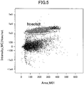
- the cell population of the nucleated cells in which nuclei and SMN protein are labeled is a population including cells having a nuclear fluorescence intensity of approximately 1 ⁇ 10 5 or more and a SMN-protein fluorescence intensity of approximately 1 ⁇ 10 3 or more when the nuclei and SMN protein are labeled with a fluorescent dye as described later in Example 1 of the present description to detect the fluorescence intensities as described later in Example 1.
- a fluorescent dye as described later in Example 1 of the present description to detect the fluorescence intensities as described later in Example 1.
- Such cells are determined as intact cells.
- cells keeping the cell form and also keeping the aspect ratio (width-to-height ratio) and surface area under microscope observation are determined as intact cells.
- the criteria of determining intact cells or determining a cell population including intact cells vary depending on the method for labeling nuclei and SMN protein in nucleated cells. However, the determinations can be made as appropriate based on the results of detecting nuclei and SMN protein in nucleated cells on the basis of a labeling reagent to be used.
- the nucleated cells to be detected preferably include lymphocytes, monocytes, granulocytes, hematopoietic stem cells, and vascular endothelium progenitor cells.
- a fluorescent dye for labeling SMN protein and a fluorescent dye for labeling the nuclei are respectively referred to as a first fluorescent dye and a second fluorescent dye in some cases.
- the step of selecting a cell population of the nucleated cells in which nuclei and SMN protein are labeled comprises measuring, in a single device, a first fluorescence intensity emitted by the first fluorescent dye and a second fluorescence intensity emitted by the second fluorescent dye.
- the step of selecting a cell population of the nucleated cells in which nuclei and SMN protein are labeled comprises measuring, in a single device, a first fluorescence intensity emitted by the first fluorescent dye and a second fluorescence intensity emitted by the second fluorescent dye, and representing the measured first and second fluorescence intensities in two-dimensional coordinates.
- the sample derived from blood obtained from a test subject which is used in the method of the present invention, should contain nucleated cells derived from the blood.
- whole blood peripheral blood
- nucleated cells it can be said that whole blood contains a cell population rich in diversity and thus heterogeneous.
- human blood cell fractions constantly vary, it is preferable to minimize the influence of the variation in blood cell fractions on the amount of the SMN proteins expressed in blood.
- nucleated cells in blood can also be classified into multiple clusters by using one or more surface antigen markers and the amount of the SMN protein expressed can be measured in each of the clusters.
- the method of the present invention may comprise the steps of: classifying nucleated cells in blood into multiple clusters, for example, 2 to 4 clusters, by using one or more surface antigen markers; and selecting a predetermined cell population from the clusters. This is advantageous because it is possible to reduce the influence of the variation in blood cell fractions on the amount of the SMN proteins expressed in blood.
- the method of the present invention comprises the step of selecting a cell population of the nucleated cells in which nuclei and SMN protein are labeled
- the present invention may comprise the step of selecting a predetermined cell population from the clusters before, during, or after this selection step.
- the method of the present invention may comprise the step of selecting a cell population of the nucleated cells in which nuclei and SMN protein are labeled, the cell population being selected from multiple clusters, for example, 2 to 4 clusters, classified from the nucleated cells by using one or more surface antigen markers.
- the surface antigen markers which can be used in the method of the present invention include, but are not particularly limited to, CD45, CD66, CD14, CD3, CD19, CD33, CD123, CD34, CD11c, CD25, HLA-DR, and the like.
- the surface antigen markers may be used alone, or two or more thereof may be used together.
- CD45 is generally known as a panhemocyte marker; CD66 as a neutrophil marker; CD3 as a T cell marker; CD19 as a B cell marker; and CD14 as a monocyte marker.
- nucleated cells in blood may be classified into multiple clusters, for example, 2 to 4 clusters, based on side scatter (SSC).
- nucleated cells in blood can be classified into multiple clusters, for example, 2 to 4 clusters, by using one or more surface antigen markers and based on side scatter (SSC).
- the method of the present invention may comprise the step of selecting a cell population of the nucleated cells in which nuclei and SMN protein are labeled, the cell population being selected from multiple clusters, for example, 2 to 4 clusters, classified from the nucleated cells by using one or more surface antigen markers and based on side scatter (SSC).
- One embodiment of the method of the present invention may comprise selecting a cell population of granulocytes or a cell population including granulocytes.
- One embodiment of the method of the present invention may comprise selecting a cell population of monocytes or a cell population including monocytes.
- One embodiment of the method of the present invention may comprise selecting a cell population of B cells or a cell population including B cells.
- One embodiment of the method of the present invention may comprise selecting a cell population of T cells or a cell population including T cells.
- the method for detecting SMN protein expression of the present invention comprises a step of detecting SMN protein expression based on the label for the SMN protein in the cell population selected on the basis of the labels on the nuclei and the SMN protein in the nucleated cells.
- the method of the present invention makes it possible to enhance the sensitivity of detecting the level of the SMN protein expressed, by selecting a particular cell population and detecting the SMN protein expression in the cell population as described above. Moreover, in the method of the present invention, detection data based on the label for the SMN protein referred at the time of selecting the particular cell population can be used when the SMN protein expression is detected based on the label for the SMN protein in the selected cell population.
- the step of selecting a cell population of the nucleated cells in which nuclei and SMN protein are labeled and the step of detecting SMN protein expression based on the label for the SMN protein in the selected cell population are performed by flow cytometry.
- Flow cytometry is suitable for quick treatments.
- the step of selecting a cell population of the nucleated cells in which nuclei and SMN protein are labeled and the step of detecting SMN protein expression based on the label for the SMN protein in the selected cell population are performed by imaging flow cytometry. Since pictures of cells are taken at the same time in imaging flow cytometry, the fluorescence acquisition speed therein is set slower than those in general flow cytometry. Hence, more detailed analyses are possible.
- flow cytometry and the imaging flow cytometry can be performed using commercially available devices according to manufacturer's instruction manuals thereof.
- the method for detecting SMN protein expression of the present invention may comprise a step of comparing SMN protein expressions among subjects, for example, a childhood SMA patient, a carrier thereof, and a healthy person, specifically between a childhood SMA patient and a carrier thereof or between a childhood SMA patient and a healthy person.
- a difference in the amount of the SMN protein expressed is found between the childhood SMA patient and the carrier or between the childhood SMA patient and the healthy person, so that it is possible to classify the subject as a childhood SMA patient, a carrier, and a healthy person.
- the method for detecting SMN protein expression of the present invention can also be used in a screening method for an agent for treating childhood SMA.
- the blood to be detected is obtained from a test subject
- the step of detecting SMN protein expression comprises measuring an amount of the SMN proteins expressed
- the detection method may comprise a step of comparing the amount of the SMN protein expressed with a control.
- the control may be selected from the following (a) to (c):
- the phrase "measured in the same manner as” should be construed to allow some modification that does not substantially influence the measurement of the amount of the SMN protein expressed in comparing the amounts of the SMN protein expressed.
- the phrase does not mean, for example, that the step of washing cells and so on are also completely the same.
- the amount of the SMN protein expressed can be measured as appropriate by using techniques conventionally known in the technical field of the present invention, such as calibration curve method.
- one embodiment of the present invention comprises such a comparison step as described above, and also comprises a step of diagnosing the test subject based on the comparison result, and thus it can also be said that one embodiment of the present invention relates to a method for diagnosing childhood SMA.
- one embodiment of the present invention comprises such a comparison step as described above, and also comprises a step of selecting a drug or a drug candidate against childhood SMA based on the comparison result, and thus it can also be said that one embodiment of the present invention relates to a screening method for a drug or a drug candidate against childhood SMA.
- PBMCs peripheral blood mononuclear cells
- the ELISA method has disadvantages in that: cells are disrupted for the analysis; since the analyses have to be performed at the same time before and after the drug administration, the SMN protein may break down due to cell storage, freezing, or thawing treatments; and dead cells and extracellular SMN protein are also detected at the same time.
- PBMCs are constituted of CD4+ and CD8+ T cells (approximately 65%), B cells and natural killer cells (approximately 28%), as well as CD14+ monocytes, granulocytes, and dendritic cells (approximately 7% in total), granulocytes, which are major constituents in blood, have a high relative density and precipitate to a lower layer during centrifugation. Hence, most of the granulocytes are not included in PBMCs which are located in an upper layer due to the relative density.
- cells can be analyzed while keeping the shape, specimens can be treated on the same day (i.e., the freezing or thawing treatment is not necessary), and only the SMN protein in viable cells can be detected.
- the SMN protein analysis can be performed on a desired cell population (blood cell fractions), making it possible to reduce the influence of the variation in blood cell fractions on the amount of the SMN protein expressed.
- a blood sample obtained from a healthy person was collected into a heparin tube, then overlaid on a Ficoll solution, and centrifuged at 2000 rpm for 15 minutes. Thereby, peripheral blood mononuclear cells were separated and collected. Subsequently, a lymphoblast transformation treatment was performed by a transformation method using EB virus.
- a blood sample obtained from a SMA type I patient was also subjected to the same treatment performed on the blood sample obtained from the healthy person.
- lymphoblasts derived from the peripheral blood mononuclear cells of the healthy person obtained in (1) above were collected into a centrifuge tube.
- the cells were fixed with a 4% paraformaldehyde-phosphate buffer solution. Then, the cells were washed with a phosphate buffer solution and subsequently centrifuged at 500 x g for 5 minutes to remove the supernatant.
- BD Phosflow TM Perm Buffer II (BD Biosciences) was added to the resultant and left standing on ice for 30 minutes.
- BD Biosciences BD pharmingen Stain Buffer
- a primary antibody Purified Mouse Anti-SMN (Mouse monoclonal antibody, clone8/SMN, BD Biosciences) was then added for the reaction at room temperature for 60 minutes.
- the cells were washed with a phosphate buffer solution, and a secondary antibody (AlexaFluor 488 goat anti-mouse IgG (H+L), highly cross-absorbed, Life Technology) was added for the reaction at room temperature for 60 minutes while shielding light. The resultant was washed with a phosphate buffer solution. Then, Hoechst 33342 (Molecular Probes) was added to stain nuclei in the cells. Further, the cells were washed with a phosphate buffer solution. Subsequently, the SMN protein expression was measured by imaging flow cytometry (FlowSight(registered trademark), Amnis).
- a secondary antibody AlexaFluor 488 goat anti-mouse IgG (H+L), highly cross-absorbed, Life Technology
- lymphoblasts derived from the peripheral blood mononuclear cells of the SMA type I patient obtained in (1) above were also measured for the SMN protein expression as in the case of the healthy person.
- SMN protein expression intensities were conducted by selecting cell populations 1 (populations 1) shown in Figs. 1 and 2 .
- the cell populations shown in Figs. 1 and 2 were obtained by using 10000 cells from each of both the healthy person and the SMA type I patient.
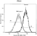
- the graph in Fig. 3 shows distributions of fluorescence intensities of cell populations selected from 20000 cells from each of both the healthy person and the SMA type I patient.
- Table 3 shows the numbers of cells analyzed for the fluorescence intensities shown in Fig. 3 and the medians of the fluorescence intensities.
- Fig. 3 and Table 3 show the results obtained by using a total of 20000 cells for the imaging flow cytometry for each of both the healthy person and the SMA type I patient.
- Table 3 Count (the number of cells) Median Normal control (whole) 13417 66530.04 Normal control (R2) 12201 70913.87 Normal control (R3) 1215 21155 SMA type I patient (whole) 11451 10385 SMA type I patient (R2) 984 35985.43 SMA type I patient (R3) 10449 9562.4
- the method of the present invention is capable of sensitive detection of SMN protein expression.
- R2 and R3 in Fig. 3 were to compare the fluorescence intensities by removing regions where the expression amounts overlapped between the normal control and the SMA type I patient for gating, and to show a difference between the two groups.
- frequency values on the vertical axis represent relative frequencies.
- the values used are relative values obtained according to proportions of cell populations having fluorescence intensities among the total cell populations.
- Samples containing nucleated cells derived from blood of a carrier and a SMA type I patient were also prepared by the lysis method as in the case of the healthy person.
- BD Phosflow TM Perm Buffer II (BD Biosciences) was added as a cell membrane permeation reagent to the nucleated cells of the healthy person obtained in (1) above, and left standing on ice for 30 minutes. And then, the cells were washed with BD pharmingen Stain Buffer (BD Biosciences), and Milli-Mark TM (anti-SMN antibody: Anti-SMN-FITC clone 2B1, Millipore) was then added for the reaction at room temperature for 60 minutes.
- Milli-Mark TM anti-SMN antibody: Anti-SMN-FITC clone 2B1, Millipore
- the cells were washed with a phosphate buffer solution, and Hoechst 33342 (Molecular Probes) was added to stain nuclei in the cells. Further, the cells were washed with a phosphate buffer solution. Then, the SMN protein expression was measured by imaging flow cytometry (FlowSight(registered trademark), Amnis).
- the samples of the carrier and the SMA type I patient obtained in (1) above were also measured for the SMN protein expression as in the case of the healthy person.
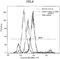
- SMN protein expression intensities of intact cell groups in which nuclei and SMN protein were sufficiently fluorescence-labeled were analyzed in the same manner as in Example 1 above, except that 10000 cells from each of the healthy person, the carrier, and the SMA type I patient were used.
- Fig. 4 shows the results.
- Table 4 shows the numbers of cells analyzed for the fluorescence intensities shown in Fig. 4 and the medians of the fluorescence intensities.
- Table 4 Count (the number of cells) Median Normal control 7341 7936.19 Carrier 5616 3571.02 SMA type I patient 7810 1781.21
- the method of the present invention is capable of detecting a difference in the amount of the SMN protein expressed among a healthy person, a carrier, and a childhood SMA patient.
- Samples were prepared according to the following staining protocol. 1. Collect 2 mL of peripheral blood obtained from a healthy person into a tube. Add reagents thereto such that 2 mL of the peripheral blood contains 80 ⁇ L of an Fc receptor blocking reagent (Clear Back, MTG-001, MBL) and 100 ⁇ L of Hoechst 33342 (Molecular Probes) which has been diluted to 5 ⁇ g/mL with PBS. 2. Seal the tube, and slowly rotate it at room temperature for 15 minutes in the dark. 3. Place 500 ⁇ L of the peripheral blood thus treated into each of four 15-mL conical tubes. 4.
- Fc receptor blocking reagent Clear Back, MTG-001, MBL
- Hoechst 33342 Molecular Probes
- a cell membrane permeation reagent (BD Phosflow TM Perm/Wash Buffer I), and incubate the resultant at room temperature for 20 minutes. 12. Centrifuge the resultant in the same manner as in 8 above, and wash the cells twice with 2 mL of FBS/NaN 3 containing PBS (BD stain buffer 554656). 13. Place equal volumes of the cell suspension into two 1.5-mL microtubes, so that one tube contains approximately 5 x 10 5 cells/45 ⁇ L. 14.
- a cell population was further selected from the R1 cluster by utilizing CD33 and CD66abce ( Fig. 8 ).
- 99.5% (the number of cells: 30060) of the R1 cluster in which SMN protein were labeled (the number of cells: 30198) were CD33+ CD66abce+ cells (neutrophil-like cells).
- the analysis was performed on fluorescence intensities emitted by the SMN protein labeling reagent in these CD33+ CD66abce+ cells ( Fig. 9 ).
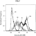
- the geometric mean of the fluorescence intensities emitted by the SMN protein labeling reagent was 33679.5.
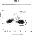
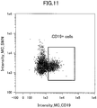
- cell populations were further selected from the R2 cluster by utilizing CD3 and SMN protein or by utilizing CD19 and SMN protein, and the analysis was performed on fluorescence intensities emitted by the SMN protein labeling reagent ( Figs. 10 and 11 ).
- fluorescence intensities emitted by the SMN protein labeling reagent Figs. 10 and 11 .
- 63.6% (the number of cells: 8197) of the R2 cluster in which SMN protein were labeled (the number of cells: 12882) were CD3+ cells (T cells), while 9.55% (the number of cells: 1230) thereof were CD19+ cells (B cells).
- the geometric mean of the fluorescence intensities emitted by the SMN protein labeling reagent in the CD3+ cells shown in Fig. 10 was 1782.23
- the geometric mean of the fluorescence intensities emitted by the SMN protein labeling reagent in the CD19+ cells shown in Fig. 11 was 2103.25.
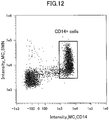
- a cell population was further selected from the R3 cluster by utilizing CD14 and SMN protein, and the analysis was performed on fluorescence intensities emitted by the SMN protein labeling reagent ( Fig. 12 ).
- fluorescence intensities emitted by the SMN protein labeling reagent Fig. 12
- 61% the number of cells: 2495
- the number of cells: 4088 were CD14+ cells (monocyte-like cells).
- the geometric mean of the fluorescence intensities emitted by the SMN protein labeling reagent in the CD14+ cells was 13606.79.
- the expressed amount of the SMN protein which is a ubiquitous protein, is increased in blood cells in the order of granulocytes (neutrophils, basophils, eosinophils) > monocytes > B cells and T cells. Accordingly, since the conventional SMN protein quantitative analysis by the ELISA method using PBMCs can hardly analyze granulocytes expressing a large amount of SMN protein, it is conceivable that the SMN protein expression cannot be analyzed sufficiently.
- the method of the present invention is capable of analyzing blood cells including granulocytes expressing a large amount of SMN protein, and also capable of analysis focusing on only granulocytes. These indicate that the method of the present invention is excellent in detecting SMN protein expression.
- the method for completely curing SMA has not been established yet, and SMA is one of diseases designated as intractable by the country.
- the present invention is quite usefully utilized in evaluating a method which can be a SMA curing method.
- the method of the present invention can be used in evaluating a drug or a drug candidate against SMA, and can be used in screening for a drug or a drug candidate against SMA.
- the present invention is usefully utilized in SMA diagnosis, too.
Landscapes
- Health & Medical Sciences (AREA)
- Life Sciences & Earth Sciences (AREA)
- Engineering & Computer Science (AREA)
- Immunology (AREA)
- Chemical & Material Sciences (AREA)
- Hematology (AREA)
- Biomedical Technology (AREA)
- Molecular Biology (AREA)
- Urology & Nephrology (AREA)
- Physics & Mathematics (AREA)
- General Health & Medical Sciences (AREA)
- Pathology (AREA)
- Biochemistry (AREA)
- Analytical Chemistry (AREA)
- General Physics & Mathematics (AREA)
- Cell Biology (AREA)
- Medicinal Chemistry (AREA)
- Food Science & Technology (AREA)
- Biotechnology (AREA)
- Microbiology (AREA)
- Proteomics, Peptides & Aminoacids (AREA)
- Chemical Kinetics & Catalysis (AREA)
- Optics & Photonics (AREA)
- Nuclear Medicine, Radiotherapy & Molecular Imaging (AREA)
- Zoology (AREA)
- Tropical Medicine & Parasitology (AREA)
- Virology (AREA)
- Neurology (AREA)
- Neurosurgery (AREA)
- Investigating Or Analysing Biological Materials (AREA)
- Measuring Or Testing Involving Enzymes Or Micro-Organisms (AREA)
Applications Claiming Priority (2)
| Application Number | Priority Date | Filing Date | Title |
|---|---|---|---|
| JP2014076985 | 2014-04-03 | ||
| PCT/JP2015/060662 WO2015152410A1 (fr) | 2014-04-03 | 2015-04-03 | Procédé de détection de l'expression de la protéine smn |
Publications (3)
| Publication Number | Publication Date |
|---|---|
| EP3128327A1 true EP3128327A1 (fr) | 2017-02-08 |
| EP3128327A4 EP3128327A4 (fr) | 2017-08-23 |
| EP3128327B1 EP3128327B1 (fr) | 2018-12-12 |
Family
ID=54240717
Family Applications (1)
| Application Number | Title | Priority Date | Filing Date |
|---|---|---|---|
| EP15773282.7A Active EP3128327B1 (fr) | 2014-04-03 | 2015-04-03 | Procédé de détection de l'expression de la protéine smn |
Country Status (5)
| Country | Link |
|---|---|
| US (1) | US20170115297A1 (fr) |
| EP (1) | EP3128327B1 (fr) |
| JP (1) | JP6115979B2 (fr) |
| CN (1) | CN106255886B (fr) |
| WO (1) | WO2015152410A1 (fr) |
Cited By (1)
| Publication number | Priority date | Publication date | Assignee | Title |
|---|---|---|---|---|
| EP4057004A1 (fr) * | 2021-03-12 | 2022-09-14 | Tokyo Women's Medical University | Procédé d'acquisition d'informations sur l'amyotrophie spinale |
Families Citing this family (2)
| Publication number | Priority date | Publication date | Assignee | Title |
|---|---|---|---|---|
| JP6671664B2 (ja) * | 2016-05-24 | 2020-03-25 | 学校法人東京女子医科大学 | Smnタンパク質の核内構造体の発現解析方法 |
| JP6956402B2 (ja) * | 2017-06-27 | 2021-11-02 | 国立大学法人弘前大学 | 解析方法 |
Family Cites Families (7)
| Publication number | Priority date | Publication date | Assignee | Title |
|---|---|---|---|---|
| US5627040A (en) * | 1991-08-28 | 1997-05-06 | Becton Dickinson And Company | Flow cytometric method for autoclustering cells |
| CN1287151C (zh) * | 2005-01-04 | 2006-11-29 | 济南市中心医院 | 流式细胞仪检测专用溶血方法 |
| CN1810835A (zh) * | 2006-02-27 | 2006-08-02 | 福建医科大学附属第一医院 | 兔抗人smn全长多克隆抗体的优选与提取法 |
| WO2009151546A2 (fr) * | 2008-05-27 | 2009-12-17 | Ptc Therapeutics, Inc. | Procédés de traitement d'une atrophie musculaire spinale |
| TWI350312B (en) * | 2009-03-16 | 2011-10-11 | Univ Kaohsiung Medical | Method for determining smn gene transfer and intragenic mutations |
| WO2011032109A1 (fr) * | 2009-09-11 | 2011-03-17 | Sma Foundation | Biomarqueurs de l'atrophie musculaire spinale |
| RU2013148071A (ru) * | 2011-04-01 | 2015-05-10 | Президент Энд Феллоуз Ов Харвард Колледж | Аналогичное диализу терапевтическое (dlt) устройство |
-
2015
- 2015-04-03 WO PCT/JP2015/060662 patent/WO2015152410A1/fr not_active Ceased
- 2015-04-03 US US15/300,456 patent/US20170115297A1/en not_active Abandoned
- 2015-04-03 JP JP2016511649A patent/JP6115979B2/ja active Active
- 2015-04-03 EP EP15773282.7A patent/EP3128327B1/fr active Active
- 2015-04-03 CN CN201580022715.XA patent/CN106255886B/zh active Active
Cited By (1)
| Publication number | Priority date | Publication date | Assignee | Title |
|---|---|---|---|---|
| EP4057004A1 (fr) * | 2021-03-12 | 2022-09-14 | Tokyo Women's Medical University | Procédé d'acquisition d'informations sur l'amyotrophie spinale |
Also Published As
| Publication number | Publication date |
|---|---|
| JP6115979B2 (ja) | 2017-04-19 |
| WO2015152410A1 (fr) | 2015-10-08 |
| JPWO2015152410A1 (ja) | 2017-04-13 |
| CN106255886A (zh) | 2016-12-21 |
| EP3128327B1 (fr) | 2018-12-12 |
| US20170115297A1 (en) | 2017-04-27 |
| EP3128327A4 (fr) | 2017-08-23 |
| CN106255886B (zh) | 2018-06-08 |
Similar Documents
| Publication | Publication Date | Title |
|---|---|---|
| CN103502816B (zh) | 用于监测整个身体中组织稳态扰乱的方法和手段 | |
| KR102268963B1 (ko) | 머신러닝 기법으로 말초혈액 내 암 면역력을 평가하여 암을 진단하거나 치료반응을 예측하는 방법 및 이를 이용한 진단키트 | |
| WO2012134813A1 (fr) | Procédés et compositions pour identifier une maladie résiduelle minimale associée à une leucémie lymphoblastique aiguë | |
| EP3467497B1 (fr) | Procédé d'analyse d'expression de corps nucléaire de la protéine smn | |
| CN109187941B (zh) | Cd4+cd70+t细胞亚群在制备辅助诊断极重型再生障碍性贫血试剂盒中的应用 | |
| Jamaly et al. | Elevated plasma levels of P‐selectin glycoprotein ligand‐1‐positive microvesicles in patients with unprovoked venous thromboembolism | |
| EP3128327B1 (fr) | Procédé de détection de l'expression de la protéine smn | |
| JP5932797B2 (ja) | 炎症状態の診断及び/又は予後の方法 | |
| Claireaux et al. | Deep profiling of antigen-specific B cells from different pathogens identifies novel compartments in the IgG memory B cell and antibody-secreting cell lineages | |
| CN118534130A (zh) | 一种用于脓毒症判别的单核细胞cd39检测试剂盒 | |
| EP3146333B1 (fr) | Diagnostic de la leucémie myélomonocytique chronique (cmml) par cytométrie de flux | |
| WO2023101599A2 (fr) | Méthode de tri d'une population de macrophages hétérogènes | |
| US20230028910A1 (en) | Method for diagnosing cutaneous t-cell lymphoma diseases | |
| JP6956402B2 (ja) | 解析方法 | |
| Sharaby | AI-driven approach for diagnosis of renal transplant rejection based on biomarkers identification and integration. | |
| EP4057004A1 (fr) | Procédé d'acquisition d'informations sur l'amyotrophie spinale | |
| US20240060965A1 (en) | Car therapy response prediction | |
| Rodríguez-Lorenzo et al. | PERIVENTRICULAR CD56BRIGHT NK CELL | |
| CN118465265A (zh) | 与急性髓系白血病造血干细胞移植术后临床结局相关的生物标志物及其应用 | |
| van Dongen et al. | Clinical Cell Analysis of the European Society for Clinical Cell Analysis (ESCCA) and the Iberian Society for Cytometry (SIC) | |
| CN112955743A (zh) | 用于肾脏移植后bk病毒性肾病风险分层的方法 | |
| Kwek et al. | Ex Vivo Single-Cell Profiling of Acute Myocardial Infarction Patients Reveals Disproportionate Cd66b+ Cell Secretion Response | |
| Roubanis et al. | Stepwise single-cell-resolved deep immunophenotyping pipeline to characterize immune heterogeneity and functionality in health and disease |
Legal Events
| Date | Code | Title | Description |
|---|---|---|---|
| STAA | Information on the status of an ep patent application or granted ep patent |
Free format text: STATUS: THE INTERNATIONAL PUBLICATION HAS BEEN MADE |
|
| PUAI | Public reference made under article 153(3) epc to a published international application that has entered the european phase |
Free format text: ORIGINAL CODE: 0009012 |
|
| STAA | Information on the status of an ep patent application or granted ep patent |
Free format text: STATUS: REQUEST FOR EXAMINATION WAS MADE |
|
| 17P | Request for examination filed |
Effective date: 20161011 |
|
| AK | Designated contracting states |
Kind code of ref document: A1 Designated state(s): AL AT BE BG CH CY CZ DE DK EE ES FI FR GB GR HR HU IE IS IT LI LT LU LV MC MK MT NL NO PL PT RO RS SE SI SK SM TR |
|
| AX | Request for extension of the european patent |
Extension state: BA ME |
|
| DAV | Request for validation of the european patent (deleted) | ||
| DAX | Request for extension of the european patent (deleted) | ||
| A4 | Supplementary search report drawn up and despatched |
Effective date: 20170721 |
|
| RIC1 | Information provided on ipc code assigned before grant |
Ipc: G01N 33/49 20060101ALI20170717BHEP Ipc: G01N 21/64 20060101ALI20170717BHEP Ipc: G01N 33/48 20060101ALI20170717BHEP Ipc: C07K 14/435 20060101ALI20170717BHEP Ipc: G01N 33/68 20060101AFI20170717BHEP |
|
| STAA | Information on the status of an ep patent application or granted ep patent |
Free format text: STATUS: EXAMINATION IS IN PROGRESS |
|
| 17Q | First examination report despatched |
Effective date: 20180302 |
|
| GRAP | Despatch of communication of intention to grant a patent |
Free format text: ORIGINAL CODE: EPIDOSNIGR1 |
|
| STAA | Information on the status of an ep patent application or granted ep patent |
Free format text: STATUS: GRANT OF PATENT IS INTENDED |
|
| INTG | Intention to grant announced |
Effective date: 20180821 |
|
| GRAS | Grant fee paid |
Free format text: ORIGINAL CODE: EPIDOSNIGR3 |
|
| GRAA | (expected) grant |
Free format text: ORIGINAL CODE: 0009210 |
|
| STAA | Information on the status of an ep patent application or granted ep patent |
Free format text: STATUS: THE PATENT HAS BEEN GRANTED |
|
| AK | Designated contracting states |
Kind code of ref document: B1 Designated state(s): AL AT BE BG CH CY CZ DE DK EE ES FI FR GB GR HR HU IE IS IT LI LT LU LV MC MK MT NL NO PL PT RO RS SE SI SK SM TR |
|
| REG | Reference to a national code |
Ref country code: GB Ref legal event code: FG4D |
|
| REG | Reference to a national code |
Ref country code: CH Ref legal event code: EP |
|
| REG | Reference to a national code |
Ref country code: AT Ref legal event code: REF Ref document number: 1076712 Country of ref document: AT Kind code of ref document: T Effective date: 20181215 |
|
| REG | Reference to a national code |
Ref country code: DE Ref legal event code: R096 Ref document number: 602015021458 Country of ref document: DE |
|
| REG | Reference to a national code |
Ref country code: IE Ref legal event code: FG4D |
|
| REG | Reference to a national code |
Ref country code: NL Ref legal event code: MP Effective date: 20181212 |
|
| REG | Reference to a national code |
Ref country code: LT Ref legal event code: MG4D |
|
| PG25 | Lapsed in a contracting state [announced via postgrant information from national office to epo] |
Ref country code: LT Free format text: LAPSE BECAUSE OF FAILURE TO SUBMIT A TRANSLATION OF THE DESCRIPTION OR TO PAY THE FEE WITHIN THE PRESCRIBED TIME-LIMIT Effective date: 20181212 Ref country code: FI Free format text: LAPSE BECAUSE OF FAILURE TO SUBMIT A TRANSLATION OF THE DESCRIPTION OR TO PAY THE FEE WITHIN THE PRESCRIBED TIME-LIMIT Effective date: 20181212 Ref country code: NO Free format text: LAPSE BECAUSE OF FAILURE TO SUBMIT A TRANSLATION OF THE DESCRIPTION OR TO PAY THE FEE WITHIN THE PRESCRIBED TIME-LIMIT Effective date: 20190312 Ref country code: LV Free format text: LAPSE BECAUSE OF FAILURE TO SUBMIT A TRANSLATION OF THE DESCRIPTION OR TO PAY THE FEE WITHIN THE PRESCRIBED TIME-LIMIT Effective date: 20181212 Ref country code: HR Free format text: LAPSE BECAUSE OF FAILURE TO SUBMIT A TRANSLATION OF THE DESCRIPTION OR TO PAY THE FEE WITHIN THE PRESCRIBED TIME-LIMIT Effective date: 20181212 Ref country code: ES Free format text: LAPSE BECAUSE OF FAILURE TO SUBMIT A TRANSLATION OF THE DESCRIPTION OR TO PAY THE FEE WITHIN THE PRESCRIBED TIME-LIMIT Effective date: 20181212 Ref country code: BG Free format text: LAPSE BECAUSE OF FAILURE TO SUBMIT A TRANSLATION OF THE DESCRIPTION OR TO PAY THE FEE WITHIN THE PRESCRIBED TIME-LIMIT Effective date: 20190312 |
|
| REG | Reference to a national code |
Ref country code: AT Ref legal event code: MK05 Ref document number: 1076712 Country of ref document: AT Kind code of ref document: T Effective date: 20181212 |
|
| PG25 | Lapsed in a contracting state [announced via postgrant information from national office to epo] |
Ref country code: AL Free format text: LAPSE BECAUSE OF FAILURE TO SUBMIT A TRANSLATION OF THE DESCRIPTION OR TO PAY THE FEE WITHIN THE PRESCRIBED TIME-LIMIT Effective date: 20181212 Ref country code: SE Free format text: LAPSE BECAUSE OF FAILURE TO SUBMIT A TRANSLATION OF THE DESCRIPTION OR TO PAY THE FEE WITHIN THE PRESCRIBED TIME-LIMIT Effective date: 20181212 Ref country code: GR Free format text: LAPSE BECAUSE OF FAILURE TO SUBMIT A TRANSLATION OF THE DESCRIPTION OR TO PAY THE FEE WITHIN THE PRESCRIBED TIME-LIMIT Effective date: 20190313 Ref country code: RS Free format text: LAPSE BECAUSE OF FAILURE TO SUBMIT A TRANSLATION OF THE DESCRIPTION OR TO PAY THE FEE WITHIN THE PRESCRIBED TIME-LIMIT Effective date: 20181212 |
|
| PG25 | Lapsed in a contracting state [announced via postgrant information from national office to epo] |
Ref country code: NL Free format text: LAPSE BECAUSE OF FAILURE TO SUBMIT A TRANSLATION OF THE DESCRIPTION OR TO PAY THE FEE WITHIN THE PRESCRIBED TIME-LIMIT Effective date: 20181212 |
|
| PG25 | Lapsed in a contracting state [announced via postgrant information from national office to epo] |
Ref country code: PT Free format text: LAPSE BECAUSE OF FAILURE TO SUBMIT A TRANSLATION OF THE DESCRIPTION OR TO PAY THE FEE WITHIN THE PRESCRIBED TIME-LIMIT Effective date: 20190412 Ref country code: CZ Free format text: LAPSE BECAUSE OF FAILURE TO SUBMIT A TRANSLATION OF THE DESCRIPTION OR TO PAY THE FEE WITHIN THE PRESCRIBED TIME-LIMIT Effective date: 20181212 Ref country code: PL Free format text: LAPSE BECAUSE OF FAILURE TO SUBMIT A TRANSLATION OF THE DESCRIPTION OR TO PAY THE FEE WITHIN THE PRESCRIBED TIME-LIMIT Effective date: 20181212 Ref country code: IT Free format text: LAPSE BECAUSE OF FAILURE TO SUBMIT A TRANSLATION OF THE DESCRIPTION OR TO PAY THE FEE WITHIN THE PRESCRIBED TIME-LIMIT Effective date: 20181212 |
|
| PG25 | Lapsed in a contracting state [announced via postgrant information from national office to epo] |
Ref country code: RO Free format text: LAPSE BECAUSE OF FAILURE TO SUBMIT A TRANSLATION OF THE DESCRIPTION OR TO PAY THE FEE WITHIN THE PRESCRIBED TIME-LIMIT Effective date: 20181212 Ref country code: SK Free format text: LAPSE BECAUSE OF FAILURE TO SUBMIT A TRANSLATION OF THE DESCRIPTION OR TO PAY THE FEE WITHIN THE PRESCRIBED TIME-LIMIT Effective date: 20181212 Ref country code: IS Free format text: LAPSE BECAUSE OF FAILURE TO SUBMIT A TRANSLATION OF THE DESCRIPTION OR TO PAY THE FEE WITHIN THE PRESCRIBED TIME-LIMIT Effective date: 20190412 Ref country code: SM Free format text: LAPSE BECAUSE OF FAILURE TO SUBMIT A TRANSLATION OF THE DESCRIPTION OR TO PAY THE FEE WITHIN THE PRESCRIBED TIME-LIMIT Effective date: 20181212 Ref country code: EE Free format text: LAPSE BECAUSE OF FAILURE TO SUBMIT A TRANSLATION OF THE DESCRIPTION OR TO PAY THE FEE WITHIN THE PRESCRIBED TIME-LIMIT Effective date: 20181212 |
|
| REG | Reference to a national code |
Ref country code: DE Ref legal event code: R097 Ref document number: 602015021458 Country of ref document: DE |
|
| PLBE | No opposition filed within time limit |
Free format text: ORIGINAL CODE: 0009261 |
|
| STAA | Information on the status of an ep patent application or granted ep patent |
Free format text: STATUS: NO OPPOSITION FILED WITHIN TIME LIMIT |
|
| PG25 | Lapsed in a contracting state [announced via postgrant information from national office to epo] |
Ref country code: AT Free format text: LAPSE BECAUSE OF FAILURE TO SUBMIT A TRANSLATION OF THE DESCRIPTION OR TO PAY THE FEE WITHIN THE PRESCRIBED TIME-LIMIT Effective date: 20181212 Ref country code: DK Free format text: LAPSE BECAUSE OF FAILURE TO SUBMIT A TRANSLATION OF THE DESCRIPTION OR TO PAY THE FEE WITHIN THE PRESCRIBED TIME-LIMIT Effective date: 20181212 Ref country code: SI Free format text: LAPSE BECAUSE OF FAILURE TO SUBMIT A TRANSLATION OF THE DESCRIPTION OR TO PAY THE FEE WITHIN THE PRESCRIBED TIME-LIMIT Effective date: 20181212 |
|
| 26N | No opposition filed |
Effective date: 20190913 |
|
| REG | Reference to a national code |
Ref country code: CH Ref legal event code: PL |
|
| REG | Reference to a national code |
Ref country code: BE Ref legal event code: MM Effective date: 20190430 |
|
| PG25 | Lapsed in a contracting state [announced via postgrant information from national office to epo] |
Ref country code: LU Free format text: LAPSE BECAUSE OF NON-PAYMENT OF DUE FEES Effective date: 20190403 Ref country code: MC Free format text: LAPSE BECAUSE OF FAILURE TO SUBMIT A TRANSLATION OF THE DESCRIPTION OR TO PAY THE FEE WITHIN THE PRESCRIBED TIME-LIMIT Effective date: 20181212 |
|
| PG25 | Lapsed in a contracting state [announced via postgrant information from national office to epo] |
Ref country code: LI Free format text: LAPSE BECAUSE OF NON-PAYMENT OF DUE FEES Effective date: 20190430 Ref country code: CH Free format text: LAPSE BECAUSE OF NON-PAYMENT OF DUE FEES Effective date: 20190430 |
|
| PG25 | Lapsed in a contracting state [announced via postgrant information from national office to epo] |
Ref country code: BE Free format text: LAPSE BECAUSE OF NON-PAYMENT OF DUE FEES Effective date: 20190430 |
|
| PG25 | Lapsed in a contracting state [announced via postgrant information from national office to epo] |
Ref country code: TR Free format text: LAPSE BECAUSE OF FAILURE TO SUBMIT A TRANSLATION OF THE DESCRIPTION OR TO PAY THE FEE WITHIN THE PRESCRIBED TIME-LIMIT Effective date: 20181212 |
|
| PG25 | Lapsed in a contracting state [announced via postgrant information from national office to epo] |
Ref country code: IE Free format text: LAPSE BECAUSE OF NON-PAYMENT OF DUE FEES Effective date: 20190403 |
|
| REG | Reference to a national code |
Ref country code: DE Ref legal event code: R082 Ref document number: 602015021458 Country of ref document: DE Representative=s name: HL KEMPNER PATENTANWAELTE, SOLICITORS (ENGLAND, DE Ref country code: DE Ref legal event code: R082 Ref document number: 602015021458 Country of ref document: DE Representative=s name: HL KEMPNER PATENTANWALT, RECHTSANWALT, SOLICIT, DE Ref country code: DE Ref legal event code: R082 Ref document number: 602015021458 Country of ref document: DE Representative=s name: HL KEMPNER PARTG MBB, DE |
|
| PG25 | Lapsed in a contracting state [announced via postgrant information from national office to epo] |
Ref country code: CY Free format text: LAPSE BECAUSE OF FAILURE TO SUBMIT A TRANSLATION OF THE DESCRIPTION OR TO PAY THE FEE WITHIN THE PRESCRIBED TIME-LIMIT Effective date: 20181212 |
|
| PG25 | Lapsed in a contracting state [announced via postgrant information from national office to epo] |
Ref country code: HU Free format text: LAPSE BECAUSE OF FAILURE TO SUBMIT A TRANSLATION OF THE DESCRIPTION OR TO PAY THE FEE WITHIN THE PRESCRIBED TIME-LIMIT; INVALID AB INITIO Effective date: 20150403 Ref country code: MT Free format text: LAPSE BECAUSE OF FAILURE TO SUBMIT A TRANSLATION OF THE DESCRIPTION OR TO PAY THE FEE WITHIN THE PRESCRIBED TIME-LIMIT Effective date: 20181212 |
|
| PG25 | Lapsed in a contracting state [announced via postgrant information from national office to epo] |
Ref country code: MK Free format text: LAPSE BECAUSE OF FAILURE TO SUBMIT A TRANSLATION OF THE DESCRIPTION OR TO PAY THE FEE WITHIN THE PRESCRIBED TIME-LIMIT Effective date: 20181212 |
|
| PGFP | Annual fee paid to national office [announced via postgrant information from national office to epo] |
Ref country code: DE Payment date: 20250422 Year of fee payment: 11 |
|
| PGFP | Annual fee paid to national office [announced via postgrant information from national office to epo] |
Ref country code: GB Payment date: 20250423 Year of fee payment: 11 |
|
| PGFP | Annual fee paid to national office [announced via postgrant information from national office to epo] |
Ref country code: FR Payment date: 20250425 Year of fee payment: 11 |