EP3134439B1 - Molécules d'anticorps anti-psyk et leur utilisation pour la thérapie ciblée sur syk - Google Patents
Molécules d'anticorps anti-psyk et leur utilisation pour la thérapie ciblée sur syk Download PDFInfo
- Publication number
- EP3134439B1 EP3134439B1 EP15720854.7A EP15720854A EP3134439B1 EP 3134439 B1 EP3134439 B1 EP 3134439B1 EP 15720854 A EP15720854 A EP 15720854A EP 3134439 B1 EP3134439 B1 EP 3134439B1
- Authority
- EP
- European Patent Office
- Prior art keywords
- psyk
- syk
- antibody
- cell
- antibody molecule
- Prior art date
- Legal status (The legal status is an assumption and is not a legal conclusion. Google has not performed a legal analysis and makes no representation as to the accuracy of the status listed.)
- Not-in-force
Links
Images












Classifications
-
- C—CHEMISTRY; METALLURGY
- C07—ORGANIC CHEMISTRY
- C07K—PEPTIDES
- C07K16/00—Immunoglobulins [IG], e.g. monoclonal or polyclonal antibodies
- C07K16/40—Immunoglobulins [IG], e.g. monoclonal or polyclonal antibodies against enzymes
-
- A—HUMAN NECESSITIES
- A61—MEDICAL OR VETERINARY SCIENCE; HYGIENE
- A61P—SPECIFIC THERAPEUTIC ACTIVITY OF CHEMICAL COMPOUNDS OR MEDICINAL PREPARATIONS
- A61P35/00—Antineoplastic agents
- A61P35/02—Antineoplastic agents specific for leukemia
-
- A—HUMAN NECESSITIES
- A61—MEDICAL OR VETERINARY SCIENCE; HYGIENE
- A61P—SPECIFIC THERAPEUTIC ACTIVITY OF CHEMICAL COMPOUNDS OR MEDICINAL PREPARATIONS
- A61P43/00—Drugs for specific purposes, not provided for in groups A61P1/00-A61P41/00
-
- C—CHEMISTRY; METALLURGY
- C07—ORGANIC CHEMISTRY
- C07K—PEPTIDES
- C07K16/00—Immunoglobulins [IG], e.g. monoclonal or polyclonal antibodies
- C07K16/18—Immunoglobulins [IG], e.g. monoclonal or polyclonal antibodies against material from animals or humans
- C07K16/28—Immunoglobulins [IG], e.g. monoclonal or polyclonal antibodies against material from animals or humans against receptors, cell surface antigens or cell surface determinants
- C07K16/30—Immunoglobulins [IG], e.g. monoclonal or polyclonal antibodies against material from animals or humans against receptors, cell surface antigens or cell surface determinants from tumour cells
-
- A—HUMAN NECESSITIES
- A61—MEDICAL OR VETERINARY SCIENCE; HYGIENE
- A61K—PREPARATIONS FOR MEDICAL, DENTAL OR TOILETRY PURPOSES
- A61K39/00—Medicinal preparations containing antigens or antibodies
- A61K2039/505—Medicinal preparations containing antigens or antibodies comprising antibodies
-
- C—CHEMISTRY; METALLURGY
- C07—ORGANIC CHEMISTRY
- C07K—PEPTIDES
- C07K2317/00—Immunoglobulins specific features
- C07K2317/30—Immunoglobulins specific features characterized by aspects of specificity or valency
- C07K2317/34—Identification of a linear epitope shorter than 20 amino acid residues or of a conformational epitope defined by amino acid residues
-
- C—CHEMISTRY; METALLURGY
- C07—ORGANIC CHEMISTRY
- C07K—PEPTIDES
- C07K2317/00—Immunoglobulins specific features
- C07K2317/70—Immunoglobulins specific features characterized by effect upon binding to a cell or to an antigen
- C07K2317/76—Antagonist effect on antigen, e.g. neutralization or inhibition of binding
Definitions
- the invention relates to antibody molecules which bind pSYK, and methods for using the same for diagnosis, prognosis, to select patients for treatment with a SYK-targeted therapy, or evaluate the pharmacodynamic profile of a SYK-targeted therapy.
- Spleen tyrosine kinase is a 72 kDa non-receptor cytoplasmic tyrosine kinase.
- SYK has a primary amino acid sequence similar to that of zeta-associated protein-70 (ZAP-70) and is involved in receptor-mediated signal transduction.
- ZAP-70 zeta-associated protein-70
- the N-terminal domain of SYK contains two Src-homology 2 (SH2) domains, which bind to diphosphorylated immunoreceptor tyrosine-based activation motifs (ITAMs) found in the cytoplasmic signaling domains of many immunoreceptor complexes.
- ITAMs diphosphorylated immunoreceptor tyrosine-based activation motifs
- the C-terminus contains the catalytic domain, and includes several catalytic loop autophosphorylation sites that are responsible for receptor-induced SYK activation and subsequent downstream signal propagation.
- SYK is expressed in many cell types involved in adaptive and innate immunity, including lymphocytes (B cells, T cells, and NK cells), granulocytes (basophils, neutrophils, and eosinophils), monocytes, macrophages, dendritic cells, and mast cells.
- B cells lymphocytes
- T cells T cells
- NK cells granulocytes (basophils, neutrophils, and eosinophils), monocytes, macrophages, dendritic cells, and mast cells.
- SYK is expressed in other cell types, including airway epithelium and fibroblasts in the upper respiratory system. See, e.g., Martin Turner et al., Immunology Today (2000) 21(3):148-54 ; and Michael P. Sanderson et al., Inflammation
- SYK is involved in various signal transduction cascades in cells of the hematopoietic lineage including those involved in B-cell receptor (BCR) activation, B cell migration, and B cell polarization.
- BCR B-cell receptor
- Abnormal SYK activation has been implicated in several hematopoietic malignancies including acute myeloid leukemia (AML), chronic lymphocytic leukemia (CLL), peripheral T-cell lymphoma (PTCL), follicular lymphoma, mantle cell lymphoma and diffuse large B-cell lymphoma (DLBCL).
- AML acute myeloid leukemia
- CLL chronic lymphocytic leukemia
- PTCL peripheral T-cell lymphoma
- follicular lymphoma mantle cell lymphoma
- DLBCL diffuse large B-cell lymphoma
- the disclosure is based, at least in part, on the discovery of novel anti-phospho-spleen tyrosine kinase (pSYK) antibodies.
- the present disclosure relates to prognosis and selecting for treatment of cancer by detection and/or measurement of pSYK by methods described herein.
- the disclosure further relates to the discovery that subjects with cancer respond to treatment with a SYK inhibitor.
- the invention generally relates to increased expression of pSYK, e.g., SYK phosphorylated at tyrosine 525 and/or 526 (pSYK Y525/526) in biological samples comprising cells obtained from subjects with cancer.
- the invention relates to treating cancer patients with a SYK inhibitor if a sample from the patient demonstrates an elevated level of pSYK Y525/526.
- the invention provides anti-pSYK antibody molecules comprising three heavy chain complementarity determining regions (CDR1, CDR2, and CDR3) comprising amino acid sequences SEQ ID NOs:11, 12 and 13, respectively; and three light chain complementarity determining regions (CDR1, CDR2, and CDR3) comprising amino acid sequences SEQ ID NOs:14, 15 and 16, respectively.
- the anti-pSYK antibody molecules of the invention are monoclonal antibodies.
- the anti-pSYK antibody molecules of the invention are rabbit or rabbit-derived antibodies, preferably rabbit monoclonal antibodies.
- the anti-pSYK antibody molecules of the invention comprise a heavy chain variable region comprising an amino acid sequence according to SEQ ID NO:8, and a light chain variable region comprising an amino acid sequence according to SEQ ID NO:10. In some embodiments, the anti-pSYK antibody molecules of the invention are conjugated to a detectable label.
- the detectable label is: (i) selected from the group consisting of horseradish peroxidase (HRP), alkaline phosphatase, galactosidase, glucoamylase, lysozyme, saccharide oxidases, heterocyclic oxidases, coupled with an enzyme that employs hydrogen peroxide to oxidize a dye, biotin/avidin, spin labels, bacteriophage labels, and stable free radicals; (ii) a fluorophore selected from fluorescein or a derivatives thereof, rhodamine or a derivative thereof, dansyl, umbelliferone, a luciferase, luciferin, and 2,3-dihydrophthalazinediones; or (iii) a radioactive agent selected from the group consisting of 32 P, 3 H, 14 C, 188 Rh , 43 K, 52 Fe, 57 Co, 67 Cu, 67 Ga, 68 Ga, 77
- the invention provides isolated nucleic acid sequences that encode the anti-pSYK antibody molecules of the invention.
- the invention also provides cells comprising the isolated nucleic acid sequence of the invention.
- the invention provides methods of producing the anti-pSYK antibody molecule the inventon, comprising culturing the cell of the invention under conditions that allow production of the anti-pSYK antibody molecule.
- the invention provides SYK-targeted therapeutic agents for use in a method of treating a patient having a disease characterized by one or more pSYK-expressing cells, said method comprising: a. detecting pSYK protein expression in a biological sample obtained from the patient using a method comprising: contacting the biological sample with the anti-pSYK antibody molecule of any one of claims 1 to 7; and detecting formation of a complex between the anti-pSYK antibody molecule and pSYK protein; and b. administering the SYK-targeted therapeutic agent to the patient if the biological sample expresses pSYK.
- the SYK-targeted therapeutic agent for use in the invention comprises 6-((1S,2R)-2-aminocyclohexylamino)-7-fluoro-4-(1-methyl-1H-pyrazol-4-yl)-1H-pyrrolo[3,4-c]pyridin-3(2H)-one or a pharmaceutically acceptable salt thereof.
- the patient is a cancer patient, and wherein the biological sample is a sample from tumor cells obtained from the cancer patient.
- the disease is acute myeloid leukemia, or diffuse large B-cell lymphoma (DLBCL).
- the invention provides methods of evaluating the pharmacodynamics of a SYK-targeted therapy, said method comprising the steps of: a. contacting the anti-pSYK antibody molecule of the invention with a biological sample comprising one or more cells suspected of expressing pSYK from a patient who has received the SYK-targeted therapy according to a dosing regimen; b. detecting formation of a complex between the anti-pSYK antibody molecule and pSYK protein in the biological sample; c. quantifying pSYK expression in the biological sample to determine a pSYK expression level; and d. comparing the pSYK expression level against a database comprising the SYK-targeted therapy.
- the method further comprises adjusting the dosing regimen based on the pSYK expression level.
- an anti-pSYK antibody molecule or any embodiment thereof in the context of the invention, shall mean the anti-SYK antibody molecule comprising the three heavy chain CDRs and the three light chain CDRs, as defined above.
- the anti-pSYK antibody molecules may be useful as naked antibody molecules and as components of immunoconjugates. Accordingly, in another aspect, the invention features immunoconjugates comprising an anti-pSYK antibody molecule of the invention and a therapeutic agent or label. The invention also features methods of using the anti-pSYK antibody molecules and immunoconjucates described herein, e.g., for detection of pSYK and of cells or tissues that express pSYK. Such methods are useful, inter alia, for diagnosis, prognosis, imaging, or staging of a SYK-mediated disease.
- the invention features methods of selecting a subject for treatment with a SYK-targeted therapy, e.g., an anti-pSYK antibody therapy or a therapeutic regimen comprising a therapeutic agent such as a SYK inhibitor.
- a SYK-targeted therapeutic agent for use in a method of treating a patient having a disease characterized by one or more pSYK-expressing cells, the method comprising determining if a subject having a disease is a potential candidate for a SYK-targeted therapy, e.g., a SYK-targeted therapy described herein.
- the treatment includes acquiring knowledge and/or evaluating a sample or subject to determine pSYK expression levels, and if the sample or subject expresses pSYK, then administering a SYK-targeted therapy, e.g., a SYK targeted therapy described herein such as a small molecule inhibitor of SYK.
- a SYK-targeted therapy e.g., a SYK targeted therapy described herein such as a small molecule inhibitor of SYK.
- the method features generating a personalized treatment report, e.g., a SYK targeted treatment report, by obtaining a sample from a subject and determining pSYK expression levels or activation status, e.g., by a detection or measurement method described herein such as using an anti-pSYK antibody described herein, and based upon the determination, selecting a targeted treatment for the subject.
- Anti-pSYK antibodies e.g., the anti-pSYK antibodies of the invention, are also useful for evaluating the pharmacodynamics of a SYK-targeted therapy.
- the dosage of the SYK-targeted therapy may be adjusted based on the level of pSYK expression.
- the invention also relates to isolated and/or recombinant nucleic acids encoding anti-pSYK antibody molecule amino acid sequences, as well as host cells comprising such nucleic acids, and methods for producing the anti-pSYK antibody molecules.
- reaction mixtures and kits comprising the anti-pSYK antibodies, e.g., an immunoconjugate, described herein, as well as in vitro assays, e.g., comprising an anti-pSYK antibody described herein, to detect pSYK expression.
- the invention provides an anti-pSYK antibody molecule comprising three heavy chain complementarity determining regions (CDR1, CDR2, and CDR3) comprising amino acid sequences SEQ ID NOs: 11, 12 and 13, respectively; and three light chain complementarity determining regions (CDR1, CDR2, and CDR3) comprising amino acid sequences SEQ ID NOs: 14, 15 and 16, respectively.
- the anti-pSYK antibody molecule is a monoclonal antibody.
- the anti-pSYK antibody molecule is a rabbit or rabbit-derived antibody.
- the antibody is a rabbit monoclonal antibody.
- the anti-pSYK antibody molecule further comprises a heavy chain variable region comprising an amino acid sequence according to SEQ ID NO:8, and a light chain variable region comprising an amino acid sequence according to SEQ ID NO:10.
- the anti-pSYK antibody molecule is conjugated to a detectable label.
- the detectable label is selected from the group consisting of horseradish peroxidase (HRP), alkaline phosphatase, galactosidase, glucoamylase, lysozyme, saccharide oxidases, heterocyclic oxidases, coupled with an enzyme that employs hydrogen peroxide to oxidize a dye, biotin/avidin, spin labels, bacteriophage labels, and stable free radicals.
- HRP horseradish peroxidase
- alkaline phosphatase galactosidase
- glucoamylase glucoamylase
- lysozyme saccharide oxidases
- heterocyclic oxidases coupled with an enzyme that employs hydrogen peroxide to oxidize a dye, biotin/avidin, spin labels, bacteriophage labels, and stable free radicals.
- the detectable label is a fluorophores selected from fluorescein or a derivatives thereof, rhodamine or a derivative thereof, dansyl, umbelliferone, a luceriferase, luciferin, and 2,3-dihydrophthalazinediones.
- the detectable label is a radioactive agent selected from the group consisting of 32 P, 3 H, 14 C, 188 Rh, 43 K, 52 Fe, 57 Co, 67 Cu, 67Ga, 68Ga, 77 Br, 81 Rb/ 81M Kr, 87M Sr, 99 Tc, 111 In, 113M In, 123 I, 125 I, 127 Cs, 129 Cs, 131 I, 132 I, 197 Hg, 203 Pb, 206 Bi, and 213 Bi.
- the invention provides an isolated nucleic acid sequence that encodes the anti-pSYK antibody molecule. In one embodiment, the invention provides a cell comprising the isolated nucleic acid. In one embodiment, the invention provides a method of producing an anti-pSYK antibody molecule, comprising culturing the cell under conditions that allow production of an antibody molecule. Described herein is also a vector comprising one or both of the light chain and heavy chain the anti-pSYK antibody molecule.
- the invention provides the use of a SYK-targeted therapeutic agent in a method of treating a patient having a disease characterized by one or more pSYK-expressing cells, said method comprising detecting a pSYK molecule in a biological sample comprising a) contacting the biological sample with the antibody molecule and b) determining if said antibody molecule binds to said pSYK molecule.
- the method of detection comprises an immunohistochemistry assay.
- the biological sample is a tumor biopsy derived from a patient suspected of having a pSYK expressing cancer.
- the method further comprises the step of quantifying pSYK expression in said biological sample.
- the quantification of pSYK expression comprises cytoplasmic pSYK expression in said biological sample.
- the quantification step comprises an H-score approach.
- the pSYK expressing cancer is a hematological malignancy, selected from a leukemia and a lymphoma.
- the hematological malignancy is chronic lymphocytic leukemia (CLL).
- the hematological malignancy is acute myeloid leukemia.
- the hematological malignancy is diffuse large B-cell lymphoma (DLBCL).
- kits comprising the anti-pSYK antibody molecule and instructions for use.
- the kit further comprises a SYK-targeted therapeutic agent.
- the SYK-targeted therapeutic agent comprises a fused heteroaromatic pyrrolidinone.
- the SYK-targeted therapeutic agent comprises 6-((1R,2S)-2-Aminocyclohexylamino)-7-fluoro-4-(1-methyl-1H-pyrazol-4-yl)-1H-pyrrolo[3,4-c]pyridin-3(2H)-one or a pharmaceutically acceptable salt thereof.
- the SYK-targeted therapeutic agent comprises 6-((1S,2R)-2-Aminocyclohexylamino)-7-fluoro-4-(1-methyl-1H-pyrazol-4-yl)-1H-pyrrolo[3,4-c]pyridin-3(2H)-one or a pharmaceutically acceptable salt thereof.
- the SYK-targeted therapeutic agent comprises 6-((1R,2S)-2-Aminocyclohexylamino)-4-(1-(difluoromethyl)-1H-pyrazol-4-yl)-7-fluoro-1H-pyrrolo[3,4-c]pyridin-3(2H)-one or a pharmaceutically acceptable salt thereof.
- the SYK-targeted therapeutic agent comprises cis-6-(2-Aminocyclohexylamino)-7-fluoro-4-(1-methyl-1H-pyrazol-4-yl)-1H-pyrrolo[3,4-c]pyridin-3 (2H)-one or a pharmaceutically acceptable salt thereof.
- the SYK-targeted therapeutic agent comprises 6-((3R,4R)-3-aminotetrahydro-2H-pyran-4-ylamino)-4-(1-(difluoromethyl)-1H-pyrazol-4-yl)-7-fluoro-1H-pyrrolo[3,4-c]pyridin-3(2H)-one or a pharmaceutically acceptable salt thereof.
- the SYK-targeted therapeutic agent comprises 6-((3R,4R)-3-Aminotetrahydro-2H-pyran-4-ylamino)-7-fluoro-4-(3-methylisothiazol-5-yl)-1H-pyrrolo[3,4-c]pyridin-3(2H)-one or a pharmaceutically acceptable salt thereof.
- the invention provides a SYK-targeted therapeutic agent for use in a method of treating a patient having a disease characterized by one or more pSYK-expressing cells, comprising: a. detecting pSYK protein expression in a biological sample obtained from the patient; and b. administering a SYK-targeted therapeutic agent to the patient if the biological sample expresses pSYK.
- the detection step comprises the steps of: a) contacting the biological sample with an anti-pSYK antibody molecule comprising three heavy chain complementarity determining regions (CDR1, CDR2, and CDR3) comprising amino acid sequences according to SEQ ID NOs: 11, 12 and 13, respectively; and three light chain complementarity determining regions (CDR1, CDR2, and CDR3) comprising amino acid sequences according to SEQ ID NOs: 14, 15 and 16, respectively; and b) detecting formation of a complex between the anti-pSYK antibody molecule and pSYK protein.
- the detection step is performed via immunohistochemistry.
- the SYK-targeted therapeutic agent comprises a fused heteroaromatic pyrrolidinone. In some embodiments, the SYK-targeted therapeutic agent comprises 6-((1R,2S)-2-Aminocyclohexylamino)-7-fluoro-4-(1-methyl-1H-pyrazol-4-yl)-1H-pyrrolo [3,4-c]pyridin-3(2H)-one or a pharmaceutically acceptable salt thereof.
- the SYK-targeted therapeutic agent comprises 6-((1S,2R)-2-Aminocyclohexylamino)-7-fluoro-4-(1-methyl-1H-pyrazol-4-yl)-1H-pyrrolo[3,4-c]pyridin-3(2H)-one or a pharmaceutically acceptable salt thereof.
- the SYK-targeted therapeutic agent comprises 6-((1R,2S)-2-Aminocyclohexylamino)-4-(1-(difluoromethyl)-1H-pyrazol-4-yl)-7-fluoro-1H-pyrrolo[3,4-c]pyridin-3(2H)-one or a pharmaceutically acceptable salt thereof.
- the SYK-targeted therapeutic agent comprises cis-6-(2-Aminocyclohexylamino)-7-fluoro-4-(1-methyl-1H-pyrazol-4-yl)-1H-pyrrolo[3,4-c]pyridin-3 (2H)-one or a pharmaceutically acceptable salt thereof.
- the SYK-targeted therapeutic agent comprises 6-((3R,4R)-3-aminotetrahydro-2H-pyran-4-ylamino)-4-(1-(difluoromethyl)-1H-pyrazol-4-yl)-7-fluoro-1H-pyrrolo[3,4-c]pyridin-3(2H)-one or a pharmaceutically acceptable salt thereof.
- the SYK-targeted therapeutic agent comprises 6-((3R,4R)-3-Aminotetrahydro-2H-pyran-4-ylamino)-7-fluoro-4-(3-methylisotbiazol-5-yl)-1H-pyrrolo[3,4-c]pyridin-3(2H)-one or a pharmaceutically acceptable salt thereof.
- the disease characterized by one or more pSYK-expressing cells is a cancer.
- the pSYK expressing cancer is a hematological malignancy selected from a leukemia and a lymphoma.
- the hematological malignancy is chronic lymphocytic leukemia (CLL).
- the hematological malignancy is acute myeloid leukemia (AML).
- the hematological malignancy is diffuse large B-cell lymphoma (DLBCL).
- the biological sample is a cell or a tissue biopsy.
- the cell or tissue biopsy is a tumor biopsy.
- the SYK-targeted therapeutic agent for use in the invention comprises determining sensitivity of cancer cells to a SYK-targeted therapeutic agent, the method comprising the steps of: a) providing a sample from cancer cells from a patient that has cancer; b) contacting the sample with an anti-pSYK antibody molecule comprising three heavy chain complementarity determining regions (CDR1, CDR2, and CDR3) comprising amino acid sequences according to SEQ ID NOs: 11, 12 and 13, respectively; and three light chain complementarity determining regions (CDR1, CDR2, and CDR3) comprising amino acid sequences according to SEQ ID NOs: 14, 15 and 16, respectively; and c) detecting formation of a complex between the anti-pSYK antibody molecule and pSYK protein, thereby determining the sensitivity of the cancer to the SYK-targeted therapeutic agent, and/or determining if a subject is a candidate for treatment with a SYK-targeted therapy.
- the SYK-targeted therapeutic agent for use in the invention comprise evaluating whether a subject is a potential candidate for a SYK-targeted therapy, the method comprising the steps of: a) providing a sample from cancer cells from a patient that has cancer; b) contacting the sample with an anti-pSYK antibody molecule comprising three heavy chain complementarity determining regions (CDR1, CDR2, and CDR3) comprising amino acid sequences according to SEQ ID NOs: 11, 12 and 13, respectively; and three light chain complementarity determining regions (CDR1, CDR2, and CDR3) comprising amino acid sequences according to SEQ ID NOs: 14, 15 and 16, respectively; and c) detecting formation of a complex between the anti-pSYK antibody molecule and pSYK protein, thereby determining the sensitivity of the cancer to the SYK-targeted therapeutic agent, and/or determining if a subject is or identifying the subject as a candidate for treatment with a SYK-targeted therapy
- the cancer is a hematological malignancy.
- the hematological malignancy is chronic lymphocytic leukemia (CLL).
- the hematological malignancy is acute myeloid leukemia (AML).
- the hematological malignancy is diffuse large B-cell lymphoma (DLBCL).
- the SYK-targeted therapeutic agent for use in the invention further comprises the step of administering the SYK-targeted therapeutic agent to the patient.
- the SYK-targeted therapeutic agent comprises a fused heteroaromatic pyrrolidinone.
- the SYK-targeted therapeutic agent comprises 6-((1R,2S)-2-Aminocyclohexylamino)-7-fluoro-4-(1-methyl-1H-pyrazol-4-yl)-1H-pyrrolo[3,4-c]pyridin-3(2H)-one or a pharmaceutically acceptable salt thereof.
- the SYK-targeted therapeutic agent comprises 6-((1S,2R)-2-Aminocyclohexylamino)-7-fluoro-4-(1-methyl-1H-pyrazol-4-yl)-1H-pyrrolo[3,4-c]pyridin-3(2H)-one or a pharmaceutically acceptable salt thereof.
- the SYK-targeted therapeutic agent comprises 6-((1R,2S)-2-Aminocyclohexylamino)-4-(1-(difluoromethyl)-1H-pyrazol-4-yl)-7-fluoro-1H-pyrrolo[3,4-c]pyridin-3(2H)-one or a pharmaceutically acceptable salt thereof.
- the SYK-targeted therapeutic agent comprises cis-6-(2-Aminocyclohexylamino)-7-fluoro-4-(1-methyl-1H-pyrazol-4-yl)-1H-pyrrolo[3,4-c]pyridin-3 (2H)-one or a pharmaceutically acceptable salt thereof.
- the SYK-targeted therapeutic agent comprises 6-((3R,4R)-3-aminotetrahydro-2H-pyran-4-ylamino)-4-(1-(difluoromethyl)-1H-pyrazol-4-yl)-7-fluoro-1H-pyrrolo[3,4-c]pyridin-3(2H)-one or a pharmaceutically acceptable salt thereof.
- the SYK-targeted therapeutic agent comprises 6-((3R,4R)-3-Aminotetrahydro-2H-pyran-4-ylamino)-7-fluoro-4-(3-methylisothiazol-5-yl)-1H-pyrrolo[3,4-c]pyridin-3(2H)-one or a pharmaceutically acceptable salt thereof.
- a SYK-targeted therapeutic agent for treatment and the method of evaluating the pharmacodynamics of a SYK-targeted therapy use a reaction mixture comprising a biological sample and an anti-pSYK antibody molecule comprising three heavy chain complementarity determining regions (CDR1, CDR2, and CDR3) comprising amino acid sequences according to SEQ ID NOs: 11, 12 and 13, respectively; and three light chain complementarity determining regions (CDR1, CDR2, and CDR3) comprising amino acid sequences according to SEQ ID NOs: 14, 15 and 16, respectively.
- the biological sample comprises one or more cells.
- the biological sample comprises cancer cells obtained from the patient.
- the biological sample comprises a tissue sample.
- the tissue sample is a paraffin-embedded tissue sample.
- the biological sample is a primary or metastatic tumor biopsy sample.
- the biological sample is mounted on a slide.
- the cell is a chronic lymphocytic leukemia cell. In some embodiments, the cell is an acute myeloid leukemia cell. In some embodiments, the cell is a diffuse large B-cell lymphoma cell. In some embodiments, the cell is a peripheral T-cell lymphoma cell. In some embodiments, the biological sample is suspected of containing pSYK protein.
- the reaction mixture further comprises a reagent suitable for detecting formation of a complex between the anti-pSYK antibody and pSYK protein.
- the invention provides the use of a SYK-targeted therapeutic agent in a method of treating a patient having a disease characterized by one or more pSYK-expressing cells, said method comprising generating a personalized cancer treatment report, said method comprising the steps of: a) contacting a biological sample comprising one or more cancer cells obtained from a cancer patient suspected of having a pSYK-expressing cancer with an anti-pSYK antibody molecule comprising three heavy chain complementarity determining regions (CDR1, CDR2, and CDR3) comprising amino acid sequences according to SEQ ID NOs: 11, 12 and 13, respectively; and three light chain complementarity determining regions (CDR1, CDR2, and CDR3) comprising amino acid sequences according to SEQ ID NOs: 14, 15 and 16, respectively; b) detecting formation of a complex between the anti-pSYK molecule and pSYK protein in the biological sample; c) quantifying pSYK expression in the biological sample; d) comparing the pSYK
- the pSYK-expressing cancer is is a hematological malignancy selected from a leukemia and a lymphoma.
- the hematological malignancy is chronic lymphocytic leukemia (CLL).
- the hematological malignancy is acute myeloid leukemia (AML).
- the hematological malignancy is diffuse large B-cell lymphoma (DLBCL).
- the detection step is performed via immunohistochemistry.
- the quantification of pSYK expression comprises cytoplasmic pSYK expression in said biological sample.
- the quantification step comprises an H-score approach.
- the invention provides a method of evaluating the pharmacodynamics of a SYK-targeted therapy, said method comprising the steps of: a) administering to a patient a SYK-targeted therapy; b) obtaining a biological sample comprising one or more cells suspected of expressing pSYK from the patient; c) contacting the biological sample with an anti-pSYK antibody molecule comprising three heavy chain complementarity determining regions (CDR1, CDR2, and CDR3) comprising amino acid sequences according to SEQ ID NOs: 11, 12 and 13, respectively; and three light chain complementarity determining regions (CDR1, CDR2, and CDR3) comprising amino acid sequences according to SEQ ID NOs: 14, 15 and 16, respectively; d) detecting formation of a complex between the anti-pSYK molecule and pSYK protein in the biological sample; e) quantifying pSYK expression in the biological sample; f) comparing the pSYK expression level against a database comprising the C
- the pSYK-expressing cancer is a hematological malignancy selected from a leukemia and a lymphoma.
- the hematological malignancy is chronic lymphocytic leukemia (CLL).
- the hematological malignancy is acute myeloid leukemia (AML).
- the hematological malignancy is diffuse large B-cell lymphoma (DLBCL).
- the detection step is performed via immunohistochemistry.
- the quantification of pSYK expression comprises cytoplasmic pSYK expression in said biological sample.
- the quantification step comprises an H-score approach.
- the SYK-targeted therapy comprises a fused heteroaromatic pyrrolidinone.
- the SYK-targeted therapy comprises 6-((1R,2S)-2-Aminocyclohexylamino)-7-fluoro-4-(1-methyl-1H-pyrazol-4-yl)-1H-pyrrolo[3,4-c]pyridin-3(2H)-one or a pharmaceutically acceptable salt thereof
- the SYK-targeted therapy comprises 6-((1S,2R)-2-Aminocyclohexylamino)-7-fluoro-4-(1-methyl-1H-pyrazol-4-yl)-1H-pyrrolo[3,4-c]pyridin-3(2H)-one or a pharmaceutically acceptable salt thereof.
- the SYK-targeted therapy comprises 6-((1R,2S)-2-Aminocyclohexylamino)-4-(1-(difluoromethyl)-1H-pyrazol-4-yl)-7-fluoro-1H-pyrrolo[3,4-c]pyridin-3(2H)-one or a pharmaceutically acceptable salt thereof.
- the SYK-targeted therapy comprises cis-6-(2-Aminocyclohexylamino)-7-fluoro-4-(1-methyl-1H-pyrazol-4-yl)-1H-pyrrolo[3,4-c]pyridin-3 (2H)-one or a pharmaceutically acceptable salt thereof.
- the SYK-targeted therapy comprises 6-((3R,4R)-3-aminotetrahydro-2H-pyran-4-ylamin-(difluoromethyl)- 1H-pyrazol-4-yl)-7-fluoro-1H-pyrrolo[3,4-c]pyridin-3(2H)-one or a pharmaceutically acceptable salt thereof.
- the SYK-targeted therapy comprises 6-((3R,4R)-3-Aminotetrahydro-2H-pyran-4-ylamino)-7-fluoro-4-(3-methylisothiazol-5-yl)-1H-pyrrolo[3,4-c]pyridin-3(2H)-one or a pharmaceutically acceptable salt thereof.
- Disclosesd herein is also a method for identifying a compound as a SYK inhibitor, comprising: a) contacting a cell comprising pSYK with a test compound; and b) measuring the effect of the test compound on the phosphorylation or Y525/526 in the cell, and wherein the test compound is a SYK inhibitor if it inhibits the phosphorylation of Y525/526.
- Futher disclosed herein is a method for paying for the treatment of cancer with an SYK inhibitor comprising: a) recording the activation status of SYK in a patient sample comprising tumor cells, and b) paying for the SYK inhibitor treatment if the SYK activation status indicates a favorable outcome.
- the invention provides a SYK inhibitor for use in a method of treating a cancer characterized by one or more pSYK-expressing cells, comprising administering to a patient a therapeutically effective amount of said SYK inhibitor, such as a small molecule inhibitor of SYK or a pharmaceutically acceptable salt or pharmaceutical composition thereof.
- a therapeutically effective amount of said SYK inhibitor such as a small molecule inhibitor of SYK or a pharmaceutically acceptable salt or pharmaceutical composition thereof.
- the tumor sample of the cancer patient is characterized by having an elevated level of pSYK.
- the biological sample of the cancer patient e.g., tumor sample is characterized by having an elevated level of pSYK.
- the tumor sample of the cancer patient is characterized by having pSYK Y525/526, e.g., as measured by a pSYK immunohistochemistry (IHC) assay.
- the present invention provides the use of a SYK-targeted therapeutic agent in a method of treating a patient having a disease characterized by one or more pSYK-expressing cells, said method comprising continuing a therapeutic regimen comprising a SYK inhibitor, comprising obtaining after some treatment with the SYK inhibitor a second biological sample from the patient, measuring the level of pSYK and continuing the treatment if the second sample is characterized as having an elevated level of pSYK or less pSYK than in the first sample.
- the methods further include detecting an additional phosphoprotein, such as phosphorylated B-Cell Linker protein (pBLNK) and/or phosphorylated Bruton agammaglobulinemia Tyrosine Kinase (pBTK) and/or total SYK, such as SYK detected regardless of the presence of phosphate on any amino acid residue(s) (e.g., using Epitomics 1688-1 antibody).
- the methods further include detecting phosphorylated Fms-related Tyrosine Kinase 3 (pFLT3).
- the SYK-targeted therapeutic agent for use in the invention relates to therapeutic methods which further include the step of beginning, continuing, or commencing a therapy accordingly where the presence of pSYK, such as pSYK Y525/526 is measured.
- the methods include therapeutic methods which further include the step of stopping, discontinuing, altering or halting a SYK-targeted therapy accordingly where the presence or reappearance of pSYK, such as pSYK Y525/526 indicates that the patient is expected to demonstrate an unfavorable outcome with the treatment, e.g., with the SYK inhibitor, e.g., as compared to a patient identified as having a favorable outcome receiving the same therapeutic regimen.
- methods encompass analysis of a patient not yet being treated with a therapy, e.g., a SYK inhibitor, and identification of the patient for treatment and prediction of treatment outcome based upon the presence of pSYK as described herein.
- Such methods can include not being treated with the therapy, e.g., SYK inhibitor, being treated with therapy, e.g., SYK inhibitor, being treated with a SYK inhibitor in combination with one more additional therapies, being treated with an alternative therapy to a SYK inhibitor, or being treated with a more aggressive dosing and/or administration regimen of a therapy, e.g., SYK inhibitor, e.g., as compared to the dosing and/or administration regimen of a patient identified as having a favorable outcome to standard SYK inhibitor therapy.
- the invention can eliminate ineffective or inappropriate use of therapy, e.g., SYK inhibitor therapy regimens.
- An inhibitor which decreases the presence of pSYK e.g., pSYK Y526/526 (i.e., in a cell population, the inhibitor selects against cells comprising pSYK) in a manner that indicates favorable outcome of a patient having cancer
- pSYK pSYK Y526/526
- an agent which is able to decrease the viability of a tumor cell comprising pSYK or decrease an unfavorably high amount of pSYK would be a candidate therapeutic agent for treating the cancer.
- Disclosed herein is a method to evaluate whether to treat or pay for the treatment of cancer, e.g., hematological cancer or solid tumor cancer by reviewing the amount of a patient's pSYK for indication of outcome to a cancer therapy, e.g., a SYK inhibitor therapy regimen, determining whether payment should be made and paying for cancer therapy of the patient if a favorable outcome is indicated.
- a cancer therapy e.g., a SYK inhibitor therapy regimen
- SYK is a key mediator of signaling through immune receptors (B-cell and Fc receptors), proteins associated with Epstein-Barr Virus (EBV) latency and transformation, and cell-cell and cell-matrix interactions.
- SYK is activated in lymphomas/leukemias, EBV-associated tumors and other solid tumors.
- SYK, BTK and PI3K ⁇ are clinically validated targets in BCR-activated malignancies.
- SYK regulates several biological processes including innate and adaptive immunity, cell adhesion, osteoclast maturation, platelet activation and vascular development.
- SYK assembles into signaling complexes, e.g., signalosomes, with activated receptors at the plasma membrane via interaction between its SH2 domains and receptor tyrosine-phosphorylated immunoreceptor tyrosine-based activation motif (ITAM) domains.
- ITAM immunoreceptor tyrosine-based activation motif
- the phosphorylation of the ITAM domains is generally mediated by SRC subfamily kinases upon engagement of the receptor. More rarely signal transduction via SYK could be ITAM-independent.
- Direct downstream effectors phosphorylated by SYK include VAV1, phospholipase C gamma (PLC ⁇ ) such as PLCG1, PI-3-kinase, LCP2 and BLNK.
- SYK Activated upon BCR engagement, SYK phosphorylates and activates BLNK, an adapter linking the activated BCR to downstream signaling adapters and effectors. It also phosphorylates and activates PLCG1 and the PKC signaling pathway. It also phosphorylates BTK and regulates its activity in B-cell antigen receptor (BCR)-coupled signaling. SYK also plays a role in T-cell receptor signaling and innate immune response to fungal, bacterial and viral pathogens. SYK also plays also a role in non-immune processes, including vascular development where it may regulate blood and lymphatic vascular separation. SYK is required for osteoclast development and function, and functions in the activation of platelets by collagen.
- SYK is inactive and partially (incompletely) phosphorylated.
- sites such as Y296, S297, Y323, Y348, Y352, Y630, on SYK may be phosphorylated, resulting in phospho-SYK (pSYK) activation.
- pSYK phospho-SYK
- SYK binds signaling partners.
- Fully active pSYK is autophosphorylated at tyrosines Y525/526. Fully active pSYK then phosphorylates proteins downstream in the signaling cascade.
- BCR B-cell receptor
- BCR-mediated SYK activation can lead to the development of diffuse large B-cell lymphoma (DLBCL), follicular lymphoma (FL), mantle cell lymphoma (MCL) or chronic lymphocytic leukemia (CLL).
- DLBCL diffuse large B-cell lymphoma
- FL follicular lymphoma
- MCL mantle cell lymphoma
- CLL chronic lymphocytic leukemia
- Fc ⁇ receptor-mediated SYK activation can lead to the development of acute myeloid leukemia (AML) or myelodysplastic syndrome (MDS).
- AML acute myeloid leukemia
- MDS myelodysplastic syndrome
- LMP2A herpesvirus 4 latent membrane protein 2A
- pSYK has been characterized as a protein involved in various cancers and can therefore serve as a diagnostic or therapeutic target.
- Antibody molecules directed to pSYK can be used in naked or labeled form, to detect pSYK-expressing cancerous cells.
- Anti-pSYK antibody molecules of the invention can bind human pSYK.
- an anti-pSYK antibody molecule of the invention can inhibit the binding of a ligand to pSYK.
- an anti-pSYK antibody molecule of the invention does not inhibit the binding of a ligand to pSYK.
- the present invention is based, in part, on the recognition that commercially available anti-pSYK antibodies are deficient at detecting some tumor cells with activated SYK. In some embodiments, commercially available anti-pSYK antibodies are deficient at detecting pSYK Y525/526.
- commercially available anti-pSYK antibodies are deficient at detecting pSYK Y525/526 in samples obtained from patients with cancer (e.g., PTCL, DLBCL, FL, MCL, CLL, AML, MDS, nasopharyngeal carcinoma, lymphoma, gastric carcinoma, breast cancer, ovarian cancer, lung cancer (e.g., small cell lung cancer) and post-transplant lymphoproliferative disorders (PT-LPD)).
- cancer e.g., PTCL, DLBCL, FL, MCL, CLL, AML, MDS, nasopharyngeal carcinoma, lymphoma, gastric carcinoma, breast cancer, ovarian cancer, lung cancer (e.g., small cell lung cancer) and post-transplant lymphoproliferative disorders (PT-LPD)
- cancer e.g., PTCL, DLBCL, FL, MCL, CLL, AML, MDS
- nasopharyngeal carcinoma
- the antibody molecules described are improved reagents which provide an improved method of detecting cancer cells.
- the anti-SYK antibody molecules of the invention are improved reagents which provide an improved method for detecting or measuring the amount of pSYK Y525/526, such as in samples obtained from patients with cancer.
- the anti-SYK antibody molecules of the invention are improved reagents which provide an improved method of detecting and/or measuring pSYK Y525/526 in a cell-based assay, such as immunocytochemistry, immunofluorescence, or immunohistochemistry.
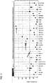
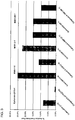
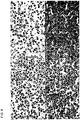
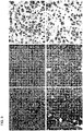
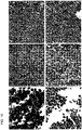
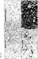
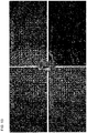
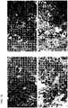
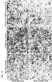
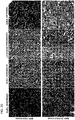
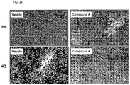
- the anti-SYK antibody molecules provide improved intensity. In certain such embodiments, the anti-SYK antibody molecules provide improved dynamic range. See, for example, Figures 2 , 3 , 7 , 14 . Quantification of the signal resulting from binding of anti-pSYK Y525/526 antibodies described herein demonstrates a greater signal-to-noise ratio and dynamic range, i.e., difference between lowest and highest values, than the signal resulting from binding of commercially available anti-pSYK Y525/526 antibodies.
- a large dynamic range of signal detection provides the ability to detect pSYK Y525/526 in samples comprising tumor cells from subjects, aged, damaged or archived tumor specimens, or tumors which do not express high amounts of pSYK Y525/526. This reduces the number of false negative results and provides higher certainty of correct therapeutic regimens.
- Specific detection and/or quantitative determination of pSYK Y525/526 using anti-pSYK Y525/526 antibodies described herein in a sample comprising tumor cells from a subject indicates fully active SYK, i.e., pSYK which causes the phosphorylation of proteins such as BTK, BLNK or PLC ⁇ , downstream in the signaling cascade. This reduces the number of false positive results, such as from detecting partially phosphorylated, partially activated or non-autophosphorylating pSYK, which may not indicate a tumor which is SYK-dependent.
- Contributing to the superiority of anti-pSYK Y525/526 antibodies described herein may be the selection using autophosphorylated pSYK, in combination with being raised against a pSYK Y525/526 peptide immunogen. Further selection comprised tests for performance of the antibodies in specific three-dimensional assays, such as immunocytochemistry (ICC), immunofluorescence (IF) and immunohistochemistry (IHC).
- ICC immunocytochemistry
- IF immunofluorescence
- IHC immunohistochemistry
- Standard techniques are used for recombinant DNA, oligonucleotide synthesis, and tissue culture and transformation and transfection (e.g., electroporation, lipofection). Enzymatic reactions and purification techniques are performed according to manufacturer's specifications or as commonly accomplished in the art or as described herein. The foregoing techniques and procedures are generally performed according to methods known in the art, e.g., as described in various general and more specific references that are cited and discussed throughout the present specification. See e.g., Sambrook et al. Molecular Cloning: A Laboratory Manual (3rd ed., Cold Spring Harbor Laboratory Press, Cold Spring Harbor, N.Y. (2000 )); Harlow, E. and Lane, D.
- SYK also known as “spleen tyrosine kinase” or “p72-Syk” protein refers to mammalian tyrosine-protein kinase SYK, such as human SYK protein.
- SYK also refers to human SYK protein phosphorylated at sites other than Y525 and/or Y526. In certain such embodiments, particular phosphorylated residues are indicated with a "p” before the residue number (referring to SEQ ID NO:2) For example, SYK phosphorylated at tyrosine residue 323 is herein referred to as SYK pY323.
- Human SYK refers to the protein shown in SEQ ID NO:2 and naturally occurring isoforms or allelic protein variants thereof.
- the allele in SEQ ID NO:2 can be encoded by the nucleic acid sequence of SYK shown in SEQ ID NO:1.
- One SYK variant lacks an in-frame exon and encodes a polypeptide variant of SEQ ID NO:2 which does not include amino acid residues 282 to 305 of SEQ ID NO:2.
- Other variants are known in the art. For example, there are several single nucleotide polymorphisms (SNPs) of human SYK, as seen in the NCBI SNP database.
- a naturally occurring allelic variant has an amino acid sequence at least 90%, 95%, 97%, 98% or 99% identical to the SYK sequence of SEQ ID NO:2.
- the transcript encodes a protein product of 635 amino acids, and is described in GenBank accession no.: NM_003177.
- the SH2 domains mediate the interaction of SYK with the phosphorylated ITAM domains of transmembrane proteins.
- SYK protein is characterized as a cytoplasmic non-receptor tyrosine kinase, and is believed to play a critical role in several biological processes including innate and adaptive immunity, cell adhesion, osteoclast maturation, platelet activation and vascular development.
- antibody molecule broadly encompass naturally-occurring forms of antibodies, e.g., polyclonal antibodies (e.g., IgG, IgA, IgM, IgE) and monoclonal and recombinant antibodies or antibody peptides such as single-chain antibodies, two-chain and multi-chain proteins, chimeric, CDR-grafted, human and humanized antibodies and multi-specific antibodies, as well as fragments and derivatives of all of the foregoing, which fragments (e.g., dAbs, scFv, Fv, Fab, F(ab)' 2 , Fab') and derivatives such as functional heavy chain antibodies, nanobodies, e.g., derivatives when paired with a complete variable region of the other chain, e.g., the light chain, it will allow binding of at least 25, 50, 75, 85 or 90% of that seen with the whole heavy and
- antibody also includes synthetic and genetically engineered variants, such as monobodies and diabodies. Although not within the term “antibody molecules,” the invention also includes derivatives such as "antibody analog(s),” other non-antibody molecule protein-based scaffolds, e.g., fusion proteins and/or immunoconjugates that use CDRs to provide specific antigen binding.
- pSYK phosphorylated Spleen Tyrosine Kinase
- An "anti-pSYK antibody molecule” refers to an antibody molecule (i.e., an antibody, antigen-binding fragment of an antibody or antibody analog) which interacts with or recognizes, e.g., binds (e.g., binds specifically) to phosphorylated SYK, e.g., human phosphorylated SYK.
- exemplary anti-pSYK antibody molecules are such as those summarized in Tables 1 and 2.
- pSYK refers in particular to SYK that has been phosphorylated at tyrosine 525 and/or 526 of SEQ ID NO:2.
- pBTK phospho-Bruton agammaglobulinemia tyrosine kinase
- pBTK or "phospho-Bruton agammaglobulinemia tyrosine kinase” means the polypeptide represented by GenPept Accession No. NP_000052, SEQ ID NO:26, or an isoform thereof, phosphorylated at at least one of its amino acid residues.
- pBTK is phosphorylated at tyrosine 551 of SEQ ID NO:26 ("BTK pY551").
- pBTK is phosphorylated at tyrosine 223 of SEQ ID NO:26 ("BTK pY223 ").
- pBLNK or "phospho-B-cell linker protein means the polypeptide represented by GenPept Accession No. NP_037446, SEQ ID NO:27 or an isoform thereof, phosphorylated at at least one of its amino acid residues.
- pBTK is phosphorylated at tyrosine 96 of SEQ ID NO:27 ("BLNK pY96").
- pBTK is phosphorylated at tyrosine 84 of SEQ ID NO:27 (“BLNK pY84").
- pFLT3 or "phospho-fms-related tyrosine kinase 3 means the polypeptide represented by GenPept Accession No. NP_004110, SEQ ID NO:28 or an isoform thereof, phosphorylated at at least one of its amino acid residues.
- pFLT3 is phosphorylated at tyrosine 591 of SEQ ID NO:28 ("FLT3 pY591").
- pFLT3 is phosphorylated at tyrosine 969 of SEQ ID NO:28 ("FLT3 pY969").
- a "SYK inhibitor,” a “SYK-targeted therapeutic agent” or “SYK targeted therapy” is a compound which inhibits activation of SYK, inhibits the activity of SYK or pSYK or inhibits the autophosphorylation of SYK.
- a SYK inhibitor inhibits the autophosphorylation of pSYK.
- the autophosphorylation inhibited by a SYK inhibitor is the autophosphorylation at tyrosine residue 525 and/or tyrosine residue 526 of SEQ ID NO:2.
- a SYK inhibitor can be one of several known in the art or may be selected from the group consisting of an ATP-competitor such as R406 or a non-hydrolyzable ATP analog, piceatannol, fostamatinib, a diaminopyrimidine carboxamide, e.g., as described in WO1999/31073 or WO2009/136995 , a pyrrolopyrimidine, e.g., as described in WO2009/131687 , and a fused heteroaromatic pyrrolidinone.
- a SYK inhibitor may be a fused heteroaromatic pyrrolidinone.
- a fused heteroaromatic pyrrolidinone may be a pyrrolopyrimidinone (e.g., a 6,7-dihydro-5H-pyrrolo[3,4-c]pyrimidin-5-one) or a pyrrolopyridinone (e.g., a 1H-pyrrolo[3,4-c]pyridine-3(2H)-one) compound described in U.S. Patent No. 8,440,689 .
- Some fused heteroaromatic pyrrolidinone compounds are described herein.
- One such fused heteroaromatic pyrrolidinone is described herein as Compound A.
- a therapeutic regimen comprising a SYK inhibitor may comprise treating a subject with a fused heteroaromatic pyrrolidinone, such as Compound A.
- antigen binding constellation of CDRs or "a number of CDRs sufficient to allow binding” (and similar language), as used herein, refers to sufficient CDRs of a chain, e.g., the heavy chain, such that when placed in a framework and paired with a complete variable region of the other chain, or with a portion of the other chain's variable region of similar length and having the same number of CDRs, e.g., the light chain, will allow binding, e.g., of at least 25, 50, 75, 85 or 90% of that seen with the whole heavy and light variable region.
- humanized antibody refers to an antibody that is derived from a non-human antibody e.g., rabbit, rodent (e.g., murine), sheep or goat, that retains or substantially retains the antigen-binding properties of the parent antibody but is less immunogenic in humans.
- Humanized as used herein is intended to include deimmunized antibodies.
- humanized antibodies include non-human CDRs and human or human derived framework and constant regions.
- modified antibody refers to antibodies that are prepared, expressed, created or isolated by recombinant means, such as antibodies expressed using a recombinant expression vector transfected into a host cell, antibodies isolated from a recombinant, combinatorial antibody library, antibodies isolated from a non-human animal (e.g., a rabbit, mouse, rat, sheep or goat) that is transgenic for human immunoglobulin genes or antibodies prepared, expressed, created or isolated by any other means that involves splicing of human immunoglobulin gene sequences to other DNA sequences.
- recombinant means such as antibodies expressed using a recombinant expression vector transfected into a host cell, antibodies isolated from a recombinant, combinatorial antibody library, antibodies isolated from a non-human animal (e.g., a rabbit, mouse, rat, sheep or goat) that is transgenic for human immunoglobulin genes or antibodies prepared, expressed, created or isolated by any other means that involves splicing of human immunoglobulin gene sequences to other
- modified antibodies include humanized, CDR grafted (e.g., an antibody having CDRs from a first antibody and a framework region from a different source, e.g., a second antibody or a consensus framework), chimeric, in vitro generated (e.g., by phage display) antibodies, and may optionally include variable or constant regions derived from human germline immunoglobulin sequences or human immunoglobulin genes or antibodies which have been prepared, expressed, created or isolated by any means that involves splicing of human immunoglobulin gene sequences to alternative immunoglobulin sequences.
- a modified antibody molecule includes an antibody molecule having a sequence change from a reference antibody.
- monospecific antibody refers to an antibody or antibody preparation that displays a single binding specificity and affinity for a particular epitope. This term includes a “monoclonal antibody” or “monoclonal antibody composition.”
- bispecific antibody or “bifunctional antibody” refers to an antibody that displays dual binding specificity for two epitopes, where each binding site differs and recognizes a different epitope.
- non-conjugated antibody and “naked antibody” are used interchangeably to refer to an antibody molecule that is not conjugated to a non-antibody moiety, e.g., an agent or a label.
- immunoconjugate refers to an antibody that is conjugated to a non-antibody moiety, e.g., an agent or a label.
- agent is used herein to denote a chemical compound, a mixture of chemical compounds, a biological macromolecule, or an extract made from biological materials.
- therapeutic agent refers to an agent that has biological activity. Exemplary therapeutic agents are chemotherapeutic agents.
- Cytotoxic agents refer to compounds which cause cell death primarily by interfering directly with the cell's functioning, including, but not limited to, alkylating agents, tumor necrosis factor inhibitors, intercalators, microtubule inhibitors, kinase inhibitors, proteasome inhibitors and topoisomerase inhibitors.
- a "toxic payload” as used herein refers to a sufficient amount of cytotoxic agent which, when delivered to a cell results in cell death. Delivery of a toxic payload may be accomplished by administration of a sufficient amount of immunoconjugate comprising an antibody or antigen binding fragment of the invention and a cytotoxic agent.
- Delivery of a toxic payload may also be accomplished by administration of a sufficient amount of an immunoconjugate comprising a cytotoxic agent, wherein the immunoconjugate comprises a secondary antibody or antigen binding fragment thereof which recognizes and binds an antibody or antigen binding fragment of the invention.
- a sequence "derived from” or “specific for a designated sequence” refers to a sequence that comprises a contiguous sequence of approximately at least 6 nucleotides or at least 2 amino acids, at least about 9 nucleotides or at least 3 amino acids, at least about 10-12 nucleotides or 4 amino acids, or at least about 15-21 nucleotides or 5-7 amino acids corresponding, i.e., identical or complementary to, e.g., a contiguous region of the designated sequence.
- the sequence comprises all of a designated nucleotide or amino acid sequence.
- sequences may be complementary (in the case of a polynucleotide sequence) or identical to a sequence region that is unique to a particular sequence as determined by techniques known in the art.
- Regions from which sequences may be derived include but are not limited to, regions encoding specific epitopes, regions encoding CDRs, regions encoding framework sequences, regions encoding constant domain regions, regions encoding variable domain regions, as well as non-translated and/or non-transcribed regions.
- the derived sequence will not necessarily be derived physically from the sequence of interest under study, but may be generated in any manner, including, but not limited to, chemical synthesis, replication, reverse transcription or transcription, that is based on the information provided by the sequence of bases in the region(s) from which the polynucleotide is derived. As such, it may represent either a sense or an antisense orientation of the original polynucleotide. In addition, combinations of regions corresponding to that of the designated sequence may be modified or combined in ways known in the art to be consistent with the intended use.
- a sequence may comprise two or more contiguous sequences which each comprise part of a designated sequence, and are interrupted with a region which is not identical to the designated sequence but is intended to represent a sequence derived from the designated sequence.
- "derived therefrom” includes an antibody molecule which is functionally or structurally related to a comparison antibody, e.g., "derived therefrom” includes an antibody molecule having similar or substantially the same sequence or structure, e.g., having the same or similar CDRs, framework or variable regions.
- “Derived therefrom” for an antibody also includes residues, e.g., one or more, e.g., 2, 3, 4, 5, 6 or more residues, which may or may not be contiguous, but are defined or identified according to a numbering scheme or homology to general antibody structure or three-dimensional proximity, i.e., within a CDR or a framework region, of a comparison sequence.
- the term "derived therefrom” is not limited to physically derived therefrom, but includes generation by any manner, e.g., by use of sequence information from a comparison antibody to design another antibody and can refer merely to sequence similarity.
- the phrase "encoded by” refers to a nucleic acid sequence that codes for a polypeptide sequence, wherein the polypeptide sequence or a portion thereof contains an amino acid sequence of at least 3 to 5 amino acids, at least 8 to 10 amino acids, or at least 15 to 20 amino acids from a polypeptide encoded by the nucleic acid sequence.
- Calculations of "homology" between two sequences can be performed as follows.
- the sequences are aligned for optimal comparison purposes (e.g., gaps can be introduced in one or both of a first and a second amino acid or nucleic acid sequence for optimal alignment and nonhomologous sequences can be disregarded for comparison purposes).
- the length of a reference sequence aligned for comparison purposes is at least 30%, 40%, or 50%, at least 60%, or at least 70%, 80%, 90%, 95%, or 100% of the length of the reference sequence.
- the amino acid residues or nucleotides at corresponding amino acid positions or nucleotide positions are then compared.
- amino acid or nucleic acid “identity” is equivalent to amino acid or nucleic acid "homology”).
- the percent identity between the two sequences is a function of the number of identical positions shared by the sequences, taking into account the number of gaps, and the length of each gap, which need to be introduced for optimal alignment of the two sequences.
- the comparison of sequences and determination of percent homology between two sequences may be accomplished using a mathematical algorithm.
- the percent homology between two amino acid sequences may be determined using any method known in the art. For example, the Needleman and Wunsch, J. Mol. Biol. 48:444-453 (1970 ), algorithm which has been incorporated into the GAP program in the GCG software package, using either a Blossum 62 matrix or a PAM250 matrix, and a gap weight of 16, 14, 12, 10, 8, 6, or 4 and a length weight of 1, 2, 3, 4, 5, or 6.
- the percent homology between two nucleotide sequences can also be determined using the GAP program in the GCG software package (Accelerys, Inc.
- An exemplary set of parameters for determination of homology are a Blossum 62 scoring matrix with a gap penalty of 12, a gap extend penalty of 4, and a frameshift gap penalty of 5.
- hybridizes under stringent conditions describes conditions for hybridization and washing.
- Guidance for performing hybridization reactions may be found in Current Protocols in Molecular Biology, John Wiley & Sons, N.Y. (1989), 6.3.1-6.3.6 . Aqueous and nonaqueous methods are described in that reference and either may be used.
- Specific hybridization conditions referred to herein are as follows: 1) low stringency hybridization conditions in 6X sodium chloride/sodium citrate (SSC) at about 45° C, followed by two washes in 0.2X SSC, 0.1% SDS at least at 50° C, (the temperature of the washes may be increased to 55° C for low stringency conditions); 2) medium stringency hybridization conditions in 6X SSC at about 45° C, followed by one or more washes in 0.2X SSC, 0.1% SDS at 60° C; 3) high stringency hybridization conditions in 6X SSC at about 45° C, followed by one or more washes in 0.2X SSC, 0.1% SDS at 65° C; and 4) very high stringency hybridization conditions are 0.5M sodium phosphate, 7% SDS at 65° C, followed by one or more washes at 0.2X SSC, 1% SDS at 65° C.
- SSC 6X sodium chloride/sodium citrate
- Very high stringency conditions are often the preferred conditions and the ones that should be used unless otherwise specified. It is understood that the antibodies and antigen binding fragment thereof of the invention may have additional conservative or non-essential amino acid substitutions, which do not have a substantial effect on the polypeptide functions. Whether or not a particular substitution will be tolerated, i.e., will not adversely affect desired biological properties, such as binding activity, may be determined as described in Bowie, J U et al. Science 247: 1306-1310 (1990 ) or Padlan et al. FASEB J. 9: 133-139 (1995 ). A "conservative amino acid substitution” is one in which the amino acid residue is replaced with an amino acid residue having a similar side chain. Families of amino acid residues having similar side chains have been defined in the art.
- amino acids with basic side chains e.g., lysine, arginine, histidine
- acidic side chains e.g., aspartic acid, glutamic acid
- uncharged polar side chains e.g., asparagine, glutamine, serine, threonine, tyrosine, cysteine
- nonpolar side chains e.g., glycine, alanine, valine, leucine, isoleucine, proline, phenylalanine, methionine, tryptophan
- beta-branched side chains e.g., threonine, valine, isoleucine
- aromatic side chains e.g., tyrosine, phenylalanine, tryptophan, histidine
- non-essential amino acid residue is a residue that may be altered from the wild-type sequence of the binding agent, e.g., the antibody, without abolishing or, without substantially altering a biological activity, whereas an "essential" amino acid residue results in such a change.
- an essential amino acid residue may be a specificity determining residue (SDR).
- isolated refers to material that is removed from its original environment (e.g., the natural environment if it is naturally occurring).
- a naturally occurring polynucleotide or polypeptide present in a living animal is not isolated, but the same polynucleotide or DNA or polypeptide, separated from some or all of the coexisting materials in the natural system, is isolated.
- Such polynucleotide or polypeptide could be part of a vector and/or such polynucleotide or polypeptide could be part of a composition, e.g., a mixture, solution or suspension or comprising an isolated cell or a cultured cell which comprises the polynucleotide or polypeptide, and still be isolated in that the vector or composition is not part of its natural environment.
- a composition e.g., a mixture, solution or suspension or comprising an isolated cell or a cultured cell which comprises the polynucleotide or polypeptide, and still be isolated in that the vector or composition is not part of its natural environment.
- operably linked refers to a situation wherein the components described are in a relationship permitting them to function in their intended manner.
- a control sequence "operably linked" to a coding sequence is ligated in such a manner that expression of the coding sequence is achieved under conditions compatible with the control sequence.
- vector refers to a replicon in which another polynucleotide segment is attached, such as to bring about the replication and/or expression of the attached segment.
- control sequence refers to a polynucleotide sequence that is necessary to effect the expression of a coding sequence to which it is ligated. The nature of such control sequences differs depending upon the host organism. In prokaryotes, such control sequences generally include a promoter, a ribosomal binding site and terminators and, in some instances, enhancers. The term “control sequence” thus is intended to include at a minimum all components whose presence is necessary for expression, and also may include additional components whose presence is advantageous, for example, leader sequences.
- purified product refers to a preparation of the product which has been isolated from the cellular constituents with which the product is normally associated and/or from other types of cells that may be present in the sample of interest.
- epitope refers to a protein determinate capable of binding specifically to an antibody.
- Epitopic determinants usually consist of chemically active surface groupings of molecules such as amino acids or sugar side chains and usually have specific three dimensional structural characteristics, as well as specific charge characteristics.
- Some epitopes are linear epitopes while others are conformational epitopes.
- a linear epitope is an epitope wherein a contiguous amino acid primary sequence comprises the epitope recognized.
- a linear epitope typically includes at least 3, and more usually, at least 5, for example, about 8 to about 10 contiguous amino acids.
- a conformational epitope can result from at least two situations, such as: a) a linear sequence which is only exposed to antibody binding in certain protein conformations, e.g., dependent on ligand binding, or dependent on modification (e.g., phosphorylation) by signaling molecules; or b) a combination of structural features from more than one part of the protein, or in multisubunit proteins, from more than one subunit, wherein the features are in sufficiently close proximity in 3-dimensional space to participate in binding.
- isotype refers to the antibody class (e.g., IgM, IgA, IgE or IgG) that is encoded by heavy chain constant region genes.
- detectable agent As used herein, the terms “detectable agent,” “label” or “labeled” are used to refer to incorporation of a detectable marker on a polypeptide or glycoprotein. Various methods of labeling polypeptides and glycoproteins are known in the art and may be used.
- labels for polypeptides include, but are not limited to, the following: radioisotopes or radionuclides (e.g., indium ( 111 In), iodine ( 131 I or 125 I), yttrium ( 90 Y), lutetium ( 177 Lu), actinium ( 225 Ac), bismuth ( 212 Bi or 213 Bi), sulfur ( 35 S), carbon ( 14 C), tritium ( 3 H), rhodium ( 188 Rh), technetium ( 99 mTc), praseodymium, or phosphorous ( 32 P) or a positron-emitting radionuclide, e.g., carbon-11 ( 11 C), potassium-40 ( 40 K), nitrogen-13 ( 13 N), oxygen-15 ( 15 O), fluorine-18 ( 18 F), gallium-68 ( 68 Ga), and iodine- 121 ( 121 I)), fluorescent labels (e.g., FITC, rhodamine, lanthanide phosphors, fluorescent
- binding means, for an anti-pSYK antibody molecule, that the antibody molecule binds to pSYK, e.g., human phosphorylated SYK protein, with greater affinity than it does to a non-phosphorylated SYK, phosphatase-treated (PPase) SYK or pSYK or non-SYK protein, e.g., bovine serum albumin (BSA), a rat sarcoma viral oncogene homolog (RAS), or human actin.
- pSYK e.g., human phosphorylated SYK protein
- PPase phosphatase-treated SYK or pSYK or non-SYK protein
- BSA bovine serum albumin
- RAS rat sarcoma viral oncogene homolog
- an anti-pSYK molecule will have a K d for the non-phosphorylated SYK or non-SYK protein, e.g., BSA, RAS or actin, which is greater than 2, greater than 10, greater than 100, greater than 1,000 times, greater than 10 4 , greater than 10 5 , or greater than 10 6 times its K d for phosphorylated SYK, e.g., human phosphorylated SYK protein.
- K d for phosphorylated SYK and the non-phosphorylated SYK or non-SYK protein, e.g., BSA, RAS or actin should be done under the same conditions.
- an anti-pSYK antibody having specific binding to pSYK Y525/526 binds to pSYK Y525/526 with greater affinity than it does to SYK phosphorylated at any other tyrosine, serine or threonine of SEQ ID NO:2.
- affinity refers to the apparent association constant or K a .
- the K a is the reciprocal of the dissociation constant (K d ).
- An antibody may, for example, have a binding affinity of at least 10 5 , 10 6 , 10 7 , 10 8 , 10 9 , 10 10 and 10 11 M -1 for a particular target molecule. Higher affinity binding of an antibody to a first target relative to a second target may be indicated by a higher K A (or a smaller numerical value K D ) for binding the first target than the K A (or numerical value K D ) for binding the second target.
- the antibody has specificity for the first target (e.g., a protein in a first conformation or mimic thereof) relative to the second target (e.g., the same protein in a second conformation or mimic thereof; or a second protein).
- Differences in binding affinity may be at least 1.5, 2, 3, 4, 5, 10, 15, 20, 37.5, 50, 70, 80, 91, 100, 500, 1000, or 10 5 fold. Binding affinity may be determined by a variety of methods including equilibrium dialysis, equilibrium binding, gel filtration, ELISA, surface plasmon resonance, or spectroscopy (e.g., using a fluorescence assay).
- relative affinity of an anti-pSYK antibody molecule may be measured from ELISA measurements against pSYK protein, pSYK Y525/526 peptide (e.g., the immunogen used to raise anti-pSYK antibody molecules), by FACS measurements with pSYK expressing cells.
- Exemplary conditions for evaluating binding affinity are in TRIS-buffer (50mM TRIS, 150mM NaCl, 5mM CaCl 2 at pH7.5). These techniques may be used to measure the concentration of bound and free binding protein as a function of binding protein (or target) concentration.
- K A it is not always necessary to make an exact determination of K A , though, since sometimes it is sufficient to obtain a quantitative measurement of affinity, e.g., determined using a method such as ELISA or FACS analysis, is proportional to K A , and thus may be used for comparisons, such as determining whether a higher affinity is, e.g., 2-fold higher, to obtain a qualitative measurement of affinity, or to obtain an inference of affinity, e.g., by activity in a functional assay, e.g., an in vitro or in vivo assay.
- a functional assay e.g., an in vitro or in vivo assay.
- Affinity of anti-pSYK antibody molecules can also be measured using a technology such as real-time Biomolecular Interaction Analysis (BIA) (see, e.g., Sjolander, S, and Urbaniczky, C, 1991, Anal. Chem. 63:2338-2345 and Szabo et al., 1995, Curr. Opin. Struct. Biol. 5:699-705 ).
- BIOA Biomolecular Interaction Analysis
- surface plasmon resonance is a technology for studying biospecific interactions in real time, without labeling any of the interactants (e.g., BIACORETM).
- An anti-pSYK antibody (Prep A) may be diluted to an appropriate concentration (e.g., 20 g/mL) in 10 nM sodium acetate, pH 4.0 and a reference/control antibody (Prep B) may be diluted to an appropriate concentration (e.g., 10 g/mL) in 10 mM sodium acetate, pH 4.0.
- Prep A an anti-pSYK antibody
- Prep B a reference/control antibody
- Prep B may be diluted to an appropriate concentration (e.g., 10 g/mL) in 10 mM sodium acetate, pH 4.0.
- Each antibody may then be covalently immobilized to several CM4 BIACORE chips using standard amine coupling.
- Prep A antibody may be immobilized over two flow cells at around 75-100 RU while Prep B antibody may be immobilized to one flow cell at around 70-80 RU.
- the remaining fourth flow cell of each CM4 chip may be used as the reference flow cell.
- the term “treat” or “treatment” is defined as the administration of a therapeutic agent to modify a material or subject.
- treatment shall mean the use of a therapy to prevent or inhibit further tumor growth, as well as to cause shrinkage of a tumor. Such use can provide longer survival times.
- Treatment is also intended to include prevention of metastasis of tumor.
- a tumor is “inhibited” or “treated” if at least one symptom (as determined by responsiveness/non-responsiveness, time to progression, or indicators known in the art and described herein) of the cancer or tumor is alleviated, terminated, slowed, minimized, or prevented.
- any amelioration of any symptom, physical or otherwise, of a tumor pursuant to treatment using a therapeutic regimen is within the scope of the invention.
- the treatment may be to cure, heal, alleviate, relieve, alter, remedy, ameliorate, palliate, improve or affect the disorder, the symptoms of the disorder or the predisposition toward the disorder, e.g., a cancer.
- treating is believed to cause the inhibition, ablation, or killing of a cell in vitro or in vivo, or otherwise reducing capacity of a cell, e.g., an aberrant cell, to mediate a disorder, e.g., a disorder as described herein (e.g., a cancer).
- Administration of a SYK-targeted therapy can be providing an anti-pSYK antibody molecule or a SYK inhibitor to a subject, e.g., a patient, or administration, e.g., by application, to an isolated tissue or cell from a subject which is returned to the subject.
- the anti-pSYK antibody molecule or SYK inhibitor may be administered alone or in combination with a second agent.
- a subject is intended to include mammals, primates, humans and non-human animals.
- a subject may be a patient (e.g., a human patient or a veterinary patient), having a disorder, disease, or condition, such as a disorder, disease, or condition mediated by SYK, e.g., aberrant BCR-mediated SYK activation disorders (e.g., DLBCL, follicular lymphoma, mantle cell lymphoma, or chronic lymphocytic leukemia), aberrant Fc ⁇ receptor-mediated SYK activation disorders (e.g., acute myeloid leukemia or myelodysplastic syndrome) or aberrant LMP2A-mediated SYK activation disorders (e.g., nasopharyngeal carcinoma, lymphoma, or gastric cancer) as described above.
- BCR-mediated SYK activation disorders
- the disorder, disease, or condition is related to abnormal cell growth, including hematological cancer, leukemia, lymphoma or myeloma malignancies, such as acute myeloid leukemia (AML), B-cell chronic lymphocytic leukemia (BCLL), B-cell lymphoma (e.g., mantle cell lymphoma (MCL), diffuse large B-cell lymphoma (DLBCL)), follicular lymphoma, and T-cell lymphoma (e.g., peripheral T-cell lymphoma), as well as epithelial cancers (i.e., carcinomas), such as lung cancer (small cell lung cancer and non-small cell lung cancer), pancreatic cancer, and colon cancer.
- AML acute myeloid leukemia
- BLL B-cell chronic lymphocytic leukemia
- B-cell lymphoma e.g., mantle cell lymphoma (MCL), diffuse large B-cell lymphoma (DLBCL)
- the condition may include other types of cancer, including leukemia (chronic myelogenous leukemia and chronic lymphocytic leukemia (CLL)) and breast cancer, genitourinary cancer, skin cancer, bone cancer, prostate cancer, and liver cancer; brain cancer; cancer of the larynx, gall bladder, rectum, parathyroid, thyroid, adrenal, neural tissue, bladder, head, neck, stomach, bronchi, and kidneys; basal cell carcinoma, squamous cell carcinoma, metastatic skin carcinoma, osteosarcoma, Ewing's sarcoma, veticulum cell sarcoma, and Kaposi's sarcoma; myeloma, EBV-associated tumors and other solid tumors, giant cell tumor, islet cell tumor, acute and chronic lymphocytic and granulocytic tumors, hairy-cell tumor, adenoma, medullary carcinoma,
- leukemia chronic myelogenous leukemia and chronic lymphocytic leukemia
- the disorder, disease or condition is selected from PTCL, DLBCL, FL, MCL, CLL, AML, MDS, nasopharyngeal carcinoma, lymphoma, gastric carcinoma, breast cancer, ovarian cancer, lung cancer (e.g., small cell lung cancer) and PT-LPD.
- the DLBCL is classified as a subtype selected from the group consisting of germinal center B cell-like (GCB) subtype, activated B cell-like (ABC) subtype and non-germinal center B cell-like (non-GCB) subtype.
- the condition may include other diseases related to abnormal cell growth, including non-malignant proliferative diseases such as benign prostatic hypertrophy, restinosis, hyperplasia, synovial proliferation disorder, retinopathy or other neovascular disorders of the eye, among others.
- the condition may include a symptom of such SYK expressing, pSYK expressing or SYK-activating conditions; or a predisposition toward such SYK-expressing, pSYK expressing or SYK-activating conditions.
- SYK, BTK and PI3K ⁇ are clinically validated targets in BCR-activated malignancies.
- non-human animals of the invention includes all non-human vertebrates, e.g., non-human mammals and non-mammals, such as non-human primates, sheep, dog, cow, chickens, amphibians, reptiles, mouse, rat, rabbit or goat etc., unless otherwise noted.
- subject excludes one or more or all of a mouse, rat, rabbit or goat.
- an amount of an anti-pSYK antibody molecule or a therapeutic agent "effective” or “sufficient” to treat a disorder refers to an amount of the antibody molecule which is effective, upon single or multiple dose administration to a subject, (a) to cause a detectable decrease in the severity of the disorder or disease state being treated; (b) to ameliorate or alleviate the patient's symptoms of the disease or disorder; or (c) to slow or prevent advancement of, or otherwise stabilize or prolong stabilization of, the disorder or disease state being treated (e.g., prevent additional tumor growth of a cancer) or a cell, e.g., cancer cell (e.g., a SYK-expressing, pSYK expressing or SYK-activated tumor cell), or in prolonging curing, alleviating, relieving or improving a subject with a disorder as described herein beyond that expected in the absence of such treatment.
- cancer cell e.g., a SYK-expressing, pSYK expressing or SYK-activated tumor cell
- inhibiting the growth of the tumor or cancer refers to slowing, interrupting, arresting or stopping its growth and/or metastases and does not necessarily indicate a total elimination of the tumor growth.
- a specific dosage and treatment regimen for any particular patient may depend upon a variety of factors, including the activity of the specific compound employed, the age, body weight, general health, sex, and diet of the patient, time of administration, rate of excretion, drug combinations, the judgment of the treating physician, and the severity of the particular disease being treated.
- the H-score approach provides optimal data resolution for determining variation in intensity and tumor percentage of staining within and among tumor types. It also provides a good tool for determining thresholds for positive staining.
- the percentage of cells (0-100) within a tumor with staining intensities ranging from 0-3+ are provided. With the instant method, scores with intensities of 0, 0.5, 1, 2 and 3 were provided. Depending on the marker, 0.5 staining may be scored as positive or negative, and reflects light but perceptible staining for the marker.
- H score % tumor * 1 + % tumor * 2 + % tumor * 3 . For example, if a tumor is 20% negative (0), 30% +1, 10% +2, 40% +3, this would give an H score of 170.
- the maximum H-score is 300 (100% * +3), per sub-cellular localization (i.e., apical or cytoplasmic), if 100% of tumor cells label with 3+ intensity.
- the total H-score alone was not be used to compare samples, but evaluated in addition to a review of the break-down of the percentage of cells at each intensity. For example, a score of 90 could represent 90% of tumor cells staining with 1+ intensity or 30% of cells with 3+ intensity. These samples have the same H-score but very different pSYK expression.
- the percentage of cells to be scored at each intensity may vary, but are normally scored in increments of 10%; however, a small percentage of scoring of a single component may be estimated at 1% and 5% as well in order to demonstrate that some level of staining is present.
- apical staining may be considered for evaluating at low level increments, such as 1 and 5%.
- the anti-pSYK antibody molecules of the invention are useful, inter alia, to detect pSYK expression.
- the anti-pSYK antibody molecules are useful for detecting autophosphorylated pSYK.
- the anti-pSYK antibody molecules are useful for detecting pSYK Y525/526.
- the anti-pSYK antibody molecules are useful for detecting activated pSYK, e.g., SYK which can lead to the phosphorylation of BLNK, BTK, or PLC ⁇ .
- the anti-pSYK antibody molecules may include non-human anti-pSYK antibody molecules (e.g., non-human and non- murine antibody molecules) that specifically bind to pSYK, e.g., with a binding affinity of at least 10 3 , 10 4 , 10 5 , 10 6 , 10 7 ,10 8 , 10 9 , 10 10 or 10 11 M "1 for pSYK, e.g., for pSYK Y525/526.
- the anti-pSYK antibody molecule may be a non-human, non-murine and non-rat antibody molecule, e.g., a rabbit anti-pSYK antibody molecule, e.g., as described herein.
- the invention relates to anti-pSYK antibody molecules that include features such as those summarized in Tables 1 and 2. In other aspects, the invention relates to anti-pSYK antibody molecules that include features such as those summarized in Tables 3, 4, 5 and/or 6.
- the anti-pSYK antibody molecule is a rabbit hybridoma antibody and is one of antibody MIL81-1-8. In an embodiment, the anti-pSYK antibody molecule is derived from antibody MIL81-1-8. In another embodiment, the anti-pSYK antibody molecule is a rabbit hybridoma antibody and is MIL81-2-1. In an embodiment, the anti-pSYK antibody molecule is derived from antibody MIL81-2-1. In another embodiment, the anti-pSYK antibody molecule is a rabbit hybridoma antibody and is MIL81-99-1. In an embodiment, the anti-pSYK antibody molecule is derived from antibody MIL81-99-1.
- an anti-pSYK antibody molecule has an affinity for pSYK, e.g., as measured by direct binding or competition binding assays.
- the anti-pSYK antibody molecule has a K d of less than 1x10 -6 M, less than 1x10 -7 M, less than 1x10 -8 M, less than 1x10 -9 M, less than 1x10 -10 M, less than 1x 10 -11 M, less than 1x10 -12 M, or less than 1x10 -13 M.
- the antibody molecule is an IgG, or antigen-binding fragment thereof, and has a K d of less than 1x10 -6 M, less than 1x10 -7 M, less than 1x10 -8 M, or less than 1x10 -9 M.
- an anti-pSYK antibody molecule e.g., a MIL81-1-8 antibody or antibody derived therefrom has a K d of about 80 to about 200 pM, about 50 to about 400 pM, about 100 to about 300 pM, about 100 to about 150 pM or about 120 pM.
- an anti-pSYK antibody molecule e.g., a MIL81-1-8 antibody or antibody derived therefrom has a k a of about 0.9 to about 1.25x10 5 M -1 s -1 , or about 1.1x10 s M -1 s -1 .
- the antibody molecule is an ScFv and has a K d of less than 1x10 -6 M, less than 1x10 -7 M, less than 1x10 -8 M, less than 1x10 -9 M, less than 1x10 -10 M, 1x10 -11 M, less than 1x10 -12 M, or less than 1x10 -13 M.
- the naturally occurring mammalian antibody structural unit is typified by a tetramer.
- Each tetramer is composed of two pairs of polypeptide chains, each pair having one "light” (about 25 kDa) and one "heavy" chain (about 50-70 kDa).
- the amino-terminal portion of each chain includes a variable region of about 100 to 110 or more amino acids primarily responsible for antigen recognition.
- the carboxy-terminal portion of each chain defines a constant region primarily responsible for effector function.
- Human light chains can be classified as kappa and lambda light chains.
- Heavy chains may be classified as mu, delta, gamma, alpha, or epsilon, and define the antibody's isotype as IgM, IgD, IgG, IgA, and IgE, respectively.
- the variable and constant regions are joined by a "J" region of about 12 or more amino acids, with the heavy chain also including a "D” region of about 10 more amino acids. See generally, Fundamental Immunology Ch. 7 (Paul, W., ed., 2nd ed. Raven Press, N.Y. (1989 )).
- the variable regions of each light/heavy chain pair form the antibody binding site.
- the isotypes for the anti-pSYK antibody molecules are IgG immunoglobulins, which may be classified into four subclasses, IgGl, IgG2, IgG3 and IgG4, having different gamma heavy chains. Most therapeutic antibodies are human, chimeric, or humanized antibodies of the IgG1 isotype. In a particular embodiment, the anti-pSYK antibody molecule is a rabbit IgG antibody.
- variable regions of each heavy and light chain pair form the antigen binding site.
- an intact IgG antibody has two binding sites which are the same.
- bifunctional or bispecific antibodies are artificial hybrid constructs which have two different heavy/light chain pairs, resulting in two different binding sites.
- the chains all exhibit the same general structure of relatively conserved framework regions (FR) joined by three hypervariable regions, also called complementarity determining regions or CDRs.
- the CDRs from the two chains of each pair are aligned by the framework regions, enabling binding to a specific epitope.
- both light and heavy chains comprise the domains FR1, CDR1, FR2, CDR2, FR3, CDR3 and FR4.
- the assignment of amino acids to each domain is in accordance with the definitions of Kabat Sequences of Proteins of Immunological Interest (National Institutes of Health, Bethesda, Md. (1987 and 1991 )), or Chothia & Lesk J. Mol. Biol.
- CDRs are referred to for each of the heavy (HCDR1, HCDR2, HCDR3) and light (LCDR1, LCDR2, LCDR3) chains.
- An anti-pSYK antibody molecule can comprise all, or an antigen binding subset of the CDRs, of one or both, the heavy and light chain, of one of the above-referenced rabbit antibodies.
- Amino acid sequences of rabbit hybridoma antibodies, including variable regions and CDRs, may be found in Table 3 and Table 5.
- the antibody molecule may include one or both of: (a) one, two, three, or an antigen binding number of, light chain CDRs (LCDR1, LCDR2 and/or LCDR3) of one of the above-referenced rabbit hybridoma antibodies, or the CDR(s) may comprise an amino acid sequence of one or more or all of LCDR1-3 as follows: LCDR1, or modified LCDR1 wherein one to seven amino acids are conservatively substituted) LCDR2, or modified LCDR2 wherein one or two amino acids are conservatively substituted); or LCDR3, or modified LCDR3 wherein one or two amino acids are conservatively substituted; and (b) one, two, three, or an antigen binding number of, heavy chain CDRs (HCDR1, HCDR2 and/or HCDR3) of one of the above-referenced rabbit hybridoma antibodies.
- HCDR1, HCDR2 and/or HCDR3 an antigen binding number of, heavy chain CDRs
- the CDR(s) may comprise an amino acid sequence of one or more or all of HCDR1-3 as follows: HCDR1, or modified HCDR1 wherein one or two amino acids are conservatively substituted; HCDR2, or modified HCDR2 wherein one to four amino acids are conservatively substituted; or HCDR3, or modified HCDR3 wherein one or two amino acids are conservatively substituted.
- Useful immunogens for production of anti-pSYK antibody molecules include pSYK e.g., human pSYK-expressing cells; membrane fractions of pSYK-expressing cells; recombinant cells expressing pSYK; isolated or purified pSYK, e.g., human pSYK protein (e.g., biochemically isolated pSYK, or a portion thereof (e.g., a portion or peptide comprising the phosphorylation sites of pSYK, e.g., comprising at least about 8, 10, 12, 14, 16, 20, 24, 28 or 32 amino acid residues of SEQ ID NO:2)).
- pSYK e.g., human pSYK-expressing cells
- membrane fractions of pSYK-expressing cells e.g., recombinant cells expressing pSYK
- isolated or purified pSYK e.g., human pSYK protein (e.g.
- an immunogen to generate an anti-pSYK antibody is a synthetic phosphopeptide corresponding to residues surrounding Tyr525/526 of human SYK.
- the peptide immunogen is amino acids 520 to 529 of SEQ ID NO:2, modified with phosphate on tyrosine 525 and/or 526.
- Immunogens may be fused to heterologous sequences to aid in biochemical manipulation, purification, immunization or antibody titer measurement.
- Such immunogens may comprise a portion of pSYK, e.g., the phosphorylation domain, and a portion comprising a non-pSYK polypeptide.
- a fusion protein for ease of purification or immobilization onto a solid support, e.g., an affinity column or a microtiter plate or other suitable assay substrate/chip.
- a fusion moiety can add a domain, e.g., glutathione-S -transferase/kinase (GST), which can bind glutathione; an Fc region of an immunoglobulin, which can bind to protein A or protein G; amino acid residues, e.g., two, three, four, five, or six histidine residues which can bind nickel or cobalt on an affinity column; an epitope tag, e.g., a portion of c-myc oncogene (myc-tag), a FLAG tag ( U.S. Pat. No.
- GST glutathione-S -transferase/kinase
- an Fc region of an immunoglobulin which can bind to protein A or protein G
- amino acid residues e.g., two, three, four, five, or six histidine residues which can bind nickel or cobalt on an affinity column
- an epitope tag e.g.,
- a peptide immunogen has a terminal cysteine so the peptide may be conjugated to a hapten or carrier protein. In some examples, the peptide immunogen has an N-terminal cysteine.
- Immunogens which comprise the Fc portion of an immunoglobulin can hold the pSYK, either in solution or attached to a cell, in a configuration which allows structural access to pSYK epitopes by the host immune surveillance components for efficient antibody generation. Because immunoglobulin heavy chains comprising the Fc regions associate into dimers through interchain disulfide bonds, immunogens resulting from fusion with Fc regions are dimers.
- This adjuvant function can trigger specific antibodies against both Fc and pSYK epitopes.
- Fc-reactive antibodies may be identified and discarded during screening.
- the Fc portion may have a wild type sequence or a sequence which is mutated to modify effector function.
- a mutated constant region may be incorporated into a fusion protein to minimize binding to Fc receptors and/or ability to fix complement (see e.g.
- lysine 235 and glycine 237 are mutated, e.g., to alanine.
- An immunogen/fusion protein with Fc-mutated IgG can have reduced interaction with Fc receptors in the host.
- Useful epitopes e.g., reference epitopes, from the pSYK molecule, to which the anti-pSYK antibody molecules, e.g., rabbit monoclonal antibodies, or humanized versions thereof, as described herein, may bind, may be found in permeabilized cells, membrane fractions, cell lysates, and in tissue sections.
- an epitope for an anti-pSYK antibody molecule may reside within, or include a residue(s) from, residues 500-550 of SEQ ID NO:2; residues 510-540 of SEQ ID NO:2; residues 515-530 of SEQ ID NO:2; or fragments thereof that bind an anti-pSYK antibody molecule of the invention, e.g., a MIL81-1-8-binding fragment thereof.
- Such fragments may comprise residues 525/526 and surrounding residues of SEQ ID NO:2, and may be phosphorylated e.g. phosphorylated at tyrosine 525 and/or phosphorylated tyrosine 526.
- an epitope for an anti-pSYK antibody molecule is a conformational epitope further comprising one or more additional amino acid residues in the SYK amino acid sequence, i.e., selected from about residue 1 to 635 of SEQ ID NO:2.
- Antibodies raised against such epitopes or the phosphorylation domain may be useful as therapeutic or diagnostic antibodies, as described herein.
- the anti-pSYK antibody molecule has one or more of the following properties: a) it competes for binding, e.g., binding to cytoplasmic pSYK or purified pSYK, with one of the above-referenced anti-pSYK antibody molecules summarized in Tables 1 and 2 or rabbit hybridoma antibodies (e.g., MIL81-1-8, MIL81-2-1 or MIL81-99-1); b) it binds to the same, or substantially the same, epitope on pSYK as one of the above-referenced anti-pSYK antibody molecules summarized in Tables 1 and 2, or rabbit hybridoma antibodies (e.g., MIL81-1-8, MIL81-2-1 or MIL81-99-1).
- the antibody binds the same epitope, as determined by one or more of a peptide array assay or by binding to truncation mutants, chimeras or point mutants of SEQ ID NO:2 expressed in the cytoplasm or membrane preparations, e.g., as those assays are described herein; c) it binds to an epitope which has at least 1, 2, 3, 4, 5, 8, 10, 15 or 20 contiguous amino acid residues in common with the epitope of one of the above-referenced anti-pSYK antibody molecules summarized in Tables 1 and 2, or rabbit hybridoma antibodies (e.g., MIL81-1-8, MIL81-2-1 or MIL81-99-1); d) it binds a region of human pSYK that is bound by an anti-pSYK antibody of the invention, wherein the region e.g., cytoplasmic region, is 10-15, 10-20, 20-30, or 20-40 residues in length, and binding is determined, e.
- the anti-pSYK antibody molecule binds the phosphorylation domain of human pSYK.
- an anti-pSYK antibody molecule may bind the human pSYK portion defined by amino acid residues 500-550, 510-540, 515-530 or 520-529 of SEQ ID NO:2.
- an anti-pSYK antibody molecule binds the phosphorylation site at amino acid residues 525 and/or 526 of SEQ ID NO:2; or e) it binds to a reference epitope described herein.
- the anti-pSYK antibody molecule binds the SYK sequence: RADEN-pY-pY-KAQ (amino acids 520 to 529 of SEQ ID NO:2, modified with phosphate on tyrosine 525 and 526).
- the antibody molecule binds a conformational epitope. In other embodiments, an antibody molecule binds a linear epitope.
- the anti-pSYK antibody molecules may be polyclonal antibodies, monoclonal antibodies, monospecific antibodies, chimeric antibodies (See U.S. Pat. No. 6,020,153 ) or humanized antibodies or antibody fragments or derivatives thereof. Synthetic and genetically engineered variants (See U.S. Pat. No. 6,331,415 ) of any of the foregoing are also contemplated by the present invention.
- Monoclonal antibodies may be produced by a variety of techniques, including conventional murine monoclonal antibody methodology (e.g., the standard somatic cell hybridization technique of Kohler and Milstein, Nature 256: 495 (1975 ); see generally, Harlow, E. and Lane, D.
- Immunization with protein e.g., pSYK or a soluble portion, or fusion protein comprising a portion of pSYK, such as immunogen peptide, SEQ ID NO:25, or cells or membrane fractions therefrom, e.g., cells expressing surface-exposed pSYK or a portion thereof, may be performed with the immunogen prepared for injection in a manner to induce a response, e.g., with adjuvant, e.g., complete Freund's adjuvant.
- adjuvant e.g., complete Freund's adjuvant.
- suitable adjuvants include TITERMAX GOLD® adjuvant (CYTRX Corporation, Los Angeles, Calif.) and alum.
- Small peptide immunogens such as SEQ ID NO:25 may be linked to a larger molecule, such as keyhole limpet hemocyanin (KLH).
- KLH keyhole limpet hemocyanin
- Mice or rabbits may be injected in a number of manners, e.g., subcutaneous, intravenous or intramuscular at a number of sites, e.g., in the peritoneum (i.p.), base of the tail, or foot pad, or a combination of sites, e.g., iP and base of tail (BIP).
- Booster injections can include the same or a different immunogen and can additionally include adjuvant, e.g., incomplete Freund's adjuvant.
- DNA e.g., DNA encoding SYK or a portion thereof or fusion protein comprising pSYK or a portion thereof may be injected using gene gun technology.
- DNA is loaded onto microscopic gold particles and injected into mice or rabbits at frequent intervals over a brief period.
- a hybridoma is produced by fusing a suitable cell from an immortal cell line (e.g., a myeloma cell line such as SP2/0, P3X63Ag8.653 or a heteromyeloma) with antibody-producing cells.
- an immortal cell line e.g., a myeloma cell line such as SP2/0, P3X63Ag8.653 or a heteromyeloma
- Antibody-producing cells may be obtained from the peripheral blood or the spleen or lymph nodes, of humans, human-antibody transgenic animals or other suitable animals (e.g., rabbits) immunized with the antigen of interest.
- Cells that produce antibodies of human origin e.g., a human antibody
- suitable methods for example, fusion of a human antibody-producing cell and a heteromyeloma or trioma, or immortalization of an activated human B cell via infection with Epstein Barr virus.
- the fusion is performed using the fusion partner cell line 240E-1 from U.S. Patent No. 5,675,063 or 240E-W from U.S. Patent No. 7,429,487 .
- the fused or immortalized antibody-producing cells may be isolated using selective culture conditions, and cloned by limiting dilution.
- Cells which produce antibodies with the desired specificity may be identified using a suitable assay (e.g., ELISA (e.g., with immunogen, immobilized on the microtiter well) or by FACS on a cell expressing pSYK or a portion thereof).
- ELISA e.g., with immunogen, immobilized on the microtiter well
- FACS FACS on a cell expressing pSYK or a portion thereof.
- the pSYK-immunogen comprises a fusion moiety that is an affinity reagent
- this moiety can allow the fusion protein comprising pSYK or a portion thereof to be bound to a matrix, e.g., protein G-coated, streptavidin-coated, glutathione-derivatized or antibody-coated microtitre plates or assay chips, which are then combined with the immune serum or conditioned medium from a hybridoma or antibody-expressing recombinant cell, and the mixture incubated under conditions conducive to complex formation (e.g., at physiological conditions for salt and pH). Following incubation, the microtitre plate wells or chip cells are washed to remove any unbound components and binding by anti-pSYK antibody is measured.
- a matrix e.g., protein G-coated, streptavidin-coated, glutathione-derivatized or antibody-coated microtitre plates or assay chips
- the screening to identify the anti-pSYK antibody measures binding to the immunogen. In some examples, the screening to identify the anti-pSYK antibody measures binding to pSYK. In some examples, the pSYK is GST-tagged recombinant SYK. In some examples, the GST-tagged recombinant SYK is autophosphorylated. In some examples, the pSYK is a component of a lysate from a cell comprising activated SYK.
- Control reagents useful to identify anti-pSYK antibodies and ensure specificity include using unstimulated cells, sample comprising SYK phosphorylated at sites other than Y525 or Y526 or phosphatase-treated sample, such as a sample treated with T-cell protein phosphatase (New England Biolabs, Ipswich, MA).
- the screening to identify the anti-pSYK antibody compares the binding to a composition comprising autophosphorylated pSYK with binding to a composition comprising phosphatase treated pSYK and selects an antibody whose binding is reduced or eliminated in the phosphatase-treated composition.
- the antibodies of the present invention are humanized antibodies.
- the advantage of humanized antibodies is that they potentially decrease or eliminate the immunogenicity of the antibody in a subject recipient, thereby permitting an increase in the bioavailability and a reduction in the possibility of adverse immune reaction, thus potentially enabling multiple antibody administrations.
- Humanized antibody molecules may minimize the immunogenic and allergic responses intrinsic to non-human or non-human-derivatized mAbs and thus to increase the efficacy and safety of the antibodies administered to human subjects.
- the use of humanized antibody molecules may provide a substantial advantage in the treatment of chronic and recurring human diseases, such as inflammation, autoimmunity, and cancer, which require repeated antibody administrations.
- humanized antibodies with reduced immunogenicity may be accomplished in connection with techniques of humanization and display techniques using appropriate libraries.
- antibodies from non-human species such as mice, rats, rabbits, sheep, goats, etc.
- the antibody of interest may be engineered by recombinant DNA techniques to substitute the CH1, CH2, CH3, hinge domains, and/or the framework domain with the corresponding human sequence (see WO 92/02190 and U.S. Pat. Nos.
- phage display technology may be used to produce human antibodies or antibodies from other species, as well as antibody fragments in vitro, from immunoglobulin variable (V) domain genes, e.g., from repertoires from unimmunized donors.
- V domain genes are cloned in-frame into either a major or minor coat protein gene of a filamentous bacteriophage, such as M13 or fd, and displayed as functional antibody fragments on the surface of the phage particle.
- the filamentous particle contains a single-stranded DNA copy of the phage genome, selections based on the functional properties of the antibody also result in selection of the gene encoding the antibody exhibiting those properties.
- the phage mimics some of the properties of the B cell. Phage display may be performed in a variety of formats; for their review see, e.g., Johnson and Chiswell, Current Opinion in Structural Biology, 3:564-571 (1993 ).
- V-gene segments may be used for phage display.
- Clackson et al. Nature, 352:624-628 (1991 ) isolated a diverse array of anti-oxazolone antibodies from a small random combinatorial library of V genes derived from the spleens of immunized mice.
- a repertoire of V genes from unimmunized human donors may be constructed and antibodies to a diverse array of antigens (including self-antigens) may be isolated essentially following the techniques described by Marks et al., J. Mol. Biol, 222:581-597 (1991 ), or Griffith et al, EMBO J., 12:725-734 (1993 ). See, also, U.S. Pat. Nos.
- Display libraries may contain antibodies or antigen-binding fragments of antibodies that contain artificial amino acid sequences.
- the library may contain Fab fragments which contain artificial CDRs (e.g., random amino acid sequences) and human framework regions. (See, for example, U.S. Pat. No. 6,300,064 (Knappik, et al. ).)
- the sequences of human constant region genes may be found in Kabat et al. (1991) Sequences of Proteins of Immunological Interest, N.I.H. publication no. 91-3242 , the NCBI IgBLAST database or the Abysis antibody database maintained by the bioinformatics group at University College London, UK. Human C region genes are readily available from known clones. The choice of isotype will be guided by the desired effector functions, such as complement fixation, or activity in antibody-dependent cellular cytotoxicity. Isotypes may be IgGl, IgG2, IgG3 or IgG4. In particular embodiments, antibody molecules of the invention are IgG1 and IgG2. In certain embodiments, the antibody molecules of the invention are IgG2. Either of the human light chain constant regions, kappa or lambda, may be used. The chimeric, humanized antibody is then expressed by conventional methods.
- an anti-pSYK antibody molecule of the invention may draw antibody-dependent cellular cytotoxicity (ADCC) to a cell expressing pSYK, e.g., a tumor cell.
- ADCC antibody-dependent cellular cytotoxicity
- Antibodies with the IgG1 and IgG3 isotypes are useful for eliciting effector function in an antibody-dependent cytotoxic capacity, due to their ability to bind the Fc receptor.
- Antibodies with the IgG2 and IgG4 isotypes are useful to minimize an ADCC response because of their low ability to bind the Fc receptor.
- substitutions in the Fc region or changes in the glycosylation composition of an antibody may be made to enhance the ability of Fc receptors to recognize, bind, and/or mediate cytotoxicity of cells to which anti-pSYK antibodies bind (see, e.g., U.S. Pat. Nos. 7,317,091 , 5,624,821 and publications including WO 00/42072 , Shields, et al. J. Biol. Chem. 276:6591-6604 (2001 ), Lazar et al. Proc. Natl. Acad. Sci. U.S.A.
- the antibody or antigen-binding fragment may include amino acid substitutions or replacements that alter or tailor function (e.g., effector function).
- a constant region of human origin e.g., ⁇ 1 constant region, ⁇ 2 constant region
- the amino acid sequence of a constant region of human origin that contains such amino acid substitutions or replacements is at least about 95% identical over the full length to the amino acid sequence of the unaltered constant region of human origin, or at least about 99% identical over the full length to the amino acid sequence of the unaltered constant region of human origin.
- effector functions may also be altered by modulating the glycosylation pattern of the antibody.
- altering is meant deleting one or more carbohydrate moieties found in the antibody, and/or adding one or more glycosylation sites that are not present in the antibody.
- antibodies with enhanced ADCC activities with a mature carbohydrate structure that lacks fucose attached to an Fc region of the antibody are described in U.S. Patent Application Publication No. 2003/0157108 (Presta ). See also U.S. Patent Application Publication No. 2004/0093621 (Kyowa Hakko Kogyo Co., Ltd).
- Glycofi has also developed yeast cell lines capable of producing specific glycoforms of antibodies.
- Humanized antibodies may also be made using a CDR-grafted approach. Techniques of generation of such humanized antibodies are known in the art. Generally, humanized antibodies are produced by obtaining nucleic acid sequences that encode the variable heavy and variable light sequences of an antibody that binds to pSYK, identifying the complementary determining region or "CDR" in the variable heavy and variable light sequences and grafting the CDR nucleic acid sequences on to human framework nucleic acid sequences. (See, for example, U.S. Pat. Nos. 4,816,567 and 5,225,539 ). The location of the CDRs and framework residues may be determined (see, Kabat, E. A., et al.
- Anti-pSYK antibody molecules described herein have the CDR amino acid sequences and nucleic acid sequences encoding CDRs listed in Tables 5 and 6. In some embodiments sequences from Tables 5 and 6 may be incorporated into molecules which recognize pSYK for use in the therapeutic or diagnostic methods described herein.
- the human framework that is selected is one that is suitable for in vivo administration, meaning that it does not exhibit immunogenicity.
- a suitable framework region may be selected from an antibody of human origin having at least about 65%, at least about 70%, at least about 80%, at least about 90% or at least about 95% amino acid sequence identity over the length of the framework region within the amino acid sequence of the equivalent portion (e.g., framework region) of the donor antibody, e.g., an anti-pSYK antibody molecule.
- Amino acid sequence identity may be determined using a suitable amino acid sequence alignment algorithm, such as CLUSTAL W, using the default parameters. ( Thompson J. D. et al., Nucleic Acids Res. 22:4673-4680 (1994 )).
- the amino acid sequences encoding the CDRs are identified and the corresponding nucleic acid sequences grafted on to selected human FRs. This may be done using known primers and linkers, the selection of which are known in the art. All of the CDRs of a particular human antibody may be replaced with at least a portion of a non-human CDR or only some of the CDRs may be replaced with non-human CDRs. It is only necessary to replace the number of CDRs required for binding of the humanized antibody to a predetermined antigen.
- the resulting "humanized" variable heavy and variable light sequences are expressed to produce a humanized Fv or humanized antibody that binds to pSYK.
- the CDR-grafted (e.g., humanized) antibody binds a pSYK protein with an affinity similar to, substantially the same as, or better than that of the donor antibody.
- the humanized variable heavy and light sequences are expressed as a fusion protein with human constant domain sequences so an intact antibody that binds to pSYK is obtained.
- a humanized Fv antibody may be produced that does not contain the constant sequences.
- humanized anti-SYK antibody molecules in which specific amino acids have been substituted, deleted or added.
- humanized antibodies may have amino acid substitutions in the framework region, such as to improve binding to the antigen.
- a selected, small number of acceptor framework residues of the humanized immunoglobulin chain may be replaced by the corresponding donor amino acids.
- Locations of the substitutions include amino acid residues adjacent to the CDR, or which are capable of interacting with a CDR (see e.g., U.S. Pat. No. 5,585,089 or 5,859,205 ).
- the acceptor framework may be a mature human antibody framework sequence or a consensus sequence.
- the term "consensus sequence” refers to the sequence found most frequently, or devised from the most common residues at each position in a sequence in a region among related family members.
- a number of human antibody consensus sequences are available, including consensus sequences for the different subgroups of human variable regions (see, Kabat, E. A., et al., Sequences of Proteins of Immunological Interest, Fifth Edition, U.S. Department of Health and Human Services, U.S. Government Printing Office (1991 )).
- the Kabat database and its applications are freely available on line, e.g. via IgBLAST at the National Center for Biotechnology Information, Bethesda, Md. (also see, Johnson, G. and Wu, T. T., Nucleic Acids Research 29:205-206 (2001 )).
- the anti-pSYK antibody molecule includes other humanized antibodies which may also be modified by specific deletion of human T cell epitopes or "deimmunization" by the methods disclosed in PCT Publication Nos. WO 98/52976 and WO 00/34317 . Briefly, the rabbit, or other non-human species, heavy and light chain variable regions of an anti-pSYK antibody may be analyzed for peptides that bind to MHC Class II; these peptides represent potential T-cell epitopes.
- peptide threading For detection of potential T-cell epitopes, a computer modeling approach termed "peptide threading" may be applied, and in addition a database of human MHC class II binding peptides may be searched for motifs present in the rabbit VH and VL sequences, as described in PCT Publication Nos. WO 98/52976 and WO 00/34317 . These motifs bind to any of the 18 major MHC class II DR allotypes, and thus constitute potential T cell epitopes. Potential T-cell epitopes detected may be eliminated by substituting small numbers of amino acid residues in the variable regions or by single amino acid substitutions.
- the mutagenized variable sequence may, optionally, be fused to a human constant region, e.g., human IgGl, IgG2 or K (kappa) constant regions.
- Reduction of an immunogenic response by a CDR-grafted antibody may be achieved by changes, e.g., deletions, substitutions, of amino acid residues in CDRs (skyi et al. Methods 36:25-34 (2005 ), U.S. Pat. No. 6,818,749 , Tan et al. J. Immunol. 169:1119-1125 (2006 )).
- residues at positions involved in contact with the antigen would not be changed.
- the specificity determining residues (SDRs) are in positions which display high levels of variability among antibodies. Consensus sequences derived, e.g., by the Clustal method ( Higgins D. G.
- the SDRs are the following, at least the first residue or in some embodiments, the first four residues of heavy chain CDR1; at least the N-terminal portion, e.g., the first seven, ten or 13 residues of heavy chain CDR2; nearly all of heavy chain CDR3; the C-terminal portion, e.g., after residue six, eight, or nine of light chain CDR1; about the first, middle and/or last residue of light chain CDR2; and most of light chain CDR3, or at least after residue two or three.
- such SDR residues in CDRs of the anti-pSYK antibody molecules are less amenable to changes, e.g., from rabbit residues to human consensus residues than are residues in other residues of the CDRs or the framework regions.
- Anti-pSYK antibodies that are not intact antibodies are also disclosed. Such antibodies may be derived from any of the antibodies described above.
- Useful antibody molecules of this type include (i) a Fab fragment, a monovalent fragment consisting of the VL, VH, CL and CHI domains; (ii) a F(ab') 2 fragment, a bivalent fragment comprising two Fab fragments linked by a disulfide bridge at the hinge region; (iii) an Fd fragment consisting of the VH and CHI domains; (iv) an Fv fragment consisting of the VL and VH domains of a single arm of an antibody, (v) a dAb fragment ( Ward et al., Nature 341:544-546 (1989 )), which consists of a VH domain; (vii) a single domain functional heavy chain antibody, which consists of a VHH domain (known as a nanobody) see e.g., Cortez-Retamozo, et al., Cancer Res.
- an isolated CDR e.g., one or more isolated CDRs together with sufficient framework to provide an antigen binding fragment.
- VL and VH are coded for by separate genes, they may be joined, using recombinant methods, by a synthetic linker that enables them to be made as a single protein chain in which the VL and VH regions pair to form monovalent molecules (known as single chain Fv (scFv); see e.g., Bird et al. Science 242:423-426 (1988 ); and Huston et al. Proc. Natl. Acad. Sci.
- scFv single chain Fv
- Such single chain antibodies are also intended to be encompassed within the term "antigen-binding fragment" of an antibody.
- antibody fragments are obtained using conventional techniques known to those with skill in the art, and the fragments are screened for utility in the same manner as are intact antibodies.
- Antibody fragments such as Fv, F(ab') 2 and Fab may be prepared by cleavage of the intact protein, e.g. by protease or chemical cleavage.
- all of the CDRs sequences or both the heavy and light chain may be used in another antibody molecule, e.g., in a CDR-grafted, humanized, or chimeric antibody molecule.
- Embodiments include an antibody molecule that comprises sufficient CDRs, e.g., all six CDRs from one of the rabbit hybridoma antibodies described herein to allow binding to cytoplasmic pSYK.
- the CDRs e.g., all of the HCDRs, or all of the LCDRs, or all six, are embedded in human or human derived framework region(s).
- human framework regions include human germline framework sequences, human germline sequences that have been affinity matured (either in vivo or in vitro), or synthetic human sequences, e.g., consensus sequences.
- the heavy chain framework is an IgG1 or IgG2 framework.
- the light chain framework is a kappa framework.
- the anti-pSYK antibody molecule e.g., a CDR-grafted or humanized antibody molecule
- comprises sufficient CDRs e.g., all six CDRs from one of the antibodies described herein, e.g., sequences listed in Table 5, to allow binding to pSYK.
- an anti-pSYK antibody molecule comprises CDRs from MIL81-1-8.
- Suitable organic moieties intended to increase the in vivo serum half-life of the antibody may include one, two or more linear or branched moiety selected from a hydrophilic polymeric group (e.g., a linear or a branched polymer (e.g., a polyalkane glycol such as polyethylene glycol, monomethoxy-polyethylene glycol and the like), a carbohydrate (e.g., a dextran, a cellulose, a polysaccharide and the like), a polymer of a hydrophilic amino acid (e.g., polylysine, polyaspartate and the like), a polyalkane oxide and polyvinyl pyrrolidone), a fatty acid group (e.g., a mono-carboxylic acid or a di-carboxylic acid), a fatty acid ester group, a lipid group (e.g., a linear or branched polymer (e.g., a polyalkane
- the organic moiety is bound to a predetermined site where the organic moiety does not impair the function (e.g., decrease the antigen binding affinity) of the resulting immunoconjugate compared to the non-conjugated antibody moiety.
- the organic moiety may have a molecular weight of about 500 Da to about 50,000 Da, about 2000, about 5000, about 10,000 or about 20,000 Da. Examples and methods for modifying polypeptides, e.g., antibodies, with organic moieties may be found, for example, in U.S. Pat. Nos. 4,179,337 and 5,612,460 , PCT Publication Nos. WO 95/06058 and WO 00/26256 , and U.S. Patent Application Publication No. 20030026805 .
- An anti-pSYK antibody molecule may comprise all, or an antigen binding fragment of the variable region, of one or both, the heavy and light chain, of one of the above-referenced rabbit hybridoma antibodies.
- the light chain amino acid sequence of SEQ ID NO:10 may differ from one of the reference amino acid sequence(s) referred to in (a)(i-ii) by as many as 1, 2, 3, 4, 5, 10, or 15 residues. In certain embodiments the differences are conservative substitutions. In certain embodiments, the differences are in the framework regions. In an embodiment the heavy chain amino acid sequence of SEQ ID NO: 8 may differ from one of the reference amino acid sequence(s) referred to in (b)(i-ii) by as many as 1, 2, 3, 4, 5, 10, or 15 residues. In certain embodiments the differences are conservative substitutions. In certain embodiments the differences are in the framework regions.
- the anti-pSYK antibody molecule comprises both of: (a) a light chain amino acid sequence of all, or an antigen binding fragment of, either, (i) a light chain variable region amino acid sequence from Table 3, SEQ ID NO:10, or (ii) a light chain variable region amino acid encoded by a nucleotide sequence from Table 4, e.g., SEQ ID NO:9; and (b) a heavy chain amino acid sequence of all, or an antigen binding fragment of, either (i) a heavy chain variable region amino acid sequence from Table 3, SEQ ID NO:8, or (ii) a heavy chain amino acid sequence encoded by a nucleotide sequence from Table 4, e.g., SEQ ID NO:7.
- the anti-pSYK antibody molecule comprises one or both of: a) a light chain variable region, or an antigen binding fragment thereof, having at least 85, 90, 95, 97 or 99% homology with the light chain variable region of an anti-pSYK antibody molecule of the invention; and (b) a heavy chain variable region, or an antigen binding fragment thereof, having at least 85, 90, 95, 97 or 99% homology with the heavy chain variable region of an anti-pSYK antibody molecule of the invention.
- Amino acid sequences of the variable regions of the anti-pSYK antibodies of the invention can be found in Table 3.
- consensus sequences encoding the heavy and light chain J regions may be used to design oligonucleotides for use as primers to introduce useful restriction sites into the J region for subsequent linkage of V region segments to human C region segments.
- C region cDNA may be modified by site directed mutagenesis to place a restriction site at the analogous position in the human sequence.
- Expression vectors include plasmids, retroviruses, cosmids, YACs, EBV derived episomes, and the like.
- a convenient vector is one that encodes a functionally complete human CH or CL immunoglobulin sequence, with appropriate restriction sites engineered so that any VH or VL sequence may be easily inserted and expressed.
- splicing usually occurs between the splice donor site in the inserted J region and the splice acceptor site preceding the human C region, and also at the splice regions that occur within the human CH exons.
- Suitable expression vectors may contain a number of components, for example, an origin of replication, a selectable marker gene, one or more expression control elements, such as a transcription control element (e.g., promoter, enhancer, terminator) and/or one or more translation signals, a signal sequence or leader sequence, and the like.
- a transcription control element e.g., promoter, enhancer, terminator
- translation signals e.g., a signal sequence or leader sequence, and the like.
- Polyadenylation and transcription termination occur at native chromosomal sites downstream of the coding regions.
- the resulting chimeric antibody may be joined to any strong promoter.
- suitable vectors include those that are suitable for mammalian hosts and based on viral replication systems, such as simian virus 40 (SV40), Rous sarcoma virus (RSV), adenovirus 2, bovine papilloma virus (BPV), papovavirus BK mutant (BKV), or mouse and human cytomegalovirus (CMV), and moloney murine leukemia virus (MMLV), native Ig promoters, etc.
- SV40 simian virus 40
- RSV Rous sarcoma virus
- BPV bovine papilloma virus
- BKV papovavirus BK mutant
- CMV mouse and human cytomegalovirus
- MMLV moloney murine leukemia virus
- Suitable vectors are known in the art, including vectors which are maintained in single copy or multiple copies, or which become integrated into the host cell chromosome, e.g., via LTRs, or via artificial chromosomes engineered with multiple integration sites ( Lindenbaum et al. Nucleic Acids Res. 32:el72 (2004 ), Kennard et al. Biotechnol. Bioeng. Online May 20, 2009 ). Additional examples of suitable vectors are listed in a later section.
- an expression vector comprising a nucleic acid encoding an antibody, antigen-binding fragment of an antibody (e.g., a humanized, chimeric antibody or antigen-binding fragment of any of the foregoing), antibody chain (e.g., heavy chain, light chain) or antigen-binding portion of an antibody chain that binds a pSYK protein.
- an antibody e.g., a humanized, chimeric antibody or antigen-binding fragment of any of the foregoing
- antibody chain e.g., heavy chain, light chain
- antigen-binding portion of an antibody chain that binds a pSYK protein binds a pSYK protein.
- any antibody produced that is inactive due to improper folding may be renaturable according to known methods ( Kim and Baldwin, “Specific Intermediates in the Folding Reactions of Small Proteins and the Mechanism of Protein Folding", Ann. Rev. Biochem. 51, pp. 459-89 (1982 )). It is possible that the host cells will produce portions of intact antibodies, such as light chain dimers or heavy chain dimers, which also are antibody homologs according to the present invention.
- antibodies or antibodies from human or non-human species may be generated through display-type technologies, including, without limitation, phage display, retroviral display, ribosomal display, and other techniques, using techniques well known in the art and the resulting molecules may be subjected to additional maturation, such as affinity maturation, as such techniques are known in the art.
- display-type technologies including, without limitation, phage display, retroviral display, ribosomal display, and other techniques, using techniques well known in the art and the resulting molecules may be subjected to additional maturation, such as affinity maturation, as such techniques are known in the art.
- antibodies that are generated need not initially possess a particular desired isotype but, rather, the antibody as generated may possess any isotype and still possess desired binding to the pSYK molecule.
- the antibody produced by the MIL81-1-8 rabbit hybridoma has a rabbit IgG isotype.
- the isotype of the antibody may be switched thereafter, e.g., to human IgG1, IgG2, or IgG3, using conventional techniques that are known in the art. Such techniques include the use of direct recombinant techniques (see e.g., U.S. Pat. No. 4,816,397 ), cell-cell fusion techniques (see e.g., U.S. Pat No 5,916,771 ), among others.
- a myeloma or other cell line is prepared that possesses a heavy chain with any desired isotype and another myeloma or other cell line is prepared that possesses the light chain.
- Such cells may, thereafter, be fused and a cell line expressing an intact antibody may be isolated.
- antibody candidates are generated that meet desired "structural" attributes as discussed above, they may generally be provided with at least certain additional “functional” attributes that are desired through isotype switching.
- variable region or antigen binding fragment thereof may be coupled to a constant region (or fragment thereof) other than the constant region it was generated with, e.g., a constant region (or fragment thereof) from another antibody or to a synthetic constant region (or fragment thereof).
- constant region is an IgGl or IgG2 constant region (or fragment thereof). Sequence changes may be made in the variable or constant regions to modify effector activity of the antibody molecule.
- the antibodies that are produced and characterized herein with respect to pSYK provide for the design of other therapeutic or diagnostic modalities including other antibodies, other antagonists, or chemical moieties other than antibodies.
- Such modalities include, without limitation, antibodies having similar binding activity or functionality, advanced antibody therapeutic and diagnostic agents, such as bispecific antibodies, immunoconjugates, and radiolabeled agents, generation of peptide agents, particularly intrabodies, and small molecules.
- advanced antibody therapeutic and diagnostic agents such as bispecific antibodies, immunoconjugates, and radiolabeled agents, generation of peptide agents, particularly intrabodies, and small molecules.
- the effector function of the antibodies of the invention may be changed by isotype switching to an IgG1, IgG2, IgG3, IgG4, IgD, IgAl, IgA2, IgE, or IgM for various in vivo uses such as for therapy or imaging.
- bispecific antibodies may be generated that comprise (i) two antibodies, one with a specificity to pSYK and another to a second molecule that are conjugated together, (ii) a single antibody that has one chain specific to pSYK and a second chain specific to a second molecule, or (iii) a single chain antibody that has specificity to pSYK and the other molecule.
- Such bispecific antibodies may be generated using techniques that are known. For example, bispecific antibodies may be produced by crosslinking two or more antibodies (of the same type or of different types).
- Suitable crosslinkers include those that are heterobifunctional, having two distinctly reactive groups separated by an appropriate spacer (e.g., m-maleimidobenzoyl-N-hydroxysuccinimide ester) or homobifunctional (e.g., disuccinimidyl suberate).
- an appropriate spacer e.g., m-maleimidobenzoyl-N-hydroxysuccinimide ester
- homobifunctional e.g., disuccinimidyl suberate.
- linkers are available from Pierce Chemical Company, Rockford, 111. See also, e.g., Fanger et al. Immunomethods 4:72-81 (1994 ) and Winter and Harris Immunol Today 14:43-46 (1993 ) and Wright et al. Crit. Reviews in Immunol. 12125-168 (1992 ) and in connection with (iii) see e.g., Traunecker et al. Int. J.
- the present invention relates to polynucleotide and polypeptide sequences that encode for or represent the anti-SYK antibody molecules of the invention.
- Such polynucleotides encode for both the variable and constant regions of each of the heavy and light chains, although other combinations are also contemplated herein in accordance with the compositions described herein.
- Disclosed herein are also oligonucleotide fragments derived from the disclosed polynucleotides and nucleic acid sequences complementary to these polynucleotides.
- the polynucleotides can be in the form of RNA or DNA. Polynucleotides in the form of DNA, cDNA, genomic DNA, nucleic acid analogs and synthetic DNA are within the scope of the present invention.
- the DNA may be double-stranded or single-stranded, and if single stranded, may be the coding (sense) strand or non-coding (anti-sense) strand.
- the coding sequence that encodes the polypeptide may be identical to the coding sequence provided herein or may be a different coding sequence which coding sequence, as a result of the redundancy or degeneracy of the genetic code, encodes the same polypeptide as the DNA provided herein.
- polynucleotides encode at least one heavy chain variable region and at least one light chain variable region of the present invention, e.g., as summarized in Table 4.
- the present invention also includes variant polynucleotides containing modifications such as polynucleotide deletions, substitutions or additions, and any polypeptide modification resulting from the variant polynucleotide sequence.
- a polynucleotide of the present invention may also have a coding sequence that is a variant of the coding sequence provided herein.
- a variant polynucleotide may have at least 50%, 60%, 70%, 75%, 80%, 85%, 90%, 95% or 97% identity with a polynucleotide listed in Table 4.
- the variant polynucleotide encodes for an anti-pSYK antibody molecule.
- the present disclosure further relates to polypeptides that represent the antibodies of the present invention as well as fragments, analogs and derivatives of such polypeptides.
- the polypeptides may be recombinant polypeptides, naturally produced polypeptides or synthetic polypeptides.
- the fragment, derivative or analogs of the polypeptides may be one in which one or more of the amino acid residues is substituted with a conserved or non-conserved amino acid residue and such substituted amino acid residue may or may not be one encoded by the genetic code; or it may be one in which one or more of the amino acid residues includes a substituent group; or it may be one in which the polypeptide is fused with another compound, such as a compound to increase the half-life of the polypeptide (for example, polyethylene glycol); or it may be one in which the additional amino acids are fused to the polypeptide, such as a leader or secretory sequence or a sequence that is employed for purification of the polypeptide or a proprotein sequence.
- the polypeptides of the invention may be partially purified, or purified, product.
- a polypeptide disclosed herein can have an amino acid sequence that is identical to that of the antibodies described herein, e.g., summarized in Tables 2 or 3, or that is different by minor variations due to one or more amino acid substitutions.
- the variation may be a "conservative change" typically in the range of about 1 to 5 amino acids, wherein the substituted amino acid has similar structural or chemical properties, e.g., replacement of leucine with isoleucine or threonine with serine; replacement of lysine with arginine or histidine.
- variations may include nonconservative changes, e.g., replacement of a glycine with a tryptophan.
- Similar minor variations may also include amino acid deletions or insertions or both.
- the invention features, isolated nucleic acids encoding the anti-pSYK antibody molecules of the invention.
- Nucleic acids may encode one or more of an antibody molecule, a heavy chain, a light chain, a light chain variable region, a heavy chain variable region, portions of the heavy chains and light chains of the antibody molecules described herein (e.g., a light chain variable region fragment which when paired with a full length heavy chain variable region is antigen binding, or a heavy chain variable region fragment which when paired with a full length light chain variable region is antigen binding), and CDRs.
- Examples include such nucleic acids disposed in vectors, e.g., expression vectors.
- the invention encompasses antibody molecules produced by host cells, e.g., expressing the antibody molecules encoded by such plasmids.
- a vector e.g., an expression vector, comprising one or both of: sequences encoding a light chain variable region, e.g., a light chain variable region described in Table 3, e.g., a sequence listed in Table 4, an antigen binding fragment thereof, or one, two or three CDRs from a light chain (and optionally a framework region), described herein, e.g., CDRs described in Table 5, e.g., a CDR encoding sequence in Table 6; and sequences encoding a heavy chain variable region, e.g., a heavy chain variable region described in Table 3, e.g., a sequence listed in Table 4, an antigen binding fragment thereof, or one, two or three CDRs from a heavy chain (and optionally a framework region), described herein, e.g., CDRs described in Table 5, e.g., a CDR encoding sequence in Table 6.
- polynucleotides encode at least one heavy chain variable region or at least one light chain variable region of the antibodies of the present invention. In certain embodiments provided herein, polynucleotides may encode at least one heavy chain variable region and one light chain variable region of the antibodies of the present invention.
- the anti-pSYK antibody molecule comprises one or both of: (a) a light chain variable region, or an antigen binding fragment thereof, encoded by a nucleic acid that hybridizes under selected stringency conditions with, (i) the complement of an anti-pSYK antibody molecule-encoding-nucleic acid sequence described herein, e.g., in Table 4, or (ii) any nucleic acid sequence that encodes a light chain of an anti-pSYK antibody molecule the invention, e.g., one of the above-referenced rabbit antibodies summarized in Tables 1 and 2; and (b) a heavy chain variable region, or an antigen binding fragment thereof, encoded by a nucleic acid that hybridizes under selected stringency conditions with, (i) the complement of an anti-pSYK antibody molecule-encoding-nucleic acid sequence described herein, e.g., in Table 4, or (ii) any nucleic acid sequence that encodes a heavy chain of an anti-pS
- selected stringency conditions are high stringency or very high stringency conditions, e.g., as those conditions are described herein.
- the present invention also provides isolated nucleic acid sequences that encode the anti-SYK antibody molecule of the present invention, host cells which are genetically engineered with vectors comprising isolated nucleic acid sequences of the present invention and the production of the antibodies of the present invention by recombinant techniques.
- the appropriate DNA sequence may be inserted into the vector by a variety of procedures. In general, the DNA sequence is inserted into appropriate restriction endonuclease sites by procedures known in the art.
- the polynucleotide sequence in the expression vector is operatively linked to an appropriate expression control sequence (i.e. promoter) to direct mRNA synthesis.
- appropriate expression control sequence i.e. promoter
- promoters include, but are not limited to, the Rous sarcoma virus LTR or the early or late SV40 promoter, the E. coli lac or trp, the phage lambda PL promoter and other promoters known to control expression of genes in prokaryotic (e.g., tac, T3, T7 promoters for E.
- the expression vector also contains a ribosome binding site for translation initiation and a transcription terminator.
- the vector may also include appropriate sequences for amplifying expression.
- the vector may contain enhancers, which are transcription-stimulating DNA sequences of viral origin, such as those derived form simian virus such as SV40, polyoma virus, cytomegalovirus, bovine papilloma virus or Moloney sarcoma virus, or genomic, origin.
- the vector may also contain an origin of replication.
- the vector may be constructed to contain an exogenous origin of replication or, such an origin of replication may be derived from SV40 or another viral source, or by the host cell chromosomal replication mechanism.
- the vectors optionally contain a marker gene for selection of transfected host cells such as dihydrofolate reductase marker genes to permit selection with methotrexate in a variety of hosts, or antibiotics, such as ⁇ -lactamase gene (ampicillin resistance), Tet gene (for tetracycline resistance) used in prokaryotic cells or neomycin, GA418 (geneticin, a neomycin-derivative) gpt (mycophenolic acid), ampicillin, or hygromycin resistance genes, or genes which complement a genetic lesion of the host cells such as the absence of thymidine kinase, hypoxanthine phosphoribosyl transferase, dihydrofolate reductase, etc.
- one or more polynucleotide sequences that encode for the light and heavy chain variable regions and light and heavy chain constant regions of the antibodies of the present invention should be incorporated into a vector.
- Polynucleotide sequences encoding the light and heavy chains of the antibodies of the present invention may be incorporated into one or multiple vectors and then incorporated into the host cells.
- Suitable expression vectors for expression in mammalian cells include, for example, pCDM8, pcDNAl.l/amp, pcDNA3.1, pRc/RSV, pEF-1 (Invitrogen Life Technologies, Carlsbad, Calif.), pCMV-SCRIPT, pFB, pSG5, pXTl (Stratagene, La Jolla, Calif.), pCDEF3 ( Goldman, L.
- Expression vectors which are suitable for use in various expression hosts, such as prokaryotic cells (E. coli), insect cells (Drosophila Schnieder S2 cells, Sf9) and yeast (P. methanolica, P. pastoris, S. cerevisiae) are also available.
- Exemplary vectors are pLKTOK58 (wild type IgGl Fc sequence) and pLKTOK59 (mutated IgGl Fc sequence) (see U.S. Patent Application publication no. 20060147445 ).
- anti-SYK antibody molecules in accordance with the present invention may be expressed in cell lines other than hybridoma cell lines. Sequences encoding the cDNAs or genomic clones for the particular antibodies may be used for a suitable mammalian or nonmammalian host cells.
- Transformation may be by any known method for introducing polynucleotides into a host cell, including, for example packaging the polynucleotide in a virus (or into a viral vector) and transducing a host cell with the virus (or vector) or by transfection procedures known in the art, for introducing heterologous polynucleotides into mammalian cells, e.g., dextran-mediated transfection, calcium phosphate precipitation, polybrene mediated transfection, protoplast fusion, electroporation, encapsulation of the polynucleotide(s) into liposomes and direct microinjection of the DNA molecule.
- the transformation procedure used depends upon the host to be transformed.
- Methods for introduction of heterologous polynucleotides into mammalian cells include, but are not limited to, dextran-mediated transfection, calcium phosphate precipitation, polybrene mediated transfection, protoplast fusion, electroporation, particle bombardment, encapsulation of the polynucleotide(s) in liposomes, peptide conjugates, dendrimers, and direct microinjection of the DNA into nuclei.
- the invention features, a host cell comprising a nucleic acid described herein.
- the cell expresses an antibody molecule, or component thereof, described herein.
- Still further embodiment provides a method of producing an antibody molecule, e.g., an anti-pSYK antibody molecule described herein, e.g. a rabbit antibody molecule, or a humanized version thereof, comprising maintaining the host cell under conditions appropriate for expression, whereby immunoglobulin chain(s) are expressed and an antibody molecule is produced.
- An additional embodiment provides a host cell comprising any of the foregoing expression vectors encoding heavy and light chain antibody sequences.
- the host cell may be a eukaryotic cell, e.g., a mammalian cell, an insect cell, a yeast cell, or a prokaryotic cell, e.g., E. coli.
- the mammalian cell may be a cultured cell or a cell line.
- Exemplary mammalian cells include lymphocytic cell lines (e.g., NSO), Chinese hamster ovary cells (CHO), COS cells.
- the cultured host cell is a CHO cell comprising nucleic acid sequences encoding a MIL81-1-8 antibody molecule.
- the host cell is Hybridoma MIL81-1-8.
- cells include oocyte cells, and cells from a transgenic animal, e.g., mammary epithelial cell.
- nucleic acids encoding an antibody molecule described herein may be expressed in a transgenic nonhuman animal.
- Mammalian cell lines available as hosts for expression are known in the art and include many immortalized cell lines available from the American Type Culture Collection (ATCC, Manassas, VA), including but not limited to Chinese hamster ovary (CHO) cells, NSO cells, HeLa cells, baby hamster kidney (BHK) cells, monkey kidney cells (COS), human hepatocellular carcinoma cells (e.g., Hep G2), human embryonic kidney (HEK) 293 cells (e.g., 293-6E cells, see, e.g., U.S. Patent No. 8,551,774 ) and a number of other cell lines.
- ATCC American Type Culture Collection
- VA Manassas, VA
- ATCC American Type Culture Collection
- VA American Type Culture Collection
- Non-mammalian cells including but not limited to bacterial, yeast, insect, and plants may also be used to express recombinant antibodies.
- Site directed mutagenesis of the antibody CH2 domain to eliminate glycosylation may be used in order to prevent changes in either the immunogenicity, pharmacokinetic, and/or effector functions resulting from non-human glycosylation.
- the expression methods are selected by determining which system generates the highest expression levels and produce antibodies with constitutive pSYK binding properties.
- a still further embodiment provides a method of producing an anti-pSYK antibody molecule, e.g., a rabbit antibody molecule or a humanized version thereof, comprising maintaining the host cell comprising nucleic acids described herein, e.g., one or more nucleic acid sequence listed in Table 4 or 6, under conditions appropriate for expression of an immunoglobulin, whereby immunoglobulin chains, are expressed and an antibody molecule, e.g., a rabbit antibody molecule, or a humanized version thereof, that binds pSYK, or a fragment or variant thereof, is produced.
- methods of expression of antibody molecules include the use of host cells wherein a first recombinant nucleic acid molecule encoding an antibody molecule, e.g., a rabbit antibody light chain or a humanized version thereof, and a second recombinant nucleic acid molecule encoding an antibody molecule, e.g., a rabbit antibody heavy chain or a humanized version thereof, are comprised in a single expression vector. In other examples, they are in separate vectors.
- the method may further comprise the step of isolating or recovering the antibody, antigen-binding fragment of an antibody, antibody chain or antigen-binding fragment of an antibody chain, if desired.
- a nucleic acid molecule i.e., one or more nucleic acid molecules
- an expression construct i.e., one or more constructs comprising such nucleic acid molecule(s)
- a suitable host cell to create a recombinant host cell using any method appropriate to the host cell selected (e.g., transformation, transfection, electroporation, infection), such that the nucleic acid molecule(s) are operably linked to one or more expression control elements (e.g., in a vector, in a construct created by processes in the cell, integrated into the host cell genome).
- the resulting recombinant host cell may be maintained under conditions suitable for expression (e.g., in the presence of an inducer, in a suitable non-human animal, in suitable culture media supplemented with appropriate salts, growth factors, antibiotics, nutritional supplements, etc.), whereby the encoded polypeptide(s) are produced.
- the encoded protein may be isolated or recovered (e.g., from the animal, the host cell, medium, milk). This process encompasses expression in a host cell of a transgenic non-human animal (see, e.g., WO 92/03918 , GenPharm International) or plant.
- antibodies of the invention may be enhanced using a number of known techniques.
- glutamine synthetase and DHFR gene expression systems are common approaches for enhancing expression under certain conditions.
- High expressing cell clones may be identified using conventional techniques, such as limited dilution cloning, Microdrop technology, or any other methods known in the art.
- the GS system is discussed in whole or part in connection with European Patent Nos. 0 216 846 , 0 256 055 , and 0 323 997 and European Patent Application No. 89303964.4 .
- a recombinant expression vector encoding both the antibody heavy chain and the antibody light chain is introduced into dhfr-CHO cells by calcium phosphate-mediated transfection.
- the antibody heavy and light chain genes are each operatively linked to enhancer/promoter regulatory elements (e.g., derived from SV40, CMV, adenovirus and the like, such as a CMV enhancer/ AdMLP promoter regulatory element or an SV40 enhancer/ AdMLP promoter regulatory element) to drive high levels of transcription of the genes.
- enhancer/promoter regulatory elements e.g., derived from SV40, CMV, adenovirus and the like, such as a CMV enhancer/ AdMLP promoter regulatory element or an SV40 enhancer/ AdMLP promoter regulatory element
- the recombinant expression vector also carries a DHFR gene, which allows for selection of CHO cells that have been transfected with the vector using methotrexate selection/amplification.
- the selected transformant host cells are cultured to allow for expression of the antibody heavy and light chains and intact antibody is recovered from the culture medium. Standard molecular biology techniques are used to prepare the recombinant expression vector, transfect the host cells, select for transformants, culture the host cells and recover the antibody from the culture medium.
- Antibodies of the invention may also be produced transgenically through the generation of a mammal or plant that is transgenic for the immunoglobulin heavy and light chain sequences of interest and production of the antibody in a recoverable form therefrom.
- antibodies may be produced in, and recovered from, the milk of goats, cows, or other mammals. See, e.g., U.S. Pat. Nos. 5,827,690 , 5,756,687 , 5,750,172 , and 5,741,957 .
- antibodies, antigen-binding fragments, antibody chains and antigen-binding portions thereof described herein also may be produced in a suitable in vitro expression system, by chemical synthesis or by any other suitable method.
- anti-pSYK antibodies described herein may be functionally linked by any suitable method (e.g., chemical coupling, genetic fusion, noncovalent association or otherwise) to one or more non-antibody molecular entities.
- Fusion proteins may be produced in which an anti-pSYK antibody molecule as described herein and a non-antibody moiety are components of a single continuous polypeptide chain.
- the non-antibody moiety may be located N-terminally, C-terminally, or internally, with respect to the antibody moiety.
- some embodiments may be produced by the insertion of a nucleic acid encoding immunoglobulin sequences into a suitable expression vector, such as a pET vector (e.g., pET-15b, Novagen), a phage vector (e.g., pCNATAB 5 E, Pharmacia), or other vector, e.g., pRIT2T Protein A fusion vector, Pharmacia).
- a suitable expression vector such as a pET vector (e.g., pET-15b, Novagen), a phage vector (e.g., pCNATAB 5 E, Pharmacia), or other vector, e.g., pRIT2T Protein A fusion vector,
- the resulting construct may be expressed to produce antibody chains that comprise a non-antibody moiety (e.g., Histidine tag, E tag, or Protein A IgG binding domain). Fusion proteins may be isolated or recovered using any suitable technique, such as chromatography using a suitable affinity matrix (see, e.g., Current Protocols in Molecular Biology (Ausubel, F. M et al., eds., Vol. 2, Suppl. 26, pp. 16.4.1-16.7.8 (1991 )).
- the invention provides anti-pSYK antibody molecules which are directed to and, in certain embodiments, are internalized into cells, e.g., permeabilized cells. They are capable of delivering therapeutic agents or detectable agents to or into cells expressing pSYK, but not to or into cells where the target is not expressed.
- the invention also provides anti-pSYK immunoconjugates comprising an anti-pSYK antibody molecule as described herein, which is conjugated to a therapeutic agent or a detectable agent.
- the affinity for pSYK of an anti-pSYK immunoconjugate is at least 10, 25, 50, 75, 80, 90, or 95% of that for the unconjugated antibody.
- the anti-pSYK antibody molecule e.g., an immunoconjugate
- the anti-pSYK antibody molecule may be modified to act as an immunoconjugate utilizing techniques that are known in the art. See e.g., Vitetta Immunol Today 14:252 (1993 ). See also U.S. Pat. No. 5,194,594 .
- the preparation of radiolabeled antibodies may also be readily prepared utilizing techniques that are known in the art. See e.g., Junghans et al. in Cancer Chemotherapy and Biotherapy 655-686 (2nd edition, Chafner and Longo, eds., Lippincott Raven (1996 )). See also U.S. Pat. Nos. 4,681,581 , 4,735,210 , 5,101,827 , 5,102,990 (U.S. Re. Pat. No. 35,500 ), 5,648,471 , and 5,697,902 .
- Rabbit monoclonal anti-pSYK antibodies were generated as is discussed in more detail in the Examples. Briefly, rabbit monoclonal antibodies MIL81-1-8, MIL81-2-1 and MIL81-99-1 were generated by traditional immunization technology in rabbits. True rabbit-rabbit hybridomas were generated at Epitomics (Burlingame, CA) by fusing isolated B-cells from an immunized rabbit with Epitomics' fusion partner cell line (see U.S. Patent No. 7,429,487 ). Specificity of the antibodies against pSYK was tested by several methods, including ELISA, western blot, immunofluorescence, AlphaLISA, immunocytochemistry and immunohistochemistry.
- Table 1 summarizes the sequences of rabbit monoclonal anti-pSYK antibody MIL81-1-8, generated using an immunogen comprising a synthetic phosphopeptide corresponding to residues surrounding Tyr525/526 of human SYK and selected from screens comprising pSYK.
- amino acid and nucleic acid sequences for each of the CDRs of the heavy and light chains for anti-pSYK antibodies are shown in Tables 5 and 6, respectively.
- Sequencing of the CDRs allowed determination of the abundance of residues that might serve as toxin conjugation sites.
- an unpaired free cysteine in the antigen binding region could be a site for auristatin conjugation and a lysine could be a site for maytansine conjugation.
- Toxin conjugation to an amino acid of the CDR would raise the concern of altering the binding affinity of the antibody to pSYK.
- the CDRs lack an amino acid which may be conjugated to a therapeutic agent.
- Table 1 Summary of SEQ ID NOs for heavy and light chains of anti-pSYK rabbit mAb mAb IgG Chain Nucleic Acid SEQ ID NO Amino Acid SEQ ID NO MIL81-1-8 3H3 Heavy 3 4 MIL81-1-8 3L2 Light 5 6
- Anti-pSYK antibody molecules used in methods described herein, e.g., in the in vitro and in vivo detection, e.g., diagnostic, staging, or imaging methods, may be directly or indirectly labeled with a detectable agent to facilitate detection of the bound or unbound binding agent.
- Indirect labeling includes contacting with a secondary antibody, such as an anti-rabbit antibody, which binds the anti-pSYK antibody, e.g., the Fc portion of the antibody.
- the secondary antibody may be conjugated to a detectable agent or to a moiety which binds to a label or enzymatically converts a substance into a detectable substance.
- Suitable detectable agents include various biologically active enzymes, ligands, prosthetic groups, fluorescent materials, luminescent materials, chemiluminescent materials, bioluminescent materials, chromophoric materials, electron dense materials, paramagnetic (e.g., nuclear magnetic resonance active) materials, and radioactive materials.
- the anti-pSYK antibody molecule is coupled to a radioactive ion, e.g., indium ( 111 In), iodine ( 131 I or 125 I), yttrium ( 90 Y), lutetium ( 177 Lu), actinium ( 225 Ac), bismuth ( 212 Bi or 213 Bi), sulfur ( 35 S), carbon ( 14 C), tritium ( 3 H), rhodium ( 188 Rh), technetium ( 99 mTc), praseodymium, or phosphorous ( 32 P); or a positron-emitting radionuclide, e.g., carbon-11 ( 11 C), potassium-40 ( 40 K), nitrogen-13 ( 13 N), oxygen-15 ( 15 O), fluorine-18 ( 18 F), gallium ( 68 Ga), and iodine-121 ( 121 I).
- a radioactive ion e.g., indium ( 111 In), iodine ( 131 I or 125 I), yttrium
- Exemplary labels include fluorophores such as rare earth chelates, such as comprising europium, samarium or terbium, or fluorescein and its derivatives, rhodamine and its derivatives, dansyl, umbelliferone, luceriferases, e.g., firefly luciferase and bacterial luciferase ( U.S. Pat. No. 4,737,456 ), luciferin, cyanine (Cy) fluorescent dyes such as Cy3, or Cy5), Alexa Fluor 488, Alexa Fluor 592, Oregon green and 2,3-dihydrophthalazinediones.
- fluorophores such as rare earth chelates, such as comprising europium, samarium or terbium, or fluorescein and its derivatives, rhodamine and its derivatives, dansyl, umbelliferone, luceriferases, e.g., firefly luciferas
- exemplary labels for direct or indirect detection of an anti-pSYK antibody include moieties which enzymatically convert a substance into a detectable substance, such as horseradish peroxidase (HRP), alkaline phosphatase, galactosidase, glucoamylase, lysozyme, saccharide oxidases, e.g., glucose oxidase, galactose oxidase, and glucose 6-phosphate dehydrogenase, heterocyclic oxidases such as uricase and xanthine oxidase, coupled with an enzyme that employs hydrogen peroxide to oxidize a dye precursor such as HRP, lactoperoxidase, or microperoxidase, moieties which bind to a detectable substance, such as biotin which can bind to an avidin-conjugated label, spin labels, bacteriophage labels, stable free radicals, and the like.
- HRP horseradish peroxidas
- Fluorophore and chromophore labeled antibody molecules may be prepared from standard moieties known in the art. Since antibodies and other proteins absorb light having wavelengths up to about 310 nm, the fluorescent moieties should be selected to have substantial absorption at wavelengths above 310 nm or above 400 nm. A variety of suitable fluorescent compounds and chromophores are described by Stryer Science, 162:526 (1968 ) and Brand, L. et al. Annual Review of Biochemistry, 41 : 843-868 (1972 ). The antibodies may be labeled with fluorescent chromophore groups by conventional procedures such as those disclosed in U.S. Pat. Nos. 3,940,475 , 4,289,747 , and 4,376,110 .
- fluorescers having a number of the desirable properties described above is the xanthene dyes, which include the fluoresceins derived from 3,6-dihydroxy-9-henylxanthhydrol and resamines and rhodamines derived from 3,6-diamino-9-phenylxanthydrol and lissanime rhodamine B.
- the rhodamine and fluorescein derivatives of 9-o-carboxyphenylxanthhydrol have a 9-o-carboxyphenyl group.
- Fluorescein compounds having reactive coupling groups such as amino and isothiocyanate groups such as fluorescein isothiocyanate and fluorescamine are readily available.
- Another group of fluorescent compounds are the naphthylamines, having an amino group in the ⁇ or ⁇ position.
- Labeled antibody molecules may be used, for example, diagnostically and/or experimentally in a number of contexts, including (i) to isolate a predetermined antigen by standard techniques, such as affinity chromatography or immunoprecipitation; (ii) to detect a predetermined antigen (e.g., in a cellular lysate, tissue specimen or cell supernatant) in order to evaluate the abundance and pattern of expression of the protein; (iii) to monitor protein levels in tissue as part of a clinical testing procedure, e.g., to determine the efficacy of a given treatment regimen.
- a predetermined antigen e.g., in a cellular lysate, tissue specimen or cell supernatant
- An assay for detecting pSYK may include a primary antibody enhancer for diluting the pSYK antibody in preparation for a detection assay.
- the primary antibody enhancer may be an anti-rabbit secondary antibody raised in a species other than rabbit (e.g., human, rat, goat, mouse, etc.) having the same isotype as the MIL81-1-8 rabbit mAb (rabbit IgG) or a similar reagent that is suitable to amplify the MIL81-1-8 signal.
- Several enhancers are commercially available (e.g., Pierce Biotechnology, Inc).
- An assay amplification system such as Tyramide Signal Amplification (TSA) system, can be used to increase signal intensity and reduce background in both immunohistochemistry and immunofluorescent staining techniques.
- TSA systems are commercially available (e.g. Life Technologies, Grand Island, NY or PerkinElmer Co., Waltham, MA, Ventana Medical Systems, Arlington, AZ).
- the TSA signal can be further amplified by a further secondary detection of the TSA (using a haptenized electron donor), such as in the AMP HQ method (Ventana Medical Systems, Arlington, AZ).
- the anti-pSYK antibodies and immunoconjugates described herein may be used to detect the presence or absence of pSYK, e.g., to detect the presence or absence of pSYK in an ex vivo biological sample obtained from a subject (i.e., in vitro detection), or to detect the presence or distribution or absence of pSYK in a subject (i.e., in vivo detection). Such detection methods are useful to detect or diagnose a variety of disorders, or to guide therapeutic or economic decisions or actions.
- the term "detecting” as used herein encompasses quantitative or qualitative detection.
- Detecting pSYK or SYK phosphoprotein means detecting intact pSYK protein or detecting a portion of the pSYK protein that comprises the epitope to which the anti-pSYK antibody molecule binds.
- the invention comprises a method of detecting pSYK expression in a biological sample such as a cell or tissue, e.g., a tumor cell, or a tumor having one or more cells that express pSYK.
- the method comprises: contacting a biological sample with an anti-pSYK antibody molecule of the invention (e.g., MIL81-1-8), under conditions which allow formation of a complex between the anti-pSYK antibody molecule and pSYK protein; and detecting formation of a complex between the anti-pSYK antibody molecule and pSYK protein, to thereby detect the presence of pSYK protein, e.g., to detect a pSYK expressing cell or tumor.
- an anti-pSYK antibody molecule of the invention e.g., MIL81-1-8
- the anti-pSYK antibody molecule is an immunoconjugate comprising a detectable label.
- the detectable label may be a radioactive agent.
- the detectable label is a non-radioactive agent (e.g. a fluorophore or a chromophore as described above).
- a sample for use in an assay to measure pSYK can be a biological sample comprising cells obtained from a patient afflicted with cancer.
- the biological sample includes normal and/or cancerous cells or tissues that express pSYK.
- the normal and/or cancerous cells or tissues may express pSYK at higher levels relative to other cells or tissues.
- a sample can contain tumor cells or normal cells from the tissue or organ of cancer origin or a mixture of tumor cells and normal cells.
- a sample can be from a vicinity of a tumor tissue, such as a lymph node or from a distant tissue, such as liver or bone, to which the cancer may spread by metastasis.
- the cancerous cells are leukemia cells, e.g., cells from AML or CLL, lymphoma cells, e.g., cells from DLBCL or FL, or solid tumor cells, e.g., cells from nasopharyngeal carcinoma or gastric carcinoma.
- the cancerous cells are selected from PTCL, DLBCL, FL, MCL, CLL, AML, MDS, nasopharyngeal carcinoma, lymphoma, gastric carcinoma, breast cancer, ovarian cancer, lung cancer (e.g., small cell lung cancer) and PT-LPD cells.
- the cancerous cells are selected from DLBCL, AML, and CLL cells.
- the DLBCL cells are selected from the group consisting of GCB DLBCL cells, ABC DLBCL cells and non-GCB DLBCL cells.
- the disorder is a cell proliferative disorder, such as a cancer, tumor, lymphoma or leukemia.
- the cell proliferative disorder is selected from PTCL, DLBCL, FL, MCL, CLL, AML, MDS, nasopharyngeal carcinoma, lymphoma, gastric carcinoma, breast cancer, ovarian cancer, lung cancer (e.g., small cell lung cancer) and PT-LPD.
- a cell proliferative disorder is AML, CLL or DLBCL.
- the DLBCL disorder is classified by subtype.
- the DLBCL subtype is GCB subtype.
- the DLBCL subtype is the ABC subtype.
- the DLBCL subtype is the non-GCB subtype.
- the invention comprises a method for detecting the presence or absence of pSYK protein in a biological sample in vitro (e.g., in a cell or tissue biopsy obtained from a subject).
- the method comprises: (i) contacting a biological sample obtained from a subject with an anti-pSYK antibody molecule or immunoconjugate thereof and (ii) detecting formation of a complex between the anti-pSYK antibody molecule and pSYK protein. Complex formation is indicative of the presence or level of pSYK protein in the biological sample, whereas no complex formation is indicative of the absence of pSYK protein in the biological sample.
- Tissue samples to assay for pSYK can be obtained from a patient who is a candidate for treatment as described herein or from a commercial source.
- Commercial sources of tissue samples include PhenoPath Laboratories, PLLC, Seattle, WA; Proteogenix SAS, Obershausbergen, France; and U.S. Biomax, Inc., Rockville, MD.
- Exemplary biological samples for methods comprising a cell, cells, tissue or body fluid, and isolates thereof, such as an inflammatory exudate, blood, serum, plasma, urine, sputum, bone marrow aspirate, nipple aspirate, bowel fluid, stool sample or material obtained by swabbing mucosal tissues, such as of the mouth, throat or cervix.
- the biological sample comprises a cancerous cell(s) or tissue.
- the sample may be a tumor biopsy, such as a lymph node biopsy, or from a tissue sample from any metastatic site thereof.
- the sample is a blood sample or derived from a blood sample.
- the blood sample comprises tumor cells released from a tumor, e.g., from lymphocytosis.
- the biological sample may be blood or another fluid, where the fluid comprises a cancer cell.
- a biological sample may be obtained using any of a number of methods in the art. Further, a biological sample may be treated with a fixative such as formaldehyde and embedded in paraffin and sectioned for use. Alternatively, fresh or frozen tissue may be employed. In other embodiments, fine-needle aspirates may be used.
- the biological sample is a cell or a tissue biopsy. In certain embodiments, the cell or tissue biopsy is a tumor biopsy. In some embodiments, the tumor biopsy is a needle biopsy.
- hematological tumors of the bone marrow e.g., myeloma tumors or leukemias
- primary analysis of the tumor can be performed on bone marrow samples, e.g., samples which comprise myeloma tumor or leukemia tumor cells.
- some tumor cells e.g., clonotypic tumor cells, circulating endothelial cells
- G-CSF granulocyte-colony stimulating factor
- noninvasive samples e.g., for in vitro measurement of markers to determine outcome of treatment, can include peripheral blood samples. Accordingly, cells within peripheral blood can be tested to determine the amount of pSYK.
- Blood collection containers can comprise an anti-coagulant, e.g., heparin or ethylenediaminetetraacetic acid (EDTA), sodium citrate or citrate solutions with additives to preserve blood integrity, such as dextrose or albumin or buffers, e.g., phosphate or Ringer's.
- an anti-coagulant e.g., heparin or ethylenediaminetetraacetic acid (EDTA)
- EDTA ethylenediaminetetraacetic acid
- a protein stabilizer e.g., an agent that inhibits proteases, such as serine proteases, cysteine proteases, etc. can be added to the sample.
- protease inhibitors such as cocktails of inhibitors for more than one protease are readily available.
- a DNA stabilizer e.g., an agent that inhibits DNAse, such as a DNAse I inhibitor
- an RNA stabilizer e.g., an agent that inhibits RNAse, such as an inhibitor or RNAse A, RNAse B, or RNAse C
- RNAse or RNAse inhibitors such as cocktails of inhibitors for more than one DNAse or RNAse are readily available.
- Examples of blood collection containers comprising a stabilizer are PAXGENE® tubes (PREANALYTIX, Valencia, CA), useful for RNA stabilization upon blood collection, and CELLSAVE Preservation tubes (Janssen Diagnostics, LLC), useful for the stabilization of circulating tumor cells upon blood collection.
- Peripheral blood samples or tumor exudates can be modified, e.g., fractionated, sorted or concentrated (e.g., to result in samples enriched with tumor or depleted of tumor (e.g., for a reference sample)).
- modified samples include clonotypic myeloma cells, which can be collected by e.g., negative selection, e.g., separation of white blood cells from red blood cells (e.g., differential centrifugation through a dense sugar or polymer solution (e.g., FICOLL® solution (Amersham Biosciences division of GE healthcare, Piscataway, NJ) or HISTOPAQUE®-1077 solution, Sigma-Aldrich Biotechnology LP and Sigma-Aldrich Co., St.
- negative selection e.g., separation of white blood cells from red blood cells (e.g., differential centrifugation through a dense sugar or polymer solution (e.g., FICOLL® solution (Amersham Biosciences division of GE healthcare, Piscataway, NJ) or HISTOPAQUE®-1077 solution, Sigma-Aldrich Biotechnology LP and Sigma-Aldrich Co., St.
- FICOLL® solution Amersham Biosciences division of GE healthcare, Piscat
- a selection agent e.g., a reagent which binds to a tumor cell marker, a B-cell marker or myeloid progenitor marker, such as CD5, CD19, CD20, CD34, CD38, CD117, CD138, CD133, or ZAP70 for direct isolation (e.g., the application of a magnetic field to solutions of cells comprising magnetic beads (e.g., from Miltenyi Biotec, Auburn, CA) which bind to the B cell markers) or fluorescent-activated cell sorting).
- the differential centrifugation concentrates a cell layer comprising tumor cells.
- the tumor cell layer can be isolated and fixed for pSYK detection.
- Non-myeloma samples e.g., tumor exudates from solid tumors
- can be treated by similar methods as myeloma samples to enrich for tumor cells e.g., using tumor cell selection markers known in the art.
- selection by binding to a marker selected from CD5, CD19 and CD20 can enrich a tumor cell in a sample from a cancer patient, such as a lymphoma patient, e.g., a CML cancer patient.
- selection by binding to a marker selected from CD34 and CD117 can enrich a tumor cell in a sample from a cancer patient, e.g., a myeloid tumor patient, e.g., an AML cancer patient.
- the sample e.g., tumor, e.g., biopsy or bone marrow, blood or modified blood, (e.g., a sample comprising tumor cells), tumor exudate and/or the reference, e.g., matched control (e.g., germline or nontumor), sample can be subjected to a variety of well-known post-collection preparative and storage techniques (e.g., lysis, nucleic acid and/or protein extraction, or isolation, fixation, storage, freezing, ultrafiltration, concentration, evaporation, centrifugation, etc .) prior to detecting or assessing the amount of pSYK in the sample.
- post-collection preparative and storage techniques e.g., lysis, nucleic acid and/or protein extraction, or isolation, fixation, storage, freezing, ultrafiltration, concentration, evaporation, centrifugation, etc .
- a test cell or tissue is obtained from an individual suspected of having a disorder associated with pSYK expression. In certain embodiments, a test cell or tissue is obtained from an individual suspected of having a disorder associated with increased expression of pSYK.
- the level of pSYK, in a sample from the subject, or in the subject is compared with a reference level, e.g., the level of pSYK in a control material, e.g., a normal cell of the same tissue origin as the subject's cell or a cell having pSYK at levels comparable to such a normal cell.
- a control, reference sample for normal characteristic e.g., unphosphorylated SYK or SYK phosphorylated at a residue that is not Y525 or Y526, can be obtained from skin or a buccal swab of the patient.
- a typical sample comprising tumor cells is a biopsy of the primary tumor or neighboring lymph nodes.
- Solid tumor samples obtained by less invasive means e.g., shed or scraped from the tumor site include a cervical smear (e.g., from a cervical cancer patient), tumor exudate, e.g., lymph fluid, cystic fluid, nipple aspirate (e.g., from a breast cancer patient), ascites fluid, pleural fluid, sputum (e.g., from lung cancer patient), gynecological fluids (e.g., from an ovarian cancer patient), urine, stool (e.g., for colon cancer).
- a control, reference sample for normal SYK expression can be obtained from blood of the patient, a skin sample, or a buccal swab from the patient.
- the method may comprise, e.g., providing a diagnosis, a prognosis, an evaluation of the efficacy of treatment, or the staging of a disorder in response to the detected level of pSYK.
- a higher level of pSYK in the sample or subject, as compared to the control material indicates the presence of a disorder associated with increased expression of pSYK.
- a higher level of pSYK in the sample or subject e.g., 1.2 times, 1.5 times, 2.0 times, 3 times, 4 times, 5 times or 10 times or more times the level, as compared to the control material, may also indicate the relative lack of efficacy of a treatment, a relatively poorer prognosis, or a later stage of disease.
- the level of pSYK may also be used to evaluate or select future treatment, e.g., the need for more or less aggressive treatment, or the need to switch from one treatment regimen to another.
- the methods further comprise selecting a SYK-targeted therapy, e.g., a SYK-targeted therapy described herein, based, at least in part, on the determined pSYK levels, and optionally administering the selected SYK-targeted therapy to the subject.
- Anti-pSYK antibody molecule such as a small molecule inhibitor of SYK and pSYK may be detected by measuring or visualizing either the antibody (or antibody fragment) bound to the pSYK antigen or unbound antibody molecule.
- a small molecule inhibitor of SYK and pSYK may be detected by measuring or visualizing either the antibody (or antibody fragment) bound to the pSYK antigen or unbound antibody molecule.
- One having ordinary skill in the art can readily appreciate the multitude of ways to detect binding of anti-pSYK antibodies to pSYK. Some methods are included in the Examples described herein.
- Such methods include, but are not limited to, antigen-binding assays that are known in the art, such as western blots, radioimmunoassays (RIA), ELISA (enzyme linked immunosorbent assay), or variations thereof, such as ALPHALISA® immunoassay (PerkinElmer, Waltham, MA), "sandwich” Immunoassays or variations thereof, such as DELFIA® immunodiagnostics system (PerkinElmer, Waltham MA), immunoprecipitation assays, fluorescent immunoassays, protein A immunoassays, flow cytometry, and immunohistochemistry (IHC).
- antigen-binding assays that are known in the art, such as western blots, radioimmunoassays (RIA), ELISA (enzyme linked immunosorbent assay), or variations thereof, such as ALPHALISA® immunoassay (PerkinElmer, Waltham, MA), "sandwich” Immunoassays or variations thereof,
- the method of pSYK detection is selected from flow cytometry, IHC, and fluorescence activated cell sorting (FACS). In certain embodiments, the method of pSYK detection is selected from flow cytometry and IHC. In certain embodiments, the method of pSYK detection is flow cytometry. In certain embodiments, the method of pSYK detection is IHC. In some embodiments, the IHC method uses signal enhancement or amplification, such as tyramide signal amplification, horseradish peroxidase multimer amplification, such as AMP HQ system (Ventana Medical Systems, Arlington, AZ) or tyramide signal amplification with AMP HQ amplification.
- signal enhancement or amplification such as tyramide signal amplification, horseradish peroxidase multimer amplification, such as AMP HQ system (Ventana Medical Systems, Arlington, AZ) or tyramide signal amplification with AMP HQ amplification.
- pSYK is detected or measured by immunohistochemistry using an anti-pSYK antibody of the invention.
- Immunohistochemistry techniques may be used to identify and essentially stain cells that express pSYK. Such "staining" also allows for analysis of metastatic migration.
- Anti-pSYK antibodies such as those described herein are contacted with fixed cells and the pSYK present in the cells reacts with the antibodies.
- the antibodies are detectably labeled or detected indirectly using labeled second antibody or protein A to stain the cells.
- the MIL81-1-8 antibody is used in an IHC assay to detect or measure pSYK expression in a biological sample.
- antigen binding assays some variation can be due to possible variation in sample preparation methods and sample quality. Since the method contemplates the use of archival samples, some degradation may occur prior to the assay. A balance can be struck between detecting pSYK, e.g., pSYK Y525/526 and keeping a high signal relative to background staining by several art-known immunochemistry, such as IHC, techniques. For example, the practitioner optionally may undertake pre-assay choices and/or IHC assay choices.
- pre-assay choices include choice of antibody, the step of eliminating cross-reaction, e.g., by preadsorbtion of the antibody preparation against a protein whose detection is not desired, such as normal sample, e.g., normal human tissue or inactive SYK, choice of label or development reagent, and the method of fixation of the biological sample.
- IHC assay choices include method or choice of reagent for blocking non-specific binding, whether to heat the slide (e.g., 72°C or 95°C) to open the conformation of the fixed structures prior to antibody binding and the method or choice of reagent for washing tissue sections, e.g., washing with a solution comprising a nonionic detergent or surfactant, such as polysorbate, such as TWEEN 20.
- a nonionic detergent or surfactant such as polysorbate, such as TWEEN 20.
- IHC can be performed by automated machinery, such as Ventana BENCHMARKTM slide staining platform or Ventana Discovery XT (Ventana Medical Systems, Inc., Tuscon, AZ) or Leica BOND RX (Leica Biosystems, Buffalo Grove, IL or suitable diagnostic autostaining platform).
- the automated IHC assay is a useful tool for screening cancer patients for pSYK expressing tumors as a clinical trial enrollment criterion for a SYK-targeted cancer therapeutic, and generally as a screening tool for selecting patients (e.g., cancer patients) who should receive a SYK-targeted therapy.
- a pSYK antigen binding assay comprises treating the sample with a protein blocking reagent to reduce binding to background structures, e.g., proteins or structures which do not comprise pSYK Y525/526.
- a protein blocking reagent to reduce binding to background structures, e.g., proteins or structures which do not comprise pSYK Y525/526.
- background structures e.g., proteins or structures which do not comprise pSYK Y525/526.
- background structures e.g., proteins or structures which do not comprise pSYK Y525/526.
- background structures e.g., proteins or structures which do not comprise pSYK Y525/526.
- background structures e.g., proteins or structures which do not comprise pSYK Y525/526.
- examples include rabbit IgG, such as IgG2, BSA, casein, HiBlock (Perkin Elmer, Waltham, MA) or protein block (Dak
- an alkaline phosphatase blocking reagent such as EGTA, glycerophosphate or phenanthroline can inhibit dephosphorylation of pSYK.
- the sample undergoes decalcification, such as with EDTA.
- FET fluorescence energy transfer
- a fluorophore label on the first, "donor” molecule is selected such that, upon excitation with incident light of appropriate wavelength, its emitted fluorescent energy will be absorbed by a fluorescent label on a second "acceptor” molecule, which in turn is able to fluoresce due to the absorbed energy.
- the "donor” protein molecule may simply utilize the natural fluorescent energy of tryptophan residues. Labels are chosen that emit different wavelengths of light, such that the "acceptor” molecule label may be differentiated from that of the "donor". Since the efficiency of energy transfer between the labels is related to the distance separating the molecules, spatial relationships between the molecules may be assessed.
- an FET binding event may be conveniently measured through standard fluorometric detection means well known in the art (e.g., using a fluorimeter).
- determination of the ability of an antibody molecule to recognize pSYK may be accomplished without labeling either assay component (pSYK or antibody molecule) by utilizing a technology such as real-time Biomolecular Interaction Analysis (BIA) (see, e.g., Sjolander, S, and Urbaniczky, C, 1991, Anal. Chem. 63:2338-2345 and Szabo et al., 1995, Curr. Opin. Struct. Biol. 5:699-705 ).
- BIOA Biomolecular Interaction Analysis
- surface plasmon resonance is a technology for studying biospecific interactions in real time, without labeling any of the interactants (e.g., BIACORETM).
- Such a program can reduce noise from non-specific sample variations, can integrate the strength of the signal, can calculate the input derived from colors of multiple labels to determine the specific contribution by the specific label for detecting the pSYK antibody molecule and/or to quantify a label for a second marker, such as pBTK or pBLNK, being detected in the assay.
- Examples of computer programs, e.g., image analysis software, for IHC include Definiens Tissue Studio (Definiens Inc, Carlsbad, CA) or APERIO® SCANSCOPETM slide scanner or EPATHOLOGYTM system (Leica Biosystems, Buffalo Grove, IL). Quantification methods, including computer-assisted quantification, can be calibrated using a calibration sample.
- a calibration sample can be prepared before the staining procedure or prepared during the staining procedure, e.g., to use as an assay control.
- calibration samples include an unstained sample or a sample to remain unstained or stained with normal IgG during the staining procedure; a sample stained by a non-pSYK antibody, such as a total SYK antibody, or a sample for staining with a non-pSYK antibody during the staining procedure; a phosphatase-treated sample stained by a pSYK antibody, such as a sample treated with T-cell protein phosphatase (New England Biolabs, Ipswich, MA), or a sample for pretreating with phosphatase during the staining procedure; a sample with high pSYK amounts, such as cell pellets of pervanadate treated or BCR-crosslinked lymphoma, such as WSU-DLCL2 cells, xenografts of TMD8 lymphoma tumors ( Tohd
- OCI-LY10 tumor cell National Cancer Institute, Bethesda, MD
- xenografts or a sample of cells, such as NIH-3T3 cells, expressing a negative control protein such as a kinase dead (kd) mutant of SYK or RAS, such as cells transfected with a RAS recombinant construct, such as a chimera with RAS, e.g., ITK-RAS, pre-stained by a pSYK antibody or for staining with a pSYK antibody during the staining procedure.
- a calibration sample may help adjust the sensitivity of the practitioner, such as a cytotechnologist or pathologist, the scanner or the image analysis software by prespecifying the levels expected for positive or strong staining and for negative, weak or background staining.
- an assay to detect pSYK in a sample indicates pSYK presence if pSYK is detected in the cytoplasm. In some embodiments, an assay to detect pSYK in a sample indicates pSYK presence if pSYK is detected in the cytoplasm and the cell membrane. In some embodiments, an assay to detect pSYK in a sample indicates pSYK presence if pSYK is detected in the nucleus and the cytoplasm.
- an assay to detect pSYK in a sample can limit the detection of false positive results.
- an assay to detect pSYK in a sample indicates pSYK absence if pSYK is detected only in the nucleus.
- an assay to detect pSYK in a sample indicates pSYK absence if pSYK is detected only in the cell membrane.
- an assay to detect pSYK in a sample indicates pSYK absence if pSYK is detected in the membrane or nucleus and a further step of contacting a portion of the sample with a reagent which binds total SYK does not detect total SYK.
- an assay to detect pSYK in a sample indicates pSYK absence if pSYK is detected in the membrane or nucleus and a further step of contacting an anti-pSYK antibody on a portion of the sample after phosphatase treatment of the portion does not prevent binding by the anti-pSYK antibody.
- an assay to detect pSYK in a sample indicates pSYK absence if pSYK is detected in the membrane or nucleus and a further step of staining a portion of the sample with anti-pSYK antibody preadsorbed with the pY525/526 peptide immunogen does not prevent binding by the preadsorbed anti-pSYK antibody.
- pSYK expression in a cell is quantified by measuring the amount of pSYK detected in the cytoplasm.
- pSYK expression in a sample comprising cells is quantified by subtracting background staining, e.g., staining of nuclei or non-tumor cells, from positive staining, e.g., tumor cell cytoplasm staining.
- pSYK expression in a sample comprising cells is quantified by the formula (N p + N sp )/N total , wherein N p is number of positive pixels, N sp is the number of strong positive pixels and N total is the number of strong positive pixels + positive pixels + weak positive pixels + negative pixels.
- the quantification is performed on IHC samples at a magnification of 10x to 200x or 10x to 50x. In some embodiments, the quantification is performed on IHC samples at a magnification of 20x.
- the disclosure features a reaction mixture that includes an antibody molecule described herein (e.g., MIL81-1-8, MIL81-2-1 or MIL81-99-1) or an immunoconjugate that includes an antibody molecule described herein and, e.g., a label and a biological sample, e.g., a biological sample described herein.
- the reaction mixture may include an antibody molecule described herein (e.g., MIL81-1-8, MIL81-2-1 or MIL81-99-1) or an immunoconjugate that includes an antibody molecule described herein and, e.g., a label and pSYK obtained or isolated from a biological sample, e.g., a biological sample described herein.
- a method comprises detecting binding of an anti-pSYK antibody to pSYK in a membrane preparation obtained from a cell comprising pSYK. In other embodiments, a method further comprises detecting binding of a cell by a reagent which detects a non-SYK marker expressed on its surface.
- a non-SYK marker can be selected from a tumor cell marker, a B-cell marker or myeloid progenitor marker, such as CD5, CD19, CD20, CD79, such as mutated CD79 (e.g., as bound by Leica Biosystems PA0192 antibody), CD34, CD38, CD117, CD138, CD133, LMP2A and ZAP70.
- a method comprising detecting pSYK further comprises detecting CD34 and/or CD117.
- the method comprises contacting a cell with an anti-pSYK antibody under conditions permissive for binding of the anti-pSYK antibody to pSYK, such as under permeabilization conditions, and detecting whether a complex is formed between the anti-pSYK antibody and pSYK.
- An exemplary assay for detecting binding of an anti-pSYK antibody to pSYK under permeabilization conditions is an immunofluorescence assay, such as a flow cytometry assay or a fluorescence-activated cell sorting, "FACS" assay, optionally including gating using a cell surface molecule, such as CD34 or CD117.
- the invention provides a SYK-targeted therapeutic agent for use in a method of treating a patient having a disease characterized by one or more pSYK-expressing cells, the method comprising a method for detecting the presence or absence of pSYK-expressing cells or tissues in vivo.
- the method includes (i) administering to a subject (e.g., a patient having a cancer) an anti-pSYK antibody molecule of the invention (i.e., MIL81-1-8), or antigen binding fragment thereof, such as an antibody or antigen binding fragment thereof conjugated to a detectable label or marker; (ii) exposing the subject to a means for detecting said detectable label or marker to the pSYK-expressing tissues or cells.
- Such in vivo methods may be used for evaluation, diagnosis, staging and/or prognosis of a patient having a disorder such as cancer.
- the method comprises: (i) administering to a subject, an anti-pSYK antibody molecule or immunoconjugate thereof; and (ii) detecting formation of a complex between the anti-pSYK antibody molecule and pSYK protein. Complex formation is indicative of the presence or level of pSYK in the subject whereas no complex formation is indicative of the absence of pSYK in the subject.
- Such individuals may be diagnosed as having metastasized pSYK-expressing cancer and the metastasized pSYK-expressing cancer cells may be detected by administering to the individual, such as by intravenous administration, a pharmaceutical composition that comprises a pharmaceutically acceptable carrier or diluent and a conjugated compound that comprises an anti-pSYK antibody molecule and an active moiety wherein the active moiety is a radioactive agent, and detecting the presence of a localized accumulation or aggregation of radioactivity, indicating the presence of cells expressing pSYK.
- a pharmaceutical composition that comprises a pharmaceutically acceptable carrier or diluent and a conjugated compound that comprises an anti-pSYK antibody molecule and an active moiety wherein the active moiety is a radioactive agent, and detecting the presence of a localized accumulation or aggregation of radioactivity, indicating the presence of cells expressing pSYK.
- the pharmaceutical composition comprises a pharmaceutically acceptable carrier or diluent and a conjugated compound that comprises an anti-pSYK antibody molecule and an active moiety wherein the active moiety is a radioactive agent and the anti-pSYK antibody molecule is the MIL81-1-8 antibody described herein, or fragments or derivatives thereof.
- radionuclides may be conjugated to an anti-pSYK antibody molecule of the invention for use as an imaging agent in in vivo imaging procedures.
- Imaging agents are useful diagnostic procedures as well as the procedures used to identify the location of metastasized cells.
- individuals may be diagnosed as having metastasized cancer and the metastasized cancer cells may be detected by administering to the individual, such as by intravenous administration, a pharmaceutical composition that comprises a pharmaceutically acceptable carrier or diluent and a conjugated compound that comprises an anti-pSYK antibody molecule of the invention and an active moiety wherein the active moiety is a radionuclide and detecting the presence of a localized accumulation or aggregation of radioactivity, indicating the presence of cells that express pSYK.
- a pharmaceutical composition that comprises a pharmaceutically acceptable carrier or diluent and a conjugated compound that comprises an anti-pSYK antibody molecule of the invention and an active moiety wherein the active moiety is a radionuclide and detecting the presence of a localized accumulation or aggregation of radioactivity, indicating the presence of cells that express pSYK.
- Imaging may be performed by many procedures well-known to those having ordinary skill in the art and the appropriate imaging agent useful in such procedures may be conjugated to an anti-pSYK antibody molecule of the invention by well-known means.
- labels useful for diagnostic imaging in accordance with the present invention are radiolabels such as 32 P, 3 H, 14 C, 188 Rh, 43 K, 52 Fe, 57 CO, 67 Cu, 67 Ga, 68 Ga, 77 Br, 81 Rb/ 81 MKr, 87M Sr, 99 Tc, 111 In, 113M In, 123 I, 125 I, 127 Cs, 129 Cs, 1 31 I, 1 32 I, 197 Hg, 2 03 Pb and 2 06 Bi, and 213 Bi; fluorescent labels such as fluorescein and rhodamine; nuclear magnetic resonance active labels; positron emitting isotopes of oxygen, nitrogen, iron, carbon, or gallium (e.g., 68 Ga, 18 F) detectable by a single photon emission computed
- Short-range radiation emitters such as isotopes detectable by short-range detector probes, such as a transrectal probe
- Imaging may also be performed, for example, by radioscintigraphy, nuclear magnetic resonance imaging (MRI) or computed tomography (CT scan). Imaging by CT scan may employ a heavy metal such as iron chelates.
- MRI scanning may employ chelates of gadolinium or manganese.
- the antibody may be labeled with such reagents using techniques known in the art. For example, Magerstadt, M. (1991) Antibody Conjugates And Malignant Disease, CRC Press, Boca Raton, F la.; and Barchel, S. W. and Rhodes, B. H., (1983) Radioimaging and Radiotherapy, Elsevier, NY, N.Y ., teach the conjugation of various therapeutic and diagnostic radionuclides to amino acids of antibodies. Such reactions may be applied to conjugate radionuclides to anti-pSYK antibody molecules of the invention with an appropriate chelating agent and/or linker.
- the antibody is administered to the patient, is localized to the tumor bearing the antigen with which the antibody reacts, and is detected or "imaged" in vivo using known techniques such as radionuclear scanning using e.g., a gamma camera or emission tomography or computed tomography. See e.g., A. R. Bradwell et al., "Developments in Antibody Imaging", Monoclonal Antibodies for Cancer Detection and Therapy, R. W. Baldwin et al., (eds.), pp 65-85 (Academic Press 1985 ).
- positron emission transaxial tomography scanner such as designated Pet VI located at Brookhaven National Laboratory, may be used where the radiolabel emits positrons (e.g., 11 C, 18 F, 15 O, and 13 N, 68 Ga).
- the invention comprises methods for determining the dose, e.g., radiation dose, that different tissues are exposed to when a subject, e.g., a human subject, is administered an anti-pSYK antibody molecule that is conjugated to a radioactive isotope.
- a subject e.g., a human subject
- an anti-pSYK antibody molecule that is conjugated to a radioactive isotope.
- the method includes: (i) administering an anti-pSYK antibody molecule as described herein, e.g., an anti-pSYK antibody molecule, that is labeled with a radioactive isotope to a subject; (ii) measuring the amount of radioactive isotope located in different tissues, e.g., tumor, or blood, at various time points until some or all of the radioactive isotope has been eliminated from the body of the subject; and (iii) calculating the total dose of radiation received by each tissue analyzed.
- the measurements may be taken at scheduled time points, e.g., day 1, 2, 3, 5, 7, and 12, following administration (at day 0) of the radioactively labeled anti-pSYK antibody molecule to the subject.
- the concentration of radioisotope present in a given tissue, integrated over time, and multiplied by the specific activity of the radioisotope may be used to calculate the dose that a given tissue receives.
- Pharmacological information generated using anti-pSYK antibody molecules labeled with one radioactive isotope e.g., a gamma-emitter, e.g., 111 In may be used to calculate the expected dose that the same tissue would receive from a different radioactive isotope which cannot be easily measured, e.g., a beta-emitter, e.g., 90 Y.
- the in vitro and in vivo diagnostic methods described herein are useful to inform whether a patient having a proliferative disease such as cancer, should be treated or not with a SYK-targeted therapy, based on the presence or absence, respectively, of pSYK expression or the level of pSYK expression on the surface of or within the patient's cells or tissue.
- a patient having one more cells that express pSYK on the cell surface or within the cell may be a candidate for treatment with a SYK-targeted therapy.
- the invention provides a SYK-targeted therapeutic agent for use in a method of treating a patient having a disease characterized by one or more pSYK-expressing cells, the method comprising a method of determining sensitivity of a patient that has or is suspected of having a pSYK-expressing disease or disorder to a SYK-targeted therapy, comprising the steps of: (i) contacting a biological sample obtained from a subject with an anti-pSYK antibody molecule; (ii) detecting formation of a complex between the anti-pSYK antibody molecule and pSYK protein; wherein complex formation is indicative of the presence or level of pSYK protein in the biological sample, whereas no complex formation or non-specific complex formation is indicative of the absence of pSYK protein in the biological sample, thereby determining the sensitivity of the patient to a SYK-targeted therapy.
- complex formation between the anti-pSYK antibody molecule and pSYK protein in the biological sample is detected via immunohistochemistry or immunofluorescence using an antibody molecule described herein, e.g., the MIL81-1-8 antibody described herein.
- the anti-pSYK antibody is used in combination with one or more additional antibodies to determine sensitivity of the patient to a SYK-targeted therapy.
- antibodies to be used in combination with the anti-pSYK antibody include, but are not limted to, an anti-pBTK antibody, such as a BTK pY551 antibody (ABCAM®, Catalog No.
- an anti-pBLNK antibody such as a BLNK pY96 antibody (Cell Signaling Technologies, Catalog No. 3601). Additional antibodies include antibodies to FLT3, VAV1, PLCG1, PI-3-kinase (PI3K ⁇ and PI3K ⁇ / ⁇ ), and LCP2.
- antibodies which may be used in combination with the anti-pSYK antibody include a non-SYK antibody, such as an antibody to a cell surface molecule, e.g., selected from CD5, CD19, CD20, CD79, such as mutated CD79 (e.g., as bound by Leica Biosystems PA0192 antibody), CD34, CD38, CD117, CD138, CD133, LMP2A and ZAP70.
- the anti-pSYK antibody is used in a method of evaluating the pharmacodynamics of a SYK-targeted therapy , wherein the method comprises some or all of the following steps: administering to a patient a SYK-target therapy; obtaining a biological sample comprising one or more cells suspected of expressing pSYK from the patient; contacting the biological sample with an anti-pSYK antibody; detecting formation of a complex between the anti-pSYK molecule and pSYK protein in the biological sample; quantifying pSYK expression in the biological sample, and, optionally one or more control or calibration samples; comparing the pSYK expression level against a database comprising pSYK expression levels in calibration samples or in studies of SYK-targeted therapy; and optionally, adjusting the dosing regimen based on the pSYK expression level.
- the invention provides a SYK-targeted therapeutic agent for use in a method of treating a patient having a disease characterized by one or more pSYK-expressing cells (e.g., a hematological malignancy such as chronic lymphocytic leukemia, acute myeloid leukemia, diffuse large B-cell lymphoma, or peripheral T-cell lymphoma), comprising: a. detecting pSYK protein expression in a biological sample obtained from the patient (e.g., a cell, a tissue biopsy, or a tumor biopsy); and b.
- a biological sample obtained from the patient (e.g., a cell, a tissue biopsy, or a tumor biopsy); and b.
- the detection step comprises: i) contacting the biological sample with an anti-pSYK antibody molecule comprising three heavy chain complementarity determining regions (CDR1, CDR2, and CDR3) comprising amino acid sequences according to SEQ ID NOs: 11, 12 and 13, respectively; and three light chain complementarity determining regions (CDR1, CDR2, and CDR3) comprising amino acid sequences according to SEQ ID NOs: 14, 15 and 16, respectively; and ii) detecting formation of a complex between the anti-pSYK antibody molecule and pSYK protein.
- the detection step is performed using immunohistochemistry.
- the invention provides a SYK-targeted therapeutic agent for use in a method of treating a patient having a disease characterized by one or more pSYK-expressing cells (e.g., a hematological malignancy such as chronic lymphocytic leukemia, acute myeloid leukemia, diffuse large B-cell lymphoma, or peripheral T-cell lymphoma), comprising a. detecting pSYK protein expression in a biological sample of the patient, b. administering administering a SYK-targeted therapeutic agent to the patient (e.g., a small molecule inhibitor of SYK).
- a SYK-targeted therapeutic agent e.g., a small molecule inhibitor of SYK
- the invention provides a SYK-targeted therapeutic agent for use in a method of treating a patient having a hematological malignancy selected from chronic lymphocytic leukemia, acute myeloid leukemia, diffuse large B-cell lymphoma, and peripheral T-cell lymphoma, comprising a detection step and administering a compound as disclosed herein.
- the diffuse large B-cell lymphoma is the germinal center B cell-like (GCB) subtype.
- the diffuse large B-cell lymphoma is the activated B cell-like (ABC) subtype.
- the diffuse large B-cell lymphoma is the non-germinal center B cell-like (non-GCB) subtype.
- the invention encompasses determining a SYK inhibition therapy regimen for treating a tumor in a patient comprising determining in a sample obtained from the patient the level of pSYK by contacting the sample with an anti-pSYK antibody molecule comprising three heavy chain complementarity determining regions (CDR1, CDR2, and CDR3) comprising amino acid sequences according to SEQ ID NOs: 11, 12 and 13, respectively; and three light chain complementarity determining regions (CDR1, CDR2, and CDR3) comprising amino acid sequences according to SEQ ID NOs: 14, 15 and 16, respectively; and ii) detecting formation of a complex between the anti-pSYK antibody molecule and pSYK protein.
- CDR1, CDR2, and CDR3 three heavy chain complementarity determining regions
- CDR1, CDR2, and CDR3 comprising amino acid sequences according to SEQ ID NOs
- a medicament e.g., a SYK inhibitor
- a disorder or cancer characterized as having pSYK expression, e.g., expression detected by the antibodies and methods described herein.
- Exemplary diseases/disorders that may be evaluated (e.g., diagnosed) and/or treated using the companion diagnostic methods described herein include, but are not limited to disorders, diseases, and conditions related to abnormal cell growth, including hematological malignancies, such as acute myeloid leukemia, B-cell chronic lymphocytic leukemia (BCLL), B-cell lymphoma (e.g., mantle cell lymphoma, follicular lymphoma, diffuse large B-cell lymphoma (DLBCL) (e.g., GCB DLBCL, ABC DLBCL or non GCB DLBCL), and T-cell lymphoma (e.g., peripheral T-cell lymphoma), as well as epithelial cancers (i.e., carcinomas), such as lung cancer (small cell lung cancer and non-small cell lung cancer), pancreatic cancer, and colon cancer.
- hematological malignancies such as acute myeloid leukemia, B-cell chronic lymphocytic leuk
- the condition may include other types of cancer, including leukemia (chronic myelogenous leukemia and chronic lymphocytic leukemia (CLL); breast cancer, genitourinary cancer, skin cancer, bone cancer, prostate cancer, and liver cancer; brain cancer; cancer of the larynx, gall bladder, rectum, parathyroid, thyroid, adrenal, neural tissue, bladder, head, neck, stomach, bronchi, and kidneys; basal cell carcinoma, squamous cell carcinoma, metastatic skin carcinoma, osteosarcoma, Ewing's sarcoma, veticulum cell sarcoma, and Kaposi's sarcoma; myeloma, EBV-associated tumors and other solid tumors, giant cell tumor, islet cell tumor, acute and chronic lymphocytic and granulocytic tumors, hairy-cell tumor, adenoma, medullary carcinoma, phe
- leukemia chronic myelogenous leukemia and chronic lymphocytic leuk
- the condition may include other diseases related to abnormal cell growth, including non-malignant proliferative diseases such as benign prostatic hypertrophy, restinosis, hyperplasia, synovial proliferation disorder, retinopathy or other neovascular disorders of the eye, among others.
- the condition may include a symptom of such SYK expressing conditions; or a predisposition toward such SYK-expressing conditions.
- the disease characterized by one or more pSYK-expressing cells is selected from PTCL, DLBCL, FL, MCL, CLL, AML, MDS, nasopharyngeal carcinoma, lymphoma, gastric carcinoma, breast cancer, ovarian cancer, lung cancer (e.g., small cell lung cancer) and PT-LPD.
- the DLBCL is the GCB subtype, the ABC subtype or the non GCB subtype.
- the disease characterized by one or more pSYK-expressing cells is a cancer, such as a hematological malignancy selected from a leukemia and a lymphoma.
- the hematological malignancy is chronic lymphocytic leukemia (CLL). In certain such embodiments, the hematological malignancy is acute myeloid leukemia (AML). In certain such embodiments, the hematological malignancy is diffuse large B-cell lymphoma (DLBCL). In certain such embodiments, the disease characterized by one or more pSYK-expressing cells is selected from CLL, AML, DLBCL and EBV-lymphoid or solid tumor malignancy. In certain embodiments, the disease characterized by one or more pSYK-expressing cells is selected from CLL, AML, and DLBCL.
- the invention encompasses the determination of whether to treat a patient with a SYK-targeted therapy.
- the methods disclosed herein also allow for the generation of a personalized treatment report, e.g., a personalized cancer treatment report, e.g., with a SYK-targeted therapy described herein, e.g., based on the presence or level of pSYK in a sample from tumor cells from a patient.
- the methods also determine payment for treatment of the disorder or cancer.
- Such methods may further comprise paying for treatment, e.g., treatment with a SYK-targeted therapy, of a patient whose cancer is characterized as having pSYK or having an increased level of pSYK, e.g., as indicated in the personalized cancer treatment report.
- treatment e.g., treatment with a SYK-targeted therapy
- the SYK-targeted therapeutic agent is anti-pSYK human IgGl monoclonal antibody conjugated to a cytotoxic agent, wherein the mAb includes a light chain variable region (VL) having the three light chain complementarity determining regions (CDR1, CDR2, and CDR3) and a heavy chain variable region (VH) having the three heavy chain complementarity determining regions (CDR1, CDR2, and CDR3) listed in Tables 5 (amino acid sequences) and 6 (corresponding nucleic acid sequences), and a heavy chain variable region and light chain variable region listed in Tables 3 (amino acid sequences) and 4 (corresponding nucleic acid sequence).
- VL light chain variable region
- CDR1, CDR2, and CDR3 three light chain complementarity determining regions
- VH heavy chain variable region having the three heavy chain complementarity determining regions
- the SYK-targeted therapy is a SYK antagonist or a SYK inhibitor.
- the SYK-targeted therapy is a peptide antagonist, an ATP competitor or a small molecule inhibitor of SYK.
- the SYK-targeted therapeutic agent comprises a SYK inhibiting compound, such as a small molecule inhibitor of SYK.
- the SYK-targeted therapy is a small molecule inhibitor of SYK.
- the small molecule inhibitor of SYK comprises a fused heteroaromatic pyrrolidinone such as a pyrrolopyrimidinone (e.g., a 6,7-dihydro-5H-pyrrolo[3,4-c]pyrimidin-5-one) or pyrrolopyridinone (e.g., a 1H-pyrrolo[3,4-c]pyridine-3(2H)-one).
- a fused heteroaromatic pyrrolidinone such as a pyrrolopyrimidinone (e.g., a 6,7-dihydro-5H-pyrrolo[3,4-c]pyrimidin-5-one) or pyrrolopyridinone (e.g., a 1H-pyrrolo[3,4-c]pyridine-3(2H)-one).
- the small molecule inhibitor of SYK may be found in US 8,440,689 .
- the small molecule inhibitor of SYK comprises one or more of the following compounds:
- the small molecule inhibitor of SYK is 6-((1R,2S)-2-Aminocyclohexylamino)-7-fluoro-4-(1-methyl-1H-pyrazol-4-yl)-1H-pyrrolo[3,4-c]pyridin-3(2H)-one or a pharmaceutically acceptable salt thereof (Compound A).
- the small molecule inhibitor of SYK is 6-((1S,2R)-2-Aminocyclohexylamino)-7-fluoro-4-(1-methyl-1H-pyrazol-4-yl)-1H-pyrrolo[3,4-c]pyridin-3(2H)-one or a pharmaceutically acceptable salt thereof.
- the small molecule inhibitor of SYK is 6-((1R,2S)-2-Aminocyclohexylamino)-4-(1-(difluoromethyl)-1H-pyrazol-4-yl)-7-fluoro-1H-pyrrolo[3,4-c]pyridin-3(2H)-one or a pharmaceutically acceptable salt thereof.
- the small molecule inhibitor of SYK is cis-6-(2-Aminocyclohexylamino)-7-fluoro-4-(1-methyl-1H-pyrazol-4-yl)-1H-pyrrolo[3,4-c]pyridin-3 (2H)-one or a pharmaceutically acceptable salt thereof.
- the small molecule inhibitor of SYK is 6-((3R,4R)-3-aminotetrahydro-2H-pyran-4-ylamino)-4-(1-(difluoromethyl)-1H-pyrazol-4-yl)-7-fluoro-1H-pyrrolo[3,4-c]pyridin-3(2H)-one or a pharmaceutically acceptable salt thereof.
- the small molecule inhibitor of SYK is 6-((3R,4R)-3-Aminotetrahydro-2H-pyran-4-ylamino)-7-fluoro-4-(3-methylisothiazol-5-yl)-1H-pyrrolo[3,4-c]pyridin-3(2H)-one or a pharmaceutically acceptable salt thereof.
- such small molecule inhibitors of SYK may be administered orally (such as by administering tablets or capsules), intravenously, subcutaneously, or by any other suitable method.
- administration may be twice daily or once daily.
- administration may be seven, six, five, four, three, two, or one time per week.
- one or more inhibitors of SYK, BTK, BLNK, FLT3, VAV1, PLCG1, PI-3-kinase (PI3K ⁇ and PI3K ⁇ / ⁇ ), and LCP2, or any combination thereof are administered to the patient.
- an inhibitor of SYK may be administered in combination with an inhibitor of FLT3.
- an inhibitor such as a small molecule inhibitor, may act as an inhibitor of both SYK and FLT3.
- the antibody molecules described herein permit assessment of the presence of a pSYK protein in normal versus neoplastic tissues, through which the presence or severity of disease, disease progress and/or the efficacy of therapy may be assessed. For example, therapy may be monitored and efficacy assessed.
- a pSYK protein may be detected and/or measured in a first sample obtained from a subject having a proliferative disease and therapy may be initiated. Later, a second sample may be obtained from the subject and pSYK protein in the sample may be detected and/or measured. A decrease in the quantity of pSYK protein detected or measured in the second sample may be indicative of therapeutic efficacy.
- kits comprising an anti-pSYK antibody molecule or immunoconjugate as described herein.
- a “kit” is any article of manufacture (e.g. a package or container) comprising at least one reagent, e.g. an antibody described herein, for specifically detecting pSYK.
- kits comprising liposome compositions comprising an anti-pSYK antibody molecule or immunoconjugate.
- the kit may include one or more other elements including: instructions for use; other reagents, e.g., a label, a therapeutic agent, or an agent useful for chelating, or otherwise coupling, an antibody to a label or therapeutic agent, or a radioprotective composition; devices or other materials for preparing the antibody for administration; pharmaceutically acceptable carriers; and devices or other materials for administration to a subject.
- Instructions for use may include instructions for diagnostic applications of the anti-pSYK antibody molecule or immunoconjugate to detect pSYK, in vitro, e.g., in a sample, e.g., a biopsy, fluid or cells from a patient having a cancer, or in vivo.
- the instructions may include guidance for therapeutic application including suggested dosages and/or modes of administration, e.g., in a patient with a cancer.
- Other instructions may include instructions on coupling of the antibody to a chelator, a label or a therapeutic agent, or for purification of a conjugated antibody, e.g., from unreacted conjugation components.
- the kit may include a label, e.g., any of the labels described herein and optionally may further include an amplification reagent.
- the kit may include a therapeutic agent, e.g., a therapeutic agent described herein.
- the antibody will be reacted with other components, e.g., a chelator or a label or therapeutic agent, e.g., a radioisotope, e.g., yttrium or lutetium.
- the kit may include one or more of a reaction vessel to carry out the reaction or a separation device, e.g., a chromatographic column, for use in separating the finished product from starting materials or reaction intermediates.
- the kit may further contain at least one additional reagent, such as a diagnostic or therapeutic agent, e.g., a diagnostic or therapeutic agent as described herein, and/or one or more additional anti-pSYK antibody molecules or immunoconjugates, formulated as appropriate, in one or more separate pharmaceutical preparations.
- the kit may further contain at least one additional reagent to detect at least one additional protein.
- the additional protein may be pBTK, such as a BTK pY551, pBLNK, such as BLNK pY96 or pFLT3.
- the additional protein may be FLT3, VAV1, PLCG1, PI-3-kinase (PI3K ⁇ and PI3K ⁇ / ⁇ ) or LCP2.
- the additional protein may be a cell surface molecule, e.g., CD5, CD19, CD20, CD79, such as mutated CD79, CD34, CD38, CD117, CD138, CD133, LMP2A or ZAP70.
- the kit may comprise a pSYK antibody and a non-pSYK antibody, such as an antibody to an additional protein.
- the kit may further contain a reagent which stabilizes the sample, such as a protein stabilizer, a RNA stabilizer, a DNA stabilizer or a phosphate stabilizer.
- the kit may further contain a calibration sample as described herein.
- the kit may further contain a reagent to confirm specific staining, such as an immunogen peptide, e.g., SEQ ID NO:25, for preadsorption of pSYK antibody, or phosphatase.
- the kit may further contain a reagent for identifying tumor subtype.
- the reagent for identifying tumor subtype identifies a diffuse B-cell lymphoma subtype.
- the diffuse large B-cell lymphoma subtype is the germinal center B cell-like (GCB) subtype.
- the diffuse large B-cell lymphoma subtype is the activated B cell-like (ABC) subtype.
- the diffuse large B-cell lymphoma subtype is the non-germinal center B cell-like (non-GCB) subtype.
- the kit may further contain a radioprotectant.
- the radiolytic nature of isotopes e.g., 90 Yttrium ( 90 Y) is known.
- radioprotectants may be included, e.g., in the reaction buffer, as long as such radioprotectants are benign, meaning that they do not inhibit or otherwise adversely affect the labeling reaction, e.g., of an isotope, such as of 90 Y, to the antibody.
- the formulation buffer may include a radioprotectant such as human serum albumin (HSA) or ascorbate, which minimize radiolysis due to yttrium or other strong radionuclides.
- HSA human serum albumin
- radioprotectants are known in the art and may also be used in the formulation buffer of the present invention, i.e., free radical scavengers (phenol, sulfites, glutathione, cysteine, gentisic acid, nicotinic acid, ascorbyl palmitate, glycerol, HOP(O)H 2 , sodium formaldehyde sulfoxylate, Na 2 S 2 O, Na 2 S 2 O 3 , and SO 2 , etc.).
- free radical scavengers phenol, sulfites, glutathione, cysteine, gentisic acid, nicotinic acid, ascorbyl palmitate, glycerol, HOP(O)H 2 , sodium formaldehyde sulfoxylate, Na 2 S 2 O, Na 2 S 2 O 3 , and SO 2 , etc.
- a disclosed kit is one useful for radiolabeling a chelator-conjugated protein or peptide with a therapeutic radioisotope for administration to a patient.
- the kit includes (i) a vial containing chelator-conjugated antibody, (ii) a vial containing formulation buffer for stabilizing and administering the radiolabeled antibody to a patient, and (iii) instructions for performing the radiolabeling procedure.
- the kit provides for exposing a chelator-conjugated antibody to the radioisotope or a salt thereof for a sufficient amount of time under amiable conditions, e.g., as recommended in the instructions.
- a radiolabeled antibody having sufficient purity, specific activity and binding specificity is produced.
- the radiolabeled antibody may be diluted to an appropriate concentration, e.g., in formulation buffer, and administered directly to the patient with or without further purification.
- the chelator-conjugated antibody may be supplied in lyophilized form.
- SYK inhibitors i.e., candidate or test compounds or agents (e.g., proteins, peptides, peptidomimetics, peptoids, small molecules or other drugs) which have a inhibitory effect on, for example, SYK phosphorylation, expression or SYK pathway activity, or have a stimulatory or inhibitory effect on, for example, the expression or activity of a SYK substrate or proteins in the SYK pathway, or on the expression or activity of a downstream effector of SYK function, e.g., Akt/mTOR or NF- ⁇ B.
- SYK inhibitors i.e., candidate or test compounds or agents (e.g., proteins, peptides, peptidomimetics, peptoids, small molecules or other drugs) which have a inhibitory effect on, for example, SYK phosphorylation, expression or SYK pathway activity, or have a stimulatory or inhibitory effect on, for example, the expression or activity of a SYK substrate or proteins in
- Compounds thus identified can be used to modulate the activity of the target (e.g., pSYK) in a therapeutic protocol, to elaborate the biological function of the target, or to identify compounds that disrupt pSYK interactions.
- Compounds, e.g., SYK inhibitors can be identified that cause the death, apoptosis or senescence of cells, e.g., cells from a hematological tumor or a solid tumor (e.g., PTCL, DLBCL (e.g., GCB subtype, ABC subtype or non-GCB subtype), FL, MCL, CLL, AML, MDS, nasopharyngeal carcinoma, lymphoma, gastric carcinoma, breast cancer, ovarian cancer, lung cancer (e.g., small cell lung cancer) and PT-LPD), or a cell line, e.g., cells grown from an explant of a tumor from a patient nonresponsive to Compound A, which have a mutant SYK
- the assay can identify compounds which modulate one or more activity of a SYK, e.g., the ability to bind a ligand, e.g., selected from the group consisting of an ITAM domain, CD79a, CD79b, ATP, BLNK, the ability to bind a nucleotide, e.g., ATP or ADP; the ability to hydrolyze a nucleotide, e.g., ATP; the ability to bind CD79, the ability to bind a signalosome; the ability to phosphorylate tyrosine; the ability to autophosphorylate; the ability to phosphorylate BLNK; the ability to control the cell cycle, the ability to regulate cell signaling; and/or the ability to support tumor cell survival.
- a SYK e.g., the ability to bind a ligand, e.g., selected from the group consisting of an ITAM domain, CD79a, CD79b, ATP, BLNK, the
- test compounds can be obtained using any of the numerous approaches in combinatorial library methods known in the art, including: biological libraries; peptoid libraries (libraries of molecules having the functionalities of peptides, but with a novel, non-peptide backbone which are resistant to enzymatic degradation but which nevertheless remain bioactive; see, e.g., Zuckermann et al. (1994) J. Med. Chem. 37:2678-85 ); spatially addressable parallel solid phase or solution phase libraries; synthetic library methods requiring deconvolution; the 'one-bead one-compound' library method; and synthetic library methods using affinity chromatography selection.
- an assay is a cell-based assay in which a cell which expresses SYK protein, pSYK or biologically active portion thereof or ITK-SYK ( Streubel et al. 2006 Leukemia 20:313-318 ) is contacted with a test compound, and the ability of the test compound to modulate SYK activity or the viability of the cell is determined.
- an in vitro cell-based assay is conducted on cells grown under nutrient-poor, e.g., low serum or low glucose, conditions.
- Determining the ability of the test compound to modulate SYK activity can be accomplished in a SYK functional assay, such as by monitoring, for example, the ability SYK to bind a ligand, e.g., selected from the group consisting of an ITAM domain, CD79a, CD79b, ATP, BLNK, the ability to bind a nucleotide, e.g., ATP or ADP; the ability of SYK to hydrolyze a nucleotide, e.g., ATP; the ability to bind CD79, the ability of SYK to bind a signalosome; the ability of SYK to phosphorylate tyrosine; the ability of SYK to autophosphorylate; the ability of SYK to phosphorylate BTK; the ability of SYK to phosphorylate BLNK; the ability of SYK to control the cell cycle, the ability of the cell to regulate cell signaling, and/or tumor cell survival.
- a ligand
- the effect of the test compound can be compared to a control cell not exposed to the test compound, a cell comprising a kinase dead mutant of SYK, such as kinase dead ITK-SYK, or a cell comprising a molecule which does not signal through the BCR pathway, such as RAS, a cell comprising ITK-RAS.
- a control cell not exposed to the test compound a cell comprising a kinase dead mutant of SYK, such as kinase dead ITK-SYK, or a cell comprising a molecule which does not signal through the BCR pathway, such as RAS, a cell comprising ITK-RAS.
- a SYK inhibitor e.g., a fused heteroaromatic pyrrolidinone, such as a pyrrolopyrimidinone (e.g., a 6,7-dihydro-5H-pyrrolo[3,4-c]pyrimidin-5-one) or a pyrrolopyridinone (e.g., a 1H-pyrrolo[3,4-c]pyridine-3(2H)-one), such as Compound A, to which the cell comprising the pSYK has resistance.
- a SYK inhibitor e.g., a fused heteroaromatic pyrrolidinone, such as a pyrrolopyrimidinone (e.g., a 6,7-dihydro-5H-pyrrolo[3,4-c]pyrimidin-5-one) or a pyrrolopyridinone (e.g., a 1H-pyrrolo[3,4-c]pyridine-3(2H)-one), such as Com
- the assay can determine the ability of the test compound to modulate a variant of an enzyme structurally or mechanistically similar to pSYK in a drug resistant cell line in vitro or in vivo, e.g, in a xenograft tumor model.
- the compound is identified as modulator of drug resistance or a SYK inhibitor agent when the cell viability or cell growth is decreased.
- information e.g., about the SYK activation status of a patient's tumor, e.g., the presence or amount of pSYK measured as described herein, or about whether a patient is expected to have a favorable outcome
- a third party e.g., a hospital, clinic, a government entity, reimbursing party or insurance company (e.g., a life insurance company).
- a third party e.g., a hospital, clinic, a government entity, reimbursing party or insurance company (e.g., a life insurance company).
- a third party e.g., a hospital, clinic, a government entity, reimbursing party or insurance company (e.g., a life insurance company).
- choice of medical procedure whether to pay for a medical procedure, payment by a reimbursing party, or cost for a service or insurance can be function of the information.
- the third party receives the information, makes a determination based at least in part on the information, and optionally communicates the information or makes a choice of procedure, payment, level of payment, coverage, etc. based on the information.
- the presence or amount of pSYK, such as p525/526 is determined as described herein.
- the method may further comprise paying for a SYK therapy or billing for an insurance premium.
- a premium for insurance (e.g., life or medical) is evaluated as a function of information about the presence or level of pSYK associated with treatment outcome (e.g., the informative amount). For example, premiums can be increased (e.g., by a certain percentage) if the pSYK of a patient's cancer described herein is different between an insured candidate (or a candidate seeking insurance coverage) and a reference value (e.g., a non-afflicted person) or a reference sample, e.g., matched control. Premiums can also be scaled depending on the result of evaluating pSYK as described herein.
- premiums can be assessed to distribute risk, e.g., as a function of pSYK, e.g., the result of evaluating a pSYK as described herein.
- premiums are assessed as a function of actuarial data that is obtained from patients that have known treatment outcomes.
- Information about SYK activity or pSYK expression e.g., the result of evaluating a pSYK described herein (e.g., the informative amount), can be used, e.g., in an underwriting process for life insurance. The information can be incorporated into a profile about a subject.
- An insurance policy can be recommended as a function of the information on pSYK, e.g., the result of evaluating a pSYK described herein, along with one or more other items of information in the profile.
- An insurance premium or risk assessment can also be evaluated as function of the marker or marker set information. In one implementation, points are assigned on the basis of expected treatment outcome.
- information about SYK, its activity or amount of pSYK is analyzed by a function that determines whether to authorize the transfer of funds to pay for a service or treatment provided to a subject (or make another decision referred to herein) and/or to pay for the service or treatment.
- the results of analyzing pSYK described herein may indicate that a subject is expected to have a favorable outcome, suggesting that a treatment course is needed, thereby triggering a result that indicates or causes authorization to pay or pays for a service or treatment provided to a subject.
- pSYK amount measured in a sample comprising tumor cells e.g., cells from a hematological cancer, such as AML, CLL or DLBCL, detected as described herein is determined and payment is authorized or is made if the pSYK amount identifies a favorable outcome.
- an entity e.g., a hospital, care giver, government entity, or an insurance company or other entity which pays for, or reimburses medical expenses, can use the result of a method described herein to determine whether a party, e.g., a party other than the subject patient, will pay for services (e.g., a particular therapy) or treatment provided to the patient.
- a first entity e.g., an insurance company
- a first entity e.g., an insurance company
- the disclosure features a method of providing data.
- the method includes providing data described herein, e.g., generated by a method described herein, to provide a record, e.g., a record described herein, for determining if a payment will be provided.
- the data is provided by computer, compact disc, telephone, facsimile, email, or letter.
- the data is provided by a first party to a second party.
- the first party is selected from the subject, a healthcare provider, a treating physician, a health maintenance organization (HMO), a hospital, a governmental entity, or an entity which sells or supplies the drug.
- HMO health maintenance organization
- the second party is a third party payor, an insurance company, employer, employer sponsored health plan, HMO, or governmental entity.
- the first party is selected from the subject, a healthcare provider, a treating physician, an HMO, a hospital, an insurance company, or an entity which sells or supplies the drug and the second party is a governmental entity.
- the first party is selected from the subject, a healthcare provider, a treating physician, an HMO, a hospital, an insurance company, or an entity which sells or supplies the drug and the second party is an insurance company.
- the disclosure features a record (e.g., computer readable record) or a personalized treatment report, e.g., a personalized cancer treatment report, which includes a list and value of SYK, e.g., activity, presence of p525/526 or amount of p525/526 for a patient.
- the record includes more than one value for each aspect of SYK or pSYK values obtained at different times of treatment, e.g, before treatment and after treatment.
- the ability of compounds to inhibit SYK activity may be assessed using a variety of methods, including in vitro and in vivo assays.
- An example of such an assay is disclosed in US 8,440,689 .
- the following in vitro assay measures a test compound's ability to inhibit SYK-mediated phosphorylation of a FAM-labeled SYK-specific substrate (5FAM-KKKKEEIYFFFG-NH2, SEQ ID NO:23).
- SYK protein was prepared from cDNA encoding human spleen tyrosine kinase and was expressed in insect cells using a baculovirus expression vector.
- the cDNA was purchased from Open Biosystems.
- the SYK kinase domain (residues 356-635) was amplified via PCR and cloned into plasmid pFastBacl (Invitrogen) at BamHI/XbaI sites.
- Recombinant plasmid encoding Met-Ala-Lys-SYK(356-635)-HHHHHH (SEQ ID NO:24) was sequenced and transformed into E. coli DH10Bac strain.
- the recombinant bacmid DNA was isolated and transfected into Sf9 insect cells.
- Recombinant virus was harvested 72 h after transfection.
- High titer viral stock was prepared by infecting Sf9 cells at a multiplicity of infection (MOI) of approximately 0.01.
- the frozen Sf9 cells (10 L) were broken into small ( ⁇ 1 cm) particles and suspended in a lysis buffer (300 mL) containing 20 mM Tris (pH 7.6), 0.25 mM TCEP, 100 mM NaCl, 5% glycerol and a protease inhibitor. The suspension was stirred at RT until completely thawed, lysed an additional 2-4 min on a rotary blade homogenizer, and then centrifuged at 4200 g for 1 h.
- the supernatant was poured through cheese cloth and combined with a nickel chelating resin (PROBOND RESINTM, Invitrogen) which was pre-equilibrated in a wash buffer containing 10 mM Tris (pH 7.6), 0.25 mM TCEP, 300 mM NaCl, 5% glycerol, and 20 mM imidazole. The mixture was agitated for 3 h in a cold room and then centrifuged at 900 g for 10 min.
- PROBOND RESINTM nickel chelating resin
- the resin was dispersed in wash buffer (50 mL), centrifuged for 10 min at 900 g, re-dispersed in a small amount of wash buffer (5 mL), and then poured into a disposable Poly-Prep chromatography column, through which wash buffer was passed by gravity until no protein is observed in coomassie buffer (about 120 mL of wash buffer).
- An elution buffer (30 mL) containing 10 mM HEPES (pH 7.4), 150 mM NaCl, 10% glycerol, 5 mM DTT, and 400 mM imidazole was used to elute the SYK protein from the resin.
- the eluate was concentrated (5 mL) and further purified on a Superdex 200 column (1.2 mL/min for 160 min, 10 mM HEPES (pH 7.4), 10 mM NaCl, 10 mM MgCl, 0.1 mM EDTA, and 0.25 mM TCEP).
- the chromatographed fractions were run on SDS-PAGE and the requisite fractions were pooled and concentrated.
- Final delivery buffer was 10 mM HEPES (pH 7.4), 10 mM Methione, 150 mM NaCl, 10% glycerol, 5 mM DTT.
- SYK inhibition was determined using a black 384 well plate format in buffer containing 50 mM HEPES, 10 mM NaCl, 10 mM MgCl2, 0.2 mM EDTA, 0.01% EDA (Brij 35), 1 mM DTT, and 0.1 mg/ml BSA at pH 7.3.
- Each test compound was prepared in DMSO using 2-fold serial dilutions for 11 data points, which were added to the buffer so that each dilution contained 3% DMSO.
- Corresponding IC50 values may be calculated by non-linear curve fitting of the compound concentrations and percent of inhibition to the standard IC50 equation and reported as pIC50, i.e., -log(IC50), where IC50 is molar concentration.
- Rabbit monoclonal antibodies against the pSYK protein were generated using the RabMAb service provided by Epitomics (Burlingame, CA).
- New Zealand White rabbits (2.5-3 months old, with body weight around 2.5 Kg, ID nos. 4784, 4785, 4786 and 4787) were immunized with a peptide covering residues 520-529 of hSYK and residues corresponding to tyrosine 525 and tyrosine 526 are phosphorylated (C-RADEN-pY-pY-KAQ, SEQ ID NO:25).
- This peptide was linked to KLH via the sulfhydryl moiety of the cysteine added to the N-terminus. Immunization was accomplished using conventional immunization techniques and employed adjuvant (SigmaAldrich, St. Louis, MO).
- CFA complete Freunds adjuvant
- SFA modified adjuvant
- PEG polyethylene glycol
- lymphocytes from this rabbit were used to produce a commercially available antibody (Epitomics 2175-1).
- Other lymphocytes from this rabbit were frozen and stored and later used for fusion and generation of pSYK antibodies described herein.
- the phosphopeptide Y525/526 immunogen was prepared by Epitomics. When tested by ELISA, this material was bound by Epitomics 2175-1, but was not bound by anti-hSYK antibody (Epitomics 1688-1).
- GST-hSYK was expressed in baculovirus-infected Sf9 cells and affinity-purified by glutathione Sepharose affinity resin.
- IVK in vitro kinase
- isolated GST-SYK was incubated in 1 mM ATP, 1 mM MgCl 2 for 1 hr at room temperature to allow autophosphorylation to occur. The reaction was stopped by the addition of 5 mM EDTA and a small aliquot removed for treatment with 0.1% SDS, stored at 4° and used for AlphaScreen analysis or stored at -80° without further purification. This sample was later used at Epitomics for screening assays of hybridoma.
- T-cell protein phosphatase T-cell protein phosphatase (TC-PTP, New England Biolabs), a tyrosine-specific phosphatase, for confirming specificity of antibodies to pSYK.
- T-PTP T-cell protein phosphatase
- This material PPase-pSYK or de-pSYK retains its binding to an anti-hSYK antibody (Epitomics 1688-1 or Cell Signaling Technologies #2712) but loses most binding to Epitomics 2175-1 or Cell Signaling Technologies #2710.
- WSU-DLCL2 human diffuse large B-cell lymphoma
- B-cell receptor AffiniPure F(ab') 2 fragment of goat anti-human Ig, cat #109-006-127, JacksonImmuno Research Laboratories, Inc., West Grove, PA
- pervanadate treatment 25 ⁇ L into 1mL of cells of the following; 50 ⁇ L Na 3 VO 4 , 5.5 ⁇ L 9.1M H 2 O 2 (31%), 944.5 ⁇ l H 2 O) over a 60 min time course.
- LY10 OCI-LY10 cells were used as lysates without or with stimulation with crosslinking or pervanadate as described above. Ly10 cells have weak SYK expression compared to WSUDLCL2 cells ( Figure 1 ). This weak SYK and/or pSYK expression was confirmed in a western blot study comparing staining with different SYK and pSYK antibodies (pSYK, Epitomics 2175-1 and CST 2710; SYK pY352, CST 2717; SYK pY323, Epitomics 2173 and CST 2715) in 3 cell lines-WSU-DLCL2, LY10 and HBL1, untreated, treated for 10 min with crosslinker or treated with pervanadate, 2.5 nM, 10 min on WSU-DLCL2 cells and 5 nM for 30 min on Ly10 cells.
- An ELISA assay was performed at Epitomics (Burlingame, CA). The ELISA assays tested for binding by Epitomics 2175-1 compared to binding by the anti-SYK antibody (Epitomics 1688-1, raised against a peptide corresponding to a sequence near the kinase domain of SYK).
- a subsequent confirmatory screen was performed by ELISA using plates coated with 50 ng of GST-SYK-p and GST-SYK-np. 30 clones were confirmed positive against GST-SYK-p and among them 29 were identified as GST-SYK-p specific, i.e., they were negative against negative control protein.
- the 30 multi-clone supernatants were subjected to further testing by western blot, Alpha LISA, and immunofluorescence.
- AlphaLISA® luminescence proximity assay (PerkinElmer Inc., Waltham, MA) was performed in duplicate on the AlphaScreen 96-well microtiter plates. For each individual hybridoma supernatant duplicate 25 ⁇ l of 2 nM SYK sample listed (IVK and PPase treated recombinant GST-SYK samples) was added to 5 ⁇ l of hybridoma supernatant and 25 ⁇ l of 25 ⁇ g/ml of glutathione donor (PerkinElmer # 6765300) and Protein A acceptor beads (PerkinElmer #6760137M) in HiBlock (PerkinElmer #AL004C) for 2 hrs at room temperature.
- Immunofluorescence assays of the multi-clone supernatants was performed on HeLa cells transfected for 24 hr with lipid:DNA complex (FuGENE 6, 3:1 ratio) of one of two versions of IL2-inducible T-cell kinase (ITK)-SYK fusion protein construct, wild-type (wt, Streubel et al. 2006 Leukemia 20:313-318 ) and kinase dead (kd) mutant thereof and plated at 3,000 cells per well of 96-well plates. Prior to the assay, the cells were treated 15 min with pervanadate, then fixed for antibody fluorescence staining by standard methods.
- the good supernatants were 96, 55, 59, 63, 70, 84, 85 and 87.
- Some multiclone supernatants also were screened on western blots of cell lysates. Binding to SYK in lysates of WSU-DLCL2 cells and LY10 cells were compared. Antibodies from multiclones 1 and 2 moderately bound SYK on the sample from stimulated WSU-DLCL2 cells. Antibodies from multiclone 99 strongly bound SYK on the sample from stimulated WSU-DLCL2 cells and moderately bound SYK on the sample from stimulated LY10 cells. These antibodies also bound some extra bands in both the unstimulated and stimulated cells of both types. The commercial anti-pSYK Y525/526 antibody strongly bound SYK in the samples from both types of stimulated cells and did not bind any band in the samples from unstimulated cells.
- Immunocytochemistry was performed on WSU-DLCL2 samples untreated or activated by Fab crosslinking.
- Fab crosslinking is performed by adding Fab to the cells while they are in culture. Fab crosslinks the surface proteins of the cell which activates the SYK pathway.
- a cell pellet of 200 x 10 6 cells was formalin fixed as a pellet, paraffin embedded and sectioned for staining.
- the negative control was NIH 3T3 ITK-RAS (ITK fused to RAS).
- the supernatants were tested for binding the cell samples without or with prior heat treatment to expose epitopes (95°C, 8 min).
- the 9 supernatants chosen for a good staining pattern in the ICC assay were screened using the PHTX-95L (primary human lymphoma tumor).
- PHTX-95L primary human lymphoma tumor
- NOD SCID mice are implanted with primary human tumor pieces for growth to approx 200 mm 3 size.
- Formalin-fixed paraffin-embedded (FFPE) sections of the tumors were stained following standard Ventana Production protocol using the Ventana Discovery XT Automated IHC/ISH research slide staining system (Ventana Medical Systems, Inc. Arlington, AZ).
- the IHC assay for multiclone supernatant 81-19 IHC was performed following a general protocol which used heat for incubating the antibody and OMNIMAPTM anti-rabbit HRP reagent (Ventana 760-4311); and staining for binding of all other supernatants followed a general protocol which did not use heat for incubating the antibody and OMNIMAPTM anti-rabbit HRP reagent (Ventana 760-4311).
- Dako protein block catalog # X0909
- Dako antibody diluent catalog# S0809 was used to dilute the Epitomics 2175-1 antibody.
- 81-1 and 81-2 had a decrease in staining intensity when comparing untreated versus treated xenografts.
- 81-99 there was a difference between treated and untreated. Staining was very strong, although significant background was observed. However, with purification and titration, it has the possibility to give the staining differential needed. The above supernatants were judged suitable for further consideration.
- AlphaLISA assays of the subclones were performed in duplicate using 25 ⁇ l of a 2nM SYK sample diluted by 1 ⁇ 2 in final assay conditions sample (either recombinant in vitro autophosphorylated sample or with the same sample treated with a protein phosphatase , 5 ⁇ l of hybridoma supernatant and 25 ⁇ g/ml of glutathione donor (PerkinElmer: # 6765300) and Protein A acceptor beads (PerkinElmer #6760137M) in HiBlock (PerkinElmer #AL004C) for 2 hrs at room temperature. The results are in the table below.
- IHC assays of the subclones were performed using Ventana Discovery XT following staining protocol followed a general protocol which did not use heat for incubating the antibody and OMNIMAPTM anti-rabbit HRP reagent (Ventana 760-4311) for all subclones and IHC using pSYK Y525/526 antibody, Epitomics 2175-1, was stained following a general protocol which used heat for incubating the antibody and ULTRAMAPTM anti-rabbit HRP reagent (Ventana 760-4315) as described in fourth level screening above).
- the hybridoma cells for subclones #81.1-8, 81.2-1 and 81.99-1 were selected for large scale-up and purification.
- Hybridomas were grown in suspension in either CELLINETM two-compartment disposable bioreactors (Integra Biosciences Corp., Hudson, NH) or standard tissue culture flasks. Culture supernatant underwent one-step purification (protein A chromatography followed by buffer exchange) to generate antibody for the later studies.
- Quantitative analysis was performed with Aperio Spectrum analysis following Postitive Pixel Count v9 Algorithm at 20x magnification.
- the Positive pixel analysis was used rather than a signal-to-noise ratio because of program difficulties defining true cytoplasmic staining from background for positive amounts below 5%.
- the table below has the results of the quantitive analysis.
- An IHC assay using the MIL81-1-8 antibody was developed to evaluate pSYK expression in HBL1 xenograft tissue and several primary human tissue xenografts (PHTX-95L, OCI LY10) derived from diffuse large B-cell lymphoma (DLBCL) patient samples in female SCID mice.
- pSYK protein levels in Formalin-Fixed, Paraffin-Embedded (FFPE) tissues were assessed on 5 m thick sections and incubated with MIL81-1-8 antibody (3.5 g/mL) for 1 hour on the Ventana Medical Systems (Tucson, AZ) Discovery XT automated stainer.
- Antibodies were incubated with OmniMap goat anti-rabbit HRP secondary antibody (Ventana Medical Systems) for 16 minutes and developed with the 3,3'-diaminobexidine (DAB) substrate map system. Slides were counterstained with hematoxylin and imaged using the Aperio whole slide scanning system (XT).
- OmniMap goat anti-rabbit HRP secondary antibody Ventana Medical Systems
- DAB 3,3'-diaminobexidine
- MIL81-1-8 antibody was compared to commercial pSYK antibody Epitomics pSYK Catalog No. 2175-1 (Y525/526) for staining specificity. MIL81-1-8 was able to be used at a higher dilution/lower concentration and had lower background (see Figure 4 staining a PHTX95L xenograft; MIL81-1-8 at 1:3200 minimal background; 2175 at 1:25, high background). This high background by 2175 was not eliminated by treatment of xenograft-bearing mice with a SYK inhibitor ( Figure 5 -HBL1 xenograft; Figure 6 -Ly10 xenograft; summarized in Figure 7 ).
- the 2175 antibody was unable to distinguish tumors from untreated and Syk-inhibitor treated HBL1 or Ly-10 tumors ( Figure 7 ).
- the MIL81-1-8 pSYK antibody was able to specifically detect the presence of pSYK in the medium- (HBL1) and low-expressing (Ly10) xenografts.
- a detectable trend toward reduction of pSYK staining also was seen by the anti-pSYK MIL81-1-8 antibody in medium- and low-expressing xenograft samples from mice treated with a SYK inhibitor ( Figures 5 to 7 ).
- MIL81-1-8 staining judged as positive, specific for pSYK was cytoplasmic (see, e.g., Figures 3 , 12 ). Sometimes, a nuclear stain is detected with MIL81-1-8 antibody. This staining is considered to be off-target because it is not detected in samples stained with total SYK antibody ( Figure 8 ). This phenomenon is not limited to pSYK antibody, because it also was seen with anti-SYK p323 antibody ( Figure 8 ).
- Nuclear staining by MIL81-1-8 antibody also was seen in old (at least 3 years old) archived samples, such as cores from tissue microarrays (see Figure 9 -DLBCL TMA), cells from the interior of tissue samples ( Figures 11 and 13 ) and samples from normal lymphoid tissues (spleen on Figure 10 , lymph node on Figure 20 ). This periphery sample cytoplasmic staining-interior sample nuclear staining also was seen with SYK pY323 antibody ( Figures 12 vs 8 and 13). Samples tested the instant study included samples that were cut and stored and samples that were cut fresh.
- Validation of the specificity MIL81-1-8 on lymphoma cells used a pellet of pervanadate-treated WSU-DLCL cells, PHTX-95L xenograft or a TMA core of a DLBCL tumor. When peptide immunogen was added to the antibody prior to incubation with the tissue, the pSYK IHC staining was blocked ( Figure 9 ).
- Another validation of the specificity of MIL81-1-8 staining used phosphatase treatment of the slides prior to antibody incubation. The phosphatase treatment eliminated epitopes for the pSYK antibody to bind, except for the nuclear staining of normal spleen ( Figure 10 ). Due to this nuclear staining of phosphatase-treated samples, staining of only nuclei in samples was generally judged to be off-target and thus pSYK-negative.
- MIL81-1-8 staining is much stronger and cleaner in recently biopsied DLBCL tissues than Epitomics 2175-1 giving MIL81-1-8 greater probability of identifying low pSYK Y525/6 expressing patients ( Figures 14 and 15 ).
- the MIL81-1-8 antibody was cloned into a recombinant vector for production by transient transfection in mammalian cells and for sequencing.
- cDNAs for both variable regions of heavy and light chains were RT-PCR amplified and cloned into pTT5 vector (National Research Council of Canada, see U.S. Pat. Application Publication Nos. 20050170450 or 20100261275 ) for production by transient transfection in mammalian cells and for sequencing. Five clones were produced. The plasmid is amplified in E. coli dH5 ⁇ bacteria to to obtain sufficient material for transfection into mammalian cells ( U.S. Patent No. 8,551,774 ). The plasmid holds only one chain, so a plasmid comprising the light chain is co-transfected with a plasmid comprising the heavy chain. After initial small scale transfection in 293 cells, the culture supernatants were tested by ELISA and sequenced. All the clones had the same sequences.
- Binding activity of the molecular clone antibody (MIL81-1-8 3H3/3L2) was confirmed by western blot.
- Samples included on the blot were GST-tagged recombinant SYK produced in the baculo/Sf9 system.
- the recombinant SYK was either untreated, treated with phosphatase or subjected to the in vitro kinase reaction (IVK) as described above.
- Other samples included lysates from WSU-DLCL2 cells, either resting or stimulated by 10 min crosslinking of the B-cell receptor. All samples bound the anti-hSYK antibody (Epitomics #1688).
- Epitomics 2175-1 bound all except the phosphatase-treated recombinant SYK and the unstimulated WSU-DLCL2 samples.
- the IVK sample had a stronger band than the untreated recombinant SYK.
- the molecular clone of the 81-1-8 antibody also stained the IVK-treated SYK band more intensely than the untreated SYK band. This antibody also stained the band from stimulated WSU-DLCL2 cells (and some extra bands). It slightly stained the band from phosphatase and the unstimulated WSU-DLCL2 cells.
- the molecular clone antibody also was assessed by IHC.
- the immunohistochemical staining of antibody purified from MIL81-1-8 hybridoma supernatant as described above was compared to staining by MIL81-1-8 3H3/3L2 clone antibody purified from culture supernatant of transiently co-transfected HEK293 cells (also one-step purification -protein A chromatography followed by buffer exchange).
- the samples used to compare these antibodies included WSU-DLBCL Cell pellets, Xenograft samples from TMD8 lymphoma cell line (DLBCL)-implanted mice (untreated or treated by a single dose of an inhibitor of SYK kinase activity (Compound A) administered orally at 120 mg/kg. Two hours after dosing, tumors were collected for 10% formalin fixation and processing for IHC.)
- the molecular clone stains untreated samples and the membranous intensity is minutely weaker than the hybridoma in the treated samples ( Figure 18 ). For both antibodies, the stain was diminished on the samples from Compound A treated TMD8 xenograft mice.
- Both the molecular clone and the hybridoma antibodies stained HBL1 to a moderate extent and strongly stained PHTX95L untreated samples. For both antibodies, the stain was diminished on the samples from Compound A treated PHTX95L xenograft mice ( Figure 19 ).
- the molecular clone antibody was compared to the hybridoma antibody for staining biopsies of human tissues. There were three DLBCL patient samples and one normal lymph node sample (non-tumorigenic) from a patient.
- the molecular clone nuclear intensity is remarkably reduced compared to staining by the hybridoma antibody, allowing for more cytoplasmic and membranous stain to be seen ( Figure 20 ). If nuclear staining would be considered off-target and lead to a determination of absence of pSYK, then a version of MIL81-1-8, with less nuclear relative to cytoplasmic or membranous staining could lead to fewer false negative results.
- the molecular clones staining pattern appears to be very similar to the hybridoma staining pattern. All the same regions and areas are staining for all cell pellets, xenografts and human biopsies tested.
- the molecular clone does appear to stain the membrane with a slightly weaker intensity, but its off-target nuclear staining is remarkably reduced. This allows easier viewing of the cytoplasmic and membrane staining.
- the molecular clone antibody appears to be slightly better than the hybridoma antibody.
- Example 7 Compound A Demonstrates Anti-tumor Activity in DLBCL Models by Inhibiting SYK
- Compound A is an investigational inhibitor of SYK that is currently being evaluated in a Phase I clinical trial.
- Compound A inhibits SYK with an IC50 of 3.2nM and has the ability to inhibit cellular proliferation in a subset of relevant models with an EC50 between 25 to 400 nM.
- Compound A was found to inhibit cellular proliferation with an EC50 between 25 nM to 4 ⁇ M ( Figure 21 ).
- Enzyme Activity/IC 50 (nM) SYK FLT3 ZAP70 VEGFR JAK3 Compound A 3.2 4.6 87 140 110
- Compound A showed anti-tumor activity in DLBCL cell-line xenograft models representing ABC (OCI-LY-10 (TGI 50%), HBL-1 and a primary human tumor xenograft model, PHTX-95L (TGI 70% at 120 mg/kg), GCB (OCI-LY-19 (TGI 37%)) and non-ABC/GCB (WSU (TGI approximately 50%)) subtypes.
- ABC OCI-LY-10
- HBL-1 a primary human tumor xenograft model
- PHTX-95L TGI 70% at 120 mg/kg
- GCB OCI-LY-19
- WSU TGI approximately 50%
- Compound A was administered orally once daily for 21 days to SCID mice bearing OCI-LY10 ABC DLBCL xenografts ( Figure 22 ), PHTX-95L primary DLBCL tumor xenografts ( Figure 23 ), and orally once daily for 14 days to femle SCID mice bearing OCI-LY19 GCB DLBCL xenografts ( Figure 24 ).
- pSYK525) and pBLNK (pBLNK65) expression were assessed following single doses of 120 mg/kg of Compound A in NOD SCID mice bearing PHTX-95L primary DLBCL xenografts. Tumors were harvested over time, paraffin-embedded, and stained by immunohistochemistry for pSYK, pBLNK, and cleaved caspase 3 (Cl-Casp-3). Time dependent inhibition of these phospho-proteins and also increase in expression of cleaved caspase 3, an apoptosis marker, was observed in the DLBCL models studied here.
- Antibodies were incubated with OmniMap goat anti-rabbit HRP secondary antibody (Ventana Medical Systems) for 16 minutes.
- a Discovery AMP TSA HQ kit (Ventana Medical Systems) was applied for 16 minutes followed by 16 minute incubation with the Discovery anti-HQ HRP (Ventana Medical Systems) antibody.
- the staining was then developed with the 3,3'-diaminobexidine (DAB) substrate map system. Slides were counterstained with hematoxylin and imaged using the Aperio whole slide scanning system (XT).
- a comparative staining was obtained with the MIL81-1-8 3H3/3L2 antibody using an automated protocol on a Leica automated stainer.
- AMP HQ was able to identify membrane staining with less cytoplasmic background, even when used at a higher dilution (standard (Leica) IHC used 1:3200 dilution and amplified (AMP HQ) IHC used 1:10,000 dilution)(see Figure 25 staining PHTX-95L).
- AMP HQ allowed clear assessment of the reduction of staining in a sample of primary human xenograft from an animal treated with (Compound A) compared to vehicle. Also the nuclear off target staining see in human DLBCL biopsies was greatly reduced allowing the determination of positive staining regions to be more easily identified ( Figure 26 ).
- AMP HQ MIL81-1-8 staining was determined to be much stronger and cleaner in human DLBCL biopsied tissue and DLBCL xenograft tissue than the Leica IHC method, increasing the ability to identify pSYK Y525/6 expressing patients.
- PBMC peripheral blood mononuclear cells
- FICOLL® solution Amersham Biosciences division of GE healthcare, Piscataway, NJ
- the cloudy white layer was then extracted from the rest and fixed in 10% neutral buffered formalin for a minimum of 12 hours.
- the cells were embedded in histogel and then processed into a paraffin block.
- AML PBMC blocks were assessed on 5 ⁇ m sections and incubated with MIL81-1-8 3H3/3L2 (0.10 ⁇ g/mL) for 1 hour on the Leica Bond RX (Leica Microsystems, Wetzlar, Germany) automated stainer.
- Antibodies were biotinylated with a goat anti-rabbit secondary antibody (Vector Labrotories). This biotinylated complex was then stained with a Tyramide Signal Amplification (TSA) molecule conjugated to a Alexa Fluor® 488 (Life Technologies) which convalently bound to the tissue immediately adjacent to the pSYK Y525/6 antigen.
- TSA Tyramide Signal Amplification
- the tissue was heated to 95°C in a solution of pH 6.0 sodium citrate which stripped off the MIL81-1-8 3H3/3L2 and biotinylated secondary leaving only the convalently bound TSA and Alexa Fluor®.
- the slide was incubated with a cocktail of CD34 and CD117 commercially available antibodies (CD34 ab81289 and CD117 ab32363, Abcam, Cambridge, MA) for 1 hour.
- the antibodies were biotinylated with a goat anti-rabbit secondary antibody (Vector Labrotories). This biotinylated complex was then stained with a Tyramide Signal Amplification (TSA) molecule conjugated to Alexa Fluor® 594 (Life Technologies) which convalently bound to the tissue immediately adjacent to the CD34/CD117 antigens.
- TSA Tyramide Signal Amplification
- the nuclear dye Hoeschst 33342 was added for 5 minutes allowing the cell nuclei to be visible.
- the slides were coverslipped using Prolong Gold (Life Technologies) mounting media and imaged with either Nikon WSI microscope or Aperio whole slide scanner (FL).
- the CD34/CD117 staining identified the AML tumor blasts and the cells that colocalized with MIL81-1-8 3H3/3L2 showed the AML tumor blasts that were pSYK Y525/6 upregulated. Analysis was done using 6 fields of view and Definiens Imgaing Analysis Software (Definiens, Kunststoff, Germany).
- MV-4-11 AML cell pellet known to have low CD34 expression
- NCI-H82 lung cell pellet negative control for pSYK and CD34/CD117
- KG-1 xenografts positive control for pSYK and CD34/117.
- MV-4-11 showed high levels of pSYK and low levels of CD34/CD117
- NCI-H82 showed no staining with either anibody
- the KG-1 xenograft showed high levels of both pSYK Y525/6 and CD34/CD117 which were the expected results ( Figure 27 ).
Landscapes
- Health & Medical Sciences (AREA)
- Chemical & Material Sciences (AREA)
- Organic Chemistry (AREA)
- Life Sciences & Earth Sciences (AREA)
- General Health & Medical Sciences (AREA)
- Medicinal Chemistry (AREA)
- Immunology (AREA)
- Veterinary Medicine (AREA)
- Proteomics, Peptides & Aminoacids (AREA)
- Pharmacology & Pharmacy (AREA)
- General Chemical & Material Sciences (AREA)
- Animal Behavior & Ethology (AREA)
- Chemical Kinetics & Catalysis (AREA)
- Public Health (AREA)
- Nuclear Medicine, Radiotherapy & Molecular Imaging (AREA)
- Molecular Biology (AREA)
- Genetics & Genomics (AREA)
- Biophysics (AREA)
- Biochemistry (AREA)
- Bioinformatics & Cheminformatics (AREA)
- Cell Biology (AREA)
- Oncology (AREA)
- Hematology (AREA)
- Engineering & Computer Science (AREA)
- Medicines Containing Antibodies Or Antigens For Use As Internal Diagnostic Agents (AREA)
- Pharmaceuticals Containing Other Organic And Inorganic Compounds (AREA)
- Peptides Or Proteins (AREA)
- Measuring Or Testing Involving Enzymes Or Micro-Organisms (AREA)
- Preparation Of Compounds By Using Micro-Organisms (AREA)
- Micro-Organisms Or Cultivation Processes Thereof (AREA)
- Medicines That Contain Protein Lipid Enzymes And Other Medicines (AREA)
Claims (15)
- Molécule d'anticorps anti-phospho-tyrosine kinase splénique (pSYK) comprenant trois régions déterminant la complémentarité de la chaîne lourde (CDR1, CDR2 et CDR3) comprenant les séquences d'acides aminés SEQ ID NOs: 11, 12 et 13, respectivement; et trois régions déterminant la complémentarité de la chaîne légère (CDR1, CDR2 et CDR3) comprenant les séquences d'acides aminés SEQ ID NOs: 14, 15 et 16, respectivement.
- Molécule d'anticorps anti-pSYK selon la revendication 1, ladite molécule d'anticorps anti-pSYK étant un anticorps monoclonal.
- Molécule d'anticorps anti-pSYK selon la revendication 1, ladite molécule d'anticorps anti-pSYK étant un anticorps de lapin ou dérivé de lapin, de préférence un anticorps monoclonal de lapin.
- Molécule d'anticorps anti-pSYK selon la revendication 1, comprenant une région variable de chaîne lourde comprenant une séquence d'acides aminés selon SEQ ID NO: 8, et une région variable de chaîne légère comprenant une séquence d'acides aminés selon SEQ ID NO: 10.
- Molécule d'anticorps anti-pSYK selon l'une quelconque des revendications 1 à 4, ladite molécule d'anticorps anti-pSYK étant conjuguée à un marqueur détectable.
- Molécule d'anticorps anti-pSYK selon la revendication 5, où ledit marqueur est:(i) choisi dans le groupe constitué par la peroxydase de raifort (HRP), la phosphatase alcaline, la galactosidase, la glucoamylase, le lysozyme, les glucide oxydases, les oxydases hétérocycliques, couplées à une enzyme qui emploie du peroxyde d'hydrogène pour oxyder un colorant, la biotine/avidine, les marqueurs de spin, les marqueurs de bactériophages, et les radicaux libres stables;(ii) un fluorophore choisi parmi la fluorescéine ou un dérivé de celle-ci, la rhodamine ou un dérivé de celle-ci, le dansyle, l'ombelliférone, une lucériferase, la luciférine, et les 2,3-dihydrophtalazinediones; ou(iii) un agent radioactif choisi dans le groupe constitué par 32P, 3H, 14C, 188Rh, 43K, 52Pe, 57CO, 67Cu, 67Ga, 68Ga, 77Br, 81Rb/81MKr, 87MSr, 99Tc, 111In, 113MIn, 123I, 125I, 127Cs, 129Cs, 131I, 132I, 197Hg, 203Pb, 206Bi, et 213Bi.
- Séquence d'acides nucléiques isolée qui code pour la molécule d'anticorps anti-pSYK selon l'une quelconque des revendications 1 ou 4.
- Cellule comprenant la séquence d'acides nucléiques isolée selon la revendication 7.
- Méthode de production de la molécule d'anticorps anti-pSYK selon l'une quelconque des revendications 1 ou 4, comprenant la culture de la cellule selon la revendication 8 dans des conditions qui permettent la production de la molécule d'anticorps anti-pSYK.
- Agent thérapeutique ciblé sur SYK pour l'utilisation dans une méthode de traitement d'un patient ayant une maladie caractérisée par une ou plusieurs cellules exprimant pSYK, ladite méthode comprenant:a. la détection d'une expression de la protéine pSYK dans un échantillon biologique obtenu à partir du patient en utilisant une méthode comprenant:la mise en contact de l'échantillon biologique avec la molécule d'anticorps anti-pSYK selon l'une quelconque des revendications 1 à 7; etla détection de la formation d'un complexe entre la molécule d'anticorps anti-pSYK et la protéine pSYK; etb. l'administration de l'agent thérapeutique ciblé sur SYK au patient si l'échantillon biologique exprime pSYK.
- Agent thérapeutique ciblé sur SYK pour l'utilisation selon la revendication 10, comprenant de la 6-((1S,2R)-2-aminocyclohexylamino)-7-fluoro-4-(1-méthyl-1H-pyrazol-4-yl)-1H-pyrrolo[3,4-c]pyridin-3(2H)-one ou un sel pharmaceutiquement acceptable de celle-ci.
- Agent thérapeutique ciblé sur SYK pour l'utilisation selon la revendication 10 ou 11, où le patient est un patient atteint de cancer, et où l'échantillon biologique est un échantillon provenant de cellules tumorales obtenues à partir du patient atteint de cancer.
- Agent thérapeutique ciblé sur SYK pour l'utilisation selon la revendication 10 ou 11, où ladite maladie est une leucémie myéloïde aiguë, ou un lymphome diffus à grandes cellules B (DLBCL).
- Méthode d'évaluation de la pharmacocinétique d'une thérapie ciblée sur SYK, ladite méthode comprenant les étapes consistant à:a. mettre en contact la molécule d'anticorps anti-pSYK selon l'une quelconque des revendications 1 à 6 avec un échantillon biologique comprenant une ou plusieurs cellules suspectées d'exprimer pSYK provenant d'un patient qui a reçu la thérapie ciblée sur SYK selon un schéma posologique;b. détecter la formation d'un complexe entre la molécule d'anticorps anti-pSYK et une protéine pSYK dans l'échantillon biologique;c. quantifier l'expression de pSYK dans l'échantillon biologique afin de déterminer un niveau d'expression de pSYK; etd. comparer le niveau d'expression de pSYK avec une base de données comprenant la thérapie ciblée sur SYK.
- Méthode selon la revendication 14, comprenant en outre l'ajustement du schéma posologique sur la base du niveau d'expression de pSYK.
Applications Claiming Priority (3)
| Application Number | Priority Date | Filing Date | Title |
|---|---|---|---|
| US201461982098P | 2014-04-21 | 2014-04-21 | |
| US201462004496P | 2014-05-29 | 2014-05-29 | |
| PCT/US2015/026806 WO2015164330A1 (fr) | 2014-04-21 | 2015-04-21 | Molécules d'anticorps anti-psyk et leur utilisation pour la thérapie ciblée sur syk |
Publications (2)
| Publication Number | Publication Date |
|---|---|
| EP3134439A1 EP3134439A1 (fr) | 2017-03-01 |
| EP3134439B1 true EP3134439B1 (fr) | 2018-12-26 |
Family
ID=53053107
Family Applications (1)
| Application Number | Title | Priority Date | Filing Date |
|---|---|---|---|
| EP15720854.7A Not-in-force EP3134439B1 (fr) | 2014-04-21 | 2015-04-21 | Molécules d'anticorps anti-psyk et leur utilisation pour la thérapie ciblée sur syk |
Country Status (3)
| Country | Link |
|---|---|
| EP (1) | EP3134439B1 (fr) |
| JP (1) | JP2017518737A (fr) |
| WO (1) | WO2015164330A1 (fr) |
Families Citing this family (4)
| Publication number | Priority date | Publication date | Assignee | Title |
|---|---|---|---|---|
| US20190231757A1 (en) * | 2016-07-13 | 2019-08-01 | Takeda Pharmaceutical Company Limited | Combination of spleen tyrosine kinase inhibitors and other therapeutic agents |
| EP3490568A4 (fr) | 2016-07-29 | 2020-04-15 | New York University | Traitement d'une tumeur solide par ciblage de signalisation de dectine 1 |
| CA3080120C (fr) | 2017-10-27 | 2023-11-21 | New York University | Anticorps anti-galectine-9 et leurs utilisations |
| CN114341178B (zh) * | 2019-07-02 | 2024-05-07 | 机敏医药股份有限公司 | Ccl14的抗体和检测 |
Family Cites Families (72)
| Publication number | Priority date | Publication date | Assignee | Title |
|---|---|---|---|---|
| US3940475A (en) | 1970-06-11 | 1976-02-24 | Biological Developments, Inc. | Radioimmune method of assaying quantitatively for a hapten |
| US4179337A (en) | 1973-07-20 | 1979-12-18 | Davis Frank F | Non-immunogenic polypeptides |
| US4289747A (en) | 1978-12-26 | 1981-09-15 | E-Y Laboratories, Inc. | Immunological determination using lectin |
| US4376110A (en) | 1980-08-04 | 1983-03-08 | Hybritech, Incorporated | Immunometric assays using monoclonal antibodies |
| GB8308235D0 (en) | 1983-03-25 | 1983-05-05 | Celltech Ltd | Polypeptides |
| US4816567A (en) | 1983-04-08 | 1989-03-28 | Genentech, Inc. | Recombinant immunoglobin preparations |
| US4681581A (en) | 1983-12-05 | 1987-07-21 | Coates Fredrica V | Adjustable size diaper and folding method therefor |
| US4703004A (en) | 1984-01-24 | 1987-10-27 | Immunex Corporation | Synthesis of protein with an identification peptide |
| US4683195A (en) | 1986-01-30 | 1987-07-28 | Cetus Corporation | Process for amplifying, detecting, and/or-cloning nucleic acid sequences |
| US4683202A (en) | 1985-03-28 | 1987-07-28 | Cetus Corporation | Process for amplifying nucleic acid sequences |
| EP0216846B2 (fr) | 1985-04-01 | 1995-04-26 | Celltech Limited | Lignee cellulaire de myelomes transformee et procede d'expression d'un gene codant un polypeptide eucaryotique employant cette lignee |
| US4737456A (en) | 1985-05-09 | 1988-04-12 | Syntex (U.S.A.) Inc. | Reducing interference in ligand-receptor binding assays |
| US5101827A (en) | 1985-07-05 | 1992-04-07 | Immunomedics, Inc. | Lymphographic and organ imaging method and kit |
| US4735210A (en) | 1985-07-05 | 1988-04-05 | Immunomedics, Inc. | Lymphographic and organ imaging method and kit |
| US5776093A (en) | 1985-07-05 | 1998-07-07 | Immunomedics, Inc. | Method for imaging and treating organs and tissues |
| GB8601597D0 (en) | 1986-01-23 | 1986-02-26 | Wilson R H | Nucleotide sequences |
| US4868103A (en) | 1986-02-19 | 1989-09-19 | Enzo Biochem, Inc. | Analyte detection by means of energy transfer |
| US5225539A (en) | 1986-03-27 | 1993-07-06 | Medical Research Council | Recombinant altered antibodies and methods of making altered antibodies |
| JP3101690B2 (ja) | 1987-03-18 | 2000-10-23 | エス・ビィ・2・インコーポレイテッド | 変性抗体の、または変性抗体に関する改良 |
| US5750172A (en) | 1987-06-23 | 1998-05-12 | Pharming B.V. | Transgenic non human mammal milk |
| GB8717430D0 (en) | 1987-07-23 | 1987-08-26 | Celltech Ltd | Recombinant dna product |
| US5648471A (en) | 1987-12-03 | 1997-07-15 | Centocor, Inc. | One vial method for labeling antibodies with Technetium-99m |
| GB8800077D0 (en) | 1988-01-05 | 1988-02-10 | Ciba Geigy Ag | Novel chimeric antibodies |
| WO1989007142A1 (fr) | 1988-02-05 | 1989-08-10 | Morrison Sherie L | Anticorps a region constante a modification de domaine |
| US5223409A (en) | 1988-09-02 | 1993-06-29 | Protein Engineering Corp. | Directed evolution of novel binding proteins |
| US5530101A (en) | 1988-12-28 | 1996-06-25 | Protein Design Labs, Inc. | Humanized immunoglobulins |
| US5324844A (en) | 1989-04-19 | 1994-06-28 | Enzon, Inc. | Active carbonates of polyalkylene oxides for modification of polypeptides |
| DK0486622T3 (da) | 1989-08-09 | 1999-07-19 | Rhomed Inc | Direkte radiomærkning af antistoffer og andre proteiner med technetium eller rhenium |
| US5633076A (en) | 1989-12-01 | 1997-05-27 | Pharming Bv | Method of producing a transgenic bovine or transgenic bovine embryo |
| US5859205A (en) | 1989-12-21 | 1999-01-12 | Celltech Limited | Humanised antibodies |
| US5165424A (en) | 1990-08-09 | 1992-11-24 | Silverman Harvey N | Method and system for whitening teeth |
| ATE158021T1 (de) | 1990-08-29 | 1997-09-15 | Genpharm Int | Produktion und nützung nicht-menschliche transgentiere zur produktion heterologe antikörper |
| US5194594A (en) | 1990-09-07 | 1993-03-16 | Techniclone, Inc. | Modified antibodies |
| EP0519596B1 (fr) | 1991-05-17 | 2005-02-23 | Merck & Co. Inc. | Procédé pour réduire l'immunogénicité des domaines variables d'anticorps |
| US5565332A (en) | 1991-09-23 | 1996-10-15 | Medical Research Council | Production of chimeric antibodies - a combinatorial approach |
| US5777085A (en) | 1991-12-20 | 1998-07-07 | Protein Design Labs, Inc. | Humanized antibodies reactive with GPIIB/IIIA |
| EP0552108B1 (fr) | 1992-01-17 | 1999-11-10 | Lakowicz, Joseph R. | Essai fluoro-immunologique impliquant un transfert d'énergie par phase-modulation |
| ATE208374T1 (de) | 1992-02-18 | 2001-11-15 | Otsuka Kagaku Kk | Beta-laktam und cepham verbindungen und ihre herstellung |
| US5714350A (en) | 1992-03-09 | 1998-02-03 | Protein Design Labs, Inc. | Increasing antibody affinity by altering glycosylation in the immunoglobulin variable region |
| US5733743A (en) | 1992-03-24 | 1998-03-31 | Cambridge Antibody Technology Limited | Methods for producing members of specific binding pairs |
| US5573905A (en) | 1992-03-30 | 1996-11-12 | The Scripps Research Institute | Encoded combinatorial chemical libraries |
| EP0627932B1 (fr) | 1992-11-04 | 2002-05-08 | City Of Hope | Structure d'anticorps |
| WO1994019935A1 (fr) | 1993-03-09 | 1994-09-15 | Genzyme Corporation | Isolement de constituants d'interet du lait |
| AU691811B2 (en) | 1993-06-16 | 1998-05-28 | Celltech Therapeutics Limited | Antibodies |
| GB9317618D0 (en) | 1993-08-24 | 1993-10-06 | Royal Free Hosp School Med | Polymer modifications |
| US5827690A (en) | 1993-12-20 | 1998-10-27 | Genzyme Transgenics Corporatiion | Transgenic production of antibodies in milk |
| US5675063A (en) | 1995-02-28 | 1997-10-07 | Loyola University Of Chicago | Immortalized rabbit hybridoma fusion partner |
| ES2176484T3 (es) | 1995-08-18 | 2002-12-01 | Morphosys Ag | Bancos de proteinas/(poli)peptidos. |
| US5834597A (en) | 1996-05-20 | 1998-11-10 | Protein Design Labs, Inc. | Mutated nonactivating IgG2 domains and anti CD3 antibodies incorporating the same |
| US5916771A (en) | 1996-10-11 | 1999-06-29 | Abgenix, Inc. | Production of a multimeric protein by cell fusion method |
| EP0983303B1 (fr) | 1997-05-21 | 2006-03-08 | Biovation Limited | Procede de production de proteines non immunogenes |
| EP1054004B1 (fr) | 1997-12-15 | 2008-07-16 | Astellas Pharma Inc. | Nouveaux derives de pyrimidine-5-carboxamide |
| US6197582B1 (en) | 1998-03-18 | 2001-03-06 | The Trustees Of Columbia University In The City Of New York | Development of human monoclonal antibodies and uses thereof |
| US6818749B1 (en) | 1998-10-31 | 2004-11-16 | The United States Of America As Represented By The Department Of Health And Human Services | Variants of humanized anti carcinoma monoclonal antibody cc49 |
| CA2353082A1 (fr) | 1998-11-03 | 2000-05-11 | Centocor Inc. | Anticorps et fragments d'anticorps modifies avec une duree d'activite accrue |
| AU776910B2 (en) | 1998-12-08 | 2004-09-23 | Merck Patent Gesellschaft Mit Beschrankter Haftung | Modifying protein immunogenicity |
| PL209392B1 (pl) | 1999-01-15 | 2011-08-31 | Genentech Inc | Przeciwciało, komórka gospodarza, sposób wytwarzania przeciwciała oraz zastosowanie przeciwciała |
| GB0013810D0 (en) | 2000-06-06 | 2000-07-26 | Celltech Chiroscience Ltd | Biological products |
| AU2002257440A1 (en) | 2001-05-07 | 2002-11-18 | National Research Council Of Canada | Enhanced production of recombinant proteins by transient transfection of suspension-growing mammalian cells |
| US7053202B2 (en) | 2001-10-19 | 2006-05-30 | Millennium Pharmaceuticals, Inc. | Immunoglobulin DNA cassette molecules, monobody constructs, methods of production, and methods of use therefor |
| CA2463879C (fr) | 2001-10-25 | 2012-12-04 | Genentech, Inc. | Compositions de glycoproteine |
| US20040093621A1 (en) | 2001-12-25 | 2004-05-13 | Kyowa Hakko Kogyo Co., Ltd | Antibody composition which specifically binds to CD20 |
| US7317091B2 (en) | 2002-03-01 | 2008-01-08 | Xencor, Inc. | Optimized Fc variants |
| US7402409B2 (en) | 2003-01-23 | 2008-07-22 | Epitomics, Inc. | Cell fusion method |
| US7462697B2 (en) | 2004-11-08 | 2008-12-09 | Epitomics, Inc. | Methods for antibody engineering |
| EP1861498A4 (fr) | 2005-03-17 | 2009-06-24 | Ca Nat Research Council | Vecteurs d'expression pour l'expression genetique transitoire et cellules mammaliennes les exprimant |
| US7429487B2 (en) | 2005-07-05 | 2008-09-30 | Epitomics, Inc. | Fusion partner for production of monoclonal rabbit antibodies |
| WO2009073975A1 (fr) | 2007-12-10 | 2009-06-18 | National Research Council Of Canada | Production de protéines interférons de recombinaison |
| SG165655A1 (en) | 2008-04-16 | 2010-11-29 | Portola Pharm Inc | 2, 6-diamino- pyrimidin- 5-yl-carboxamides as syk or JAK kinases inhibitors |
| AU2009238590A1 (en) | 2008-04-22 | 2009-10-29 | Portola Pharmaceuticals, Inc. | Inhibitors of protein kinases |
| WO2011045352A2 (fr) * | 2009-10-15 | 2011-04-21 | Novartis Forschungsstiftung | Tyrosine kinase de la rate et cancers du cerveau |
| NZ601267A (en) | 2009-12-23 | 2014-03-28 | Takeda Pharmaceutical | Fused heteroaromatic pyrrolidinones as syk inhibitors |
-
2015
- 2015-04-21 JP JP2016564022A patent/JP2017518737A/ja active Pending
- 2015-04-21 EP EP15720854.7A patent/EP3134439B1/fr not_active Not-in-force
- 2015-04-21 WO PCT/US2015/026806 patent/WO2015164330A1/fr not_active Ceased
Non-Patent Citations (1)
| Title |
|---|
| None * |
Also Published As
| Publication number | Publication date |
|---|---|
| JP2017518737A (ja) | 2017-07-13 |
| EP3134439A1 (fr) | 2017-03-01 |
| WO2015164330A1 (fr) | 2015-10-29 |
Similar Documents
| Publication | Publication Date | Title |
|---|---|---|
| RU2715038C2 (ru) | Антитела анти-pd-l1 и способы их диагностического применения | |
| US9662405B2 (en) | Method of treating cancer by administering chimeric antigen receptor comprising anti-GCC antibody | |
| US20210109099A1 (en) | Assays for detecting t cell immune subsets and methods of use thereof | |
| US8212009B2 (en) | Methods and compounds for lymphoma cell detection and isolation | |
| JP6472746B2 (ja) | 抗gcc抗体分子、およびgcc標的化療法への感受性を試験するためのその使用 | |
| HK1243738A1 (zh) | 用於ox40激动剂治疗的功效预测和评估的方法和生物标志物 | |
| JP7330196B2 (ja) | がん薬物応答性を予測する方法 | |
| CN104755496A (zh) | 作为癌症治疗和诊断靶标的ly75 | |
| EP3134439B1 (fr) | Molécules d'anticorps anti-psyk et leur utilisation pour la thérapie ciblée sur syk | |
| US20170037151A1 (en) | Anti-psyk antibody molecules and use of same for syk-targeted therapy | |
| WO2014140330A1 (fr) | Anticorps d'internalisation anti-ddr1 et leur utilisation médicale | |
| HK40114573A (zh) | 治疗pd-1和pd-l1相关疾患的生物标志物和方法 | |
| HK40012200A (en) | Biomarkers and methods of treating pd-1 and pd-l1 related conditions | |
| HK1238715A1 (en) | Assays for detecting t cell immune subsets and methods of use thereof | |
| HK1236961B (zh) | 抗pd-l1抗體及其診斷用途 | |
| HK40012200B (zh) | 治疗pd-1和pd-l1相关疾患的生物标志物和方法 | |
| HK1236961A1 (en) | Anti-pd-l1 antibodies and diagnostic uses thereof | |
| HK1212769A1 (zh) | 治疗pd-1和pd-l1相关疾患的生物标志物和方法 |
Legal Events
| Date | Code | Title | Description |
|---|---|---|---|
| STAA | Information on the status of an ep patent application or granted ep patent |
Free format text: STATUS: THE INTERNATIONAL PUBLICATION HAS BEEN MADE |
|
| PUAI | Public reference made under article 153(3) epc to a published international application that has entered the european phase |
Free format text: ORIGINAL CODE: 0009012 |
|
| STAA | Information on the status of an ep patent application or granted ep patent |
Free format text: STATUS: REQUEST FOR EXAMINATION WAS MADE |
|
| 17P | Request for examination filed |
Effective date: 20161027 |
|
| AK | Designated contracting states |
Kind code of ref document: A1 Designated state(s): AL AT BE BG CH CY CZ DE DK EE ES FI FR GB GR HR HU IE IS IT LI LT LU LV MC MK MT NL NO PL PT RO RS SE SI SK SM TR |
|
| AX | Request for extension of the european patent |
Extension state: BA ME |
|
| DAV | Request for validation of the european patent (deleted) | ||
| DAX | Request for extension of the european patent (deleted) | ||
| STAA | Information on the status of an ep patent application or granted ep patent |
Free format text: STATUS: EXAMINATION IS IN PROGRESS |
|
| 17Q | First examination report despatched |
Effective date: 20171013 |
|
| GRAP | Despatch of communication of intention to grant a patent |
Free format text: ORIGINAL CODE: EPIDOSNIGR1 |
|
| STAA | Information on the status of an ep patent application or granted ep patent |
Free format text: STATUS: GRANT OF PATENT IS INTENDED |
|
| INTG | Intention to grant announced |
Effective date: 20180703 |
|
| GRAJ | Information related to disapproval of communication of intention to grant by the applicant or resumption of examination proceedings by the epo deleted |
Free format text: ORIGINAL CODE: EPIDOSDIGR1 |
|
| STAA | Information on the status of an ep patent application or granted ep patent |
Free format text: STATUS: EXAMINATION IS IN PROGRESS |
|
| GRAR | Information related to intention to grant a patent recorded |
Free format text: ORIGINAL CODE: EPIDOSNIGR71 |
|
| GRAS | Grant fee paid |
Free format text: ORIGINAL CODE: EPIDOSNIGR3 |
|
| STAA | Information on the status of an ep patent application or granted ep patent |
Free format text: STATUS: GRANT OF PATENT IS INTENDED |
|
| GRAA | (expected) grant |
Free format text: ORIGINAL CODE: 0009210 |
|
| STAA | Information on the status of an ep patent application or granted ep patent |
Free format text: STATUS: THE PATENT HAS BEEN GRANTED |
|
| INTC | Intention to grant announced (deleted) | ||
| AK | Designated contracting states |
Kind code of ref document: B1 Designated state(s): AL AT BE BG CH CY CZ DE DK EE ES FI FR GB GR HR HU IE IS IT LI LT LU LV MC MK MT NL NO PL PT RO RS SE SI SK SM TR |
|
| INTG | Intention to grant announced |
Effective date: 20181120 |
|
| REG | Reference to a national code |
Ref country code: GB Ref legal event code: FG4D |
|
| REG | Reference to a national code |
Ref country code: CH Ref legal event code: EP |
|
| REG | Reference to a national code |
Ref country code: AT Ref legal event code: REF Ref document number: 1081275 Country of ref document: AT Kind code of ref document: T Effective date: 20190115 |
|
| REG | Reference to a national code |
Ref country code: DE Ref legal event code: R096 Ref document number: 602015022268 Country of ref document: DE |
|
| REG | Reference to a national code |
Ref country code: IE Ref legal event code: FG4D |
|
| REG | Reference to a national code |
Ref country code: CH Ref legal event code: NV Representative=s name: KIRKER AND CIE S.A., CH |
|
| PG25 | Lapsed in a contracting state [announced via postgrant information from national office to epo] |
Ref country code: BG Free format text: LAPSE BECAUSE OF FAILURE TO SUBMIT A TRANSLATION OF THE DESCRIPTION OR TO PAY THE FEE WITHIN THE PRESCRIBED TIME-LIMIT Effective date: 20190326 Ref country code: NO Free format text: LAPSE BECAUSE OF FAILURE TO SUBMIT A TRANSLATION OF THE DESCRIPTION OR TO PAY THE FEE WITHIN THE PRESCRIBED TIME-LIMIT Effective date: 20190326 Ref country code: HR Free format text: LAPSE BECAUSE OF FAILURE TO SUBMIT A TRANSLATION OF THE DESCRIPTION OR TO PAY THE FEE WITHIN THE PRESCRIBED TIME-LIMIT Effective date: 20181226 Ref country code: LT Free format text: LAPSE BECAUSE OF FAILURE TO SUBMIT A TRANSLATION OF THE DESCRIPTION OR TO PAY THE FEE WITHIN THE PRESCRIBED TIME-LIMIT Effective date: 20181226 Ref country code: LV Free format text: LAPSE BECAUSE OF FAILURE TO SUBMIT A TRANSLATION OF THE DESCRIPTION OR TO PAY THE FEE WITHIN THE PRESCRIBED TIME-LIMIT Effective date: 20181226 Ref country code: FI Free format text: LAPSE BECAUSE OF FAILURE TO SUBMIT A TRANSLATION OF THE DESCRIPTION OR TO PAY THE FEE WITHIN THE PRESCRIBED TIME-LIMIT Effective date: 20181226 |
|
| REG | Reference to a national code |
Ref country code: NL Ref legal event code: MP Effective date: 20181226 |
|
| REG | Reference to a national code |
Ref country code: LT Ref legal event code: MG4D |
|
| PG25 | Lapsed in a contracting state [announced via postgrant information from national office to epo] |
Ref country code: AL Free format text: LAPSE BECAUSE OF FAILURE TO SUBMIT A TRANSLATION OF THE DESCRIPTION OR TO PAY THE FEE WITHIN THE PRESCRIBED TIME-LIMIT Effective date: 20181226 Ref country code: RS Free format text: LAPSE BECAUSE OF FAILURE TO SUBMIT A TRANSLATION OF THE DESCRIPTION OR TO PAY THE FEE WITHIN THE PRESCRIBED TIME-LIMIT Effective date: 20181226 Ref country code: SE Free format text: LAPSE BECAUSE OF FAILURE TO SUBMIT A TRANSLATION OF THE DESCRIPTION OR TO PAY THE FEE WITHIN THE PRESCRIBED TIME-LIMIT Effective date: 20181226 Ref country code: GR Free format text: LAPSE BECAUSE OF FAILURE TO SUBMIT A TRANSLATION OF THE DESCRIPTION OR TO PAY THE FEE WITHIN THE PRESCRIBED TIME-LIMIT Effective date: 20190327 |
|
| REG | Reference to a national code |
Ref country code: AT Ref legal event code: MK05 Ref document number: 1081275 Country of ref document: AT Kind code of ref document: T Effective date: 20181226 |
|
| PG25 | Lapsed in a contracting state [announced via postgrant information from national office to epo] |
Ref country code: NL Free format text: LAPSE BECAUSE OF FAILURE TO SUBMIT A TRANSLATION OF THE DESCRIPTION OR TO PAY THE FEE WITHIN THE PRESCRIBED TIME-LIMIT Effective date: 20181226 |
|
| PG25 | Lapsed in a contracting state [announced via postgrant information from national office to epo] |
Ref country code: PL Free format text: LAPSE BECAUSE OF FAILURE TO SUBMIT A TRANSLATION OF THE DESCRIPTION OR TO PAY THE FEE WITHIN THE PRESCRIBED TIME-LIMIT Effective date: 20181226 Ref country code: IT Free format text: LAPSE BECAUSE OF FAILURE TO SUBMIT A TRANSLATION OF THE DESCRIPTION OR TO PAY THE FEE WITHIN THE PRESCRIBED TIME-LIMIT Effective date: 20181226 Ref country code: PT Free format text: LAPSE BECAUSE OF FAILURE TO SUBMIT A TRANSLATION OF THE DESCRIPTION OR TO PAY THE FEE WITHIN THE PRESCRIBED TIME-LIMIT Effective date: 20190426 Ref country code: CZ Free format text: LAPSE BECAUSE OF FAILURE TO SUBMIT A TRANSLATION OF THE DESCRIPTION OR TO PAY THE FEE WITHIN THE PRESCRIBED TIME-LIMIT Effective date: 20181226 Ref country code: ES Free format text: LAPSE BECAUSE OF FAILURE TO SUBMIT A TRANSLATION OF THE DESCRIPTION OR TO PAY THE FEE WITHIN THE PRESCRIBED TIME-LIMIT Effective date: 20181226 |
|
| PGFP | Annual fee paid to national office [announced via postgrant information from national office to epo] |
Ref country code: DE Payment date: 20190429 Year of fee payment: 5 |
|
| PG25 | Lapsed in a contracting state [announced via postgrant information from national office to epo] |
Ref country code: RO Free format text: LAPSE BECAUSE OF FAILURE TO SUBMIT A TRANSLATION OF THE DESCRIPTION OR TO PAY THE FEE WITHIN THE PRESCRIBED TIME-LIMIT Effective date: 20181226 Ref country code: IS Free format text: LAPSE BECAUSE OF FAILURE TO SUBMIT A TRANSLATION OF THE DESCRIPTION OR TO PAY THE FEE WITHIN THE PRESCRIBED TIME-LIMIT Effective date: 20190426 Ref country code: SK Free format text: LAPSE BECAUSE OF FAILURE TO SUBMIT A TRANSLATION OF THE DESCRIPTION OR TO PAY THE FEE WITHIN THE PRESCRIBED TIME-LIMIT Effective date: 20181226 Ref country code: EE Free format text: LAPSE BECAUSE OF FAILURE TO SUBMIT A TRANSLATION OF THE DESCRIPTION OR TO PAY THE FEE WITHIN THE PRESCRIBED TIME-LIMIT Effective date: 20181226 Ref country code: SM Free format text: LAPSE BECAUSE OF FAILURE TO SUBMIT A TRANSLATION OF THE DESCRIPTION OR TO PAY THE FEE WITHIN THE PRESCRIBED TIME-LIMIT Effective date: 20181226 |
|
| PGFP | Annual fee paid to national office [announced via postgrant information from national office to epo] |
Ref country code: FR Payment date: 20190425 Year of fee payment: 5 |
|
| REG | Reference to a national code |
Ref country code: DE Ref legal event code: R097 Ref document number: 602015022268 Country of ref document: DE |
|
| PGFP | Annual fee paid to national office [announced via postgrant information from national office to epo] |
Ref country code: CH Payment date: 20190501 Year of fee payment: 5 |
|
| PG25 | Lapsed in a contracting state [announced via postgrant information from national office to epo] |
Ref country code: AT Free format text: LAPSE BECAUSE OF FAILURE TO SUBMIT A TRANSLATION OF THE DESCRIPTION OR TO PAY THE FEE WITHIN THE PRESCRIBED TIME-LIMIT Effective date: 20181226 Ref country code: DK Free format text: LAPSE BECAUSE OF FAILURE TO SUBMIT A TRANSLATION OF THE DESCRIPTION OR TO PAY THE FEE WITHIN THE PRESCRIBED TIME-LIMIT Effective date: 20181226 |
|
| PGFP | Annual fee paid to national office [announced via postgrant information from national office to epo] |
Ref country code: GB Payment date: 20190429 Year of fee payment: 5 |
|
| PLBE | No opposition filed within time limit |
Free format text: ORIGINAL CODE: 0009261 |
|
| STAA | Information on the status of an ep patent application or granted ep patent |
Free format text: STATUS: NO OPPOSITION FILED WITHIN TIME LIMIT |
|
| 26N | No opposition filed |
Effective date: 20190927 |
|
| REG | Reference to a national code |
Ref country code: BE Ref legal event code: MM Effective date: 20190430 |
|
| PG25 | Lapsed in a contracting state [announced via postgrant information from national office to epo] |
Ref country code: LU Free format text: LAPSE BECAUSE OF NON-PAYMENT OF DUE FEES Effective date: 20190421 Ref country code: MC Free format text: LAPSE BECAUSE OF FAILURE TO SUBMIT A TRANSLATION OF THE DESCRIPTION OR TO PAY THE FEE WITHIN THE PRESCRIBED TIME-LIMIT Effective date: 20181226 |
|
| PG25 | Lapsed in a contracting state [announced via postgrant information from national office to epo] |
Ref country code: BE Free format text: LAPSE BECAUSE OF NON-PAYMENT OF DUE FEES Effective date: 20190430 Ref country code: SI Free format text: LAPSE BECAUSE OF FAILURE TO SUBMIT A TRANSLATION OF THE DESCRIPTION OR TO PAY THE FEE WITHIN THE PRESCRIBED TIME-LIMIT Effective date: 20181226 |
|
| PG25 | Lapsed in a contracting state [announced via postgrant information from national office to epo] |
Ref country code: TR Free format text: LAPSE BECAUSE OF FAILURE TO SUBMIT A TRANSLATION OF THE DESCRIPTION OR TO PAY THE FEE WITHIN THE PRESCRIBED TIME-LIMIT Effective date: 20181226 |
|
| PG25 | Lapsed in a contracting state [announced via postgrant information from national office to epo] |
Ref country code: IE Free format text: LAPSE BECAUSE OF NON-PAYMENT OF DUE FEES Effective date: 20190421 |
|
| REG | Reference to a national code |
Ref country code: DE Ref legal event code: R119 Ref document number: 602015022268 Country of ref document: DE |
|
| REG | Reference to a national code |
Ref country code: CH Ref legal event code: PL |
|
| PG25 | Lapsed in a contracting state [announced via postgrant information from national office to epo] |
Ref country code: DE Free format text: LAPSE BECAUSE OF NON-PAYMENT OF DUE FEES Effective date: 20201103 Ref country code: CH Free format text: LAPSE BECAUSE OF NON-PAYMENT OF DUE FEES Effective date: 20200430 Ref country code: LI Free format text: LAPSE BECAUSE OF NON-PAYMENT OF DUE FEES Effective date: 20200430 Ref country code: FR Free format text: LAPSE BECAUSE OF NON-PAYMENT OF DUE FEES Effective date: 20200430 |
|
| GBPC | Gb: european patent ceased through non-payment of renewal fee |
Effective date: 20200421 |
|
| PG25 | Lapsed in a contracting state [announced via postgrant information from national office to epo] |
Ref country code: GB Free format text: LAPSE BECAUSE OF NON-PAYMENT OF DUE FEES Effective date: 20200421 |
|
| PG25 | Lapsed in a contracting state [announced via postgrant information from national office to epo] |
Ref country code: CY Free format text: LAPSE BECAUSE OF FAILURE TO SUBMIT A TRANSLATION OF THE DESCRIPTION OR TO PAY THE FEE WITHIN THE PRESCRIBED TIME-LIMIT Effective date: 20181226 |
|
| PG25 | Lapsed in a contracting state [announced via postgrant information from national office to epo] |
Ref country code: HU Free format text: LAPSE BECAUSE OF FAILURE TO SUBMIT A TRANSLATION OF THE DESCRIPTION OR TO PAY THE FEE WITHIN THE PRESCRIBED TIME-LIMIT; INVALID AB INITIO Effective date: 20150421 Ref country code: MT Free format text: LAPSE BECAUSE OF FAILURE TO SUBMIT A TRANSLATION OF THE DESCRIPTION OR TO PAY THE FEE WITHIN THE PRESCRIBED TIME-LIMIT Effective date: 20181226 |
|
| PG25 | Lapsed in a contracting state [announced via postgrant information from national office to epo] |
Ref country code: MK Free format text: LAPSE BECAUSE OF FAILURE TO SUBMIT A TRANSLATION OF THE DESCRIPTION OR TO PAY THE FEE WITHIN THE PRESCRIBED TIME-LIMIT Effective date: 20181226 |