EP3136097A1 - Dispositif d'analyse immunochromatographique, procédé d'analyse immunochromatographique et kit d'analyse immunochromatographique - Google Patents
Dispositif d'analyse immunochromatographique, procédé d'analyse immunochromatographique et kit d'analyse immunochromatographique Download PDFInfo
- Publication number
- EP3136097A1 EP3136097A1 EP15782442.6A EP15782442A EP3136097A1 EP 3136097 A1 EP3136097 A1 EP 3136097A1 EP 15782442 A EP15782442 A EP 15782442A EP 3136097 A1 EP3136097 A1 EP 3136097A1
- Authority
- EP
- European Patent Office
- Prior art keywords
- analyte
- immunochromatographic analysis
- sample addition
- analysis device
- hba1c
- Prior art date
- Legal status (The legal status is an assumption and is not a legal conclusion. Google has not performed a legal analysis and makes no representation as to the accuracy of the status listed.)
- Withdrawn
Links
Images



Classifications
-
- G—PHYSICS
- G01—MEASURING; TESTING
- G01N—INVESTIGATING OR ANALYSING MATERIALS BY DETERMINING THEIR CHEMICAL OR PHYSICAL PROPERTIES
- G01N33/00—Investigating or analysing materials by specific methods not covered by groups G01N1/00 - G01N31/00
- G01N33/48—Biological material, e.g. blood, urine; Haemocytometers
- G01N33/50—Chemical analysis of biological material, e.g. blood, urine; Testing involving biospecific ligand binding methods; Immunological testing
- G01N33/68—Chemical analysis of biological material, e.g. blood, urine; Testing involving biospecific ligand binding methods; Immunological testing involving proteins, peptides or amino acids
- G01N33/6893—Chemical analysis of biological material, e.g. blood, urine; Testing involving biospecific ligand binding methods; Immunological testing involving proteins, peptides or amino acids related to diseases not provided for elsewhere
-
- G—PHYSICS
- G01—MEASURING; TESTING
- G01N—INVESTIGATING OR ANALYSING MATERIALS BY DETERMINING THEIR CHEMICAL OR PHYSICAL PROPERTIES
- G01N33/00—Investigating or analysing materials by specific methods not covered by groups G01N1/00 - G01N31/00
- G01N33/48—Biological material, e.g. blood, urine; Haemocytometers
- G01N33/50—Chemical analysis of biological material, e.g. blood, urine; Testing involving biospecific ligand binding methods; Immunological testing
- G01N33/53—Immunoassay; Biospecific binding assay; Materials therefor
- G01N33/543—Immunoassay; Biospecific binding assay; Materials therefor with an insoluble carrier for immobilising immunochemicals
- G01N33/54366—Apparatus specially adapted for solid-phase testing
- G01N33/54386—Analytical elements
- G01N33/54387—Immunochromatographic test strips
- G01N33/54388—Immunochromatographic test strips based on lateral flow
-
- G—PHYSICS
- G01—MEASURING; TESTING
- G01N—INVESTIGATING OR ANALYSING MATERIALS BY DETERMINING THEIR CHEMICAL OR PHYSICAL PROPERTIES
- G01N33/00—Investigating or analysing materials by specific methods not covered by groups G01N1/00 - G01N31/00
- G01N33/48—Biological material, e.g. blood, urine; Haemocytometers
- G01N33/50—Chemical analysis of biological material, e.g. blood, urine; Testing involving biospecific ligand binding methods; Immunological testing
- G01N33/72—Chemical analysis of biological material, e.g. blood, urine; Testing involving biospecific ligand binding methods; Immunological testing involving blood pigments, e.g. haemoglobin, bilirubin or other porphyrins; involving occult blood
- G01N33/721—Haemoglobin
- G01N33/723—Glycosylated haemoglobin
-
- G—PHYSICS
- G01—MEASURING; TESTING
- G01N—INVESTIGATING OR ANALYSING MATERIALS BY DETERMINING THEIR CHEMICAL OR PHYSICAL PROPERTIES
- G01N2440/00—Post-translational modifications [PTMs] in chemical analysis of biological material
- G01N2440/38—Post-translational modifications [PTMs] in chemical analysis of biological material addition of carbohydrates, e.g. glycosylation, glycation
-
- G—PHYSICS
- G01—MEASURING; TESTING
- G01N—INVESTIGATING OR ANALYSING MATERIALS BY DETERMINING THEIR CHEMICAL OR PHYSICAL PROPERTIES
- G01N2800/00—Detection or diagnosis of diseases
- G01N2800/04—Endocrine or metabolic disorders
- G01N2800/042—Disorders of carbohydrate metabolism, e.g. diabetes, glucose metabolism
Definitions
- the present invention relates to an immunochromatographic analysis device, an immunochromatographic analysis method, and an immunochromatographic analysis kit.
- Measurement of various components contained in an analyte such as blood or urine is clinically extremely important for ascertaining the physical conditions of a patient, and conventionally, various measurement methods are adopted according to the components.
- an immunochromatographic analysis device and analysis method in which a detection target contained in an analyte is caused to develop color by an immune reaction, and the developed color signal is confirmed.
- the diagnosis of diabetes has been made by measuring the blood glucose level.
- concentration of glycated hemoglobin (glycohemoglobin) in which a sugar is bound to hemoglobin in the blood particularly hemoglobin A1c (hereinafter referred to as "HbA1c") in which the N-terminal valine residue of the hemoglobin ⁇ chain has been glycated with respect to the amount of total hemoglobin reflects the average blood glucose level in the past 1 to 2 months, and therefore has begun to be used as an index suitable for diagnosis of diabetes or follow-up observation of diabetes.
- HbA1c The measurement of HbA1c is conventionally performed by an HPLC method, a capillary electrophoresis method, an enzymatic method, an immunological measurement method, or the like, however, these methods require expert knowledge of a specific device or analysis, and therefore have a problem that the value of HbA1c cannot be easily known in small-sized hospitals, at home, etc.
- HbA1c blood level of HbA1c
- a positive or negative determination of diabetes is made by also simultaneously measuring hemoglobin other than HbA1c, and ascertaining the ratio of the amount of HbA1c to the amount of total hemoglobin including HbA1c and hemoglobin other than HbA1c.
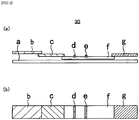
- an immunochromatographic analysis device 30 As an immunochromatographic analysis device used here, as shown in a cross-sectional view of FIG. 3(a) and a plan view of FIG. 3(b) , an immunochromatographic analysis device 30 is configured such that, on a plastic adhesive sheet a, a sample pad b, a pad c containing a particle-labeled antibody labeled with a labeling substance, an antibody-immobilized membrane f, and a water absorption pad g are provided in this order along the longitudinal direction of the device, respectively, and on the antibody-immobilized membrane f, an anti-HbA1c antibody-coated part d coated with an anti-HbA1c antibody and an anti-HbA0 antibody-coated part e coated with an anti-HbA0 antibody are provided, respectively.
- the sample solution further developed on the antibody-immobilized membrane f reaches the anti-HbA0 antibody-coated part e, and only HbA0 in the sample solution reacts and is detected (S407).
- Other forms of hemoglobin do not react and migrate to the water absorption pad g (S408). In this manner, HbA0 and HbA1c in the sample solution are detected respectively.
- Patent Document 1 JP-A-2012-251789
- a step of mixing blood collected and a component for exposing the N-terminal of the hemoglobin ⁇ chain on the surface of a protein (N-terminal exposure agent) S401
- a step of preparing a sample solution by leaving the resulting mixture to stand for a few minutes S402
- a step of dropping the sample solution onto a sample pad b S403
- a step of further preparing a developing solution separately and dropping the developing solution onto the sample pad are included, and therefore, two solutions: the sample solution and the developing solution are needed, and also it is necessary to separately drop these solutions onto the sample pad b. Accordingly, it has problems that the measurement preparation and operations are complicated, and also the measurement takes time, and the efficiency is low.
- an object of the present invention is to solve the above-mentioned conventional problems and provide an immunochromatographic analysis device, an immunochromatographic analysis method, and an immunochromatographic analysis kit capable of measuring various components contained in various analytes such as blood and urine efficiently in a short measurement time without the need for complicated measurement preparation and operations.
- the present invention is as follows.
- the immunochromatographic analysis device, the immunochromatographic analysis method, and the immunochromatographic analysis kit of the present invention are configured such that a sample addition part contains an anionic surfactant, and therefore, unlike the conventional art, two solutions: a sample solution and a developing solution are not needed, and it is only necessary to drop one solution: an analyte-containing solution obtained by diluting an analyte containing a detection target with an analyte dilution solution onto the sample addition part.
- an anionic surfactant is incorporated in the sample addition part, and therefore, it is not necessary to include a step of exposing an epitope of a detection target before adding an analyte-containing solution to the sample addition part. Therefore, according to the present invention, various components contained in various analytes such as blood and urine can be measured efficiently in a short measurement time without the need for complicated measurement preparation and operations.
- Examples of the analyte to be used in the present invention include blood samples such as blood, plasma, and serum, urine, saliva, spinal fluid, sweat, tear, amniotic fluid, nipple discharge fluid, nasal discharge, sputum, a nasal swab, a pharyngeal swab, exudate from the skin, and extracts from tissues, cells, and feces.
- blood samples such as blood, plasma, and serum, urine, saliva, spinal fluid, sweat, tear, amniotic fluid, nipple discharge fluid, nasal discharge, sputum, a nasal swab, a pharyngeal swab, exudate from the skin, and extracts from tissues, cells, and feces.
- Examples of the detection target in the present invention include tumor marker substances such as ⁇ -fetoprotein (AFP) and carcinoembryonic antigens (CEA), serum proteins such as ferritin, prostate-specific antigens (PSA), and immunoglobulin G (IgG), hormone-related substances such as rheumatoid factors, growth hormone (GH), and adrenocorticotropic hormone (ACTH), influenza viruses, adenoviruses, bacterial antigens such as Chlamydia antigen and Streptococcus pyogenes antigen, and hemoglobin-derived proteins.
- tumor marker substances such as ⁇ -fetoprotein (AFP) and carcinoembryonic antigens (CEA), serum proteins such as ferritin, prostate-specific antigens (PSA), and immunoglobulin G (IgG), hormone-related substances such as rheumatoid factors, growth hormone (GH), and adrenocorticotropic hormone (ACTH)
- influenza viruses
- Examples of the detection target in the present invention preferably, a detection target in blood, include hemoglobin-derived proteins, plasma proteins, lipoproteins, secretory proteins, hormones, complements, lipids, cholesterol, sugars, other components in the body, drugs administered to the body, and metabolites thereof.
- the detection target is preferably glycated proteins such as glycated albumin and glycated hemoglobin in blood, more preferably HbA1c.
- glycated proteins such as glycated albumin and glycated hemoglobin in blood
- HbA1c glycated proteins
- hemoglobin other than HbA1c is used as a substance to serve as a standard for making a positive or negative determination, however, the present invention is not limited to the following example.
- the immunochromatographic analysis device of the present invention includes a sample addition part, a labeling substance retaining part, a chromatography medium part having a detection part supported thereon, and an absorption part.
- FIG. 1(a) and FIG. 1(b) are schematic views for explaining one example of the immunochromatographic analysis device of the present invention, and FIG. 1(a) is a cross-sectional view, and FIG. 1 (b) is a plan view.
- an immunochromatographic analysis device 1 includes a sample addition part 12, a labeling substance retaining part 13 containing an anti-hemoglobin antibody labeled with a labeling substance, a chromatography medium part 14, and an absorption part 15, which are provided on a plastic adhesive sheet 11 in this order along the longitudinal direction of the kit, respectively.
- an anti-HbA1c antibody-coated part 16 coated with an anti-HbA1c antibody, an anti-hemoglobin antibody-coated part 17 coated with an anti-hemoglobin antibody, and an anti-IgG antibody-coated part 18 coated with an anti-IgG antibody as a control are provided respectively.
- the plastic adhesive sheet 11 serves as a base material of the immunochromatographic analysis device 1, and one surface is made to serve as an adhesive surface by coating one surface with an adhesive or by sticking an adhesive tape to one surface, and part or the entire of the respective constituent parts described below are closely adhered onto the adhesive surface.
- a material of the plastic adhesive sheet 11 a material which is impermeable to a sample and also is impermeable to moisture may be appropriately selected.
- the sample addition part 12 can be composed of a porous sheet having properties such that it quickly absorbs the below-mentioned analyte-containing solution, but has a low ability to retain the analyte-containing solution so that the analyte-containing solution quickly migrates to a region where an antigen-antibody reaction occurs.
- porous sheet examples include a pad, a fiber, a membrane, etc. composed of glass fiber, cellulose, polyethylene terephthalate, polyurethane, polyacetate, cellulose acetate, nitrocellulose, nylon, a cotton cloth, etc.
- sample addition part 12 is preferably formed using one member selected from the group consisting of glass fiber, cellulose, and polyethylene terephthalate.
- the sample addition part 12 contains an anionic surfactant.
- anionic surfactant examples include alkyl sulfates such as sodium dodecyl sulfate (SDS), polyoxyethylene alkyl ether sulfates such as sodium polyoxyethylene lauryl ether sulfate, alkyl benzene sulfonates such as sodium dodecyl benzene sulfonate, acylamino acid salts such as lauroyl methyl alanine and sodium N-lauroyl sarcosine, sodium dialkyl sulfosuccinate, a sodium salt of ⁇ -naphthalene sulfonate formalin condensate, and a special polycarboxylic acid-based polymer surfactant.
- SDS sodium dodecyl sulfate
- polyoxyethylene alkyl ether sulfates such as sodium polyoxyethylene lauryl ether sulfate
- alkyl sulfates such as sodium dodecyl sulfate (SDS) are particularly preferred from the viewpoint of improving the effect of the present invention.
- the anionic surfactant can be contained in an amount of, for example, 8 to 800 ⁇ g, and is contained in an amount of preferably 10 to 500 ⁇ g, more preferably 16 to 480 ⁇ g. Further, in the sample addition part 12, the anionic surfactant can be contained in an amount of 5 to 500 ⁇ g/cm 2 , and is contained in an amount of preferably 6 to 313 ⁇ g/cm 2 , more preferably 10 to 300 ⁇ g/cm 2 .
- a means such as freeze-drying, hot-air drying, natural drying, or the like may be appropriately adopted.
- the anionic surfactant has a function to expose an epitope of HbA1c in collected blood on the surface of a hemoglobin protein. Specifically, the anionic surfactant functions as a component for exposing the N-terminal of the hemoglobin ⁇ chain on the surface of a protein (N-terminal exposure agent).
- the anionic surfactant is incorporated in the sample addition part 12, and therefore, it is not necessary to include a step of exposing an epitope of a detection target before adding the below-mentioned analyte-containing solution to the sample addition part.
- the labeling substance retaining part 13 retains, for example, an anti-hemoglobin antibody labeled with a labeling substance.
- the anti-hemoglobin antibody in the labeling substance retaining part 13 can specifically bind to hemoglobin in an analyte-containing solution.
- Examples of such an antibody include a polyclonal antibody and a monoclonal antibody.
- the monoclonal antibody and the polyclonal antibody or fragments thereof are known and available, and can be prepared by a known method.
- an animal species that produces the antibody include a human being, a mouse, a rat, a rabbit, and a goat.
- the immunoglobulin may be any of IgG, IgM, IgA, IgE, and IgD.
- the monoclonal antibody is obtained according to a conventional method as follows.
- the spleen cells and myeloma cells of a mouse immunized with an antigen are fused, and a hybridoma which produces a desired antibody is selected, and a monoclonal antibody produced from this hybridoma is obtained (see, for example, the method of Kohler and Milstein [Nature, 256 (1975), 495-497 ]).
- the polyclonal antibody is obtained by separating a desired antibody from an antiserum obtained by immunizing an antibody-producing animal (for example, a human being, a mouse, a rat, a rabbit, a goat, a horse, etc.) with an antigen according to a conventional method.
- an antibody-producing animal for example, a human being, a mouse, a rat, a rabbit, a goat, a horse, etc.
- the labeling substance a colored substance capable of visually confirming coloration is preferred, and a substance known in the art can be appropriately adopted.
- examples thereof include colloidal metal particles, colloidal non-metal particles, colored latex, and an enzyme label, however, colloidal metal particles whose color hardly fades even if time has passed are particularly preferred from the viewpoint of stability of the label.
- colloidal metal particles examples include colloidal gold, platinum, copper, silver, and palladium, and other than these, particles obtained by mixing these colloidal metals.
- colloidal gold particles are preferred from the viewpoint that the particles having an appropriate particle diameter exhibit red color.
- the average particle diameter of the colloidal metal particles is in the range of, for example, 1 to 500 nm, from the viewpoint of obtaining a strong color tone, preferably 10 nm to 150 nm, more preferably 20 to 100 nm.
- colloidal non-metal particles colloidal selenium and the like can be exemplified.
- the colloidal metal particles and the colloidal non-metal particles can be prepared according to a conventional method, and at this time, the particle diameter is adjusted so as to exhibit a desired color tone. In addition, it is also possible to use a commercially available product.
- Examples of the colored latex include high-molecular weight polymer particles such as polystyrene particles colored with a coloring agent exhibiting red or blue color, and such colored latex can be prepared according to a conventional method.
- Examples of the enzyme label include peroxidase, alkaline phosphatase, glucose oxidase, and galactosidase. In the case where the enzyme label is used, a substrate for the enzyme and, according to need, a color-developing reagent are allowed to act on the enzyme, and the color developed by the reaction is detected.
- the preparation of the antibody labeled with a labeling substance can be performed according to a known method.
- a known method such as physical adsorption or chemical binding can be employed.
- the antibody is added to a solution in which gold particles are colloidally dispersed to cause a physical adsorption, and thereafter, a blocking protein such as a bovine serum albumin solution is added thereto to block the surface of the particle to which the antibody is not bound, whereby the preparation can be performed.
- a known sandwich method, a competition method, a method combining these methods can be adopted for the antigen-antibody reaction.
- Examples of a material of the labeling substance retaining part 13 include glass fiber, cellulose, polyethylene terephthalate, polyurethane, polyacetate, nylon, and a cotton cloth.
- the chromatography medium part 14 may be any as long as it can absorb a sample analyte by a capillary phenomenon and allows the sample analyte to migrate therein, and can be composed of, for example, nitrocellulose, cellulose acetate, nylon, polyether sulfone, polyvinyl alcohol, polyester, glass fiber, polyolefin, cellulose, a mixed fiber thereof, or the like.
- the anti-HbA1c antibody-coated part 16, the anti-hemoglobin antibody-coated part 17, and the anti-IgG antibody-coated part 18 provided on the chromatography medium part 14 are composed of, for example, a material capable of supporting and fixing each antibody, and examples of the material include nitrocellulose.
- Examples of a material of the absorption part 15 include a material having an ability to quickly absorb an excess amount of an analyte-containing solution, and cellulose fiber, a glass filter paper, or the like is used.
- the immunochromatographic analysis method of the present invention is a method for detecting a detection target contained in an analyte using the immunochromatographic analysis device described above, and includes the following steps (1) to (4):
- an analyte-containing solution is prepared by diluting an analyte with an analyte dilution solution.
- the analyte dilution solution can act as a developing solution for developing an analyte and a labeling substance in the chromatography medium part as a mobile phase in the step (3).
- the analyte dilution solution preferably contains a nonionic surfactant from the viewpoint that it has favorable developing properties and also does not inhibit the antigen-antibody reaction.
- nonionic surfactant examples include polyoxyethylene alkyl ethers, polyoxyethylene/polyoxypropylene alkyl ethers, polyoxyethylene sorbitan fatty acid esters, polyoxyethylene p-t-octylphenyl ethers, polyoxyethylene p-t-nonylphenyl ethers, alkyl polyglucosides, fatty acid diethanolamide, and alkyl monoglyceryl ethers.
- the nonionic surfactant can be contained in a proportion of, for example, 0.01 to 5 mass%, and is contained preferably in a proportion of 0.03 to 3.0 mass%, more preferably in a proportion of 0.3 to 3.0 mass%.
- an additive such as a buffer or an inorganic salt may be added as needed.
- the ratio of the anionic surfactant contained in the sample addition part 12 to the nonionic surfactant contained in the analyte dilution solution is set to a given value, and specifically, the ratio of the anionic surfactant to the nonionic surfactant (mass ratio) is preferably from 9:1 to 1:90, more preferably from 3:1 to 1:60, further more preferably from 1:2 to 1:50, and most preferably from 1:2 to 1:34.
- analyte dilution solution is mixed with blood collected from a patient, whereby an analyte-containing solution is prepared and dropped onto the sample addition part 12.
- a site where blood is collected from a patient include a finger, a gum, an arm vein, and an ear.
- HbA1c contained in the analyte-containing solution reaches the labeling substance retaining part 13 by a capillary phenomenon while exposing the N-terminal of the hemoglobin ⁇ chain on the surface of a protein by the anionic surfactant contained in the sample addition part 12.
- hemoglobin in the analyte-containing solution having reached the labeling substance retaining part 13 is subjected to an antigen-antibody reaction with the anti-hemoglobin antibody labeled with a labeling substance, whereby a complex is formed. That is, HbA1c, which is the detection target, is also recognized by the labeling substance retained in the labeling substance retaining part 13.
- the analyte and the labeling substance that is, the analyte-containing solution and the complex of the anti-hemoglobin antibody and hemoglobin are developed in the chromatography medium part 14 as a mobile phase.
- the nonionic surfactant contained in the analyte-containing solution exhibits an effect that the developing properties in the development are improved and also the subsequent antigen-antibody reaction is not inhibited as described above.
- HbA1c which is the detection target in the mobile phase developed in the chromatography medium part 14 reaches the anti-HbA1c antibody-coated part 16, and HbA1c reacts with the anti-HbA1c antibody while it is passing through the part and is immobilized thereon.
- Hemoglobin other than HbA1c and the analyte-containing solution do not react and pass through the anti-HbA1c antibody-coated part 16, however, when reaching the anti-hemoglobin antibody-coated part 17, hemoglobin other than HbA1c reacts with the anti-hemoglobin antibody and is immobilized thereon.
- the labeling substance which does not react with the antigen or the labeling substance which does not react in the antibody-coated parts 16 and 17 react with the anti-IgG antibody in the anti-IgG antibody-coated part 18 and is immobilized thereon, and develops color as a control showing that the development is performed normally.
- the anti-HbA1c antibody and the anti-hemoglobin antibody may be either a monoclonal antibody or a polyclonal antibody as described above.
- HbA1c and hemoglobin other than HbA1c are caused to simultaneously develop color and the determination is made by comparison between the degrees of color development of HbA1c and hemoglobin other than HbA1c.
- the developed color signals of HbA1c and hemoglobin other than HbA1c are compared, and in the case where the developed color signal of HbA1c is higher than that of hemoglobin other than HbA1c, a positive determination can be made. On the other hand, in the case where the developed color signal of HbA1c is equal to or lower than that of hemoglobin other than HbA1c, a negative determination can be made.
- the immunochromatographic analysis kit of the present invention includes the immunochromatographic analysis device of the present invention, which includes the sample addition part 12 containing an anionic surfactant, the labeling substance retaining part 13, the chromatography medium part 14 having a detection part supported thereon, and the absorption part 15, and the analyte dilution solution, which is for diluting and developing an analyte, and contains a nonionic surfactant.
- FIG. 2 is a flowchart when measuring the concentration of HbA1c with respect to the amount of total hemoglobin in blood using the immunochromatographic analysis kit of the present invention.
- an analyte-containing solution is prepared (S201).
- An appropriate amount of the obtained analyte-containing solution is dropped onto the sample addition part 12 (S202).
- the analyte-containing solution is developed on the labeling substance retaining part 13 by a capillary phenomenon (S203).
- the analyte-containing solution developed on the labeling substance retaining part 13 reaches the anti-HbA1c antibody-coated part 16 supported on the chromatography medium part 14, and only HbA1c in the sample solution reacts in the part and is detected (S204).
- the anionic surfactant to serve as the N-terminal exposure agent is incorporated in the sample addition part 12, and therefore, unlike the conventional art, it is not necessary to prepare two solutions: a sample solution containing an N-terminal exposure agent and a developing solution, nor to drop these two solutions separately onto the sample addition part 12.
- An immunochromatographic analysis device 1 as shown in FIGS. 1(a) and 1(b) was prepared according to the following procedure.
- a membrane As a membrane, a sheet composed of nitrocellulose (manufactured by Millipore, Inc., trade name: HF 12250 mm x 25 mm) was used.
- An anti-HbA1c monoclonal antibody, an anti-hemoglobin monoclonal antibody, or an anti-IgG monoclonal antibody was diluted to a concentration of 1.0 mg/ml with a 10 mM phosphate buffer solution (pH 7.4) containing 5 mass% sucrose and 5 mass% isopropanol.
- the diluted solutions (150 ⁇ L) were applied to different places with a width of 1 mm on the membrane by an antibody applicator (manufactured by BioDot, Inc.), followed by drying at 50°C for 30 minutes and then drying at room temperature overnight, whereby an anti-HbA1c antibody-coated part 16, an anti-hemoglobin antibody-coated part 17, and an anti-IgG antibody-coated part 18 were provided, respectively, on a chromatography medium part 14.
- an antibody applicator manufactured by BioDot, Inc.
- a colloidal gold suspension manufactured by Tanaka Kikinzoku Kogyo K.K., average particle diameter: 40 nm
- a Tris buffer solution pH 8.5
- PEG-SH manufactured by NOF Corporation, trade name: SUNBRIGHT ME-200SH, molecular weight: 20,000
- PEG-SH concentration after addition 0.001 mass%
- the resulting mixture was left to stand at room temperature for 10 minutes. Thereafter, the mixture was thoroughly stirred, and then centrifuged at 8000 x g for 15 minutes. After removing the supernatant, 0.1 mL of a phosphate buffer solution (pH 7.4) containing 1 mass% bovine serum albumin was added thereto, whereby a labeling substance solution was prepared.
- a solution obtained by adding 100 ⁇ L of a phosphate buffer solution (pH 9.0) containing a 25 mass% aqueous solution of trehalose to 220 ⁇ L of the labeling substance solution prepared above was added uniformly to a 8 mm x 100 mm glass fiber pad (manufactured by Millipore, Inc.), followed by drying in a vacuum dryer, whereby a labeling substance retaining part 13 was prepared.
- a labeling substance retaining part 13 was prepared on a plastic adhesive sheet 11, the sample addition part 12, the labeling substance retaining part 13 labeled with a labeling substance, and the chromatography medium part 14 prepared above were stuck, and a general-purpose absorption part 15 was further stuck.
- the resulting material was cut to a width of 5 mm by a cutter, whereby an immunochromatographic analysis device 1 was prepared. At this time, the amount of SDS contained per device was 96 ⁇ g.
- An analyte-containing solution was prepared by stirring the respective components according to the following formulation.
- nonionic surfactant a mixture of Triton X-100 (trade name, manufactured by SIGMA, Inc.) and Tween 20 (trade name, manufactured by Wako Pure Chemical Industries, Ltd.) at a mass ratio of 1:5: 1.2 mass%
- Blood containing HbA1c at different concentrations was collected by pricking the finger of each of healthy adult males and diabetic male patients.
- the blood and the analyte dilution solution were mixed at 1:1,000 (former : latter (volume ratio)), whereby an analyte-containing solution was prepared.
- Example 1 The procedure of Example 1 was repeated except that the constituent material of the sample addition part 12 was changed from the glass fiber pad in Example 1 to a cellulose fiber pad (manufactured by Asahi Kasei Fibers Corporation, trade name: SR-601). The result is shown in Table 1.
- Example 1 The procedure of Example 1 was repeated except that the constituent material of the sample addition part 12 was changed from the glass fiber pad in Example 1 to a polyethylene terephthalate (PET) pad (manufactured by Asahi Kasei Fibers Corporation, trade name: Bemliese A-01). The result is shown in Table 1.
- PET polyethylene terephthalate
- Example 1 The procedure of Example 1 was repeated except that the constituent material of the sample addition part 12 was changed from the glass fiber pad in Example 1 to a nitrocellulose membrane (manufactured by Millipore, Inc., trade name: HF 120). The result is shown in Table 1.
- Example 1 The procedure of Example 1 was repeated except that unlike Example 1, a sample addition part free from SDS was used, and 10 ⁇ L of a sample solution prepared by adding blood to an extraction solution containing an N-terminal exposure agent having the following composition and leaving the resulting mixture to stand for 2 minutes and 100 ⁇ L of a developing solution having the following composition were separately dropped onto the sample addition part 12. The result is shown in Table 1.
- composition of extraction solution as an anionic surfactant, sodium dodecyl sulfate: 1.0 mass%, as an additive, sodium thiocyanate: 100 mM, as an additive, EDTA: 5 mM, balance: water
- composition of developing solution as a nonionic surfactant, a mixture of Triton X-100 (trade name, manufactured by SIGMA, Inc.) and Tween 20 (trade name, manufactured by Wako Pure Chemical Industries, Ltd.) at a mass ratio of 1:5: 1.2 mass%, as a buffer, a Bicine buffer solution: 50 mM, as an inorganic salt, potassium chloride: 0.6 mass%, as an additive, casein sodium: 2.0 mass%, balance: water
- Example 1 Concentration of HbA1c 5.5% 7.0% 9.0%
- Example 2 (cellulose fiber pad + SDS) ⁇ + ++
- Example 3 (PET pad + SDS) ⁇ ⁇ +
- Example 4 (nitrocellulose membrane + SDS) ⁇ + + Reference Example 1 (extraction solution + developing solution) + ++ +++ Reference Example 2 (only developing solution) - - ⁇ Reference Example 3 (developing solution + 0.1% SDS) - - + Reference Example 4 (developing solution + 0.2% SDS) - ⁇ + Reference Example 5 (developing solution + 0.4% SDS) - - ⁇
- Example 2 The procedure of Example 1 was repeated except that the content per device of SDS to be added to the sample addition part 12 and the content of the nonionic surfactant to be contained in the analyte dilution solution in Example 1 were changed to the values shown in Table 2. The results are shown in Table 2.
- Table 1 and Table 2 reveal that by using the immunochromatographic analysis device of the present invention, unlike Reference Example 1 of the conventional art, two solutions: a sample solution and a developing solution are not needed, and it is only necessary to drop one solution: an analyte-containing solution onto the sample addition part, and it is possible to measure various components contained in various analytes such as blood and urine efficiently in a short measurement time equivalently to Reference Example 1 of the conventional art without the need for complicated measurement preparation and operations.
- Reference Example 2 is an example in which the sample addition part free from SDS was used, and only the developing solution was used, and therefore, although the concentration of HbA1c was high, the degree of color development was poor.
- Reference Examples 3 to 5 are examples in which the sample addition part free from SDS was used, and SDS was added to the developing solution, however, although the degree of color development was improved as compared with Reference Example 2, a satisfactory level could not yet be reached.
Landscapes
- Health & Medical Sciences (AREA)
- Life Sciences & Earth Sciences (AREA)
- Immunology (AREA)
- Engineering & Computer Science (AREA)
- Hematology (AREA)
- Molecular Biology (AREA)
- Biomedical Technology (AREA)
- Chemical & Material Sciences (AREA)
- Urology & Nephrology (AREA)
- Biotechnology (AREA)
- Biochemistry (AREA)
- Cell Biology (AREA)
- Food Science & Technology (AREA)
- Medicinal Chemistry (AREA)
- Physics & Mathematics (AREA)
- Analytical Chemistry (AREA)
- Microbiology (AREA)
- General Health & Medical Sciences (AREA)
- General Physics & Mathematics (AREA)
- Pathology (AREA)
- Proteomics, Peptides & Aminoacids (AREA)
- Investigating Or Analysing Biological Materials (AREA)
- Automatic Analysis And Handling Materials Therefor (AREA)
Applications Claiming Priority (2)
| Application Number | Priority Date | Filing Date | Title |
|---|---|---|---|
| JP2014091510A JP5767362B1 (ja) | 2014-04-25 | 2014-04-25 | 免疫クロマト分析装置、免疫クロマト分析方法及び免疫クロマト分析キット |
| PCT/JP2015/062308 WO2015163384A1 (fr) | 2014-04-25 | 2015-04-22 | Dispositif d'analyse immunochromatographique, procédé d'analyse immunochromatographique et kit d'analyse immunochromatographique |
Publications (2)
| Publication Number | Publication Date |
|---|---|
| EP3136097A1 true EP3136097A1 (fr) | 2017-03-01 |
| EP3136097A4 EP3136097A4 (fr) | 2017-05-10 |
Family
ID=53888041
Family Applications (1)
| Application Number | Title | Priority Date | Filing Date |
|---|---|---|---|
| EP15782442.6A Withdrawn EP3136097A4 (fr) | 2014-04-25 | 2015-04-22 | Dispositif d'analyse immunochromatographique, procédé d'analyse immunochromatographique et kit d'analyse immunochromatographique |
Country Status (8)
| Country | Link |
|---|---|
| US (1) | US20170045510A1 (fr) |
| EP (1) | EP3136097A4 (fr) |
| JP (1) | JP5767362B1 (fr) |
| CN (1) | CN105021823A (fr) |
| BR (1) | BR112016024730A2 (fr) |
| RU (1) | RU2016146092A (fr) |
| TW (1) | TWI554759B (fr) |
| WO (1) | WO2015163384A1 (fr) |
Cited By (2)
| Publication number | Priority date | Publication date | Assignee | Title |
|---|---|---|---|---|
| EP3139163A4 (fr) * | 2014-04-30 | 2017-05-03 | Tanaka Kikinzoku Kogyo K.K. | Nécessaire, dispositif et procédé d'analyse immunochromotographique |
| EP3368901A1 (fr) * | 2015-10-29 | 2018-09-05 | Thomas Bruderer | Procédé d'immuno-essai soustractif et bandelette de dosage d'immunochromatographie à écoulement latéral pour réaliser le procédé |
Families Citing this family (7)
| Publication number | Priority date | Publication date | Assignee | Title |
|---|---|---|---|---|
| JP6675834B2 (ja) * | 2015-05-21 | 2020-04-08 | デンカ生研株式会社 | イムノクロマト試験片及びそれを用いたイムノクロマト法 |
| JP7020413B2 (ja) * | 2016-07-29 | 2022-02-16 | ミナリスメディカル株式会社 | 糖化ヘモグロビンの測定方法 |
| US11353453B2 (en) | 2017-04-06 | 2022-06-07 | Asahi Kasei Kabushiki Kaisha | Hydrophilic colored cellulose microparticles |
| WO2019138898A1 (fr) * | 2018-01-09 | 2019-07-18 | 東洋紡株式会社 | Éprouvette d'immunochromatographie, kit de mesure et procédé de mesure |
| WO2019138899A1 (fr) * | 2018-01-11 | 2019-07-18 | 東洋紡株式会社 | Liquide de dilution d'échantillon de mesure, trousse, et procédé de mesure |
| CN109540846B (zh) * | 2019-01-04 | 2022-04-12 | 无锡博慧斯生物医药科技有限公司 | 一种测量糖化的蛋白质的方法 |
| CN114280312B (zh) * | 2020-09-27 | 2023-09-15 | 河北特温特生物科技发展有限公司 | 一种用于免疫荧光层析检测的全血分离膜及其制备方法和应用 |
Family Cites Families (11)
| Publication number | Priority date | Publication date | Assignee | Title |
|---|---|---|---|---|
| US20030180967A1 (en) * | 2002-03-19 | 2003-09-25 | Matsushita Electric Industrial Co., Ltd. | Immunoassay method, test strip for use therein, and immunoassay apparatus |
| JP2003344397A (ja) * | 2002-03-19 | 2003-12-03 | Matsushita Electric Ind Co Ltd | 免疫学的測定方法、それに用いる試験片、および免疫学的測定装置 |
| EP1637883B1 (fr) * | 2003-05-27 | 2009-11-25 | Mitsubishi Kagaku Iatron, Inc. | Procede d'imunochromatographie |
| CA2514010A1 (fr) * | 2004-08-03 | 2006-02-03 | Axis-Shield Diagnostics Limited | Analyse |
| CN101360997B (zh) * | 2005-11-18 | 2013-05-01 | 美国政府健康及人类服务部,疾病控制和预防中心 | 改性心磷脂及其应用 |
| JP4428670B2 (ja) * | 2008-03-06 | 2010-03-10 | Tanakaホールディングス株式会社 | 免疫学的測定法、キット及び展開溶媒 |
| WO2009137055A1 (fr) * | 2008-05-05 | 2009-11-12 | Los Alamos National Security, Llc | Microréseaux à écoulement latéral à base de nanocristaux et systèmes de détection d’un signal de faible tension |
| EP2350671A1 (fr) * | 2008-10-24 | 2011-08-03 | Boditechmed Inc. | Système de mesure quantitative de la glycohémoglobine et procédé de mesure de la teneur en glycohémoglobine l'utilisant |
| WO2010067612A1 (fr) * | 2008-12-11 | 2010-06-17 | 積水メディカル株式会社 | Procédé de prétraitement d'un échantillon contenant de l'hémoglobine glycosylée |
| JP5852329B2 (ja) * | 2011-05-31 | 2016-02-03 | 積水メディカル株式会社 | HbA1cの免疫学的測定方法 |
| JP5775197B1 (ja) * | 2014-04-30 | 2015-09-09 | 田中貴金属工業株式会社 | 免疫クロマト分析キット及び免疫クロマト分析方法 |
-
2014
- 2014-04-25 JP JP2014091510A patent/JP5767362B1/ja not_active Expired - Fee Related
-
2015
- 2015-04-22 EP EP15782442.6A patent/EP3136097A4/fr not_active Withdrawn
- 2015-04-22 RU RU2016146092A patent/RU2016146092A/ru unknown
- 2015-04-22 BR BR112016024730A patent/BR112016024730A2/pt not_active IP Right Cessation
- 2015-04-22 WO PCT/JP2015/062308 patent/WO2015163384A1/fr not_active Ceased
- 2015-04-22 US US15/306,511 patent/US20170045510A1/en not_active Abandoned
- 2015-04-24 TW TW104113183A patent/TWI554759B/zh not_active IP Right Cessation
- 2015-04-24 CN CN201510202401.XA patent/CN105021823A/zh active Pending
Cited By (2)
| Publication number | Priority date | Publication date | Assignee | Title |
|---|---|---|---|---|
| EP3139163A4 (fr) * | 2014-04-30 | 2017-05-03 | Tanaka Kikinzoku Kogyo K.K. | Nécessaire, dispositif et procédé d'analyse immunochromotographique |
| EP3368901A1 (fr) * | 2015-10-29 | 2018-09-05 | Thomas Bruderer | Procédé d'immuno-essai soustractif et bandelette de dosage d'immunochromatographie à écoulement latéral pour réaliser le procédé |
Also Published As
| Publication number | Publication date |
|---|---|
| EP3136097A4 (fr) | 2017-05-10 |
| JP5767362B1 (ja) | 2015-08-19 |
| RU2016146092A (ru) | 2018-05-28 |
| JP2015210167A (ja) | 2015-11-24 |
| WO2015163384A1 (fr) | 2015-10-29 |
| BR112016024730A2 (pt) | 2017-08-15 |
| RU2016146092A3 (fr) | 2018-05-28 |
| US20170045510A1 (en) | 2017-02-16 |
| TW201541079A (zh) | 2015-11-01 |
| CN105021823A (zh) | 2015-11-04 |
| TWI554759B (zh) | 2016-10-21 |
Similar Documents
| Publication | Publication Date | Title |
|---|---|---|
| EP3136097A1 (fr) | Dispositif d'analyse immunochromatographique, procédé d'analyse immunochromatographique et kit d'analyse immunochromatographique | |
| US20170059589A1 (en) | Immunochromatographic analysis kit, immunochromatographic analysis device, and immunochromatographic analysis method | |
| EP3153858B1 (fr) | Procédé pour éliminer un phénomène de prozone dans un réactif d'immuno-essai | |
| EP2602619B1 (fr) | Composition de réactif d'immunochromatographie et procédé de mesure utilisant celle-ci | |
| EP2829879B1 (fr) | Procédé de détection d'immunochromatographie | |
| EP3128324B1 (fr) | Procédé de dosage immunochromatographique | |
| EP3096139A1 (fr) | Méthode, dispositif et kit d'analyse immunochromatographique | |
| EP4249508A1 (fr) | Kit et procédé de détection de sars-cov-2 | |
| EP3407065A1 (fr) | Milieu pour chromatographe | |
| CN102621310B (zh) | 胶体金层析法抗u1-rnp抗体检测试纸及其制备方法 |
Legal Events
| Date | Code | Title | Description |
|---|---|---|---|
| PUAI | Public reference made under article 153(3) epc to a published international application that has entered the european phase |
Free format text: ORIGINAL CODE: 0009012 |
|
| 17P | Request for examination filed |
Effective date: 20161024 |
|
| AK | Designated contracting states |
Kind code of ref document: A1 Designated state(s): AL AT BE BG CH CY CZ DE DK EE ES FI FR GB GR HR HU IE IS IT LI LT LU LV MC MK MT NL NO PL PT RO RS SE SI SK SM TR |
|
| AX | Request for extension of the european patent |
Extension state: BA ME |
|
| A4 | Supplementary search report drawn up and despatched |
Effective date: 20170411 |
|
| RIC1 | Information provided on ipc code assigned before grant |
Ipc: G01N 33/72 20060101AFI20170405BHEP Ipc: G01N 33/558 20060101ALI20170405BHEP |
|
| DAV | Request for validation of the european patent (deleted) | ||
| DAX | Request for extension of the european patent (deleted) | ||
| 17Q | First examination report despatched |
Effective date: 20180123 |
|
| GRAP | Despatch of communication of intention to grant a patent |
Free format text: ORIGINAL CODE: EPIDOSNIGR1 |
|
| INTG | Intention to grant announced |
Effective date: 20180912 |
|
| STAA | Information on the status of an ep patent application or granted ep patent |
Free format text: STATUS: THE APPLICATION IS DEEMED TO BE WITHDRAWN |
|
| 18D | Application deemed to be withdrawn |
Effective date: 20190123 |