EP3141602A1 - Cassette d'expression de polypeptide hétérogène - Google Patents
Cassette d'expression de polypeptide hétérogène Download PDFInfo
- Publication number
- EP3141602A1 EP3141602A1 EP15785407.6A EP15785407A EP3141602A1 EP 3141602 A1 EP3141602 A1 EP 3141602A1 EP 15785407 A EP15785407 A EP 15785407A EP 3141602 A1 EP3141602 A1 EP 3141602A1
- Authority
- EP
- European Patent Office
- Prior art keywords
- scfv
- antibody
- seq
- bacterium
- bifidobacterium
- Prior art date
- Legal status (The legal status is an assumption and is not a legal conclusion. Google has not performed a legal analysis and makes no representation as to the accuracy of the status listed.)
- Withdrawn
Links
Images









Classifications
-
- C—CHEMISTRY; METALLURGY
- C12—BIOCHEMISTRY; BEER; SPIRITS; WINE; VINEGAR; MICROBIOLOGY; ENZYMOLOGY; MUTATION OR GENETIC ENGINEERING
- C12N—MICROORGANISMS OR ENZYMES; COMPOSITIONS THEREOF; PROPAGATING, PRESERVING, OR MAINTAINING MICROORGANISMS; MUTATION OR GENETIC ENGINEERING; CULTURE MEDIA
- C12N15/00—Mutation or genetic engineering; DNA or RNA concerning genetic engineering, vectors, e.g. plasmids, or their isolation, preparation or purification; Use of hosts therefor
- C12N15/09—Recombinant DNA-technology
- C12N15/63—Introduction of foreign genetic material using vectors; Vectors; Use of hosts therefor; Regulation of expression
- C12N15/74—Vectors or expression systems specially adapted for prokaryotic hosts other than E. coli, e.g. Lactobacillus, Micromonospora
- C12N15/746—Vectors or expression systems specially adapted for prokaryotic hosts other than E. coli, e.g. Lactobacillus, Micromonospora for lactic acid bacteria (Streptococcus; Lactococcus; Lactobacillus; Pediococcus; Enterococcus; Leuconostoc; Propionibacterium; Bifidobacterium; Sporolactobacillus)
-
- A—HUMAN NECESSITIES
- A61—MEDICAL OR VETERINARY SCIENCE; HYGIENE
- A61K—PREPARATIONS FOR MEDICAL, DENTAL OR TOILETRY PURPOSES
- A61K35/00—Medicinal preparations containing materials or reaction products thereof with undetermined constitution
- A61K35/66—Microorganisms or materials therefrom
- A61K35/74—Bacteria
-
- A—HUMAN NECESSITIES
- A61—MEDICAL OR VETERINARY SCIENCE; HYGIENE
- A61K—PREPARATIONS FOR MEDICAL, DENTAL OR TOILETRY PURPOSES
- A61K35/00—Medicinal preparations containing materials or reaction products thereof with undetermined constitution
- A61K35/66—Microorganisms or materials therefrom
- A61K35/74—Bacteria
- A61K35/741—Probiotics
- A61K35/744—Lactic acid bacteria, e.g. enterococci, pediococci, lactococci, streptococci or leuconostocs
- A61K35/745—Bifidobacteria
-
- A—HUMAN NECESSITIES
- A61—MEDICAL OR VETERINARY SCIENCE; HYGIENE
- A61K—PREPARATIONS FOR MEDICAL, DENTAL OR TOILETRY PURPOSES
- A61K39/00—Medicinal preparations containing antigens or antibodies
- A61K39/395—Antibodies; Immunoglobulins; Immune serum, e.g. antilymphocytic serum
-
- A—HUMAN NECESSITIES
- A61—MEDICAL OR VETERINARY SCIENCE; HYGIENE
- A61P—SPECIFIC THERAPEUTIC ACTIVITY OF CHEMICAL COMPOUNDS OR MEDICINAL PREPARATIONS
- A61P35/00—Antineoplastic agents
-
- A—HUMAN NECESSITIES
- A61—MEDICAL OR VETERINARY SCIENCE; HYGIENE
- A61P—SPECIFIC THERAPEUTIC ACTIVITY OF CHEMICAL COMPOUNDS OR MEDICINAL PREPARATIONS
- A61P37/00—Drugs for immunological or allergic disorders
- A61P37/02—Immunomodulators
- A61P37/04—Immunostimulants
-
- C—CHEMISTRY; METALLURGY
- C07—ORGANIC CHEMISTRY
- C07K—PEPTIDES
- C07K16/00—Immunoglobulins [IG], e.g. monoclonal or polyclonal antibodies
- C07K16/18—Immunoglobulins [IG], e.g. monoclonal or polyclonal antibodies against material from animals or humans
- C07K16/28—Immunoglobulins [IG], e.g. monoclonal or polyclonal antibodies against material from animals or humans against receptors, cell surface antigens or cell surface determinants
- C07K16/2803—Immunoglobulins [IG], e.g. monoclonal or polyclonal antibodies against material from animals or humans against receptors, cell surface antigens or cell surface determinants against the immunoglobulin superfamily
- C07K16/2818—Immunoglobulins [IG], e.g. monoclonal or polyclonal antibodies against material from animals or humans against receptors, cell surface antigens or cell surface determinants against the immunoglobulin superfamily against CD28 or CD152
-
- C—CHEMISTRY; METALLURGY
- C07—ORGANIC CHEMISTRY
- C07K—PEPTIDES
- C07K16/00—Immunoglobulins [IG], e.g. monoclonal or polyclonal antibodies
- C07K16/18—Immunoglobulins [IG], e.g. monoclonal or polyclonal antibodies against material from animals or humans
- C07K16/28—Immunoglobulins [IG], e.g. monoclonal or polyclonal antibodies against material from animals or humans against receptors, cell surface antigens or cell surface determinants
- C07K16/2896—Immunoglobulins [IG], e.g. monoclonal or polyclonal antibodies against material from animals or humans against receptors, cell surface antigens or cell surface determinants against molecules with a "CD"-designation, not provided for elsewhere
-
- C—CHEMISTRY; METALLURGY
- C07—ORGANIC CHEMISTRY
- C07K—PEPTIDES
- C07K2317/00—Immunoglobulins specific features
- C07K2317/10—Immunoglobulins specific features characterized by their source of isolation or production
- C07K2317/14—Specific host cells or culture conditions, e.g. components, pH or temperature
-
- C—CHEMISTRY; METALLURGY
- C07—ORGANIC CHEMISTRY
- C07K—PEPTIDES
- C07K2317/00—Immunoglobulins specific features
- C07K2317/60—Immunoglobulins specific features characterized by non-natural combinations of immunoglobulin fragments
- C07K2317/62—Immunoglobulins specific features characterized by non-natural combinations of immunoglobulin fragments comprising only variable region components
- C07K2317/622—Single chain antibody (scFv)
-
- C—CHEMISTRY; METALLURGY
- C07—ORGANIC CHEMISTRY
- C07K—PEPTIDES
- C07K2319/00—Fusion polypeptide
- C07K2319/01—Fusion polypeptide containing a localisation/targetting motif
- C07K2319/02—Fusion polypeptide containing a localisation/targetting motif containing a signal sequence
-
- C—CHEMISTRY; METALLURGY
- C12—BIOCHEMISTRY; BEER; SPIRITS; WINE; VINEGAR; MICROBIOLOGY; ENZYMOLOGY; MUTATION OR GENETIC ENGINEERING
- C12N—MICROORGANISMS OR ENZYMES; COMPOSITIONS THEREOF; PROPAGATING, PRESERVING, OR MAINTAINING MICROORGANISMS; MUTATION OR GENETIC ENGINEERING; CULTURE MEDIA
- C12N2830/00—Vector systems having a special element relevant for transcription
- C12N2830/36—Vector systems having a special element relevant for transcription being a transcription termination element
Definitions
- the present invention relates to an expression cassette comprising a promoter DNA, a DNA encoding a secretory signal peptide, a DNA encoding a linker peptide, a DNA encoding a heterogeneous polypeptide and a terminator DNA sequentially in this order and expressed in a bacterium of the genus Bifidobacterium.
- the present invention also relates to a vector comprising the expression cassette, a bacterium of the genus Bifidobacterium transformed with the vector and a pharmaceutical composition comprising the bacterium.
- an anaerobic enteric bacterium if it is systemically administered, is accumulated in a hypoxic solid tumor.
- a transformed microorganism which can express a gene encoding a protein having an anti-tumor activity or a protein having an activity to convert a precursor of an anti-tumor agent into the anti-tumor agent at a target site of disease (see, for example, Patent Document 1) has been proposed.
- an expression cassette which comprises a promoter, a DNA encoding a signal sequence, a DNA encoding a polypeptide or a cloning site for inserting the DNA
- a recombinant obligatory anaerobic gram-positive bacterium which comprises a nucleic acid encoding a fusion protein comprising a signal peptide, a single-chain antibody and one or more heterogeneous polypeptides and which can express the nucleic acid therein (see, for example, Patent Document 3) have been proposed.
- signal peptides proposed in the above documents are found in membrane proteins and secretory proteins.
- a signal peptide having better secretion efficiency is desirably identified.
- An object of the present invention is to provide, in producing a heterogeneous polypeptide by a bacterium of the genus Bifidobacterium, a signal peptide which enables to secrete the heterogeneous polypeptide with high efficiency outside the bacterial cell; a heterogeneous polypeptide expression cassette, which enables to secrete the heterogeneous polypeptide outside the bacterial cell with high efficiency by using such a signal peptide; a heterogeneous polypeptide expression vector having the expression cassette integrated therein; and a bacterium of the genus Bifidobacterium, which is transformed with the heterogeneous polypeptide expression vector and can secrete the heterogeneous polypeptide.
- the present inventors have already developed an anti-cancer therapy using a bacterium of the genus Bifidobacterium, which is transformed with a vector prepared by integrating a gene for cytosine deaminase, i.e., an enzyme for converting 5-fluoro-cytosine into 5-fluorouracil having an antitumor activity, and which specifically accumulates and grows in solid cancer.
- a vector prepared by integrating a gene for cytosine deaminase i.e., an enzyme for converting 5-fluoro-cytosine into 5-fluorouracil having an antitumor activity, and which specifically accumulates and grows in solid cancer.
- Bifidobacterium longum transformed with a vector in which an expression cassette comprising a promoter DNA functioning in a bacterium of the genus Bifidobacterium; a DNA encoding the secretory signal peptide as mentioned above; a DNA encoding a linker peptide; a DNA encoding a single-chain antibody(scFv antibody) having an antitumor activity; and a terminator DNA functioning in the bacterium of the genus Bifidobacterium, sequentially in this order, is integrated, secretes the scFv antibody outside the bacterial cell with high efficiency.
- the present invention is more specifically as follows.
- a therapeutic method comprising administering an effective amount of bacterium of the genus Bifidobacterium according to [9] or [10]; use of the bacterium of the genus Bifidobacterium according to [9] or [10] for preparing a therapeutic agent; and the bacterium of the genus Bifidobacterium according to [9] or [10] for use in treating a disease can be mentioned.
- the present invention it is possible to provide a bacterium of the genus Bifidobacterium capable of secreting a heterogeneous polypeptide outside the bacterial cell with high efficiency by using an expression cassette excellent in secretion of the heterogeneous polypeptide.
- the heterogeneous polypeptide is an antibody having an anti-cancer effect, such as PD-1 and CTLA-4, the bacterium of the genus Bifidobacterium is extremely useful as an anti-cancer agent.
- the expression cassette of the present invention is not particularly limited as long as it is an expression cassette sequentially comprising (1) a promoter DNA functioning in a bacterium of the genus Bifidobacterium, (2) a DNA encoding a secretory signal peptide, (3) a DNA encoding a linker peptide, (4) a DNA encoding a heterogeneous polypeptide, and (5) a terminator DNA functioning in a bacterium of the genus Bifidobacterium, in which the secretory signal peptide consists of a) an amino acid sequence represented by any one of SEQ ID Nos: 6, 7, 8, 9, 10, 11, 12, 13, 14, 15 and 16, or b) an amino acid sequence obtained by deletion, replacement or addition of one or several of amino acids in the amino acid sequence represented by any one of SEQ ID Nos: 6, 7, 8, 9, 10, 11, 12, 13, 14, 15 and 16; and constituting a peptide functioning as a secretory signal peptide in the bacterium of the genus Bifid
- the promoter DNA is not particularly limited as long as it is a promoter DNA functioning in a bacterium of the genus Bifidobacterium.
- Hu promoter DNA which is a promoter involved in expression of a gene encoding histone-like DNA binding protein derived from Bifidobacterium longum; a promoter DNA for Gap gene derived from Bifidobacterium breve ( Biotechnol. Lett. 2008 30: 1983-1988 ); a promoter DNA for AmyB gene derived from Bifidobacterium longum ( Biotechnol. Lett. 2006 28: 163-168 ); 16S rRNA Promoter DNA ( Biotechnol. Lett.
- secretory signal peptide examples include a) a peptide consisting of an amino acid sequence represented by any one of SEQ ID Nos. 6, 7, 8, 9, 10, 11, 12, 13, 14, 15 and 16; namely, SP7, SP45, SP50, SP52, SP55, SP58, SP64, SP66, SP67, SP68, SP69 respectively, and b) a peptide consisting of an amino acid sequence obtained by deletion, replacement or addition of one or several of amino acids in the amino acid sequence represented by any one of SEQ ID Nos. 6, 7, 8, 9, 10, 11, 12, 13, 14, 15 and 16 and functioning as a secretory signal peptide in a bacterium of the genus Bifidobacterium (mutated secretory signal peptide).
- an amino acid sequence obtained by deletion, replacement or addition of one or several of amino acids refers to an amino acid sequence obtained by deletion, replacement or addition of e.g., 1 to 5, preferably 1 to 3, more preferably 1 to 2 and further preferably a single amino acid.
- the amino acid sequence of the mutated secretory signal peptide has a sequence identity of preferably 90% or more with any of the amino acid sequence represented by SEQ ID Nos: 6, 7, 8, 9, 10, 11, 12, 13, 14, 15 and 16, more preferably 95% or more and further preferably, 98% or more.
- DNA encoding a secretory signal peptide consisting of the amino acid sequence described in a) above is not particularly limited as long as it is DNA having a nucleotide sequence corresponding to the amino acid sequence described in a). Accordingly, examples of DNA include, although different DNAs due to codon degeneracy are included, DNA consisting of a nucleotide sequence represented by any one of SEQ ID Nos: 116, 117, 118, 119, 120, 121, 122, 123, 124, 125 and 126. These DNAs can be prepared by a method known to those skilled in the art, such as chemical synthesis and genetic engineering procedure.
- DNA encoding a secretory signal peptide consisting of the amino acid sequence described in b) above is not particularly limited as long as it is DNA having a nucleotide sequence corresponding to the amino acid sequence described in b). Accordingly, different DNAs due to codon degeneracy are included. These DNAs molecules can be also prepared by a method known to those skilled in the art such as chemical synthesis, genetic engineering procedure and mutation induction.
- mutated DNAs can be obtained by introducing a mutation into these DNAs, by a method of bringing a mutagenic drug into contact with DNA consisting of a nucleotide sequence represented by any of SEQ ID Nos: 116, 117, 118, 119, 120, 121, 122, 123, 124, 125 and 126; a UV irradiation method; and/or genetic engineering techniques.
- One of the genetic engineering procedures i.e., site-specific mutagenesis, is useful since a specific mutation can be introduced into a specific site, and carried out in accordance with the method described in e.g., Molecular Cloning: A laboratory Manual, 2nd Ed., Cold Spring Harbor Laboratory, Cold Spring Harbor, NY., 1989 ., Current Protocols in Molecular Biology, Supplement 1 to 38, John Wiley & Sons (1987-1997 ).
- the secretory signal peptide is preferably used in conjunction with a linker peptide in the form of a signal peptide-linker conjugate, in view of expression/secretion efficiency of a heterogeneous polypeptide.
- the linker peptide to be used herein is not particularly limited as long as it is a peptide that can be conjugated to the C terminal of a secretory signal peptide as mentioned above and successfully enhance expression/secretion efficiency of a heterogeneous polypeptide.
- a peptide consisting of an amino acid sequence having 1 to 30 amino acid residues can be mentioned.
- a linker peptide consisting of amino acid sequence having any number of amino acids within the range of the 1st to 30th amino acid, such as a linker peptide consisting of the 1st to 5th, 1st to 10th, 1st to 15th or 1st to 20th, is mentioned.
- DNA encoding the secretory signal peptide-linker conjugate of the present invention refers to a DNA region from the 5' end of the DNA (2) encoding a secretory signal peptide to the 3' end of the DNA (3) encoding a linker peptide. Since the 3' end of DNA (2) encoding a secretory signal peptide is connected to the 5' end of DNA (3) encoding a linker peptide, the secretory signal peptide-linker conjugate is hereinafter sometimes referred to as SPxLy (where x represents the number of a secretory signal peptide designated in the specification; and y represents the number of amino acid residues of a linker peptide).
- SPxLy the secretory signal peptide-linker conjugate
- the following 11 types can be specifically mentioned.
- the 5' end of DNA encoding a linker peptide which is obtained by deletion of amino acid residues of 0 to 29 (e.g., 10 to 29, 15 to 29, 20 to 29, or 25 to 29) from the C terminal of a linker peptide consisting of the amino acid sequence represented by SEQ ID No: 25, is connected to the 3' end of DNA encoding a secretory signal peptide consisting of the amino acid sequence represented by SEQ ID No: 6.
- linker peptide which is obtained by deletion of amino acid residues of 0 to 29 (e.g., 10 to 29, 15 to 29, 20 to 29, or 25 to 29) from the C terminal of a linker peptide consisting of the amino acid sequence represented by any one of SEQ ID Nos: 26 to 35 is correspondingly connected to the 3' end of DNA encoding a secretory signal peptide consisting of the amino acid sequence represented by any of SEQ ID Nos: 7 to 16.
- DNA encoding the secretory signal peptide-linker conjugate can be prepared by a method known to those skilled in the art such as chemical synthesis and genetic engineering procedure based on the nucleotide sequence information represented by any of, for example, SEQ ID No: 116 + SEQ ID No: 127 (SP7L30), SEQ ID No: 117 + SEQ ID No: 128 (SP45L30), SEQ ID No: 118 + SEQ ID No: 129 (SP50L30), SEQ ID No: 119 + SEQ ID No: 130 (SP52L30), SEQ ID No: 120 + SEQ ID No: 131 (SP55L30), SEQ ID No: 121 + SEQ ID No: 132 (SP58L30), SEQ ID No: 122 + SEQ ID No: 133 (SP64L30), SEQ ID No: 123 + SEQ ID No: 134 (SP66L30), SEQ ID No: 124 + SEQ ID No: 135 (SP67L30), SEQ ID No: 125 +
- reference symbol "y" of "Ly” mentioned above can be represented by any of 30, 29, 28, 27, 26, 25, 24, 23, 22, 21, 20, 19, 18, 17, 16, 15, 14, 13, 12, 11, 10, 9, 8, 7, 6, 5, 4, 3, 2 and 1.
- secretory signal peptide-linker conjugates by which successful secretion of anti-hPD-1scFv03 was observed include SP69L1, SP69L2, SP69L3, SP69L4, SP69L5, SP69L6, SP69L7, SP69L8, SP69L9, SP69L10, SP69L15, SP69L20; SP7L20, SP45L20, SP50L20, SP52L20, SP55L20, SP58L20, SP64L20, SP66L20, SP67L20, SP68L20; SP7L5, SP7L10, SP7L15, SP45L5, SP45L10, SP45L15, SP50L5, SP50L10, SP50L15, SP50L5, SP50L10, SP50L15, SP
- secretory signal peptide-linker conjugate used there include SP7L5, SP7L20, SP68L5, SP68L20, SP69L5, SP69L20, SP45L20, SP50L20, SP52L20, SP55L20, SP58L20, SP64L20, SP66L20 and SP67L20.
- SP7L5, SP68L5 and SP69L5 secreting anti-hPD-1scFv03 binding to PD-1 in a larger amount can be preferably mentioned.
- secretory signal peptide-linker conjugate used there include SP69L5, SP69L20, SP50L5, SP64L5, SP67L10, SP68L1, SP68L5, SP69L1 and SP69L7.
- SP50L5, SP68L1 and SP69L1 are preferable.
- SP50L5 inhibits 50% of binding between hPD-1 and PD-L1 at a low concentration (antibody concentration (IC50) is low) is particularly preferable.
- secretory signal peptide-linker conjugate used there include SP7L20, SP45L20, SP50L20, SP52L20, SP55L20, SP58L20, SP64L20, SP66L20, SP67L20, SP68L20, SP69L20, SP45L0, SP45L1, SP45L2, SP45L3, SP45L4, SP45L5, SP50L0, SP50L1, SP50L2, SP50L3, SP50L4, SP50L5, SP64L0, SP64L1, SP64L2, SP64L3, SP64L4, SP64L5, SP68L1, SP68L2, SP68L3, SP68L4, SP7L20, SP45L20, SP50L20, SP52L20, SP55L20, SP58L20, SP64L20, SP66L20, SP67L20, SP68L20, SP69L20, SP45L0, SP45L1, SP45L2, SP45L3, SP45L4, SP45L5, SP50L0, SP50L1, SP50L2,
- the DNA encoding a heterogeneous polypeptide as mentioned above is not particularly limited as long as it can be expressed in the expression cassette of the present invention and is not derived from a bacterium of the genus Bifidobacterium.
- Examples thereof include DNAs encoding a cytokines such as interferon (IFN)- ⁇ , ⁇ , ⁇ , granulocyte-macrophage colony-stimulating factor (GM-CSF), interleukins (IL)-1 ⁇ , IL-1 ⁇ , IL-2, IL-3, IL-4, IL-6, IL-7, IL-10, IL-12, IL-13, IL-15, IL-18, IL-27, tumor necrosis factor (TNF)- ⁇ , lymphotoxin (LT)- ⁇ , granulocyte colony-stimulating factor (G-CSF), macrophage colony-stimulating factor (M-CSF), macrophage migration inhibitory factor (MIF); and DNAs encoding an angiogenesis
- DNA encoding a cytosine deaminase which is an enzyme of converting a prodrug of 5-fluorouracil, i.e., 5-fluoro-cytosine, to 5-fluorouracil
- DNA encoding an antibody polypeptide can be used.
- DNA encoding an affinity tag such as a histidine tag can be appropriately added for the convenience of isolating the polypeptide.
- the antibody examples include an antibody targeting an interleukin-6 (IL-6) receptor for use in an antirheumatic drug and an antibody targeting ⁇ 4-integrin for use in a multiple sclerosis therapeutic agent and antibodies targeting CD20, CD33, PD-1, CTLA-4, CD80 or CD86 and having an anti-cancer effect.
- IL-6 interleukin-6
- the anti-PD-1 antibody can be preferably mentioned as an example, since the anti-PD-1 antibody binds to a PD-1 receptor expressed on an activated lymphocyte (T cell, B cell) to inhibit binding of PD-L1 or PD-L2 expressed by a cancer cell to the PD-1 receptor, with the result that an immune reaction with a tumor cell is augmented.
- T cell activated lymphocyte
- B cell activated lymphocyte
- the anti-CTLA-4 antibody can be preferably mentioned as an example, since the anti-CTLA-4 antibody suppresses the function of CTLA-4, known as a molecule suppressing an autoimmune function to augment an anti-tumor immune response and inhibit the binding of CTLA-4 to CD80 and CD86 expressed on an antigen-presenting cell, with the result that negative downregulation of immune response induced by interaction of these molecules is conceivably inhibited.
- DNA encoding the antibody polypeptide examples include DNA encoding a chimera antibody, a humanized antibody, Fab, Fab', F (ab') 2 and a scFv antibody; however, DNA encoding a scFv antibody is preferable, since it can recognize and bind to a target substance by itself; it is not excessively large in view of molecular weight and it can be expressed even if it is introduced into a bacterium of the genus Bifidobacterium cell.
- DNA encoding a heterogeneous polypeptide including these antibodies can be prepared by a method known in the art such as chemical synthesis and genetic engineering procedure based on its sequence information appropriately obtained from documents publicly known and database such as GenBank.
- the terminator DNA is not particularly limited as long as it is terminator DNA functioning in a bacterium of the genus Bifidobacterium. Specific examples thereof include Hu terminator and T572 terminator derived from Bifidobacterium longum ( J Microbiol Biotechnol. 2012 Dec; 22 (12) : 1714-23 ).
- a method for preparing an expression cassette of the present invention preferably an expression cassette comprising DNA encoding a linker peptide sequentially comprises
- the expression cassette of the present invention can be prepared in accordance with a method described in a commercially available experimental certificate, for example, Gene Manual (Kodansha Ltd. ), Gene Manipulation Experimental Technique edited by Yasutaka Takagi, (Kodansha Ltd. ), Molecular Cloning [Cold Spring Harbor Laboratory (1982 )], Molecular Cloning, second edition [Cold Spring Harbor Laboratory (1989 )], Methods in Enzymology 194 (1991 ) and Gene Experimental Technique by Yeast, Experimental Medicine, Supplementary Volume, Yodosha (1994 ).
- the vector of the present invention is not particularly limited, as long as it is a vector which allows to express a heterogeneous polypeptide when the expression cassette of the present invention is inserted in the vector and a bacterium of the genus Bifidobacterium is transformed with the vector.
- One or more expression cassette of the present invention can be inserted into the vector.
- a plasmid replication unit functioning in a bacterium of the genus Bifidobacterium is included in the vector of the present invention. Examples thereof include replication units such as pTB6 ( Biosci Biotechnol Biochem. 2005 Feb; 69(2): 422-5 ); pMB1 ( Lett Appl Microbiol.
- a pTB6 replication unit consisting of a pTB6 derived OriV region and RepB gene can be preferably mentioned.
- a marker gene such as an antibiotic resistant gene can be added to the above vector.
- marker gene examples include spectinomycin, chloramphenicol, erythromycin and ampicillin.
- Examples of a bacterium of the genus Bifidobacterium according to the present invention include Bifidobacterium longum, Bifidobacterium breve ( B. breve ) , Bifidobacterium adolescentis (B. adolescentis), Bifidobacterium bifidum (B. bifidum), Bifidobacterium pseudolongum (B. pseudolongum ) , Bifidobacterium thermophirum (B. thermophirum ) , Bifidobacterium infantis (B. infantis ), Bifidobacterium animalis (B. animalis ), Bifidobacterium angulatum (B.
- Bifidobacterium asteroides Bifidobacterium asteroides
- Bifidobacterium boum Bifidobacterium catenulatum
- Bifidobacterium choerinum Bifidobacterium coryneforme
- Bifidobacterium cuniculi Bifidobacterium denticolens
- Bifidobacterium dentium Bifidobacterium gallicum
- Bifidobacterium gallinarum B. gallinarum
- Bifidobacterium globosum B. globosum
- Bifidobacterium indicum B. indicum
- Bifidobacterium inopinatum B. inopinatum
- Bifidobacterium lactis B. lactis
- Bifidobacterium lactentis B. lactentis
- Bifidobacterium magnum B. magnum
- Bifidobacterium merycicum B. merycicum
- Bifidobacterium minimum B.
- Bifidobacterium Mongolia Enns B. Mongolia Enns
- Bifidobacterium parvulorum B. parvulorum
- Bifidobacterium pseudocatenulatum B. pseudocatenulatum
- Bifidobacterium psychraerophilum B. psychraerophilum
- Bifidobacterium pullorum B. pullorum
- Bifidobacterium ruminale B. ruminale
- Bifidobacterium ruminantium Bifidobacterium saeculare
- Bifidobacterium scardovii B. scardovii
- Bifidobacterium subtile B. subtile
- Bifidobacterium suis B. suis
- Bifidobacterium thermacidophilum B. thermacidophilum
- Bifidobacterium longum Bifidobacterium breve, Bifidobacterium adolescentis, Bifidobacterium bifidum and Bifidobacterium infantis are preferably used as a host cell since they are known to be endemic bacteria in the intestines of humans regardless of age, and Bifidobacterium longum is more preferably used. These bacteria are all commercially available or can be easily obtained from depositories.
- the strain of each bacterium is not particularly limited.
- Examples of the strain of Bifidobacterium longum include Bifidobacterium longum 105-A strain, Bifidobacterium longum aE-194b strain, Bifidobacterium longum bs-601 strain, Bifidobacterium longum M101-2 strain and Bifidobacterium longum ATCC-15707 strain. Of them, Bifidobacterium longum 105-A strain is preferable.
- Examples of the strain of B. breve include B. breve standard strain (JCM1192), B. breve aS-1 strain and B. breve I-53-8W strain. Of them, B. breve standard strain and B.
- Examples of the strain of B. infantis include B. infantis standard strain (JCM1222) and B. infantis I-10-5 strain.
- Examples of the strain of B. lactentis include B. lactentis standard strain (JCM1210).
- Examples of the strain of Bifidobacterium bifidum include Bifidobacterium bifidum ATCC-11863 strain.
- the vectors, transformed bacterium of the genus Bifidobacterium, etc. of the present invention can be prepared in accordance with a method described in a commercially available experimental certificate, for example, Gene Manual (Kodansha Ltd. ), Gene Manipulation Experimental Technique edited by Yasutaka Takagi (Kodansha Ltd. ), Molecular Cloning [Cold Spring Harbor Laboratory (1982 )], Molecular Cloning, second edition [Cold Spring Harbor Laboratory (1989 )], Methods in Enzymology 194 (1991 ) and Gene Experimental Technique by Yeast, Experimental Medicine, Supplementary Volume, Yodosha (1994 ).
- a transformed bacterium of the genus Bifidobacterium as mentioned above does not proliferate in normal tissues but proliferates only in tumor tissues under an anaerobic environment and can express a polypeptide useful for therapy within tumor tissues. Accordingly, such a transformed bacterium of the genus Bifidobacterium brings about a tumor growth suppression effect and a tumor volume suppression effect, preferably a complete regression action (effect) against a tumor under an anaerobic environment, and thus can be used as a pharmaceutical composition effective for treating a solid tumor, particularly as an anti-cancer agent.
- the pharmaceutical composition of the present invention is not particularly limited as long as it contains a bacterium of the genus Bifidobacterium of the present invention as mentioned above capable of secreting a heterogeneous polypeptide, preferably an antibody having an anti-cancer effect, as active ingredient, and can contain optional ingredients such as a pharmacologically acceptable carrier, an excipient and a diluent, as long as the optional ingredients do not suppress the function and effect of the polypeptide to be secreted.
- examples of the applicable cancer include colorectal cancer, head and neck cancer, breast cancer, lung cancer, esophageal cancer, stomach cancer, liver cancer, gallbladder cancer, bile duct cancer, pancreatic cancer, pancreatic islet cell carcinoma, choriocarcinoma, colon cancer, renal cell cancer, adrenal cortical cancer, bladder cancer, testis cancer, prostate cancer, testicular cancer, ovarian cancer, uterine cancer, choriocarcinoma, thyroid cancer, squamous cell cancer, skin cancer, brain tumor, malignant carcinoid tumor, osteosarcoma, soft tissue sarcoma, neuroblastoma, Wilms' tumor, retinoblastoma and melanoma.
- a liquid preparation or solid preparation is mentioned.
- the liquid preparation can be produced by purifying the culture of the bacterium of the genus Bifidobacterium of the present invention, optionally adding appropriate saline, a replacement fluid or pharmaceutical additive(s), and charging ampoules or vials with the resultant suspension.
- the solid preparation can be produced by adding a suitable protective agent to a liquid preparation and charging ampoules or vials with the resultant solution, and subjecting them to lyophilization or by adding a suitable protective agent to a liquid preparation, subjecting lyophilization and thereafter charging ampoules or vials with the resultant material.
- a method for administering the pharmaceutical composition of the present invention both oral administration and parenteral administration can be employed; however, parenteral administration is preferable. Examples thereof include intravenous administration and local administration.
- the dosage amount of a pharmaceutical composition of the present invention is not particularly limited as long as it is sufficient for the bacterium of the genus Bifidobacterium of the present invention to grow at a site of disease and express an effective therapeutic amount of active antibody.
- the dosage can be appropriately selected depending upon the extent of the disease and the weight, age and gender of a patient; and can be appropriately increased or decreased depending upon the degree of improvement.
- the dosage amount is preferably as low as possible within the range where a requisite therapeutic effect can be obtained, from an economic point of view and in order to avoid a side effect as much as possible.
- an injectable preparation having a lowest possible concentration is injected in a plurality of divided doses or diluted with an appropriate replacement fluid and continuously injected.
- an injectable preparation having a lowest possible concentration is injected in a plurality of divided doses or diluted with an appropriate replacement fluid and continuously injected.
- bacterial cells of the bacterium of the genus Bifidobacterium of the present invention 10 4 to 10 12 cfu of the bacterial cells per body weight (1 kg), is administered once or in a plurality of divided doses per day for one to several days, continuously or at appropriate intervals.
- a preparation containing the bacterial cells of bacterium of the genus Bifidobacterium of the present invention in an amount of 10 4 to 10 10 cfu/mL is administered directly in a dose of 1 to 1000 mL per adult or diluted with an appropriate replacement fluid and administered once or in a plurality of divided portions per day continuously for one to several days.
- bacterial cells are required to be colonized in the whole diseased tissue as much as possible and proliferate there. Because of this, a highly concentrated injection is desirably injected to a plurality of sites in the diseased tissue.
- 10 4 to 10 12 cfu of the bacterial cells per body weight (1 kg) is administered once or in a plurality of divided doses per day, if necessary, for one to several days, continuously or at appropriate intervals.
- a preparation containing the bacterial cells of the bacterium of the genus Bifidobacterium of the present invention in an amount of 10 4 to 10 10 cfu/mL is administered directly in a dose of 0.1 to 100 mL per adult in several divided portions per day, if necessary, continuously for one to a plurality of days.
- Anti-PD-1-1 scFv antibody secretory plasmids were prepared by using secretory signal peptides known in the art and a novel secretory signal peptide.
- PCR primers used in insert and vector amplification Insert Forward primer Reverse primer PCR product (bp) PD-1-1 lns_PD-1-1_F1 lns_PD-1-1_R1 747 PD-1-2 lns-PD-1-2_F1 lns_PD-1-2_R1 759 PD-1-3 lns_PD-1-3_F1 lns_PD-1-3_R1 771 CTLA-4-1 lns_CTLA-4-1_F1 lns_CTLA-4-1_R1 774 CTLA-4-2 lns_CTLA-4-1_F1 lns_CTLA-4-2_R1 756
- Vector Forward primer Reverse primer PCR product (bp) Vec-SP3L20 TGA_Hu_Terminator_F vec-SP3L20-PD-1-1_R1 4019 Vec-SP7L20-1 TGA_
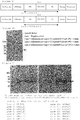
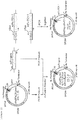
- An expression cassette for an anti-PD-1 scFv antibody was, as shown in Figure 1(a) , constituted of Hu promoter DNA (derived from Bifidobacterium longum ) , a DNA encoding a secretory signal peptide, a DNA encoding a linker peptide following the secretory signal peptide, a DNA encoding an anti-PD-1 scFv antibody (containing a heavy chain sequence, a linker ((GGGGS) 3 ), a light chain sequence), a His tag sequence and a Hu terminator (derived from Bifidobacterium longum ) .
- Hu promoter DNA derived from Bifidobacterium longum
- a DNA encoding a secretory signal peptide a DNA encoding a linker peptide following the secretory signal peptide
- a DNA encoding an anti-PD-1 scFv antibody containing a heavy chain sequence, a linker ((GGGGS)
- anti-PD-1 scFv antibodies containing nucleotide sequences containing a heavy chain sequence, a linker ((GGGGS) 3 ) and a light chain sequence
- two anti-human PD-1 scFv antibodies and a single anti-mouse PD-1 scFv antibody were used, with reference to the documents shown in Table 3 below.
- SEQ ID No: 139 (249 amino acids) Heavy chain sequence(1-360 nucleotides) (GGGGS) 3 linker (361-405 nucleotides) Light chain sequence (406-729 nucleotides) Histidine tag (730-747 nucleotides) scFv-PD-1-3 (Anti-mouse PD-1 antibody) SEQ ID No: 3 (762 nucleotides sequence) Japanese Patent No. 4249013 SEQ ID No: 140 (253 amino acids) Heavy chain sequence(1-366 nucleotides) (GGGGS) 3 linker (367-411 nucleotides) Light chain sequence (412-741 nucleotides) Histidine tag (742-759 nucleotides)
- the genes of the anti-PD-1 scFv antibodies were separately subcloned in a plasmid for E . coli, i.e., pUC57 by GenScript Japan Inc. and provided as artificially synthesized plasmids, namely, pUC57-scFv-PD-1-1, pUC57-scFv-PD-1-2 and pUC57-scFv-PD-1-3, respectively.
- secretory signal peptide-linker conjugates namely SP3L22, SP7L20 and SP23L27, were used.
- a known sequence represented by SEQ ID No: 8 described in International Publication No. WO2011/093467 was used.
- SP23L27 a known sequence represented by SEQ ID No: 25 described in International Publication No. WO2011/093467 was used.
- SP7L20 a nucleotide sequence, which was prepared by adding a 66-nucleotide: ATGGCGTTGATGATGAGCGTTAAGACTATTATTTCCACATCAGTGGCGATTATCGCCA CGGGTGCC (starting with ATG) to the upstream of the 5' side of a nucleotide sequence represented by SEQ ID No: 12 described in International Publication No.
- WO2011/093467 (used as a basic sequence), and represented by SEQ ID No: 81, was used.
- a nucleotide sequence was added because the SP7 was estimated to have a secretory signal peptide sequence at the N terminal of a protein when SP7 was analyzed by a signal sequence estimation program, Signal P 4.1 server (http://www.cbs.dtu.dk/services/SignalP/), which is a program of estimating the presence or absence of a secretory signal.
- the linker peptide sequence following SP7 was 20 amino acid residues at the C terminal side of SP7.
- scFv-gene is PD-1.
- pHuSP3L22-scFv-PD-1-1 In preparing a plasmid, pHuSP3L22-scFv-PD-1-1, first, a plasmid, pHuSP3L20-scFv-PD-1-1 was prepared.
- PCR amplification was performed by using a plasmid, pUC57-scFv-PD-1-1 (500 pg) as a template and a primer set consisting of Ins_PD-1-1_F1 primer and Ins_PD-1-1_R1 primer listed in Table 1 and Table 2.
- the primer sequences were designed such that an insert fragment and a vector fragment were mutually overlapped at the end of 15 bp.
- the PCR amplification was performed by setting each primer concentration at 0.2 ⁇ M and the reaction volume at 30 ⁇ L and using PrimeSTAR HS (Premix) kit (manufactured by Takara Bio Inc.).
- a cycle consisting of a reaction at 98°C for 10 seconds, a reaction at 55°C for 5 seconds and a reaction at 72°C for 60 seconds was repeated 30 times.
- the PCR product obtained by the amplification was subjected to electrophoresis on 2% agarose gel and purified by QIAquick Gel Extraction Kit (manufactured by QIAGEN) to prepare an anti-PD-1-1 scFv antibody insert.
- PCR amplification was performed by using a linearized vector fragment (500 pg) represented by SEQ ID No: 20 as a template and a primer set consisting of TGA_Hu_Terminator_F primer and vec-SP3L20-PD-1-1_R1 primer listed in Table 1 and Table 2 above, setting each primer concentration at 0.2 ⁇ M and the reaction volume at 30 ⁇ L and using PrimeSTAR HS (Premix) kit.
- a cycle consisting of a reaction at 98°C for 10 seconds, a reaction at 55°C for 5 seconds and a reaction at 72°C for 5 minutes was repeated 30 times.
- the PCR product obtained by the amplification was subjected to electrophoresis on 0.8% agarose gel and purified by QIAquick Gel Extraction Kit (manufactured by QIAGEN) to prepare 5'-Hu terminator-pTB6rep unit-SPCMr-pUCori-Hu promoter-SP3L20-3' vector fragment.
- the linearized vector fragment is the nucleotide sequence consisting of the 4th nucleotide to the 3845th nucleotide of the nucleotide sequence represented by SEQ ID No: 20 and constituted of:
- the vector fragment and an anti-PD-1-1 scFv antibody insert prepared above were ligated by use of In-Fusion (registered trademark) HD Cloning Kit (manufactured by Takara Bio Inc.). More specifically, in a microtube, the vector and the insert were added at a molar ratio of 1: 5 and thereafter 2 ⁇ L of 5 ⁇ In-Fusion HD Enzyme premix was added. The volume of the reaction solution was adjusted to be 10 ⁇ L. The reaction solution was kept at 50°C for 15 minutes. The procedure described in the instruction of the kit except the aforementioned procedure was performed to prepare an infusion reaction solution.
- In-Fusion registered trademark
- HD Cloning Kit manufactured by Takara Bio Inc.
- E. coli HST08 competent cell (manufactured by Takara Bio Inc.) was transformed in accordance with the instruction thereof. After transformation, the bacterial suspension was spread onto a 75 ⁇ g/mL spectinomycin-containing LB agar medium and subjected to static culture at 37°C overnight. E. coli colonies formed on the agar medium were cultured in a 75 ⁇ g/mL spectinomycin-containing LB liquid medium at 30°C overnight. From the cultured medium, a plasmid was extracted by use of QIAprep Spin Miniprep Kit (manufactured by QIAGEN).
- An anti-PD-1-1 scFv antibody expression cassette (containing Hu promoter to Hu terminator) of the plasmid extracted was subjected to a sequencing reaction by BigDye (registered trademark) Terminator v3.1 Cycle Sequencing Kit (manufactured by Applied Biosystems) for determining the sequence thereof.
- the extracted plasmid was designated as pHuSP3L20-scFv-PD-1-1.
- Ins_PD-1-1_F1 primer and SP3L22_R1 primer listed in Table 1 and Table 2 was phosphorylated with T4 polynucleotide kinase (manufactured by Takara Bio Inc.) in accordance with the instruction.
- Ins_PD-1-1_F1 phosphorylated primer and SP3L22_R1 phosphorylated primer each were adjusted so as to have a final concentration of 20 ⁇ M with 0.1 ⁇ TE buffer (1mM Tris-HCl, 0.1mM EDTA, pH7.5).
- PCR amplification was performed by using a plasmid, pHuSP3L20-scFv-PD-1-1 (500 pg) obtained above as a template and a primer set consisting of Ins_PD-1-1_F1 phosphorylated primer and SP3L22_R1 phosphorylated primer, setting each primer concentration at 0.2 ⁇ M and the reaction volume at 30 ⁇ L and using PrimeSTAR HS (Premix) kit.
- a cycle consisting of a reaction at 98°C for 10 seconds, a reaction at 55°C for 5 seconds and a reaction at 72°C for 5 minutes was repeated 30 times.
- the PCR product obtained by amplification was subjected to electrophoresis on 0.8% agarose gel, purified by QIAquick Gel Extraction Kit to prepare a linearized fragment pHuSP3L22-scFv-PD-1-1 vector.
- the linearized vector fragment obtained above was subjected to a ligation reaction using T4 DNA Ligase (manufactured by Thermo Fisher Scientific Inc.) in accordance with the instruction.
- T4 DNA Ligase manufactured by Thermo Fisher Scientific Inc.
- the accompanying 10 ⁇ buffer (2 ⁇ L) for T4 DNA ligase was added and the volume of a reaction solution was set to be 20 ⁇ L. This was subjected to a ligation reaction at 22°C for 10 minutes to prepare a ligation reaction solution.
- E. coli HST08 competent cell was transformed. After transformation, a bacterial suspension was spread onto a 75 ⁇ g/mL spectinomycin-containing LB agar medium and subjected to static culture at 37°C overnight. Transformation was carried out in accordance with the conditions described in the instruction for E . coli HST08 competent cell as outlined below. E. coli colonies formed on the agar medium were cultured in a 75 ⁇ g/mL spectinomycin-containing LB liquid medium at 30°C overnight. From this, a plasmid was extracted by use of QIAprep Spin Miniprep Kit.
- the anti-PD-1-1 scFv antibody expression cassette (containing Hu promoter to Hu terminator) of the extracted plasmid was subjected to a sequencing reaction by use of BigDye (registered trademark) Terminator v3.1 Cycle Sequencing Kit for determining the sequence thereof.
- the extracted plasmid was designated as pHuSP3L22-scFv-PD-1-1.
- pHuSP3L22-scFv-PD-1-1 1000 ng
- Bifidobacterium longum 105-A strain was transformed by the electroporation system (Gene Pulser II, manufactured by Bio-Rad Laboratories, Inc.).
- electroporation system Gene Pulser II, manufactured by Bio-Rad Laboratories, Inc.
- a solution mixture of IMR liquid medium 800 ⁇ L
- a vitamin C-added solution 50 ⁇ L was poured in a cuvette (2 mm gap) and this was collected to a 2 mL-microtube sterilized.
- Each of the resultant bacterium suspensions kept warm was spread onto a 75 ⁇ g/mL spectinomycin-containing IMR agar medium. These plates were placed in an airtight container together with a de-oxygen and carbon dioxide gas generating agent as mentioned above, and cultured in an incubator set at 37°C for 2 days.
- spectinomycin-containing IMR agar medium A part of the colonies formed on the spectinomycin-containing IMR agar medium was picked up, streaked on a 75 ⁇ g/mL spectinomycin-containing BL-bS agar medium (BL agar medium containing no equine defibrinated blood), placed in an airtight container together with a de-oxygen and carbon dioxide gas generating agent and cultured in an incubator set at 37°C for one day to obtain Bifidobacterium longum 105-A/pHuSP3L22-scFv-PD-1-1 strain.
- An anti-PD-1-1 scFv antibody insert was prepared in the same procedure as in the above section: [Preparation of plasmid, pHuSP3L20-scFv-PD-1-1], (Preparation of anti-PD-1-1 scFv antibody insert).
- PCR amplification was performed by using a linearized vector fragment (500 pg) represented by SEQ ID No: 21 as a template and a primer set consisting of TGA_Hu_Terminator_F primer and vec-SP7L20-PD-1-1_R1 primer described in Table 1 and Table 2 above, setting each primer concentration at 0.2 ⁇ M and the reaction volume at 30 ⁇ L and using PrimeSTAR HS (Premix) kit.
- a cycle consisting of a reaction at 98°C for 10 seconds, a reaction at 55°C for 5 seconds and a reaction at 72°C for 5 minutes was repeated 30 times.
- the PCR product obtained by the amplification was subjected to electrophoresis on 0.8% agarose gel and purified by QIAquick Gel Extraction Kit to prepare a 5'-Hu terminator-pTB6rep unit-SPCMr-pUCori-Hu promoter-SP7L20-3' vector fragment (1).
- the linearized vector fragment is the nucleotide sequence consisting of the 4th nucleotide to the 3845th nucleotide of the nucleotide sequence represented by SEQ ID No: 21 and constituted of:
- An infusion reaction solution was prepared in the same procedure as in the infusion reaction in the above section: [Preparation of plasmid, pHuSP3L20-scFv-PD-1-1] except that the vector fragment and the anti-PD-1-1 scFv antibody insert prepared above were used.
- E. coli HST08 competent cell was transformed and the anti-PD-1-1 scFv antibody expression cassette (containing Hu promoter to Hu terminator) of the extracted plasmid was subjected to a sequencing reaction in the same procedure as in the above section: [Preparation of plasmid, pHuSP3L20-scFv-PD-1-1], (Transformation of E. coli and sequencing of plasmid DNA) except that the infusion reaction solution (5 ⁇ L) prepared above was used.
- the extracted plasmid was designated as pHuSP7L20-scFv-PD-1-1.
- Bifidobacterium longum 105-A strain was transformed in the same procedure in the above section: [Preparation of pHuSP3L22-scFv-PD-1-1], (Transformation of bacterium of the genus Bifidobacterium ) except that DNA of a plasmid, pHuSP7L20-scFv-PD-1-1 (1000 ng) extracted from E. coli transformed above was used to obtain Bifidobacterium longum 105-A/pHuSP7L20-scFv-PD-1-1 strain.
- a plasmid, pHuSP23L27-scFv-PD-1-1 first, a plasmid, pHuSP23L20-scFv-PD-1-1 was prepared.
- An anti-PD-1-1 scFv antibody insert was prepared in the same procedure as in the above section: [Preparation of plasmid, pHuSP3L20-scFv-PD-1-1], (Preparation of an anti-PD-1-1 scFv antibody insert).
- PCR amplification was performed by using the linearized vector fragment (500 pg) represented by SEQ ID No: 22 as a template and a primer set consisting of TGA_Hu_Terminator_F primer and vec-SP23L20-PD-1-1_R1 primer listed in Table 1 and Table 2 above, setting each primer concentration at 0.2 ⁇ M and the reaction volume at 30 ⁇ L and using PrimeSTAR HS (Premix) kit.
- a cycle consisting of a reaction at 98°C for 10 seconds, a reaction at 55°C for 5 seconds and a reaction at 72°C for 5 minutes was repeated 30 times.
- the PCR product obtained by the amplification was subjected to electrophoresis on 0.8% agarose gel and purified by QIAquick Gel Extraction Kit to prepare a 5'-Hu terminator-pTB6rep unit-SPCMr-pUCori-Hu promoter-SP23L20-3' vector fragment.
- the linearized vector fragment is the nucleotide sequence consisting of the 4th nucleotide to the 3845th nucleotide of the nucleotide sequence represented by SEQ ID No: 22 and constituted of:
- An infusion reaction solution was prepared in the same procedure as in the infusion reaction in the above section: [Preparation of plasmid, pHuSP3L20-scFv-PD-1-1] except that the vector fragment and the anti-PD-1-1 scFv antibody insert prepared above were used.
- E. coli HST08 competent cell was transformed and the anti-PD-1-1 scFv antibody expression cassette (containing Hu promoter to Hu terminator) of the extracted plasmid was subjected to a sequencing reaction (for determining the sequence) in the same procedure as in the above section: [Preparation of plasmid, pHuSP3L20-scFv-PD-1-1], (Transformation of E . coli and sequencing of plasmid DNA) except that the infusion reaction solution (5 ⁇ L) prepared above was used.
- the extracted plasmid was designated as pHuSP23L20-scFv-PD-1-1.
- Ins_PD-1-1_F1 primer and SP23L27_R1 primer listed in Table 1 and Table 2 was phosphorylated by using T4 polynucleotide kinase in accordance with the instruction.
- Ins_PD-1-1_F1 phosphorylated primer and SP23L27_R1 phosphorylated primer each were adjusted so as to have a final concentration of 20 ⁇ M, with 0.1 ⁇ TE buffer (1mM Tris-HCl, 0.1mM EDTA, pH7.5).
- PCR amplification was performed by using a plasmid, pHuSP23L20-scFv-PD-1-1 (500 pg) obtained above as a template, a primer set consisting of Ins_PD-1-1_F1 phosphorylated primer and SP23L27_R1 phosphorylated primer, setting a final concentration at 0.2 ⁇ M and the reaction volume at 30 ⁇ L, and using PrimeSTAR HS (Premix) kit.
- a cycle consisting of a reaction at 98°C for 10 seconds, a reaction at 55°C for 5 seconds and a reaction at 72°C for 5 minutes was repeated 30 times.
- the PCR product obtained by the amplification was subjected to electrophoresis on 0.8% agarose gel and purified by QIAquick Gel Extraction Kit to prepare a linearized fragment of pHuSP23L27-scFv-PD-1-1 vector.
- the vector fragment obtained above was subjected to a ligation reaction to prepare a ligation reaction solution in the same procedure as in the above section: [Preparation of plasmid, pHuSP3L22-scFv-PD-1-1], (Ligation reaction).
- E. coli HST08 competent cell was transformed by using the above ligation reaction solution (10 ⁇ L) and subjected to a sequencing reaction in the same manner as in the above section: [Preparation of plasmid, pHuSP3L22-scFv-PD-1-1], (Transformation of E. coli and sequencing of plasmid DNA).
- the extracted plasmid was designated as pHuSP23L27-scFv-PD-1-1.
- Bifidobacterium longum 105-A strain was transformed in the same procedure as in the above section: [Preparation of plasmid, pHuSP3L22-scFv-PD-1-1], (Transformation of bacterium of the genus Bifidobacterium) to obtain Bifidobacterium longum 105-A/pHuSP23L27-scFv-PD-1-1 strain.
- a streak culture of each of the recombinant bacterium of the genus Bifidobacterium strains on a 75 ⁇ g/mL spectinomycin-containing BL-bS agar medium was inoculated in MRS (manufactured by Becton, Dickinson and Company) liquid medium (10 mL) supplemented with spectinomycin (a final concentration of 75 ⁇ g/mL), 100 ⁇ L of a vitamin C-added solution (a solution (100 mL) containing ascorbic acid (35 g), L-cysteine hydrochloride monohydrate (2 g) and sodium carbonate (11 g)), and anaerobically cultured at 37°C for 24 hours to obtain an activated culture solution.
- MRS manufactured by Becton, Dickinson and Company
- the activated culture solution was centrifuged and the culture supernatant was collected. Protein in the culture supernatant was allowed to precipitate with trichloroacetic acid (TCA, manufactured by Wako Pure Chemical Industries Ltd.), washed with acetone, dissolved in SDS-PAGE buffer and subjected to a heat treatment at 95°C for 3 minutes to obtain a culture supernatant concentrate.
- TCA trichloroacetic acid
- SDS-PAGE buffer a heat treatment at 95°C for 3 minutes to obtain a culture supernatant concentrate.
- Bifidobacterium longum 105-A/pBEshuttle strain was subjected to the same operation and used as a negative control.
- the culture supernatant concentrate (corresponding to 1 mL of culture solution) was subjected to electrophoresis by Mini-PROTEAN (registered trademark) TGXTM gel (4 to 20%) (manufactured by Bio-Rad).
- Mini-PROTEAN registered trademark
- TGXTM gel (4 to 20%) (manufactured by Bio-Rad).
- the gel was transferred onto a PVDF membrane (iBlot Transfer Stacks, manufactured by Life Technologies) by use of an iBlot transfer device (manufactured by Life Technologies).
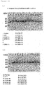
- the membrane was subjected to blocking (2% ECL Prime Blocking agent (manufactured by GE Healthcare Japan) in TTBS), and then to a reaction with a mouse histidine tag antibody (THE HIS Tag Antibody, mAb, Mouse, manufactured by GenScript Japan Inc.) used as a primary antibody and an ECL-peroxidase-labeled anti-mouse antibody (manufactured by GE Healthcare Japan) used as a secondary antibody, and allowed to emit light by use of Western Lightning Ultra (manufactured by PerkinElmer Co., Ltd.). The light-emission was analyzed by an imaging analyzer (Fluor SMax, manufactured by Bio-Rad). The results are shown in Figure 4 .
- Lane M shows a marker
- lane 1 shows a negative control
- lane 2 shows an antibody secreted from a bacterium of the genus Bifidobacterium transformed with pHuSP3L22-scFv-PD-1-1 in lane 2
- lane 3 shows an antibody secreted from a bacterium of the genus Bifidobacterium transformed with pHuSP7L20-scFv-PD-1-1
- lane 4 shows an antibody secreted from a bacterium of the genus Bifidobacterium transformed with pHuSP23L27-scFv-PD-1-1.
- TM transmembrane region
- amino acid sequences of Bifidobacterium longum 105A strain corresponding to the amino acid sequences of the 33 types of peptides selected, the sequence from the N terminal of each of the amino acid sequences (of the proteins) up to the amino acid which is estimated as a protease cleavage putative site by Signal P analysis was determined as a secretory signal peptide sequence. Furthermore, 20 amino acid residues following from the C terminal of each of the signal sequences was specified as the linker peptide sequence.
- PCR amplification was performed by using Bifidobacterium longum 105-A strain (120 pg) as a template and each of the primer sets listed in Table 4 and Table 5. Individual primer sequences were designed such that an insert fragment and a vector fragment were overlapped at the end of 15 bp.
- the PCR amplification was performed by setting a primer concentration at 0.2 ⁇ M and the reaction volume at 20 ⁇ L and using PrimeSTAR HS (Premix) kit.
- PrimeSTAR HS Premix
- a cycle consisting of a reaction at 98°C for 10 seconds, a reaction at 65°C for 5 seconds and a reaction at 72°C for 20 seconds was repeated 30 times, followed by an elongation reaction at 72°C for 30 seconds. In this manner, inserts consisting of 33 types of secretory signal peptide-linker conjugates were obtained by amplification.
- PCR amplification was performed by using pHuSP7L20-scFv-PD-1-1 (2.5 ng) mentioned above as a template, and a primer set consisting of PD-1-scFv_vec_F1 primer and Hu_Vec_R1 primer listed in Table 4 and Table 5 above, setting a final concentration at 0.2 ⁇ M and the reaction volume at 50 ⁇ L, and using PrimeSTAR HS (Premix) kit.
- a cycle consisting of a reaction at 98°C for 10 seconds, a reaction at 65°C for 5 seconds and a reaction at 72°C for 4 minutes and 45 seconds was repeated 30 times followed by an elongation reaction at 72°C for 30 seconds.
- a vector fragment 5'-scFv-PD-1-1-Hu terminator-pTB6rep unit-SPCMr-pUCori-Hu promoter-3', was prepared.
- the vector fragment prepared in the above and each of 33 types secretory signal peptide-linker conjugate inserts were ligated by use of In-Fusion (registered trademark) HD Cloning Kit. More specifically, in a microtube, the vector and an insert were added at a molar ratio of 1: 2, and 5 ⁇ In-Fusion HD Enzyme premix (2 ⁇ L) provided in the kit was added. The volume of the mixture was adjusted with 0.1 ⁇ TE buffer (1 mM Tris-HCl, 0.1 mM EDTA, pH7.5) so as to be 10 ⁇ L. This solution was kept warm at 37°C for 15 minutes and further kept warm at 50°C for 15 minutes. The procedure as described in the instruction of the kit except the aforementioned procedure was performed to prepare 33 types of infusion reaction solutions.
- In-Fusion registered trademark
- HD Cloning Kit More specifically, in a microtube, the vector and an insert were added at a molar ratio of 1: 2, and 5 ⁇ In-Fusion HD Enzyme
- E. coli HST16CR competent cells manufactured by Takara Bio Inc.
- each of the bacterial suspensions was spread onto a 75 ⁇ g/mL spectinomycin-containing LB agar medium and subjected to static culture at 37°C overnight.
- the transformation was performed in accordance with the conditions described in the instruction of E. coli HST16CR competent cell as outlined below.
- E. coli colonies formed on the agar medium were cultured in a 75 ⁇ g/mL spectinomycin-containing LB liquid medium at 37°C overnight. From the culture medium, a plasmid was extracted by use of QIAprep Spin Miniprep Kit.
- the anti-PD-1-1 scFv antibody expression cassette (containing Hu promoter to Hu terminator) of the extracted plasmid was subjected to a sequencing reaction by BigDye (registered trademark) Terminator v3.1 Cycle Sequencing Kit for determining the sequence thereof.
- secretion of an anti-PD-1-1 scFv antibody was not observed in bacterium of the genus Bifidobacterium transformed with plasmids in which secretory signal peptide-linker conjugates (SP42L20, SP48L20, SP62L20, (which respectively consist of secretory signal peptide sequences (SP42, SP48, SP62) and subsequent linker peptide sequences of 20 amino acid residues) were inserted.
- secretory signal peptide-linker conjugates SP42L20, SP48L20, SP62L20, (which respectively consist of secretory signal peptide sequences (SP42, SP48, SP62) and subsequent linker peptide sequences of 20 amino acid residues
- an anti-PD-1-2 scFv antibody secretory plasmid pHuSP7L20-scFv-PD-1-2 having a secretory signal peptide-linker conjugate, SP7L20, was constructed.
- the details of the construction are as follows.
- PCR amplification was performed by using the artificial plasmid, pUC57-scFv-PD-1-2 (500 pg) synthesized above as a template and a primer set consisting of Ins_PD-1-2_F1 primer and Ins_PD-1-2_R1 primer listed in Table 1 and Table 2 above.
- the primer sequences were designed such that an insert fragment and a vector fragment were mutually overlapped at the end of 15 bp.
- the PCR reaction solution used herein was prepared so as to contain a primer in a concentration of 0.2 ⁇ M in a reaction volume of 30 ⁇ L by using PrimeSTAR HS (Premix) kit.
- a cycle consisting of a reaction at 98°C for 10 seconds, a reaction at 55°C for 5 seconds and a reaction at 72°C for 60 seconds was repeated 30 times.
- the PCR product obtained by the amplification was subjected to electrophoresis on 2% agarose gel and purified by QIAquick Gel Extraction Kit to prepare an anti-PD-1-2 scFv antibody insert.
- PCR amplification was performed by using a linearized vector fragment (500 pg) represented by SEQ ID No: 23 as a template and a primer set consisting of TGA_Hu_Terminator_F primer and vec-SP7L20-PD-1-2_R1 primer listed in Table 1 and Table 2 above, setting a final concentration at 0.2 ⁇ M and the reaction volume at 30 ⁇ L and using PrimeSTAR HS (Premix) kit.
- a cycle consisting of a reaction at 98°C for 10 seconds, a reaction at 55°C for 5 seconds and a reaction at 72°C for 5 minutes was repeated 30 times.
- the PCR product obtained by the amplification was subjected to electrophoresis on 0.8% agarose gel and purified by QIAquick Gel Extraction Kit to prepare a 5'-Hu terminator-pTB6rep unit-SPCMr-pUCori-Hu promoter-SP7L20-3' vector fragment (2).
- the linearized vector fragment is the nucleotide sequence consisting of the 4th nucleotide to the 3845th nucleotide of the nucleotide sequence represented by SEQ ID No: 23 and constituted of:
- An infusion reaction solution was prepared in the same procedure as in the infusion reaction in the above section: [Preparation of plasmid, pHuSP3L20-scFv-PD-1-1] except that the vector fragment and anti-PD-1-2 scFv antibody insert prepared above were used.
- E. coli HST08 competent cell was transformed in the same procedure as in Example 1, [Preparation of plasmid, pHuSP3L20-scFv-PD-1-1], (Transformation of E. coli and sequencing of plasmid DNA) except that the infusion reaction solution (5 ⁇ L) prepared above was used.
- the anti-PD-1-2 scFv antibody expression cassette (containing Hu promoter to Hu terminator) of the plasmid extracted was subjected to a sequencing reaction for determining the sequence thereof.
- the extracted plasmid was designated as pHuSP7L20-scFv-PD-1-2.
- Bifidobacterium longum 105-A strain was transformed in the same procedure as in the above section: [Preparation of plasmid, pHuSP3L22-scFv-PD-1-1], (Transformation of bacterium of the genus Bifidobacterium ) to obtain Bifidobacterium longum 105-A/pHuSP7L20-scFv-PD-1-2 strain.
- An anti-PD-1-3 scFv antibody was selected as a heterogeneous peptide and an anti-PD-1-3 scFv antibody secretory plasmid, pHuSP7L20-scFv-PD-1-3 having a secretory signal peptide-linker conjugate, SP7L20 was constructed. The details of the construction are as follows.
- An anti-PD-1-3 scFv antibody insert was prepared in the same procedure as in the above section: [Preparation of pHuSP7L20-scFv-PD-1-2], (Preparation of an anti-PD-1-2 scFv antibody insert) except that the artificial plasmid, pUC57-scFv-PD-1-3 (500 pg) synthesized above was used as a template and a primer set consisting of Ins_PD-1-3_F1 primer and Ins_PD-1-3_R1 primer listed in Table 1 and Table 2 was used.
- a 5'-Hu terminator-pTB6rep unit-SPCMr-pUCori-Hu promoter-SP7L20-3' vector fragment (3) was prepared in the same procedure in [Preparation of pHuSP7L20-scFv-PD-1-2], (Preparation of vector fragment) except that the linearized vector fragment (500 pg) represented by SEQ ID No: 24 was used as a template and a primer set consisting of TGA_Hu_Terminator_F primer and vec-SP7L20-PD-1-3_R1 primer listed in Table 1 and Table 2 above was used.
- the linearized vector fragment is the nucleotide sequence consisting of the 4th nucleotide to the 3845th nucleotide of the nucleotide sequence represented by SEQ ID No: 24 and constituted of:
- An infusion reaction solution was prepared in the same procedure as in the infusion reaction in the above section: [Preparation of plasmid, pHuSP3L20-scFv-PD-1-1] except that the vector fragment and the anti-PD-1-3 scFv antibody insert prepared above were used.
- E. coli HST08 competent cell was transformed in the same procedure as in Example 1, [Preparation of plasmid, pHuSP3L20-scFv-PD-1-1], (Transformation of E. coli and sequencing of plasmid DNA) except that the infusion reaction solution (5 ⁇ L) prepared above was used.
- the anti-PD-1-3 scFv antibody expression cassette (containing Hu promoter to Hu terminator) of the plasmid extracted was subjected to a sequencing reaction for determining the sequence thereof.
- the extracted plasmid was designated as pHuSP7L20-scFv-PD-1-3.
- Bifidobacterium longum 105-A strain was transformed in the same procedure as in the above section: [Preparation of plasmid, pHuSP3L22-scFv-PD-1-1], (Transformation of bacterium of the genus Bifidobacterium ) to obtain Bifidobacterium longum 105-A/pHuSP7L20-scFv-PD-1-3 strain.
- An Expression cassette for anti-CTLA-4 scFv antibody is, as shown in Figure 1(b) , constituted of Hu promoter DNA (derived from Bifidobacterium longum ), a DNA encoding a secretory signal peptide, a DNA encoding a linker peptide following the secretory signal peptide, a DNA encoding an anti-CTLA-4 scFv antibody (containing a heavy chain sequence, linker ((GGGGS) 3 ) and a light chain sequence), a His tag sequence and a Hu terminator (derived from Bifidobacterium longum ) .
- Hu promoter DNA derived from Bifidobacterium longum
- a DNA encoding a secretory signal peptide a DNA encoding a linker peptide following the secretory signal peptide
- a DNA encoding an anti-CTLA-4 scFv antibody containing a heavy chain sequence, linker ((GGGGS) 3 )
- anti-CTLA-4 scFv antibody containing nucleotide sequence containing a heavy chain sequence, a linker ((GGGGS) 3 ) and a light chain sequence
- two types of anti-human CTLA-4 scFv antibodies as shown below was used, with reference to the document listed in Table 6 below.
- Table 6 Antibody Reference document scFv-CTLA-4-1 SEQ ID No: 4 (765 nucleotide sequence) Japanese Patent No.
- SEQ ID No: 141 (254 amino acids) 3793693 Heavy chain sequence (1-378 nucleotides) (GGGGS) 3 linker (379-423 nucleotides) Light chain sequence (424-744 nucleotides) Histidine tag (745-762 nucleotides) scFv-CTLA-4-2 SEQ ID No: 5 (747 nucleotide sequence) Japanese Patent No. SEQ ID No: 142 (248 amino acids) 4093757 Heavy chain sequence (1-357 nucleotides) (GGGGS) 3 linker (358-402 nucleotides) Light chain sequence (403-726 nucleotides) Histidine tag (727-744 nucleotides)
- scFv antibody genes namely, scFv-CTLA-4-1 represented by SEQ ID No: 4 and scFv-CTLA-4-2 represented by SEQ ID No: 5, were separately subcloned in plasmids for E. coli pUC57 by GenScript Japan Inc. In this manner, artificial plasmids pUC57-scFv-CTLA-4-1 and pUC57-scFv-CTLA-4-2 were obtained by gene synthesis.
- PCR amplification was performed by using the plasmid, pUC57-scFv-CTLA-4-1 (500 pg) as a template, and a primer set consisting of Ins_CTLA-4-1_F1 primer and Ins_CTLA-4-1_R1 primer listed in Table 1 and Table 2 above.
- the primer sequences were designed such that an insert fragment and a vector fragment were mutually overlapped at the end of 15 bp.
- the amplification was performed by PrimeSTAR HS (Premix) (manufactured by Takara Bio Inc.) kit using a PCR reaction solution, which was prepared so as to satisfy a primer concentration of 0.2 ⁇ M and a reaction volume of 30 ⁇ L.
- a cycle consisting of a reaction at 98°C for 10 seconds, a reaction at 55°C for 5 seconds and a reaction at 72°C for 60 seconds was repeated 30 times.
- the insert PCR product obtained by the amplification was subjected to electrophoresis on 2% agarose gel and purified by QIAquick Gel Extraction Kit to prepare an anti-CTLA-4-1 scFv antibody insert.
- a 5'-Hu terminator-pTB6rep unit-SPCMr-pUCori-Hu promoter-SP7L20-3' vector fragment (1) was prepared in the same procedure as in the above section: [Preparation of pHuSP7L20-scFv-PD-1-1], (Preparation of vector fragment).
- An infusion reaction solution was prepared in the same procedure as in the infusion reaction in the above section: [Preparation of plasmid, pHuSP3L20-scFv-PD-1-1] except that the vector fragment and anti-CTLA-4-1 scFv antibody insert prepared above were used.
- E. coli HST08 competent cell was transformed in the same procedure as in the above section: [Preparation of plasmid, pHuSP3L20-scFv-PD-1-1], (Transformation of E . coli and sequencing of plasmid DNA) except that the infusion reaction solution (5 ⁇ L) prepared above was used.
- the anti-CTLA-4-1 scFv antibody expression cassette (containing Hu promoter to Hu terminator) of the plasmid extracted was subjected to a sequencing reaction for determining the sequence thereof.
- the extracted plasmid was designated as pHuSP7L20-scFv-CTLA-4-1.
- Bifidobacterium longum 105-A strain was transformed in the same procedure as in the above section: [Preparation of pHuSP3L22-scFv-PD-1-1], (Transformation of bacterium of the genus Bifidobacterium) except that DNA of a plasmid, pHuSP7L20-scFv-CTLA-4-1 (1000 ng), extracted from E . coli transformed above was used to obtain Bifidobacterium longum 105-A/pHuSP7L20-scFv-CTLA-4-1 strain.
- An anti-CTLA-4-2 scFv antibody secretory plasmid, pHuSP7L20-scFv-CTLA-4-2, having a secretory signal peptide-linker conjugate (SP7L20) was constructed. The details of the construction are as follows.
- An anti-CTLA-4-2 scFv antibody insert was purified in the same procedure as in the above section: (Preparation of anti-CTLA-4-1 scFv antibody insert) except that the plasmid, pUC57-scFv-CTLA-4-2 obtained above was used as a template and a primer set consisting of Ins_CTLA-4-1_F1 primer and Ins_CTLA-4-2_R1 primer set listed in Table 1 and Table 2 above was used.
- a 5'-Hu terminator-pTB6rep unit-SPCMr-pUCori-Hu promoter-SP7L20-3' vector fragment (1) was prepared in the same procedure as in the above section: [Preparation of pHuSP7L20-scFv-PD-1-1], (Preparation of vector fragment).
- An infusion reaction solution was prepared in the same procedure as in the infusion reaction in the above section: [Preparation of plasmid, pHuSP3L20-scFv-PD-1-1] except that the vector fragment and the anti-CTLA-4-2 scFv antibody insert prepared above were used.
- E. coli HST08 competent cell was transformed in the same procedure as in Example 1, [Preparation of plasmid, pHuSP3L20-scFv-PD-1-1], (Transformation of E. coli and sequencing of plasmid DNA) except that the infusion reaction solution (5 ⁇ L) prepared above was used.
- the anti-CTLA-4-2 scFv antibody expression cassette (containing Hu promoter to Hu terminator) of the plasmid extracted was subjected to a sequencing reaction for determining the sequence thereof.
- the extracted plasmid was designated as pHuSP7L20-scFv-CTLA-4-2.
- Bifidobacterium longum 105-A strain was transformed in the same procedure as in the above section: [Preparation of pHuSP3L22-scFv-PD-1-1], (Transformation of bacterium of the genus Bifidobacterium) except that a plasmid, pHuSP7L20-scFv-CTLA-4-2 (1000 ng) extracted from E. coli transformed above was used to obtain Bifidobacterium longum 105-A/pHuSP7L20-scFv-CTLA-4-2 strain.
- an anti-PD-1-1 scFv antibody was secreted in the culture supernatant of Bifidobacterium longum 105-A/pHuSP7L20-scFv-PD-1-1 strain; an anti-PD-1-2 scFv antibody was secreted in the culture supernatant of Bifidobacterium longum 105-A/pHuSP7L20-scFv-PD-1-2 strain; an anti-PD-1-3 scFv antibody was secreted in the culture supernatant of Bifidobacterium longum 105-A/pHuSP7L20-scFv-PD-1-3 strain; anti-CTLA-4-1 scFv antibody was secreted in the culture supernatant of Bifidobacterium longum 105-A/pHuSP7L20-scFv-CTLA-4-1 strain; and anti-CTLA-4-2 scFv antibody was secreted in the culture supernatant of B
- a scFv antibody was purified from each of Bifidobacterium longum 105-A/pHuSP7L20-scFv-PD-1-1 strain, Bifidobacterium longum 105-A/pHuSP7L20-scFv-PD-1-2 strain, Bifidobacterium longum 105-A/pHuSP7L20-scFv-PD-1-3 strain, Bifidobacterium longum 105-A/pHuSP7L20-scFv-CTLA-4-1 strain and Bifidobacterium longum 105-A/pHuSP7L20-scFv-CTLA-4-2 strain, by the method shown below.
- Each of the recombinant bacterium of the genus Bifidobacterium strains was inoculated in MRS culture medium (10 mL) supplemented with spectinomycin (a final concentration of 75 ⁇ g/mL) and a vitamin C-added solution (100 ⁇ L) and anaerobically cultured at 37°C for 24 hours.
- a culture medium containing DMEM: MRS (9: 1) was prepared by adding spectinomycin (a final concentration of 75 ⁇ g/mL) and a vitamin C-added solution (500 ⁇ L per culture medium 100 mL). To this medium, the culture solution obtained above (0.5% of the amount of the medium) was inoculated and anaerobically cultured at 37°C for 18 hours.
- the culture supernatant was obtained by centrifugation.
- ammonium sulfate was added little by little while stirring the culture supernatant so as to obtain a saturation of 80%.
- the solution obtained was stirred at 4°C overnight to perform salting out.
- the solution was centrifuged and the precipitate was recovered.
- a histidine tag fused protein was purified by a purification kit for a histidine tagged protein (TALON resin, manufactured by Takara Bio Inc.).
- the solution of the purified protein was concentrated by ultrapurification (Amicon Ultra-0.5, manufactured by Merck Millipore Corporation).
- the concentration of the purified protein was measured by the Bradford method (Coomassie Plus Protein Assay manufactured by Thermo Scientific).
- the purified scFv antibody was aliquoted, subjected to SDS-PAGE and then stained with Coomassie brilliant blue (SimplyBlue (registered trademark) Safe Stain, manufactured by Life Technologies).
- Coomassie brilliant blue SimplyBlue (registered trademark) Safe Stain, manufactured by Life Technologies
- hPD-1 Recombinant Human PD-1 Fc Chimera, manufactured by R&D Systems, Inc.
- 1 ⁇ PBS 1 ⁇ g/mL
- hPD-1 Recombinant Human PD-1 Fc Chimera, manufactured by R&D Systems, Inc.
- the anti-PD-1-2 scFv antibody was adjusted so as to have a concentration of 1000 ng/mL, 100 ng/mL, 10 ng/mL and 1 ng/mL with a signal enhancing reagent (Signal Enhancer HIKARI, manufactured by Nacalai Tesque). Each solution (100 ⁇ L) was dispensed in the plates to which blocking treatment was already applied.
- An anti-CTLA-4-1 scFv antibody and an anti-CTLA-4-2 scFv antibody purified from a bacterium of the genus Bifidobacterium were subjected to the same operation as above and used as a negative control(s).
- a signal enhancing reagent 100 ⁇ L alone was dispensed. After the plates were sealed and incubated at room temperature for 2 hours, immobilized hPD-1 was allowed to react with each of the scFv antibodies. After the solution was removed, 350 ⁇ L of 1 ⁇ PBS was dispensed and the solution was removed. This operation was repeated three times to carry out washing.
- a secondary antibody (The His-Tag Antibody, manufactured by GenScript) was diluted 2500 fold with a signal enhancing reagent.
- the dilution solution (100 ⁇ L) was dispensed in the plates. The plates were sealed and incubated at room temperature for 2 hours. After the solution was removed, 350 ⁇ L of 1 ⁇ PBS was dispensed and the solution was removed. This operation was repeated three times to carry out washing.
- a tertiary antibody (Biotin anti-mouse IgG, manufactured by Biolegend) was diluted 20,000 fold with a signal enhancing reagent.
- the dilution solution (100 ⁇ L) was dispensed in the plates. The plates were sealed and incubated at room temperature for 30 minutes. After the solution was removed, 350 ⁇ L of 1 ⁇ PBS was dispensed and the solution was removed. This operation was repeated three times to carry out washing.
- An avidin-biotinylated enzyme complex (Vectastain ABC Kit, manufactured by Vector), more specifically, three drops of each of Solution A and Solution B, were added to a signal enhancing reagent (7.5 mL).
- This solution (100 ⁇ L) was dispensed in the plates and the plates were sealed and incubated at room temperature for 30 minutes. After the solution was removed, 350 ⁇ L of 1 ⁇ PBS was dispensed and the solution was removed. This operation was repeated three times to carry out washing.
- Color Solution A and Color Solution B serving as a detection reagent were mixed in equal amounts. This solution (200 ⁇ L) was dispensed in the plates. The plates were sealed and incubated at room temperature for 20 minutes. Accurately 20 minutes later, 50 ⁇ L of a Stop solution (manufactured by R&D Systems) was each added to terminate a color reaction. Absorbance at 450 nm was measured, whereas absorbance at 570 nm was measured as a control.
- the anti-PD-1-2 scFv antibody purified from a bacterium of the genus Bifidobacterium bound to hPD-1 in a concentration dependent manner.
- the anti-CTLA-4-1 scFv antibody and anti-CTLA-4-2 scFv antibody similarly purified from a bacterium of the genus Bifidobacterium did not bind to hPD-1. From the results, it was verified that the anti-PD-1-2 scFv antibody (anti-human PD-1 antibody) secreted from the bacterium of the genus Bifidobacterium specifically binds to hPD-1.
- an anti-CTLA-4-1 scFv antibody and an anti-CTLA-4-2 scFv antibody purified from the above culture supernatants whether the antibodies bind to human CTLA-4 (hCTLA-4) (Recombinant Human CTLA-4-Fc Chimera, carrier-free, manufactured by BioLegend, Inc.) was checked in the same procedure as in the above section: [Verification of the presence or absence of binding between human PD-1 and anti-PD-1 scFv antibody or anti-CTLA-4 scFv antibody].
- the anti-CTLA-4-1 scFv antibody and anti-CTLA-4-2 scFv antibody bound to hCTLA-4 in a concentration dependent manner.
- the anti-PD-1-2 scFv antibody similarly purified from the bacterium of the genus Bifidobacterium did not bind to hCTLA-4. From the results, it was verified that the anti-CTLA-4-1 scFv antibody and anti-CTLA-4-2 scFv antibody (both are anti-human CTLA-4 antibodies) secreted from a bacterium of the genus Bifidobacterium specifically bind to hCTLA-4.
- the anti-PD-1-3 scFv antibody purified from a bacterium of the genus Bifidobacterium was diluted with a signal enhancing reagent (Signal Enhancer HIKARI, manufactured by Nacalai Tesque) to prepare solutions having a concentration of 1000 ng/mL, 100 ng/mL and 10 ng/mL. Each solution (100 ⁇ L) was dispensed in the plates to which blocking treatment was already applied.
- a signal enhancing reagent Signal enhancing reagent
- An anti-PD-1-2 scFv antibody purified from a bacterium of the genus Bifidobacterium and anti-CTLA-4-2 scFv antibody purified from a bacterium of the genus Bifidobacterium were subjected to the same operation as above and used as a control.
- a signal enhancing reagent 100 ⁇ L alone was dispensed.
- immobilized mPD-1 was allowed to react with the anti-PD-1-3 scFv antibody.
- 350 ⁇ L of 1 ⁇ PBS was dispensed and the solution was removed. This operation was repeated three times to carry out washing.
- a secondary antibody (Anti-His-tag mAb-Biotin, MBL) was diluted 2,000 fold with a signal enhancing reagent.
- the dilution solution 100 ⁇ L was dispensed to the plates. The plates were sealed and incubated at room temperature for 2 hours. After the solution was removed, 350 ⁇ L of 1 ⁇ PBS was dispensed and the solution was removed. This operation was repeated three times to carry out washing.
- Solution A and Solution B of a kit each (three drops) were added to a signal enhancing reagent (7.5 mL). This solution (100 ⁇ L) was dispensed in the plates. The plates were sealed and incubated at room temperature for 30 minutes. After the solution was removed, 350 ⁇ L of 1 ⁇ PBS was dispensed and the solution was removed. This operation was repeated three times to carry out washing.
- Color Solution A and Color Solution B serving as a detection reagent were mixed in equal amounts. This mixture (200 ⁇ L) was dispensed in the plates. The plates were sealed and incubated at room temperature for 20 minutes. Accurately 20 minutes later, 50 ⁇ L of a Stop solution (manufactured by R&D Systems) was added to terminate a color reaction.
- the anti-PD-1-3 scFv antibody bound to mPD-1 in a concentration dependent manner.
- the anti-PD-1-2 scFv antibody and anti-CTLA-4-2 scFv antibody similarly purified did not bind to mPD-1. From the results, it was verified that the anti-PD-1-3 scFv antibody (anti-mouse PD-1 antibody) secreted from the bacterium of the genus Bifidobacterium specifically binds to mPD-1.
- a positive control i.e., anti-CTLA-4-2 scFv antibody bound to hCTLA-4 in a concentration dependent manner but the anti-PD-1-3 scFv antibody did not bind.
- An anti-human PD-1 antibody i.e., anti-PD-1-2 scFv antibody, did not bind. From these results, it was also verified that the binding between mPD-1 and the anti-PD-1-3 scFv antibody found in [Example 13] is specific.
- human PD-1 over-expressing Jurkat cells were collected in a 15-mL Falcon tube (manufactured by Becton, Dickinson and Company) and the number of cells were counted by a counting chamber. The cells were dispensed in 1.5 mL-tubes (manufactured by Ina-optika corporation) so as to contain 1 ⁇ 10 5 cells/mL/tube. The human PD-1 over-expressing HEK293T cells, culture supernatant was removed. Cells were washed twice with PBS (Ca 2+ , Mg 2+ -free phosphate buffer).
- PD-1 over-expressing Jurkat cell and HEK293T cells dispensed to the 1.5 mL tubes were spun by a micro refrigerated centrifuge (manufactured by TOMY SEIKO CO., LTD.) at 5000 rpm and 4°C for one minute. After centrifugation, the supernatants were removed. The remaining cell pellets in the tube were washed twice with 0.5 mL of PBS.
- An anti-PD-1-2 scFv antibody and LEAF purified anti-human PD-1 antibody (clone: EH12.2H7) (manufactured by BioLegend) serving as a positive control were respectively added in a concentration of 10 ⁇ g/mL and in an amount of 50 ⁇ L.
- FACS buffer PBS containing 1% BSA and 0.1% NaN 3
- Biotinylated protein L 50 ⁇ L (manufactured by Pierce) diluted with sterilized ultrapure water to 1 ⁇ g/mL was added to the 1.5 mL tube, stirred well by pipetting and incubated on ice for 30 minutes.
- FACS buffer PBS containing 1% BSA and 0.1% NaN 3
- FACS buffer PBS containing 1% BSA and 0.1% NaN 3
- the anti-CTLA-4-1 scFv antibody and anti-CTLA-4-2 scFv antibody respectively secreted from recombinant bacteria of the genus Bifidobacterium prepared by Examples 7 and 8 were used to check the competitive inhibitory activity of these antibodies against binding between human CD80 (hCD80) or human CD86 (hCD86) to human CTLA-4 (hCTLA-4).
- hCD80 human CD80
- hCD86 human CD86
- hCTLA-4 human CTLA-4
- an anti-PD-1-2 scFv antibody was used as a negative control.
- hCTLA-4 Recombinant Human CTLA-4-Fc Chimera, carrier-free, manufactured by BioLegend
- hCD80 Recombinant Human B7-1/CD80 Fc Chimera, manufactured by R&D Systems
- hCD86 Recombinant Human B7-2/CD86 Fc Chimera, manufactured by R&D Systems
- the anti-CTLA-4-1 antibody and anti-CTLA-4-2 scFv antibody purified from bacteria of the genus Bifidobacterium each were adjusted with a signal enhancing reagent to have a concentration of 20000 ng/mL, 2000 ng/mL, 200 ng/mL, 20 ng/mL and 2ng/mL and mixed with hCD80 and hCD86 (each was controlled to be 110 ⁇ L) in equal amounts. Thereafter, 100 ⁇ L of each of the solutions was dispensed to the plates to which blocking treatment was already applied.
- the anti-PD-1-2 scFv antibody purified from a bacterium of the genus Bifidobacterium and serving as a negative control was similarly adjusted as mentioned above.
- a secondary antibody (Human B7-1/CD80 Biotinylated Antibody, manufactured by R&D Systems) against hCD80 and a secondary antibody (Biotinylated Anti-human B7-2 Antibody, manufactured by R&D Systems) against hCD86 were adjusted to have a concentration of 2.5 ⁇ g/mL and 0.5 ⁇ g/mL with a signal enhancing reagent. This (100 ⁇ L) was dispensed. The plates were sealed and incubated at room temperature for 30 minutes. After the solution was removed, 350 ⁇ L of 1 ⁇ PBS was dispensed and the solution was removed. This operation was repeated three times to carry out washing.
- Color Solution A and Color Solution B serving as a detection reagent were mixed in equal amounts. This mixture (200 ⁇ L) was dispensed to the plates. The plates were sealed and incubated at room temperature for 20 minutes. Accurately 20 minutes later, 50 ⁇ L of a Stop solution (manufactured by R&D Systems) was added to terminate a color reaction. Absorbance at 450 nm was measured, whereas absorbance at 570 nm was measured as a control. The measurement results are shown in Table 11 and Table 12 and the inhibitory rates are shown in Figure 13 and Figure 14 .
- the inhibitory rate of binding of hCD80 to hCTLA-4 was 61.7% and 73.1% in the case of 1 ⁇ g/mL anti-CTLA-4-1 scFv antibody and anti-CTLA-4-2 scFv antibody, respectively, and 94.1% and 99.3%, respectively in the case of 10 ⁇ g/mL.
- the inhibitory rates of binding of hCD86 to hCTLA-4 by 1 ⁇ g/mL anti-CTLA-4-1 scFv antibody and anti-CTLA-4-2 scFv antibody were 83.7% and 90.9%, respectively.
- the inhibitory rates thereof by 10 ⁇ g/mL of anti-CTLA-4-1 scFv antibody and anti-CTLA-4-2 scFv antibody were 96.0% and 97.7%, respectively. From the above results, it was demonstrated that the anti-CTLA-4-1 scFv antibody and anti-CTLA-4-2 scFv antibody both competitively inhibit binding of hCD80 and hCD86 (1 ⁇ g/mL) to hCTLA-4 in a concentration of 1 ⁇ g/mL or more.
- the mPD-L1 (Recombinant Mouse B7-H1/PD-L1 Fc Chimera, manufactured by R&D Systems) was diluted with a signal enhancing reagent (Signal Enhancer HIKARI, manufactured by Nacalai Tesque) to prepare a solution having a concentration of 2000 ng/mL.
- a signal enhancing reagent Synchronization Enhancer HIKARI, manufactured by Nacalai Tesque
- the anti-mouse PD-1 scFv antibody purified from Bifidobacterium longum 105-A/pHuSP7L20-scFv-PD-1-3 strain was diluted with a signal enhancing reagent to prepare solutions having a concentration of 20000 ng/mL, 2000 ng/mL, 200 ng/mL and 20 ng/mL and mixed with the mPD-L1 in the equal amounts.
- the solutions each (100 ⁇ L) were dispensed to the plates to which blocking treatment was already applied.
- the anti-human CTLA-4-2 scFv antibody purified from a bacterium of the genus Bifidobacterium was similarly prepared as mentioned above as a negative control.
- a secondary antibody (Biotin anti-mouse CD274, manufactured by Biolegend) against mPD-L1 was diluted with a signal enhancing reagent to prepare a solution having a concentration of 10 ng/mL. This solution (100 ⁇ L) was dispensed. The plates were sealed and incubated at room temperature for 30 minutes. After the solution was removed, 350 ⁇ L of 1 ⁇ PBS was dispensed and the solution was removed. This operation was repeated three times to carry out washing.
- the avidin-biotinylated enzyme complex (Vectastain ABC Kit, manufactured by Vector), more specifically, Solution A and Solution B, each (a single drop) were added to a signal enhancing reagent (2.5 mL).
- This solution (100 ⁇ L) was dispensed to the plates. The plates were sealed and incubated at room temperature for 30 minutes. After the solution was removed, 350 ⁇ L of 1 ⁇ PBS was dispensed and the solution was removed. This operation was repeated three times to carry out washing.
- Color Solution A and Color Solution B serving as a detection reagent were mixed in equal amounts. This mixture (200 ⁇ L) was dispensed to the plates. The plates were sealed and incubated at room temperature for 20 minutes. Accurately 20 minutes later, 50 ⁇ L of a Stop solution (manufactured by R&D Systems) was added to terminate a color reaction. Absorbance at 450 nm was measured, whereas absorbance at 570 nm was measured as a control. The measurement results are shown in Table 13 below and Figure 15 .
- the spleen was excised out from Balb/c mouse and spleen cells were collected. Thereafter, CD4 positive cells were labeled with Biotin anti-mouse CD4 Antibody (Biolegend). After excessive antibody was washed away, Streptavidin Particles Plus-DM (manufactured by Becton, Dickinson and Company, Japan) was added and magnetic beads were allowed to bind to the CD4 positive cells. The magnetic beads were separated by a magnet and the cells attracted by the magnet were collected as CD4 positive cells and suspended in RPMI-1640 culture medium containing 10% fetal bovine serum, 8 mM HEPES and 50 ⁇ M 2-mercaptoethanol to prepare CD4 positive cells (2 ⁇ 10 6 cells/mL).
- a purified anti-mouse CD3 ⁇ antibody (manufactured by Biolegend, hereinafter referred to also as "anti-CD3 antibody”) adjusted to be 2 ⁇ g/mL was dispensed to 48-well plates at a rate of 110 ⁇ L/well and incubated at 4°C overnight to immobilize.
- the CD4 positive cells prepared above were dispensed to immobilized plates at a rate of 500 ⁇ L/well.
- Functional Grade Anti-Mouse CD28 (manufactured by eBioscience, hereinafter referred to also as "anti-CD28 antibody”) adjusted to be 1 mg/mL was added at a rate of 1 ⁇ L/well to obtain a CD3/CD28 stimulated CD4 positive cells.
- the CD4 positive cells were dispensed to obtain a non-stimulated CD4-positive cells.
- the plate was cultured at 37°C in the conditions of 5% CO 2 for two days.
- the cell obtained by culturing for two days were removed by pipetting, transferred to a 1.5 mL tube and centrifuged at 4°C and 5000 rpm for one minute, and then the supernatants were removed. PBS (1 mL) was added and the resultant solution was centrifuged and the supernatant was removed. After this operation was repeated twice for washing, 0.9 mL of FACS buffer (PBS containing 1% BSA) was added. The solution was centrifuged and the supernatants were removed in the same manner as above.
- the binding of anti-mouse PD-1 scFv antibody to the cells was checked by using the non-stimulated CD4-positive cells obtained from the mouse spleen cell and the CD3/CD28-stimulated CD4 positive cell.
- the cells used herein were separately collected and 1 mL of 1 ⁇ PBS buffer was added and centrifuged, and the supernatants were removed. This operation was repeated twice to wash away the medium components. To the resultant cell, further FACS buffer was added and, centrifuged, and the supernatants were removed. To this, 20 ⁇ L of TruStain fcX (manufactured by Biolegend) diluted 50 fold with FACS buffer was added.
- FACS buffer (1 mL) was added and then the cells suspended with FACS buffer were transferred to a 5 mL-polystyrene round-bottom tube (manufactured by Becton, Dickinson and Company).
- PD-1 PD-1-positive cell obtained from a mouse spleen cell and a CD3/CD28-stimulated CD4 positive cell, in which CD4 and CD69 were labelled with a fluorescent antibody.
- 20 ⁇ L of FITC anti-mouse CD279 (PD-1) antibody (manufactured by Biolegend) diluted 100 fold with FACS buffer was added. The solution obtained was incubated on ice for 20 minutes. To this, 1 mL of FACS buffer was added. The solution was centrifuged and the supernatants were removed. This operation was repeated twice for washing.
- FACS buffer (1 mL) was added and then the cell suspended with FACS buffer were transferred to a 5 mL-polystyrene round-bottom tube (manufactured by Becton, Dickinson and Company).
- a propidium iodide solution diluted to a 5 ⁇ g/mL with FACS buffer was added to the suspension solution of the fluorescent labeled cells.
- the obtained solution was subjected to analysis performed by BD FACS canto II flow cytometer (Becton, Dickinson and Company) and flow cytometric analysis software Kaluza ver 1.2 (manufactured by Beckman Coulter, Inc.).
- the forward scattering (FSC) and side scattering (SSC) of the cell suspension solution were checked by the flow cytometer and gating of a cell population was performed depending upon the size and shape of the cells. Subsequently, the cell population was subjected to gating with PI and dead cells were removed to obtain a cell population.
- CD4 positive cells were subjected to gating at a wavelength of 647 nm and whether the cells express CD69 and are stimulated with CD3/CD28 was checked by PE. Expression of mPD-1 was checked and binding of the anti-mouse PD-1 scFv antibody to mPD-1 was verified by an increase of FITC positive cells (means that mPD-1 is expressed or anti-mouse PD-1 scFv antibody is bound) in the CD4 positive cell population.
- the anti-mPD-1 antibody specifically binds to the CD3/CD28 stimulated CD-positive cells and expression of PD-1 on the cell surface was verified. Also, as shown in Figure 16C and Figure 16D , it was verified that anti-mouse PD-1 scFv antibody purified from the anti-mouse PD-1 scFv antibody-secreting bacterium of the genus Bifidobacterium specifically binds to the mPD-1 expressing cells (CD3/CD28-stimulated CD4 positive cell).
- Bifidobacterium longum 105-A/pHuSP7L20-scFv-PD-1-3 strain was intravenously administered and colonization of the strain in the tumor and local secretion of an anti-mouse PD-1 scFv antibody were verified by immunohistochemical staining.
- Mouse large-intestine cancer cell strain CT-26 was cultured in RPMI1640 culture medium (manufactured by Wako Pure Chemical Industries Ltd.) containing 10% of FBS (manufactured by EQUITECH-BIO, INC.) and grafted to 6 weeks old, female BALB/c mice (manufactured by Japan SLC, Inc.) to prepare cancer-bearing mice.
- the cancer-bearing mice having a tumor size of 30.18 to 138.16 mm 3 were divided into 3 groups (Day 1, each consisting of 3 mice) as shown in the Table below.
- the first group and second group were used for verifying secretion of an anti-mouse PD-1 scFv antibody from a recombinant bacterium of the genus Bifidobacterium in a tumor tissue; whereas, the third group was used for verifying colonization of the recombinant bacterium of the genus Bifidobacterium in the tumor tissue.
- a briefly prepared frozen product of an anti-mouse PD-1 scFv antibody-secreting bacterium of the genus Bifidobacterium namely, Bifidobacterium longum 105-A/pHuSP7L20-scFv-PD-1-3 strain
- Bifidobacterium longum 105-A/pHuSP7L20-scFv-PD-1-3 strain was administered in an amount of 1.0 ⁇ 10 9 cfu from the tail vein.
- 1 mL of a 10% maltose solution was administered at a frequency of two times a day for 5 days.
- Seven days after the administration of the bacterium of the genus Bifidobacterium a tumor was excised out.
- the tumor of the third group was cryopreserved and then viable bacterial count was obtained.
- the tumors of the first group and second group were embedded with O.C.T. compound (manufactured by Sakura Finetek Japan Co. Ltd.) and frozen. These embedded and frozen tissues were sliced by a cryostat microtome, Leica CM1900 (manufactured by Leica) and thin-slice slide samples were prepared and subjected to histological staining.
- an anaerobic diluting solution was added and homogenized.
- the homogenate was appropriately diluted with the anaerobic diluting solution, spread onto BLFS agar medium (BL agar medium containing 250 ⁇ g/mL 5-fluorouracil and 30 ⁇ g/mL spectinomycin) and cultured in anaerobic conditions at 37°C for 3 days.
- the number of colonies formed on the BLFS agar medium was counted and the viable bacterial count of the recombinant bacterium of the genus Bifidobacterium in the tumor was calculated. The results are shown in the Table 14 below.
- the thin-slice slides were dried in air and soaked in a 4% PFA (manufactured by Wako Pure Chemical Industries Ltd.) for 10 minutes for fixation. After fixation, the thin slice slides were washed with pure water for one minute and washed three times with 1 ⁇ PBS (-) for 5 minutes. Water around the tissue was wiped out and the tissue was enclosed by a line drawn by Dako pen (manufactured by Dako) and then 3% BSA-PBS was added dropwise to the tissue and allowed to react for 60 minutes to inhibit non-specific binding.
- PFA manufactured by Wako Pure Chemical Industries Ltd.
- An Anti-His-tag mAb-Alexa Fluor (registered trademark) 488 (manufactured by MBL) antibody reaction solution was diluted 1000 fold with 3% BSA-PBS, added dropwise onto the tissue and allowed to react at 4°C overnight. After the antibody reaction, the tissue was washed with 1 ⁇ PBS (-) for 5 minutes three times and mounted with VECTASHIELD (R) Mounting Medium with DAPI. The stained slice was observed by a microscope DM5000B (Manufactured by Leica) and an image thereof was photographed. The resultant images are shown in Figure 17 .
- CT26 cancer-bearing model mice were prepared in the same manner as in Example 19.
- the cancer-bearing mice having a tumor size of 40.51 to 88.03 mm 3 were divided into 5 groups (Day 0, each consisting of 7 mice) as shown in the Table 15 below.
- the first group is a non-administration control group; the second group was a single agent administration (i.v.) group with an anti-mouse PD-1 scFv antibody-secreting strain (Bifidobacterium longum 105-A/pHuSP7L20-scFv-PD-1-3 strain); the third group was a single agent administration (i.t.) group with an anti-mPD-1 antibody (manufactured by BioXcell, Clone: RPM1-14); the fourth group was a single agent administration (i.t.) group with an anti-mCTLA-4 antibody (manufactured by BioXcell, Clone: 9D9); and the fifth group was a combined administration of two agents group with the anti-mouse PD-1 scFv antibody-secreting bacterium of the genus Bifidobacterium (i.v.) and the anti-mCTLA-4 antibody (i.t.).
- mice of the second group and fifth group were administered a 10% maltose solution (1 mL) at a frequency of two times a day from Day 1 to Day 5 and Day 8 to Day 12.
- the diameter of tumors was periodically measured (Day 0, 3, 7, 10, 14, 17, 22) in all groups.
- the tumors were excised out on Day 22 and weight of the tumors was measured.
- a change of tumor volume is shown in Table 16 below.
- the results of a statistical test of tumor volumes in CT26 cancer-bearing mice are shown in Table 17, and changes in CT26 tumor volume of single agent administration groups and the non-administration group are shown in Figure 18 .
- the volume of a tumor is lower than the non-administration group (first group) at all time points and a significant growth suppression effect was observed at all time points (Table 17). Furthermore, the tumor growth suppression expressed by the ratio of the administration group relative to the non-administration group (T/C) was 0.34 or less on and after Day 10.
- the anti-mPD-1 antibody single agent administration group third group
- a significantly high suppression effect on tumor volume was observed at all time points than in the non-administration group (first group) and the T/C ratio was 0.15 or less on and after Day 10.
- the anti-mCTLA-4 antibody intratumoral single agent administration group fourth group
- a significantly higher inhibitory effect on tumor volume than in the non-administration group was found at all time points and the T/C ratio was 0.22 or less on and after Day 10.
- the T/C ratios of the anti-mouse PD-1 scFv antibody-secreting bacterium of the genus Bifidobacterium administration group, anti-mPD-1 antibody administration group, anti-mCTLA-4 antibody administration group and combined administration of two agents group were 0.31, 0.12, 0.10 and 0.04, respectively.
- the anti-mouse PD-1 scFv antibody secreted from a bacterium of the genus Bifidobacterium has an activity of competitively inhibiting binding between mPD-1 and mPD-L1 in the in-vitro test (Example 17), and inhibits the negative signal transduction between PD-1 on a T cell and PD-L1 on a tumor cell in the same mechanism also within a tumor to activate the T cell, with the result that an anti-tumor effect is presumably produced.
- the effect of combined administration of the anti-mouse PD-1 scFv antibody-secreting bacterium of the genus Bifidobacterium and the anti-mCTLA-4 antibody was verified. From this, usefulness of a bacterium of the genus Bifidobacterium simultaneously expressing both a scFv antibody inhibiting CTLA-4 and a scFv antibody inhibiting PD-1 was suggested.
- An anti-human PD-1scFv03 secretory plasmid, pHuSP7L20-hPD-1scFv03 having a secretory signal SP7 and a subsequent linker consisting of 20 amino acid residues was constructed.
- the secretory signal and the subsequent linker in this plasmid were replaced respectively with SP69 and a subsequent linker consisting of 20 amino acid residues corresponding to SP69 to construct an anti-human PD-1scFv03 secretory plasmid, pHuSP69L20-hPD-1scFv03.
- plasmids were constructed in the same manner as above except that the length of the linker was changed from 20 amino acid residues to L0 to 10 and 15 amino acid residues (L0, L1, L2, L3, L4, L5, L6, L7, L8, L9, L10 and L15).
- the primers used herein are shown in Table 21 below.
- the nucleotide sequence of DNA encoding the anti-human PD-1scFv03 secretion expression cassette which is a cassette comprising (1) to (5) DNA: (1) Hu promoter DNA, (2) a DNA encoding a signal peptide-linker conjugate, SP7L20, (3) a DNA encoding the amino acid sequence of anti-hPD-1scFv03 (containing a heavy chain sequence, a linker (GGGGS) 3 and a light chain sequence), (4) a DNA encoding a His tag sequence and (5) Hu terminator DNA, sequentially in this order ((1) to (5)) from the upstream side (the 5' end) to the downstream side (the 3' end) was constructed.
- nucleotide sequence of DNA encoding a heavy chain sequence, a linker (GGGGS) 3 and a light chain sequence in the amino acid sequence of the aforementioned anti-hPD-1scFv03) was prepared with reference to the document listed in the Table 22 below.
- the gene of the anti-hPD-1scFv03 represented by SEQ ID No: 86 was subcloned in a plasmid for E. coli, i.e., pUC57 by GenScript Japan Inc. and artificially synthesized as a plasmid, namely, pUC57-hPD-1scFv03.
- SEQ ID No: 86 (729 nucleotides sequence) Japanese Patent No. 5028700 Heavy chain sequence(1-342 nucleotides) (GGGGS) 3 linker (343-387 nucleotides) Light chain sequence (388-708 nucleotides) Histidine tag (709-726 nucleotides)
- PCR amplification was performed by using the plasmid, pUC57-hPD-1scFv03 (500 pg) as a template and a primer set of Ins-hPD-1scFv03-F1 primer (forward) and Ins-hPD-1scFv03-R1 primer (reverse) listed in Table 21 above.
- the primer sequences were designed such that an insert fragment and a vector fragment were mutually overlapped at the end of 15 bp.
- the PCR amplification was performed by setting each primer concentration at 0.2 ⁇ M and the reaction volume at 30 ⁇ L and using PrimeSTAR HS (Premix) kit (manufactured by Takara Bio Inc.).
- a cycle consisting of a reaction at 98°C for 10 seconds, a reaction at 55°C for 5 seconds and a reaction at 72°C for 60 seconds was repeated 30 times.
- the PCR product obtained by the amplification was subjected to electrophoresis on 2% agarose gel and purified by QIAquick Gel Extraction Kit (manufactured by QIAGEN) to prepare about 0.7 kbp of anti-hPD-1scFv03 insert fragment (1).
- PCR amplification was performed by using the linearized vector fragment (500 pg) represented by SEQ ID No: 87 as a template and a primer set of TGA-Hu-Terminator-F primer (forward) and vec-SP7L20-R1 primer (reverse) listed in Table 21 above.
- the PCR amplification was performed by setting each primer concentration at 0.2 ⁇ M and the reaction volume at 30 ⁇ L and using PrimeSTAR HS (Premix) kit.
- a cycle consisting of a reaction at 98°C for 10 seconds, a reaction at 55°C for 5 seconds and a reaction at 72°C for 5 minutes was repeated 30 times.
- the PCR product obtained by the amplification was subjected to electrophoresis on 0.8% agarose gel and purified by QIAquick Gel Extraction Kit to prepare about 4.0 kbp of 5'-Hu terminator-pTB6rep unit-SPCMr-pUCori-Hu promoter-SP7L20-3' vector fragment (1).
- the vector fragment (1) prepared above and the anti-hPD-1scFv03 insert fragment (1) were ligated by use of In-Fusion (registered trademark) HD Cloning kit (manufactured by Takara Bio Inc.). More specifically, the above vector within the kit and the insert were added in a molar ratio of 1: 5 to a microtube and then 2 ⁇ L of 5 ⁇ In-Fusion (registered trademark) HD Enzyme premix (manufactured by Takara Bio Inc.) was added. The reaction solution, after the volume of the solution was adjusted to be 10 ⁇ L, was kept at 50°C for 15 minutes. The procedure described in the instruction of the kit except the aforementioned procedure was performed to prepare an infusion reaction solution 1.
- In-Fusion registered trademark
- HD Enzyme premix manufactured by Takara Bio Inc.
- E. coli HST16CR competent cell was transformed by use of the infusion reaction solution (5 ⁇ L) prepared above in accordance with the instruction. After the transformation, a bacterial suspension was spread onto a 75 ⁇ g/mL spectinomycin-containing LB agar medium and subjected to a static culture performed at 37°C overnight. Colonies formed on the agar medium were subjected to shaking culture performed in a 75 ⁇ g/mL spectinomycin-containing LB liquid medium at 30°C overnight. From the culture, a plasmid was extracted by use of QIAprep Spin Miniprep Kit (manufactured by QIAGEN).
- a vector fragment was prepared by PCR amplification using a plasmid, pHuSP7L20-hPD-1scFv03 as a template and a primer set of hPD-1scFv03_Vec_F1 primer and Hu-mCCL21_Vec_R1 primer listed in Table 23 (V1) and Table 24 below at a primer concentration of 0.2 ⁇ M and a reaction volume of 50 ⁇ L and using PrimeSTAR HS (Premix) kit.
- a cycle consisting of a reaction at 98°C for 10 seconds, a reaction at 65°C for 5 seconds and a reaction at 72°C for 4 minutes and 45 seconds was repeated 30 times.
- the PCR product obtained by amplification was designated as vector fragment V1.
- PCR amplification was performed by using Bifidobacterium longum 105-A genomic DNA (800 pg) as a template and a primer set of SP69-ins_F1 primer and SP69-ins_R1_hPD1_03 primer listed in Table 23 (Ins-1) and Table 24 below.
- the primer sequences were designed such that an insert fragment and a vector fragment were mutually overlapped at the end of 15 bp.
- the PCR amplification was performed in the same manner as in the above vector. However, the elongation reaction time at 72°C was set at 20 seconds.
- the PCR product obtained by amplification was designated as insert fragment Ins-1.
- Vector fragment V1 and insert fragment Ins-1 prepared above were ligated by use of In-Fusion HD Cloning Kit. More specifically, in a microtube, the vector fragment and the insert fragment were added in a molar ratio of 1: 2 and then 5 ⁇ In-Fusion (registered trademark) HD Enzyme premix (2 ⁇ L) and Cloning Enhancer (1 ⁇ L) were added. The reaction solution, after the volume of the solution was adjusted to be 10 ⁇ L, was kept at 37°C for 15 minutes and then at 50°C for 15 minutes. The procedure described in the instruction of the kit except the aforementioned procedure was performed to prepare an infusion reaction solution.
- In-Fusion HD Cloning Kit More specifically, in a microtube, the vector fragment and the insert fragment were added in a molar ratio of 1: 2 and then 5 ⁇ In-Fusion (registered trademark) HD Enzyme premix (2 ⁇ L) and Cloning Enhancer (1 ⁇ L) were added. The reaction solution, after the volume of the solution was adjusted to
- E. coli HST16CR competent cell (manufactured by Takara Bio Inc.) was transformed by using the infusion reaction solution (1 ⁇ L) in accordance with the instruction. After the transformation, a bacterial suspension was spread onto a 75 ⁇ g/mL spectinomycin-containing LB agar medium and subjected to a static culture performed at 37°C overnight. Colonies formed on the agar medium were subjected to shaking culture performed in a 75 ⁇ g/mL spectinomycin-containing LB liquid medium at 37°C overnight. From the culture, a plasmid was extracted by use of QIAprep Spin Miniprep Kit (manufactured by QIAGE N Genomics Inc.).
- a plasmid prepared by shortening the linker of pHuSP69L20-hPD-1scFv03 from the 3' end thereof to prepare DNA encoding 0 to 10 and 15 amino acids, namely, pHuSP69Ly-hPD-1scFv03 (y 0, 1, 2, 3, 4, 5, 6, 7, 8, 9, 10, 15), as follows.
- vector fragment V1 As a vector fragment, vector fragment V1 as prepared above was used.
- An insert fragment was prepared in the same manner as in preparation of insert fragment Ins-1 mentioned above except that a plasmid, pHuSP69L20-hPD-1scFv03 was used as a template and a primer set listed in Table 23 (Ins-L0 to 10, Ins-L15) were used.
- the PCR product amplified were designated as insert fragments Ins-L0, Ins-L1, Ins-L2, Ins-L3, Ins-L4, Ins-L5, Ins-L6, Ins-L7, Ins-L8, Ins-L9, Ins-L10 and Ins-L15.
- E. coli was transformed by using the infusion reaction solution prepared above and plasmid DNA was sequenced in the same manner as in preparing pHuSP69L20-hPD-1scFv03.
- pHuSP69Ly-hPD-1scFv03 (L0, L1, L2, L3, L4, L5, L6, L7, L8, L9, L10, L15 and L20) prepared above, Bifidobacterium longum 105-A strain was transformed by an electroporation system (Gene Pulser II, manufactured by Bio-Rad Laboratories). After electric shock (2kV, 25 ⁇ F, 200 ⁇ ) was applied, a solution mixture of IMR liquid medium (800 ⁇ L) and a vitamin C-added solution (50 ⁇ L) was immediately added in a cuvette (2 mm gap) and this was collected in a sterilized 2 mL-microtube.
- a streak culture of each of the recombinant bacterium of the genus Bifidobacterium strains on the 75 ⁇ g/mL spectinomycin-containing BL-bS agar medium was inoculated in MRS (manufactured by Becton, Dickinson and Company) liquid medium (10 mL) supplemented with spectinomycin (a final concentration of 75 ⁇ g/mL), 100 ⁇ L of vitamin C-added solution (a solution (100 mL) containing ascorbic acid (35 g), L-cysteine hydrochloride monohydrate (2 g) and sodium carbonate (11 g)) and anaerobically cultured at 37°C for 24 hours to obtain an activated culture solution.
- MRS manufactured by Becton, Dickinson and Company
- the culture solution was centrifuged and the culture supernatant was collected. Protein in the culture supernatant was allowed to precipitate with trichloroacetic acid (TCA, manufactured by Wako Pure Chemical Industries Ltd.), washed with acetone, dissolved in SDS-PAGE buffer and subjected to a heat treatment at 95°C for 3 minutes to obtain a culture supernatant concentrate.
- TCA trichloroacetic acid
- the culture supernatant concentrate (corresponding to 1 mL of culture solution) was subjected to electrophoresis on by Mini-PROTEAN (registered trademark) TGXTM gel (4 to 20%) (manufactured by Bio-Rad).
- Mini-PROTEAN registered trademark
- TGXTM gel 4 to 20%
- the gel was transferred onto a PVDF membrane (iBlot Transfer Stacks, manufactured by Life Technologies) by use of Trans-Blot Turbo (manufactured by Bio-Rad).
- the membrane was subjected to a blocking treatment (2% ECL Prime Blocking agent (manufactured by GE Healthcare Japan) in TTBS), subjected to a reaction using a mouse histidine tag antibody (THE HIS Tag Antibody, mAb, Mouse, manufactured by GenScript Japan Inc.) as a primary antibody and an ECL-peroxidase-labeled anti-mouse antibody (manufactured by GE Healthcare Japan) as a secondary antibody and allowed to emit light by use of Western Lightning Ultra (manufactured by PerkinElmer Co., Ltd.). This was analyzed by an imaging analyzer (myECL Imager manufactured by Thermo Scientific or Fluor S Max, manufactured by Bio-Rad). The results are shown in Figure 21 .
- anti-hPD-1scFv03 was detected in all strains except Bifidobacterium longum 105-A/pHuSP69L0-hPD-1scFv03 strain having a linker L0.
- the amount of anti-hPD-1scFv03 secretion tended to decrease as the length of a linker reduced.
- Bifidobacterium longum 105-A/pHuSP69L20-hPD-1scFv03 strain and Bifidobacterium longum 105-A/pHuSP69L5-hPD-1scFv03 were cultured in the same manner as in Example 22. Note that the scale of culture was increased to 200 mL.
- the culture supernatant was obtained by centrifugation.
- ammonium sulfate was added little by little while stirring the culture supernatant so as to obtain a saturation of 80%.
- the solution obtained was stirred at 4°C overnight to perform salting out.
- the solution was centrifuged and the precipitate was recovered.
- a histidine-tag fused protein was purified by a purification kit for a histidine-tagged protein (TALON resin, manufactured by Takara Bio Inc).
- the solution of the purified protein was concentrated by ultrapurification (Amicon Ultra-0.5, NMWL: 10,000, manufactured by Merck Millipore Corporation).
- the concentration of the purified protein was measured by the Bradford method (Coomassie Plus Protein Assay manufactured by Thermo Scientific). Albumin was used as the concentration standard.
- the purified scFv antibody was aliquoted, subjected to SDS-PAGE and stained with Coomassie brilliant blue (SimplyBlueTM Safe Stain, Life Technologies). As a result, it was verified that each of the anti-hPD-1scFv03 scFv antibodies is purified with a purity of about 90%.
- hPD-1 Recombinant Human PD-1 Fc Chimera, manufactured by R&D Systems, Inc.
- 1 ⁇ PBS 1 ⁇ g/mL
- hPD-1 Recombinant Human PD-1 Fc Chimera, manufactured by R&D Systems, Inc.
- the anti-hPD-1scFv03 was diluted with a signal enhancing reagent (Signal Enhancer HIKARI, manufactured by Nacalai Tesque) to prepare solutions having a concentration of 5ng/mL, 10 ng/mL, 20 ng/mL and 40 ng/mL. Each solution (100 ⁇ L) was dispensed to the plates to which blocking treatment was already applied. To blank wells, a signal enhancing reagent (100 ⁇ L) alone was added. After the plates were sealed and incubated at room temperature for 2 hours, immobilized hPD-1 was allowed to react with each of scFv antibodies. After the solution was removed, 350 ⁇ L of 1 ⁇ PBS was dispensed and the solution was removed. This operation was repeated three times to carry out washing.
- a signal enhancing reagent Simuental Enhancer HIKARI, manufactured by Nacalai Tesque
- a secondary antibody (Anti-His-tag-Biotin, manufactured by MBL) was diluted 2000 fold with a signal enhancing reagent.
- the dilution solution (100 ⁇ L) was dispensed to the plates. The plates were sealed and incubated at room temperature for 30 minutes. After the solution was removed, 350 ⁇ L of 1 ⁇ PBS was dispensed and the solution was removed. This operation was repeated three times to carry out washing.
- An avidin-biotinylated enzyme complex (Vectastain ABC Kit, manufactured by Vector), more specifically, Solution A and Solution B, each (three drops) were added to a signal enhancing reagent (7.5 mL).
- This solution mixture (100 ⁇ L) was dispensed to the plates and the plates were sealed and incubated at room temperature for 30 minutes. After the solution was removed, 350 ⁇ L of 1 ⁇ PBS was dispensed to the plates and the solution was removed. This operation was repeated three times to carry out washing.
- Color Solution A and Color Solution B (manufactured by R&D Systems) serving as a detection reagent were mixed in equal amounts.
- This solution 200 ⁇ L was dispensed to the plates. The plates were sealed and incubated at room temperature for 20 minutes. Accurately 20 minutes later, 50 ⁇ L of a Stop solution (manufactured by R&D Systems) was added to the plates to terminate a color reaction. Absorbance at 450 nm was measured, whereas absorbance at 570 nm was measured as a control.
- Example 23 Using anti-hPD-1scFv03 different in linker length purified in Example 23, competitive (binding) inhibitory activity against the binding of human PD-L1 to human PD-1 was verified by ELISA. As a negative control, an anti-human CTLA-4-2 scFv antibody was used.
- hPD-1 Recombinant Human PD-1 Fc Chimera, manufactured by R&D Systems, Inc.
- 1 ⁇ PBS 1 ⁇ g/mL
- hPD-1 Recombinant Human PD-1 Fc Chimera, manufactured by R&D Systems, Inc.
- hPD-L1 concentration of hPD-L1 (manufactured by R&D Systems, Recombinant Human B7-H1, hPDL1) was controlled to be 40 nM with a signal enhancing reagent (Signal Enhancer HIKARI, manufactured by Nacalai Tesque).
- the concentrations of the anti-human PD-1 scFv antibody purified from each bacterium of the genus Bifidobacterium were adjusted to be 1280 nM, 640 nM, 320 nM, 160 nM, 80 nM, 40 nM, 20 nM and 10 nM with a signal enhancing reagent.
- Each of the solutions prepared was mixed with 40nM hPD-L1 in equal amounts.
- the scFv/hPD-L1 mixed solution (100 ⁇ L) was dispensed to the plates to which blocking treatment was already applied.
- an anti-human CTLA-4-2 scFv antibody purified from a bacterium of the genus Bifidobacterium was used and subjected to the same operation as above. Furthermore, to a scFv-free (no competitive inhibition is shown) well, a hPD-L1 solution (100 ⁇ L) controlled to be 20 nM with a signal enhancing reagent was dispensed. To a blank well, a signal enhancing reagent (100 ⁇ L) alone was dispensed. After the plates were sealed and incubated at room temperature for 2 hours, immobilized hPD-1 was allowed to react with hPD-L1 mixed with anti-hPD-1scFv. After the solution was removed, 350 ⁇ L of 1 ⁇ PBS was dispensed and the solution was removed. This operation was repeated three times to carry out washing.
- a secondary antibody (Biotin-anti-human CD274, PD-L1, manufactured by Biolegend) against hPD-L1 was adjusted to be 200 ng/mL with a signal enhancing reagent.
- This solution 100 ⁇ L was dispensed to the plates. The plates were sealed and incubated at room temperature for 30 minutes. After the solution was removed, 350 ⁇ L of 1 ⁇ PBS was dispensed and the solution was removed. This operation was repeated three times to carry out washing.
- An avidin-biotinylated enzyme complex (Vectastain ABC Kit, manufactured by Vector), more specifically, Solution A and Solution B, each (four drops) were added to a signal enhancing reagent (10 mL).
- This solution (100 ⁇ L) was dispensed to the plates and the plates were sealed and incubated at room temperature for 30 minutes. After the solution was removed, 350 ⁇ L of 1 ⁇ PBS was dispensed and the solution was removed. This operation was repeated three times to carry out washing.
- Color Solution A and Color Solution B serving as a detection reagent were mixed in equal amounts. This solution (200 ⁇ L) was dispensed to the plates. The plates were sealed and incubated at room temperature for 20 minutes. Accurately 20 minutes later, 50 ⁇ L of a Stop solution (manufactured by R&D Systems) was added to terminate a color reaction. Absorbance at 450 nm was measured, whereas absorbance at 570 nm was measured as a control. From the measured values, a regression formula was obtained by use of free software Image J and the antibody concentration (IC50) at which 50% of binding between PD-1 and PD-L1 is inhibited, was obtained.
- IC50 antibody concentration
- anti-hPD-1scFv03 exhibited competitive inhibitory activity against binding of human PD-L1 to human PD-1, regardless of the length of linkers (L5 and L20).
- the IC 50 values of anti-hPD-1scFv03 antibodies having SP69L5 and SP69L20 were 18.7 nM and 51.7 nM, respectively.
- the antibody having SP69L5 having a shorter exhibited competitive inhibitory activity at a lower concentration. Note that, in the negative control, i.e., anti-human CTLA-4-2 scFv antibody, competitive inhibition was not observed.
- anti-hPD-1scFv03 antibodies bound to human PD-1 regardless of the type of signal peptide. Of them, in the cases of SP7, SP50, SP64 and SP67, the amount of binding was large.
- anti-hPD-1scFv03 antibodies purified from the above Bifidobacterium longum 105-A/pHuSPxL20-hPD-1-scFv03 strains were used.
- Eleven types of secretory signal peptides (SP7, SP45, SP50, SP52, SP55, SP58, SP64, SP66, SP67, SP68 and SP69) were used with respect to anti-hPD-1-scFv03 antibody.
- Anti-hPD-1scFv03 secretory plasmids were constructed by inserting secretory signal peptide-linker conjugates different in linker length. Bifidobacterium longum 105-A strains transformed with these plasmids were prepared.
- a scFv antibody was secreted from Bifidobacterium longum 105-A strains transformed with an anti-hPD-1scFv03 secretory plasmids in which secretory signal peptide-linker conjugates, namely, SP7L5, SP7L10, SP7L15,SP7L20, SP45L5, SP45L10, SP45L15,SP45L20, SP50L5, SP50L10, SP50L15,SP50L20, SP52L5, SP52L10, SP52L15,SP52L20, SP55L5, SP55L10, SP55L15,SP55L20, SP58L15,SP58L20, SP64L5, SP64L10, SP64L15,SP64L20, SP66L5, SP66L10, SP66L15,SP66L20, SP67L5, SP67L10, SP67L15,SP67L20, SP68L5, SP68L10,
- the secretion products from SP7 series had the same molecular size regardless of the linker length designed; the secretion products from SP45 series had molecular sizes in accordance with the linker length designed; and the secretion products from SP50 series had molecular sizes in accordance with the linker length designed.
- the linker length designed was L20
- the secretion product had a small molecular size and no product was secreted in shorter linker length of (L10, L5) (see, Figure 25(c) ).
- the molecular size of a scFv antibody determined by Western Analysis was not always consistent with the linker length designed.
- the binding amounts of anti-hPD-1scFv03 antibodies to a human PD-1-immobilized plate by ELISA are shown in Table 28 below.
- the binding amount of anti-hPD-1scFv03 antibody to the human PD-1-immobilized plate was larger in linker length 5 (L5) than in linker length 20 (L20). From this, it was demonstrated that the binding amount of anti-hPD-1scFv03 antibody to human PD-1 can be increased by optimizing the length of the linker.
- secretion was observed in Bifidobacterium longum 105-A strains transformed with an anti-hPD-1scFv03 secretory plasmids in which secretory signal peptide-linker conjugates SP45L0, SP45L1, SP45L2, SP45L3, SP45L5, SP50L0, SP50L1, SP50L2, SP50L3, SP50L4, SP50L5, SP64L0, SP64L1, SP64L2, SP64L3, SP64L4, SP64L5, SP68L0, SP68L1, SP68L2, SP68L3, SP68L4, SP68L5, SP69L1, SP69L2, SP69L3, SP69L4, SP69L5, SP69L6, SP69L7, SP69L8, SP69L9 and SP69L10 were inserted.
- the amount of secretion was low (see, Figure 26(b) ).
- Secretion of the scFv antibodies was analyzed by Western Blotting using an anti-histidine tag attached antibody. The results are shown in Figure 27 .
- a ligand human PD-1 Fc chimera (manufactured by R&D) was used and dissolved in acetate pH4.5 (manufactured by GE Healthcare) to prepare a solution having a concentration of a 5 ⁇ g/mL. The solution was applied and immobilized onto a sensor chip having a gold film-surface coated with dextran having a CM group introduced therein by a standard protein immobilization method (amine coupling method).
- the anti-human PD-1 scFv antibody serving as an analyte was diluted with HBS-EP buffer (manufactured by GE Healthcare) to prepare a solution having a concentration of 12.5, 25, 50, 100 or 200 nM. Measurement of kinetics was started from a low concentration and carried out at an addition time of 60 seconds and at a dissociation time at 60 seconds and a flow rate at 30 ⁇ L/minute, in accordance with a single cycle method. The dissociation conditions of samples after measurement were the addition time of Glycin 1.5 (manufactured by GE Healthcare) : 60 seconds; and the flow rate: 30 ⁇ L/ minutes.
- the running buffer in this experiment was HBS-EP buffer. The results are shown in Table 30 below.
- SP50L5 was the most excellent since it had strong binding force (KD value is low) and slow dissociation rate (kd value is low) and subsequently SP67L10, SP68L1, SP69L1 (Sample No. 3, 4, 6) were excellent.
- Binding of anti-hCTLA-4scFv02 purified from each of the culture supernatants of Bifidobacterium longum 105-A/pP30SPxL20-hCTLA-4scFv02 strains to human CTLA-4 was verified. Absorbance at 450 nm was measured, whereas absorbance at 570 nm was measured as a control. The ELISA results of binding to human CTLA-4 are shown in Table 31 below.
- a mixture of purified scFv (10 ⁇ g/mL) and human CD80 or human CD86 (1 ⁇ g/mL) was added.
- the amount of human CD80 or human CD86 bound to human CTLA-4 was measured when anti-hCTLA-4scFv02 obtained from a Bifidobacterium longum 105-A/pP30SPxL20-hCTLA-4scFv02 strain was used.
- the bacterium of the genus Bifidobacterium and pharmaceutical composition of the present invention are useful in the field of medicines.
Landscapes
- Health & Medical Sciences (AREA)
- Life Sciences & Earth Sciences (AREA)
- Chemical & Material Sciences (AREA)
- Organic Chemistry (AREA)
- Immunology (AREA)
- Genetics & Genomics (AREA)
- General Health & Medical Sciences (AREA)
- Medicinal Chemistry (AREA)
- Engineering & Computer Science (AREA)
- Molecular Biology (AREA)
- Microbiology (AREA)
- Bioinformatics & Cheminformatics (AREA)
- Mycology (AREA)
- Animal Behavior & Ethology (AREA)
- Public Health (AREA)
- Pharmacology & Pharmacy (AREA)
- Veterinary Medicine (AREA)
- Biochemistry (AREA)
- Biophysics (AREA)
- Biomedical Technology (AREA)
- Zoology (AREA)
- Wood Science & Technology (AREA)
- General Engineering & Computer Science (AREA)
- Biotechnology (AREA)
- Epidemiology (AREA)
- Proteomics, Peptides & Aminoacids (AREA)
- Physics & Mathematics (AREA)
- Plant Pathology (AREA)
- Chemical Kinetics & Catalysis (AREA)
- General Chemical & Material Sciences (AREA)
- Nuclear Medicine, Radiotherapy & Molecular Imaging (AREA)
- Medicines Containing Material From Animals Or Micro-Organisms (AREA)
- Peptides Or Proteins (AREA)
- Medicines Containing Antibodies Or Antigens For Use As Internal Diagnostic Agents (AREA)
- Micro-Organisms Or Cultivation Processes Thereof (AREA)
- Saccharide Compounds (AREA)
- Preparation Of Compounds By Using Micro-Organisms (AREA)
Applications Claiming Priority (2)
| Application Number | Priority Date | Filing Date | Title |
|---|---|---|---|
| JP2014094869 | 2014-05-01 | ||
| PCT/JP2015/002132 WO2015166640A1 (fr) | 2014-05-01 | 2015-04-17 | Cassette d'expression de polypeptide hétérogène |
Publications (2)
| Publication Number | Publication Date |
|---|---|
| EP3141602A1 true EP3141602A1 (fr) | 2017-03-15 |
| EP3141602A4 EP3141602A4 (fr) | 2017-11-29 |
Family
ID=54358386
Family Applications (1)
| Application Number | Title | Priority Date | Filing Date |
|---|---|---|---|
| EP15785407.6A Withdrawn EP3141602A4 (fr) | 2014-05-01 | 2015-04-17 | Cassette d'expression de polypeptide hétérogène |
Country Status (8)
| Country | Link |
|---|---|
| US (1) | US10443061B2 (fr) |
| EP (1) | EP3141602A4 (fr) |
| JP (1) | JP6634625B2 (fr) |
| KR (1) | KR20170002403A (fr) |
| CN (1) | CN107002090B (fr) |
| CA (1) | CA2947525C (fr) |
| SG (2) | SG11201609032WA (fr) |
| WO (1) | WO2015166640A1 (fr) |
Cited By (3)
| Publication number | Priority date | Publication date | Assignee | Title |
|---|---|---|---|---|
| WO2017123675A1 (fr) * | 2016-01-11 | 2017-07-20 | Synlogic, Inc. | Microorganismes programmés pour produire des immunomodulateurs et des agents thérapeutiques anticancéreux dans des cellules tumorales |
| WO2018129404A1 (fr) * | 2017-01-06 | 2018-07-12 | Synlogic, Inc. | Micro-organismes programmés pour produire des immunomodulateurs et des agents thérapeutiques anticancéreux dans des cellules tumorales |
| EP4023245A4 (fr) * | 2019-08-28 | 2023-08-30 | Azusapharma Sciences, Inc. | Bifidobacterium spp. l'invention concerne l'expression et la sécrétion de bsab de type diabody |
Families Citing this family (6)
| Publication number | Priority date | Publication date | Assignee | Title |
|---|---|---|---|---|
| JP6573398B2 (ja) | 2014-05-02 | 2019-09-11 | 国立大学法人信州大学 | 抗体遺伝子の発現・分泌システム |
| DK3228706T3 (da) | 2014-12-03 | 2021-07-26 | Azusapharma Sciences Inc | Coekspressionsplasmid |
| SG11201705915VA (en) | 2015-01-19 | 2017-08-30 | Univ Shinshu | Therapeutic agent for ischemic diseases |
| CN110747154A (zh) * | 2019-09-17 | 2020-02-04 | 广州维生君生物科技有限公司 | Pd-1人源化单链抗体融合基因转化乳酸菌及其应用 |
| CN116554344A (zh) * | 2022-01-30 | 2023-08-08 | 中国科学院生物物理研究所 | 一种pd1抗体与白介素2融合的双功能分子 |
| WO2025037535A1 (fr) * | 2023-08-11 | 2025-02-20 | 株式会社Co2資源化研究所 | Gène peptidique de signal de sécrétion dérivé d'une bactérie du genre hydrogenophilus |
Family Cites Families (11)
| Publication number | Priority date | Publication date | Assignee | Title |
|---|---|---|---|---|
| US6051227A (en) * | 1995-07-25 | 2000-04-18 | The Regents Of The University Of California, Office Of Technology Transfer | Blockade of T lymphocyte down-regulation associated with CTLA-4 signaling |
| DE10101793A1 (de) * | 2001-01-17 | 2002-08-01 | Manfred Nilius | Verwendung von SLPI zur Behandlung chronisch-entzündlicher Darmerkrankungen |
| EP1227152A1 (fr) | 2001-01-30 | 2002-07-31 | Société des Produits Nestlé S.A. | Souche bactérienne et genome de bifidobacterium |
| IL149701A0 (en) * | 2001-05-23 | 2002-11-10 | Pfizer Prod Inc | Use of anti-ctla-4 antibodies |
| US20040241745A1 (en) * | 2001-07-31 | 2004-12-02 | Tasuku Honjo | Substance specific to pd-1 |
| CA2603453C (fr) | 2005-04-08 | 2015-01-27 | Yoshinori Hamaji | Bacteries resistantes au 5-fluorouracil et leur procede de production |
| US9730968B2 (en) * | 2008-04-17 | 2017-08-15 | Anaeropharma Science, Inc. | Therapeutic agent for ischemic diseases |
| WO2010126073A1 (fr) | 2009-04-28 | 2010-11-04 | 協和発酵キリン株式会社 | Cassette d’expression pour sécrétion/expression de polypeptide et son utilisation |
| CN101608172B (zh) * | 2009-07-24 | 2013-01-23 | 南京华贞生物医药科技有限公司 | 一种用于减肥降脂的转化体及其制备方法 |
| KR101850162B1 (ko) | 2010-01-29 | 2018-04-18 | 가부시키가이샤 아네로파마·사이엔스 | 형질전환용 플라스미드 |
| KR20150037953A (ko) | 2012-07-13 | 2015-04-08 | 각코호진 데이쿄 헤이세이 다이가쿠 | 항종양제, 종양 검출용 마커 및 경구 백신제 |
-
2015
- 2015-04-17 KR KR1020167030618A patent/KR20170002403A/ko not_active Ceased
- 2015-04-17 CA CA2947525A patent/CA2947525C/fr active Active
- 2015-04-17 CN CN201580029678.5A patent/CN107002090B/zh active Active
- 2015-04-17 SG SG11201609032WA patent/SG11201609032WA/en unknown
- 2015-04-17 JP JP2016515853A patent/JP6634625B2/ja active Active
- 2015-04-17 EP EP15785407.6A patent/EP3141602A4/fr not_active Withdrawn
- 2015-04-17 WO PCT/JP2015/002132 patent/WO2015166640A1/fr not_active Ceased
- 2015-04-17 SG SG10201809450VA patent/SG10201809450VA/en unknown
- 2015-04-17 US US15/308,179 patent/US10443061B2/en active Active
Cited By (5)
| Publication number | Priority date | Publication date | Assignee | Title |
|---|---|---|---|---|
| WO2017123675A1 (fr) * | 2016-01-11 | 2017-07-20 | Synlogic, Inc. | Microorganismes programmés pour produire des immunomodulateurs et des agents thérapeutiques anticancéreux dans des cellules tumorales |
| US11723932B2 (en) | 2016-01-11 | 2023-08-15 | Synlogic Operating Company, Inc. | Microorganisms programmed to produce immune modulators and anti-cancer therapeutics in tumor cells |
| WO2018129404A1 (fr) * | 2017-01-06 | 2018-07-12 | Synlogic, Inc. | Micro-organismes programmés pour produire des immunomodulateurs et des agents thérapeutiques anticancéreux dans des cellules tumorales |
| US11471494B2 (en) | 2017-01-06 | 2022-10-18 | Synlogic Operating Company, Inc. | Microorganisms programmed to produce immune modulators and anti-cancer therapeutics in tumor cells |
| EP4023245A4 (fr) * | 2019-08-28 | 2023-08-30 | Azusapharma Sciences, Inc. | Bifidobacterium spp. l'invention concerne l'expression et la sécrétion de bsab de type diabody |
Also Published As
| Publication number | Publication date |
|---|---|
| SG10201809450VA (en) | 2018-11-29 |
| JPWO2015166640A1 (ja) | 2017-04-20 |
| EP3141602A4 (fr) | 2017-11-29 |
| SG11201609032WA (en) | 2016-12-29 |
| US10443061B2 (en) | 2019-10-15 |
| KR20170002403A (ko) | 2017-01-06 |
| CN107002090B (zh) | 2021-09-10 |
| CA2947525A1 (fr) | 2015-11-05 |
| JP6634625B2 (ja) | 2020-01-22 |
| WO2015166640A1 (fr) | 2015-11-05 |
| CA2947525C (fr) | 2023-09-05 |
| US20170183667A1 (en) | 2017-06-29 |
| CN107002090A (zh) | 2017-08-01 |
Similar Documents
| Publication | Publication Date | Title |
|---|---|---|
| EP3141602A1 (fr) | Cassette d'expression de polypeptide hétérogène | |
| CN109862912A (zh) | 携带双特异性T细胞衔接器(BiTE)的腺病毒 | |
| EP3362472A1 (fr) | Compositions et méthodes d'inhibition d'antigènes spécifiques à une lignée | |
| CN115925989B (zh) | 长效的双靶点嵌合抗原受体、核酸分子、重组载体、细胞及其应用 | |
| EP2910636B1 (fr) | Plasmide de transformation | |
| CN106279434A (zh) | 工程化cd20靶向性的nkt细胞及其制备方法和应用 | |
| EP3228706B1 (fr) | Plasmide de coexpression | |
| CN105384824A (zh) | 嵌合抗原受体及其基因和重组表达载体、工程化her2靶向性的nkt细胞及其应用 | |
| KR20170128254A (ko) | Cd1d를 표적화하는 단일 도메인 항체 | |
| CN101580817A (zh) | 含有细胞因子诱导杀伤细胞的细胞群的制造方法 | |
| WO2023123195A1 (fr) | Gène cible de cellule immunitaire modifié pouvant être régulé, procédé de préparation et son utilisation | |
| JP6920786B2 (ja) | 免疫ウイルス療法のためのrnaウイルス | |
| US20220281998A1 (en) | Bifidobacterium spp. expressing and secreting diabody-type bsab | |
| CN119242666A (zh) | 一种基因工程化细菌外泌体囊泡及在制备治疗肿瘤的药物中的应用 | |
| US11866493B2 (en) | Single-chain variable fragment of Met monoclonal antibody and methods of use in CAR T cell therapy | |
| EP3138907B1 (fr) | Système d'expression-sécrétion de gène d'anticorps | |
| EP4458956A1 (fr) | Récepteur antigénique chimérique à nanocorps anti-mésothéline et son utilisation | |
| CN120417889A (zh) | 工程化嵌合融合蛋白组合物及其使用方法 | |
| CN116655804A (zh) | 一种用于激活嵌合抗原受体效应细胞的中间模块分子及其应用 | |
| EP4211152A1 (fr) | Récepteur antigénique chimérique (car) à domaine transmembranaire de cd28 | |
| CN119462935B (zh) | 一种泛肿瘤抗原抗体d6及其应用 | |
| EP3833743B1 (fr) | Cellules t recombinantes restreintes au cd1 pour le traitement de la tuberculose | |
| CN121852328A (zh) | 一种自分泌免疫细胞衔接器的CAR-γδT细胞及制备方法 | |
| CN118574843A (zh) | 以可检测的亲和力结合特异性肽的抗原识别构建体和对kras具有抗原特异性的t细胞受体以及相应的核酸序列、载体、宿主细胞、药物组合物和试剂盒 |
Legal Events
| Date | Code | Title | Description |
|---|---|---|---|
| STAA | Information on the status of an ep patent application or granted ep patent |
Free format text: STATUS: THE INTERNATIONAL PUBLICATION HAS BEEN MADE |
|
| PUAI | Public reference made under article 153(3) epc to a published international application that has entered the european phase |
Free format text: ORIGINAL CODE: 0009012 |
|
| STAA | Information on the status of an ep patent application or granted ep patent |
Free format text: STATUS: REQUEST FOR EXAMINATION WAS MADE |
|
| 17P | Request for examination filed |
Effective date: 20161031 |
|
| AK | Designated contracting states |
Kind code of ref document: A1 Designated state(s): AL AT BE BG CH CY CZ DE DK EE ES FI FR GB GR HR HU IE IS IT LI LT LU LV MC MK MT NL NO PL PT RO RS SE SI SK SM TR |
|
| AX | Request for extension of the european patent |
Extension state: BA ME |
|
| DAV | Request for validation of the european patent (deleted) | ||
| DAX | Request for extension of the european patent (deleted) | ||
| A4 | Supplementary search report drawn up and despatched |
Effective date: 20171027 |
|
| RIC1 | Information provided on ipc code assigned before grant |
Ipc: C12N 15/62 20060101ALI20171020BHEP Ipc: C07K 16/28 20060101ALI20171020BHEP Ipc: A61P 35/00 20060101ALI20171020BHEP Ipc: A61K 35/74 20150101ALI20171020BHEP Ipc: C12N 15/09 20060101AFI20171020BHEP Ipc: A61K 35/745 20150101ALI20171020BHEP Ipc: C07K 16/18 20060101ALI20171020BHEP Ipc: C12N 15/74 20060101ALI20171020BHEP Ipc: A61K 39/395 20060101ALI20171020BHEP |
|
| RAP1 | Party data changed (applicant data changed or rights of an application transferred) |
Owner name: ANAEROPHARMA SCIENCE, INC. |
|
| STAA | Information on the status of an ep patent application or granted ep patent |
Free format text: STATUS: EXAMINATION IS IN PROGRESS |
|
| 17Q | First examination report despatched |
Effective date: 20181221 |
|
| RAP1 | Party data changed (applicant data changed or rights of an application transferred) |
Owner name: AZUSAPHARMA SCIENCES, INC. |
|
| GRAP | Despatch of communication of intention to grant a patent |
Free format text: ORIGINAL CODE: EPIDOSNIGR1 |
|
| STAA | Information on the status of an ep patent application or granted ep patent |
Free format text: STATUS: GRANT OF PATENT IS INTENDED |
|
| INTG | Intention to grant announced |
Effective date: 20240606 |
|
| STAA | Information on the status of an ep patent application or granted ep patent |
Free format text: STATUS: THE APPLICATION IS DEEMED TO BE WITHDRAWN |
|
| 18D | Application deemed to be withdrawn |
Effective date: 20241008 |