EP3149212B1 - Elektrochemische verfahren und verbindungen zur detektion von enzymen - Google Patents
Elektrochemische verfahren und verbindungen zur detektion von enzymen Download PDFInfo
- Publication number
- EP3149212B1 EP3149212B1 EP15799039.1A EP15799039A EP3149212B1 EP 3149212 B1 EP3149212 B1 EP 3149212B1 EP 15799039 A EP15799039 A EP 15799039A EP 3149212 B1 EP3149212 B1 EP 3149212B1
- Authority
- EP
- European Patent Office
- Prior art keywords
- glucose
- enzyme
- electrochemically active
- occurrence
- solution
- Prior art date
- Legal status (The legal status is an assumption and is not a legal conclusion. Google has not performed a legal analysis and makes no representation as to the accuracy of the status listed.)
- Not-in-force
Links
- 102000004190 Enzymes Human genes 0.000 title claims description 120
- 108090000790 Enzymes Proteins 0.000 title claims description 120
- 150000001875 compounds Chemical class 0.000 title claims description 49
- 238000001514 detection method Methods 0.000 title description 41
- 238000002848 electrochemical method Methods 0.000 title description 6
- 239000000523 sample Substances 0.000 claims description 166
- 102000005348 Neuraminidase Human genes 0.000 claims description 100
- 108010006232 Neuraminidase Proteins 0.000 claims description 100
- 238000000034 method Methods 0.000 claims description 88
- 239000000758 substrate Substances 0.000 claims description 83
- 125000000956 methoxy group Chemical group [H]C([H])([H])O* 0.000 claims description 29
- 125000005647 linker group Chemical group 0.000 claims description 26
- 238000003776 cleavage reaction Methods 0.000 claims description 21
- 230000007017 scission Effects 0.000 claims description 21
- 230000002255 enzymatic effect Effects 0.000 claims description 16
- BDHFUVZGWQCTTF-UHFFFAOYSA-M sulfonate Chemical compound [O-]S(=O)=O BDHFUVZGWQCTTF-UHFFFAOYSA-M 0.000 claims description 12
- 229910052739 hydrogen Inorganic materials 0.000 claims description 8
- WQZGKKKJIJFFOK-GASJEMHNSA-N Glucose Natural products OC[C@H]1OC(O)[C@H](O)[C@@H](O)[C@@H]1O WQZGKKKJIJFFOK-GASJEMHNSA-N 0.000 description 135
- 229960001031 glucose Drugs 0.000 description 135
- WQZGKKKJIJFFOK-VFUOTHLCSA-N beta-D-glucose Chemical compound OC[C@H]1O[C@@H](O)[C@H](O)[C@@H](O)[C@@H]1O WQZGKKKJIJFFOK-VFUOTHLCSA-N 0.000 description 130
- 239000008103 glucose Substances 0.000 description 117
- 229940088598 enzyme Drugs 0.000 description 114
- LFQSCWFLJHTTHZ-UHFFFAOYSA-N Ethanol Chemical compound CCO LFQSCWFLJHTTHZ-UHFFFAOYSA-N 0.000 description 83
- 239000000243 solution Substances 0.000 description 82
- OKKJLVBELUTLKV-UHFFFAOYSA-N Methanol Chemical compound OC OKKJLVBELUTLKV-UHFFFAOYSA-N 0.000 description 73
- HEDRZPFGACZZDS-MICDWDOJSA-N Trichloro(2H)methane Chemical compound [2H]C(Cl)(Cl)Cl HEDRZPFGACZZDS-MICDWDOJSA-N 0.000 description 67
- YMWUJEATGCHHMB-UHFFFAOYSA-N Dichloromethane Chemical compound ClCCl YMWUJEATGCHHMB-UHFFFAOYSA-N 0.000 description 59
- 241000712461 unidentified influenza virus Species 0.000 description 53
- 206010022000 influenza Diseases 0.000 description 44
- 241000700605 Viruses Species 0.000 description 41
- 238000006243 chemical reaction Methods 0.000 description 41
- 244000052769 pathogen Species 0.000 description 41
- XEKOWRVHYACXOJ-UHFFFAOYSA-N Ethyl acetate Chemical compound CCOC(C)=O XEKOWRVHYACXOJ-UHFFFAOYSA-N 0.000 description 40
- 125000002496 methyl group Chemical group [H]C([H])([H])* 0.000 description 39
- VLKZOEOYAKHREP-UHFFFAOYSA-N n-Hexane Chemical compound CCCCCC VLKZOEOYAKHREP-UHFFFAOYSA-N 0.000 description 39
- 230000003612 virological effect Effects 0.000 description 39
- 239000011734 sodium Substances 0.000 description 38
- 238000003556 assay Methods 0.000 description 37
- DCYOBGZUOMKFPA-UHFFFAOYSA-N iron(2+);iron(3+);octadecacyanide Chemical group [Fe+2].[Fe+2].[Fe+2].[Fe+3].[Fe+3].[Fe+3].[Fe+3].N#[C-].N#[C-].N#[C-].N#[C-].N#[C-].N#[C-].N#[C-].N#[C-].N#[C-].N#[C-].N#[C-].N#[C-].N#[C-].N#[C-].N#[C-].N#[C-].N#[C-].N#[C-] DCYOBGZUOMKFPA-UHFFFAOYSA-N 0.000 description 36
- 229960003351 prussian blue Drugs 0.000 description 35
- 239000013225 prussian blue Substances 0.000 description 35
- 238000004809 thin layer chromatography Methods 0.000 description 32
- 238000005160 1H NMR spectroscopy Methods 0.000 description 31
- 230000001717 pathogenic effect Effects 0.000 description 28
- 239000011541 reaction mixture Substances 0.000 description 28
- 238000006460 hydrolysis reaction Methods 0.000 description 27
- 238000001644 13C nuclear magnetic resonance spectroscopy Methods 0.000 description 26
- 108010015776 Glucose oxidase Proteins 0.000 description 26
- 239000004366 Glucose oxidase Substances 0.000 description 26
- 229940116332 glucose oxidase Drugs 0.000 description 26
- 235000019420 glucose oxidase Nutrition 0.000 description 26
- ZMANZCXQSJIPKH-UHFFFAOYSA-N Triethylamine Chemical compound CCN(CC)CC ZMANZCXQSJIPKH-UHFFFAOYSA-N 0.000 description 25
- 150000008505 β-D-glucopyranosides Chemical class 0.000 description 25
- WQZGKKKJIJFFOK-SVZMEOIVSA-N (+)-Galactose Chemical group OC[C@H]1OC(O)[C@H](O)[C@@H](O)[C@H]1O WQZGKKKJIJFFOK-SVZMEOIVSA-N 0.000 description 24
- 0 CCC(C)[C@@](*)C(C(*)[C@@](C)O)OF Chemical compound CCC(C)[C@@](*)C(C(*)[C@@](C)O)OF 0.000 description 24
- 230000007062 hydrolysis Effects 0.000 description 24
- SQVRNKJHWKZAKO-UHFFFAOYSA-N beta-N-Acetyl-D-neuraminic acid Natural products CC(=O)NC1C(O)CC(O)(C(O)=O)OC1C(O)C(O)CO SQVRNKJHWKZAKO-UHFFFAOYSA-N 0.000 description 22
- 108090000915 Aminopeptidases Proteins 0.000 description 21
- 102000004400 Aminopeptidases Human genes 0.000 description 21
- 208000037265 diseases, disorders, signs and symptoms Diseases 0.000 description 21
- 230000002829 reductive effect Effects 0.000 description 21
- 239000003814 drug Substances 0.000 description 20
- 229940079593 drug Drugs 0.000 description 19
- 238000012360 testing method Methods 0.000 description 19
- HEMHJVSKTPXQMS-UHFFFAOYSA-M Sodium hydroxide Chemical compound [OH-].[Na+] HEMHJVSKTPXQMS-UHFFFAOYSA-M 0.000 description 18
- WQDUMFSSJAZKTM-UHFFFAOYSA-N Sodium methoxide Chemical compound [Na+].[O-]C WQDUMFSSJAZKTM-UHFFFAOYSA-N 0.000 description 18
- 235000019439 ethyl acetate Nutrition 0.000 description 18
- SQVRNKJHWKZAKO-OQPLDHBCSA-N sialic acid Chemical group CC(=O)N[C@@H]1[C@@H](O)C[C@@](O)(C(O)=O)OC1[C@H](O)[C@H](O)CO SQVRNKJHWKZAKO-OQPLDHBCSA-N 0.000 description 18
- 102000006635 beta-lactamase Human genes 0.000 description 17
- 239000002904 solvent Substances 0.000 description 17
- MHAJPDPJQMAIIY-UHFFFAOYSA-N Hydrogen peroxide Chemical compound OO MHAJPDPJQMAIIY-UHFFFAOYSA-N 0.000 description 16
- ZMXDDKWLCZADIW-UHFFFAOYSA-N N,N-Dimethylformamide Chemical compound CN(C)C=O ZMXDDKWLCZADIW-UHFFFAOYSA-N 0.000 description 16
- 239000003443 antiviral agent Substances 0.000 description 16
- 239000000872 buffer Substances 0.000 description 16
- 229920001661 Chitosan Polymers 0.000 description 14
- 108090000204 Dipeptidase 1 Proteins 0.000 description 14
- QTBSBXVTEAMEQO-UHFFFAOYSA-N Acetic acid Chemical compound CC(O)=O QTBSBXVTEAMEQO-UHFFFAOYSA-N 0.000 description 13
- 241000894006 Bacteria Species 0.000 description 13
- 239000010410 layer Substances 0.000 description 13
- 230000004044 response Effects 0.000 description 13
- VYPSYNLAJGMNEJ-UHFFFAOYSA-N Silicium dioxide Chemical compound O=[Si]=O VYPSYNLAJGMNEJ-UHFFFAOYSA-N 0.000 description 12
- 210000004027 cell Anatomy 0.000 description 12
- 238000002484 cyclic voltammetry Methods 0.000 description 12
- 201000010099 disease Diseases 0.000 description 12
- 230000000694 effects Effects 0.000 description 12
- 125000002887 hydroxy group Chemical group [H]O* 0.000 description 12
- XLYOFNOQVPJJNP-UHFFFAOYSA-N water Substances O XLYOFNOQVPJJNP-UHFFFAOYSA-N 0.000 description 12
- VHYFNPMBLIVWCW-UHFFFAOYSA-N 4-Dimethylaminopyridine Chemical compound CN(C)C1=CC=NC=C1 VHYFNPMBLIVWCW-UHFFFAOYSA-N 0.000 description 11
- 241000588724 Escherichia coli Species 0.000 description 10
- 102000005744 Glycoside Hydrolases Human genes 0.000 description 10
- 108010031186 Glycoside Hydrolases Proteins 0.000 description 10
- 238000000576 coating method Methods 0.000 description 10
- LOKCTEFSRHRXRJ-UHFFFAOYSA-I dipotassium trisodium dihydrogen phosphate hydrogen phosphate dichloride Chemical compound P(=O)(O)(O)[O-].[K+].P(=O)(O)([O-])[O-].[Na+].[Na+].[Cl-].[K+].[Cl-].[Na+] LOKCTEFSRHRXRJ-UHFFFAOYSA-I 0.000 description 10
- 238000007812 electrochemical assay Methods 0.000 description 10
- 239000000203 mixture Substances 0.000 description 10
- 239000002953 phosphate buffered saline Substances 0.000 description 10
- 238000000746 purification Methods 0.000 description 10
- 229920005989 resin Polymers 0.000 description 10
- 239000011347 resin Substances 0.000 description 10
- 230000035945 sensitivity Effects 0.000 description 10
- 239000007787 solid Substances 0.000 description 10
- 210000004369 blood Anatomy 0.000 description 9
- 239000008280 blood Substances 0.000 description 9
- -1 carbapenamases Proteins 0.000 description 9
- 239000011248 coating agent Substances 0.000 description 9
- 208000035475 disorder Diseases 0.000 description 9
- 239000012086 standard solution Substances 0.000 description 9
- 108010055851 Acetylglucosaminidase Proteins 0.000 description 8
- 241000233866 Fungi Species 0.000 description 8
- VEXZGXHMUGYJMC-UHFFFAOYSA-N Hydrochloric acid Chemical compound Cl VEXZGXHMUGYJMC-UHFFFAOYSA-N 0.000 description 8
- LQZMLBORDGWNPD-UHFFFAOYSA-N N-iodosuccinimide Chemical compound IN1C(=O)CCC1=O LQZMLBORDGWNPD-UHFFFAOYSA-N 0.000 description 8
- 102100030122 Protein O-GlcNAcase Human genes 0.000 description 8
- 230000002378 acidificating effect Effects 0.000 description 8
- 229940121357 antivirals Drugs 0.000 description 8
- 230000001580 bacterial effect Effects 0.000 description 8
- 108010005774 beta-Galactosidase Proteins 0.000 description 8
- 102000005936 beta-Galactosidase Human genes 0.000 description 8
- 238000000835 electrochemical detection Methods 0.000 description 8
- KTWOOEGAPBSYNW-UHFFFAOYSA-N ferrocene Chemical compound [Fe+2].C=1C=C[CH-]C=1.C=1C=C[CH-]C=1 KTWOOEGAPBSYNW-UHFFFAOYSA-N 0.000 description 8
- YMAWOPBAYDPSLA-UHFFFAOYSA-N glycylglycine Chemical compound [NH3+]CC(=O)NCC([O-])=O YMAWOPBAYDPSLA-UHFFFAOYSA-N 0.000 description 8
- 210000000265 leukocyte Anatomy 0.000 description 8
- 239000000047 product Substances 0.000 description 8
- 239000000725 suspension Substances 0.000 description 8
- BCNZYOJHNLTNEZ-UHFFFAOYSA-N tert-butyldimethylsilyl chloride Chemical compound CC(C)(C)[Si](C)(C)Cl BCNZYOJHNLTNEZ-UHFFFAOYSA-N 0.000 description 8
- ARAIBEBZBOPLMB-UFGQHTETSA-N zanamivir Chemical compound CC(=O)N[C@@H]1[C@@H](N=C(N)N)C=C(C(O)=O)O[C@H]1[C@H](O)[C@H](O)CO ARAIBEBZBOPLMB-UFGQHTETSA-N 0.000 description 8
- 229960001028 zanamivir Drugs 0.000 description 8
- WFDIJRYMOXRFFG-UHFFFAOYSA-N Acetic anhydride Chemical compound CC(=O)OC(C)=O WFDIJRYMOXRFFG-UHFFFAOYSA-N 0.000 description 7
- 125000002353 D-glucosyl group Chemical group C1([C@H](O)[C@@H](O)[C@H](O)[C@H](O1)CO)* 0.000 description 7
- 102000053187 Glucuronidase Human genes 0.000 description 7
- 108010060309 Glucuronidase Proteins 0.000 description 7
- SQVRNKJHWKZAKO-PFQGKNLYSA-N N-acetyl-beta-neuraminic acid Chemical class CC(=O)N[C@@H]1[C@@H](O)C[C@@](O)(C(O)=O)O[C@H]1[C@H](O)[C@H](O)CO SQVRNKJHWKZAKO-PFQGKNLYSA-N 0.000 description 7
- QAOWNCQODCNURD-UHFFFAOYSA-N Sulfuric acid Chemical compound OS(O)(=O)=O QAOWNCQODCNURD-UHFFFAOYSA-N 0.000 description 7
- 206010012601 diabetes mellitus Diseases 0.000 description 7
- 239000002245 particle Substances 0.000 description 7
- 125000005629 sialic acid group Chemical group 0.000 description 7
- 229910000104 sodium hydride Inorganic materials 0.000 description 7
- 239000012085 test solution Substances 0.000 description 7
- LNFJGCJNPDUWDR-BTVCFUMJSA-N (2R,3S,4R,5R)-2,3,4,5,6-pentahydroxyhexanal 2,2,2-trichloroacetamide Chemical compound OC(=N)C(Cl)(Cl)Cl.OC[C@@H](O)[C@@H](O)[C@H](O)[C@@H](O)C=O LNFJGCJNPDUWDR-BTVCFUMJSA-N 0.000 description 6
- UPQQXPKAYZYUKO-UHFFFAOYSA-N 2,2,2-trichloroacetamide Chemical compound OC(=N)C(Cl)(Cl)Cl UPQQXPKAYZYUKO-UHFFFAOYSA-N 0.000 description 6
- WEVYAHXRMPXWCK-UHFFFAOYSA-N Acetonitrile Chemical compound CC#N WEVYAHXRMPXWCK-UHFFFAOYSA-N 0.000 description 6
- XKRFYHLGVUSROY-UHFFFAOYSA-N Argon Chemical compound [Ar] XKRFYHLGVUSROY-UHFFFAOYSA-N 0.000 description 6
- 108010059892 Cellulase Proteins 0.000 description 6
- 108090000371 Esterases Proteins 0.000 description 6
- 241000725303 Human immunodeficiency virus Species 0.000 description 6
- 102100034343 Integrase Human genes 0.000 description 6
- 108700019535 Phosphoprotein Phosphatases Proteins 0.000 description 6
- JUJWROOIHBZHMG-UHFFFAOYSA-N Pyridine Chemical compound C1=CC=NC=C1 JUJWROOIHBZHMG-UHFFFAOYSA-N 0.000 description 6
- 108010092799 RNA-directed DNA polymerase Proteins 0.000 description 6
- YXFVVABEGXRONW-UHFFFAOYSA-N Toluene Chemical compound CC1=CC=CC=C1 YXFVVABEGXRONW-UHFFFAOYSA-N 0.000 description 6
- 239000012131 assay buffer Substances 0.000 description 6
- 230000008901 benefit Effects 0.000 description 6
- AGEZXYOZHKGVCM-UHFFFAOYSA-N benzyl bromide Chemical compound BrCC1=CC=CC=C1 AGEZXYOZHKGVCM-UHFFFAOYSA-N 0.000 description 6
- 229920001429 chelating resin Polymers 0.000 description 6
- 239000003153 chemical reaction reagent Substances 0.000 description 6
- 238000003818 flash chromatography Methods 0.000 description 6
- 235000019162 flavin adenine dinucleotide Nutrition 0.000 description 6
- 239000011714 flavin adenine dinucleotide Substances 0.000 description 6
- 108010000849 leukocyte esterase Proteins 0.000 description 6
- 230000003287 optical effect Effects 0.000 description 6
- VSZGPKBBMSAYNT-RRFJBIMHSA-N oseltamivir Chemical compound CCOC(=O)C1=C[C@@H](OC(CC)CC)[C@H](NC(C)=O)[C@@H](N)C1 VSZGPKBBMSAYNT-RRFJBIMHSA-N 0.000 description 6
- 229910052760 oxygen Inorganic materials 0.000 description 6
- 239000000126 substance Substances 0.000 description 6
- ITMCEJHCFYSIIV-UHFFFAOYSA-N triflic acid Chemical compound OS(=O)(=O)C(F)(F)F ITMCEJHCFYSIIV-UHFFFAOYSA-N 0.000 description 6
- FTVLMFQEYACZNP-UHFFFAOYSA-N trimethylsilyl trifluoromethanesulfonate Chemical compound C[Si](C)(C)OS(=O)(=O)C(F)(F)F FTVLMFQEYACZNP-UHFFFAOYSA-N 0.000 description 6
- 239000003643 water by type Substances 0.000 description 6
- 229960000549 4-dimethylaminophenol Drugs 0.000 description 5
- 108030000961 Aminopeptidase Y Proteins 0.000 description 5
- 229930186147 Cephalosporin Natural products 0.000 description 5
- 102000003886 Glycoproteins Human genes 0.000 description 5
- 108090000288 Glycoproteins Proteins 0.000 description 5
- GRYLNZFGIOXLOG-UHFFFAOYSA-N Nitric acid Chemical compound O[N+]([O-])=O GRYLNZFGIOXLOG-UHFFFAOYSA-N 0.000 description 5
- 102000045595 Phosphoprotein Phosphatases Human genes 0.000 description 5
- 230000003115 biocidal effect Effects 0.000 description 5
- 230000015572 biosynthetic process Effects 0.000 description 5
- 229940124587 cephalosporin Drugs 0.000 description 5
- 150000001780 cephalosporins Chemical class 0.000 description 5
- IWJBVMJWSPZNJH-UQGZVRACSA-N ethyl glucuronide Chemical compound CCO[C@@H]1O[C@H](C(O)=O)[C@@H](O)[C@H](O)[C@H]1O IWJBVMJWSPZNJH-UQGZVRACSA-N 0.000 description 5
- 238000011534 incubation Methods 0.000 description 5
- 208000037797 influenza A Diseases 0.000 description 5
- 239000000463 material Substances 0.000 description 5
- 239000011159 matrix material Substances 0.000 description 5
- 238000005259 measurement Methods 0.000 description 5
- 230000007246 mechanism Effects 0.000 description 5
- 239000012528 membrane Substances 0.000 description 5
- 229910017604 nitric acid Inorganic materials 0.000 description 5
- 239000012044 organic layer Substances 0.000 description 5
- 229960003752 oseltamivir Drugs 0.000 description 5
- 239000012088 reference solution Substances 0.000 description 5
- 239000000741 silica gel Substances 0.000 description 5
- 229910002027 silica gel Inorganic materials 0.000 description 5
- AKHNMLFCWUSKQB-UHFFFAOYSA-L sodium thiosulfate Chemical compound [Na+].[Na+].[O-]S([O-])(=O)=S AKHNMLFCWUSKQB-UHFFFAOYSA-L 0.000 description 5
- 239000002689 soil Substances 0.000 description 5
- 241000894007 species Species 0.000 description 5
- 238000010186 staining Methods 0.000 description 5
- 201000008827 tuberculosis Diseases 0.000 description 5
- LTDMWBCMUUSQGW-YFKPBYRVSA-N (2s)-2-amino-4-methoxy-4-methylpentanoic acid Chemical compound COC(C)(C)C[C@H](N)C(O)=O LTDMWBCMUUSQGW-YFKPBYRVSA-N 0.000 description 4
- 108010025188 Alcohol oxidase Proteins 0.000 description 4
- 102100032126 Aminopeptidase B Human genes 0.000 description 4
- 239000002028 Biomass Substances 0.000 description 4
- YPZRHBJKEMOYQH-UYBVJOGSSA-L FADH2(2-) Chemical compound C1=NC2=C(N)N=CN=C2N1[C@@H]([C@H](O)[C@@H]1O)O[C@@H]1COP([O-])(=O)OP([O-])(=O)OC[C@@H](O)[C@@H](O)[C@@H](O)CN1C(NC(=O)NC2=O)=C2NC2=C1C=C(C)C(C)=C2 YPZRHBJKEMOYQH-UYBVJOGSSA-L 0.000 description 4
- 229930186217 Glycolipid Natural products 0.000 description 4
- 108010008488 Glycylglycine Proteins 0.000 description 4
- PMMYEEVYMWASQN-DMTCNVIQSA-N Hydroxyproline Chemical compound O[C@H]1CN[C@H](C(O)=O)C1 PMMYEEVYMWASQN-DMTCNVIQSA-N 0.000 description 4
- 241000187479 Mycobacterium tuberculosis Species 0.000 description 4
- 241000588652 Neisseria gonorrhoeae Species 0.000 description 4
- MTCFGRXMJLQNBG-UHFFFAOYSA-N Serine Natural products OCC(N)C(O)=O MTCFGRXMJLQNBG-UHFFFAOYSA-N 0.000 description 4
- 208000019802 Sexually transmitted disease Diseases 0.000 description 4
- UIIMBOGNXHQVGW-UHFFFAOYSA-M Sodium bicarbonate Chemical class [Na+].OC([O-])=O UIIMBOGNXHQVGW-UHFFFAOYSA-M 0.000 description 4
- 230000004913 activation Effects 0.000 description 4
- 108090000449 aminopeptidase B Proteins 0.000 description 4
- 230000002223 anti-pathogen Effects 0.000 description 4
- 239000012300 argon atmosphere Substances 0.000 description 4
- 239000012267 brine Substances 0.000 description 4
- 230000008859 change Effects 0.000 description 4
- 238000004587 chromatography analysis Methods 0.000 description 4
- 238000004440 column chromatography Methods 0.000 description 4
- 239000012043 crude product Substances 0.000 description 4
- 230000034994 death Effects 0.000 description 4
- 231100000517 death Toxicity 0.000 description 4
- 238000002405 diagnostic procedure Methods 0.000 description 4
- PMMYEEVYMWASQN-UHFFFAOYSA-N dl-hydroxyproline Natural products OC1C[NH2+]C(C([O-])=O)C1 PMMYEEVYMWASQN-UHFFFAOYSA-N 0.000 description 4
- 238000004070 electrodeposition Methods 0.000 description 4
- 238000005516 engineering process Methods 0.000 description 4
- 235000013305 food Nutrition 0.000 description 4
- 230000002538 fungal effect Effects 0.000 description 4
- 229930182830 galactose Natural products 0.000 description 4
- 125000002642 gamma-glutamyl group Chemical group 0.000 description 4
- 239000000499 gel Substances 0.000 description 4
- 229910021397 glassy carbon Inorganic materials 0.000 description 4
- 108010081292 glycyl aminopeptidase Proteins 0.000 description 4
- 229940043257 glycylglycine Drugs 0.000 description 4
- 229960002591 hydroxyproline Drugs 0.000 description 4
- 150000008146 mannosides Chemical class 0.000 description 4
- 238000001819 mass spectrum Methods 0.000 description 4
- BASFCYQUMIYNBI-UHFFFAOYSA-N platinum Chemical compound [Pt] BASFCYQUMIYNBI-UHFFFAOYSA-N 0.000 description 4
- 238000011160 research Methods 0.000 description 4
- HPALAKNZSZLMCH-UHFFFAOYSA-M sodium;chloride;hydrate Chemical compound O.[Na+].[Cl-] HPALAKNZSZLMCH-UHFFFAOYSA-M 0.000 description 4
- FGMPLJWBKKVCDB-UHFFFAOYSA-N trans-L-hydroxy-proline Natural products ON1CCCC1C(O)=O FGMPLJWBKKVCDB-UHFFFAOYSA-N 0.000 description 4
- 229940070710 valerate Drugs 0.000 description 4
- NQPDZGIKBAWPEJ-UHFFFAOYSA-N valeric acid Chemical compound CCCCC(O)=O NQPDZGIKBAWPEJ-UHFFFAOYSA-N 0.000 description 4
- LQXKHFZRJYXXFA-QMMMGPOBSA-N (2s)-2-[(4-methylphenyl)sulfonylamino]propanoic acid Chemical compound OC(=O)[C@H](C)NS(=O)(=O)C1=CC=C(C)C=C1 LQXKHFZRJYXXFA-QMMMGPOBSA-N 0.000 description 3
- NWUYHJFMYQTDRP-UHFFFAOYSA-N 1,2-bis(ethenyl)benzene;1-ethenyl-2-ethylbenzene;styrene Chemical compound C=CC1=CC=CC=C1.CCC1=CC=CC=C1C=C.C=CC1=CC=CC=C1C=C NWUYHJFMYQTDRP-UHFFFAOYSA-N 0.000 description 3
- WVDDGKGOMKODPV-UHFFFAOYSA-N Benzyl alcohol Chemical compound OCC1=CC=CC=C1 WVDDGKGOMKODPV-UHFFFAOYSA-N 0.000 description 3
- 108020004256 Beta-lactamase Proteins 0.000 description 3
- 241000606153 Chlamydia trachomatis Species 0.000 description 3
- WQZGKKKJIJFFOK-QTVWNMPRSA-N D-mannopyranose Chemical compound OC[C@H]1OC(O)[C@@H](O)[C@@H](O)[C@@H]1O WQZGKKKJIJFFOK-QTVWNMPRSA-N 0.000 description 3
- 239000006144 Dulbecco’s modified Eagle's medium Substances 0.000 description 3
- 229910021578 Iron(III) chloride Inorganic materials 0.000 description 3
- AFVFQIVMOAPDHO-UHFFFAOYSA-N Methanesulfonic acid Chemical compound CS(O)(=O)=O AFVFQIVMOAPDHO-UHFFFAOYSA-N 0.000 description 3
- JGFZNNIVVJXRND-UHFFFAOYSA-N N,N-Diisopropylethylamine (DIPEA) Chemical compound CCN(C(C)C)C(C)C JGFZNNIVVJXRND-UHFFFAOYSA-N 0.000 description 3
- 229910018162 SeO2 Inorganic materials 0.000 description 3
- PMZURENOXWZQFD-UHFFFAOYSA-L Sodium Sulfate Chemical compound [Na+].[Na+].[O-]S([O-])(=O)=O PMZURENOXWZQFD-UHFFFAOYSA-L 0.000 description 3
- WQZGKKKJIJFFOK-PHYPRBDBSA-N alpha-D-galactose Chemical compound OC[C@H]1O[C@H](O)[C@H](O)[C@@H](O)[C@H]1O WQZGKKKJIJFFOK-PHYPRBDBSA-N 0.000 description 3
- 230000000840 anti-viral effect Effects 0.000 description 3
- 229910052786 argon Inorganic materials 0.000 description 3
- QVGXLLKOCUKJST-UHFFFAOYSA-N atomic oxygen Chemical compound [O] QVGXLLKOCUKJST-UHFFFAOYSA-N 0.000 description 3
- 125000001797 benzyl group Chemical group [H]C1=C([H])C([H])=C(C([H])=C1[H])C([H])([H])* 0.000 description 3
- 229940041011 carbapenems Drugs 0.000 description 3
- 238000001460 carbon-13 nuclear magnetic resonance spectrum Methods 0.000 description 3
- 230000003197 catalytic effect Effects 0.000 description 3
- 238000012512 characterization method Methods 0.000 description 3
- 229940038705 chlamydia trachomatis Drugs 0.000 description 3
- 239000012141 concentrate Substances 0.000 description 3
- 238000011161 development Methods 0.000 description 3
- 239000003651 drinking water Substances 0.000 description 3
- 235000020188 drinking water Nutrition 0.000 description 3
- 238000000119 electrospray ionisation mass spectrum Methods 0.000 description 3
- 230000007717 exclusion Effects 0.000 description 3
- 238000002474 experimental method Methods 0.000 description 3
- 239000012091 fetal bovine serum Substances 0.000 description 3
- 239000012530 fluid Substances 0.000 description 3
- 238000006206 glycosylation reaction Methods 0.000 description 3
- 238000010438 heat treatment Methods 0.000 description 3
- 239000003456 ion exchange resin Substances 0.000 description 3
- 229920003303 ion-exchange polymer Polymers 0.000 description 3
- RBTARNINKXHZNM-UHFFFAOYSA-K iron trichloride Chemical compound Cl[Fe](Cl)Cl RBTARNINKXHZNM-UHFFFAOYSA-K 0.000 description 3
- 239000007788 liquid Substances 0.000 description 3
- 238000004519 manufacturing process Methods 0.000 description 3
- 238000002156 mixing Methods 0.000 description 3
- 239000013642 negative control Substances 0.000 description 3
- 239000002777 nucleoside Substances 0.000 description 3
- NIHNNTQXNPWCJQ-UHFFFAOYSA-N o-biphenylenemethane Natural products C1=CC=C2CC3=CC=CC=C3C2=C1 NIHNNTQXNPWCJQ-UHFFFAOYSA-N 0.000 description 3
- 230000003647 oxidation Effects 0.000 description 3
- 238000007254 oxidation reaction Methods 0.000 description 3
- 239000001301 oxygen Substances 0.000 description 3
- KDLHZDBZIXYQEI-UHFFFAOYSA-N palladium Substances [Pd] KDLHZDBZIXYQEI-UHFFFAOYSA-N 0.000 description 3
- NXJCBFBQEVOTOW-UHFFFAOYSA-L palladium(2+);dihydroxide Chemical compound O[Pd]O NXJCBFBQEVOTOW-UHFFFAOYSA-L 0.000 description 3
- 125000001997 phenyl group Chemical group [H]C1=C([H])C([H])=C(*)C([H])=C1[H] 0.000 description 3
- 229920001467 poly(styrenesulfonates) Polymers 0.000 description 3
- 229920000642 polymer Polymers 0.000 description 3
- 230000008569 process Effects 0.000 description 3
- 238000000425 proton nuclear magnetic resonance spectrum Methods 0.000 description 3
- UMJSCPRVCHMLSP-UHFFFAOYSA-N pyridine Natural products COC1=CC=CN=C1 UMJSCPRVCHMLSP-UHFFFAOYSA-N 0.000 description 3
- 238000003753 real-time PCR Methods 0.000 description 3
- JPJALAQPGMAKDF-UHFFFAOYSA-N selenium dioxide Chemical compound O=[Se]=O JPJALAQPGMAKDF-UHFFFAOYSA-N 0.000 description 3
- 238000010898 silica gel chromatography Methods 0.000 description 3
- 229910000108 silver(I,III) oxide Inorganic materials 0.000 description 3
- 229910052938 sodium sulfate Inorganic materials 0.000 description 3
- 238000001179 sorption measurement Methods 0.000 description 3
- 238000001228 spectrum Methods 0.000 description 3
- 108060007951 sulfatase Proteins 0.000 description 3
- 238000003786 synthesis reaction Methods 0.000 description 3
- 229940086542 triethylamine Drugs 0.000 description 3
- 239000001226 triphosphate Substances 0.000 description 3
- 235000011178 triphosphate Nutrition 0.000 description 3
- 210000002700 urine Anatomy 0.000 description 3
- RGHHSNMVTDWUBI-UHFFFAOYSA-N 4-hydroxybenzaldehyde Chemical compound OC1=CC=C(C=O)C=C1 RGHHSNMVTDWUBI-UHFFFAOYSA-N 0.000 description 2
- MZMREVQAHLYKDY-JTQLQIEISA-N 7-amino-3-[(2S)-2,6-diaminohexanoyl]-4-(trifluoromethyl)chromen-2-one Chemical compound C1=C(N)C=C2OC(=O)C(C(=O)[C@@H](N)CCCCN)=C(C(F)(F)F)C2=C1 MZMREVQAHLYKDY-JTQLQIEISA-N 0.000 description 2
- VHUUQVKOLVNVRT-UHFFFAOYSA-N Ammonium hydroxide Chemical compound [NH4+].[OH-] VHUUQVKOLVNVRT-UHFFFAOYSA-N 0.000 description 2
- 241000271566 Aves Species 0.000 description 2
- CPELXLSAUQHCOX-UHFFFAOYSA-M Bromide Chemical compound [Br-] CPELXLSAUQHCOX-UHFFFAOYSA-M 0.000 description 2
- OKTJSMMVPCPJKN-UHFFFAOYSA-N Carbon Chemical compound [C] OKTJSMMVPCPJKN-UHFFFAOYSA-N 0.000 description 2
- 229920000742 Cotton Polymers 0.000 description 2
- IAJILQKETJEXLJ-UHFFFAOYSA-N Galacturonsaeure Natural products O=CC(O)C(O)C(O)C(O)C(O)=O IAJILQKETJEXLJ-UHFFFAOYSA-N 0.000 description 2
- 206010018429 Glucose tolerance impaired Diseases 0.000 description 2
- 241000712431 Influenza A virus Species 0.000 description 2
- 206010023424 Kidney infection Diseases 0.000 description 2
- QNRRHYPPQFELSF-CNYIRLTGSA-N Laninamivir Chemical compound OC[C@@H](O)[C@@H](OC)[C@@H]1OC(C(O)=O)=C[C@H](N=C(N)N)[C@H]1NC(C)=O QNRRHYPPQFELSF-CNYIRLTGSA-N 0.000 description 2
- 238000005481 NMR spectroscopy Methods 0.000 description 2
- 239000007832 Na2SO4 Substances 0.000 description 2
- 229940123424 Neuraminidase inhibitor Drugs 0.000 description 2
- 102000035195 Peptidases Human genes 0.000 description 2
- 108091005804 Peptidases Proteins 0.000 description 2
- 206010037596 Pyelonephritis Diseases 0.000 description 2
- KEAYESYHFKHZAL-UHFFFAOYSA-N Sodium Chemical compound [Na] KEAYESYHFKHZAL-UHFFFAOYSA-N 0.000 description 2
- 241000191967 Staphylococcus aureus Species 0.000 description 2
- 102000005262 Sulfatase Human genes 0.000 description 2
- XSQUKJJJFZCRTK-UHFFFAOYSA-N Urea Chemical compound NC(N)=O XSQUKJJJFZCRTK-UHFFFAOYSA-N 0.000 description 2
- 241000607598 Vibrio Species 0.000 description 2
- 108020000999 Viral RNA Proteins 0.000 description 2
- 230000009471 action Effects 0.000 description 2
- 230000003213 activating effect Effects 0.000 description 2
- GZCGUPFRVQAUEE-SLPGGIOYSA-N aldehydo-D-glucose Chemical compound OC[C@@H](O)[C@@H](O)[C@H](O)[C@@H](O)C=O GZCGUPFRVQAUEE-SLPGGIOYSA-N 0.000 description 2
- IAJILQKETJEXLJ-QTBDOELSSA-N aldehydo-D-glucuronic acid Chemical compound O=C[C@H](O)[C@@H](O)[C@H](O)[C@H](O)C(O)=O IAJILQKETJEXLJ-QTBDOELSSA-N 0.000 description 2
- 102000019199 alpha-Mannosidase Human genes 0.000 description 2
- 108010012864 alpha-Mannosidase Proteins 0.000 description 2
- PNEYBMLMFCGWSK-UHFFFAOYSA-N aluminium oxide Inorganic materials [O-2].[O-2].[O-2].[Al+3].[Al+3] PNEYBMLMFCGWSK-UHFFFAOYSA-N 0.000 description 2
- 238000004458 analytical method Methods 0.000 description 2
- 239000003242 anti bacterial agent Substances 0.000 description 2
- 229940088710 antibiotic agent Drugs 0.000 description 2
- 239000000427 antigen Substances 0.000 description 2
- 102000036639 antigens Human genes 0.000 description 2
- 108091007433 antigens Proteins 0.000 description 2
- 238000013459 approach Methods 0.000 description 2
- 239000012298 atmosphere Substances 0.000 description 2
- 208000033847 bacterial urinary tract infection Diseases 0.000 description 2
- 239000003782 beta lactam antibiotic agent Substances 0.000 description 2
- MSWZFWKMSRAUBD-UHFFFAOYSA-N beta-D-galactosamine Natural products NC1C(O)OC(CO)C(O)C1O MSWZFWKMSRAUBD-UHFFFAOYSA-N 0.000 description 2
- 239000007853 buffer solution Substances 0.000 description 2
- 150000001720 carbohydrates Chemical class 0.000 description 2
- 229910052799 carbon Inorganic materials 0.000 description 2
- 150000001782 cephems Chemical class 0.000 description 2
- HVYWMOMLDIMFJA-DPAQBDIFSA-N cholesterol Chemical compound C1C=C2C[C@@H](O)CC[C@]2(C)[C@@H]2[C@@H]1[C@@H]1CC[C@H]([C@H](C)CCCC(C)C)[C@@]1(C)CC2 HVYWMOMLDIMFJA-DPAQBDIFSA-N 0.000 description 2
- 229940125904 compound 1 Drugs 0.000 description 2
- 229940125782 compound 2 Drugs 0.000 description 2
- 229940126214 compound 3 Drugs 0.000 description 2
- 239000000470 constituent Substances 0.000 description 2
- 238000011109 contamination Methods 0.000 description 2
- JZCCFEFSEZPSOG-UHFFFAOYSA-L copper(II) sulfate pentahydrate Chemical compound O.O.O.O.O.[Cu+2].[O-]S([O-])(=O)=O JZCCFEFSEZPSOG-UHFFFAOYSA-L 0.000 description 2
- 238000004132 cross linking Methods 0.000 description 2
- 230000001351 cycling effect Effects 0.000 description 2
- 201000003146 cystitis Diseases 0.000 description 2
- 230000003247 decreasing effect Effects 0.000 description 2
- 238000003745 diagnosis Methods 0.000 description 2
- 230000008034 disappearance Effects 0.000 description 2
- 239000003480 eluent Substances 0.000 description 2
- OAYLNYINCPYISS-UHFFFAOYSA-N ethyl acetate;hexane Chemical compound CCCCCC.CCOC(C)=O OAYLNYINCPYISS-UHFFFAOYSA-N 0.000 description 2
- 230000005284 excitation Effects 0.000 description 2
- 230000006870 function Effects 0.000 description 2
- 125000002791 glucosyl group Chemical group C1([C@H](O)[C@@H](O)[C@H](O)[C@H](O1)CO)* 0.000 description 2
- 229940097043 glucuronic acid Drugs 0.000 description 2
- 150000004676 glycans Chemical class 0.000 description 2
- 230000035931 haemagglutination Effects 0.000 description 2
- 230000036541 health Effects 0.000 description 2
- 230000000951 immunodiffusion Effects 0.000 description 2
- 208000037798 influenza B Diseases 0.000 description 2
- 238000011850 initial investigation Methods 0.000 description 2
- 150000002500 ions Chemical class 0.000 description 2
- 238000002955 isolation Methods 0.000 description 2
- 238000002372 labelling Methods 0.000 description 2
- 229950004244 laninamivir Drugs 0.000 description 2
- 238000002386 leaching Methods 0.000 description 2
- 230000000670 limiting effect Effects 0.000 description 2
- 238000012417 linear regression Methods 0.000 description 2
- 230000007774 longterm Effects 0.000 description 2
- 238000004949 mass spectrometry Methods 0.000 description 2
- 230000000813 microbial effect Effects 0.000 description 2
- 230000004048 modification Effects 0.000 description 2
- 238000012986 modification Methods 0.000 description 2
- 229940041009 monobactams Drugs 0.000 description 2
- PSHKMPUSSFXUIA-UHFFFAOYSA-N n,n-dimethylpyridin-2-amine Chemical compound CN(C)C1=CC=CC=N1 PSHKMPUSSFXUIA-UHFFFAOYSA-N 0.000 description 2
- 230000007935 neutral effect Effects 0.000 description 2
- 150000002959 penams Chemical class 0.000 description 2
- 150000002960 penicillins Chemical class 0.000 description 2
- XRQDFNLINLXZLB-CKIKVBCHSA-N peramivir Chemical compound CCC(CC)[C@H](NC(C)=O)[C@@H]1[C@H](O)[C@@H](C(O)=O)C[C@H]1NC(N)=N XRQDFNLINLXZLB-CKIKVBCHSA-N 0.000 description 2
- 229960001084 peramivir Drugs 0.000 description 2
- WVDDGKGOMKODPV-ZQBYOMGUSA-N phenyl(114C)methanol Chemical compound O[14CH2]C1=CC=CC=C1 WVDDGKGOMKODPV-ZQBYOMGUSA-N 0.000 description 2
- 229910052697 platinum Inorganic materials 0.000 description 2
- 238000005498 polishing Methods 0.000 description 2
- 239000013641 positive control Substances 0.000 description 2
- 239000000843 powder Substances 0.000 description 2
- 238000002360 preparation method Methods 0.000 description 2
- 238000012545 processing Methods 0.000 description 2
- 238000011002 quantification Methods 0.000 description 2
- 230000009467 reduction Effects 0.000 description 2
- 239000002911 sialidase inhibitor Substances 0.000 description 2
- 239000012312 sodium hydride Substances 0.000 description 2
- AWDBHOZBRXWRKS-UHFFFAOYSA-N tetrapotassium;iron(6+);hexacyanide Chemical compound [K+].[K+].[K+].[K+].[Fe+6].N#[C-].N#[C-].N#[C-].N#[C-].N#[C-].N#[C-] AWDBHOZBRXWRKS-UHFFFAOYSA-N 0.000 description 2
- 238000011282 treatment Methods 0.000 description 2
- 230000007306 turnover Effects 0.000 description 2
- 239000002132 β-lactam antibiotic Substances 0.000 description 2
- 229940124586 β-lactam antibiotics Drugs 0.000 description 2
- LCTORNIWLGOBPB-DVKNGEFBSA-N (2s,3r,4s,5s,6r)-2-amino-6-(hydroxymethyl)oxane-2,3,4,5-tetrol Chemical compound N[C@@]1(O)O[C@H](CO)[C@@H](O)[C@H](O)[C@H]1O LCTORNIWLGOBPB-DVKNGEFBSA-N 0.000 description 1
- 125000004169 (C1-C6) alkyl group Chemical group 0.000 description 1
- 125000006528 (C2-C6) alkyl group Chemical group 0.000 description 1
- MSWZFWKMSRAUBD-GASJEMHNSA-N 2-amino-2-deoxy-D-galactopyranose Chemical compound N[C@H]1C(O)O[C@H](CO)[C@H](O)[C@@H]1O MSWZFWKMSRAUBD-GASJEMHNSA-N 0.000 description 1
- MSWZFWKMSRAUBD-IVMDWMLBSA-N 2-amino-2-deoxy-D-glucopyranose Chemical compound N[C@H]1C(O)O[C@H](CO)[C@@H](O)[C@@H]1O MSWZFWKMSRAUBD-IVMDWMLBSA-N 0.000 description 1
- 241000607534 Aeromonas Species 0.000 description 1
- 102100025683 Alkaline phosphatase, tissue-nonspecific isozyme Human genes 0.000 description 1
- GUBGYTABKSRVRQ-XLOQQCSPSA-N Alpha-Lactose Chemical compound O[C@@H]1[C@@H](O)[C@@H](O)[C@@H](CO)O[C@H]1O[C@@H]1[C@@H](CO)O[C@H](O)[C@H](O)[C@H]1O GUBGYTABKSRVRQ-XLOQQCSPSA-N 0.000 description 1
- USFZMSVCRYTOJT-UHFFFAOYSA-N Ammonium acetate Chemical compound N.CC(O)=O USFZMSVCRYTOJT-UHFFFAOYSA-N 0.000 description 1
- 102000009133 Arylsulfatases Human genes 0.000 description 1
- 101000904208 Aspergillus niger Glucose oxidase Proteins 0.000 description 1
- 208000035143 Bacterial infection Diseases 0.000 description 1
- 229920002498 Beta-glucan Polymers 0.000 description 1
- 108091003079 Bovine Serum Albumin Proteins 0.000 description 1
- 101150041968 CDC13 gene Proteins 0.000 description 1
- 241000282465 Canis Species 0.000 description 1
- 206010061041 Chlamydial infection Diseases 0.000 description 1
- 241000511343 Chondrostoma nasus Species 0.000 description 1
- 241000193468 Clostridium perfringens Species 0.000 description 1
- 108010065152 Coagulase Proteins 0.000 description 1
- RGHNJXZEOKUKBD-SQOUGZDYSA-N D-gluconic acid Chemical compound OC[C@@H](O)[C@@H](O)[C@H](O)[C@@H](O)C(O)=O RGHNJXZEOKUKBD-SQOUGZDYSA-N 0.000 description 1
- RGHNJXZEOKUKBD-UHFFFAOYSA-N D-gluconic acid Natural products OCC(O)C(O)C(O)C(O)C(O)=O RGHNJXZEOKUKBD-UHFFFAOYSA-N 0.000 description 1
- PHOQVHQSTUBQQK-SQOUGZDYSA-N D-glucono-1,5-lactone Chemical compound OC[C@H]1OC(=O)[C@H](O)[C@@H](O)[C@@H]1O PHOQVHQSTUBQQK-SQOUGZDYSA-N 0.000 description 1
- 241000305071 Enterobacterales Species 0.000 description 1
- 241000588921 Enterobacteriaceae Species 0.000 description 1
- 206010016952 Food poisoning Diseases 0.000 description 1
- 208000019331 Foodborne disease Diseases 0.000 description 1
- 229930091371 Fructose Natural products 0.000 description 1
- RFSUNEUAIZKAJO-ARQDHWQXSA-N Fructose Chemical compound OC[C@H]1O[C@](O)(CO)[C@@H](O)[C@@H]1O RFSUNEUAIZKAJO-ARQDHWQXSA-N 0.000 description 1
- 239000005715 Fructose Substances 0.000 description 1
- 229910003597 H2SeO3 Inorganic materials 0.000 description 1
- 208000031886 HIV Infections Diseases 0.000 description 1
- 108010078851 HIV Reverse Transcriptase Proteins 0.000 description 1
- 101710154606 Hemagglutinin Proteins 0.000 description 1
- 241000282412 Homo Species 0.000 description 1
- 102000004867 Hydro-Lyases Human genes 0.000 description 1
- 108090001042 Hydro-Lyases Proteins 0.000 description 1
- UFHFLCQGNIYNRP-UHFFFAOYSA-N Hydrogen Chemical compound [H][H] UFHFLCQGNIYNRP-UHFFFAOYSA-N 0.000 description 1
- 102000004157 Hydrolases Human genes 0.000 description 1
- 108090000604 Hydrolases Proteins 0.000 description 1
- 108010093096 Immobilized Enzymes Proteins 0.000 description 1
- 206010061598 Immunodeficiency Diseases 0.000 description 1
- JVTAAEKCZFNVCJ-UHFFFAOYSA-M Lactate Chemical compound CC(O)C([O-])=O JVTAAEKCZFNVCJ-UHFFFAOYSA-M 0.000 description 1
- GUBGYTABKSRVRQ-QKKXKWKRSA-N Lactose Natural products OC[C@H]1O[C@@H](O[C@H]2[C@H](O)[C@@H](O)C(O)O[C@@H]2CO)[C@H](O)[C@@H](O)[C@H]1O GUBGYTABKSRVRQ-QKKXKWKRSA-N 0.000 description 1
- 101710180643 Leishmanolysin Proteins 0.000 description 1
- 108090001060 Lipase Proteins 0.000 description 1
- 102000004882 Lipase Human genes 0.000 description 1
- 239000004367 Lipase Substances 0.000 description 1
- 241001465754 Metazoa Species 0.000 description 1
- RJQXTJLFIWVMTO-TYNCELHUSA-N Methicillin Chemical compound COC1=CC=CC(OC)=C1C(=O)N[C@@H]1C(=O)N2[C@@H](C(O)=O)C(C)(C)S[C@@H]21 RJQXTJLFIWVMTO-TYNCELHUSA-N 0.000 description 1
- 101900164864 Mycobacterium tuberculosis Beta-lactamase Proteins 0.000 description 1
- BNSTVBLCTRZUDD-CBQIKETKSA-N N-acetyl-D-glucoseamine Chemical group CC(=O)N[C@@]1(O)O[C@H](CO)[C@@H](O)[C@H](O)[C@H]1O BNSTVBLCTRZUDD-CBQIKETKSA-N 0.000 description 1
- 206010028885 Necrotising fasciitis Diseases 0.000 description 1
- 206010028980 Neoplasm Diseases 0.000 description 1
- YGGOPXMYYSMWNK-MZDPTMEVSA-N O=C[C@H](O)[C@@H](O)[C@H](O)[C@H](O)CO.C(C)(=O)N[C@@H]1[C@H](CC(C(O)=O)(O)O[C@H]1[C@H](O)[C@H](O)CO)O Chemical class O=C[C@H](O)[C@@H](O)[C@H](O)[C@H](O)CO.C(C)(=O)N[C@@H]1[C@H](CC(C(O)=O)(O)O[C@H]1[C@H](O)[C@H](O)CO)O YGGOPXMYYSMWNK-MZDPTMEVSA-N 0.000 description 1
- 101710093908 Outer capsid protein VP4 Proteins 0.000 description 1
- 101710135467 Outer capsid protein sigma-1 Proteins 0.000 description 1
- 102000003992 Peroxidases Human genes 0.000 description 1
- 108010003781 Phosphoamidase Proteins 0.000 description 1
- 208000001280 Prediabetic State Diseases 0.000 description 1
- 239000004365 Protease Substances 0.000 description 1
- 101710176177 Protein A56 Proteins 0.000 description 1
- 240000004808 Saccharomyces cerevisiae Species 0.000 description 1
- 229910018143 SeO3 Inorganic materials 0.000 description 1
- 206010040047 Sepsis Diseases 0.000 description 1
- 108010079723 Shiga Toxin Proteins 0.000 description 1
- 241000700584 Simplexvirus Species 0.000 description 1
- 229920004890 Triton X-100 Polymers 0.000 description 1
- 239000013504 Triton X-100 Substances 0.000 description 1
- 241000607626 Vibrio cholerae Species 0.000 description 1
- 241000607493 Vibrionaceae Species 0.000 description 1
- 108010093857 Viral Hemagglutinins Proteins 0.000 description 1
- 208000036142 Viral infection Diseases 0.000 description 1
- 241000710886 West Nile virus Species 0.000 description 1
- 230000001464 adherent effect Effects 0.000 description 1
- 230000002776 aggregation Effects 0.000 description 1
- 238000004220 aggregation Methods 0.000 description 1
- 150000001298 alcohols Chemical class 0.000 description 1
- 150000001299 aldehydes Chemical class 0.000 description 1
- 125000000217 alkyl group Chemical group 0.000 description 1
- WQZGKKKJIJFFOK-DVKNGEFBSA-N alpha-D-glucose Chemical compound OC[C@H]1O[C@H](O)[C@H](O)[C@@H](O)[C@@H]1O WQZGKKKJIJFFOK-DVKNGEFBSA-N 0.000 description 1
- 102000012086 alpha-L-Fucosidase Human genes 0.000 description 1
- 108010061314 alpha-L-Fucosidase Proteins 0.000 description 1
- 125000003277 amino group Chemical group 0.000 description 1
- 235000011114 ammonium hydroxide Nutrition 0.000 description 1
- 238000004082 amperometric method Methods 0.000 description 1
- 239000012491 analyte Substances 0.000 description 1
- 230000000798 anti-retroviral effect Effects 0.000 description 1
- 238000011225 antiretroviral therapy Methods 0.000 description 1
- 229940124522 antiretrovirals Drugs 0.000 description 1
- 239000012062 aqueous buffer Substances 0.000 description 1
- 239000007864 aqueous solution Substances 0.000 description 1
- 238000011948 assay development Methods 0.000 description 1
- 208000022362 bacterial infectious disease Diseases 0.000 description 1
- 239000012472 biological sample Substances 0.000 description 1
- 230000005540 biological transmission Effects 0.000 description 1
- 229920001222 biopolymer Polymers 0.000 description 1
- 210000001124 body fluid Anatomy 0.000 description 1
- 125000000484 butyl group Chemical group [H]C([*])([H])C([H])([H])C([H])([H])C([H])([H])[H] 0.000 description 1
- 201000011510 cancer Diseases 0.000 description 1
- 239000004202 carbamide Substances 0.000 description 1
- YZBQHRLRFGPBSL-RXMQYKEDSA-N carbapenem Chemical compound C1C=CN2C(=O)C[C@H]21 YZBQHRLRFGPBSL-RXMQYKEDSA-N 0.000 description 1
- 108010068385 carbapenemase Proteins 0.000 description 1
- 125000003178 carboxy group Chemical group [H]OC(*)=O 0.000 description 1
- 239000003054 catalyst Substances 0.000 description 1
- 238000004113 cell culture Methods 0.000 description 1
- 150000001793 charged compounds Chemical class 0.000 description 1
- 238000007385 chemical modification Methods 0.000 description 1
- 238000011976 chest X-ray Methods 0.000 description 1
- 235000012000 cholesterol Nutrition 0.000 description 1
- 238000003759 clinical diagnosis Methods 0.000 description 1
- 239000011247 coating layer Substances 0.000 description 1
- 239000002299 complementary DNA Substances 0.000 description 1
- 229940125773 compound 10 Drugs 0.000 description 1
- 229940125898 compound 5 Drugs 0.000 description 1
- 238000012790 confirmation Methods 0.000 description 1
- ARUVKPQLZAKDPS-UHFFFAOYSA-L copper(II) sulfate Chemical compound [Cu+2].[O-][S+2]([O-])([O-])[O-] ARUVKPQLZAKDPS-UHFFFAOYSA-L 0.000 description 1
- 229910000366 copper(II) sulfate Inorganic materials 0.000 description 1
- 239000013078 crystal Substances 0.000 description 1
- 125000004122 cyclic group Chemical group 0.000 description 1
- 230000002498 deadly effect Effects 0.000 description 1
- 230000007547 defect Effects 0.000 description 1
- 230000001419 dependent effect Effects 0.000 description 1
- 238000003795 desorption Methods 0.000 description 1
- 230000010460 detection of virus Effects 0.000 description 1
- ZBCBWPMODOFKDW-UHFFFAOYSA-N diethanolamine Chemical compound OCCNCCO ZBCBWPMODOFKDW-UHFFFAOYSA-N 0.000 description 1
- 238000010790 dilution Methods 0.000 description 1
- 239000012895 dilution Substances 0.000 description 1
- ZOMNIUBKTOKEHS-UHFFFAOYSA-L dimercury dichloride Chemical class Cl[Hg][Hg]Cl ZOMNIUBKTOKEHS-UHFFFAOYSA-L 0.000 description 1
- 150000002016 disaccharides Chemical class 0.000 description 1
- 238000004090 dissolution Methods 0.000 description 1
- 239000012153 distilled water Substances 0.000 description 1
- 238000007876 drug discovery Methods 0.000 description 1
- 238000001035 drying Methods 0.000 description 1
- 239000000428 dust Substances 0.000 description 1
- 235000013601 eggs Nutrition 0.000 description 1
- 238000003487 electrochemical reaction Methods 0.000 description 1
- 239000003792 electrolyte Substances 0.000 description 1
- 238000000132 electrospray ionisation Methods 0.000 description 1
- 210000002257 embryonic structure Anatomy 0.000 description 1
- 230000007071 enzymatic hydrolysis Effects 0.000 description 1
- 238000006047 enzymatic hydrolysis reaction Methods 0.000 description 1
- 238000006911 enzymatic reaction Methods 0.000 description 1
- 238000001952 enzyme assay Methods 0.000 description 1
- 239000002532 enzyme inhibitor Substances 0.000 description 1
- 125000001495 ethyl group Chemical group [H]C([H])([H])C([H])([H])* 0.000 description 1
- 230000002550 fecal effect Effects 0.000 description 1
- 238000001914 filtration Methods 0.000 description 1
- VWWQXMAJTJZDQX-UYBVJOGSSA-N flavin adenine dinucleotide Chemical compound C1=NC2=C(N)N=CN=C2N1[C@@H]([C@H](O)[C@@H]1O)O[C@@H]1CO[P@](O)(=O)O[P@@](O)(=O)OC[C@@H](O)[C@@H](O)[C@@H](O)CN1C2=NC(=O)NC(=O)C2=NC2=C1C=C(C)C(C)=C2 VWWQXMAJTJZDQX-UYBVJOGSSA-N 0.000 description 1
- 229940093632 flavin-adenine dinucleotide Drugs 0.000 description 1
- 125000003983 fluorenyl group Chemical group C1(=CC=CC=2C3=CC=CC=C3CC12)* 0.000 description 1
- 238000007421 fluorometric assay Methods 0.000 description 1
- 239000006260 foam Substances 0.000 description 1
- 239000003205 fragrance Substances 0.000 description 1
- 125000000524 functional group Chemical group 0.000 description 1
- 239000007789 gas Substances 0.000 description 1
- 230000002068 genetic effect Effects 0.000 description 1
- 235000012208 gluconic acid Nutrition 0.000 description 1
- 229950006191 gluconic acid Drugs 0.000 description 1
- 235000012209 glucono delta-lactone Nutrition 0.000 description 1
- 229960003681 gluconolactone Drugs 0.000 description 1
- 229960002442 glucosamine Drugs 0.000 description 1
- 150000002303 glucose derivatives Chemical class 0.000 description 1
- 239000003292 glue Substances 0.000 description 1
- 229930182470 glycoside Natural products 0.000 description 1
- PCHJSUWPFVWCPO-UHFFFAOYSA-N gold Chemical compound [Au] PCHJSUWPFVWCPO-UHFFFAOYSA-N 0.000 description 1
- 229910052737 gold Inorganic materials 0.000 description 1
- 239000010931 gold Substances 0.000 description 1
- 238000003306 harvesting Methods 0.000 description 1
- 239000000185 hemagglutinin Substances 0.000 description 1
- 125000004051 hexyl group Chemical group [H]C([H])([H])C([H])([H])C([H])([H])C([H])([H])C([H])([H])C([H])([H])* 0.000 description 1
- 238000004896 high resolution mass spectrometry Methods 0.000 description 1
- 238000013537 high throughput screening Methods 0.000 description 1
- 238000010842 high-capacity cDNA reverse transcription kit Methods 0.000 description 1
- 239000008240 homogeneous mixture Substances 0.000 description 1
- 230000003301 hydrolyzing effect Effects 0.000 description 1
- 125000000687 hydroquinonyl group Chemical group C1(O)=C(C=C(O)C=C1)* 0.000 description 1
- 230000001900 immune effect Effects 0.000 description 1
- 238000003126 immunogold labeling Methods 0.000 description 1
- 208000037799 influenza C Diseases 0.000 description 1
- 239000003112 inhibitor Substances 0.000 description 1
- 230000005764 inhibitory process Effects 0.000 description 1
- 230000002452 interceptive effect Effects 0.000 description 1
- 230000002427 irreversible effect Effects 0.000 description 1
- ZLVXBBHTMQJRSX-VMGNSXQWSA-N jdtic Chemical compound C1([C@]2(C)CCN(C[C@@H]2C)C[C@H](C(C)C)NC(=O)[C@@H]2NCC3=CC(O)=CC=C3C2)=CC=CC(O)=C1 ZLVXBBHTMQJRSX-VMGNSXQWSA-N 0.000 description 1
- 210000003734 kidney Anatomy 0.000 description 1
- 239000008101 lactose Substances 0.000 description 1
- 150000002611 lead compounds Chemical class 0.000 description 1
- 235000019421 lipase Nutrition 0.000 description 1
- 238000004020 luminiscence type Methods 0.000 description 1
- 235000013372 meat Nutrition 0.000 description 1
- 230000001404 mediated effect Effects 0.000 description 1
- 239000002207 metabolite Substances 0.000 description 1
- VNWKTOKETHGBQD-UHFFFAOYSA-N methane Natural products C VNWKTOKETHGBQD-UHFFFAOYSA-N 0.000 description 1
- 229940098779 methanesulfonic acid Drugs 0.000 description 1
- 229960003085 meticillin Drugs 0.000 description 1
- 244000005700 microbiome Species 0.000 description 1
- 238000012544 monitoring process Methods 0.000 description 1
- 150000002772 monosaccharides Chemical class 0.000 description 1
- 210000003097 mucus Anatomy 0.000 description 1
- 230000035772 mutation Effects 0.000 description 1
- LNOPIUAQISRISI-UHFFFAOYSA-N n'-hydroxy-2-propan-2-ylsulfonylethanimidamide Chemical compound CC(C)S(=O)(=O)CC(N)=NO LNOPIUAQISRISI-UHFFFAOYSA-N 0.000 description 1
- QCTHLCFVVACBSA-JVNHZCFISA-N n-[(2s,3r,4r,5s,6r)-4,5-dihydroxy-6-(hydroxymethyl)-2-(4-methyl-2-oxochromen-7-yl)oxyoxan-3-yl]acetamide Chemical compound CC(=O)N[C@@H]1[C@@H](O)[C@H](O)[C@@H](CO)O[C@H]1OC1=CC=C(C(C)=CC(=O)O2)C2=C1 QCTHLCFVVACBSA-JVNHZCFISA-N 0.000 description 1
- 239000002086 nanomaterial Substances 0.000 description 1
- 201000009240 nasopharyngitis Diseases 0.000 description 1
- 201000007970 necrotizing fasciitis Diseases 0.000 description 1
- 229920001542 oligosaccharide Polymers 0.000 description 1
- 150000002482 oligosaccharides Chemical class 0.000 description 1
- 238000007410 oral glucose tolerance test Methods 0.000 description 1
- 244000045947 parasite Species 0.000 description 1
- 125000001147 pentyl group Chemical group C(CCCC)* 0.000 description 1
- 108040007629 peroxidase activity proteins Proteins 0.000 description 1
- 230000007505 plaque formation Effects 0.000 description 1
- 229920001282 polysaccharide Polymers 0.000 description 1
- 239000005017 polysaccharide Substances 0.000 description 1
- 201000009104 prediabetes syndrome Diseases 0.000 description 1
- 238000003825 pressing Methods 0.000 description 1
- 230000002265 prevention Effects 0.000 description 1
- 125000001436 propyl group Chemical group [H]C([*])([H])C([H])([H])C([H])([H])[H] 0.000 description 1
- 235000019833 protease Nutrition 0.000 description 1
- 235000019419 proteases Nutrition 0.000 description 1
- 108090000623 proteins and genes Proteins 0.000 description 1
- 230000005180 public health Effects 0.000 description 1
- 238000010791 quenching Methods 0.000 description 1
- 238000012124 rapid diagnostic test Methods 0.000 description 1
- 230000009257 reactivity Effects 0.000 description 1
- 238000011897 real-time detection Methods 0.000 description 1
- 238000011946 reduction process Methods 0.000 description 1
- 238000010992 reflux Methods 0.000 description 1
- 230000000241 respiratory effect Effects 0.000 description 1
- 230000027756 respiratory electron transport chain Effects 0.000 description 1
- 210000002345 respiratory system Anatomy 0.000 description 1
- 238000012340 reverse transcriptase PCR Methods 0.000 description 1
- 230000002441 reversible effect Effects 0.000 description 1
- 210000003296 saliva Anatomy 0.000 description 1
- 239000012047 saturated solution Substances 0.000 description 1
- 238000012216 screening Methods 0.000 description 1
- 230000001953 sensory effect Effects 0.000 description 1
- 238000013207 serial dilution Methods 0.000 description 1
- 239000010865 sewage Substances 0.000 description 1
- 239000000377 silicon dioxide Substances 0.000 description 1
- VFWRGKJLLYDFBY-UHFFFAOYSA-N silver;hydrate Chemical compound O.[Ag].[Ag] VFWRGKJLLYDFBY-UHFFFAOYSA-N 0.000 description 1
- 239000002002 slurry Substances 0.000 description 1
- 229910000030 sodium bicarbonate Inorganic materials 0.000 description 1
- WBHQBSYUUJJSRZ-UHFFFAOYSA-M sodium bisulfate Chemical compound [Na+].OS([O-])(=O)=O WBHQBSYUUJJSRZ-UHFFFAOYSA-M 0.000 description 1
- 229910000342 sodium bisulfate Inorganic materials 0.000 description 1
- 229910000033 sodium borohydride Inorganic materials 0.000 description 1
- 239000012279 sodium borohydride Substances 0.000 description 1
- GEHJYWRUCIMESM-UHFFFAOYSA-L sodium sulfite Chemical compound [Na+].[Na+].[O-]S([O-])=O GEHJYWRUCIMESM-UHFFFAOYSA-L 0.000 description 1
- 235000011152 sodium sulphate Nutrition 0.000 description 1
- 235000019345 sodium thiosulphate Nutrition 0.000 description 1
- NNNXBDLJYKMDAI-NLSRWXBQSA-M sodium;(2s,4s,5r,6r)-5-acetamido-4-hydroxy-2-(4-methyl-2-oxochromen-7-yl)oxy-6-[(1r,2r)-1,2,3-trihydroxypropyl]oxane-2-carboxylate Chemical compound [Na+].O1[C@@H]([C@H](O)[C@H](O)CO)[C@H](NC(=O)C)[C@@H](O)C[C@@]1(C([O-])=O)OC1=CC=C(C(C)=CC(=O)O2)C2=C1 NNNXBDLJYKMDAI-NLSRWXBQSA-M 0.000 description 1
- 238000000527 sonication Methods 0.000 description 1
- 230000003595 spectral effect Effects 0.000 description 1
- 238000010183 spectrum analysis Methods 0.000 description 1
- 238000010561 standard procedure Methods 0.000 description 1
- 239000011550 stock solution Substances 0.000 description 1
- 239000001117 sulphuric acid Substances 0.000 description 1
- 235000011149 sulphuric acid Nutrition 0.000 description 1
- 230000008685 targeting Effects 0.000 description 1
- 238000010998 test method Methods 0.000 description 1
- 229940124597 therapeutic agent Drugs 0.000 description 1
- 238000003325 tomography Methods 0.000 description 1
- 230000006098 transglycosylation Effects 0.000 description 1
- 238000005918 transglycosylation reaction Methods 0.000 description 1
- 238000011269 treatment regimen Methods 0.000 description 1
- 150000004043 trisaccharides Chemical class 0.000 description 1
- 239000012588 trypsin Substances 0.000 description 1
- 229960001005 tuberculin Drugs 0.000 description 1
- 229960005486 vaccine Drugs 0.000 description 1
- 230000007502 viral entry Effects 0.000 description 1
- 230000009385 viral infection Effects 0.000 description 1
- 210000002845 virion Anatomy 0.000 description 1
- 238000004832 voltammetry Methods 0.000 description 1
- 238000005406 washing Methods 0.000 description 1
- 239000002023 wood Substances 0.000 description 1
- 229940030186 xpect Drugs 0.000 description 1
- 150000008501 α-D-glucopyranosides Chemical class 0.000 description 1
Images





Classifications
-
- G—PHYSICS
- G01—MEASURING; TESTING
- G01N—INVESTIGATING OR ANALYSING MATERIALS BY DETERMINING THEIR CHEMICAL OR PHYSICAL PROPERTIES
- G01N27/00—Investigating or analysing materials by the use of electric, electrochemical, or magnetic means
- G01N27/26—Investigating or analysing materials by the use of electric, electrochemical, or magnetic means by investigating electrochemical variables; by using electrolysis or electrophoresis
- G01N27/28—Electrolytic cell components
- G01N27/30—Electrodes, e.g. test electrodes; Half-cells
- G01N27/327—Biochemical electrodes, e.g. electrical or mechanical details for in vitro measurements
- G01N27/3275—Sensing specific biomolecules, e.g. nucleic acid strands, based on an electrode surface reaction
-
- C—CHEMISTRY; METALLURGY
- C07—ORGANIC CHEMISTRY
- C07H—SUGARS; DERIVATIVES THEREOF; NUCLEOSIDES; NUCLEOTIDES; NUCLEIC ACIDS
- C07H15/00—Compounds containing hydrocarbon or substituted hydrocarbon radicals directly attached to hetero atoms of saccharide radicals
- C07H15/02—Acyclic radicals, not substituted by cyclic structures
- C07H15/04—Acyclic radicals, not substituted by cyclic structures attached to an oxygen atom of the saccharide radical
-
- C—CHEMISTRY; METALLURGY
- C07—ORGANIC CHEMISTRY
- C07H—SUGARS; DERIVATIVES THEREOF; NUCLEOSIDES; NUCLEOTIDES; NUCLEIC ACIDS
- C07H15/00—Compounds containing hydrocarbon or substituted hydrocarbon radicals directly attached to hetero atoms of saccharide radicals
- C07H15/18—Acyclic radicals, substituted by carbocyclic rings
-
- C—CHEMISTRY; METALLURGY
- C07—ORGANIC CHEMISTRY
- C07H—SUGARS; DERIVATIVES THEREOF; NUCLEOSIDES; NUCLEOTIDES; NUCLEIC ACIDS
- C07H15/00—Compounds containing hydrocarbon or substituted hydrocarbon radicals directly attached to hetero atoms of saccharide radicals
- C07H15/20—Carbocyclic rings
- C07H15/203—Monocyclic carbocyclic rings other than cyclohexane rings; Bicyclic carbocyclic ring systems
-
- C—CHEMISTRY; METALLURGY
- C07—ORGANIC CHEMISTRY
- C07H—SUGARS; DERIVATIVES THEREOF; NUCLEOSIDES; NUCLEOTIDES; NUCLEIC ACIDS
- C07H17/00—Compounds containing heterocyclic radicals directly attached to hetero atoms of saccharide radicals
- C07H17/04—Heterocyclic radicals containing only oxygen as ring hetero atoms
- C07H17/06—Benzopyran radicals
- C07H17/065—Benzo[b]pyrans
- C07H17/075—Benzo[b]pyran-2-ones
-
- C—CHEMISTRY; METALLURGY
- C07—ORGANIC CHEMISTRY
- C07H—SUGARS; DERIVATIVES THEREOF; NUCLEOSIDES; NUCLEOTIDES; NUCLEIC ACIDS
- C07H3/00—Compounds containing only hydrogen atoms and saccharide radicals having only carbon, hydrogen, and oxygen atoms
- C07H3/04—Disaccharides
-
- C—CHEMISTRY; METALLURGY
- C12—BIOCHEMISTRY; BEER; SPIRITS; WINE; VINEGAR; MICROBIOLOGY; ENZYMOLOGY; MUTATION OR GENETIC ENGINEERING
- C12Q—MEASURING OR TESTING PROCESSES INVOLVING ENZYMES, NUCLEIC ACIDS OR MICROORGANISMS; COMPOSITIONS OR TEST PAPERS THEREFOR; PROCESSES OF PREPARING SUCH COMPOSITIONS; CONDITION-RESPONSIVE CONTROL IN MICROBIOLOGICAL OR ENZYMOLOGICAL PROCESSES
- C12Q1/00—Measuring or testing processes involving enzymes, nucleic acids or microorganisms; Compositions therefor; Processes of preparing such compositions
- C12Q1/34—Measuring or testing processes involving enzymes, nucleic acids or microorganisms; Compositions therefor; Processes of preparing such compositions involving hydrolase
- C12Q1/40—Measuring or testing processes involving enzymes, nucleic acids or microorganisms; Compositions therefor; Processes of preparing such compositions involving hydrolase involving amylase
-
- C—CHEMISTRY; METALLURGY
- C12—BIOCHEMISTRY; BEER; SPIRITS; WINE; VINEGAR; MICROBIOLOGY; ENZYMOLOGY; MUTATION OR GENETIC ENGINEERING
- C12Y—ENZYMES
- C12Y302/00—Hydrolases acting on glycosyl compounds, i.e. glycosylases (3.2)
- C12Y302/01—Glycosidases, i.e. enzymes hydrolysing O- and S-glycosyl compounds (3.2.1)
- C12Y302/01018—Exo-alpha-sialidase (3.2.1.18), i.e. trans-sialidase
-
- G—PHYSICS
- G01—MEASURING; TESTING
- G01N—INVESTIGATING OR ANALYSING MATERIALS BY DETERMINING THEIR CHEMICAL OR PHYSICAL PROPERTIES
- G01N2333/00—Assays involving biological materials from specific organisms or of a specific nature
- G01N2333/005—Assays involving biological materials from specific organisms or of a specific nature from viruses
- G01N2333/08—RNA viruses
- G01N2333/11—Orthomyxoviridae, e.g. influenza virus
-
- G—PHYSICS
- G01—MEASURING; TESTING
- G01N—INVESTIGATING OR ANALYSING MATERIALS BY DETERMINING THEIR CHEMICAL OR PHYSICAL PROPERTIES
- G01N2333/00—Assays involving biological materials from specific organisms or of a specific nature
- G01N2333/90—Enzymes; Proenzymes
- G01N2333/914—Hydrolases (3)
- G01N2333/924—Hydrolases (3) acting on glycosyl compounds (3.2)
-
- G—PHYSICS
- G01—MEASURING; TESTING
- G01N—INVESTIGATING OR ANALYSING MATERIALS BY DETERMINING THEIR CHEMICAL OR PHYSICAL PROPERTIES
- G01N27/00—Investigating or analysing materials by the use of electric, electrochemical, or magnetic means
- G01N27/26—Investigating or analysing materials by the use of electric, electrochemical, or magnetic means by investigating electrochemical variables; by using electrolysis or electrophoresis
- G01N27/28—Electrolytic cell components
- G01N27/30—Electrodes, e.g. test electrodes; Half-cells
- G01N27/307—Disposable laminated or multilayered electrodes
-
- G—PHYSICS
- G01—MEASURING; TESTING
- G01N—INVESTIGATING OR ANALYSING MATERIALS BY DETERMINING THEIR CHEMICAL OR PHYSICAL PROPERTIES
- G01N27/00—Investigating or analysing materials by the use of electric, electrochemical, or magnetic means
- G01N27/26—Investigating or analysing materials by the use of electric, electrochemical, or magnetic means by investigating electrochemical variables; by using electrolysis or electrophoresis
- G01N27/28—Electrolytic cell components
- G01N27/30—Electrodes, e.g. test electrodes; Half-cells
- G01N27/327—Biochemical electrodes, e.g. electrical or mechanical details for in vitro measurements
- G01N27/3271—Amperometric enzyme electrodes for analytes in body fluids, e.g. glucose in blood
- G01N27/3272—Test elements therefor, i.e. disposable laminated substrates with electrodes, reagent and channels
Definitions
- Epidemic viral infections are responsible for significant worldwide loss of life and income in human illnesses ranging from the common cold to life-threatening influenza, West Nile, and HIV infections. Timely detection, diagnosis and treatment are key in limiting spread of disease in epidemic, pandemic and epizootic settings. Rapid screening and diagnostic methods are particularly useful in reducing patient suffering and population risk. Similarly, therapeutic agents that rapidly inhibit viral assembly and propagation are particularly useful in treatment regimens.
- Influenza has emerged as a potentially significant risk to human populations. Influenza can infect as much as 5-15% of the world population annually, resulting in 3-5 million cases of severe illness and up to 500,000 deaths each year. In the U.S. alone, flu epidemics lead to approximately 300,000 influenza-related hospital admissions and 36,000 influenza-related deaths annually in addition to an estimated cost of $12 billion per year.
- Avian strains have also crossed into humans, and there is growing evidence that the human-to-human transmission of avian strains has occurred in the past. The pandemic strains can kill far more people, for example, the 1918 strain has been reported to kill ⁇ 50 million people worldwide, more than people killed during the first world war.
- Virology test methods for detection and confirmation of influenza infection in a virus-secure reference laboratory are time consuming, high-risk and laborious (i.e., involving 4-7 days isolation of the virus in embryonated eggs; harvesting allantoic fluids from dead or dying embryos; testing the fluid in hemagglutination and hemagglutination inhibition tests, immunodiffusion; and, eventual subtyping of the virus in the fluid by hemagglutinin and neuraminidase in overnight immunodiffusion assays using specially prepared monospecific antisera).
- Present subtyping of influenza involves identifying each of 16 different possible viral hemagglutinin proteins in combination with 9 different possible viral neuraminidase proteins.
- Rapid immunodiagnostic tests for influenza antigens have been developed, and include BINAXNOW FluA and FluB (Binax, Inc., Portland, Me.), DIRECTIGEN Flu A+B (Becton Dickinson, Franklin Lakes, N.J.), FLU OIA (Biostar Inc., Boulder, Colo.), QUICKVUE (Quidel, Sand Diego, Calif.), INFLU AB QUICK (Denka Sieken Co., Ltd., Japan) and XPECT FLU A & B (Remel Inc., Lenexa, Kans.).
- RT-PCR reverse-transcriptase PCR-based diagnostics
- US2012/0122079 discloses a method for of detecting a pathogen with neuraminidase activity by detecting the presence or absence of an odor by smell, wherein the presence of the odor results from release of the odorant moiety.
- US2008/0286758 discloses reagents and kits for detection of influenza virus. The method according to US2008/0286758 are based on chemiluminescent assays and N-acetyl-neuraminic acid-luciferin is for example used as chemiluminescent substrate.
- WO9109972 describes also a chromogenic assay for the detection of human influenza virus.
- WO9109972 exploits a chromogenic modified N-acetylneuraminic acid substrate, wherein the chromogenic group exhibits a distinct color when cleaved from the substrate.
- WO2008009046 discloses a method for the detection of the amount of an enzyme of interest expressed by a microorganism. The method according to WO2008009046 utilizes an electrochemical sensor and a second enzyme which acts on a substrate or product resulting in a change in electric potential at the working electrode.
- the present invention provides compounds defined by Formula I as described below, a method of electrochemically detecting a neuraminidase enzyme, and a kit comprising a device for collecting a sample comprising a neuraminidase enzyme and a probe comprising a compound defined be Formula I.
- compositions and methods for the electrochemical detection of enzymes such as enzymes that are indicative of disease, disorders, or pathogens, such as viruses, bacteria, and fungi, or other disorders.
- the methods involve providing a probe comprising a substrate that releases an electrochemically active moiety when cleaved by an enzyme.
- the enzyme reacts with the probe to liberate the electrochemically active moiety.
- the free electrochemically active moiety can then be detected by suitable conventional means.
- probes for use in conjunction with the methods described herein the probes can release multiple electrochemical molecules upon cleavage by a single enzyme to improve sensitivity.
- influenza virus can be detected using a substrate for viral neuraminidase.
- HIV can be detected using a substrate for reverse transcriptase.
- Mycobacterium tuberculosis can be detected using a substrate for ⁇ -lactamase or sulfatase.
- Other bacteria, such as antibiotic resistance Staphylococcus aureus be detected using a substrate for ⁇ -lactamase.
- Escherichia coli be detected using a substrate for ⁇ -glucuronidase.
- Carbapenem-resistant Enterobacteriaceae (CRE) can be detected using a substrate for carbapenamases.
- Bacteria of the Vibrio and Aeronomas species can be detected using a substrate for lysyl aminopeptidase.
- Chlamydia trachomatis can be detected using a substrate for ⁇ mannoside.
- ⁇ -Galactosidase, ⁇ -Glutamyl aminopeptidase, hydroxyproline aminopeptidase, Arginine aminopeptidase, Serine aminopeptidase, glycyl-glycine aminopeptidase, phosphatase, valerate esterase, 4-Methoxyleucine aminopeptidase and glycine aminopeptidase can be used to differentiate between a number of sexually transmitted diseases (STDs), such as N. Gonorrhoeae.
- Beta lactamases are enzymes released by pathogens. They destroy beta lactam antibiotics e.g.
- Penams penicillin derivatives
- cephalosporins cephems
- monobactams and carbapenems.
- Glycosidases are produced by a number of pathogens. Fungal biomass in soil can be detected using a substrate for ⁇ -N-acetylglucosaminidase or endo 1,4- ⁇ -glucanase. Pathogens can be detected using a substrate for peptidase, sulfatase, phosphatase, esterase, or combination thereof.
- the enzyme is indicative of a disease or disorder.
- white blood cells WBC
- Leukocyte esterase an enzyme called Leukocyte esterase, which can be detected using N-tosyl-L-alanine as the substrate.
- methods for electrochemically detecting an influenza virus in a sample can comprise providing a probe comprising a substrate for a viral neuraminidase covalently linked to an electrochemically active moiety via a glycosidic bond, contacting the sample with the probe under conditions effective for enzymatic cleavage of the glycosidic bond by the viral neuraminidase, and electrochemically detecting the electrochemically active moiety enzymatically cleaved by the viral neuraminidase (e.g., determining the concentration of electrochemically active moiety enzymatically cleaved by the viral neuraminidase via electrochemical means).
- These methods can be used in point-of-care diagnostic assays for the detection of pathogen (e.g., to identify the strain of pathogen infecting a patient in a healthcare setting).
- the electrochemical methods described herein can also be used to assess the susceptibility of a pathogen to a drug, such as the susceptibility of a virus to an antiviral drug (e.g., a neuraminidase inhibitor such as laninamivir, oseltamivir, zanamivir, or peramivir).
- an antiviral drug e.g., a neuraminidase inhibitor such as laninamivir, oseltamivir, zanamivir, or peramivir.
- the probes can include a substrate for a pathogen-specific enzyme, such as viral neuraminidase (e.g., a sialic acid residue), reverse transcriptase, ⁇ -lactamase C, ⁇ -lactamase, ⁇ -glucuronidase, carbapenamases, lysyl aminopeptidase, ⁇ mannoside, ⁇ -N-acetylglucosaminidase, endo 1,4- ⁇ -glucanase, ⁇ -Galactosidase, ⁇ -Glutamyl aminopeptidase, hydroxyproline aminopeptidase, Arginine aminopeptidase, Serine aminopeptidase, glycyl-glycine aminopeptidase, phosphatase, valerate esterase, 4-Methoxyleucine aminopeptidase, glycine aminopeptidase, glycosida
- viral neuraminidase e.g.,
- the substrate can be covalently linked (optionally via a linking moiety) to an electrochemically active moiety (e.g., a glucose residue, an alcohol, etc.) via a glycosidic bond.
- an electrochemically active moiety e.g., a glucose residue, an alcohol, etc.
- the probe can be a multimeric probe that includes a plurality of electrochemically active moieties covalently linked to the substrate by a polyvalent linking moiety.
- the present invention relates to a compound defined by Formula I wherein
- compositions and methods for the electrochemical detection of enzymes such as enzymes that are indicative of disease, disorders, or pathogens, such as viruses, bacteria, and fungi, or other disorders.
- the methods can employ a probe that releases an electrochemically active moiety when exposed to an enzyme present in a sample.
- the electrochemically active moiety released in the sample can then be monitored using electrochemical techniques. By comparison to standard curves, one can determine the concentration of enzyme in the sample.
- These methods can be used in rapid diagnostic assays (e.g., in point-of-care and/or laboratory settings) for the detection of disease, disorders, or pathogens.
- Methods for electrochemically detecting an enzyme in a sample can comprise providing a probe comprising a substrate for the enzyme covalently linked to an electrochemically active moiety via a bond, such as a glycosidic bond, contacting the sample with the probe under conditions effective for enzymatic cleavage of the bond by the viral the enzyme, and electrochemically detecting the electrochemically active moiety enzymatically cleaved by the enzyme.
- a bond such as a glycosidic bond
- pathogens examples include their enzymes and their fluorescence/ chemiluminescence substrates, are given in Table 1A.
- Table 1A Pathogens of interest, enzymes and their fluorescent/chemi luminescence substrates.
- TB Mycobacterium tuberculosis
- TB Mycobacterium tuberculosis
- Mycobacterium tuberculosis the leading causes of death (approximately 2 million) worldwide.
- TB is spread through air from person to person and about 9 million people are infected with Mycobacterium tuberculosis.
- the best way to prevent the spread is timely detection of the bacteria and treatment with the appropriate antibiotic.
- Over usage of the antibiotics has resulted in the evolution of antibiotic resistant tuberculosis, therefore, it is very important that a rapid detection method be used to detect the bacteria and treat it before it can spread to others.
- tuberculosis There are many detection methods for detecting tuberculosis, such as chest X-ray, tissue culture, tuberculin skin testing, acid-fast staining, PCR, and immunologic testing. These diagnostic methods have their limitations with respect to time, accuracy, sensitivity, and cost. Therefore, there is an immediate need for point-of-care diagnostic method for detecting Mycobacterium tuberculosis (antibiotic resistant and non-resistant strains) rapidly, accurately, and in a cost effective manner. Substrates for ⁇ -lactamase C can be used to detect TB.
- E.coli is one of the leading causes of food poisoning, especially Shiga toxin producing E.coli.
- E.coli contamination in food also has significant economic repercussions.
- Testing produce, meat, and other sources of food for the presence of E.coli rapidly can prevent dissemination and rapid spread of E.coli within a population.
- There are efficient detection methods such as PCR and substrates with fluorogenic probes, however, these techniques are expensive and cannot be used everywhere as they require expensive equipment and trained personnel. Therefore, an efficient, user-friendly, and low-cost method, which can be used by everyone, will help in detecting E.coli in food that can be prevented from being spread.
- Substrates for ⁇ -glucuronidase can be used to detect E.coli.
- Methicillin Resistant Staphylococcus Aureus is commonly seen in the respiratory tract and on the skin, which is relatively harmless; however in immunocompromised individuals, it poses a high risk for sepsis. MRSA is also a leading cause for necrotizing fasciitis (flesh eating bacteria). Efficient diagnostic tools such as PCR and fluorometric analysis help in detecting antibiotic resistant bacteria, however it is critical to determine the bacterial infection early to administer the correct antibiotics and isolate the patient in hospitals. ⁇ -lactamase and coagulase (which is known to cleave a fluorescent substrate, N-t-BOC-val-pro-arg-7-amido-4-methylcoumarin) are present in most strains of MRSA.
- Detecting fungal biomass in soil is indicative of enzymatic activity in soil, which is important to study ecological diversity.
- Fluorometric and quantitative PCR are effective tools in quantifying fungal biomass in the soil to determine the microbial diversity.
- These diagnostic tests although effective they cannot be used in the field, therefore, there is a need to develop rapid detecting tools that are easy to use and will take as little expertise as possible that can be used in the field in remote areas.
- Substrates for ⁇ -N-acetylglucosaminidase and 1,4- ⁇ -glucanase can be used to detect fungi ( Figure 6 ).
- HIV Human immunodeficiency virus
- PCR is very efficient at measuring low amounts of the virus, but expensive and not realistic for daily testing.
- HIV infected patients respond well to antiretroviral therapy, however, constant monitoring of the viral load is very important for physicians to modify the dosing regimen to reduce side effects and limit antiviral resistance.
- Analogues of the substrate for reverse transcriptase (which is nucleoside triphosphate) can be used for detection of HIV. Since antiretrovirals inhibit reverse transcriptase, drug susceptibility can also be measured rapidly and other pathogens such as Herpes simplex virus can also be detected using this substrate.
- the pathogen is an influenza virus, which can be detected, for example, using a substrate for neuraminidase.
- the influenza virus can be any strain of the virus that exhibits a neuraminidase (NA) enzyme on its surface.
- NA neuraminidase
- the influenza virus can be influenza A, influenza B, or influenza C.
- the influenza virus can be strain (also referred to herein as a subtype or serotype), including but not limited to, H1N1, H2N2, H3N2, H5N1, H7N7, H1N2, H9N2, H7N2, H7N3, H10N7, or H7N9.
- the influenza virus can be an influenza strain that expresses an N1, N2, N3, or N7 subtype of influenza NA.
- influenza virus can be an influenza strain that expresses an N1 or N2 subtype of influenza NA.
- the sample can comprise, for example, a nasal swab or a throat swab collected from a subject (e.g., a human or animal) infected with an influenza virus.
- the probe for detecting influenza virus can therefore include a substrate for a viral neuraminidase covalently linked to an electrochemically active moiety via a glycosidic bond.
- the substrate for a viral neuraminidase comprises a sialic acid residue (e.g., N-acetylneuraminic acid).
- the enzyme is indicative of a disease or disorder.
- white blood cells have an enzyme called Leukocyte esterase.
- Leukocyte esterase an enzyme called Leukocyte esterase.
- the enzyme can be detected using substrates that release glucose or galactose or any electrochemically active molecule.
- the probes can include a substrate for an enzyme of interest covalently bound to an electrochemically active moiety, such that electrochemically active moiety is released upon reaction of the substrate with the enzyme of interest.
- the substrate for the enzyme of interest can be covalently bound to the electrochemically active moiety via a linking moiety.
- the probe can be a multimeric probe that includes a plurality of electrochemically active moieties covalently linked to the substrate by a polyvalent linking moiety.
- the substrate can include any suitable enzyme substrate.
- the substrate can comprise a substrate for a pathogen-specific enzyme, such as viral neuraminidase (e.g., a sialic acid residue), reverse transcriptase, ⁇ -lactamase C, ⁇ -lactamase, ⁇ -glucuronidase, carbapenamases, lysyl aminopeptidase, ⁇ mannoside, ⁇ -N-acetylglucosaminidase, endo 1,4- ⁇ -glucanase, ⁇ -Galactosidase, ⁇ -Glutamyl aminopeptidase, hydroxyproline aminopeptidase, Arginine aminopeptidase, Serine aminopeptidase, glycyl-glycine aminopeptidase, phosphatase, valerate esterase, 4-Methoxyleucine aminopeptidase, glycine aminopeptidase, glycosidases, ⁇ -N
- the substrate can be chosen from N-acetylneuraminic acid or a derivative or analog thereof, nucleoside triphosphate or a derivative or analog thereof, cephalosporins or derivatives or analogs thereof, carbapenems or derivatives or analogs thereof, glucuronic acid or a derivative or analog thereof, N-tosyl-L-alanine or a derivative or analog thereof, mannose or a derivative or analog thereof, galactose or a derivative or analog thereof, glucose or a derivative or analog thereof, glucosamine or a derivative or analog thereof, or galactosamine or a derivative or analog thereof.
- the probe is a glucose sensor configured to detect glucose in the range of about 0.001 ⁇ M -100 mM, including about 0.011 ⁇ M -1,000 ⁇ M.
- the electrochemically active moiety of the disclosed probes for pathogen detection can be any chemical moiety that is capable of generating an electrochemical signal, directly or by virtue of a mediator, in an electrochemical method such as an amperometric or voltammetric method.
- the electrochemically active moiety can be a moiety capable of existing in at least two distinct redox states.
- Suitable electrochemically active moieties are known in the art, and include, for example hydroquinone moieties, fluorene moieties, alcohols (e.g., ethanol), and saccharides (e.g., monosaccharides, disaccharides, trisaccharides).
- the electrochemically active moiety is chosen from a saccharide such as glucose, galactose, mannose, lactose, or fructose.
- the electrochemically active moiety can comprise a C 1 -C 6 alcohol, such as ethanol.
- the electrochemically active moiety comprises a moiety that is electrochemically silent when not enzymatically cleaved by the enzyme of interest (e.g., a viral neuraminidase).
- the electrochemically active moiety can comprise a moiety that become electrochemically detectable following enzymatic cleavage by virtue of reaction with an activating molecule.
- the activating molecule can be an enzyme that reacts with the cleaved electrochemically active moiety to generate a species that is electrochemically detected, either directly or via a redox mediator (e.g., ferrocene, Prussian blue (PB), or potassium hexacyanoferrate).
- a redox mediator e.g., ferrocene, Prussian blue (PB), or potassium hexacyanoferrate
- the electrochemically active moiety can comprise glucose or a derivative thereof. Once cleaved, the glucose or glucose derivative can react with glucose oxidase, forming hydrogen peroxide.
- the hydrogen peroxide can be electrochemically detected directly or by way of a redox mediator such as PB.
- electrochemically active moieties can be referred to as enzymatically-activated electrochemically active moieties.
- the electrochemically active moiety can optionally be directly bound to the substrate (e.g., via a glycosidic bond). In other embodiments, the electrochemically active moiety can optionally be covalently bound to the substrate via a linking moiety. In some cases, the linking moiety can be a bivalent linking moiety. In these cases, the probe can include a single substrate bound to a single electrochemically active moiety via a linking moiety. In other cases, the linking moiety can be a polyvalent linking moiety. In these cases, the probe can include a single substrate bound to a plurality of electrochemically active moieties via a linking moiety.
- the probe can include a single substrate bound to from 2 to 16 electrochemically active moieties (e.g., bound to from 2 to 8 electrochemically active moieties, or bound to from 2 to 4 electrochemically active moieties) via a polyvalent linking moiety.
- Suitable linking moieties are known in the art.
- the substrate can comprise N-acetylneuraminic acid or a derivative or analog thereof.
- the probe can be a compound defined by Formula I wherein
- L can be absent.
- the probe can be a compound defined by Formula IA wherein A, R 1 , R 2 , R 3 , and E are as defined above with respect to Formula I.
- L is present, and n is at least 1 (e.g., at least 2, at least 3, at least 4, at least 5, at least 6, at least 7, at least 8, at least 9, at least 10, at least 11, at least 12, at least 13, at least 14, or at least 15). In some embodiments of Formula I, L is present, and n is 16 or less (e.g., 15 or less, 14 or less, 13 or less, 12 or less, 11 or less, 10 or less, 9 or less, 8 or less, 7 or less, 6 or less, 5 or less, 4 or less, 3 or less, or 2 or less).
- n can be an integer ranging from any of the minimum values described above to any of the maximum values described above.
- n some embodiments of Formula I, L is present, and n can be an integer from 1 to 16 (e.g., an integer from 1 to 8, an integer from 1 to 4, an integer from 2 to 16, an integer from 2 to 8, or an integer from 2 to 4).
- the L can be a bivalent linking moiety.
- the probe can include a single substrate bound to a single electrochemically active moiety via a linking moiety. Suitable bivalent linking moieties are known in the art.
- L can be a bivalent linking group chosen from one of the following
- the L can be a polyvalent linking moiety.
- the probe can include a single substrate bound to a plurality of electrochemically active moieties via the polyvalent linking moiety.
- L can be a polyvalent linking group chosen from one of the following
- E can comprise a moiety defined by the general formula shown below where R 4 is a C 1 -C 6 alkyl group.
- R 4 can be a methyl group.
- R 4 can be an ethyl group.
- R 4 can be a propyl group.
- R 4 can be a butyl group.
- R 4 can be a pentyl group.
- R 4 can be a hexyl group.
- R 4 can be a C 2 -C 6 alkyl group.
- R 4 can be a C 2 -C 4 alkyl group.
- X can be, in each occurrence -OH.
- X is, in each occurrence -OH.
- R 1 and R 2 are, in each occurrence, -H.
- A is -OH.
- the probe comprises a compound shown below.
- Methods can involve contacting a sample comprising an enzyme with a probe that includes a substrate for the enzyme under conditions effective for enzymatic cleavage of the covalent linkage between the substrate and an electrochemically active moiety.
- a sample comprising an enzyme can be contacted with a probe that includes a substrate for the enzyme under conditions effective for enzymatic cleavage of the covalent linkage between the substrate and an electrochemically active moiety.
- methods can involve contacting a sample comprising a viral neuraminidase (e.g., a sample comprising influenza virus) with a probe defined by Formula I or Formula IA under conditions effective for enzymatic cleavage of the glycosidic bond through which the electrochemically active moiety is bound to the substrate.
- the sample can be contacted with (e.g., incubated with) the probe in an aqueous buffer (e.g., a buffer having a pH of from 5.0 to 11, or a buffer having a pH of from 5.5 to 7.5) for a period of time effective to provide for a suitable level of enzymatic cleavage to occur.
- an aqueous buffer e.g., a buffer having a pH of from 5.0 to 11, or a buffer having a pH of from 5.5 to 7.5
- the incubation time can be varied based on a number of factors, including the chemical structure of the probe (e.g., the reactivity of the glycosidic bond towards enzymatic cleavage), the concentration or potential concentration of enzyme (or virus) in the sample, the identity of the enzyme (e.g., the identity of the influenza virus) present in the sample (e.g., the nature and activity/catalytic turnover of the NA present on the viral surface), etc.
- the sample and the probe can be incubated for from 1 minute to 24 hours (e.g., from 1 minute to 5 hours, from 1 minute to 1 hour, or from 1 hour to 5 hours).
- the cleaved electrochemically active moiety can be electrochemically detected using any suitable method. Suitable methods for electrochemically detecting such moieties are known in the art. For example, in the case of glucose, the amount of glucose in the sample resulting from enzymatic cleavage of the probe can be electrochemically determined using methods known in the art for glucose detection, as described below. By comparison to standard curves, one can determine the concentration of an enzyme (and by extension, the concentration of a pathogen such as influenza) in the sample. In the case of influenza, one can also use these methods to determine the strain of influenza virus in the sample. These methods can be used in point-of-care diagnostic assays for the detection of enzymes (and by extension, pathogens such as influenza).
- the electrochemical methods described herein can also be used to assess the susceptibility of a pathogen to an antipathogen drug.
- This process can involve (i) preparing a first solution comprising the sample and an antipathogen drug; (ii) preparing a second solution comprising the sample; (iii) contacting the first solution and the second solution with the probe under conditions effective for enzymatic cleavage of the bond by the pathogen-specific enzyme; (iv) electrochemically determining the concentration of the electrochemically active moiety enzymatically cleaved by the pathogen-specific enzyme in the first solution and the second solution; and (v) assessing the susceptibility of the pathogen in the sample to the antipathogen drug, wherein a decrease in the concentration of the electrochemically active moiety enzymatically cleaved by the pathogen-specific enzyme in the first solution relative to the second solution indicates the susceptibility of the pathogen in the sample to the antipathogen drug.
- the methods described herein can be used to assess the susceptibility of an influenza virus to an antiviral drug (e.g., a neuraminidase inhibitor such as laninamivir, oseltamivir, zanamivir, or peramivir).
- an antiviral drug e.g., a neuraminidase inhibitor such as laninamivir, oseltamivir, zanamivir, or peramivir.
- This process can involve collecting a sample, such as a nasal or throat swab from the subject.
- the sample can then be used to prepare a reference solution (a first solution comprising the sample) and one or more test solutions (each of which comprises the sample and an antiviral drug to be tested).
- the reference solution and the one or more test solutions are then each contact with a probe under conditions effective for enzymatic cleavage of the glycosidic bond by the viral neuraminidase.
- the concentration of the electrochemically active moiety enzymatically cleaved by the viral neuraminidase in the reference solution and the one or more test solutions can then be electrochemically determined.
- concentration of the electrochemically active moiety enzymatically cleaved by the viral neuraminidase in the reference solution the concentration of the electrochemically active moiety enzymatically cleaved by the viral neuraminidase in each test solution, one can assess the susceptibility of the influenza virus in the sample to the antiviral drug added to the test solution.
- a decrease in the concentration of the electrochemically active moiety enzymatically cleaved by the viral neuraminidase in the test solution relative to the reference solution indicates the susceptibility of the influenza virus in the sample to the antiviral drug added to the test solution.
- Such methods can be used in clinical settings, in the privacy of a patient's home, or in the field, for example, to select the best antiviral drug for administration to a patient infected with influenza.
- Such methods can also be used in drug discovery efforts, for example, to screen potential antiviral drugs for activity against an influenza virus.
- kits for the detection of an enzyme of interest can include a device for collecting a sample comprising an enzyme of interest (e.g., a needle, swab, or pipette) and a probe comprising a substrate for the enzyme of interest covalently linked to an electrochemically active moiety via a bond that is cleavable by the enzyme of interest.
- the probe can be provided in a buffer solution.
- the kit can further comprise a volume of buffer in which the sample and probe can be combined and reacted.
- the kit can further include a device for detecting the electrochemically active moiety.
- kits can include a device for collecting a sample comprising an enzyme of interest (e.g., a throat swab or a nasal swab or nasopharyngeal wash) from a subject and a probe defined by Formula I or Formula IA above.
- an enzyme of interest e.g., a throat swab or a nasal swab or nasopharyngeal wash
- the probe defined by Formula I and Formula IA can be provided in a buffer solution or provided as a solid and buffer can be added to it before running the test
- the kit can further comprise a volume of buffer in which the sample from the subject and the probe defined above can be combined and reacted.
- the kit can further include a device for detecting the electrochemically active moiety.
- Example 1 Electrochemical Biosensor for the Detection of Influenza Viruses
- NA Neuraminidase
- ⁇ -2,6-sialylglucose a substrate that can be recognized and hydrolyzed by viral NA was synthesized.
- the hydrolysis product of ⁇ -2,6-sialylglucose i.e., D-glucose
- D-glucose D-glucose
- Fluorometric enzyme assays can be used to detect influenza viruses.
- fluorometric assays for the detection of influenza viruses can employ a fluorogenic reagent, such as 2'-(4-methylumberlliferyl)- ⁇ -D-N-acetylneuraminic acid (4-MUNANA), as a NA substrate.
- a fluorogenic reagent such as 2'-(4-methylumberlliferyl)- ⁇ -D-N-acetylneuraminic acid (4-MUNANA)
- 4-MUNANA 2'-(4-methylumberlliferyl)- ⁇ -D-N-acetylneuraminic acid
- the NA enzyme cleaves the sialic acid from this substrate and releases the enzymatic hydrolysis product 4-methylumberlliferone, which can be readily measured using by measuring the fluorescence intensity as a function of time and concentration. See Scheme 1 below.
- the excitation and emission of 4-methylumberlliferone are 365nm and 450nm respectively, which are significantly different compared to the excitation and emission of MUNANA (315nm and 374nm, respectively). Therefore, the interference of MUNANA can be ignored, and the fluorescence intensity at 450nm can be used to determine the concentration of 4-methylumberlliferone resulted from the hydrolysis of MUNANA.
- optical detection can be subject to misinterpretation if not performed by trained personnel and appropriate laboratory skills using instruments.
- optical detection methods can have interferences, as mucus from nasal and throat swabs can resonate at the same frequency, thereby interfering with the optical signal. Therefore, non-optical methods, such as electrochemical methods, that can be used to detect influenza viruses offer significant potential advantages, particularly for rapid diagnostic assays (e.g., in point-of-care settings) and/or in conjunction with samples such as nasal swabs.
- Electrochemical biosensors offer several advantages over other assay platforms, making them attractive candidates for real world applications, especially in low resource settings. Electrochemical assays can be designed to be label free, which greatly simplifies assay development, avoids possible change in target binding profile caused by labeling and eliminates variations in labeling efficiency. Sensitivity can be very high with small amount of analytes Electrochemical settings are easy to miniaturize and can be engineered into a portable device. Unlike many optically-based assays, turbid media can be used, thus eliminating the need for sample preparation steps. Therefore, clinical samples including urine, saliva, nasal and throat swabs, and blood can be tested directly. Indeed, measuring blood glucose was primarily done using optical methods before electrochemical assays that led to the first glucose meters became available. Today, optical methods for glucose determination have largely been replaced by electrochemical glucose meters.
- the selectivity and sensitivity of the assay can be greatly increased, especially when the sample is complex and/or includes multiple analytes.
- the response time of electrochemical biosensors is usually very short. For example, seconds of wait time is sufficient using commercial glucose monitors. Low cost and long-term stability also make electrochemical biosensor are further advantages of an electrochemical biosensor.
- electrochemical biosensors utilize the chemical reaction between immobilized biomolecules, usually enzymes, or consumption of electrons to produce measurable electrical property change, such as current, conductance, potential and ionic strength.
- a basic electrochemical setting consists of three electrodes: working electrode, reference electrode and counter electrode. Suitable electrodes can be made from platinum, gold and carbon, since they are inert, biocompatible, highly conductive and readily modified.
- Electrochemical biosensors have broad applications in clinical diagnosis, covering a variety of metabolites such as glucose, lactate, cholesterol and urea, etc.
- Other targets include antigens, antibodies and cancer markers.
- electrochemical sensing has been extended to more complex targets: pathogens, e.g., E. coli and HIV virus, by targeting at DNA or RNA.
- pathogens e.g., E. coli and HIV virus
- electrochemical sensing of glucose biosensor is the most successful example as it is currently being used by pre-diabetic and diabetic patients to monitor blood glucose concentration.
- Diabetes is one of the most common threats of human health. According to American Diabetes Association, there are 25.8 million people who have diabetes in the United States, along with 79 million people with pre-diabetes. Blood glucose level is the indicator of diabetes and needs to be monitored closely. If blood glucose is between 70 -120 mg/dl, patients are not diabetic. If the glucose level is above 126mg/dl from the fasting plasma glucose test, or 200mg/dl from the oral glucose tolerance test, patients are considered diabetic. Therefore a lot of progress has been made towards the development of a user friendly, portable glucose biosensor and these sensors are commercially available.
- glucose oxidase converts D-glucose into D-gluconic acid with the presence of oxygen and also produces hydrogen peroxide, as shown in the following reaction:
- the enzyme provides the specificity needed for eliminating false positives or negatives.
- ferrocene and its derivatives are the most widely used class of mediators in the development of chemically modified electrodes due to the following features: relatively low molecular mass, rapid responses to many electroactive substances, being pH-independent, stability in both oxidized and reduced forms at low potential and having fast electron transfer.
- ferrocene usually has to be covalently linked to chitosan in order to prevent leakage, a common problem seen in low molecular weight materials.
- Ferrocene functions as artificial electron acceptor/donor or mediator by taking place of oxygen in the enzymatic reaction.
- glucose oxidase two electrons are first transferred from glucose to FAD to form FADH 2 , and then to ferrocene and convert it to the reduced form, which can be oxidized at the electrode surface and produce a current.
- the current is directly proportional to the glucose concentration in solution. The reaction is shown below.
- FAD Flavin adenine dinucleotide;
- FADH 2 reduced form of FAD;
- Fc Ferrocene.
- Fc + oxidized form of ferrocene.
- Prussian blue is also called “artificial peroxidase", which can catalyze hydrogen peroxide electrochemical reaction at lower potential, thus chosen as another candidate of mediators.
- the catalytic mechanism of PB is shown as following:
- Sol-gel coating techniques can readily produce the desired immobilization and coating of the electrodes. These coating techniques utilize sol-gel-derived materials to embed enzyme onto electrodes, which possesses the following features: physically rigid, chemically inert and thermally stable in both aqueous and organic solution.
- Chitosan (CS), a complex polysaccharide comprising of repeating units of randomly distributed ⁇ -(1,4)-linked D-glucoseamine and N-acetyl-D-glucoseamine units can be used as a polymer for purposes of enzyme immobilization.
- Chitosan can be used in enzyme immobilization due to its excellent film-forming ability, biocompatibility, well mechanical strength, cheapness and susceptibility to chemical modification. Chitosan becomes insoluble when the pH is above 6.3, which greatly increases the stability of immobilized enzyme layer, thus the assay for detecting glucose should be done under neutral or weak basic condition.
- probes were developed that can generate a measurable electrical signal upon exposure to an influenza virus.
- the probes include a sialic acid residue bound to an electrochemically active moiety via a glycosidic bond.
- the electrochemically active moiety can be selected so as to be electrochemically silent when bound to the sialic acid; however, once cleaved from the sialic acid by an NA present on the surface of an influenza virus, the electrochemically active moiety can generate an electrochemical signal that can easily be measured.
- ⁇ -2,6-sialylglucose was targeted for initial investigation.
- the ⁇ -2,6-sialylglucose contains terminal sialic acid, which can be recognized and hydrolyzed by viral neuraminidase or intact influenza viruses.
- the intact probe itself will not result in electrochemical signal, however, the hydrolysis product ⁇ -D-glucose can be detected electrochemically as described above.
- the presence of electrochemical signal indicates whether an unknown sample contains influenza viruses.
- the glucose concentration in the hydrolysis sample falls in the linear range of detection, the amount of glucose can be quantified.
- the NA unit defined as the ability of hydrolyzing ⁇ -2,6-sialylglucose in given time can be measured.
- the concentration of glucose will be dependent on the amount of probe added, the concentration of the virus, the nature and activity/catalytic turnover of the NA (e.g., N1 vs. N2 vs. N7) present on the viral surface, etc. Therefore, though sensors operating on the same principles as commercial glucose sensors can be used in conjunction with the methods and probes described herein, the glucose sensors are configured to exhibit a sensory response to a wider range of glucose concentrations. They may also have a lower limit of detection than commercial systems.
- a glucose sensor configured to detect glucose in the range of 0.011-1,000 ⁇ M was prepared.
- a suitable sensor could be prepared using a procedure that coelectrodeposits a PB/Bi 2 Se 3 hybrid film onto an electrode surface and incorporates GOD by CS immobilization. The reported detection limit of glucose using this sensor was 3.8 ⁇ M. Also, the modified surface was stable at mild acidic condition, consistent with the maximum activity of viral NA.
- reaction mixture was stirred for 12 hours at rt. Pyridine was removed under reduced pressure, and compound 3 was used directly for the next reaction without further purification.
- a solution of crude compound 3 in DMF (10 ml) at 0°C was stirred under argon atmosphere for 30 min.
- BnBr (2.25 ml, 18.5 mmol) was added in a dropwise manner and reaction mixture was stirred for 3 hours. After completion of reaction, as determined by TLC, the reaction mixture was diluted with EtOAc and then washed with water and brine.
- a glassy carbon electrode (GCE) was polished and cleaned thoroughly prior to the modification following standard procedures. Briefly, the GCE was polished on a nylon-polishing pad using alumina micropolish powder of different particle size, following the sequence of 1.0 ⁇ m, 0.3 ⁇ m and 0.05 ⁇ m with rinses in between. The polished GCE then was washed ultrasonically in HNO 3 (50% in H 2 O), ethanol (50% in H 2 O) and DI H 2 O. The clean surface of GCE was dried under continuous airflow.
- HNO 3 50% in H 2 O
- ethanol 50% in H 2 O
- DI H 2 O DI H 2 O
- a PB solution was prepared by dissolving K 3 Fe(CN) 6 and FeCl 3 in 10mM HCl containing 0.1M KCl, reaching a final concentration of 2mM.
- Electrodepositing Bi 2 Se 3 solution was a mixture of 2mM Bi(NO) 3 and 3mM SeO 2 in diluted nitric acid (14% in H 2 O).
- Coelectrodepositing solution was obtained by mixing PB solution and Bi 2 Se 3 solution in the ratio of 2:1.
- Activation solution was 0.1M KCl in 10mM HCl.
- 0.5% CS solution was prepared by dissolving 50mg CS in 10ml of 2% acetic acid with sonication. 2mg of glucose oxidase was mixed with 50ul of CS solution and pipetted gently to obtain a homogenous mixture. All solutions were prepared fresh daily.
- the hybrid film of PB/Bi 2 Se 3 was electrodeposited on the GCE surface by cyclic voltammetry (CV) with a scan range of -0.2V to +0.6V and scan rate of 20mV/s for 30 cycles.
- the modified electrode was rinsed with DI H 2 O and activated in activation solution by CV with a scan range of -0.05V to +0.35V and scan rate of 50mV/s for 30 cycles, followed by rinse and air-dry procedure.
- a serial 2-fold dilution of D-(+)-glucose solution in 25mM PBS containing 0.1M KCl (pH 6.8), from 800 ⁇ M to 6.25 ⁇ M, was prepared to determine the linear measurement range of GOD modified PB/Bi 2 Se 3 /GCE. Current was measured continuously in 200 seconds with applied potential of 0.0V. The end point current was recorded to establish the standard curve.
- the substrate/NA or virus mixture was incubated at 37°C. NA or influenza virus was removed by filtration using Amicon-ultra centrifugal filter with a 300K cut-off membrane. The glucose concentration then was measured amperometrically following the same procedure as the glucose standard solution.
- Cyclic voltammograms were employed to obtain stable PB/Bi 2 Se 3 hybrid films.
- the nanostructure of PB particles that are co-deposited on the electrode surface was more compact and smaller in size.
- SEM scanning electron microscope
- the pure PB film showed serious aggregation, which enlarged the effective particle size furthermore; on the contrary, a much smoother surface without obvious defect was observed when PB was co-electrodeposited.
- the alkaline-resistant Bi 2 Se 3 particles can prevent PB from leaching under mild basic condition, thus expand the pH range of running assays.
- the CV technique allows dissolution and electrodeposition film occur alternatively during the oxidation and reduction process, as a result, the deposited film is compact and can firmly adherent to the electrode surface.
- the PB redox peak pair increases gradually as the cycle of scan increases, indicating the continuous growth of PB layer.
- 30 cycles of scan has been found to be suitable, resulting in a PB layer with suitable thickness that provides sufficient stability as well as sensitivity.
- 50 cycles or more of scan resulted in a much thicker PB layer, but reduced the sensitivity in detecting hydrogen peroxide.
- the scan rate is also a factor in the co-electrodeposition process. Although a higher scan rate accelerates the coating process, the resulting deposited PB layer is less compact and unstable.
- the PB layer formed at a scan rate of 100mV/s decomposes with only a few assays in KCl solution.
- GODs from different sources have various optimal pH ranges.
- GOD from fungi and yeast such as Aspergillus niger GOD which was used in fabricating our glucose biosensor, is most active in an acidic to neutral pH range.
- CS stays insoluble when pH is above 6.3.
- a buffer with the pH of 6.9 was chosen.
- the amperometric response of PB/Bi 2 Se 3 coated GCE at different concentration of glucose is shown in Figure 2 .
- Excellent linear response with a correlation coefficient of 0.998 is observed in the range of 12.5 ⁇ M to 800 ⁇ M.
- the detection limit was found to be sufficient for detecting glucose from influenza virus hydrolysis samples.
- Influenza neuraminidases are enzymes that can hydrolyze terminal sialic acid from glycoproteins or glycolipids.
- the ability of influenza NA to hydrolyze the probe was investigated by mass spectroscopy. To ensure a complete hydrolysis, the probe was incubated with NA overnight. At a physiological pH 7.40 and room temperature at overnight incubation, both the probe peak and hydrolysis product sialic acid peak could be seen, indicating an incomplete hydrolysis. However, when the pH was adjusted to 5.68 and temperature was increased to 37°C which was optimal for NA, the substrate peak could no longer be observed. The same condition was applied for intact influenza virus hydrolysis and the mass spectrum showed complete hydrolysis for both H1N1 and H3N2 viruses.
- the molecular ion peak of both ⁇ -2,6-sialylglucose and sialic acid could be clearly seen in the mass spectrum, however, the glucose peak was hardly observed. The reason is that glucose lacks functional groups such as carboxyl group or amine group, which can be easily ionized.
- the mass spectrum obtained from a sample containing equal amount of sialic acid and glucose showed strong peak of sialic acid but very tiny peak of glucose. Thus, the appearance of the sialic acid peak and the disappearance of ⁇ -2,6-sialylglucose peak could be used to assess the completion of probe hydrolysis.
- the concentration of glucose from the hydrolysis of ⁇ -2,6-sialylglucose by recombinant NA or intact influenza viruses were measured amperometrically.
- the hydrolysis reaction was carried out in 10mM NH 4 OAc buffer with a pH of 5.68 and the reaction mixture then was diluted in PBS buffer containing 0.1 KCl as support electrolyte for electrochemical sensing using 1 mM of the substrate.
- Amperometric responses of overnight hydrolysis samples by NA or influenza viruses are shown in Figure 3 .
- the concentrations of glucose in these hydrolysis samples was calculated by applying the current value into the linear regression equation obtained from the glucose standard curve.
- the hydrolysis reaction was also allowed to proceed for varying lengths of time.
- the hydrolysis of ⁇ -2,6-sialylglucose by H1N1 virus was stopped at 1h, 5h and overnight respectively and the current was recorded.
- the 1h and 5h samples showed currents comparable to the 100 ⁇ M glucose standard solution while the overnight sample showed significantly lower current. This result indicated that the hydrolysis reaction was completed in 1h, however, elongation of incubation time decreased the glucose concentration, presumably because the released glucose is consumed by the virus or is degraded by an unknown mechanism if left overnight. Summary of current and calculated glucose concentration in all hydrolysis samples is shown in Table 2 below. Table 2.
- Influenza viruses could be readily detected using ⁇ -2,6-sialylglucose as a probe. Mass spectroscopy confirmed that ⁇ -2,6-sialylglucose was hydrolyzed by both recombinant NA and intact influenza viruses. The hydrolysis product ⁇ -D-glucose could then be detected quantified using electrochemical methods.
- Example 2 Point of Care Electrochemical Assay to Detect Influenza Viruses and Measure Drug Susceptibility
- N N Dimethyl formamide, DMF; Ethyl acetate, EtOAc; Dichloromethane, DCM; Thin layer chromatography, TLC; Methanol, MeOH; Ethanol, EtOH; Sodium hydride, NaH; Benzyl bromide, BnBr; Triethylamine, NEt 3 ; tert-Butyldimethylsilyl chloride, TBSCl; 4-Dimethylaminepyridine, DMAP; Sulphuric acid, H 2 SO 4 ,; Trifluoromethanesulfonic acid, TfOH; N-Iodosuccinimide, NIS; Sodium methoxide, NaOMe; Palladium hydroxide, Pd(OH) 2 ; Sodium hydroxide, NaOH; Sodium thiosulfate, Na 2 S 2 O 3 ; Hydrochloric acid, HCl; Glucose oxidase, GOD.
- reaction mixture was stirred at rt and the reaction progress was monitored by TLC using two solvent systems: EtOAc-hexanes (1:9) and EtOAc-hexanes (1:1). After completion of reaction as monitored by TLC, the reaction mixture was diluted with EtOAc (20 mL) and washed with saturated NaHCO 3 (2 x 20 mL) and brine (2 x 20 mL). The organic layer was dried over NaSO 4 , concentrated under reduced pressure and subjected to flash chromatography to give 2 ⁇ (0.55 g, 27% over three steps) and 2 ⁇ (0.40 g, 20% over three steps). Spectral data is in complete agreement with reported value ( Fokt, I. et al. Carbohydr Res 368, 111-119 (2013 )).
- reaction mixture was diluted with DCM (20 mL), washed with aqueous solution of Na 2 S 2 O 3 (2 x 10 mL) dried over Na 2 SO 4 and concentrated under reduced pressure. The residue was subjected to flash silica gel column chromatography eluting with (hexanes:EtOAc 7:3) to afford the 4 ⁇ (0.27 g, 75 %) as a white solid.
- 4 ⁇ was synthesized in a manner from donor 3 (0.24 g, 0.38 mmol) and 2 ⁇ (0.20 g, 0.35 mmol) similar to that of 4 ⁇ and the product was purified using flash silica gel column chromatography eluting with hexanes:Et 2 OAc (7:3) to afford the compound 4 ⁇ (0.27 g, 75 %) as a yellowish solid.
- reaction was neutralized using Amberlite® IR 120 (H+) resin, filter, concentrate and subjected to P-2 gel column to furnish ⁇ / ⁇ mixture of the title compound 5 (0.013 g, 93 % over three steps) (54:46, ratio determined by 1 H NMR spectroscopy).
- MDCK Mesh-Darby canine kidney cells were purchased from ATCC® (CCL-34TM, Manassas, VA) and maintained in Dulbecco's Modified Eagle Medium (DMEM, Gibco, Grand Island NY) supplemented with 10% Fetal Bovine Serum (FBS Gibco, Grand Island NY). Influenza A virus strains used in this study were obtained from BEI Resources (Manassas, VA).
- Nasal and Throat Sample Collection Nasal and throat samples were collected from four healthy volunteers. Dry sterile cotton tipped swabs were used for each sample collection. Samples were stored in phosphate buffered saline (PBS) at 4 °C until subsequent use.
- PBS phosphate buffered saline
- Virus titers of different influenza strains were determined using standard plaque assays in MDCK cells to quantify the amount of virus. MDCK cells were grown to confluency in 6-well plates. Once confluent, media was removed from the cells and were washed three times with plain DMEM to remove residual FBS. Virus suspension was serially diluted 10-fold and added to duplicate wells at 400 ⁇ L/ well. Virus was adsorbed for 1 h at 37 °C in a 5.0% CO 2 incubator. One-hour post-adsorption, virus suspension was removed and Avicel (1.2%, 2.0 mL, FMC Biopolymer) supplemented with 2 ⁇ g/mL TPCK-trypsin (Sigma-Aldrich, St.
- Electrochemical Assay using the 3 electrode system The initial proof of principle studies were performed using three-electrode system ( Wu, S., et al. Biosens Bioelectron 38, 289-294 (2012 )) ( Figure 7A, 7B ). Briefly, a glassy carbon working electrode (GCE, 3.0 mm in diameter), a saturated calomel reference electrode, and a platinum counter electrode. Fabrication of the electrode was performed by polishing the surface of the glassy carbon electrode on the alumina slurries to form a clear mirror followed by washing with distilled water and drying with N 2 . The electrode was coated with coating solution achieved by mixing 1ml of solution A (2 mM Bi (NO 3 ).
- Glucose oxidase (GOD) was dissolved in 1 mL of 0.5% chitosan solution (prepared in 2% acetic acid) and the resulting GOD (10 ⁇ L) solution was dropped on the surface of the electrode to form a GOD layer.
- the GOD layer was air dried to form a firm coated layer on the working electrode.
- 100-200 ⁇ L of virus stock (PFU determined by plaque assays) was incubated with SG1 (500 ⁇ L, 1x10⁻³ M) for 1 h.
- SG1 500 ⁇ L, 1x10⁻³ M
- final solution volume was adjusted to 1 mL.
- Virus was inactivated using UV light or 1% Triton X-100 (Sigma Aldrich, St. Louis MO). The amount of glucose released by NA cleavage of SG1 was detected by glassy carbon electrode using electrochemical analyzer (CH Instruments Ltd, China) at rt. An amperometric i-t curve at a working potential of 0.00 V was recorded and the current at 100 seconds (when stable) is reported. Increase in peak current correlates with increased glucose concentration, which in turn, reflects the activity of influenza virus neuraminidase.
- Electrochemical assay using the printed electrodes were purchased from CH instruments, Austin, Texas, USA, ( Figure 10A ) and coated and activated the surface using the solutions as described above. GOD was coated on the surface using chitosan as previously described. Next, 100 ⁇ L of virus containing solution was mixed with SG1 (100 ⁇ L, 2 x10 -3 M) for 1 h at 37 °C. 20 ⁇ L of this solution was dropped onto the surface of the test area of printed electrode to test for the presence of glucose.
- Influenza has two major surface glycoproteins, Hemagluttinin (HA) and Neuraminidase (NA), the former is implicated in viral entry and the latter is the enzyme that cleaves N-acetyl neuraminic acid (sialic acid) from the surface of host cells to facilitate release of viral progeny ( von Itzstein, M. Nat Rev Drug Discov 6, 967-974 (2007 )).
- HA Hemagluttinin
- NA Neuraminidase
- NA is present as a tetramer with four active sites, there are approximately 200-400 individual units capable of cleaving sialic acids, which makes it a suitable target for biosensing applications.
- NA cleaves terminal sialic acids present on glycoproteins and glycolipids
- a sialic acid derivative (SG1) was synthesized where sialic acid is attached to the 6 position of D-glucose ( Figure 4 ).
- a three electrode electrochemical cell was developed comprising of a reference, working and counter electrode, and a standard curve was developed ( Wu, S., et al. Biosens Bioelectron 38, 289-294 (2012 )).
- Glucose oxidase was coated onto the working electrode using chitosan as glue. A suitable promoter was used to amplify the current. This electrochemical cell was used to develop a standard curve with different concentrations of glucose ( Figures 7A and 7B ).
- SG1 0.5 mM was dissolved in PBS buffer and tested for the presence of glucose. In the absence of enzymes, there is no glucose released ( Figure 5A , sample N, negative control, no NA or virus added ) .
- membrane free influenza viral NA from two different strains N1 from strain H5N1 A/Anhui/1/2005 and N2 from strain A/Babol/36/2005 was incubated for 2 hours at 23 °C in PBS buffer with SG1.
- influenza strains H1N1 A/Brisbane/59/2007, H3N2 A/Aichi/2/1968, and H3N2 A/HongKong/8/68
- H1N1 A/Brisbane/59/2007 H3N2 A/Aichi/2/1968
- H3N2 A/HongKong/8/68 three influenza strains were tested, which were quantified by plaque assays and rRT-PCR ( Figure 8 ).
- the mass spectra of the control where there is no virus or NA revealed a peak at m/z 470.19 (M+1 positive ion) corresponding to uncleaved SG1. A new peak emerges at m/z, 181.0711 (M+1, positive ion) corresponding to glucose when virus or NA is added.
- bacterial NA from Clostridium perfringens (Sigma Aldrich, St. Louis, MO) was introduced to SG1 ( Figure 5C , sample BNA ) .
- Zanamivir was premixed with bacterial NA, NA still cleaved SG1 because the antivirals are specific for viral NA ( Figure 5C , sample BNA + Z). Therefore, the assay can distinguish between BNA and viral NA using the antivirals.
- This limit of detection and range is 10 2 and 10 2 -10 8 PFU/ml, respectively. Since multiple studies have reported that patients (number>50) suffering from influenza typically harbor 10 3 -10 8 PFU/ml in the nase/throat ( Lee, N., et al. J Infect Dis 200, 492-500 (2009 ); Ngaosuwankul, N., et al. Virol J 7, 75 (2010 ); Suess, T., et al. PLoS ONE 7, e51653 (2012 ); Zhang, A., et al. Euro Surveill 18, 20657 (2013 )), this assay could be useful for realtime detection of influenza in a primary care setting. Table 3.
- b rRT-PCR was performed using 100 ⁇ L of virus. c 100 ⁇ L of virus was mixed with 100 ⁇ L of PBS buffer with SG1 for 1 hour at 37 °C. Glucose concentration was determined using 20 ⁇ L of this mixture.
- an electrochemical assay that releases glucose upon introduction of influenza viruses.
- the assay can be used to measure drug susceptibility, which is a significant advantage over current genotypic and phenotypic methods that take time, resources, and a laboratory environment with specialized equipment and trained personnel ( Okomo-Adhiambo, M., et al. Influenza and other respiratory viruses 7 Suppl 1, 44-49 (2013 )).
- the assay can be integrated into current glucose meters by calibrating the instruments to test nasal or throat swabs for influenza. Since glucose meters are user friendly, ubiquitous, the test strips inexpensive and disposable, this method has great potential to improve clinical decisions and minimize disease burden due to influenza.
- this concept is unique because, any enzyme, e.g.
- beta lactamase present in certain drug resistant bacterial strains can be detected by blood glucose meters provided there are sufficient copies of the enzyme and the drugs being tested are enzyme inhibitors.
- Example 3 Probes that will release glucose (or any electrochemical molecule) on reaction with an enzyme present in a pathogen or a host.
- Chlamydia trachomatis Chlamydia trachomatis:
- R D-Glucose or D-Galactose or any other electrochemically active moiety (e.g., -OCH 2 CH 3 ).
- D-glucose and D-galactose have 5 hydroxyl groups.
- attachment is at position 6 of glucose.
- attachment can be made any hydroxyl group (e.g., at the 2, 3, or 4 position).
- Suitable probes for all of these enzymes include, but are not limited to, glycosidase, aminopeptidase (arylamidase), phosphatase, phosphoamidase, protease, lipase, esterase, and aryl sulfatase.
- ⁇ -Galactosidase ⁇ -Glutamyl aminopeptidase, hydroxyproline aminopeptidase, Arginine aminopeptidase, Serine aminopeptidase, glycyl-glycine aminopeptidase, phosphatase, valerate esterase, 4-Methoxyleucine aminopeptidase and glycine aminopeptidase can be used to differentiate between a number of sexually transmitted diseases (STDs).
- STDs sexually transmitted diseases
- Probes for each of these enzymes can be prepared using the approaches described herein.
- the probes can be used in combination to indicate the presence of one or more of these diagnostic enzymes in a sample.
- probes for each of the enzymes can be exposed to a test sample. If the electrochemically active moiety (e.g., glucose or ethanol) is detected following contacting the probe with the sample, it will indicate presence of the enzyme.
- the amount of the electrochemically active moiety released by the probe can be quantified and the numbers can be input into a table. Analysis of the table will be used to detect the presence or absence of the pathogen.
- the entire system can be multiplexed for a point of care setting.
- R D-Glucose or D-Galactose or any other electrochemically active moiety (e.g., -OCH 2 CH 3 ).
- D-glucose and D-galactose have 5 hydroxyl groups.
- attachment is at position 6 of glucose.
- attachment can be made any hydroxyl group (e.g., at the 2, 3, or 4 position).
- R D-Glucose or D-Galactose or any other electrochemically active moiety (e.g., -OCH 2 CH 3 ).
- D-glucose and D-galactose have 5 hydroxyl groups.
- attachment is at position 6 of glucose.
- attachment can be made any hydroxyl group (e.g., at the 2, 3, or 4 position).
- WBC White blood cells
- Leukocyte esterase an enzyme called Leukocyte esterase.
- the enzyme can be detected using probes that release an electrochemically active moiety (e.g., glucose, galactose, or ethanol).
- R D-Glucose or D-Galactose or any other electrochemically active moiety (e.g., -OCH 2 CH 3 ).
- D-glucose and D-galactose have 5 hydroxyl groups.
- attachment is at position 6 of glucose.
- attachment can be made any hydroxyl group (e.g., at the 2, 3, or 4 position).
- X O or NH.
- Beta lactamases are enzymes released by pathogens. They destroy beta lactam antibiotics e.g. penicillin derivatives (penams), cephalosporins (cephems), monobactams, and carbapenems.
- Example probes for beta lactamases are illustrated below.
- R D-Glucose or D-Galactose or any other electrochemically active moiety (e.g., -OCH 2 CH 3 ).
- D-glucose and D-galactose have 5 hydroxyl groups. In the above example, attachment is at position 6 of glucose. However, attachment can be made any hydroxyl group (e.g., at the 2, 3, or 4 position).
- X O or NH.
- Glycosidases are produced by a number of pathogens. Examples of probes are given below.
- R D-Glucose or D-Galactose or any other electrochemically active moiety (e.g., -OCH 2 CH 3 ).
- D-glucose and D-galactose have 5 hydroxyl groups. In the above example, attachment is at position 6 of glucose. However, attachment can be made any hydroxyl group (e.g., at the 2, 3, or 4 position).
- X O or NH.
- the disclosed compounds can release multiple electrochemical molecules upon cleavage by a single enzyme to improve sensitivity.
- probes include a substrate for an enzyme covalently linked to a plurality of electrochemically active moieties via a polyvalent linker, such that the probe releases a plurality of electrochemically active moieties when cleaved by an enzyme.
- a representative example of such as probe is given below.
- Such probes can provide for enhanced sensitivity, as each enzymatic cleavage releases multiple electrochemically active moieties.
- Example 4 Substrates that will release ethanol on reaction with an enzyme present in a pathogen or a host.
- Example probes that release ethanol upon reaction with an enzyme are prepared. Probes containing glucuronic acid as the enzyme substrate are prepared for purposes of initial investigation; however, analogous probes employing sialic acid as the enzyme substrate can also be prepared. Following incubation with an appropriate enzyme for the substrate, the liberated ethanol could be detected electrochemically. To validate this methodology, the liberated ethanol can also detected using a standard fluorescence-based assay.
- Glycosylation reactions are performed under argon with solvents dried using a solvent purification system innovative Technology. All chemical reagents are of analytical grade, used as supplied without further purification unless indicated.
- the acidic ion exchange resin used was Amberlite® IR 120 (H + ) resin.
- Analytical thin layer chromatography (TLC) is performed on silica gel 230-400 mesh (Sicicycle). Plates are visualized under UV light, and/or by staining with acidic CeH 8 Mo 3 N 2 O 12 followed by heating. Column chromatography is performed on silica gel (230-400 mesh). 1 H and 13 C NMR spectra are recorded on Bruker 400MHz spectrometer.
- a similar three electrode system is used for proof-of-principle study. Briefly, the working electrode is coated with a coating solution by mixing 2ml of solution A (2 mM Bi (NO 3 ). 5H 2 O and 3mM SeO 2 in 1:20 diluted nitric acid) and 4 ml of solution B (2mM K 3 Fe(CN) 6 , 2mM FeCl 3 , 0.1M KCl and 10mM HC1) followed by cyclic voltammetry scanning from +0.60 V to -0.20 V at 20 mVs -1 for 30 cycles.
- solution A 2 mM Bi (NO 3 ). 5H 2 O and 3mM SeO 2 in 1:20 diluted nitric acid
- solution B (2mM K 3 Fe(CN) 6 , 2mM FeCl 3 , 0.1M KCl and 10mM HC1) followed by cyclic voltammetry scanning from +0.60 V to -0.20 V at 20 mVs -1 for 30 cycles.
- the working electrode is activated by cycling from 0.35 V to -0.05 V at 50mVs -1 for 30 times in solution C (0.1M KCl and 10mM HCl). 5 mg of alcohol oxidase is dissolved in 1 mL of 0.5% chitosan solution and the resulting alcohol oxidase (10 ⁇ L) solution is dropped on the surface of the electrode to form an alcohol oxidase layer. The alcohol oxidase layer is air dried to form a firm coated layer on the working electrode.
- a solution of enzymes ( ⁇ -galactosidase, ⁇ -glucoronidase, ⁇ -lactamase, and NA, 100 ⁇ L) is then added to a solution of probe (100 ⁇ L, 2 mM in DI water). The resulting mixture is incubated at 37°C for 1h. The three electrode system is then used to electrochemically measure the amount of ethanol in 20 ⁇ L of this solution.
- Ethanol standard solutions for fluorometric detection are prepared via serial dilution. Briefly, 50 ⁇ L of a 17.15 N ethanol standard is diluted with 808.7 ⁇ L of ethanol assay buffer to generate a 1 ⁇ mol/ ⁇ L standard solution. 10 ⁇ L of the 1 ⁇ mol/ ⁇ L standard solution is diluted with 990 ⁇ L of ethanol assay buffer to generate a 10 nmol/ ⁇ L standard solution. 10 ⁇ L of the 10 nmol/ ⁇ L standard solution is diluted with 990 ⁇ L of ethanol assay buffer to generate a 0.1 nmol/ ⁇ L solution. 0, 2, 4, 6, 8, 10 ⁇ L of the 0.1 nmol/ ⁇ L standard solution is then added into a 96 well plate, and ethanol assay buffer is added to each well to bring the volume to 50 ⁇ L.
- Probe is dissolved in ethanol assay buffer to generate a 40 ⁇ M probe solution.
- 25 ⁇ L probe solution, 25 ⁇ L enzymes (5 units, 200 u/ml), and 50 ⁇ L Master Reaction Mix are added to each well.
- the resulting solution is mixed well using a pipette, and incubated at 37°C for 1 h.
- Glycosidases are important enzymes that degrade oligosaccharides and the glycan components of glycoproteins and glycolipids. These hydrolyases catalyze the cleavage of glycoside bonds and have been widely used in a number of industrial applications, such as food processing, wood processing for paper and pulp products and as catalysts to form glycosidic bonds via transglycosylation or reverse hydrolysis. Numerous bacteria, fungi, parasites and viruses, express glycosidases to degrade glyco oligo/polymers. Glycosidases are also excreted in bodily fluids. Therefore, detection of glycosidases are often used to report presence/absence of a pathogen or indicate disease states.
- detection of coliforms in drinking water, recreational pools and other sources is performed by testing for ⁇ -galactosidase and/or ⁇ -glucuronidase.
- detection of ⁇ -mannosidase has been used to identify Chlamydia trachomatis infections.
Landscapes
- Chemical & Material Sciences (AREA)
- Organic Chemistry (AREA)
- Life Sciences & Earth Sciences (AREA)
- Health & Medical Sciences (AREA)
- Engineering & Computer Science (AREA)
- General Health & Medical Sciences (AREA)
- Molecular Biology (AREA)
- Biochemistry (AREA)
- Genetics & Genomics (AREA)
- Biotechnology (AREA)
- Wood Science & Technology (AREA)
- Zoology (AREA)
- Physics & Mathematics (AREA)
- Proteomics, Peptides & Aminoacids (AREA)
- Bioinformatics & Cheminformatics (AREA)
- Immunology (AREA)
- Analytical Chemistry (AREA)
- General Engineering & Computer Science (AREA)
- Microbiology (AREA)
- Biophysics (AREA)
- Spectroscopy & Molecular Physics (AREA)
- Chemical Kinetics & Catalysis (AREA)
- Electrochemistry (AREA)
- General Physics & Mathematics (AREA)
- Pathology (AREA)
- Crystallography & Structural Chemistry (AREA)
- Measuring Or Testing Involving Enzymes Or Micro-Organisms (AREA)
Claims (13)
- Verbindung, die durch die nachstehende Formel I definiert ist
 A -OH, -OMe, -OC(=O) CH3, -NH2-NHC(=O)CH3, -NHCH2OH oder -NHC(=NH)NH2 ist;R1 individuell für jedes Vorkommen -H, -Me oder -C(=O)CH3 ist;R2 -H oder -Me ist;R3 -OH, -OMe, -OC(=O)CH3, -NH2, -NHC(=O)CH3 oder -NHCH2OH ist;n eine ganze Zahl von 1 bis 16 ist;L ausgewählt ist aus einem von Folgendem:


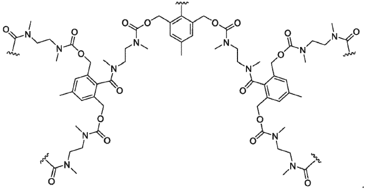 E individuell für jedes Vorkommen eine elektrochemisch aktive Einheit ist, die durch eine der nachstehend dargestellten allgemeinen Formeln definiert ist

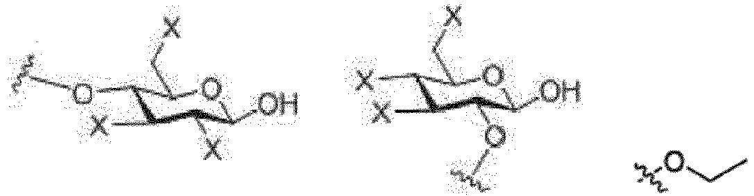
X individuell für jedes Vorkommen -OH, -OMe, -OC(=O)CH3, -F, -CF3, -CH2F, - CHF2, -Cl, -Br, -I, -H oder Sulfonat ist. - Verbindung nach Anspruch 1, wobei E eine Einheit umfasst, die durch eine der nachstehenden allgemeinen Formeln definiert ist


- Verbindung nach Anspruch 1 oder 2, wobei X bei jedem Vorkommen -OH ist.
- Verbindung nach einem der Ansprüche 1 bis 3, wobei R1 und R2 bei jedem Vorkommen -H sind.
- Verbindung nach einem der Ansprüche 1 bis 4, wobei A -OH ist.
- Verbindung nach einem der Ansprüche 1 bis 5, wobei R3 -NHC(=O)CH3 ist.
- Verbindung nach einem der Ansprüche 1 bis 6, wobei n von 2 bis 8 ist.
- Verfahren zum elektrochemischen Nachweisen eines Neuraminidaseenzyms in einer Probe, umfassend:(a) Bereitstellen einer Sonde, umfassend eine Verbindung, die durch die nachstehende Formel I definiert ist, mit einem Substrat für Neuraminidase, das über eine durch das Enzym spaltbare Bindung kovalent an eine elektrochemisch aktive Einheit gebunden ist;
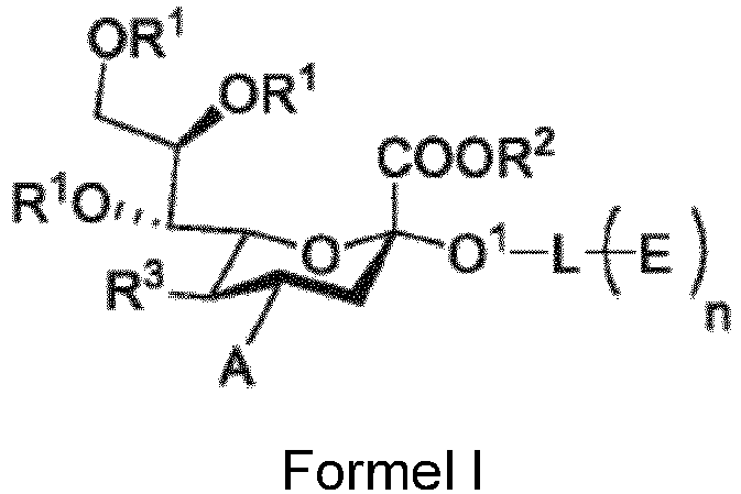 A -OH, -OMe, -OC(=O) CH3, -NH2-NHC(=O)CH3, -NHCH2OH oder -NHC(=NH)NH2 ist;R1 individuell für jedes Vorkommen -H, -Me oder -C(=O)CH3 ist;R2 -H oder -Me ist;R3 -OH, -OMe, -OC(=O)CH3, -NH2, -NHC(=O)CH3 oder -NHCH2OH ist;n eine ganze Zahl von 1 bis 16 ist;L nicht vorhanden ist oder eine Bindungseinheit ist, wobei, wenn L nicht vorhanden ist, O1 ebenfalls nicht vorhanden ist, und n 1 ist, undE individuell für jedes Vorkommen eine elektrochemisch aktive Einheit ist, die durch eine der nachstehend dargestellten allgemeinen Formeln definiert ist


X individuell für jedes Vorkommen -OH, -OMe, -OC(=O)CH3, -F, -CF3, -CH2F, -CHF2, -Cl, -Br, -I, -H oder Sulfonat ist, und(b) Inkontaktbringen der Probe mit der Sonde unter Bedingungen, die für die enzymatische Spaltung der Bindung durch das Enzym wirksam sind;(c) elektrochemisches Nachweisen der durch das Enzym enzymatisch gespaltenen elektrochemisch aktiven Einheit. - Verfahren nach Anspruch 8, wobei die elektrochemisch aktive Einheit eine Einheit umfasst, die elektrochemisch leise ist, wenn sie nicht enzymatisch durch das Enzym gespalten wird.
- Kit, umfassend:(i) eine Vorrichtung zum Sammeln einer Probe, die ein Neuraminidaseenzym von Interesse umfasst; und(ii) eine Sonde, umfassend eine Verbindung, die durch die nachstehende Formel I definiert ist, mit einem Substrat für Neuraminidase, das über eine durch das Enzym spaltbare Bindung kovalent an eine elektrochemisch aktive Einheit gebunden ist
 A -OH, -OMe, -OC(=O) CH3, -NH2-NHC(=O)CH3, -NHCH2OH oder -NHC(=NH)NH2 ist;R1 individuell für jedes Vorkommen -H, -Me oder -C(=O)CH3 ist;R2 -H oder -Me ist;R3 -OH, -OMe, -OC(=O)CH3, -NH2, -NHC(=O)CH3 oder -NHCH2OH ist;n eine ganze Zahl von 1 bis 16 ist;L nicht vorhanden ist oder eine Bindungseinheit ist, wobei, wenn L nicht vorhanden ist, O1 ebenfalls nicht vorhanden ist, und n 1 ist, undE individuell für jedes Vorkommen eine elektrochemisch aktive Einheit ist, die durch eine der nachstehend dargestellten allgemeinen Formeln definiert ist

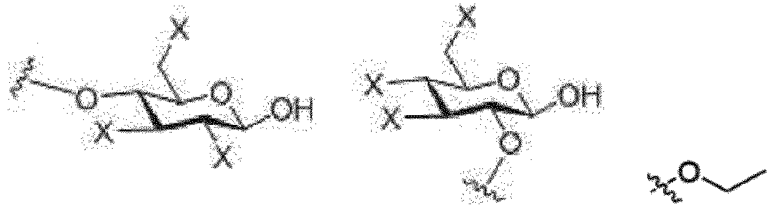
X individuell für jedes Vorkommen -OH, -OMe, -OC(=O)CH3, -F, -CF3, -CH2F, -CHF2, -Cl, -Br, -I, -H oder Sulfonat ist. - Verbindung nach Anspruch 8, wobei E eine Einheit umfasst, die durch eine der nachstehenden allgemeinen Formeln definiert ist


X individuell für jedes Vorkommen -OH, -OMe, -OC(=O)CH3, -F, -CF3, -CH2F, -CHF2, -Cl, -Br, -I, -H oder Sulfonat ist. - Verfahren nach Anspruch 8, wobei E eine durch

- Verfahren nach Anspruch 8, wobei R3 -NHC(=O)CH3 ist.
Applications Claiming Priority (3)
| Application Number | Priority Date | Filing Date | Title |
|---|---|---|---|
| US201462005407P | 2014-05-30 | 2014-05-30 | |
| US201462091205P | 2014-12-12 | 2014-12-12 | |
| PCT/US2015/033512 WO2015184442A1 (en) | 2014-05-30 | 2015-06-01 | Electrochemical methods and compounds for the detection of enzymes |
Publications (3)
| Publication Number | Publication Date |
|---|---|
| EP3149212A1 EP3149212A1 (de) | 2017-04-05 |
| EP3149212A4 EP3149212A4 (de) | 2018-04-25 |
| EP3149212B1 true EP3149212B1 (de) | 2020-08-26 |
Family
ID=54699963
Family Applications (1)
| Application Number | Title | Priority Date | Filing Date |
|---|---|---|---|
| EP15799039.1A Not-in-force EP3149212B1 (de) | 2014-05-30 | 2015-06-01 | Elektrochemische verfahren und verbindungen zur detektion von enzymen |
Country Status (3)
| Country | Link |
|---|---|
| US (2) | US10408783B2 (de) |
| EP (1) | EP3149212B1 (de) |
| WO (1) | WO2015184442A1 (de) |
Families Citing this family (9)
| Publication number | Priority date | Publication date | Assignee | Title |
|---|---|---|---|---|
| US20220017942A1 (en) * | 2016-03-22 | 2022-01-20 | Cleu Diagnostics, Llc | Compositions and methods for determining the presence of active leukocyte cells using an electrochemical assay |
| US20190064165A1 (en) * | 2016-03-22 | 2019-02-28 | Parvizi Surgical Innovation, Llc | Compositions and methods for determining the presence of active leukocyte cells using an electrochemical assay |
| US11104933B1 (en) * | 2016-03-22 | 2021-08-31 | Cleu Diagnostics, Llc | Compositions and methods for determining the presence of active leukocyte cells using an electrochemical assay |
| TWI666439B (zh) * | 2018-09-04 | 2019-07-21 | 東吳大學 | 一種檢測白血球酯酶的檢測裝置 |
| CN111187313A (zh) * | 2020-01-20 | 2020-05-22 | 山西医科大学 | 乙醇非氧化代谢物乙基葡萄糖醛酸苷和硫酸乙酯的制备方法 |
| US12405190B2 (en) * | 2020-02-04 | 2025-09-02 | Steve Naumovski | System and method for detecting airborne pathogens |
| CN113481278B (zh) * | 2021-06-15 | 2022-04-12 | 四川省食品检验研究院 | 同时测定蔗糖酶活力和果聚糖酶活力的方法 |
| US20250228878A1 (en) * | 2021-10-14 | 2025-07-17 | Owlstone Medical Limited | Method for the synthesis of evoc probes |
| CN114736255B (zh) * | 2022-05-11 | 2023-10-27 | 湖南超亟检测技术有限责任公司 | 检测β-半乳糖苷酶的黄酮衍生物荧光探针及其制备方法和应用、试剂盒及其使用方法 |
Family Cites Families (15)
| Publication number | Priority date | Publication date | Assignee | Title |
|---|---|---|---|---|
| DK0507854T3 (da) * | 1989-12-29 | 1997-05-12 | Oklahoma Med Res Found | Fremgangsmåder til diagnosticering af human-influenza og chromogene N-acetylneuraminsyresubstrater, der er modificerede i 4-positionen, til anvendelse deri |
| GB9105406D0 (en) | 1991-03-14 | 1991-05-01 | Ici Plc | Sensor devices |
| US5789385A (en) * | 1993-06-16 | 1998-08-04 | Glycomed Incorporated | Sialyl Lewisx mimetics containing phenyl backbones |
| US5658880A (en) * | 1993-06-16 | 1997-08-19 | Glycomed Incorporated | Sialic acid/fucose based medicaments |
| US6682648B1 (en) * | 1997-08-12 | 2004-01-27 | University Of Southern California | Electrochemical reporter system for detecting analytical immunoassay and molecular biology procedures |
| US5912115A (en) * | 1997-12-12 | 1999-06-15 | Akzo Nobel, N.V. | Evacuated sensor device for detecting microorganisms in blood samples, and method thereof |
| EP1331482A1 (de) | 2002-01-25 | 2003-07-30 | BMS Sensor Technology SA | Konjugat zur Bestimmung von Antigenen mittels Biosensoren |
| EP1866645A2 (de) | 2005-03-31 | 2007-12-19 | Inverness Medical Switzerland GmbH | Elektrochemischer test |
| JP2009523173A (ja) * | 2006-01-09 | 2009-06-18 | チルドレンズ ホスピタル メディカル センター | 様々な病気の治療用のアディポネクチン |
| TW200817520A (en) | 2006-07-17 | 2008-04-16 | Environmental Biotechnology Crc Pty Ltd | Detection of microorganisms and devices therefor |
| US7893272B2 (en) * | 2007-05-15 | 2011-02-22 | Cellex, Inc. | Reagents and kits for detection of influenza virus and the like |
| US20090081249A1 (en) * | 2007-08-27 | 2009-03-26 | Massachusetts Institute Of Technology | Bi-Functional Polymer-Attached Inhibitors of Influenza Virus |
| US8945943B2 (en) | 2010-05-26 | 2015-02-03 | The Board Of Trustees Of The University Of Illinois | Personal glucose meters for detection and quantification of a broad range of analytes |
| US20120122079A1 (en) | 2010-11-12 | 2012-05-17 | Los Alamos National Security, Llc | Infection detection methods and systems and related compounds and compositions |
| KR101314206B1 (ko) | 2012-04-10 | 2013-10-15 | 포항공과대학교 산학협력단 | 병원균 비브리오 콜레라 검출을 위한 탄수화물 칩 및 이의 제조 방법 |
-
2015
- 2015-06-01 US US15/314,817 patent/US10408783B2/en active Active
- 2015-06-01 WO PCT/US2015/033512 patent/WO2015184442A1/en not_active Ceased
- 2015-06-01 EP EP15799039.1A patent/EP3149212B1/de not_active Not-in-force
-
2019
- 2019-09-09 US US16/564,838 patent/US11092565B2/en not_active Expired - Fee Related
Non-Patent Citations (1)
| Title |
|---|
| None * |
Also Published As
| Publication number | Publication date |
|---|---|
| US11092565B2 (en) | 2021-08-17 |
| EP3149212A4 (de) | 2018-04-25 |
| US20170102349A1 (en) | 2017-04-13 |
| US10408783B2 (en) | 2019-09-10 |
| EP3149212A1 (de) | 2017-04-05 |
| WO2015184442A1 (en) | 2015-12-03 |
| US20200103364A1 (en) | 2020-04-02 |
Similar Documents
| Publication | Publication Date | Title |
|---|---|---|
| US11092565B2 (en) | Electrochemical methods and compounds for the detection of enzymes | |
| Fan et al. | Entropy-driven amplified electrochemiluminescence biosensor for RdRp gene of SARS-CoV-2 detection with self-assembled DNA tetrahedron scaffolds | |
| Moccia et al. | Based electrochemical peptide nucleic acid (PNA) biosensor for detection of miRNA-492: a pancreatic ductal adenocarcinoma biomarker | |
| Gao et al. | An enzyme-amplified lateral flow strip biosensor for visual detection of microRNA-224 | |
| Yuan et al. | A novel “signal on-off-super on” sandwich-type aptamer sensor of CRISPR-Cas12a coupled voltage enrichment assay for VEGF detection | |
| Danesh et al. | A novel electrochemical aptasensor based on arch-shape structure of aptamer-complimentary strand conjugate and exonuclease I for sensitive detection of streptomycin | |
| Wang et al. | Highly sensitive homogeneous electrochemical aptasensor for antibiotic residues detection based on dual recycling amplification strategy | |
| Karaballi et al. | Development of an electrochemical surface-enhanced Raman spectroscopy (EC-SERS) aptasensor for direct detection of DNA hybridization | |
| Miao et al. | An electrochemical alkaline phosphatase biosensor fabricated with two DNA probes coupled with λ exonuclease | |
| Dong et al. | Development of a novel ratiometric electrochemical sensor for monitoring β-galactosidase in Parkinson's disease model mice | |
| CN104342488B (zh) | 葡萄糖醛酸转移酶ugt1a1的特异性荧光探针及其应用 | |
| Cui et al. | Highly specific and rapid glycan based amperometric detection of influenza viruses | |
| Das et al. | Detection of enzymes, viruses, and bacteria using glucose meters | |
| Miao et al. | Nuclease assisted target recycling and spherical nucleic acids gold nanoparticles recruitment for ultrasensitive detection of microRNA | |
| Kwak et al. | Probing the neuraminidase activity of influenza virus using a cytolysin a protein nanopore | |
| CN1205011A (zh) | 4,7-二烷氧基n-乙酰基神经氨酸衍生物及其在临床样本中检测流行性感冒a型和b型病毒的方法 | |
| Fang et al. | A versatile CRISPR Cas12a-based point-of-care biosensor enabling convenient glucometer readout for ultrasensitive detection of pathogen nucleic acids | |
| Lomae et al. | Peptide nucleic acid probe-assisted paper-based electrochemical biosensor for multiplexed detection of respiratory viruses | |
| Bekhit et al. | Infection screening in biofluids with glucose test strips | |
| Mubarok et al. | Label-free electrochemical detection of neuraminidase activity: A facile whole blood diagnostic probe for infectious diseases | |
| Zhang et al. | Magnetic bead-liposome hybrids enable sensitive and portable detection of DNA methyltransferase activity using personal glucose meter | |
| CN107602643A (zh) | 一种基于萘酰亚胺的β‑葡萄糖醛酸苷酶的荧光探针及其应用 | |
| Eloi et al. | Toward a point-of-care diagnostic for specific detection of Mycobacterium tuberculosis from sputum samples | |
| CN109824743B (zh) | 一种检测N-乙酰-β-D-氨基葡萄糖苷酶的荧光探针及其应用 | |
| CN107937481B (zh) | 一种β-葡萄糖醛酸苷酶的含吲哚基的荧光探针及其应用 |
Legal Events
| Date | Code | Title | Description |
|---|---|---|---|
| STAA | Information on the status of an ep patent application or granted ep patent |
Free format text: STATUS: THE INTERNATIONAL PUBLICATION HAS BEEN MADE |
|
| PUAI | Public reference made under article 153(3) epc to a published international application that has entered the european phase |
Free format text: ORIGINAL CODE: 0009012 |
|
| STAA | Information on the status of an ep patent application or granted ep patent |
Free format text: STATUS: REQUEST FOR EXAMINATION WAS MADE |
|
| 17P | Request for examination filed |
Effective date: 20161221 |
|
| AK | Designated contracting states |
Kind code of ref document: A1 Designated state(s): AL AT BE BG CH CY CZ DE DK EE ES FI FR GB GR HR HU IE IS IT LI LT LU LV MC MK MT NL NO PL PT RO RS SE SI SK SM TR |
|
| AX | Request for extension of the european patent |
Extension state: BA ME |
|
| RIN1 | Information on inventor provided before grant (corrected) |
Inventor name: VASIREDDI, MUGDHA Inventor name: HE, YUN Inventor name: SWEENEY, JOYCE Inventor name: IYER, SURI SARANATHAN Inventor name: DINH, HIEU Inventor name: ZHANG, XIAOHU Inventor name: DHAWANE, ABASAHEB |
|
| DAV | Request for validation of the european patent (deleted) | ||
| DAX | Request for extension of the european patent (deleted) | ||
| RIC1 | Information provided on ipc code assigned before grant |
Ipc: C07H 15/04 20060101ALI20171218BHEP Ipc: C12Q 1/34 20060101ALI20171218BHEP Ipc: G01N 27/327 20060101ALI20171218BHEP Ipc: C07H 15/18 20060101ALI20171218BHEP Ipc: C07H 3/04 20060101AFI20171218BHEP Ipc: C07H 17/075 20060101ALI20171218BHEP |
|
| REG | Reference to a national code |
Ref country code: DE Ref legal event code: R079 Ref document number: 602015058075 Country of ref document: DE Free format text: PREVIOUS MAIN CLASS: C12Q0001700000 Ipc: C07H0003040000 |
|
| A4 | Supplementary search report drawn up and despatched |
Effective date: 20180328 |
|
| RIC1 | Information provided on ipc code assigned before grant |
Ipc: G01N 27/327 20060101ALI20180322BHEP Ipc: C07H 15/04 20060101ALI20180322BHEP Ipc: C07H 15/18 20060101ALI20180322BHEP Ipc: C07H 3/04 20060101AFI20180322BHEP Ipc: C07H 17/075 20060101ALI20180322BHEP Ipc: C12Q 1/34 20060101ALI20180322BHEP |
|
| STAA | Information on the status of an ep patent application or granted ep patent |
Free format text: STATUS: EXAMINATION IS IN PROGRESS |
|
| 17Q | First examination report despatched |
Effective date: 20181204 |
|
| GRAP | Despatch of communication of intention to grant a patent |
Free format text: ORIGINAL CODE: EPIDOSNIGR1 |
|
| STAA | Information on the status of an ep patent application or granted ep patent |
Free format text: STATUS: GRANT OF PATENT IS INTENDED |
|
| INTG | Intention to grant announced |
Effective date: 20200316 |
|
| GRAS | Grant fee paid |
Free format text: ORIGINAL CODE: EPIDOSNIGR3 |
|
| GRAA | (expected) grant |
Free format text: ORIGINAL CODE: 0009210 |
|
| STAA | Information on the status of an ep patent application or granted ep patent |
Free format text: STATUS: THE PATENT HAS BEEN GRANTED |
|
| AK | Designated contracting states |
Kind code of ref document: B1 Designated state(s): AL AT BE BG CH CY CZ DE DK EE ES FI FR GB GR HR HU IE IS IT LI LT LU LV MC MK MT NL NO PL PT RO RS SE SI SK SM TR |
|
| REG | Reference to a national code |
Ref country code: GB Ref legal event code: FG4D |
|
| REG | Reference to a national code |
Ref country code: CH Ref legal event code: EP |
|
| REG | Reference to a national code |
Ref country code: DE Ref legal event code: R096 Ref document number: 602015058075 Country of ref document: DE |
|
| REG | Reference to a national code |
Ref country code: AT Ref legal event code: REF Ref document number: 1306283 Country of ref document: AT Kind code of ref document: T Effective date: 20200915 |
|
| REG | Reference to a national code |
Ref country code: IE Ref legal event code: FG4D |
|
| REG | Reference to a national code |
Ref country code: LT Ref legal event code: MG4D |
|
| PG25 | Lapsed in a contracting state [announced via postgrant information from national office to epo] |
Ref country code: SE Free format text: LAPSE BECAUSE OF FAILURE TO SUBMIT A TRANSLATION OF THE DESCRIPTION OR TO PAY THE FEE WITHIN THE PRESCRIBED TIME-LIMIT Effective date: 20200826 Ref country code: HR Free format text: LAPSE BECAUSE OF FAILURE TO SUBMIT A TRANSLATION OF THE DESCRIPTION OR TO PAY THE FEE WITHIN THE PRESCRIBED TIME-LIMIT Effective date: 20200826 Ref country code: PT Free format text: LAPSE BECAUSE OF FAILURE TO SUBMIT A TRANSLATION OF THE DESCRIPTION OR TO PAY THE FEE WITHIN THE PRESCRIBED TIME-LIMIT Effective date: 20201228 Ref country code: LT Free format text: LAPSE BECAUSE OF FAILURE TO SUBMIT A TRANSLATION OF THE DESCRIPTION OR TO PAY THE FEE WITHIN THE PRESCRIBED TIME-LIMIT Effective date: 20200826 Ref country code: FI Free format text: LAPSE BECAUSE OF FAILURE TO SUBMIT A TRANSLATION OF THE DESCRIPTION OR TO PAY THE FEE WITHIN THE PRESCRIBED TIME-LIMIT Effective date: 20200826 Ref country code: BG Free format text: LAPSE BECAUSE OF FAILURE TO SUBMIT A TRANSLATION OF THE DESCRIPTION OR TO PAY THE FEE WITHIN THE PRESCRIBED TIME-LIMIT Effective date: 20201126 Ref country code: GR Free format text: LAPSE BECAUSE OF FAILURE TO SUBMIT A TRANSLATION OF THE DESCRIPTION OR TO PAY THE FEE WITHIN THE PRESCRIBED TIME-LIMIT Effective date: 20201127 Ref country code: NO Free format text: LAPSE BECAUSE OF FAILURE TO SUBMIT A TRANSLATION OF THE DESCRIPTION OR TO PAY THE FEE WITHIN THE PRESCRIBED TIME-LIMIT Effective date: 20201126 |
|
| REG | Reference to a national code |
Ref country code: NL Ref legal event code: MP Effective date: 20200826 |
|
| REG | Reference to a national code |
Ref country code: AT Ref legal event code: MK05 Ref document number: 1306283 Country of ref document: AT Kind code of ref document: T Effective date: 20200826 |
|
| PG25 | Lapsed in a contracting state [announced via postgrant information from national office to epo] |
Ref country code: RS Free format text: LAPSE BECAUSE OF FAILURE TO SUBMIT A TRANSLATION OF THE DESCRIPTION OR TO PAY THE FEE WITHIN THE PRESCRIBED TIME-LIMIT Effective date: 20200826 Ref country code: NL Free format text: LAPSE BECAUSE OF FAILURE TO SUBMIT A TRANSLATION OF THE DESCRIPTION OR TO PAY THE FEE WITHIN THE PRESCRIBED TIME-LIMIT Effective date: 20200826 Ref country code: PL Free format text: LAPSE BECAUSE OF FAILURE TO SUBMIT A TRANSLATION OF THE DESCRIPTION OR TO PAY THE FEE WITHIN THE PRESCRIBED TIME-LIMIT Effective date: 20200826 Ref country code: LV Free format text: LAPSE BECAUSE OF FAILURE TO SUBMIT A TRANSLATION OF THE DESCRIPTION OR TO PAY THE FEE WITHIN THE PRESCRIBED TIME-LIMIT Effective date: 20200826 Ref country code: IS Free format text: LAPSE BECAUSE OF FAILURE TO SUBMIT A TRANSLATION OF THE DESCRIPTION OR TO PAY THE FEE WITHIN THE PRESCRIBED TIME-LIMIT Effective date: 20201226 |
|
| PG25 | Lapsed in a contracting state [announced via postgrant information from national office to epo] |
Ref country code: EE Free format text: LAPSE BECAUSE OF FAILURE TO SUBMIT A TRANSLATION OF THE DESCRIPTION OR TO PAY THE FEE WITHIN THE PRESCRIBED TIME-LIMIT Effective date: 20200826 Ref country code: SM Free format text: LAPSE BECAUSE OF FAILURE TO SUBMIT A TRANSLATION OF THE DESCRIPTION OR TO PAY THE FEE WITHIN THE PRESCRIBED TIME-LIMIT Effective date: 20200826 Ref country code: RO Free format text: LAPSE BECAUSE OF FAILURE TO SUBMIT A TRANSLATION OF THE DESCRIPTION OR TO PAY THE FEE WITHIN THE PRESCRIBED TIME-LIMIT Effective date: 20200826 Ref country code: CZ Free format text: LAPSE BECAUSE OF FAILURE TO SUBMIT A TRANSLATION OF THE DESCRIPTION OR TO PAY THE FEE WITHIN THE PRESCRIBED TIME-LIMIT Effective date: 20200826 Ref country code: DK Free format text: LAPSE BECAUSE OF FAILURE TO SUBMIT A TRANSLATION OF THE DESCRIPTION OR TO PAY THE FEE WITHIN THE PRESCRIBED TIME-LIMIT Effective date: 20200826 |
|
| REG | Reference to a national code |
Ref country code: DE Ref legal event code: R097 Ref document number: 602015058075 Country of ref document: DE |
|
| PG25 | Lapsed in a contracting state [announced via postgrant information from national office to epo] |
Ref country code: AT Free format text: LAPSE BECAUSE OF FAILURE TO SUBMIT A TRANSLATION OF THE DESCRIPTION OR TO PAY THE FEE WITHIN THE PRESCRIBED TIME-LIMIT Effective date: 20200826 Ref country code: AL Free format text: LAPSE BECAUSE OF FAILURE TO SUBMIT A TRANSLATION OF THE DESCRIPTION OR TO PAY THE FEE WITHIN THE PRESCRIBED TIME-LIMIT Effective date: 20200826 Ref country code: ES Free format text: LAPSE BECAUSE OF FAILURE TO SUBMIT A TRANSLATION OF THE DESCRIPTION OR TO PAY THE FEE WITHIN THE PRESCRIBED TIME-LIMIT Effective date: 20200826 |
|
| PG25 | Lapsed in a contracting state [announced via postgrant information from national office to epo] |
Ref country code: SK Free format text: LAPSE BECAUSE OF FAILURE TO SUBMIT A TRANSLATION OF THE DESCRIPTION OR TO PAY THE FEE WITHIN THE PRESCRIBED TIME-LIMIT Effective date: 20200826 |
|
| PLBE | No opposition filed within time limit |
Free format text: ORIGINAL CODE: 0009261 |
|
| STAA | Information on the status of an ep patent application or granted ep patent |
Free format text: STATUS: NO OPPOSITION FILED WITHIN TIME LIMIT |
|
| PG25 | Lapsed in a contracting state [announced via postgrant information from national office to epo] |
Ref country code: IT Free format text: LAPSE BECAUSE OF FAILURE TO SUBMIT A TRANSLATION OF THE DESCRIPTION OR TO PAY THE FEE WITHIN THE PRESCRIBED TIME-LIMIT Effective date: 20200826 |
|
| 26N | No opposition filed |
Effective date: 20210527 |
|
| PG25 | Lapsed in a contracting state [announced via postgrant information from national office to epo] |
Ref country code: SI Free format text: LAPSE BECAUSE OF FAILURE TO SUBMIT A TRANSLATION OF THE DESCRIPTION OR TO PAY THE FEE WITHIN THE PRESCRIBED TIME-LIMIT Effective date: 20200826 |
|
| PG25 | Lapsed in a contracting state [announced via postgrant information from national office to epo] |
Ref country code: MC Free format text: LAPSE BECAUSE OF FAILURE TO SUBMIT A TRANSLATION OF THE DESCRIPTION OR TO PAY THE FEE WITHIN THE PRESCRIBED TIME-LIMIT Effective date: 20200826 |
|
| REG | Reference to a national code |
Ref country code: CH Ref legal event code: PL |
|
| REG | Reference to a national code |
Ref country code: BE Ref legal event code: MM Effective date: 20210630 |
|
| PG25 | Lapsed in a contracting state [announced via postgrant information from national office to epo] |
Ref country code: LU Free format text: LAPSE BECAUSE OF NON-PAYMENT OF DUE FEES Effective date: 20210601 |
|
| PG25 | Lapsed in a contracting state [announced via postgrant information from national office to epo] |
Ref country code: LI Free format text: LAPSE BECAUSE OF NON-PAYMENT OF DUE FEES Effective date: 20210630 Ref country code: IE Free format text: LAPSE BECAUSE OF NON-PAYMENT OF DUE FEES Effective date: 20210601 Ref country code: CH Free format text: LAPSE BECAUSE OF NON-PAYMENT OF DUE FEES Effective date: 20210630 |
|
| PG25 | Lapsed in a contracting state [announced via postgrant information from national office to epo] |
Ref country code: BE Free format text: LAPSE BECAUSE OF NON-PAYMENT OF DUE FEES Effective date: 20210630 |
|
| PG25 | Lapsed in a contracting state [announced via postgrant information from national office to epo] |
Ref country code: HU Free format text: LAPSE BECAUSE OF FAILURE TO SUBMIT A TRANSLATION OF THE DESCRIPTION OR TO PAY THE FEE WITHIN THE PRESCRIBED TIME-LIMIT; INVALID AB INITIO Effective date: 20150601 |
|
| PG25 | Lapsed in a contracting state [announced via postgrant information from national office to epo] |
Ref country code: CY Free format text: LAPSE BECAUSE OF FAILURE TO SUBMIT A TRANSLATION OF THE DESCRIPTION OR TO PAY THE FEE WITHIN THE PRESCRIBED TIME-LIMIT Effective date: 20200826 |
|
| P01 | Opt-out of the competence of the unified patent court (upc) registered |
Effective date: 20230530 |
|
| PGFP | Annual fee paid to national office [announced via postgrant information from national office to epo] |
Ref country code: FR Payment date: 20230626 Year of fee payment: 9 Ref country code: DE Payment date: 20230626 Year of fee payment: 9 |
|
| PGFP | Annual fee paid to national office [announced via postgrant information from national office to epo] |
Ref country code: GB Payment date: 20230627 Year of fee payment: 9 |
|
| PG25 | Lapsed in a contracting state [announced via postgrant information from national office to epo] |
Ref country code: MK Free format text: LAPSE BECAUSE OF FAILURE TO SUBMIT A TRANSLATION OF THE DESCRIPTION OR TO PAY THE FEE WITHIN THE PRESCRIBED TIME-LIMIT Effective date: 20200826 |
|
| PG25 | Lapsed in a contracting state [announced via postgrant information from national office to epo] |
Ref country code: TR Free format text: LAPSE BECAUSE OF FAILURE TO SUBMIT A TRANSLATION OF THE DESCRIPTION OR TO PAY THE FEE WITHIN THE PRESCRIBED TIME-LIMIT Effective date: 20200826 |
|
| PG25 | Lapsed in a contracting state [announced via postgrant information from national office to epo] |
Ref country code: MT Free format text: LAPSE BECAUSE OF FAILURE TO SUBMIT A TRANSLATION OF THE DESCRIPTION OR TO PAY THE FEE WITHIN THE PRESCRIBED TIME-LIMIT Effective date: 20200826 |
|
| REG | Reference to a national code |
Ref country code: DE Ref legal event code: R119 Ref document number: 602015058075 Country of ref document: DE |
|
| GBPC | Gb: european patent ceased through non-payment of renewal fee |
Effective date: 20240601 |
|
| PG25 | Lapsed in a contracting state [announced via postgrant information from national office to epo] |
Ref country code: DE Free format text: LAPSE BECAUSE OF NON-PAYMENT OF DUE FEES Effective date: 20250101 |
|
| PG25 | Lapsed in a contracting state [announced via postgrant information from national office to epo] |
Ref country code: FR Free format text: LAPSE BECAUSE OF NON-PAYMENT OF DUE FEES Effective date: 20240630 |
|
| PG25 | Lapsed in a contracting state [announced via postgrant information from national office to epo] |
Ref country code: GB Free format text: LAPSE BECAUSE OF NON-PAYMENT OF DUE FEES Effective date: 20240601 |