EP3170895A1 - Méthode de production de cellules souches pluripotentes possédant un gène codant pour le récepteur des cellules t spécifique d'un antigène - Google Patents
Méthode de production de cellules souches pluripotentes possédant un gène codant pour le récepteur des cellules t spécifique d'un antigène Download PDFInfo
- Publication number
- EP3170895A1 EP3170895A1 EP15822337.0A EP15822337A EP3170895A1 EP 3170895 A1 EP3170895 A1 EP 3170895A1 EP 15822337 A EP15822337 A EP 15822337A EP 3170895 A1 EP3170895 A1 EP 3170895A1
- Authority
- EP
- European Patent Office
- Prior art keywords
- cells
- cell
- antigen
- pluripotent stem
- specific
- Prior art date
- Legal status (The legal status is an assumption and is not a legal conclusion. Google has not performed a legal analysis and makes no representation as to the accuracy of the status listed.)
- Withdrawn
Links
- 239000000427 antigen Substances 0.000 title claims abstract description 127
- 108091007433 antigens Proteins 0.000 title claims abstract description 126
- 102000036639 antigens Human genes 0.000 title claims abstract description 126
- 210000001778 pluripotent stem cell Anatomy 0.000 title claims abstract description 44
- 238000004519 manufacturing process Methods 0.000 title description 3
- 108700042075 T-Cell Receptor Genes Proteins 0.000 title 1
- 210000004027 cell Anatomy 0.000 claims abstract description 435
- 210000001744 T-lymphocyte Anatomy 0.000 claims abstract description 233
- 101150084041 WT1 gene Proteins 0.000 claims abstract description 88
- 108700020467 WT1 Proteins 0.000 claims abstract description 85
- 108091008874 T cell receptors Proteins 0.000 claims abstract description 64
- 238000000034 method Methods 0.000 claims abstract description 59
- 241000282414 Homo sapiens Species 0.000 claims abstract description 58
- 108090000623 proteins and genes Proteins 0.000 claims abstract description 45
- 102000016266 T-Cell Antigen Receptors Human genes 0.000 claims abstract description 37
- 206010028980 Neoplasm Diseases 0.000 claims abstract description 34
- 238000009169 immunotherapy Methods 0.000 claims abstract description 29
- 230000001939 inductive effect Effects 0.000 claims abstract description 19
- 201000010099 disease Diseases 0.000 claims abstract description 14
- 208000037265 diseases, disorders, signs and symptoms Diseases 0.000 claims abstract description 14
- 208000035473 Communicable disease Diseases 0.000 claims abstract description 8
- 241000701044 Human gammaherpesvirus 4 Species 0.000 claims abstract 11
- 241000700605 Viruses Species 0.000 claims description 46
- 201000011510 cancer Diseases 0.000 claims description 22
- 238000012258 culturing Methods 0.000 claims description 8
- 102000054766 genetic haplotypes Human genes 0.000 claims description 8
- 210000004698 lymphocyte Anatomy 0.000 claims description 6
- 208000015181 infectious disease Diseases 0.000 claims description 5
- 208000023275 Autoimmune disease Diseases 0.000 claims description 2
- 206010020751 Hypersensitivity Diseases 0.000 claims description 2
- 230000007815 allergy Effects 0.000 claims description 2
- 101001136981 Homo sapiens Proteasome subunit beta type-9 Proteins 0.000 claims 1
- 102100035764 Proteasome subunit beta type-9 Human genes 0.000 claims 1
- 208000026935 allergic disease Diseases 0.000 claims 1
- 239000002609 medium Substances 0.000 description 104
- 102100022748 Wilms tumor protein Human genes 0.000 description 76
- 108090000765 processed proteins & peptides Proteins 0.000 description 65
- 101710192606 Latent membrane protein 2 Proteins 0.000 description 53
- 239000013598 vector Substances 0.000 description 32
- 210000001151 cytotoxic T lymphocyte Anatomy 0.000 description 24
- 239000006285 cell suspension Substances 0.000 description 23
- 230000008672 reprogramming Effects 0.000 description 19
- 210000000130 stem cell Anatomy 0.000 description 19
- 239000000725 suspension Substances 0.000 description 17
- 239000012737 fresh medium Substances 0.000 description 16
- 102000003812 Interleukin-15 Human genes 0.000 description 15
- 210000004443 dendritic cell Anatomy 0.000 description 14
- UCSJYZPVAKXKNQ-HZYVHMACSA-N streptomycin Chemical compound CN[C@H]1[C@H](O)[C@@H](O)[C@H](CO)O[C@H]1O[C@@H]1[C@](C=O)(O)[C@H](C)O[C@H]1O[C@@H]1[C@@H](NC(N)=N)[C@H](O)[C@@H](NC(N)=N)[C@H](O)[C@H]1O UCSJYZPVAKXKNQ-HZYVHMACSA-N 0.000 description 14
- 238000012413 Fluorescence activated cell sorting analysis Methods 0.000 description 13
- 230000031942 natural killer cell mediated cytotoxicity Effects 0.000 description 13
- 108010002586 Interleukin-7 Proteins 0.000 description 12
- XJMOSONTPMZWPB-UHFFFAOYSA-M propidium iodide Chemical compound [I-].[I-].C12=CC(N)=CC=C2C2=CC=C(N)C=C2[N+](CCC[N+](C)(CC)CC)=C1C1=CC=CC=C1 XJMOSONTPMZWPB-UHFFFAOYSA-M 0.000 description 12
- 210000001082 somatic cell Anatomy 0.000 description 12
- 102000017420 CD3 protein, epsilon/gamma/delta subunit Human genes 0.000 description 11
- 102000004127 Cytokines Human genes 0.000 description 11
- 108090000695 Cytokines Proteins 0.000 description 11
- 102100036462 Delta-like protein 1 Human genes 0.000 description 11
- 101000928537 Homo sapiens Delta-like protein 1 Proteins 0.000 description 11
- 230000004069 differentiation Effects 0.000 description 11
- 210000005259 peripheral blood Anatomy 0.000 description 11
- 239000011886 peripheral blood Substances 0.000 description 11
- 239000006143 cell culture medium Substances 0.000 description 10
- 230000001472 cytotoxic effect Effects 0.000 description 10
- 210000003958 hematopoietic stem cell Anatomy 0.000 description 10
- 239000000243 solution Substances 0.000 description 10
- 238000002054 transplantation Methods 0.000 description 10
- 230000006698 induction Effects 0.000 description 9
- 210000001519 tissue Anatomy 0.000 description 9
- 210000001239 CD8-positive, alpha-beta cytotoxic T lymphocyte Anatomy 0.000 description 8
- 108020004459 Small interfering RNA Proteins 0.000 description 8
- 239000003112 inhibitor Substances 0.000 description 8
- 208000032839 leukemia Diseases 0.000 description 8
- RXWNCPJZOCPEPQ-NVWDDTSBSA-N puromycin Chemical compound C1=CC(OC)=CC=C1C[C@H](N)C(=O)N[C@H]1[C@@H](O)[C@H](N2C3=NC=NC(=C3N=C2)N(C)C)O[C@@H]1CO RXWNCPJZOCPEPQ-NVWDDTSBSA-N 0.000 description 8
- 230000000638 stimulation Effects 0.000 description 8
- 229930182555 Penicillin Natural products 0.000 description 7
- JGSARLDLIJGVTE-MBNYWOFBSA-N Penicillin G Chemical compound N([C@H]1[C@H]2SC([C@@H](N2C1=O)C(O)=O)(C)C)C(=O)CC1=CC=CC=C1 JGSARLDLIJGVTE-MBNYWOFBSA-N 0.000 description 7
- 238000004113 cell culture Methods 0.000 description 7
- 229940079593 drug Drugs 0.000 description 7
- 239000003814 drug Substances 0.000 description 7
- 239000000203 mixture Substances 0.000 description 7
- 239000008188 pellet Substances 0.000 description 7
- 229940049954 penicillin Drugs 0.000 description 7
- 238000002360 preparation method Methods 0.000 description 7
- 229960005322 streptomycin Drugs 0.000 description 7
- 108010002350 Interleukin-2 Proteins 0.000 description 6
- 241000711408 Murine respirovirus Species 0.000 description 6
- 230000003013 cytotoxicity Effects 0.000 description 6
- 231100000135 cytotoxicity Toxicity 0.000 description 6
- 239000012636 effector Substances 0.000 description 6
- 238000000684 flow cytometry Methods 0.000 description 6
- 210000003819 peripheral blood mononuclear cell Anatomy 0.000 description 6
- 239000013612 plasmid Substances 0.000 description 6
- 210000002536 stromal cell Anatomy 0.000 description 6
- 108090000672 Annexin A5 Proteins 0.000 description 5
- 102000004121 Annexin A5 Human genes 0.000 description 5
- 239000006144 Dulbecco’s modified Eagle's medium Substances 0.000 description 5
- 206010025323 Lymphomas Diseases 0.000 description 5
- 108091027967 Small hairpin RNA Proteins 0.000 description 5
- 238000003501 co-culture Methods 0.000 description 5
- 239000002299 complementary DNA Substances 0.000 description 5
- 238000001415 gene therapy Methods 0.000 description 5
- 238000011534 incubation Methods 0.000 description 5
- 238000002560 therapeutic procedure Methods 0.000 description 5
- 102000029816 Collagenase Human genes 0.000 description 4
- 108060005980 Collagenase Proteins 0.000 description 4
- 206010059866 Drug resistance Diseases 0.000 description 4
- -1 Fbxl5 Proteins 0.000 description 4
- 108010010803 Gelatin Proteins 0.000 description 4
- 102000053187 Glucuronidase Human genes 0.000 description 4
- 108010060309 Glucuronidase Proteins 0.000 description 4
- 208000009329 Graft vs Host Disease Diseases 0.000 description 4
- 102000004144 Green Fluorescent Proteins Human genes 0.000 description 4
- 108010043121 Green Fluorescent Proteins Proteins 0.000 description 4
- 239000012981 Hank's balanced salt solution Substances 0.000 description 4
- 241000713666 Lentivirus Species 0.000 description 4
- NWIBSHFKIJFRCO-WUDYKRTCSA-N Mytomycin Chemical compound C1N2C(C(C(C)=C(N)C3=O)=O)=C3[C@@H](COC(N)=O)[C@@]2(OC)[C@@H]2[C@H]1N2 NWIBSHFKIJFRCO-WUDYKRTCSA-N 0.000 description 4
- NIJJYAXOARWZEE-UHFFFAOYSA-N Valproic acid Chemical compound CCCC(C(O)=O)CCC NIJJYAXOARWZEE-UHFFFAOYSA-N 0.000 description 4
- 210000004507 artificial chromosome Anatomy 0.000 description 4
- 210000001106 artificial yeast chromosome Anatomy 0.000 description 4
- 210000004369 blood Anatomy 0.000 description 4
- 239000008280 blood Substances 0.000 description 4
- 239000011575 calcium Substances 0.000 description 4
- 229960002424 collagenase Drugs 0.000 description 4
- 229920000159 gelatin Polymers 0.000 description 4
- 239000008273 gelatin Substances 0.000 description 4
- 235000019322 gelatine Nutrition 0.000 description 4
- 235000011852 gelatine desserts Nutrition 0.000 description 4
- 208000024908 graft versus host disease Diseases 0.000 description 4
- 239000005090 green fluorescent protein Substances 0.000 description 4
- 210000000688 human artificial chromosome Anatomy 0.000 description 4
- 238000001638 lipofection Methods 0.000 description 4
- 238000012423 maintenance Methods 0.000 description 4
- 239000003550 marker Substances 0.000 description 4
- 238000000520 microinjection Methods 0.000 description 4
- 210000001616 monocyte Anatomy 0.000 description 4
- 102000004196 processed proteins & peptides Human genes 0.000 description 4
- 102000004169 proteins and genes Human genes 0.000 description 4
- 229950010131 puromycin Drugs 0.000 description 4
- 230000009258 tissue cross reactivity Effects 0.000 description 4
- 108091032973 (ribonucleotides)n+m Proteins 0.000 description 3
- KCXVZYZYPLLWCC-UHFFFAOYSA-N EDTA Chemical compound OC(=O)CN(CC(O)=O)CCN(CC(O)=O)CC(O)=O KCXVZYZYPLLWCC-UHFFFAOYSA-N 0.000 description 3
- 108010017213 Granulocyte-Macrophage Colony-Stimulating Factor Proteins 0.000 description 3
- 102100039620 Granulocyte-macrophage colony-stimulating factor Human genes 0.000 description 3
- 102000003964 Histone deacetylase Human genes 0.000 description 3
- 108090000353 Histone deacetylase Proteins 0.000 description 3
- 108090000978 Interleukin-4 Proteins 0.000 description 3
- 241000288906 Primates Species 0.000 description 3
- 102000004142 Trypsin Human genes 0.000 description 3
- 108090000631 Trypsin Proteins 0.000 description 3
- 150000001413 amino acids Chemical group 0.000 description 3
- 230000003321 amplification Effects 0.000 description 3
- 230000000694 effects Effects 0.000 description 3
- 230000003203 everyday effect Effects 0.000 description 3
- 238000001943 fluorescence-activated cell sorting Methods 0.000 description 3
- 201000006747 infectious mononucleosis Diseases 0.000 description 3
- 238000002826 magnetic-activated cell sorting Methods 0.000 description 3
- 238000003199 nucleic acid amplification method Methods 0.000 description 3
- 239000004055 small Interfering RNA Substances 0.000 description 3
- 239000006228 supernatant Substances 0.000 description 3
- 230000008685 targeting Effects 0.000 description 3
- 239000012588 trypsin Substances 0.000 description 3
- FWMNVWWHGCHHJJ-SKKKGAJSSA-N 4-amino-1-[(2r)-6-amino-2-[[(2r)-2-[[(2r)-2-[[(2r)-2-amino-3-phenylpropanoyl]amino]-3-phenylpropanoyl]amino]-4-methylpentanoyl]amino]hexanoyl]piperidine-4-carboxylic acid Chemical compound C([C@H](C(=O)N[C@H](CC(C)C)C(=O)N[C@H](CCCCN)C(=O)N1CCC(N)(CC1)C(O)=O)NC(=O)[C@H](N)CC=1C=CC=CC=1)C1=CC=CC=C1 FWMNVWWHGCHHJJ-SKKKGAJSSA-N 0.000 description 2
- 102100038511 AT-rich interactive domain-containing protein 3A Human genes 0.000 description 2
- 208000011691 Burkitt lymphomas Diseases 0.000 description 2
- FERIUCNNQQJTOY-UHFFFAOYSA-N Butyric acid Chemical compound CCCC(O)=O FERIUCNNQQJTOY-UHFFFAOYSA-N 0.000 description 2
- 241000702421 Dependoparvovirus Species 0.000 description 2
- 108010053187 Diphtheria Toxin Proteins 0.000 description 2
- 108700007698 Genetic Terminator Regions Proteins 0.000 description 2
- 102100028972 HLA class I histocompatibility antigen, A alpha chain Human genes 0.000 description 2
- 108010075704 HLA-A Antigens Proteins 0.000 description 2
- 102100039996 Histone deacetylase 1 Human genes 0.000 description 2
- 101000808887 Homo sapiens AT-rich interactive domain-containing protein 3A Proteins 0.000 description 2
- 101001035024 Homo sapiens Histone deacetylase 1 Proteins 0.000 description 2
- 101001100327 Homo sapiens RNA-binding protein 45 Proteins 0.000 description 2
- 101000914514 Homo sapiens T-cell-specific surface glycoprotein CD28 Proteins 0.000 description 2
- 101001074035 Homo sapiens Zinc finger protein GLI2 Proteins 0.000 description 2
- ZDXPYRJPNDTMRX-VKHMYHEASA-N L-glutamine Chemical compound OC(=O)[C@@H](N)CCC(N)=O ZDXPYRJPNDTMRX-VKHMYHEASA-N 0.000 description 2
- 229930182816 L-glutamine Natural products 0.000 description 2
- 108700042652 LMP-2 Proteins 0.000 description 2
- 108700018351 Major Histocompatibility Complex Proteins 0.000 description 2
- 102100038823 RNA-binding protein 45 Human genes 0.000 description 2
- 239000006146 Roswell Park Memorial Institute medium Substances 0.000 description 2
- 101150086694 SLC22A3 gene Proteins 0.000 description 2
- 208000005718 Stomach Neoplasms Diseases 0.000 description 2
- 102100027213 T-cell-specific surface glycoprotein CD28 Human genes 0.000 description 2
- 108020004440 Thymidine kinase Proteins 0.000 description 2
- 102100035558 Zinc finger protein GLI2 Human genes 0.000 description 2
- 230000004913 activation Effects 0.000 description 2
- 239000012190 activator Substances 0.000 description 2
- 229960000723 ampicillin Drugs 0.000 description 2
- AVKUERGKIZMTKX-NJBDSQKTSA-N ampicillin Chemical compound C1([C@@H](N)C(=O)N[C@H]2[C@H]3SC([C@@H](N3C2=O)C(O)=O)(C)C)=CC=CC=C1 AVKUERGKIZMTKX-NJBDSQKTSA-N 0.000 description 2
- 230000001093 anti-cancer Effects 0.000 description 2
- 210000004436 artificial bacterial chromosome Anatomy 0.000 description 2
- QVGXLLKOCUKJST-UHFFFAOYSA-N atomic oxygen Chemical compound [O] QVGXLLKOCUKJST-UHFFFAOYSA-N 0.000 description 2
- 239000011324 bead Substances 0.000 description 2
- 230000008901 benefit Effects 0.000 description 2
- 210000000601 blood cell Anatomy 0.000 description 2
- 210000001185 bone marrow Anatomy 0.000 description 2
- 238000010322 bone marrow transplantation Methods 0.000 description 2
- 238000002659 cell therapy Methods 0.000 description 2
- 239000012228 culture supernatant Substances 0.000 description 2
- 210000004748 cultured cell Anatomy 0.000 description 2
- 231100000433 cytotoxic Toxicity 0.000 description 2
- 230000006378 damage Effects 0.000 description 2
- XEYBRNLFEZDVAW-ARSRFYASSA-N dinoprostone Chemical compound CCCCC[C@H](O)\C=C\[C@H]1[C@H](O)CC(=O)[C@@H]1C\C=C/CCCC(O)=O XEYBRNLFEZDVAW-ARSRFYASSA-N 0.000 description 2
- 229960002986 dinoprostone Drugs 0.000 description 2
- 210000001671 embryonic stem cell Anatomy 0.000 description 2
- 238000005516 engineering process Methods 0.000 description 2
- 239000003623 enhancer Substances 0.000 description 2
- 230000002708 enhancing effect Effects 0.000 description 2
- 239000003797 essential amino acid Substances 0.000 description 2
- 235000020776 essential amino acid Nutrition 0.000 description 2
- 210000004700 fetal blood Anatomy 0.000 description 2
- 230000006870 function Effects 0.000 description 2
- 206010017758 gastric cancer Diseases 0.000 description 2
- 229960002743 glutamine Drugs 0.000 description 2
- 239000001963 growth medium Substances 0.000 description 2
- 210000002443 helper t lymphocyte Anatomy 0.000 description 2
- 210000002861 immature t-cell Anatomy 0.000 description 2
- 238000011835 investigation Methods 0.000 description 2
- 238000002955 isolation Methods 0.000 description 2
- 108010028309 kalinin Proteins 0.000 description 2
- 229930027917 kanamycin Natural products 0.000 description 2
- 229960000318 kanamycin Drugs 0.000 description 2
- SBUJHOSQTJFQJX-NOAMYHISSA-N kanamycin Chemical compound O[C@@H]1[C@@H](O)[C@H](O)[C@@H](CN)O[C@@H]1O[C@H]1[C@H](O)[C@@H](O[C@@H]2[C@@H]([C@@H](N)[C@H](O)[C@@H](CO)O2)O)[C@H](N)C[C@@H]1N SBUJHOSQTJFQJX-NOAMYHISSA-N 0.000 description 2
- 229930182823 kanamycin A Natural products 0.000 description 2
- 239000002502 liposome Substances 0.000 description 2
- 230000003211 malignant effect Effects 0.000 description 2
- 210000004962 mammalian cell Anatomy 0.000 description 2
- 210000003071 memory t lymphocyte Anatomy 0.000 description 2
- 239000011325 microbead Substances 0.000 description 2
- 229960004857 mitomycin Drugs 0.000 description 2
- 108091027963 non-coding RNA Proteins 0.000 description 2
- 102000042567 non-coding RNA Human genes 0.000 description 2
- 108020004707 nucleic acids Proteins 0.000 description 2
- 102000039446 nucleic acids Human genes 0.000 description 2
- 150000007523 nucleic acids Chemical class 0.000 description 2
- 229910052760 oxygen Inorganic materials 0.000 description 2
- 239000001301 oxygen Substances 0.000 description 2
- 230000008488 polyadenylation Effects 0.000 description 2
- 239000000047 product Substances 0.000 description 2
- XEYBRNLFEZDVAW-UHFFFAOYSA-N prostaglandin E2 Natural products CCCCCC(O)C=CC1C(O)CC(=O)C1CC=CCCCC(O)=O XEYBRNLFEZDVAW-UHFFFAOYSA-N 0.000 description 2
- 230000009257 reactivity Effects 0.000 description 2
- 230000001105 regulatory effect Effects 0.000 description 2
- 238000011160 research Methods 0.000 description 2
- 230000028327 secretion Effects 0.000 description 2
- 210000002966 serum Anatomy 0.000 description 2
- 239000012679 serum free medium Substances 0.000 description 2
- 230000004936 stimulating effect Effects 0.000 description 2
- 201000011549 stomach cancer Diseases 0.000 description 2
- 230000020382 suppression by virus of host antigen processing and presentation of peptide antigen via MHC class I Effects 0.000 description 2
- 241000701161 unidentified adenovirus Species 0.000 description 2
- 241001430294 unidentified retrovirus Species 0.000 description 2
- 229960000604 valproic acid Drugs 0.000 description 2
- 230000009385 viral infection Effects 0.000 description 2
- DGVVWUTYPXICAM-UHFFFAOYSA-N β‐Mercaptoethanol Chemical compound OCCS DGVVWUTYPXICAM-UHFFFAOYSA-N 0.000 description 2
- MZOFCQQQCNRIBI-VMXHOPILSA-N (3s)-4-[[(2s)-1-[[(2s)-1-[[(1s)-1-carboxy-2-hydroxyethyl]amino]-4-methyl-1-oxopentan-2-yl]amino]-5-(diaminomethylideneamino)-1-oxopentan-2-yl]amino]-3-[[2-[[(2s)-2,6-diaminohexanoyl]amino]acetyl]amino]-4-oxobutanoic acid Chemical compound OC[C@@H](C(O)=O)NC(=O)[C@H](CC(C)C)NC(=O)[C@H](CCCN=C(N)N)NC(=O)[C@H](CC(O)=O)NC(=O)CNC(=O)[C@@H](N)CCCCN MZOFCQQQCNRIBI-VMXHOPILSA-N 0.000 description 1
- JUNHQBJCWZVSAT-BQYQJAHWSA-N (e)-n-hydroxy-3-[1-methyl-4-(4-methylbenzoyl)pyrrol-2-yl]prop-2-enamide Chemical compound C1=CC(C)=CC=C1C(=O)C1=CN(C)C(\C=C\C(=O)NO)=C1 JUNHQBJCWZVSAT-BQYQJAHWSA-N 0.000 description 1
- 102000040650 (ribonucleotides)n+m Human genes 0.000 description 1
- NMUSYJAQQFHJEW-UHFFFAOYSA-N 5-Azacytidine Natural products O=C1N=C(N)N=CN1C1C(O)C(O)C(CO)O1 NMUSYJAQQFHJEW-UHFFFAOYSA-N 0.000 description 1
- ZAYHVCMSTBRABG-UHFFFAOYSA-N 5-Methylcytidine Natural products O=C1N=C(N)C(C)=CN1C1C(O)C(O)C(CO)O1 ZAYHVCMSTBRABG-UHFFFAOYSA-N 0.000 description 1
- NMUSYJAQQFHJEW-KVTDHHQDSA-N 5-azacytidine Chemical compound O=C1N=C(N)N=CN1[C@H]1[C@H](O)[C@H](O)[C@@H](CO)O1 NMUSYJAQQFHJEW-KVTDHHQDSA-N 0.000 description 1
- ZAYHVCMSTBRABG-JXOAFFINSA-N 5-methylcytidine Chemical compound O=C1N=C(N)C(C)=CN1[C@H]1[C@H](O)[C@H](O)[C@@H](CO)O1 ZAYHVCMSTBRABG-JXOAFFINSA-N 0.000 description 1
- YXHLJMWYDTXDHS-IRFLANFNSA-N 7-aminoactinomycin D Chemical compound C[C@H]1OC(=O)[C@H](C(C)C)N(C)C(=O)CN(C)C(=O)[C@@H]2CCCN2C(=O)[C@@H](C(C)C)NC(=O)[C@H]1NC(=O)C1=C(N)C(=O)C(C)=C2OC(C(C)=C(N)C=C3C(=O)N[C@@H]4C(=O)N[C@@H](C(N5CCC[C@H]5C(=O)N(C)CC(=O)N(C)[C@@H](C(C)C)C(=O)O[C@@H]4C)=O)C(C)C)=C3N=C21 YXHLJMWYDTXDHS-IRFLANFNSA-N 0.000 description 1
- 108700012813 7-aminoactinomycin D Proteins 0.000 description 1
- 108060000903 Beta-catenin Proteins 0.000 description 1
- 102000015735 Beta-catenin Human genes 0.000 description 1
- 206010005003 Bladder cancer Diseases 0.000 description 1
- 206010006187 Breast cancer Diseases 0.000 description 1
- 208000026310 Breast neoplasm Diseases 0.000 description 1
- AQGNHMOJWBZFQQ-UHFFFAOYSA-N CT 99021 Chemical compound CC1=CNC(C=2C(=NC(NCCNC=3N=CC(=CC=3)C#N)=NC=2)C=2C(=CC(Cl)=CC=2)Cl)=N1 AQGNHMOJWBZFQQ-UHFFFAOYSA-N 0.000 description 1
- 101100257372 Caenorhabditis elegans sox-3 gene Proteins 0.000 description 1
- 208000005623 Carcinogenesis Diseases 0.000 description 1
- 201000009030 Carcinoma Diseases 0.000 description 1
- 102100025064 Cellular tumor antigen p53 Human genes 0.000 description 1
- 206010009944 Colon cancer Diseases 0.000 description 1
- 108020004414 DNA Proteins 0.000 description 1
- 102210048119 DRB1*12:01 Human genes 0.000 description 1
- 102100033575 Doublesex- and mab-3-related transcription factor B1 Human genes 0.000 description 1
- 101150099612 Esrrb gene Proteins 0.000 description 1
- 108010037362 Extracellular Matrix Proteins Proteins 0.000 description 1
- 102000010834 Extracellular Matrix Proteins Human genes 0.000 description 1
- 102100024785 Fibroblast growth factor 2 Human genes 0.000 description 1
- 108090000379 Fibroblast growth factor 2 Proteins 0.000 description 1
- 229920001917 Ficoll Polymers 0.000 description 1
- 229940123856 Glycogen synthase kinase 3 inhibitor Drugs 0.000 description 1
- 102000001398 Granzyme Human genes 0.000 description 1
- 108060005986 Granzyme Proteins 0.000 description 1
- 102210042925 HLA-A*02:01 Human genes 0.000 description 1
- 208000002250 Hematologic Neoplasms Diseases 0.000 description 1
- 102000008949 Histocompatibility Antigens Class I Human genes 0.000 description 1
- 108010088652 Histocompatibility Antigens Class I Proteins 0.000 description 1
- 102000011787 Histone Methyltransferases Human genes 0.000 description 1
- 108010036115 Histone Methyltransferases Proteins 0.000 description 1
- 208000017604 Hodgkin disease Diseases 0.000 description 1
- 208000010747 Hodgkins lymphoma Diseases 0.000 description 1
- 101000721661 Homo sapiens Cellular tumor antigen p53 Proteins 0.000 description 1
- 101000871973 Homo sapiens Doublesex- and mab-3-related transcription factor B1 Proteins 0.000 description 1
- 101000977692 Homo sapiens Iroquois-class homeodomain protein IRX-6 Proteins 0.000 description 1
- 101001018097 Homo sapiens L-selectin Proteins 0.000 description 1
- 101000946889 Homo sapiens Monocyte differentiation antigen CD14 Proteins 0.000 description 1
- 101000595669 Homo sapiens Pituitary homeobox 2 Proteins 0.000 description 1
- 101000795920 Homo sapiens T cell receptor alpha variable 12-1 Proteins 0.000 description 1
- 101000658374 Homo sapiens T cell receptor alpha variable 12-3 Proteins 0.000 description 1
- 101000772106 Homo sapiens T cell receptor alpha variable 25 Proteins 0.000 description 1
- 101000658384 Homo sapiens T cell receptor alpha variable 26-1 Proteins 0.000 description 1
- 101000658386 Homo sapiens T cell receptor beta variable 14 Proteins 0.000 description 1
- 101000939742 Homo sapiens T cell receptor beta variable 20-1 Proteins 0.000 description 1
- 101000606208 Homo sapiens T cell receptor beta variable 5-5 Proteins 0.000 description 1
- 101000844022 Homo sapiens T cell receptor beta variable 7-9 Proteins 0.000 description 1
- 101000777245 Homo sapiens Undifferentiated embryonic cell transcription factor 1 Proteins 0.000 description 1
- 101000857270 Homo sapiens Zinc finger protein GLIS1 Proteins 0.000 description 1
- 102100023527 Iroquois-class homeodomain protein IRX-6 Human genes 0.000 description 1
- 101150072501 Klf2 gene Proteins 0.000 description 1
- 108700021430 Kruppel-Like Factor 4 Proteins 0.000 description 1
- 102100033467 L-selectin Human genes 0.000 description 1
- 206010058467 Lung neoplasm malignant Diseases 0.000 description 1
- 229940124647 MEK inhibitor Drugs 0.000 description 1
- 241000124008 Mammalia Species 0.000 description 1
- 101710151321 Melanostatin Proteins 0.000 description 1
- 102100035877 Monocyte differentiation antigen CD14 Human genes 0.000 description 1
- 208000034578 Multiple myelomas Diseases 0.000 description 1
- 101000804949 Mus musculus Developmental pluripotency-associated protein 2 Proteins 0.000 description 1
- 101100310657 Mus musculus Sox1 gene Proteins 0.000 description 1
- 101100310648 Mus musculus Sox17 gene Proteins 0.000 description 1
- 101100257376 Mus musculus Sox3 gene Proteins 0.000 description 1
- 102100038895 Myc proto-oncogene protein Human genes 0.000 description 1
- 101710135898 Myc proto-oncogene protein Proteins 0.000 description 1
- 201000003793 Myelodysplastic syndrome Diseases 0.000 description 1
- 108700026495 N-Myc Proto-Oncogene Proteins 0.000 description 1
- 102100030124 N-myc proto-oncogene protein Human genes 0.000 description 1
- 101150072008 NR5A2 gene Proteins 0.000 description 1
- 208000002454 Nasopharyngeal Carcinoma Diseases 0.000 description 1
- 206010061306 Nasopharyngeal cancer Diseases 0.000 description 1
- 208000034176 Neoplasms, Germ Cell and Embryonal Diseases 0.000 description 1
- 102400000064 Neuropeptide Y Human genes 0.000 description 1
- 206010033128 Ovarian cancer Diseases 0.000 description 1
- 206010061535 Ovarian neoplasm Diseases 0.000 description 1
- KHGNFPUMBJSZSM-UHFFFAOYSA-N Perforine Natural products COC1=C2CCC(O)C(CCC(C)(C)O)(OC)C2=NC2=C1C=CO2 KHGNFPUMBJSZSM-UHFFFAOYSA-N 0.000 description 1
- 208000009565 Pharyngeal Neoplasms Diseases 0.000 description 1
- 102100036090 Pituitary homeobox 2 Human genes 0.000 description 1
- 206010035226 Plasma cell myeloma Diseases 0.000 description 1
- 206010060862 Prostate cancer Diseases 0.000 description 1
- 208000000236 Prostatic Neoplasms Diseases 0.000 description 1
- 229930185560 Pseudouridine Natural products 0.000 description 1
- PTJWIQPHWPFNBW-UHFFFAOYSA-N Pseudouridine C Natural products OC1C(O)C(CO)OC1C1=CNC(=O)NC1=O PTJWIQPHWPFNBW-UHFFFAOYSA-N 0.000 description 1
- 101100247004 Rattus norvegicus Qsox1 gene Proteins 0.000 description 1
- 208000000453 Skin Neoplasms Diseases 0.000 description 1
- FAPWRFPIFSIZLT-UHFFFAOYSA-M Sodium chloride Chemical compound [Na+].[Cl-] FAPWRFPIFSIZLT-UHFFFAOYSA-M 0.000 description 1
- 101150001847 Sox15 gene Proteins 0.000 description 1
- 102100031722 T cell receptor alpha variable 12-1 Human genes 0.000 description 1
- 102100034846 T cell receptor alpha variable 12-3 Human genes 0.000 description 1
- 102100029483 T cell receptor alpha variable 25 Human genes 0.000 description 1
- 102100034843 T cell receptor alpha variable 26-1 Human genes 0.000 description 1
- 102100034885 T cell receptor beta variable 14 Human genes 0.000 description 1
- 102100029659 T cell receptor beta variable 20-1 Human genes 0.000 description 1
- 102100039756 T cell receptor beta variable 5-5 Human genes 0.000 description 1
- 102100032192 T cell receptor beta variable 7-9 Human genes 0.000 description 1
- 101150111019 Tbx3 gene Proteins 0.000 description 1
- 101710150448 Transcriptional regulator Myc Proteins 0.000 description 1
- 102000004887 Transforming Growth Factor beta Human genes 0.000 description 1
- 108090001012 Transforming Growth Factor beta Proteins 0.000 description 1
- RTKIYFITIVXBLE-UHFFFAOYSA-N Trichostatin A Natural products ONC(=O)C=CC(C)=CC(C)C(=O)C1=CC=C(N(C)C)C=C1 RTKIYFITIVXBLE-UHFFFAOYSA-N 0.000 description 1
- 108060008682 Tumor Necrosis Factor Proteins 0.000 description 1
- 102000000852 Tumor Necrosis Factor-alpha Human genes 0.000 description 1
- 102100031278 Undifferentiated embryonic cell transcription factor 1 Human genes 0.000 description 1
- 208000007097 Urinary Bladder Neoplasms Diseases 0.000 description 1
- 208000006105 Uterine Cervical Neoplasms Diseases 0.000 description 1
- 208000002495 Uterine Neoplasms Diseases 0.000 description 1
- 208000008383 Wilms tumor Diseases 0.000 description 1
- 208000026448 Wilms tumor 1 Diseases 0.000 description 1
- 101710127857 Wilms tumor protein Proteins 0.000 description 1
- 102100025883 Zinc finger protein GLIS1 Human genes 0.000 description 1
- 230000001154 acute effect Effects 0.000 description 1
- 238000004458 analytical method Methods 0.000 description 1
- 210000000612 antigen-presenting cell Anatomy 0.000 description 1
- 229960002756 azacitidine Drugs 0.000 description 1
- WGDUUQDYDIIBKT-UHFFFAOYSA-N beta-Pseudouridine Natural products OC1OC(CN2C=CC(=O)NC2=O)C(O)C1O WGDUUQDYDIIBKT-UHFFFAOYSA-N 0.000 description 1
- 230000037396 body weight Effects 0.000 description 1
- 239000000801 calcium channel stimulating agent Substances 0.000 description 1
- 230000036952 cancer formation Effects 0.000 description 1
- 231100000504 carcinogenesis Toxicity 0.000 description 1
- 230000015556 catabolic process Effects 0.000 description 1
- 230000011712 cell development Effects 0.000 description 1
- 239000002458 cell surface marker Substances 0.000 description 1
- 238000006243 chemical reaction Methods 0.000 description 1
- 230000001684 chronic effect Effects 0.000 description 1
- 238000010367 cloning Methods 0.000 description 1
- 208000029742 colonic neoplasm Diseases 0.000 description 1
- 238000012790 confirmation Methods 0.000 description 1
- 238000007796 conventional method Methods 0.000 description 1
- 230000037029 cross reaction Effects 0.000 description 1
- 238000012136 culture method Methods 0.000 description 1
- 230000007812 deficiency Effects 0.000 description 1
- 238000006731 degradation reaction Methods 0.000 description 1
- 239000006185 dispersion Substances 0.000 description 1
- 239000003968 dna methyltransferase inhibitor Substances 0.000 description 1
- 210000003162 effector t lymphocyte Anatomy 0.000 description 1
- 210000002744 extracellular matrix Anatomy 0.000 description 1
- 102000034287 fluorescent proteins Human genes 0.000 description 1
- 108091006047 fluorescent proteins Proteins 0.000 description 1
- 239000012634 fragment Substances 0.000 description 1
- 230000004927 fusion Effects 0.000 description 1
- 238000012239 gene modification Methods 0.000 description 1
- 210000004602 germ cell Anatomy 0.000 description 1
- 201000003115 germ cell cancer Diseases 0.000 description 1
- 239000003572 glycogen synthase kinase 3 inhibitor Substances 0.000 description 1
- 230000003394 haemopoietic effect Effects 0.000 description 1
- 230000002489 hematologic effect Effects 0.000 description 1
- 125000005842 heteroatom Chemical group 0.000 description 1
- 230000036039 immunity Effects 0.000 description 1
- 210000004263 induced pluripotent stem cell Anatomy 0.000 description 1
- 230000002147 killing effect Effects 0.000 description 1
- 108010038862 laminin 10 Proteins 0.000 description 1
- 230000003902 lesion Effects 0.000 description 1
- 101150111214 lin-28 gene Proteins 0.000 description 1
- 201000007270 liver cancer Diseases 0.000 description 1
- 208000014018 liver neoplasm Diseases 0.000 description 1
- 201000005202 lung cancer Diseases 0.000 description 1
- 208000020816 lung neoplasm Diseases 0.000 description 1
- 210000001165 lymph node Anatomy 0.000 description 1
- 108010082117 matrigel Proteins 0.000 description 1
- 239000003697 methyltransferase inhibitor Substances 0.000 description 1
- 108091072810 miR-294 stem-loop Proteins 0.000 description 1
- 108091076076 miR-295 stem-loop Proteins 0.000 description 1
- 108091030789 miR-302 stem-loop Proteins 0.000 description 1
- 108091070501 miRNA Proteins 0.000 description 1
- 239000002679 microRNA Substances 0.000 description 1
- 239000002829 mitogen activated protein kinase inhibitor Substances 0.000 description 1
- FMURUEPQXKJIPS-UHFFFAOYSA-N n-(1-benzylpiperidin-4-yl)-6,7-dimethoxy-2-(4-methyl-1,4-diazepan-1-yl)quinazolin-4-amine;trihydrochloride Chemical compound Cl.Cl.Cl.C=12C=C(OC)C(OC)=CC2=NC(N2CCN(C)CCC2)=NC=1NC(CC1)CCN1CC1=CC=CC=C1 FMURUEPQXKJIPS-UHFFFAOYSA-N 0.000 description 1
- AEMBWNDIEFEPTH-UHFFFAOYSA-N n-tert-butyl-n-ethylnitrous amide Chemical compound CCN(N=O)C(C)(C)C AEMBWNDIEFEPTH-UHFFFAOYSA-N 0.000 description 1
- 201000011216 nasopharynx carcinoma Diseases 0.000 description 1
- 210000000822 natural killer cell Anatomy 0.000 description 1
- URPYMXQQVHTUDU-OFGSCBOVSA-N nucleopeptide y Chemical compound C([C@@H](C(=O)N[C@@H]([C@@H](C)CC)C(=O)N[C@@H](CC(N)=O)C(=O)N[C@@H](CC(C)C)C(=O)N[C@@H]([C@@H](C)CC)C(=O)N[C@@H]([C@@H](C)O)C(=O)N[C@@H](CCCNC(N)=N)C(=O)N[C@@H](CCC(N)=O)C(=O)N[C@@H](CCCNC(N)=N)C(=O)N[C@@H](CC=1C=CC(O)=CC=1)C(N)=O)NC(=O)[C@H](CC=1NC=NC=1)NC(=O)[C@H](CCCNC(N)=N)NC(=O)[C@H](CC(C)C)NC(=O)[C@H](C)NC(=O)[C@H](CO)NC(=O)[C@H](CC=1C=CC(O)=CC=1)NC(=O)[C@H](CC=1C=CC(O)=CC=1)NC(=O)[C@H](CCCNC(N)=N)NC(=O)[C@H](C)NC(=O)[C@H](CC(C)C)NC(=O)[C@H](CC(O)=O)NC(=O)[C@H](CCC(O)=O)NC(=O)[C@H](C)NC(=O)[C@H]1N(CCC1)C(=O)[C@H](C)NC(=O)[C@H](CC(O)=O)NC(=O)[C@H](CCC(O)=O)NC(=O)CNC(=O)[C@H]1N(CCC1)C(=O)[C@H](CC(N)=O)NC(=O)[C@H](CC(O)=O)NC(=O)[C@H]1N(CCC1)C(=O)[C@H](CCCCN)NC(=O)[C@H](CO)NC(=O)[C@H]1N(CCC1)C(=O)[C@@H](N)CC=1C=CC(O)=CC=1)C1=CC=C(O)C=C1 URPYMXQQVHTUDU-OFGSCBOVSA-N 0.000 description 1
- 230000006548 oncogenic transformation Effects 0.000 description 1
- 210000000056 organ Anatomy 0.000 description 1
- 229940094443 oxytocics prostaglandins Drugs 0.000 description 1
- 229940118537 p53 inhibitor Drugs 0.000 description 1
- 238000004806 packaging method and process Methods 0.000 description 1
- 229930192851 perforin Natural products 0.000 description 1
- 201000008006 pharynx cancer Diseases 0.000 description 1
- 238000007747 plating Methods 0.000 description 1
- 229920000724 poly(L-arginine) polymer Polymers 0.000 description 1
- 108010011110 polyarginine Proteins 0.000 description 1
- UQOQENZZLBSFKO-POPPZSFYSA-N prostaglandin J2 Chemical compound CCCCC[C@H](O)\C=C\[C@@H]1[C@@H](C\C=C/CCCC(O)=O)C=CC1=O UQOQENZZLBSFKO-POPPZSFYSA-N 0.000 description 1
- 150000003180 prostaglandins Chemical class 0.000 description 1
- PTJWIQPHWPFNBW-GBNDHIKLSA-N pseudouridine Chemical compound O[C@@H]1[C@H](O)[C@@H](CO)O[C@H]1C1=CNC(=O)NC1=O PTJWIQPHWPFNBW-GBNDHIKLSA-N 0.000 description 1
- 230000036647 reaction Effects 0.000 description 1
- 230000008707 rearrangement Effects 0.000 description 1
- 108020003175 receptors Proteins 0.000 description 1
- 102000005962 receptors Human genes 0.000 description 1
- 230000001172 regenerating effect Effects 0.000 description 1
- 210000003289 regulatory T cell Anatomy 0.000 description 1
- 230000001177 retroviral effect Effects 0.000 description 1
- 238000004904 shortening Methods 0.000 description 1
- 230000011664 signaling Effects 0.000 description 1
- 201000000849 skin cancer Diseases 0.000 description 1
- 150000003384 small molecules Chemical class 0.000 description 1
- MFBOGIVSZKQAPD-UHFFFAOYSA-M sodium butyrate Chemical compound [Na+].CCCC([O-])=O MFBOGIVSZKQAPD-UHFFFAOYSA-M 0.000 description 1
- 239000011780 sodium chloride Substances 0.000 description 1
- 239000007787 solid Substances 0.000 description 1
- 210000000952 spleen Anatomy 0.000 description 1
- 210000001541 thymus gland Anatomy 0.000 description 1
- 238000012546 transfer Methods 0.000 description 1
- RTKIYFITIVXBLE-QEQCGCAPSA-N trichostatin A Chemical compound ONC(=O)/C=C/C(/C)=C/[C@@H](C)C(=O)C1=CC=C(N(C)C)C=C1 RTKIYFITIVXBLE-QEQCGCAPSA-N 0.000 description 1
- 238000011144 upstream manufacturing Methods 0.000 description 1
- 201000005112 urinary bladder cancer Diseases 0.000 description 1
- 206010046766 uterine cancer Diseases 0.000 description 1
- 229960005486 vaccine Drugs 0.000 description 1
- 238000012795 verification Methods 0.000 description 1
Images





Classifications
-
- C—CHEMISTRY; METALLURGY
- C12—BIOCHEMISTRY; BEER; SPIRITS; WINE; VINEGAR; MICROBIOLOGY; ENZYMOLOGY; MUTATION OR GENETIC ENGINEERING
- C12N—MICROORGANISMS OR ENZYMES; COMPOSITIONS THEREOF; PROPAGATING, PRESERVING, OR MAINTAINING MICROORGANISMS; MUTATION OR GENETIC ENGINEERING; CULTURE MEDIA
- C12N5/00—Undifferentiated human, animal or plant cells, e.g. cell lines; Tissues; Cultivation or maintenance thereof; Culture media therefor
- C12N5/06—Animal cells or tissues; Human cells or tissues
- C12N5/0602—Vertebrate cells
- C12N5/0634—Cells from the blood or the immune system
- C12N5/0636—T lymphocytes
- C12N5/0638—Cytotoxic T lymphocytes [CTL] or lymphokine activated killer cells [LAK]
-
- A—HUMAN NECESSITIES
- A61—MEDICAL OR VETERINARY SCIENCE; HYGIENE
- A61K—PREPARATIONS FOR MEDICAL, DENTAL OR TOILETRY PURPOSES
- A61K40/00—Cellular immunotherapy
- A61K40/10—Cellular immunotherapy characterised by the cell type used
- A61K40/11—T-cells, e.g. tumour infiltrating lymphocytes [TIL] or regulatory T [Treg] cells; Lymphokine-activated killer [LAK] cells
-
- A—HUMAN NECESSITIES
- A61—MEDICAL OR VETERINARY SCIENCE; HYGIENE
- A61K—PREPARATIONS FOR MEDICAL, DENTAL OR TOILETRY PURPOSES
- A61K40/00—Cellular immunotherapy
- A61K40/30—Cellular immunotherapy characterised by the recombinant expression of specific molecules in the cells of the immune system
- A61K40/32—T-cell receptors [TCR]
-
- A—HUMAN NECESSITIES
- A61—MEDICAL OR VETERINARY SCIENCE; HYGIENE
- A61K—PREPARATIONS FOR MEDICAL, DENTAL OR TOILETRY PURPOSES
- A61K40/00—Cellular immunotherapy
- A61K40/40—Cellular immunotherapy characterised by antigens that are targeted or presented by cells of the immune system
- A61K40/41—Vertebrate antigens
- A61K40/42—Cancer antigens
- A61K40/4242—Transcription factors, e.g. SOX or c-MYC
- A61K40/4243—Wilms tumor 1 [WT1]
-
- A—HUMAN NECESSITIES
- A61—MEDICAL OR VETERINARY SCIENCE; HYGIENE
- A61K—PREPARATIONS FOR MEDICAL, DENTAL OR TOILETRY PURPOSES
- A61K40/00—Cellular immunotherapy
- A61K40/40—Cellular immunotherapy characterised by antigens that are targeted or presented by cells of the immune system
- A61K40/46—Viral antigens
-
- A—HUMAN NECESSITIES
- A61—MEDICAL OR VETERINARY SCIENCE; HYGIENE
- A61P—SPECIFIC THERAPEUTIC ACTIVITY OF CHEMICAL COMPOUNDS OR MEDICINAL PREPARATIONS
- A61P31/00—Antiinfectives, i.e. antibiotics, antiseptics, chemotherapeutics
- A61P31/12—Antivirals
-
- A—HUMAN NECESSITIES
- A61—MEDICAL OR VETERINARY SCIENCE; HYGIENE
- A61P—SPECIFIC THERAPEUTIC ACTIVITY OF CHEMICAL COMPOUNDS OR MEDICINAL PREPARATIONS
- A61P35/00—Antineoplastic agents
-
- A—HUMAN NECESSITIES
- A61—MEDICAL OR VETERINARY SCIENCE; HYGIENE
- A61P—SPECIFIC THERAPEUTIC ACTIVITY OF CHEMICAL COMPOUNDS OR MEDICINAL PREPARATIONS
- A61P37/00—Drugs for immunological or allergic disorders
- A61P37/08—Antiallergic agents
-
- C—CHEMISTRY; METALLURGY
- C12—BIOCHEMISTRY; BEER; SPIRITS; WINE; VINEGAR; MICROBIOLOGY; ENZYMOLOGY; MUTATION OR GENETIC ENGINEERING
- C12N—MICROORGANISMS OR ENZYMES; COMPOSITIONS THEREOF; PROPAGATING, PRESERVING, OR MAINTAINING MICROORGANISMS; MUTATION OR GENETIC ENGINEERING; CULTURE MEDIA
- C12N15/00—Mutation or genetic engineering; DNA or RNA concerning genetic engineering, vectors, e.g. plasmids, or their isolation, preparation or purification; Use of hosts therefor
- C12N15/09—Recombinant DNA-technology
-
- A—HUMAN NECESSITIES
- A61—MEDICAL OR VETERINARY SCIENCE; HYGIENE
- A61K—PREPARATIONS FOR MEDICAL, DENTAL OR TOILETRY PURPOSES
- A61K39/00—Medicinal preparations containing antigens or antibodies
- A61K2039/58—Medicinal preparations containing antigens or antibodies raising an immune response against a target which is not the antigen used for immunisation
- A61K2039/585—Medicinal preparations containing antigens or antibodies raising an immune response against a target which is not the antigen used for immunisation wherein the target is cancer
-
- C—CHEMISTRY; METALLURGY
- C12—BIOCHEMISTRY; BEER; SPIRITS; WINE; VINEGAR; MICROBIOLOGY; ENZYMOLOGY; MUTATION OR GENETIC ENGINEERING
- C12N—MICROORGANISMS OR ENZYMES; COMPOSITIONS THEREOF; PROPAGATING, PRESERVING, OR MAINTAINING MICROORGANISMS; MUTATION OR GENETIC ENGINEERING; CULTURE MEDIA
- C12N2502/00—Coculture with; Conditioned medium produced by
- C12N2502/11—Coculture with; Conditioned medium produced by blood or immune system cells
- C12N2502/1121—Dendritic cells
-
- C—CHEMISTRY; METALLURGY
- C12—BIOCHEMISTRY; BEER; SPIRITS; WINE; VINEGAR; MICROBIOLOGY; ENZYMOLOGY; MUTATION OR GENETIC ENGINEERING
- C12N—MICROORGANISMS OR ENZYMES; COMPOSITIONS THEREOF; PROPAGATING, PRESERVING, OR MAINTAINING MICROORGANISMS; MUTATION OR GENETIC ENGINEERING; CULTURE MEDIA
- C12N2506/00—Differentiation of animal cells from one lineage to another; Differentiation of pluripotent cells
- C12N2506/45—Differentiation of animal cells from one lineage to another; Differentiation of pluripotent cells from artificially induced pluripotent stem cells
Definitions
- the present application relates to a method for generating pluripotent stem cells bearing genes encoding an antigen specific T cell receptor. Further, the present application relates to a method for generating T cells from thus obtained pluripotent stem cells. The present application also relates to a cell-based immunotherapy using the T cells induced from thus generated pluripotent stem cells.
- WT1 antigen is one of cancer antigens that have been intensively studied. WT1 is expressed at high frequency in various kinds of solid cancers. WT1 plays a role in maintenance of the malignant state of the cancer and is often observed in cancer stem cells. Clinical studies of WT1 vaccines for inducing WT1-antigen specific cytotoxic T Lymphocytes (CTL) have been underwent.
- CTL cytotoxic T Lymphocytes
- TCR WT1 antigen specific T cell receptor
- Epstein-Barr(EB) virus causes various diseases such as infectious mononucleosis, malignant lymphoma or Burkitt's lymphoma, and upper pharynx cancer.
- EB virus infected cells express virus derived proteins and those proteins can be targeted as antigens. That is, the cancer expresses foreign antigen and therefore, easy for targeting in an immunotherapy.
- LMP2 Latent membrane protein 2
- TCR LMP2 antigen specific T cell receptor
- TCR genes are introduced with a retroviral vector. That is, this is a gene therapy and the TCR-introduced T cells could become cancerous.
- the endogenous TCR genes may not be completely suppressed by siRNA. Dangerous T cell reaction could unintentionally be exerted through swapping between endogenous TCRa or TCRb chain and introduced TCRa or TCRb chain.
- Collection of T-cells from the patient, gene modification and administration of the modified T cells to the patient must be conducted for every patient and therefore, previous preparation of the cells to be transplanted is difficult and the treatment takes time.
- Patent Literature 1 WO2011/096482
- An object of the present application is to provide a method for producing pluripotent stem cells that bear genes encoding a T cell receptor specific for a given antigen, especially specific for a WT1 antigen or for an EB virus associated antigen.
- an object of the present application is to provide a method for generating T cells from thus prepared pluripotent stem cells.
- an object of the present application is to provide a cell-based immunotherapy that uses the T cells generated from the pluripotent stem cells.
- an object of the present application is to provide pluripotent stem cells bearing genes encoding a T cell receptor specific for an antigen, especially, for a WT1 antigen or an EB virus associated antigen.
- a method for generating pluripotent stem cells comprising the steps of isolating or inducing WT1 antigen specific T cells or EB virus associated antigen specific T cells from a donor and inducing pluripotent stem cells from the T cells.
- the pluripotent stem cells are iPS cells
- the iPS cells established from a T cell are represented as "T-iPS cells”.
- the donor of the WT1 antigen specific T cells may be a healthy person or a WT1 antigen bearing cancer patient.
- the donor of EB virus associated antigen specific T cells may be a healthy person, a patient suffered from an EB virus associated disease or a person previously infected with EB virus but did not develop any EB virus associated disease.
- the present application provides a method for generating pluripotent stem cells that bear genes encoding a T cell receptor specific for a WT1 antigen or an EB virus associated antigen.
- iPS cells obtained by introducing the TCR genes are called as TCR-iPS cells.
- TCR-iPS cells When TCR-iPS cells are used as source for a cell-based immunotherapy, the method is called as TCR-iPS cell therapy.
- the present application further provides a method for generating T cells to be used for the cell-based immunotherapy, which comprises generating mature T cells or T progenitor cells from the pluripotent stem cells bearing genes encoding a T cell receptor specific for a WT1 antigen or an EB virus associated antigen.
- T cell progenitors or mature T cells obtained by the method of the present application may be used for the cell-based immunotherapy.
- the mature T cells obtained by this invention may be used for autograft and also for allograft to whom having HLAs that match with the donor's HLA to a predetermined extend.
- transplanting cells derived from another person does not meet the common technical knowledge regarding conventional a cell-based immunotherapy.
- bone marrow transplantation that transplants hematopoietic stem cells has been employed for treating malignant hematological cancers such as leukemia.
- HLA must match between the donor and recipient so that the bone marrow from the donor will not be rejected by the recipient.
- amino acid sequences of various protein molecules other than HLAs in the body are different between the two individuals.
- T cells from the donor could recognize the recipient as target to be attacked due to those mismatches in the recipient.
- a part of the transplanted T cells derived from the donor may fight against the recipient's cells, i.e.
- graft-versus-host disease could occur.
- HLAs between donor and recipient do not match, the graft-versus-host disease will be more severe.
- GHVDs have been observed frequently in patients who receive bone marrow transplantation. The art in the medical field recognizes that the transplantation of allogenic T cells is highly risky.
- T cells induced from pluripotent stem cells bearing genes encoding a T cell receptor specific for a desired antigen By using T cells induced from pluripotent stem cells bearing genes encoding a T cell receptor specific for a desired antigen, the inventors could unexpectedly solve the above recognized problems to some extent.
- the T-iPS cell therapy of the first embodiment does not a gene therapy and therefore, there is no risk of oncogenesis that could be occurred by introducing genes into the cells.
- CTLs re-generated from the iPS cells express only the introduced TCR genes while re-arrangement of the endogenous TCR genes are suppressed and therefore, unexpected attack by the regenerated T cells may not occur.
- expression of endogenous TCR genes in the iPS cells may be suppressed.
- TCR-iPS cells may be obtained as clonally expanded cells. A safe clone may be determined by identifying the site to which the genes are introduced and used to avoid injuring the recipient's genes.
- pluripotent stem cells bearing genes encoding a T cell receptor specific for a given antigen are generated, and T cell progenitors or mature T cells are re-generated from said pluripotent stem cells.
- T cell progenitors or mature T cells are used for the cell-based immunotherapy.
- the cells to be transplanted to the patient will be T cells having single antigen specificity and therefore, will not cause GVHD. Accordingly, the method of the present application may be used not only for autologous but also for allogenic transplantation. According to the present application, allogenic transplantation of T cells has become possible for the first time.
- the cells to be transplanted cells are expected to be fixed in the body of the patient for his/her entire life.
- regenerative therapies that use cells or tissues regenerated from iPS cell stock for allogenic transplantation, the patients need to take immune suppressing drugs for their entire life. This is disadvantageous point compared to autologous transplantation.
- T cells regenerated from T-iPS or TCR-iPS cells according to the present application the allogenically transplanted T cells are eventually rejected after a certain period.
- the cell-based immunotherapy provided by this application is advantageous than the other allogenic transplantation of the cells or tissues regenerated from iPS cells.
- T-iPS cells or TCR-iPS cells may be produced in advance. There is no need for producing T-iPS cells or TCR-iPS cells for each patient.
- a bank of T-iPS cells or TCR-iPS cells, or a bank of T cell progenitors or mature T cells produced from those iPS cells may be established.
- the present application accelerates the establishment of the therapy in which allogenic T cells are transplanted. This method has advantages not only of shortening the period for preparation of the cell-based immunotherapy but also enabling the verification of the quality of the cells before transplantation.
- Example 1 LMP2 tetramer positive-CD8 positive T cells were induced from T cells derived from a healthy volunteer.
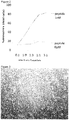
- Figure 2 shows that T cells induced by using LMP2 peptide from peripheral blood obtained from a healthy volunteer having HLA-A2402 who had previously been infected with EB virus exerted peptide specific killer activity.
- Figure 3 is a photograph of an iPS cell colony induced from a LMP2 peptide specific T cell.
- Figure 4 is a result of FACS analysis on day 13 of the differentiation of T-iPS cells established from LMP2 peptide specific T cells into T cells.
- Figure 5 is a result of FACS analysis on day 36 of the differentiation of T-iPS cells established from LMP2 peptide specific T cells into T cells.
- FIG. 6 is a result of FACS analysis on day 41 of the differentiation of T-iPS cells established from LMP2 peptide specific T cells into T cells. Generation of LMP2 specific mature T cells (CTLs) was confirmed.
- CTLs LMP2 specific mature T cells
- FIG. 7 shows LMP2 specific killer activity of the mature T cells (CTLs) regenerated from T-iPS cells established from a LMP2 peptide specific T cell.
- the killer activities in the presence (p+) or absence (p-) of LMP2 peptide were observed by using LCLs as target cells.
- Figure 8 shows natural killer cell-like activities of mature T cells regenerated from T-iPS cells established from a LMP2 peptide specific T cell.
- Figure 9 shows peptide specific cytotoxicity of CTLs regenerated from clone LMP2#1 against LCLs.
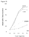
- Figure 10 shows peptide specific cytotoxicity of CTLs regenerated from clone LMP2#13 obtained in Example 2 against LCLs.
- pluripotent stem cells refer to stem cells having pluripotency, i.e. an ability to differentiate into many types of cells in the body, and self-propagation ability.
- pluripotent stem cells may include embryonic stem cells (ES cells), nuclear transfer embryonic stem cells (ntES cells), embryonic germ cells (EG cells), and induced pluripotent stem cells (iPS cells).
- ES cells embryonic stem cells
- ntES cells nuclear transfer embryonic stem cells
- EG cells embryonic germ cells
- iPS cells induced pluripotent stem cells
- pluripotent stem cells are preferably those derived from a mammal and especially from a human.
- T cells refer to cells expressing receptors for antigens called as T cell receptor (TCR).
- TCR T cell receptor
- iPS cells are induced from a T cell having a desired antigen specificity.
- T cells used as origin for iPS cells may preferably be T cells expressing at least one of CD4 and CD8, in addition to CD3.
- the preferable human T cells my include helper/regulatory T cells that are CD4 positive cells; cytotoxic T cells that are CD8 positive cells; naive T cells that are CD45RA + CD62L + cells; central memory T cells that are CD45RA - CD62L + cells, effector memory T cells that are CD45RA - CD62L - cells and terminal effector T cells that are CD45RA + CD62L - cells.
- Human T cells can be isolated from a human tissue by known procedures.
- the human tissue is not limited in particular, if the tissue contains T cells of the above-mentioned type, and examples thereof include peripheral blood, lymph node, bone marrow, thymus, spleen, umbilical cord blood, and a lesion site tissue.
- peripheral blood and umbilical cord blood are preferable since they can be derived less invasively from the human body and can be prepared with ease.
- Known procedures for isolating human T cells include, for example, flow cytometry using an antibody directing to a cell surface marker, such as CD4, and a cell sorter, as shown in the below-mentioned Examples.
- desired T cells can be isolated by detecting the secretion of a cytokine or the expression of a functional molecule as an indicator.
- T cells secrete different cytokines, depending on whether they are of the Th1 or Th2 type, and thus T cells of a desired Th type can be isolated by selecting T cells using the cytokine as an indicator.
- cytotoxic (killer) T cells can be isolated using the secretion or production of granzyme, perforin, or the like as an indicator.
- WT1 antigen specific cytotoxic T cells may be obtained by stimulating lymphocyte obtained from a human by a conventional procedure with WT1 or an epitope peptide thereof.
- Various WT1 antigen epitope peptides have been identified and the procedures for inducing WT1 specific cytotoxic T cells with those peptides have been well known.
- lymphocytes may be stimulated by cancer cells expressing a WT1 antigen.
- WT1 antigen specific cytotoxic T cells may be induced from the cells obtained from an individual who is suffered from cancer that expresses WT1 or an individual who had previously been suffered from cancer that expresses WT1.
- WT1 antigen specific cytotoxic T cells may also be induced from a healthy volunteer.
- EB virus associated antigen specific cytotoxic T cells may be induced by stimulating lymphocyte obtained from a human by a conventional procedure with an EB virus associated antigen, such as LMP2 antigen or an epitope peptide thereof.
- an EB virus associated antigen such as LMP2 antigen or an epitope peptide thereof.
- lymphocytes may be stimulated by cancer cells expressing EB virus associated antigen.
- EB virus associated antigen specific cytotoxic T cells may be induced from cells of a patient suffered from an EB virus infectious disease or an EB virus associating cancer, a healthy individual who is a carrier of EB virus or a healthy individual who have never been infected with EB virus.
- antigen specific T cells may be obtained from a patient with the infectious disease or cancer. This is because antigen specific T cells are proliferated in the body of the patient and therefore, it may be easy to detect/obtain T cells with specific reactivity.
- T-iPS cells for allogenic transplantation may be induced from a T cell having specificity for a disease obtained from a patient having the disease.
- the antigen specific T cells may also be obtained from a healthy volunteer.
- T cells obtained from a healthy volunteer the following advantages can be achieved: 1) Various antigen specific T cells may be induced from a healthy volunteer's cells and therefore, a variety of T-iPS cells having various TCR genes can be prepared in advance. 2) It is easier to collect donors from healthy people for establishing a T-iPS cell bank.
- Human T cells specific for a given antigen may be isolated from cell culture or tissue containing WT1 antigen specific or EB virus associated antigen specific T cells with an affinity column immobilized with the desired antigen.
- tetramer of an antigen-bound major histocompatibility complex may be used to isolate human T cells specific for a given antigen, such as a WT1 antigen or an EB virus associated antigen from human tissues.
- iPS cells may be induced from thus obtained T cell specific for a WT1 antigen or an EB virus associated antigen.
- the procedure for inducing pluripotent stem cells from T cells may be those taught by Vizcardo et al. , Cell Stem Cell 12, 31-36 2013 .
- T cells specific for a given antigen may be obtained from an individual who had acquired immunity against the disease to be treated and the Yamanaka factors may be introduced to the T cells to give iPS cells ( Takahashi and Yamanaka, Cell 126, 663-673 (2006 ), Takahashi et al., Cell 131, 861-872(2007 ) and Grskovic et al., Nat. Rev. Drug Dscov. 10,915-929 (2011 ).
- iPS cells can be prepared by introducing specific reprogramming factors to somatic cells.
- iPS cells are somatic cell-derived artificial stem cells having properties almost equivalent to those of ES cells ( K. Takahashi and S. Yamanaka(2006) Cell, 126:663-676 ; K. Takahashi et al. (2007), Cell, 131 :861-872 ; J. Yu et al. (2007), Science, 318:1917-1920 ; Nakagawa, M. et al., Nat. Biotechnol. 26:101-106(2008 ); and WO 2007/069666 ).
- the reprogramming factors may be constituted by genes or gene products thereof, or non-coding RNAs, which are expressed specifically in ES cells; or genes or gene products thereof, non-coding RNAs or low molecular weight compounds, which play important roles in maintenance of the undifferentiated state of ES cells.
- genes included in the reprogramming factors include Oct3/4, Sox2, Sox1, Sox3, Sox15, Sox17, Klf4, Klf2, c-Myc, N-Myc, L-Myc, Nanog, Lin28, Fbxl5, ERas, ECAT15-2, Tell, beta-catenin, Lin28b, Sall1, Sall4, Esrrb, Nr5a2, Tbx3 and Glis1, and these reprogramming factors may be used either individually or in combination.
- Examples of the combination of the reprogramming factors include those described in WO2007/069666 ; WO2008/1 18820 ; WO2009/007852 ; WO2009/032194 ; WO2009/058413 ; WO2009/057831 ; WO2009/075119 ; WO2009/079007 ; WO2009/091659 ; WO2009/101084 ; WO2009/101407 ; WO2009/102983 ; WO2009/1 14949 ; WO2009/1 17439 ; WO2009/126250 ; WO2009/126251 ; WO2009/126655 ; WO2009/157593 ; WO2010/009015 ; WO2010/033906 ; WO2010/033920 ; WO2010/042800 ; WO2010/050626 ; WO 2010/056831 ; WO2010/068955 ; WO2010/098419 ; WO2010/102267 ; WO 2010/11 14
- the reprogramming factors may be contacted with or introduced into the somatic cells by a known procedure suitable for the form of the factor to be used.
- the reprogramming factors may be introduced into somatic cells by a method such as lipofection, fusion with a cell-permeable peptide (e.g., HIV-derived TAT or polyarginine), or microinjection.
- a cell-permeable peptide e.g., HIV-derived TAT or polyarginine
- the reprogramming factors may be introduced into somatic cells by a method such as use of a vector including virus, plasmid and artificial chromosome vectors; lipofection; use of liposome; or microinjection.
- virus vector include retrovirus vectors, lentivirus vectors (these are described in Cell, 126, pp. 663-676, 2006 ; Cell, 131, pp. 861-872, 2007 ; and Science, 318, pp. 1917-1920, 2007 ), adenovirus vectors ( Science, 322, 945-949, 2008 ), adeno-associated virus vectors and Sendai virus vectors ( WO 2010/008054 ).
- Examples of the artificial chromosome vector include human artificial chromosome (HAC), yeast artificial chromosome (YAC), and bacterial artificial chromosome (BAC and PAC).
- Examples of the plasmid which may be used include plasmids for mammalian cells ( Science, 322:949-953, 2008 ).
- the vector may contain a regulatory sequence(s) such as a promoter, enhancer, ribosome binding sequence, terminator and/or polyadenylation site to enable expression of the nuclear reprogramming factors; and, as required, a sequence of a selection marker such as a drug resistance gene (e.g., kanamycin-resistant gene, ampicillin-resistant gene or puromycin-resistant gene), thymidine kinase gene or diphtheria toxin gene; a gene sequence of a reporter such as the green-fluorescent protein (GFP), ⁇ -glucuronidase (GUS) or FLAG.
- a regulatory sequence(s) such as a promoter, enhancer, ribosome binding sequence, terminator and/or polyadenylation site to enable expression of the nuclear reprogramming factors
- a selection marker such as a drug resistance gene (e.g., kanamycin-resistant gene, ampicillin-resistant gene or puromycin-resistant gene), thymidine kinase
- the vector may have LoxP sequences upstream and downstream of these sequences.
- each reprogramming factor may be introduced into somatic cells by a method such as lipofection or microinjection, and an RNA into which 5-methylcytidine and pseudouridine (TriLink Biotechnologies) were incorporated may be used in order to suppress degradation ( Warren L, (2010) Cell Stem Cell. 7:618-630 ).
- TriLink Biotechnologies TriLink Biotechnologies
- Examples of the medium for inducing iPS cells include DMEM, DMEM/F12 and DME media supplemented with 10 to 15% FBS (these media may further contain LIF, penicillin/streptomycin, puromycin, L-glutamine, non-essential amino acids, ⁇ -mercaptoethanol and/or the like, as appropriate); and commercially available media [for example, medium for culturing mouse ES cells (TX-WES medium, Thromb-X), medium for culturing primate ES cells (medium for primate ES/iPS cells, ReproCELL) and serum-free medium (mTeSR, Stemcell Technology)].
- Examples of the method to induce iPS cells include a method wherein somatic cells and reprogramming factors are brought into contact with each other at 37°C in the presence of 5% CO 2 on DMEM or DMEM/F12 medium supplemented with 10% FBS, and the cells are cultured for about 4 to 7 days, followed by plating the cells on feeder cells (e.g., mitomycin C-treated STO cells or SNL cells) and starting culture in a bFGF-containing medium for culturing primate ES cells about 10 days after the contact between the somatic cells and the reprogramming factors, thereby allowing ES-like colonies to appear about 30 to about 45 days after the contact, or later.
- feeder cells e.g., mitomycin C-treated STO cells or SNL cells
- the cells may be contacted with the reprogramming factors and cultured at 37°C in the presence of 5% CO 2 on feeder cells (e.g., mitomycin C-treated STO cells or SNL cells) in DMEM medium supplemented with 10% FBS (this medium may further contain LIF, penicillin/streptomycin, puromycin, L-glutamine, non-essential amino acids, ⁇ -mercaptoethanol and the like, as appropriate) for about 25 to about 30 days or longer, thereby allowing ES-like colonies to appear.
- feeder cells e.g., mitomycin C-treated STO cells or SNL cells
- FBS this medium may further contain LIF, penicillin/streptomycin, puromycin, L-glutamine, non-essential amino acids, ⁇ -mercaptoethanol and the like, as appropriate
- Preferred examples of the culture method include a method wherein the somatic cells themselves to be reprogrammed are used instead of the feeder cells ( Takahashi K, et al.
- iPS cells may be established using a serum-free medium ( Sun N, et al. (2009), Proc Natl Acad Sci U S A. 106: 15720-15725 ). Further, in order to enhance the establishment efficiency, iPS cells may be established under low oxygen conditions (at an oxygen concentration of 0.1 % to 15%) ( Yoshida Y, et al. (2009), Cell Stem Cell. 5:237-241 or WO2010/013845 ). The contents of the documents cited in this paragraph are herein incorporated by reference.
- HDAC histone deacetylase
- VPA valproic acid
- MC 1293 sodium butyrate
- M344 nucleic acid-based expression inhibitors
- siRNAs and shRNAs against HDAC e.g., HDAC1 siRNA Smartpool.RTM.
- MEK inhibitor e.g., PD184352, PD98059, U0126, SL327 and PD0325901
- Glycogen synthase kinase-3 inhibitor e.g., Bio and CHIR99021
- DNA methyl transferase inhibitors e.g., 5-azacytidine
- histone methyl transferase inhibitors for example, low-molecular inhibitors such as BIX-01294, and nucleic acid-based expression inhibitors such as siRNAs and shRNAs against Suv39h1, Suv39h2, SetDB1 and G9a
- L-channel calcium agonist for example, Bayk8644
- p53 inhibitor for example, siRNA and shRNA against siRNA and shRNA against
- the medium is replaced with the fresh medium once every day from Day 2 of the culture.
- the number of somatic cells used for nuclear reprogramming is not restricted, and usually within the range of about 5 ⁇ 10 3 to about 5 ⁇ 10 6 cells per 100 cm 2 area on the culture plate.
- iPS cells may be selected based on the shape of each formed colony.
- a drug resistance gene is introduced as a marker gene such that the drug resistance gene is expressed in conjunction with a gene that is expressed when a somatic cell was reprogrammed (e.g., Oct3/4 or Nanog)
- the established iPS cells can be selected by culturing the cells in a medium containing the corresponding drug (selection medium). Further, iPS cells can be selected by observation under a fluorescence microscope in cases where the marker gene is the gene of a fluorescent protein.
- T-iPS cells bear genes encoding the T cell receptor derived from the original T cell from which the iPS cells were induced.
- TCR genes specific for a WT1 antigen or an EB virus associated antigen are introduced into pluripotent stem cells.
- TCRs specific for a WT1 antigen and those for an EB virus associated antigen in relation to various cancers have been reported. For example, see Anticancer Research 32(12); 5201-5209, 2012 and Jurgens et al, Journal of Clinical Investigation, 26:22, 2006 .
- TCR genes may be obtained from T cells specific for a given antigen isolated from a patient having a cancer or an infectious disease. The TCR genes may be isolated from thus obtained T cells.
- TCR genes specific for a given antigen may be introduced into pluripotent stem cells such as iPS cells that were induced from the donor cells. For example, this procedure may be conducted as taught by Morgan R.A. et al, Science, 314:126. 2006 .
- a suitable vector bearing the TCR genes may be introduced into the iPS cells.
- TCR genes may be introduced by a vector such as virus, plasmid and artificial chromosome vectors; or by means of lipofection, liposome or microinjection.
- virus vectors include retrovirus vectors, lentivirus vectors, adenovirus vectors, adeno-associated virus vectors and Sendai virus vectors.
- artificial chromosome vector include human artificial chromosome (HAC), yeast artificial chromosome (YAC), and bacterial artificial chromosome (BAC and PAC).
- HAC human artificial chromosome
- YAC yeast artificial chromosome
- BAC and PAC bacterial artificial chromosome
- the plasmid which may be used include plasmids for mammalian cells.
- the vector may contain a regulatory sequence(s) such as a promoter, enhancer, ribosome binding sequence, terminator and/or polyadenylation site to enable expression of the TCR genes.
- a regulatory sequence such as a promoter, enhancer, ribosome binding sequence, terminator and/or polyadenylation site to enable expression of the TCR genes.
- the vector may also contain a selection marker such as a drug resistance gene (e.g., kanamycin-resistant gene, ampicillin-resistant gene or puromycin-resistant gene), thymidine kinase gene or diphtheria toxin gene; and a reporter such as the green-fluorescent protein (GFP), ⁇ -glucuronidase (GUS) or FLAG.
- GFP green-fluorescent protein
- GUS ⁇ -glucuronidase
- the pluripotent stem cells expressing the desired TCR genes are then differentiated into T cell progenitors or mature T cells.
- the procedure for differentiating pluripotent stem cells into T cells may be that taught by Timmermans et al., Journal of Immunology, 2009, 182: 6879-6888 .
- T cell progenitors may cover cells at any stages of the T cell development, from undifferentiated cells corresponding to hematopoietic stem cells to the cells at the stage just before the cells undergo positive selection/negative selection. Details of the differentiation of T cells are explained in Blood 111:1318(2008 ) and Nature Immunology 11: 585(2010 ).
- T cells are roughly divided into ⁇ T cells and ⁇ T cells.
- ⁇ T cells include killer T cells and helper T cells.
- T cells cover all types of T cells.
- T cells may cover any of T progenitor cells and mature T cells.
- T cells may be those expressing at least one of CD4 and CD8 in addition to CD3.
- a significantly high proportion of hematological malignancies and solid tumors express WT1 antigen.
- the method of the present application can be used in the cell-based immunotherapy for WT1 expressing cancers.
- T cell preparations targeting for a WT1 cancer antigen or an EB virus associated antigen, such as LMP2 are provided.
- T-iPS cells may be induced from WT1 antigen specific T cells or LMP2 antigen specific T cells obtained from a normal person and the T-iPS cells may be differentiated or re-generated into T cells. Function of the T cells re-generated from T-iPS cells may be verified with the cells obtained from the person from whom the T-iPS cells were induced and then, the T-iPS cells are stored to create a cell bank.
- the HLA of the patient with a WT1 antigen-expressing cancer may be determined and HLA-matched T-iPS cells may be chosen from the T-iPS cell bank. Then, T cells re-generated from the T-iPS cells may be used for the cell-based immunotherapy. Alternatively, T-iPS cells may be differentiated into T cells and then the T cells are frozen and stored. By the latter procedure, a cell-based immunotherapy for more patients can be started more quickly. In addition, the transferred cells are rejected eventually and therefore, there is no need to consider the risk of oncogenic transformation of the transferred cells.
- the re-generated T cells are dispersed in a suitable media such as saline or PBS and the dispersion may be administered to a patient having a certain matching level of the HLA to the donor.
- the matching level of the donor and the patient may be complete match.
- HLA haplotype homo homozygous for HLA haplotype
- HLA haplotype hetero heterozygous for HLA haplotypes
- one of the patient's HLA haplotypes should match the donor's homozygous HLA haplotype.
- the cells may be administered intravenously.
- the amount of the cells to be administered is not limited. Cells that have been differentiated into mature T cells may be intravenously administered once to several times in an amount of 10 6 -10 7 cells/kg per administration. T cell progenitors may be administered in an amount of about 1/10-1/100 of that of the mature T cells.
- the number of the cells to be administered is not limited and may be determined based on, for example, the age, sex, height and body weight of the patient and disease and conditions to be treated.
- the optimal cell number may be determined through clinical studies.
- T cells may target various antigens and therefore, the method of this application may be applied for a cell-based immunotherapy against various diseases including cancers, infectious diseases, autoimmune diseases and allergies.
- a high proportion of hematopoietic organ tumors such as leukemia, myelodysplastic syndrome, multiple myeloma, and malignant lymphoma, as well as solid tumors such as stomach cancer, colon cancer, lung cancer, breast cancer, germ cell cancer, liver cancer, skin cancer, bladder cancer, prostate cancer, uterine cancer, cervical cancer and ovarian cancer express the WT1 gene.
- CTL cells generated from WT1 specific T-iPS cells or TCR-iPS cells are effective for the cell-based immunotherapy for those WT1 gene expressing cancers.
- Epstein-Barr (EB) virus causes various diseases such as infectious mononucleosis as well as cancers such as malignant lymphoma or burkitt lymphoma and epipharyngeal carcinoma.
- CTL cells differentiated from T-iPS cells or TCR-iPS cells having TCR genes specific for LMP2 antigen, an EB virus associated antigen may be useful for the treatment of EB virus associated infectious diseases or cancers, for example, Hodgkin's disease, barkitt lymphoma, nasopharyngeal carcinoma, some types of stomach cancer, and a cell-based immunotherapy for transplantation.
- T-iPS cells (clone LMP2#1) were established from a T cell specific for LMP2 antigen derived from peripheral blood mononuclear cells of an EB virus carrier. T-iPS cells were differentiated into LMP2 antigen specific CTLs (herein after, referred to as "re-generated LMP2-CTL#1").
- EB virus infection in acute phase may cause infectious mononucleosis and sometimes cause cancer such as barkitt lymphoma.
- the donor for T cells was a healthy person who had previously been infected with EB virus. Once infected, this virus stays in the lymphocytes for entire life and therefore, the donor is an EB virus carrier. The donor is, therefore, considered to have chronic EB virus infection.
- CTL cytotoxic T Lymphocytes
- Lymphoblastoid cell line established from healthy volunteer A who had previously infected with EB virus and had HLA-A*02:06/24:02; B*39:01/40:02; C*07:02/15:02; DRB1*04:10/09:01 in the Department of Hematology and Oncology, graduate School of Medicine, Kyoto University, Kyoto, Japan was used as antigen presenting cells.
- T cells Isolation of T cells from human peripheral blood and co-culture of the T cells and dendritic cells.
- ⁇ MEM medium 500 mL FCS 125 mL 20% penicillin-streptomycin solution* 5 mL 1% hrIL-7 (stock: 10 ⁇ g/ mL) 315 ⁇ L 5 ng/mL hrFlT-3L (stock: 10 ⁇ g/mL) 315 ⁇ L 5 ng/mL hrSCF (stock: 10 ⁇ g/mL) 630 ⁇ L 10 ng/mL Total 631.26 mL *Mixture of Penicillin (10,000 U/ml) and Streptomycin (10,000 ⁇ g/ml). The final concentrations were 100 U/ml and 100 ⁇ g/ml, respectively.
- Medium C for inducing from immature T cells into mature T cells contents amount added final conc.
- ⁇ MEM medium 500 mL FCS 125 mL 20% penicillin-streptomycin solution* 5 mL 1% hrIL-7 (stock: 10 ⁇ g/ mL) 315 ⁇ L 5 ng/mL Total 630.315 mL *Mixture of Penicillin (10,000 U/ml) and Streptomycin (10,000 ⁇ g/ml). The final concentrations were 100 U/ml and 100 ⁇ g/ml, respectively.
- the medium in the OP9 stromal cell culture to be used for the co-culture was aspirated and replaced with fresh medium A.
- the medium in the iPS cell culture dish was also aspirated and 10 ml of fresh medium A was added.
- the iPS cell mass was cut with an EZ-passage roller.
- the cut iPS cell mass was suspended by using a pipetman with a 200 ul tip.
- the number of the iPS cell clusters was visually counted and approximately 600 iPS cell clusters were seeded on the OP 9 cells.
- Three or more dishes per clone of iPS cells were used, and when subculturing, the cells in all dishes were once pooled in one dish and then redistributed to the same number of dishes to reduce the disparity between the dishes.
- the cell culture medium was replaced with 20 mL of fresh medium A.
- a half of the cell culture medium was replaced with 10 mL of fresh medium A.
- a half of the cell culture medium was replaced with 10 mL of fresh medium A.
- the collagenase solution was removed by aspiration and the cells were washed with 10 mL of PBS(-). Then, 0.05% trypsin/EDTA solution was added to the dish and the dish was incubated for 20 minutes at 37°C. After the incubation, the sheet like cell aggregates peeled from the bottom of the dish and the cell aggregates were mechanically fragmented to smaller sizes by means of pipetting. Thus treated cells were added with fresh medium A 20 mL and cultured for more 45 minutes at 37°C.
- the culture medium containing the floating cells was passed through 100 ⁇ m mesh and the cells were collected. The cells were then centrifuged at 1200rpm for 7 minutes at 4°C. The obtained pellet was suspended in 10 mL of medium B. One-tenth of the suspension was separated and used for the FACS analysis. The remaining cell suspension was seeded to new dishes containing OP9/DLL1 cells. Cell suspensions obtained from several dishes were pooled and the pooled cells were seeded to the same number of new dishes.
- hematopoietic progenitor cells were contained in the obtained cells. FACS analysis was carried out using anti-CD34 antibody and anti-CD43 antibody. The results are shown in Fig. 4 . Since a sufficient number of cells could be confirmed in the CD34 low CD43 + cell fraction, it was confirmed that hematopoietic progenitor cells were induced.
- T cells Induction of T cells from hematopoietic progenitor cells.
- the obtained cells were seeded on OP9/DLL1 cells.
- cell sorting of the CD34 low CD43 + cell fraction was not performed.
- this fraction is sorted, the efficiency of differentiation of T cells could be reduced in comparison with the case where sorting was not performed due to the decrease of the cells or damage to the cells by sorting.
- the cells loosely adhered to the OP9 cells were dissociated by gently pipetting several times.
- the cells were passed through a 100 ⁇ m mesh and collected in a 50 mL conical tube.
- the tube was centrifuged at 1200 rpm for 7 minutes at 4°C.
- the pellet was dispersed in 10 mL of medium B.
- prepared cell suspension was seeded on the OP9/DLL1 cells.
- the cells loosely adhered to the OP9/DLL1 cells were dissociated by gently pipetting several times.
- the cells were passed through a 100 ⁇ m mesh and collected in a 50 mL conical tube.
- the tube was centrifuged at 1200 rpm for 7 minutes at 4°C.
- the pellet was dispersed in 10 mL of medium B.
- the cells on Day 36 were analyzed by FACS with anti CD3 antibody and LMP2 tetramer.
- Results are shown in Figure 5 .
- CD3 + cells were observed and a part of the cells were differentiated into CD3 + LMP2 tetramer positive cells.
- LMP2 positive T cells were confirmed with flow cytometry and then, the cells were added with IL-15 so that the cells are differentiated into mature killer T cells or CD8SP cells.
- the T cells were dispersed in medium C and seeded on the fresh OP9/DLL1 cell layer in each well of a 24-well plate at a density of 3 ⁇ 10 5 cells/well.
- IL-15 was added to each well to give final concentration of 10 ng/mL.
- LMP2 peptide specific CTLs were induced according to the procedure of Example 1 from a healthy volunteer other than healthy volunteer A from whom PBMC were obtained in Example 1.
- T-iPS cells (clone LMP2#13) were established from the CTL and then, the T-iPS cells were differentiated into CD8 single positive T cells (re-generated LMP2-CTL#13).
- the LMP2 peptide used in Example 1 was also used in this example.
- the antigen specific killer activity of the re-generated CTL cells against the peptide-loaded LCL cells as target cells was determined. Result is shown in Example 10.
- Example 2 The healthy volunteer in Example 2 had previously been infected with EB virus and was EBNA antibody positive, and had HLA-A*02:10/24:02 ; B*07:02/40:06; C*07:02/08:01; DRB1*04:05/04:05.
- the regenerated LMP2-CTL#13 showed high antigen specific cytotoxicity against the peptide-loaded LCLs.
- WT1 antigen specific cytotoxic T cells were induced from peripheral blood of a healthy volunteer, and T-iPS cells (clone WT#9) were established from the CTL. Then, WT1 antigen specific mature T cells (re-generated WT1-CTL(#9)) were induced from the T-iPS cells.
- This example comprises the following steps:
- the medium used is as follows. [Table 5] Medium for T cells (T cell medium): Amount Final conc. RPMI 45ml human AB serum 5ml 10% Total 50ml ii) The WT1 antigen peptide used is as follows.
- the LCL having HLA-A2402 which had been established from a healthy volunteer in the Department of Hematology and Oncology, graduate School of Medicine, Kyoto University, Kyoto, Japan was used.
- ⁇ MEM medium 500 mL FCS 125 mL 20% penicillin-streptomycin solution* 5 mL 1% hrIL-7 (stock: 10 ⁇ g/ mL) 315 ⁇ L 5 ng/mL hrFlT-3L (stock: 10 ⁇ g/mL) 315 ⁇ L 5 ng/mL hrSCF (stock: 10 ⁇ g/mL) 630 ⁇ L 10 ng/mL Total 631.26 mL *Mixture of Penicillin (10,000 U/ml) and Streptomycin (10,000 ⁇ g/ml). The final concentrations were 100 U/ml and 100 ⁇ g/ml, respectively.
- the medium in the OP9 stromal cell culture to be used for the co-culture was aspirated and replaced with fresh medium A.
- the medium in the iPS cell culture dish was also aspirated and 10 ml of fresh medium A was added.
- the iPS cell mass was cut with an EZ-passage roller.
- the cut iPS cell mass was suspended by means of a pipetman with a 200 ul tip.
- the number of the iPS cell clusters was visually counted and approximately 600 iPS cell clusters were seeded on the OP 9 cells.
- Three or more dishes per clone of iPS cells were used, and when subculturing, the cells in all dishes were once pooled in one dish and then redistributed to the same number of dishes to reduce the disparity between the dishes.
- the cell culture medium was replaced with 20 mL of fresh medium A.
- a half of the cell culture medium was replaced with 10 mL of fresh medium A.
- a half of the cell culture medium was replaced with 10 mL of fresh medium A.
- HBSS + Mg + Ca
- 10 mL of Collagenase IV 250U in HBSS (+Mg+Ca) solution was added to the dish and incubated for 45 minutes at 37°C.
- the collagenase solution was removed by aspiration and the cells were washed with 10 mL of PBS(-). Then, 0.05% trypsin/EDTA solution was added to the dish and the dish was incubated for 20 minutes at 37°C. After the incubation, the sheet like cell aggregates peeled from the bottom of the dish and the cell aggregates were mechanically fragmented to smaller sizes by means of pipetting.
- treated cells were added with fresh medium A 20 mL and cultured for more 45 minutes at 37°C.
- the culture medium containing the floating cells was passed through 100 ⁇ m mesh and the cells were collected. The cells were then centrifuged at 1200rpm for 7 minutes at 4°C. The obtained pellet was suspended in 10 mL of medium B.
- One-tenth of the suspension was separated and used for the FACS analysis.
- the remaining cell suspension was seeded to new dishes containing OP9/DLL1 cells. Cell suspensions obtained from several dishes were pooled and the pooled cells were seeded to the same number of new dishes.
- hematopoietic progenitor cells were contained in the obtained cells. FACS analysis was carried out using anti-CD34 antibody, anti-CD43 antibody. The results are shown in Fig. 4 . Since a sufficient number of cells could be confirmed in the CD34 low CD43 + cell fraction, it was confirmed that hematopoietic progenitor cells were induced.
- T cells Induction of T cells from hematopoietic progenitor cells.
- the obtained cells were seeded on OP9/DLL1 cells.
- cell sorting of the CD34 low CD43 + cell fraction was not performed.
- this fraction is sorted, the efficiency of differentiation of T cells could be reduced in comparison with the case where sorting is not performed due to the decrease of the cells or damage to the cells by sorting.
- the cells loosely adhered to the OP9 cells were gently dissociated by pipetting several times.
- the cells were passed through a 100 ⁇ m mesh and collected in a 50 mL conical tube.
- the tube was centrifuged at 1200 rpm for 7 minutes at 4°C.
- the pellet was dispersed in 10 mL of medium B.
- prepared cells were seeded on the OP9/DLL1 cells.
- the cells loosely adhered to the OP9/DLL1 cells were gently dissociated by pipetting several times.
- the cells were passed through a 100 ⁇ m mesh and collected in a 50 mL conical tube.
- the tube was centrifuged at 1200 rpm for 7 minutes at 4°C.
- the pellet was dispersed in 10 mL of medium B.
- the T cells regenerated from the T-iPS cells were confirmed to exhibit the same antigen specificity as the original T cells. Further, thus regenerated T cells expressed the surface antigen that were observed in mature T cells and therefore, had the well matured functions.
- amino acid sequences of TCR alpha and beta chains of clone WT1#9 were determined by the conventional method.
- the regenerated WT1-CTL#9 showed high antigen specific cytotoxicity against the peptide-loaded LCLs ( Figure 15 ).
- WT1 peptide specific CTL cells were induced according to the procedure of Example 3 from the same healthy volunteer from whom PBMC were obtained in Example 3.
- T-iPS cells (clone WT1#3-3) were established from the CTL and then, the T-iPS cells were differentiated into CD8 single positive T cells (re-generated WT1-CTL#3-3).
- the WT1 peptide used in Example 3 was also used in this example.
- the antigen specific killer activity of the re-generated CTLs against the LCLs loaded with the peptide as target cells was examined.
- Results are shown in Figure 16 .
- the re-generated WT1-CTL#3-3 showed high antigen specific killing activity against the peptide-loaded LCLs.
- the re-generated WT1-CTLs#3-3 were cytotoxic against both cell lines THP-1 and HL60 that express WT1 antigen. The cytotoxic activities were completely blocked by the anti-HLA class I antigen. Based on the results, the re-generated WT1-CTL(#3-3) kill the leukemia cells in the antigen specific manner.
- the original iPS cells were established from peripheral blood mononuclear cells of a healthy donor in Department of Immunology, Institute for Frontier Medicinal Sciences, Kyoto University, Kyoto, Japan in the same procedure in Example 2.
- the iPS cell line has homozygous HLAs.
- Class I restricted WT1 specific TCR genes were cloned from Clone WT1#9 and Clone WT1#3-3 in Department of Immunology, Institute for Frontier Medicinal Sciences, Kyoto University, Kyoto, Japan.
- RNAs of the cells were obtained.
- Full length cDNA was obtained by using SMARTer RACE cDNA amplificatioin kit (Clontech Laboratories, Inc.) and was used as a template.
- TCR genes were amplified by using a primer targeting the 3'-end of the TCR ⁇ chain: CACAGGCTGTCTTACAATCTTGCAGATC (SEQ ID NO: 11) or of TCR ⁇ chain: CTCCACTTCCAGGGCTGCCTTCA (SEQ ID NO: 12) or TGACCTGGGATGGTTTTGGAGCTA(SEQ ID NO: 13) to obtain double stranded WT1-TCR cDNA.
- double stranded cDNA was inserted into pTA2 vector (TOYOBO, see Figure 19 ) and introduced into a cell line. The specificity of the WT1 TCR were evaluated using the transfected cells.
- CS-UbC-RfA-IRES2-Venus vector ( Figure 20 ) was obtained from Subteam for Manipulation of Cell Fate, RIKEN BioResource Center, Tsukuba, Ibaraki, Japan. WT-TCR gene was incorporated in the vector with the Gateway system to give CS-UbC-RfA-IRES2-Venus/WT1-TCR.
- CS-UbC-RfA-IRES2-Venus/WT1-TCR was introduced into LentiX-293T packaging cells with X-treamGENE9 (Roche). The medium was exchanged on the next day and on day 2, the culture supernatant was collected and used as lentiviral supernatant.
- LMP2-T-iPS cells were treated with TrypLE Select (Life Technologies) to give completely single-cell suspension. The suspension was centrifuged and the pellet was dispersed by the lentiviral supernatant, and then, the obtained suspension was centrifuged at 3000rpm at 32°C for one hour so that lentivirus infects and then, WT1-TCR was introduced into the LMP2-T-iPS cells.
Landscapes
- Health & Medical Sciences (AREA)
- Life Sciences & Earth Sciences (AREA)
- General Health & Medical Sciences (AREA)
- Engineering & Computer Science (AREA)
- Chemical & Material Sciences (AREA)
- Organic Chemistry (AREA)
- Veterinary Medicine (AREA)
- Animal Behavior & Ethology (AREA)
- Public Health (AREA)
- Biomedical Technology (AREA)
- Bioinformatics & Cheminformatics (AREA)
- Genetics & Genomics (AREA)
- Immunology (AREA)
- Wood Science & Technology (AREA)
- Biotechnology (AREA)
- Zoology (AREA)
- Epidemiology (AREA)
- Pharmacology & Pharmacy (AREA)
- Medicinal Chemistry (AREA)
- General Engineering & Computer Science (AREA)
- Nuclear Medicine, Radiotherapy & Molecular Imaging (AREA)
- Chemical Kinetics & Catalysis (AREA)
- General Chemical & Material Sciences (AREA)
- Microbiology (AREA)
- Biochemistry (AREA)
- Oncology (AREA)
- Hematology (AREA)
- Cell Biology (AREA)
- Virology (AREA)
- Physics & Mathematics (AREA)
- Communicable Diseases (AREA)
- Biophysics (AREA)
- Molecular Biology (AREA)
- Pulmonology (AREA)
- Plant Pathology (AREA)
- Micro-Organisms Or Cultivation Processes Thereof (AREA)
- Peptides Or Proteins (AREA)
- Mycology (AREA)
- Medicines Containing Material From Animals Or Micro-Organisms (AREA)
Applications Claiming Priority (3)
| Application Number | Priority Date | Filing Date | Title |
|---|---|---|---|
| US201462026358P | 2014-07-18 | 2014-07-18 | |
| US201462026348P | 2014-07-18 | 2014-07-18 | |
| PCT/JP2015/070624 WO2016010155A1 (fr) | 2014-07-18 | 2015-07-17 | Méthode de production de cellules souches pluripotentes possédant un gène codant pour le récepteur des cellules t spécifique d'un antigène |
Publications (2)
| Publication Number | Publication Date |
|---|---|
| EP3170895A1 true EP3170895A1 (fr) | 2017-05-24 |
| EP3170895A4 EP3170895A4 (fr) | 2018-07-11 |
Family
ID=55078640
Family Applications (1)
| Application Number | Title | Priority Date | Filing Date |
|---|---|---|---|
| EP15822337.0A Withdrawn EP3170895A4 (fr) | 2014-07-18 | 2015-07-17 | Méthode de production de cellules souches pluripotentes possédant un gène codant pour le récepteur des cellules t spécifique d'un antigène |
Country Status (5)
| Country | Link |
|---|---|
| US (1) | US20170267972A1 (fr) |
| EP (1) | EP3170895A4 (fr) |
| JP (1) | JPWO2016010155A1 (fr) |
| AU (1) | AU2015290561A1 (fr) |
| WO (1) | WO2016010155A1 (fr) |
Cited By (1)
| Publication number | Priority date | Publication date | Assignee | Title |
|---|---|---|---|---|
| EP3693456A4 (fr) * | 2017-10-06 | 2021-06-30 | Thyas Co. Ltd. | Procédé de production d'une colonie de lymphocytes t génétiquement diverse dérivée d'une cellule ips |
Families Citing this family (7)
| Publication number | Priority date | Publication date | Assignee | Title |
|---|---|---|---|---|
| EP3147353B1 (fr) | 2014-05-21 | 2022-03-30 | Kyoto University | Procédé de production de cellules blastiques pancréatiques et agent de traitement d'une maladie du pancréas les contenant |
| TW202020145A (zh) * | 2018-07-13 | 2020-06-01 | 國立大學法人京都大學 | γδT細胞的製造方法 |
| SG11202101789SA (en) | 2018-07-26 | 2021-03-30 | Univ Kyoto | Method for generating cells into which an exogenous antigen receptor is introduced |
| CN113939302A (zh) * | 2019-06-14 | 2022-01-14 | 赛雅思株式会社 | 医药组合物 |
| EP4219707A4 (fr) | 2020-09-24 | 2024-11-13 | Kyoto University | Procédé de production d'une cellule effectrice ayant une spécificité souhaitée |
| CN116964194A (zh) | 2021-02-05 | 2023-10-27 | 国立大学法人神户大学 | 诱导性多能干细胞衍生的γδT细胞及其产生方法 |
| CN113388612A (zh) * | 2021-06-18 | 2021-09-14 | 重庆天科雅生物科技有限公司 | 一种针对表位点为iyvlvmlvl的tcr所设计的引物及其应用 |
Family Cites Families (5)
| Publication number | Priority date | Publication date | Assignee | Title |
|---|---|---|---|---|
| JP2006306822A (ja) * | 2005-05-02 | 2006-11-09 | Japan Science & Technology Agency | 移植片拒絶反応及び移植片対宿主疾患を防ぐ移植免疫反応抑制ポリフェノール溶液及び移植免疫反応抑制方法 |
| US9206394B2 (en) * | 2010-02-03 | 2015-12-08 | The University Of Tokyo | Method for reconstructing immune function using pluripotent stem cells |
| JPWO2011096482A1 (ja) * | 2010-02-03 | 2013-06-13 | 国立大学法人 東京大学 | 多能性幹細胞を用いた免疫機能再建法 |
| EP2611905A4 (fr) * | 2010-09-01 | 2014-02-12 | Baylor College Medicine | Lymphocytes t cytotoxiques spécifiques de ebv pour le traitement du carcinome du nasopharynx (npc) locorégional |
| JP6164746B2 (ja) * | 2012-05-22 | 2017-07-19 | 国立大学法人 東京大学 | 抗原特異的t細胞の製造方法 |
-
2015
- 2015-07-17 AU AU2015290561A patent/AU2015290561A1/en not_active Abandoned
- 2015-07-17 EP EP15822337.0A patent/EP3170895A4/fr not_active Withdrawn
- 2015-07-17 WO PCT/JP2015/070624 patent/WO2016010155A1/fr not_active Ceased
- 2015-07-17 US US15/326,968 patent/US20170267972A1/en not_active Abandoned
- 2015-07-17 JP JP2016534512A patent/JPWO2016010155A1/ja not_active Withdrawn
Cited By (1)
| Publication number | Priority date | Publication date | Assignee | Title |
|---|---|---|---|---|
| EP3693456A4 (fr) * | 2017-10-06 | 2021-06-30 | Thyas Co. Ltd. | Procédé de production d'une colonie de lymphocytes t génétiquement diverse dérivée d'une cellule ips |
Also Published As
| Publication number | Publication date |
|---|---|
| WO2016010155A1 (fr) | 2016-01-21 |
| US20170267972A1 (en) | 2017-09-21 |
| AU2015290561A1 (en) | 2017-03-09 |
| EP3170895A4 (fr) | 2018-07-11 |
| JPWO2016010155A1 (ja) | 2017-04-27 |
Similar Documents
| Publication | Publication Date | Title |
|---|---|---|
| JP7440027B2 (ja) | 多能性幹細胞から免疫細胞療法用t細胞を誘導する方法 | |
| US12391921B2 (en) | Method for inducing antigen specific CD8 positive T cells | |
| JP7613712B2 (ja) | 抗原特異的t細胞の製造方法 | |
| EP3170895A1 (fr) | Méthode de production de cellules souches pluripotentes possédant un gène codant pour le récepteur des cellules t spécifique d'un antigène | |
| JP2020506713A (ja) | 多能性幹細胞から模倣自然免疫細胞を生成する方法及びキット | |
| US20170128556A1 (en) | Immunotherapy using t precursor cells derived from pluripotent stem cells having rearranged t cell receptor genes | |
| US20170296649A1 (en) | Method for inducing t cells for cell-based immunotherapy | |
| WO2017159087A1 (fr) | Procédé d'induction de cellule t spécifique de l'antigène ny-eso1 pour immunothérapie cellulaire | |
| US20220233665A1 (en) | Medicinal composition |
Legal Events
| Date | Code | Title | Description |
|---|---|---|---|
| PUAI | Public reference made under article 153(3) epc to a published international application that has entered the european phase |
Free format text: ORIGINAL CODE: 0009012 |
|
| 17P | Request for examination filed |
Effective date: 20170216 |
|
| AK | Designated contracting states |
Kind code of ref document: A1 Designated state(s): AL AT BE BG CH CY CZ DE DK EE ES FI FR GB GR HR HU IE IS IT LI LT LU LV MC MK MT NL NO PL PT RO RS SE SI SK SM TR |
|
| AX | Request for extension of the european patent |
Extension state: BA ME |
|
| DAV | Request for validation of the european patent (deleted) | ||
| DAX | Request for extension of the european patent (deleted) | ||
| RIC1 | Information provided on ipc code assigned before grant |
Ipc: C12N 5/0783 20100101AFI20180228BHEP Ipc: A61K 35/17 20150101ALI20180228BHEP Ipc: C12N 15/09 20060101ALI20180228BHEP Ipc: C12N 5/095 20100101ALI20180228BHEP Ipc: A61P 37/02 20060101ALI20180228BHEP Ipc: A61P 31/12 20060101ALI20180228BHEP Ipc: A61P 37/08 20060101ALI20180228BHEP Ipc: A61P 35/00 20060101ALI20180228BHEP |
|
| A4 | Supplementary search report drawn up and despatched |
Effective date: 20180607 |
|
| RIC1 | Information provided on ipc code assigned before grant |
Ipc: C12N 15/09 20060101ALI20180601BHEP Ipc: A61P 35/00 20060101ALI20180601BHEP Ipc: A61P 31/12 20060101ALI20180601BHEP Ipc: A61P 37/08 20060101ALI20180601BHEP Ipc: C12N 5/0783 20100101AFI20180601BHEP Ipc: C12N 5/095 20100101ALI20180601BHEP Ipc: A61P 37/02 20060101ALI20180601BHEP Ipc: A61K 35/17 20150101ALI20180601BHEP |
|
| STAA | Information on the status of an ep patent application or granted ep patent |
Free format text: STATUS: THE APPLICATION IS DEEMED TO BE WITHDRAWN |
|
| 18D | Application deemed to be withdrawn |
Effective date: 20190108 |