EP3202885A1 - Neuartige biologische aktivitätsteststruktur zur verfolgung einer einzelzelle unter verwendung von geliermitteln - Google Patents
Neuartige biologische aktivitätsteststruktur zur verfolgung einer einzelzelle unter verwendung von geliermitteln Download PDFInfo
- Publication number
- EP3202885A1 EP3202885A1 EP14903914.1A EP14903914A EP3202885A1 EP 3202885 A1 EP3202885 A1 EP 3202885A1 EP 14903914 A EP14903914 A EP 14903914A EP 3202885 A1 EP3202885 A1 EP 3202885A1
- Authority
- EP
- European Patent Office
- Prior art keywords
- culture
- thin film
- testing structure
- cfu
- structure according
- Prior art date
- Legal status (The legal status is an assumption and is not a legal conclusion. Google has not performed a legal analysis and makes no representation as to the accuracy of the status listed.)
- Pending
Links
Images





Classifications
-
- C—CHEMISTRY; METALLURGY
- C12—BIOCHEMISTRY; BEER; SPIRITS; WINE; VINEGAR; MICROBIOLOGY; ENZYMOLOGY; MUTATION OR GENETIC ENGINEERING
- C12Q—MEASURING OR TESTING PROCESSES INVOLVING ENZYMES, NUCLEIC ACIDS OR MICROORGANISMS; COMPOSITIONS OR TEST PAPERS THEREFOR; PROCESSES OF PREPARING SUCH COMPOSITIONS; CONDITION-RESPONSIVE CONTROL IN MICROBIOLOGICAL OR ENZYMOLOGICAL PROCESSES
- C12Q1/00—Measuring or testing processes involving enzymes, nucleic acids or microorganisms; Compositions therefor; Processes of preparing such compositions
- C12Q1/02—Measuring or testing processes involving enzymes, nucleic acids or microorganisms; Compositions therefor; Processes of preparing such compositions involving viable microorganisms
- C12Q1/18—Testing for antimicrobial activity of a material
-
- C—CHEMISTRY; METALLURGY
- C12—BIOCHEMISTRY; BEER; SPIRITS; WINE; VINEGAR; MICROBIOLOGY; ENZYMOLOGY; MUTATION OR GENETIC ENGINEERING
- C12M—APPARATUS FOR ENZYMOLOGY OR MICROBIOLOGY; APPARATUS FOR CULTURING MICROORGANISMS FOR PRODUCING BIOMASS, FOR GROWING CELLS OR FOR OBTAINING FERMENTATION OR METABOLIC PRODUCTS, i.e. BIOREACTORS OR FERMENTERS
- C12M1/00—Apparatus for enzymology or microbiology
- C12M1/14—Apparatus for enzymology or microbiology with means providing thin layers or with multi-level trays
-
- C—CHEMISTRY; METALLURGY
- C12—BIOCHEMISTRY; BEER; SPIRITS; WINE; VINEGAR; MICROBIOLOGY; ENZYMOLOGY; MUTATION OR GENETIC ENGINEERING
- C12M—APPARATUS FOR ENZYMOLOGY OR MICROBIOLOGY; APPARATUS FOR CULTURING MICROORGANISMS FOR PRODUCING BIOMASS, FOR GROWING CELLS OR FOR OBTAINING FERMENTATION OR METABOLIC PRODUCTS, i.e. BIOREACTORS OR FERMENTERS
- C12M1/00—Apparatus for enzymology or microbiology
- C12M1/34—Measuring or testing with condition measuring or sensing means, e.g. colony counters
-
- C—CHEMISTRY; METALLURGY
- C12—BIOCHEMISTRY; BEER; SPIRITS; WINE; VINEGAR; MICROBIOLOGY; ENZYMOLOGY; MUTATION OR GENETIC ENGINEERING
- C12M—APPARATUS FOR ENZYMOLOGY OR MICROBIOLOGY; APPARATUS FOR CULTURING MICROORGANISMS FOR PRODUCING BIOMASS, FOR GROWING CELLS OR FOR OBTAINING FERMENTATION OR METABOLIC PRODUCTS, i.e. BIOREACTORS OR FERMENTERS
- C12M1/00—Apparatus for enzymology or microbiology
- C12M1/16—Apparatus for enzymology or microbiology containing, or adapted to contain, solid media
-
- C—CHEMISTRY; METALLURGY
- C12—BIOCHEMISTRY; BEER; SPIRITS; WINE; VINEGAR; MICROBIOLOGY; ENZYMOLOGY; MUTATION OR GENETIC ENGINEERING
- C12M—APPARATUS FOR ENZYMOLOGY OR MICROBIOLOGY; APPARATUS FOR CULTURING MICROORGANISMS FOR PRODUCING BIOMASS, FOR GROWING CELLS OR FOR OBTAINING FERMENTATION OR METABOLIC PRODUCTS, i.e. BIOREACTORS OR FERMENTERS
- C12M1/00—Apparatus for enzymology or microbiology
- C12M1/16—Apparatus for enzymology or microbiology containing, or adapted to contain, solid media
- C12M1/18—Multiple fields or compartments
-
- C—CHEMISTRY; METALLURGY
- C12—BIOCHEMISTRY; BEER; SPIRITS; WINE; VINEGAR; MICROBIOLOGY; ENZYMOLOGY; MUTATION OR GENETIC ENGINEERING
- C12M—APPARATUS FOR ENZYMOLOGY OR MICROBIOLOGY; APPARATUS FOR CULTURING MICROORGANISMS FOR PRODUCING BIOMASS, FOR GROWING CELLS OR FOR OBTAINING FERMENTATION OR METABOLIC PRODUCTS, i.e. BIOREACTORS OR FERMENTERS
- C12M23/00—Constructional details, e.g. recesses, hinges
- C12M23/02—Form or structure of the vessel
- C12M23/10—Petri dish
-
- C—CHEMISTRY; METALLURGY
- C12—BIOCHEMISTRY; BEER; SPIRITS; WINE; VINEGAR; MICROBIOLOGY; ENZYMOLOGY; MUTATION OR GENETIC ENGINEERING
- C12M—APPARATUS FOR ENZYMOLOGY OR MICROBIOLOGY; APPARATUS FOR CULTURING MICROORGANISMS FOR PRODUCING BIOMASS, FOR GROWING CELLS OR FOR OBTAINING FERMENTATION OR METABOLIC PRODUCTS, i.e. BIOREACTORS OR FERMENTERS
- C12M23/00—Constructional details, e.g. recesses, hinges
- C12M23/02—Form or structure of the vessel
- C12M23/12—Well or multiwell plates
-
- C—CHEMISTRY; METALLURGY
- C12—BIOCHEMISTRY; BEER; SPIRITS; WINE; VINEGAR; MICROBIOLOGY; ENZYMOLOGY; MUTATION OR GENETIC ENGINEERING
- C12M—APPARATUS FOR ENZYMOLOGY OR MICROBIOLOGY; APPARATUS FOR CULTURING MICROORGANISMS FOR PRODUCING BIOMASS, FOR GROWING CELLS OR FOR OBTAINING FERMENTATION OR METABOLIC PRODUCTS, i.e. BIOREACTORS OR FERMENTERS
- C12M23/00—Constructional details, e.g. recesses, hinges
- C12M23/20—Material Coatings
-
- C—CHEMISTRY; METALLURGY
- C12—BIOCHEMISTRY; BEER; SPIRITS; WINE; VINEGAR; MICROBIOLOGY; ENZYMOLOGY; MUTATION OR GENETIC ENGINEERING
- C12M—APPARATUS FOR ENZYMOLOGY OR MICROBIOLOGY; APPARATUS FOR CULTURING MICROORGANISMS FOR PRODUCING BIOMASS, FOR GROWING CELLS OR FOR OBTAINING FERMENTATION OR METABOLIC PRODUCTS, i.e. BIOREACTORS OR FERMENTERS
- C12M23/00—Constructional details, e.g. recesses, hinges
- C12M23/22—Transparent or translucent parts
-
- C—CHEMISTRY; METALLURGY
- C12—BIOCHEMISTRY; BEER; SPIRITS; WINE; VINEGAR; MICROBIOLOGY; ENZYMOLOGY; MUTATION OR GENETIC ENGINEERING
- C12M—APPARATUS FOR ENZYMOLOGY OR MICROBIOLOGY; APPARATUS FOR CULTURING MICROORGANISMS FOR PRODUCING BIOMASS, FOR GROWING CELLS OR FOR OBTAINING FERMENTATION OR METABOLIC PRODUCTS, i.e. BIOREACTORS OR FERMENTERS
- C12M25/00—Means for supporting, enclosing or fixing the microorganisms, e.g. immunocoatings
- C12M25/14—Scaffolds; Matrices
-
- C—CHEMISTRY; METALLURGY
- C12—BIOCHEMISTRY; BEER; SPIRITS; WINE; VINEGAR; MICROBIOLOGY; ENZYMOLOGY; MUTATION OR GENETIC ENGINEERING
- C12M—APPARATUS FOR ENZYMOLOGY OR MICROBIOLOGY; APPARATUS FOR CULTURING MICROORGANISMS FOR PRODUCING BIOMASS, FOR GROWING CELLS OR FOR OBTAINING FERMENTATION OR METABOLIC PRODUCTS, i.e. BIOREACTORS OR FERMENTERS
- C12M29/00—Means for introduction, extraction or recirculation of materials, e.g. pumps
- C12M29/04—Filters; Permeable or porous membranes or plates, e.g. dialysis
-
- C—CHEMISTRY; METALLURGY
- C12—BIOCHEMISTRY; BEER; SPIRITS; WINE; VINEGAR; MICROBIOLOGY; ENZYMOLOGY; MUTATION OR GENETIC ENGINEERING
- C12M—APPARATUS FOR ENZYMOLOGY OR MICROBIOLOGY; APPARATUS FOR CULTURING MICROORGANISMS FOR PRODUCING BIOMASS, FOR GROWING CELLS OR FOR OBTAINING FERMENTATION OR METABOLIC PRODUCTS, i.e. BIOREACTORS OR FERMENTERS
- C12M29/00—Means for introduction, extraction or recirculation of materials, e.g. pumps
- C12M29/26—Conditioning fluids entering or exiting the reaction vessel
-
- C—CHEMISTRY; METALLURGY
- C12—BIOCHEMISTRY; BEER; SPIRITS; WINE; VINEGAR; MICROBIOLOGY; ENZYMOLOGY; MUTATION OR GENETIC ENGINEERING
- C12M—APPARATUS FOR ENZYMOLOGY OR MICROBIOLOGY; APPARATUS FOR CULTURING MICROORGANISMS FOR PRODUCING BIOMASS, FOR GROWING CELLS OR FOR OBTAINING FERMENTATION OR METABOLIC PRODUCTS, i.e. BIOREACTORS OR FERMENTERS
- C12M41/00—Means for regulation, monitoring, measurement or control, e.g. flow regulation
- C12M41/46—Means for regulation, monitoring, measurement or control, e.g. flow regulation of cellular or enzymatic activity or functionality, e.g. cell viability
-
- C—CHEMISTRY; METALLURGY
- C12—BIOCHEMISTRY; BEER; SPIRITS; WINE; VINEGAR; MICROBIOLOGY; ENZYMOLOGY; MUTATION OR GENETIC ENGINEERING
- C12Q—MEASURING OR TESTING PROCESSES INVOLVING ENZYMES, NUCLEIC ACIDS OR MICROORGANISMS; COMPOSITIONS OR TEST PAPERS THEREFOR; PROCESSES OF PREPARING SUCH COMPOSITIONS; CONDITION-RESPONSIVE CONTROL IN MICROBIOLOGICAL OR ENZYMOLOGICAL PROCESSES
- C12Q1/00—Measuring or testing processes involving enzymes, nucleic acids or microorganisms; Compositions therefor; Processes of preparing such compositions
- C12Q1/02—Measuring or testing processes involving enzymes, nucleic acids or microorganisms; Compositions therefor; Processes of preparing such compositions involving viable microorganisms
-
- C—CHEMISTRY; METALLURGY
- C12—BIOCHEMISTRY; BEER; SPIRITS; WINE; VINEGAR; MICROBIOLOGY; ENZYMOLOGY; MUTATION OR GENETIC ENGINEERING
- C12Q—MEASURING OR TESTING PROCESSES INVOLVING ENZYMES, NUCLEIC ACIDS OR MICROORGANISMS; COMPOSITIONS OR TEST PAPERS THEREFOR; PROCESSES OF PREPARING SUCH COMPOSITIONS; CONDITION-RESPONSIVE CONTROL IN MICROBIOLOGICAL OR ENZYMOLOGICAL PROCESSES
- C12Q1/00—Measuring or testing processes involving enzymes, nucleic acids or microorganisms; Compositions therefor; Processes of preparing such compositions
- C12Q1/02—Measuring or testing processes involving enzymes, nucleic acids or microorganisms; Compositions therefor; Processes of preparing such compositions involving viable microorganisms
- C12Q1/04—Determining presence or kind of microorganism; Use of selective media for testing antibiotics or bacteriocides; Compositions containing a chemical indicator therefor
-
- G—PHYSICS
- G01—MEASURING; TESTING
- G01N—INVESTIGATING OR ANALYSING MATERIALS BY DETERMINING THEIR CHEMICAL OR PHYSICAL PROPERTIES
- G01N33/00—Investigating or analysing materials by specific methods not covered by groups G01N1/00 - G01N31/00
- G01N33/15—Medicinal preparations ; Physical properties thereof, e.g. dissolubility
-
- G—PHYSICS
- G01—MEASURING; TESTING
- G01N—INVESTIGATING OR ANALYSING MATERIALS BY DETERMINING THEIR CHEMICAL OR PHYSICAL PROPERTIES
- G01N2800/00—Detection or diagnosis of diseases
- G01N2800/26—Infectious diseases, e.g. generalised sepsis
Definitions
- the present invention relates to a novel bioactivity testing structure for single cell tracking using a gelling agent and a bioactivity testing system including the testing structure.
- the present invention also relates to bioactivity testing, drug susceptibility testing, antibiotic screening, and diagnostic methods using the testing structure.
- TDR-TB tuberculosis
- DST drug susceptibility test
- General DST methods are based on culturing cells in solid and liquid media.
- mycobacterium tuberculosis MTB is inoculated into a solid medium supplemented with an antibiotic and the formation of colonies is observed by naked-eye detection. Since MTB grows slowly (the cells differentiate for about 24 hours), conventional DST methods take at least 4 to 6 weeks.
- the use of liquid media accelerates the growth of MTB to reduce the time required for DST.
- a commercial DST platform using liquid media called MGIT 960 system is available.
- the MGIT system detects a fluorescence signals when MTB grows and consumes oxygen.
- the DST time of this MGIT system is as short as approximately 1 week.
- this method causes an increase in error rate because the growth of MTB cells is not directly observed but is indirectly identified.
- a microscopic-observation drug susceptibility (MODS) method for direct observation was developed to reduce DST time.
- MODS microscopic-observation drug susceptibility
- a sputum sample together with a drug is inoculated into a 24-well plate and colony formation is monitored daily, which plays an important role in identifying whether MTB grows.
- This method can yield DST results within one week.
- single cells of MTB cannot be immobilized in liquid media, making it impossible to track the single cells.
- a microscopic observation over the entire well area is essential for the detection of colony formation and a long-term observation is inevitable.
- Inoculum effect is an unwanted phenomenon in laboratory and may lead to overestimation of in vitro resistance, causing an increase in minimal inhibitory concentration (MIC). Consequently, inoculum effect deteriorates the reproducibility of experiments. Furthermore, since MIC is a parameter determining the microbiological efficacy of antibiotics in pharmacokinetics, different MIC values by cell densities may limit the clinical evaluation of antibiotics.
- the present inventors have earnestly and intensively conducted research to develop methods for effective and rapid diagnosis of bacteria, particularly, Mycobacterium tuberculosis, DST, and screening, and as a result, invented a new chamber (referred to as a "DAC system") to make a thin layer of an agarose mixture and have open surface on top of the thin layer for supplying liquid culture media and drugs. Further, the present inventors have found that the DAC system is effective for rapid and simple DST of drugs, drug screening, and tuberculosis diagnosis. The present invention has been achieved based on these findings.
- one object of the present invention is to provide a novel bioactivity testing structure and a bioactivity testing system including the testing structure.
- a further object of the present invention is to provide methods for bioactivity testing, antibiotic susceptibility testing of bacteria, antibiotic screening, and bacterial diagnosis using the bioactivity testing structure.
- One aspect of the present invention provides a bioactivity testing structure including a plate body, one or more culture units 100 arranged on the plate body, and a base plate 110 under which a solid thin film is to be formed on the bottom surface of each culture unit.
- the bioactivity testing may be drug susceptibility, drug screening or bacterial culture diagnosis testing.
- Each of the culture units may further have a receiving recess 120 formed on top of the base plate.
- Each of the culture units may have an inlet 111 formed in the base plate and through which a mixture solution of a liquid medium containing a gelling agent and a biological agent is to be introduced.
- Each of the culture units 100 may have one or more connection members 113 through which the base plate 110 is connected to barriers(partition walls) of the plate body and cut-away grooves 114 formed between the connection members.
- Each of the culture units may have one or more through-holes 112 formed in the base plate 110.
- each of the culture units 100 has a size of 1-50 mm ⁇ 1-50 mm and a height of 1 to 50 mm.
- the receiving recess 120 has a width of 1 to 5 mm and a height of 1 to 3 mm.
- the mixture solution is solidified under the base plate to form a biological agent-immobilized solid thin film.
- the thin film may have a thickness of 1 ⁇ m to 10 mm and the density of the biological agent in the mixture solution may be from 10 1 to 10 80 cfu/ml.
- the gelling agent is not limited to a particular kind and may be agar, agarose, gelatin, alginate, collagen or fibrin.
- a bioactive agent may be further introduced into the culture unit, and more preferably, the bioactive agent is introduced in a freeze-dried form into the receiving recess.
- the plate body, the culture units, and the base plate may be made of a transparent or translucent material.
- a further aspect of the present invention provides a bioactivity testing method including: providing a mixture solution of a liquid medium containing a gelling agent and a biological agent to the bottom surface of the bioactivity testing structure; solidifying the mixture solution to form a solid thin film in which the biological agent is immobilized; supplying a bioactive agent to the solid thin film and allowing the bioactive agent to diffuse into the solid thin film; and observing the individual responses of the single cells of the biological agent to the bioactive agent or observing whether the biological agent forms colonies or the colony forming units (CFU).
- CFU colony forming units
- the method may further include imaging the individual responses of the single cells of the biological agent to the bioactive agent, the colonies of the biological agent or the colony forming units (CFU), and analyzing the images. More preferably, the method further includes determining the minimum inhibitory concentration (MIC) of the bioactive agent based on the analysis of the images.
- MIC minimum inhibitory concentration
- the present invention also provides a method for antibiotic susceptibility testing of bacteria, including: providing a mixture solution of a liquid medium containing a gelling agent and a bacterial strain to the bottom surface of the bioactivity testing structure; solidifying the mixture solution to form a solid thin film in which the bacterial strain is immobilized; supplying an antibiotic to the solid thin film and allowing the antibiotic to diffuse into the solid thin film; and observing the individual responses of the single cells of the bacterial strain to the antibiotic or observing whether the bacterial strain forms colonies or the colony forming units (CFU).
- CFU colony forming units
- the method further includes imaging the individual responses of the single cells, the colonies or the colony forming units (CFU), analyzing the images, and determining the minimum inhibitory concentration (MIC) of the antibiotic based on the analysis of the images.
- CFU colony forming units
- the bacterial strain may be Mycobacterium tuberculosis or a nontuberculous mycobacterium (NTM).
- the Mycobacterium tuberculosis may be multi-drug-resistant (MDR) tuberculosis, extensively drug-resistant (XDR) tuberculosis or totally drug-resistant (TDR) tuberculosis.
- the method shows no inoculum effect.
- Another aspect of the present invention provides an antibiotic screening method including: providing a mixture solution of a liquid medium containing a gelling agent and a bacterial strain to the bottom surface of the bioactivity testing structure; solidifying the mixture solution to form a solid thin film in which the bacterial strain is immobilized; supplying an antibiotic to the solid thin film and allowing the antibiotic to diffuse into the solid thin film; and observing the individual responses of the single cells of the bacterial strain to the antibiotic or observing whether the bacterial strain forms colonies or the colony forming units (CFU).
- CFU colony forming units
- the method may further include imaging the individual responses of the single cells, the colonies or the colony forming units (CFU), analyzing the images, and determining the minimum inhibitory concentration (MIC) of the antibiotic based on the analysis of the images.
- CFU colony forming units
- Another aspect of the present invention provides a bacterial diagnostic method including: providing a mixture solution of a liquid medium containing a gelling agent and a bacterial strain to the bottom surface of the bioactivity testing structure; solidifying the mixture solution to form a solid thin film in which the bacterial strain is immobilized; supplying a culture medium of the bacterial strain to the solid thin film and allowing the culture medium to diffuse into the solid thin film; and observing the individual responses of the single cells of the bacterial strain or observing whether the bacterial strain forms colonies or the colony forming units (CFU).
- CFU colony forming units
- the culture medium of the bacterial strain may be further supplemented with an antibiotic and the diagnostic method may be carried out simultaneously with drug susceptibility testing.
- bioactivity testing system including: the bioactivity testing structure including culture units in which a bioactive agent is provided and a base plate under which a mixture solution of a liquid medium containing a gelling agent and a biological agent is solidified to form a thin film on the bottom surface of each culture unit; a stage for supporting and observing the testing structure; and a measurement system for imaging the individual responses of the single cells of the biological agent or the formation of colonies of the biological agent when the bioactive agent is delivered by diffusion or imaging the colony forming units (CFU), analyzing the images, and determining the minimum inhibitory concentration (MIC) of the bioactive agent based on the analysis of the images.
- CFU colony forming units
- the measurement system may include an imaging system and an image processing system.
- the bioactivity testing structure of the present invention enables very rapid and simple drug susceptibility testing of bacteria, particularly Mycobacterium tuberculosis, drug screening, and bacterial diagnosis.
- the use of the testing structure enables DST and diagnosis of bacteria only by pretreatment without the need to concentrate human sputum samples irrespective of inoculum effect, ensuring rapid, accurate, and simple testing compared to conventional tuberculosis diagnosis or DST systems.
- the testing structure of the present invention simultaneously enables the diagnosis and drug susceptibility testing of tuberculosis. Therefore, the present invention provides an effective alternative to the prior art.
- Agarose was used as a MTB fixing material.
- the agarose solution was mixed with a stock solution of TB, which posed a risk of thermal shock if a high-temperature agarose solution was used. To reduce this risk, an agarose concentration of 0.5% was used because this concentration is sufficiently low to prevent solidification at 37 °C. Lower concentrations of agarose enhance the diffusion of the culture medium and drug.
- the drugs used for the DST were purchased from Sigma-Aldrich.
- the liquid culture medium was Middlebrook 7H9 broth supplemented with 10% OADC.
- DAC Disc Agarose Channel
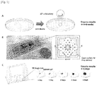
- FIG. 1 A schematic diagram of the DAC chip is shown in Fig. 1 .
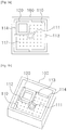
- Figs. 2a and 2b Detailed perspective and plan views of the DAC chip are illustrated in Figs. 3a and 3b .
- the DAC chip consists of a disc-shaped channel (base plate), which was loaded with the agarose and TB mixture.
- the chip also features structures to enhance the diffusion of the culture medium and drugs, such as wells that contain culture medium, drug, open spaces, and holes.
- the DAC chip consists essentially of one or more culture units arranged on a plate body and a base plate under which a solid thin film is to be formed on the bottom surface of each culture unit.
- Each of the culture units may further have an receiving recess formed on top of the disc-shaped channel (base plate).
- Each of the culture units may have an inlet formed in the base plate and through which agarose and TB mixture is to be introduced.
- Each of the culture units has one or more connection members through which the base plate is connected to barriers of the plate body and cut-away grooves formed between the connection members.
- Each of the culture units may have one or more through-holes formed in the base plate.
- the depth of the disc-shaped channel (ex. 300 ⁇ m) is determined by the height of a spacer.
- each well was designed to have the same size as that of a single well in a 24-well plate. Specifically, the diameter of the well is 11 mm, and the height of the well is 10 mm. The wells contained 1 ml of medium, which is suitable for more than 1 month of incubation.
- the chip was subjected to O 2 plasma treatment to ensure hydrophilicity and was then treated with gamma radiation for sterilization.
- TB stock solutions were prepared by collecting colonies from LJ medium using a sterilized loop. The aggregated colonies were dispersed in a U-shaped bottom tube by vortexing with 2-mm diameter glass beads. Due to safety concerns, the vortexed tube was allowed to rest for 15 min. After stabilization, 7H9 medium containing 10 % OADC was added to prepare the stock solution containing McFarland 1.5-3.0. The stock solution was mixed with 0.5 % agarose at 37 °C by vortexing.
- agarose was used as a 3D culture matrix for the DST of MTB.
- a stable 3D culture matrix was necessary for the long-term incubation of MTB.
- Fig. 4 shows a detailed diagram of a DAC chip and a DST method using the DAC chip.
- 0.5% agarose was mixed with microbeads at a ratio of 3:1 by volume in phosphate buffered saline. The mixture was loaded into the inlet of the DAC chip and 7H9 culture medium was added to the wells.
- the chip was cultured in the chamber at 37 ⁇ . The beads in the agarose matrix were well observed at the same areas for 3 weeks, indicating that the 3D culture matrix is sufficiently stable for the DST of MTB.
- H37Rv was cultured in the agarose matrix, the structure was also well maintained during single cells tracking for 3 weeks.
- the agarose matrix was properly formed in the mold.
- a 0.5 % agarose mixture containing food dye at a ratio of 3 (food dye):1 (agarose) by volume was loaded into the chip and imaged.
- the agarose matrix was well formed on the DAC chip.
- Agarose was used to immobilize TB.
- the TB drugs were dissolved in a liquid medium, 7H9 broth, to be delivered to the agarose matrix.
- concentration of drugs at the corner of the DAC chip needed to be uniform to ensure accurate DST results.
- the time required for a small molecule (molecular weight 200) to diffuse to the imaging area was calculated.
- the concentrations of the molecule in the imaging areas were the same.
- Rhodamine B mo weight, 479.02 g/mol was used to visualize the diffusion characteristics of the agarose matrix in the DAC chip because its molecular weight is similar to that of the TB drugs.
- the chips were treated with various concentrations of these molecules and imaged at 0, 10, 30, and 60 min to assess the uniformity of diffusion.
- the fluorescent signal became uniform within 30 min of loading the fluorescent dye, implying that this system is suitable for a DST (considering the cell division of MTB, which is approximately 20-24 h).
- the growth rate of MTB was the same over the entire region of the chip, which demonstrates the diffusion of the liquid culture medium and drug at the same level in the chip.
- Fig. 5 shows diffusion of the antibiotic rhodamine B from the agarose. Specifically, in Fig. 5 , A shows an image taken immediately after rhodamine B was loaded, and B , C , and D show images taken at intervals of 10 min in sequence. The diffusion time was 0.1 sec.
- Conventional DST methods rely on the measurement of MTB growth via the macroscopic observation of colony formation on a solid medium or on indirect fluorescence-based detection methods that measure oxygen consumption in liquid medium.
- the single cell growth tracking method of the present invention was used for measurement of the MTB growth under various drug conditions.
- the MTB cells on the DAC chip were tracked every day for several days.
- One imaging area 200 ⁇ m ⁇ 300 ⁇ m
- the conventional DST method on solid medium relies on the macroscopic assessment of MTB growth by an experienced experimenter. Thus, this method is subject to human error.
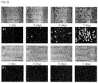
- Fig. 6 shows the time lapse images and processed images.
- A) and A') are resistant cases and B) and B') are susceptible cases.
- the formation of many colonies in the imaging area was found. After image processing, the white areas represent growing MTB colonies in the image.
- the susceptible cases there were no differences in MTB growth in the images.
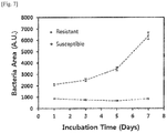
- the white area in the images was calculated by counting the pixel number. Using sequential digital data, the growth curve according to the time was plotted in Fig. 7 . By setting the threshold value, drug susceptibility of TB strains under various concentrations of TB drugs was determined.
- Fig. 7 shows quantified growth curves of the resistant and susceptible cases.
- the growth curves were constructed by measuring the growth areas of MTB in the images. Bacterial areas were continuously increased in the resistant case whereas no substantial changes were observed in the susceptible case.
- MTB DSTs are based on the estimation or measurement of the growth or non-growth of a MTB strain in the presence of a single "critical concentration" of one drug.
- the critical concentration of an anti-TB drug indicates clinically relevant resistance if growth is observed, whereas susceptible MTB strains are inhibited at the critical concentration.
- the critical concentration of each anti-TB drug is determined by the critical concentration of the MGIT because the DAC system is more similar to the liquid culture system than the solid culture system.
- DSTs were conducted using the DAC system at serial twofold concentrations near the critical concentration to acquire the minimum inhibitory concentration (MIC) and susceptibility of MTB.
- the MIC values of anti-TB drugs could be determined through sufficient tests at concentrations of the drugs. Thereafter, when the MIC value was at or below the critical concentration, the strain was considered to be susceptible, whereas resistant strains were defined by MIC values that exceed the critical concentration.
- the critical concentration of rifampin is 1.0 ⁇ g/ml in the DAC system.
- the DST using the DAC system was performed at 0.25, 0.5, 1.0, and 2.0 ⁇ g/ml concentrations of rifampin. Growth curves were plotted from the concentration of each anti-TB drug according to the incubation time. Susceptibility was determined by measuring MIC value based on the critical concentration.
- the DAC system enables rapid DST based on the concept of microfluidic channels.
- virulent standard strain of MTB, H37Rv and clinical strains of MDR and XDR strains were tested to determine the susceptibility of isoniazid, rifampicin, streptomycin, and ethambutol.
- the conventional LJ medium-based DST was used for quality control and as a comparison target because it is regarded as the 'gold standard' of DST.
- the MIC values of isoniazid, rifampicin, streptomycin, and ethambutol against H37Rv, MDR, and XDR were determined by using the DAC system.
- the susceptibility of H37Rv, MDR, and XDR to isoniazid, rifampicin, streptomycin, and ethambutol was determined below the critical concentrations or by measuring MIC values at concentrations other than the critical concentration.
- the images of all tested strains were taken at the same position every other day for 5 days using the time-lapse method.
- the images were processed using the daily time-lapse data for 5 days and were plotted to determine the MIC values of the four anti-TB drugs.
- H37Rv, MDR, and XDR TB strains were tested.
- the test concentrations were determined from the critical concentrations of the respective drugs.
- A shows MIC values of DST obtained from the DAC system and B shows susceptibility determined from the MIC data and the critical concentrations of the drugs.
- isoniazid, rifampicin, streptomycin, and ethambutol had MIC values of 0.05 ⁇ g/ml, 0.25 ⁇ g/ml, 2.0 ⁇ g/ml, and 5.0 ⁇ g/ml, respectively. From the MIC data, the strain H37Rv was found to be sensitive to isoniazid, rifampicin, streptomycin, and ethambutol. The same results were obtained also in LJ culture tests.
- the other two test strains from MDR and XDR patients were found to be resistant to all drugs in the DAC system.
- the sensitivity results were demonstrated using the LJ culture method regarded as the gold standard. All susceptibility results in the DAC system were consistent with the results from the gold standard method. From these verified results, it was demonstrated that the DAC system can reduce DST time and be used for accurate diagnosis of susceptibility and resistance to the anti-tuberculosis drugs.
- Example 2 Comparison of the DAC system with conventional culture diagnostic products
- the usefulness of the DAC system was confirmed by comparing the DAC system of the present invention with conventional TM culture diagnostic methods.
- the tuberculosis culture diagnosis or the LJ medium method used in the DST system takes 4 to 6 weeks.
- MGIT 960 (BD BACTEC TM ) is currently the fastest product for culture diagnosis of Mycobacterium tuberculosis.
- the culture diagnostic time of the DAC system according to the present invention was compared with that of the prior art.
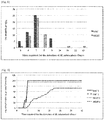
- Fig. 8 compares the times required for the detection of M. tuberculosis growth in 49 H37Rv sputum samples by the N-acetyl-L-cysteine-NaOH (NALC-NaOH) method on DAC and MGIT 960 systems.
- NALC-NaOH N-acetyl-L-cysteine-NaOH
- TTD was 5-13 days in the MGIT 960 system and 5-9 days in the DAC system.
- the highest detection level of M. tuberculosis was 7 days in the MGIT 960 and DAC systems.
- the times required for the detection of Mycobacterium tuberculosis were 1, 5, 6, 7, 8, 9, 10, 11, 12, 13, 14, 21, 28, 35, and 42 days.
- the median TTD values according to CFU values in Middlebrook 7H11 media were from 7 days (5-6 log 10 CFU/ml) to 9 days (2-3 log 10 CFU/ml) in the DAC system.
- TTD values according to CFU values in the Middlebrook 7H11 media were from 6 days (5-6 log 10 CFU/ml) to 12 days (2-3 log 10 CFU/ml) in MGIT 960 system.
- the median TTD values of M. tuberculosis in the MGIT 960 and DAC systems were 12 days and 9 days, respectively.
- Fig. 9 compares the results of the NALC-NaOH method (with concentration) or the direct inoculation method (without concentration) on the MGIT 960 and DAC systems.
- the above two sputum pretreatment method such as the DAC system culture method using the direct pretreatment method (without concentration) and the MGIT 960 culture method using the NALC-NaOH method (with concentration) method were found to have similar culture patterns.
- the median TTD was 7 days in the DAC1 (5-7 days), DAC2 (5-7 days), and MGIT1 systems (6-7 days).
- the median TTD required for the detection of M. tuberculosis from the samples was 14 days (7-24 days).
- the times required for the detection of M. tuberculosis. growth in three clinical sputum samples (3 positive samples of the 25 clinical sputum samples) and 49 H37Rv sputum samples depending on the recovery rate of mycobacteria in the DAC system are shown in the above Table 4.
- the times required for the detection of M. tuberculosis in the DAC system were 1, 5, 6, 7, 8, 9, 10, 11, 12, 13, 14, 21, 28, 35, and 42 days.
- the present inventors evaluated the antibacterial activities of rifampin, isoniazid, ethambutol, and streptomycin as primary drugs against TB using the DAC system and whether the DAC system was affected by changes in cell density. This system can draw reproducible and reliable MIC values of the drugs irrespective of inoculum size.
- M. tuberculosis was isolated from a pulmonary tuberculosis patient in the Korean Institute of Tuberculosis (KIT). For a total of 150 clinical strains including 134 pan-susceptible strains and 59 resistant strains, were identified for drug susceptibility using the L-J method before use. All isolates were freshly pre-cultured on L-J medium before use.
- Sputum samples were digested and decontaminated by the N-acetyl-L-cysteine-NaOH method described by Kent and Kubica.
- the treated samples were cultured in MGIT vials for 42 days. On the day of detection, the presence or absence of AFB in all positive tubes was examined by ZN staining and drug susceptibility testing was conducted with parallel tubes.
- E. coli strains Three clinical ESBL E. coli strains and one non-ESBL E. coli ATCC strain were used for lactamase substrate analysis.
- the E. coli strains were grown, subcultured, and analyzed on Mueller-Hinton agar (Difco) in Mueller-Hinton broth (Difco Laboratories, Detroit, MI).
- DST For the preparation of inoculation from the LJ culture, colonies up to 14 days were extracted from the grown culture. The colonies were shaken with glass beads to crush large lumps. To obtain a high density ( ⁇ 5 ⁇ 10 9 CFU/ml), the suspension was centrifuged at 3000 g for 10 min and pellets were resuspended in sterile PBS. Subsequently, the suspension was added to liquid agarose, inoculated in a bacteria-agarose mixture within the range of 10 1 to 5 ⁇ 10 8 CFU/ml, and loaded in the main channel. After solidification, 7H9 broth containing each antibiotic was applied to the chamber. All bacterial cells were imaged with an inverted optical microscope (IS71, Olympus).
- the LJ proportion method was used as a reference. All strains were tested according to the standard procedures on L-J media at concentrations of 40 ⁇ g/ml (RIF), 0.1 ⁇ g/ml (INH), 2.0 ⁇ g/ml (EMB), and 1.0 ⁇ g/ml (SM), except that they were inoculated in the range of 10 1 to 5 ⁇ 10 8 CFU/ml. Also in DST using the MGIT 960 system, all strains were tested according to the supplier recommendation except for the inoculum size.
- the MIC of cefepime against E. coli was determined according to the standard CLSI guideline except for the concentration of bacteria. 16 h after culture at 37 ⁇ , broth microdilution wells were examined.
- Fluorocillin Green (Invitrogen) was added to each well, and the plate was gently shaken at room temperature for 5 min and cultured at 37 ⁇ for 0.5 to 20 h. Changes in fluorescence were measured at an excitation wavelength of 485 nm and an emission wavelength of 530 nm.
- the culture media were filtered to remove the cells and then added to different DST sets to measure the residual efficacies of the antibiotics. Both broth microanalysis and DAC protocol were performed in the same manner as described above.
- DST was conducted against the standard strain H37Rv at various inoculum concentrations on the DAC system. Simultaneously, DST was conducted on the LJ and MGIT systems at the same concentrations as in the DAC system. TB was used at 10-fold concentrations (10 3 to 10 7 ). The inoculum size indicates the concentration of TB in agarose for the DAC system, the concentration of TB stock for the LJ system, and the final concentration of TB in the culture bottle for the MGIT system.
- MIC values were the same or different by about two-fold at various inoculum concentrations (See also Table 1).
- the standard strain H37Rv showed susceptibility to the four anti-tuberculosis drugs.
- Clinical strains express various phenotypes, particularly, in drug susceptibility.
- five clinical strains obtained from the KIT were tested at various inoculum concentrations of 10 3 to 10 7 CFU/ml, which is the concentration range proved to be effective in the test on the standard strain.
- Rifampicin, isoniazid, streptomycin, and ethambutol were used as test drugs.
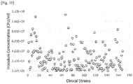
- Actual CFU values of the clinical strains are shown in Table 6. For the 131 strains, the highest concentration was 1.3 ⁇ 10 9 . .
- DST results obtained from the five clinical strains at 10 3 -10 7 CFU/ml are shown in Table 6.
- the three strains KHS, CMS, and CUD showed susceptibility to the three primary drugs.
- the DST results were the same or different by only about two-fold at 10 3 -10 7 . Therefore, susceptibility determination was not affected by changes in inoculum concentration and all strains were found to be susceptible.
- the 1088 and 131 strains were resistant to the four primary drugs. All MIC values were the same irrespective of inoculum concentration. Overall, no inoculum effect on drug susceptibility was found in the DAC system.
- the first reason is that large inoculum amounts of antibiotic degrading proteins derived from bacteria may destroy antibiotics and result in an increase in MIC value.
- the other reason is that larger inoculum amounts of antibiotic degrading proteins derived from bacteria may decrease the number of antibiotic molecules relative to single cells of bacteria and result in an increase in MIC.
- E. coli strains which are well known as a bacterial strain secreting antibiotic-degrading proteins, were tested at inoculum concentrations of 5 ⁇ 10 7 , 5 ⁇ 10 6 , and 5 ⁇ 10 5 CFU/ml. These concentrations indicate the concentrations of the bacteria in agarose for the DAC system and in liquid for the MDT system.
- Cefepime a fourth-generation cephalosporin antibiotic, was tested to demonstrate the presence of inoculum effect.
- the MIC ranges of the ESBL negative E . coli and ESBL positive E. coli were from 0.015 to 0.12 ⁇ g/ml.
- the DAC system of the present invention has the ability to control both molecular interaction and bacterial population dynamics, which are induced by excessive binding of the drug to bacteria when the cell density increases and the resulting products.
- the ability of the DAC system is because the bacterial population is fixed within the matrix and the medium containing each drug is supplied into the chamber in a gradient mode.
- This system facilitates DST without the need to strictly limit inoculation on mycobacteria because IE is not observed when it is inoculated in an inoculum amount larger than 10 7 mycobacteria per ml. Therefore, the DAC system enables accurate prediction of the microbiological efficacy of anti-tuberculosis drugs and is expected to effectively replace conventional approaches.
- the DAC system of the present invention was applied to antibiotic screening.
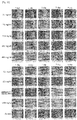
- the MIC of the drug against a first clinical strain from a TDR 102 patient was determined by treating the strain with 3.9, 7.8, 15.6, 31.2, 62.5, 125, 250, 500, and 1000 ng/ml of Q203 on the DAC system. Images in the DAC system are shown in Fig. 11 . The images were quantified and plotted. The results are shown in Fig. 12 .
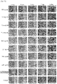
- the MIC of the drug against a second clinical strain from a TDR 103 patient was determined by treating the strain with Q203 in the same manner as described above. Images in the DAC system are shown in Fig. 13 . The images were quantified and plotted. The results are shown in Fig. 14 .
- One aspect of the present invention provides a bioactivity testing structure including a plate body, one or more culture units 100 arranged on the plate body, and a base plate 110 under which a solid thin film is to be formed on the bottom surface of each culture unit.
- Figs. 2a and 2b are perspective and plan views of a first embodiment of the testing structure according to the present invention, respectively.
- each of the culture units may have an inlet 111 formed in the base plate and through which a mixture solution of a liquid medium containing a gelling agent and a biological agent is to be introduced.
- Each of the culture units 100 may have one or more connection members 113 through which the base plate 110 is connected to barriers of the plate body and cut-away grooves 114 formed between the connection members.
- Each of the culture units may have one or more through-holes 112 formed in the base plate 110.
- a bioactive agent may be uniformly diffused into the solid thin film through the cut-away grooves 114 and the through-holes 112.
- the thickness and width of the solid thin film are determined depending on the depth and width of the receiving recess 120.
- the term "thin film” used herein refers to a thin layer that has a thickness sufficient to immobilize the biological agent and to observe the single cells.
- the thickness of the thin film may be in the range of 1 ⁇ m to 5 mm, 1 ⁇ m to 3 mm, 1 ⁇ m to 2 mm, 1 ⁇ m to 1.5 mm, 1 ⁇ m to 1 mm, 1 ⁇ m to 800 ⁇ m, 1 ⁇ m to 500 ⁇ m, 1 ⁇ m to 100 ⁇ m, 10 ⁇ m to 3 mm, 10 ⁇ m to 1 mm, 100 ⁇ m to 1 mm, 200 ⁇ m to 1 mm, or 500 ⁇ m to 1 mm, but is not particularly limited to this range.
- the thickness of the solid thin film may correspond to the size in the vertical direction of the solid thin film surface to be observed. Within the thickness range of the solid thin film defined above, the single cells of the biological agent immobilized in the solid thin film can be observed, the formation of colonies can be effectively observed or the colony forming units can be counted.
- each culture unit has a size of 1-50 mm ⁇ 1-50 mm, 1-30 mm ⁇ 1-30 mm, 5-50 mm ⁇ 5-50 mm, 5-30 mm ⁇ 5-30 mm, 10-50 mm ⁇ 10-50 mm, 10-30 mm ⁇ 10-30 mm or 1-20 mm x 1-20 mm. Most preferably, the culture unit has a size of 12 mm ⁇ 12 mm.
- each culture unit is also not limited and may vary depending on the kinds and characteristics of the biological agent and the bioactive agent.
- each culture unit may have a height of 1 to 50 mm, 1 to 30 mm, 5 to 30 mm or 5 to 15 mm.
- the diameter of the through-holes 112 is typically 1 mm or less and may be, for example, 0.99 mm or less, 0.90 mm, or less, 0.85 mm or less, 0.80 mm or less, 0.75 mm or less, 0.70 mm or less, 0.65 mm or less, 0.60 mm or less, 0.55 mm or less, 0.50 mm or less, 0.45 mm or less, 0.40 mm or less, 0.35 mm or less, 0.30 mm or less or 0.25 mm or less.
- the mixture solution of a liquid medium containing a gelling agent and a biological agent may be solidified under the base plate to form a biological agent-immobilized solid thin film.
- the liquid medium can contain about 80% or more of water or a buffer solution.
- the liquid medium can be solidified due to the presence of the gelling agent.
- the gelling agent there may be exemplified agar, agarose, gelatin, alginate, collagen or fibrin.
- agar or agarose is preferred.
- agar may be used in an amount of 0.5 to 5% by weight in the liquid medium.
- the liquid medium usually requires no nutrients. In some examples, however, the liquid medium may include nutrients.
- suitable biological agents include viruses, bacteria, fungi, algae, protozoa, parasitic pathogens, human and mammalian cells, and biofilms.
- the biological agent may grow in a liquid or solid medium and the growth thereof may be affected by the kind and concentration of a external physiologically active agent.
- the density of the biological agent in the mixture solution is not limited. Particularly, the present invention features in that the density of the biological agent is irrelevant to DST or diagnostic results. That is, the DAC system of the present invention shows no inoculum effect.
- the density of the biological agent may be in the range of 10 1 to 10 80 cfu/ml, for example, 10 70 cfu/ml or less, 10 60 cfu/ml or less, 10 50 cfu/ml or less, 10 40 cfu/ml or less, 10 30 cfu/ml or less, 10 20 cfu/ml or less, 10 10 cfu/ml or less, 10 9 cfu/ml or less, 10 8 cfu/ml or less, 10 7 cfu/ml or less, 10 6 cfu/ml or less, 10 5 cfu/ml or less or 10 4 cfu/ml or less.
- the mixture solution may be solidified under the base plate to form a biological agent-immobilized solid thin film.
- the medium As the temperature of the liquid medium decreases, the medium is solidified, and as a result, the biological agent is less mobile. Accordingly, the immobilization facilitates a continuous observation of the mobile biological agent.
- Figs. 3a and 3b are plan and perspective views of a second embodiment of a testing structure according to the present invention, respectively.
- each of the culture units may further have an receiving recess 120 formed on top of the base plate.
- the width of the receiving recess 120 is not limited and may be, preferably from 1 to 5 mm, for example, from 1 to 4.5 mm, from 1 to 4.0 mm or from 1 to 3.5 mm.
- the height of the receiving recess may be preferably from 1 to 3 mm, for example from 1 to 2.5 mm.
- the bioactive agent may be further introduced into the culture unit.
- the bioactive agent may physiologically affect the biological agent and examples thereof include: drugs such as antibiotics, anticancer agents and immunosuppressive agents, nutrients, cellular secretions, signal transducers, viruses, cells, micro RNAs, proteins, antigens, antibodies, and DNA.
- the bioactive agent in admixture with a liquid medium may be introduced into each culture unit.
- the bioactive agent may be introduced into the receiving recess 120.
- the bioactive agent may be in a freeze-dried form that is easy to transport and store.
- the plate body, the culture units, and the base plate may be partially or entirely made of a transparent or translucent material.
- the testing structure is a light-transmitting substrate, which is desirable in terms of optical imaging.
- the testing structure is not particularly limited so long as it has surface characteristics suitable for the formation of a thin film by the application of the liquid medium.
- a further aspect of the present invention provides a bioactivity testing method including: providing a mixture solution of a liquid medium containing a gelling agent and a biological agent to the bottom surface of the bioactivity testing structure; solidifying the mixture solution to form a solid thin film in which the biological agent is immobilized; supplying a bioactive agent to the solid thin film and allowing the bioactive agent to diffuse into the solid thin film; and observing the individual responses of the single cells of the biological agent to the bioactive agent or observing whether the biological agent forms colonies or the colony forming units (CFU).
- CFU colony forming units
- the method may further include imaging the individual responses of the single cells of the biological agent to the bioactive agent, the colonies of the biological agent or the colony forming units (CFU), and analyzing the images. More preferably, the method further includes determining the minimum inhibitory concentration (MIC) of the bioactive agent based on the analysis of the images.
- MIC minimum inhibitory concentration
- the optical measurement system may include an imaging system, such as a CCD or CMOS camera.
- the optical measurement system may include optical units or devices necessary for focusing and light imaging, such as a lens, an illuminator, and a light guide.
- the optical measurement system may include an image processing system for processing and analyzing image data observed by the imaging system. The optical measurement system rapidly records and analyzes changes in the growth of the biological agent observed during testing to obtain test results.
- the bioactivity testing method may be utilized in various applications, including antibiotic susceptibility testing of bacteria, antibiotic screening, and diagnosis of bacteria.
- Another aspect of the present invention provides an antibiotic screening method including: providing a mixture solution of a liquid medium containing a gelling agent and a bacterial strain to the bottom surface of the bioactivity testing structure; solidifying the mixture solution to form a solid thin film in which the bacterial strain is immobilized; supplying an antibiotic to the solid thin film and allowing the antibiotic to diffuse into the solid thin film; and observing the individual responses of the single cells of the bacterial strain to the antibiotic or observing whether the bacterial strain forms colonies or the colony forming units (CFU).
- CFU colony forming units
- the method may further include imaging the individual responses of the single cells, the colonies or the colony forming units (CFU), analyzing the images, and determining the minimum inhibitory concentration (MIC) of the antibiotic based on the analysis of the images.
- CFU colony forming units
- Another aspect of the present invention provides a bacterial diagnostic method including: providing a mixture solution of a liquid medium containing a gelling agent and a bacterial strain to the bottom surface of the bioactivity testing structure; solidifying the mixture solution to form a solid thin film in which the bacterial strain is immobilized; supplying a culture medium of the bacterial strain to the solid thin film and allowing the culture medium to diffuse into the solid thin film; and observing the individual responses of the single cells of the bacterial strain or observing whether the bacterial strain forms colonies or the colony forming units (CFU).
- CFU colony forming units
- the culture medium of the bacterial strain may be further supplemented with an antibiotic and the diagnostic method may be carried out simultaneously with drug susceptibility testing.
- the bioactivity testing may be drug susceptibility, drug screening or bacterial culture diagnosis testing.
- the bacterial strain is Mycobacterium tuberculosis. or a nontuberculous mycobacterium (NTM).
- the Mycobacterium tuberculosis may be multi-drug-resistant (MDR) tuberculosis, extensively drug-resistant (XDR) tuberculosis or totally drug-resistant (TDR) tuberculosis.
- bioactivity testing system including: the bioactivity testing structure including culture units in which a bioactive agent is provided and a base plate under which a mixture solution of a liquid medium containing a gelling agent and a biological agent is solidified to form a thin film on the bottom surface of each culture unit; a stage for supporting and observing the testing structure; and a measurement system for imaging the individual responses of the single cells of the biological agent or the formation of colonies of the biological agent when the bioactive agent is delivered by diffusion or imaging the colony forming units (CFU), analyzing the images, and determining the minimum inhibitory concentration (MIC) of the bioactive agent based on the analysis of the images.
- CFU colony forming units
- the measurement system may include an imaging system and an image processing system.
- the imaging system may be a CCD or CMOS camera.
- the image processing system is adapted to process and analyze image data observed by the imaging system.
- Culture unit 120 Receiving recess 110: Base plate 111: Inlet 112: Through-holes 113: Connection members 114: Cut-away grooves
Landscapes
- Chemical & Material Sciences (AREA)
- Health & Medical Sciences (AREA)
- Life Sciences & Earth Sciences (AREA)
- Engineering & Computer Science (AREA)
- Organic Chemistry (AREA)
- Bioinformatics & Cheminformatics (AREA)
- Zoology (AREA)
- Wood Science & Technology (AREA)
- Biotechnology (AREA)
- Biochemistry (AREA)
- General Health & Medical Sciences (AREA)
- Microbiology (AREA)
- Genetics & Genomics (AREA)
- General Engineering & Computer Science (AREA)
- Sustainable Development (AREA)
- Biomedical Technology (AREA)
- Proteomics, Peptides & Aminoacids (AREA)
- Immunology (AREA)
- Analytical Chemistry (AREA)
- Medicinal Chemistry (AREA)
- Molecular Biology (AREA)
- Biophysics (AREA)
- Physics & Mathematics (AREA)
- Clinical Laboratory Science (AREA)
- Toxicology (AREA)
- Cell Biology (AREA)
- Pharmacology & Pharmacy (AREA)
- Food Science & Technology (AREA)
- General Physics & Mathematics (AREA)
- Pathology (AREA)
- Measuring Or Testing Involving Enzymes Or Micro-Organisms (AREA)
Applications Claiming Priority (2)
| Application Number | Priority Date | Filing Date | Title |
|---|---|---|---|
| PCT/KR2014/009755 WO2016060301A1 (ko) | 2014-10-16 | 2014-10-16 | 고정화제를 이용한 단일 세포 추적을 위한 신규한 생리 활성 검사용 구조물 |
| KR1020140139700A KR101770610B1 (ko) | 2014-10-16 | 2014-10-16 | 고정화제를 이용한 단일 세포 추적을 위한 신규한 생리 활성 검사용 구조물 |
Publications (2)
| Publication Number | Publication Date |
|---|---|
| EP3202885A1 true EP3202885A1 (de) | 2017-08-09 |
| EP3202885A4 EP3202885A4 (de) | 2018-07-18 |
Family
ID=55746830
Family Applications (1)
| Application Number | Title | Priority Date | Filing Date |
|---|---|---|---|
| EP14903914.1A Pending EP3202885A4 (de) | 2014-10-16 | 2014-10-16 | Neuartige biologische aktivitätsteststruktur zur verfolgung einer einzelzelle unter verwendung von geliermitteln |
Country Status (5)
| Country | Link |
|---|---|
| US (1) | US10533207B2 (de) |
| EP (1) | EP3202885A4 (de) |
| KR (1) | KR101770610B1 (de) |
| CN (1) | CN107109331B (de) |
| WO (1) | WO2016060301A1 (de) |
Cited By (2)
| Publication number | Priority date | Publication date | Assignee | Title |
|---|---|---|---|---|
| EP3725871A4 (de) * | 2017-12-13 | 2021-09-29 | Hitachi High-Tech Corporation | Bakterielle testplatte mit darin eingeführtem antibakteriellem wirkstoff und transparente platte |
| EP4022079A4 (de) * | 2019-08-27 | 2024-07-03 | Selux Diagnostics, Inc. | Systeme und verfahren zur durchführung einer antimikrobiellen empfindlichkeitsprüfung |
Families Citing this family (10)
| Publication number | Priority date | Publication date | Assignee | Title |
|---|---|---|---|---|
| US10071141B2 (en) | 2015-05-08 | 2018-09-11 | Spectral Platforms, Inc. | Albumin-based non-covalent complexes and methods of use thereof |
| WO2018187548A2 (en) * | 2017-04-05 | 2018-10-11 | Arizona Board Of Regents On Behalf Of Arizona State University | Antimicrobial susceptibility testing with large-volume light scattering imaging and deep learning video microscopy |
| CN107130011A (zh) * | 2017-05-09 | 2017-09-05 | 上海理工大学 | 一种食源性致病菌单细胞生长过程的显微观测方法 |
| PL235845B1 (pl) * | 2017-11-10 | 2020-11-02 | Szkola Glowna Gospodarstwa Wiejskiego W Warszawie | Sposób wielostronnej oceny biozgodności materiałów |
| CA3097400A1 (en) * | 2018-04-20 | 2019-10-24 | Advanced Biological Services, Inc. | Systems and methods for treating wastewater and providing class a sludge |
| KR20200044450A (ko) * | 2018-10-19 | 2020-04-29 | 주식회사 이지다이아텍 | 세포의 격리 및 형태학적 분석 방법 |
| KR102132630B1 (ko) * | 2020-03-16 | 2020-07-10 | 주식회사 퀀타매트릭스 | 섬 구조물을 포함하는 신속한 세포배양검사 장치 |
| EP4168732A4 (de) * | 2020-06-23 | 2025-02-12 | Spectral Platforms, Inc. | Spektroskopische verfahren, reagenzien und systeme zum nachweis, zur identifizierung und zur charakterisierung von bakterien für antimikrobielle suszeptibilität |
| CN117625394B (zh) * | 2023-12-01 | 2024-12-06 | 东莞合安机电有限公司 | 一种荔枝菌菌种培养液、制备方法及培养方法 |
| CN120555166A (zh) * | 2025-04-15 | 2025-08-29 | 科茵(上海)医疗技术有限公司 | 一种生物微流芯片装置及其在微生物运动模式检测中的用途 |
Family Cites Families (18)
| Publication number | Priority date | Publication date | Assignee | Title |
|---|---|---|---|---|
| AU658079B2 (en) * | 1992-09-28 | 1995-03-30 | Becton Dickinson & Company | Cell culture insert |
| US6251624B1 (en) * | 1999-03-12 | 2001-06-26 | Akzo Nobel N.V. | Apparatus and method for detecting, quantifying and characterizing microorganisms |
| WO2004055793A1 (en) | 2002-12-18 | 2004-07-01 | Koninklijke Philips Electronics N.V. | Optical pickup apparatus. |
| US7122153B2 (en) * | 2003-01-08 | 2006-10-17 | Ho Winston Z | Self-contained microfluidic biochip and apparatus |
| EP1479760A1 (de) * | 2003-05-19 | 2004-11-24 | ProBioGen AG | Künstliches Immunorgan |
| ES2614425T3 (es) * | 2004-01-08 | 2017-05-31 | Dako Denmark A/S | Aparato y métodos para el procesamiento de muestras biológicas y un depósito para los mismos |
| KR100733914B1 (ko) * | 2005-09-22 | 2007-07-02 | 한국과학기술원 | 미세유체 기술을 이용한 3차원 세포배양 시스템 |
| WO2007053561A2 (en) | 2005-11-01 | 2007-05-10 | Rensselaer Polytechnic Institute | Three-dimensional cellular array chip and platform for toxicology assays |
| US8501462B2 (en) * | 2007-02-27 | 2013-08-06 | The Board Of Trustees Of The University Of Illinois | Insert device for multiwell plate |
| WO2009126524A2 (en) * | 2008-04-08 | 2009-10-15 | Massachusetts Institute Of Technology | Three-dimensional microfluidic platforms and methods of use thereof |
| WO2012050981A1 (en) * | 2010-09-29 | 2012-04-19 | Massachusetts Institute Of Technology | Device for high throughput investigations of cellular interactions |
| FR2972117B1 (fr) * | 2011-03-04 | 2013-12-20 | Centre Nat Rech Scient | Systeme microfluidique pour controler un profil de concentration de molecules susceptibles de stimuler une cible |
| US10526572B2 (en) * | 2011-04-01 | 2020-01-07 | EMD Millipore Corporaticn | Cell culture and invasion assay method and system |
| JP5516482B2 (ja) | 2011-04-11 | 2014-06-11 | 東京エレクトロン株式会社 | 基板搬送方法、基板搬送装置、及び塗布現像装置 |
| KR101965737B1 (ko) | 2011-10-19 | 2019-08-27 | 엘지전자 주식회사 | 공기조화기의 전동기 구동장치 |
| KR101290648B1 (ko) * | 2012-02-02 | 2013-07-29 | 서울대학교산학협력단 | 고형화제를 이용한 세균 고정, 약물 확산, 세균 단일 세포 추적과 이를 기반으로 한 빠른 약물 검사 시스템 |
| KR20140053582A (ko) | 2012-10-26 | 2014-05-08 | 단국대학교 산학협력단 | 절화의 선도유지 및 개화를 지연시키는 절화의 포장방법 |
| WO2014178692A1 (ko) * | 2013-05-02 | 2014-11-06 | 주식회사 퀀타매트릭스 | 마이크로플루이딕 멀티-웰 기반의 세포배양검사 장치 |
-
2014
- 2014-10-16 CN CN201480082659.4A patent/CN107109331B/zh active Active
- 2014-10-16 WO PCT/KR2014/009755 patent/WO2016060301A1/ko not_active Ceased
- 2014-10-16 US US15/518,668 patent/US10533207B2/en active Active
- 2014-10-16 EP EP14903914.1A patent/EP3202885A4/de active Pending
- 2014-10-16 KR KR1020140139700A patent/KR101770610B1/ko active Active
Cited By (4)
| Publication number | Priority date | Publication date | Assignee | Title |
|---|---|---|---|---|
| EP3725871A4 (de) * | 2017-12-13 | 2021-09-29 | Hitachi High-Tech Corporation | Bakterielle testplatte mit darin eingeführtem antibakteriellem wirkstoff und transparente platte |
| US11571698B2 (en) | 2017-12-13 | 2023-02-07 | Hitachi High-Tech Corporation | Bacterial test plate having antibacterial agent introduced thereinto, and transparent plate |
| EP4022079A4 (de) * | 2019-08-27 | 2024-07-03 | Selux Diagnostics, Inc. | Systeme und verfahren zur durchführung einer antimikrobiellen empfindlichkeitsprüfung |
| US12312632B2 (en) | 2019-08-27 | 2025-05-27 | SeLux Diagnostics, Inc. | Systems and methods for performing antimicrobial susceptibility testing |
Also Published As
| Publication number | Publication date |
|---|---|
| CN107109331B (zh) | 2025-02-14 |
| WO2016060301A1 (ko) | 2016-04-21 |
| KR20160045171A (ko) | 2016-04-27 |
| CN107109331A (zh) | 2017-08-29 |
| US20170233786A1 (en) | 2017-08-17 |
| EP3202885A4 (de) | 2018-07-18 |
| KR101770610B1 (ko) | 2017-08-24 |
| US10533207B2 (en) | 2020-01-14 |
Similar Documents
| Publication | Publication Date | Title |
|---|---|---|
| US10533207B2 (en) | Bioactivity testing structure for single cell tracking using gelling agents | |
| CN105209595B (zh) | 微流体多孔型细胞培养测试装置 | |
| US9399788B2 (en) | Method for inspecting susceptibility of bacteria or fungi to antimicrobial drug and system for use in the same | |
| EP3434370B1 (de) | Zellkulturtestvorrichtung basierend auf mehreren vertiefungen zur schnellen antibiotischen empfindlichkeitsprüfung | |
| US20200048680A1 (en) | Use of a fluidic device | |
| Tutty et al. | Cancer cell culture: the basics and two-dimensional cultures | |
| US11976316B2 (en) | Phenotypic characterization of cells | |
| Cerca et al. | Comparative evaluation of coagulase-negative staphylococci (CoNS) adherence to acrylic by a static method and a parallel-plate flow dynamic method | |
| Hu et al. | A digital plating platform for robust and versatile microbial detection and analysis | |
| JP6346637B2 (ja) | 薬剤耐性菌検出用ディスク試験片 | |
| Abe et al. | Precision single cell analysis to characterize host dependent antimicrobial response heterogeneity in physiological medium | |
| EP3947631B1 (de) | Kassettenanordnung | |
| Chauhan et al. | Rapid and efficient labeling by a selective organic fluorophore probe highlights heterogeneity of mycobacterial populations and persister resuscitation | |
| Miyahara et al. | Survival of ampicillin-treated uropathogenic Escherichia coli is independent of single-cell growth rates | |
| Windels et al. | Isolation of persisters enabled by ß-lactam-induced filamentation reveals their single-cell awakening characteristics | |
| Zhang | Rapid antibiotic susceptibility testing platform for direct clinical samples | |
| Annisa et al. | Viability Test of Mycobacterium Tuberculosis Bacteria Stored in Various Cryopreservation Periods | |
| 정현용 | Direct, rapid antimicrobial susceptibility testing for expediting optimal antibiotic treatment of bloodstream infection | |
| Ambastha | Developing a Microfluidic Device for Detecting Antibiotic Tolerance in Bacteria | |
| RU2255977C1 (ru) | Способ определения лекарственной чувствительности микобактерий туберкулеза | |
| Umetani et al. | Actively growing cells are the predominant persisters in exponential phase of Escherichia coli | |
| KR20230096352A (ko) | 바이오 3d 프린터를 이용한 순환 종양세포 검사방법 | |
| KR20240176080A (ko) | 바이오 의약품의 안전성 입증을 위한 신속 무균 시험 방법 및 신속 무균 시험 플랫폼 | |
| Buchar Klinovská et al. | Comparison of Coculture Models of Eukaryotic Cells With Pathogenic and/or Commensal Bacteria for Tissue Engineering | |
| EP3378945B1 (de) | Verfahren zur unterscheidung von einzelnen zellen aus einer mischung aus heterogenen zellen |
Legal Events
| Date | Code | Title | Description |
|---|---|---|---|
| STAA | Information on the status of an ep patent application or granted ep patent |
Free format text: STATUS: THE INTERNATIONAL PUBLICATION HAS BEEN MADE |
|
| PUAI | Public reference made under article 153(3) epc to a published international application that has entered the european phase |
Free format text: ORIGINAL CODE: 0009012 |
|
| STAA | Information on the status of an ep patent application or granted ep patent |
Free format text: STATUS: REQUEST FOR EXAMINATION WAS MADE |
|
| 17P | Request for examination filed |
Effective date: 20170502 |
|
| AK | Designated contracting states |
Kind code of ref document: A1 Designated state(s): AL AT BE BG CH CY CZ DE DK EE ES FI FR GB GR HR HU IE IS IT LI LT LU LV MC MK MT NL NO PL PT RO RS SE SI SK SM TR |
|
| AX | Request for extension of the european patent |
Extension state: BA ME |
|
| RAP1 | Party data changed (applicant data changed or rights of an application transferred) |
Owner name: QUANTA MATRIX CO., LTD. |
|
| RIN1 | Information on inventor provided before grant (corrected) |
Inventor name: CHOI, JUNGIL Inventor name: KIM, HYEJIN Inventor name: KIM, HEE JIN Inventor name: PARK, KYUNG-OCK Inventor name: KWON, SUNGHOON Inventor name: KIM, EUN GEUN Inventor name: KIM, HAEUN Inventor name: JEOUNG, HYEON JU Inventor name: RYOO, SUNG WEON Inventor name: YOO, JUNG HEON Inventor name: LEE, EUN HEE Inventor name: JUNG, YONG-GYUN |
|
| DAX | Request for extension of the european patent (deleted) | ||
| A4 | Supplementary search report drawn up and despatched |
Effective date: 20180620 |
|
| RIC1 | Information provided on ipc code assigned before grant |
Ipc: C12M 1/16 20060101ALI20180614BHEP Ipc: C12Q 1/02 20060101AFI20180614BHEP Ipc: C12M 1/18 20060101ALI20180614BHEP Ipc: C12M 1/14 20060101ALI20180614BHEP Ipc: C12M 1/34 20060101ALI20180614BHEP |
|
| STAA | Information on the status of an ep patent application or granted ep patent |
Free format text: STATUS: EXAMINATION IS IN PROGRESS |
|
| 17Q | First examination report despatched |
Effective date: 20200515 |