EP3207375B1 - Nachweis von igm antikörpern mit einem verbesserten immunkomplex (ic) elisa - Google Patents
Nachweis von igm antikörpern mit einem verbesserten immunkomplex (ic) elisa Download PDFInfo
- Publication number
- EP3207375B1 EP3207375B1 EP15778343.2A EP15778343A EP3207375B1 EP 3207375 B1 EP3207375 B1 EP 3207375B1 EP 15778343 A EP15778343 A EP 15778343A EP 3207375 B1 EP3207375 B1 EP 3207375B1
- Authority
- EP
- European Patent Office
- Prior art keywords
- antigen
- fcμr
- igm
- immune complexes
- antibodies
- Prior art date
- Legal status (The legal status is an assumption and is not a legal conclusion. Google has not performed a legal analysis and makes no representation as to the accuracy of the status listed.)
- Active
Links
Images




Classifications
-
- G—PHYSICS
- G01—MEASURING; TESTING
- G01N—INVESTIGATING OR ANALYSING MATERIALS BY DETERMINING THEIR CHEMICAL OR PHYSICAL PROPERTIES
- G01N33/00—Investigating or analysing materials by specific methods not covered by groups G01N1/00 - G01N31/00
- G01N33/48—Biological material, e.g. blood, urine; Haemocytometers
- G01N33/50—Chemical analysis of biological material, e.g. blood, urine; Testing involving biospecific ligand binding methods; Immunological testing
- G01N33/53—Immunoassay; Biospecific binding assay; Materials therefor
- G01N33/566—Immunoassay; Biospecific binding assay; Materials therefor using specific carrier or receptor proteins as ligand binding reagents where possible specific carrier or receptor proteins are classified with their target compounds
-
- G—PHYSICS
- G01—MEASURING; TESTING
- G01N—INVESTIGATING OR ANALYSING MATERIALS BY DETERMINING THEIR CHEMICAL OR PHYSICAL PROPERTIES
- G01N33/00—Investigating or analysing materials by specific methods not covered by groups G01N1/00 - G01N31/00
- G01N33/48—Biological material, e.g. blood, urine; Haemocytometers
- G01N33/50—Chemical analysis of biological material, e.g. blood, urine; Testing involving biospecific ligand binding methods; Immunological testing
- G01N33/68—Chemical analysis of biological material, e.g. blood, urine; Testing involving biospecific ligand binding methods; Immunological testing involving proteins, peptides or amino acids
- G01N33/6854—Immunoglobulins
-
- G—PHYSICS
- G01—MEASURING; TESTING
- G01N—INVESTIGATING OR ANALYSING MATERIALS BY DETERMINING THEIR CHEMICAL OR PHYSICAL PROPERTIES
- G01N2333/00—Assays involving biological materials from specific organisms or of a specific nature
- G01N2333/435—Assays involving biological materials from specific organisms or of a specific nature from animals; from humans
- G01N2333/705—Assays involving receptors, cell surface antigens or cell surface determinants
- G01N2333/70503—Immunoglobulin superfamily, e.g. VCAMs, PECAM, LFA-3
- G01N2333/70535—Fc-receptors, e.g. CD16, CD32, CD64 (CD2314/705F)
Definitions
- the present invention relates to the field of diagnostic or analytic methods.
- an Fc receptor for IgM heavy chains Fc ⁇ R
- a functional fragment thereof which preferably comprises the Ig-like domain of Faim 3/Toso
- IgM or IgA antibodies preferably, IgM
- immune complexes i.e., complexes formed by antigen and specific antibody.
- immune complexes may be formed by mixing an antigen with a sample comprising antibodies.
- a method for detection and quantification of IgA or IgM antibodies in the form of immune complexes is also provided. This can be useful for diagnosis of infections, e.g., with a virus.
- IgM antibodies belong to the first line of defense of the specific immune system against pathogens. IgM antibodies are produced by B-cells before class switch to IgG. Detection of IgM is thus especially suited for early diagnosis of an infection. IgM have a different structure from IgG, which leads to a particularly high avidity of the antibody, as each IgM has 10 antigen binding sites instead of 2 for IgG.
- IgM is classically detected by the IgM antibody-capture (MAC) approach, i.e., a test serum of a patient is applied to a microtitration plate previously coated with anti-human IgM, followed by application of a labelled antigen (Duermeyer W, Wielaard F. et al, 1979; Ong et al, 1989, Vorndam V., Kuno G., 1997, Balmaseda A., Guzman, M.G. et al, 2003, Vasquez, Hafner et al, 2007).
- MAC IgM antibody-capture
- EP 2 492 689 A1 describes the use of Fc ⁇ R for in vitro quantification of antibodies, in particular IgG or IgY antibodies, in the form of immune complexes.
- TOSO/Fas apoptic inhibitory molecule 3 (TOSO/FAIM3) as an Fc receptor for IgM (Fc ⁇ R), and describes the binding of IgM to Fc ⁇ R expressed by HeLa cells.
- the inventors address the problem of providing improved means for detection of IgM and/or IgA antibodies.
- the invention provides use of an Fc ⁇ R, which is immobilized on a solid support, for in vitro quantifying IgM or IgA antibodies in the form of immune complexes, wherein the immune complexes are formed by mixing an antigen with a sample comprising antibodies.
- IgM receptors Muscular et al., 1977, Hoover et al., 1981
- IgA receptor Ohno et al., 1990
- Fas apoptotic inhibitory molecule 3 (FAIM3) or TOSO (Hitoshi et al, 1998).
- FIM3 Fas apoptotic inhibitory molecule 3
- TOSO Hitoshi et al, 1998
- the former designation was incorrect, as the molecule did not induce apoptosis.
- the misinterpretation was due to use of mouse IgM anti-Fas mAb (CH11), which induced Fas-mediated apoptosis.
- Fc ⁇ R Fc receptor
- the Fc ⁇ R consists of a 107-aa V-set Ig-like domain responsible for IgM binding, an additional 127-aa extra cellular region with no known domain structure, a 21-aa transmembrane segment containing a charged His- residue, and a relatively long (118-aa) cytoplasmic tail (Kubagawa et al, 2014).
- mouse IgM bound better to the human Fc ⁇ R than human IgM.
- a soluble serum Fc ⁇ R resolved as a ⁇ 40 kDa protein, distinct from the cell surface Fc ⁇ R of ⁇ 60 kDa, is produced by both B and non-L B cells by a splicing process of the Fc ⁇ R cDNA.
- IgA-binding and IgA/ IgM-binding receptors which have been found on B-lymphocytes, and which are of special interest in the present context.
- the Fc receptor designated Fc ⁇ / ⁇ R or CD351 has been characterized in mice and humans, and binds both IgM and IgA with intermediate or high affinity. It can therefore be considered a further Fc ⁇ R.
- Human Fc ⁇ / ⁇ R is constitutively expressed on the majority of B lymphocytes and macrophages (Shibuya et al, 2000). To further characterize this receptor, it was expressed on the surface of the 293T cell line and affinity of the interactions of the receptor with IgA and IgM was measured (Yoo et al, 2011 , van der Boog et al, 2002).
- FcRI human myeloid-specific IgA Fc receptor
- CD89 human myeloid-specific IgA Fc receptor
- Fc ⁇ RI or CD89 is expressed on the surface of eosinophils, neutrophils, dendritic cells, monocytes and macrophages.
- the Fc ⁇ R may be Faim 3/Toso or a functional fragment thereof capable of binding to immune complexes, or CD351 or a functional fragment thereof capable of binding to immune complexes are of special interest. If the Fc ⁇ R is Faim 3/Toso or a fragment thereof, the antibody is of the IgM class. If Fc ⁇ R is CD351 or a fragment thereof, the antibody is of the IgM or IgA class.
- the Fc ⁇ R is Faim 3/Toso or a functional fragment thereof capable of binding to immune complexes. It may comprise an amino acid sequence having at least 80%, at least 90%), at least 95%, at least 99% or 100% sequence identity with, preferably, human Faim 3/Toso (SEQ ID NO: 1). (See Fig. 1 ).
- the fragment of the Fc ⁇ R preferably comprises an amino acid sequence having at least 80%, at least 90%, at least 95%, at least 99% or 100% sequence identity with the functional Faim 3/Toso fragment, in particular, human Faim 3/Toso fragment.
- the area of the defined sequence identity is capable of binding to immune complexes, e.g., as tested in the examples below.
- the functional fragment comprises the Ig-like domain of Faim 3/Toso, which preferably has SEQ ID NO: 2 or at least 80%, more preferable, at least 85%, at least 90%, at least 95% or at least 99% amino acid identity to SEQ ID NO: 2, or which varies from SEQ ID NO: 2 in only one or two amino acids. It may also consist or essentially consist of the Ig-like domain. In the context of this application, "essentially consist" means that at least 70%, preferably, at least 75%, at least 77%, at least 80%, at least 90% at least 95%, or at least 99% of the proteins amino acids of the protein correspond to the Ig-like domain of Faim 3/Toso.
- Amino acids not corresponding to the Ig-like domain of Faim 3 /Toso may be amino acids of a tag for purification, e.g., a His-Tag, or a linker, e.g., incorporating a cleavage site.
- the fragment does not comprise the complete extracellular domain of Faim 3/Toso.
- it does not comprise SEQ ID NO: 5, i.e., the part of Faim 3/Toso C-terminal to the Ig-like domain.
- the functional fragment of Faim 3/Toso does not comprise more than 10%, more than 20%, more than 30%, more than 40%, more than 50%, more than 60%, more than 70%, more than 80%, more than 90% of the part of Faim 3/Toso C-terminal to the Ig-like domain.
- the Fc ⁇ R does not comprise a His-Tag, in particular, if the sample may comprise anti-malaria antibodies.
- Anti-malaria-antibodies have been shown to be able to cross-react with His-tags under certain conditions.
- a His-Tag may be used for purification of the Fc ⁇ R, but it is preferably linked to the Fc ⁇ R through a protease cleavage site, so the His-Tag may be cleaved off, if needed, for example, to prevent cross-reactions with anti-malaria antibodies. If the His-Tag does not interfere with detection of immune complexes, it may be incorporated in the final product.
- His 10 -Xa-3C-Fc ⁇ R-Igl (SEQ ID NO: 3) or a protein having at least 80%, at least 90%, at least 95%, or at least 99% amino acid identity to SEQ ID NO: 3 is a preferred Fc ⁇ R of the present invention.
- the inventors also surprisingly showed that monomeric Fc ⁇ R-Ig1, as described above, preferably a protein having at least 80%, at least 85%, at least 90%, at least 95%, at least 99% or 100% amino acid identity with SEQ ID NO: 2, or, preferably, a monomeric protein having at least 80%, at least 90%, at least 95%, at least 99% or 100% amino acid identity to His 10 -Xa-3C-Fc ⁇ R-Igl (SEQ ID NO: 3), or consisting of SEQ ID NO:3, is functional in the IgM immune complex binding assay, whereas high molecular weight complexes thereof are not.
- a monomeric Fc ⁇ R-Ig1 is used, e.g., a composition comprising monomeric Fc ⁇ R-Ig1, preferably, a composition comprising at least 30%, at least 50%, at least 60%, at least 70%, at least 80%, at least 90%, at least 95%, at least 99% or 100% monomeric Fc ⁇ R-Ig1.
- the content of specific forms can be determined after detection of peaks by size exclusion chromatography and detection at 280 nm, as described below.
- the monomeric form may have a calculated molecular weight of 14-16 kDa, preferably, about 15 kDa, or 15.4 kDa.
- composition comprising a protein comprising SEQ ID NO: 2 or essentially consisting of a protein having at least 80% amino acid identity to SEQ ID NO: 2, or consisting of a protein having at least 80% amino acid identity to SEQ ID NO: 2, optionally, linked to a tag suitable for purification such as a His-Tag.
- Said composition preferably comprises at least 50%, preferably, at least 60%, at least 70%, at least 80%, at least 90%, at least 95%, at least 99% or 100% of said protein in monomeric form. % relate to mol monomer / mol total Fc ⁇ R-Igl.
- composition further comprises high molecular weight aggregates of Fc ⁇ R-Igl
- amount of Fc ⁇ R-Igl used in an assay of the invention should be adapted accordingly.
- the Fc ⁇ R may be CD351 or a functional fragment thereof capable of binding to immune complexes. It may comprise an amino acid sequence having at least 80%, at least 90%, at least 95%, at least 99% or 100% sequence identity with, preferably, human CD351.
- the fragment of the Fc ⁇ R preferably comprises an amino acid sequence having at least 80%, at least 90%, at least 95%, at least 99% or 100% sequence identity with the functional CD351 fragment, in particular, human CD351 fragment.
- the area of the defined sequence identity is capable of binding to immune complexes, e.g., as tested in the examples below.
- Fc ⁇ Rs are available. For example, they can be expressed in E. coli. Expression in mammalian expression systems, e.g., COS cells or CHO cells, is also possible. Accordingly, Fc ⁇ R may be glycosylated or non-glycosylated.
- the inventors have shown that non-glycosylated forms of Faim/Toso such as SEQ ID NO: 2, in particular, SEQ ID NO: 3, were able to bind the immune complexes and performed well in the method of the invention.
- Faim 3/Toso in particular a fragment of Faim 3/Toso based on the Ig-like domain and not including the complete part of Faim 3/Toso C-terminal to the Ig-like domain is particularly well suited to bind the immune complexes for their detection.
- the immune complexes comprise an antigen and an antibody specific for said antigen, thus allowing quantification of specific antibodies.
- the immune complexes are preferably formed by mixing an antigen with a sample comprising antibodies.
- the antigen is detectable, i.e., preferably, it is a labelled antigen, e.g., an enzyme labelled antigen (ELA), which allows for easy detection of the immune complexes.
- ELA enzyme labelled antigen
- the enzyme may be peroxidase, e.g., horseradish peroxidase. Alkaline phosphatase may also be used.
- Other labelling methods like biotinylation or labelling with a fluorescent moiety such as FITC, PE or a fluorescent protein, may also be used.
- the antigen may be unlabeled if it comprises more than one epitope.
- a second, a labelled antibody, preferably monoclonal, may then be used to detect and quantify the immune complexes.
- detection may be performed by adding an avidin- or streptavidin-labelled enzyme in a further step. Addition of a substrate of the enzyme, as well-known in the state of the art, allows for quantification of label, and consequently, of antibody in immune complexed form.
- the immune complexes may thus be quantified to determine or to quantify presence of specific antibodies in the sample.
- the antibodies which form the immune complexes will be of the IgM or IgA class, preferably, IgM.
- the composition comprising antibodies that is analysed is a sample, e.g., a biological sample, such as a sample from a patient. It may also be a sample from a subject known to have, or not to have the antibodies of interest, e.g., as a positive or negative control. Preferably, samples from a subject and a positive and/or negative control are analysed in the same test to allow direct comparison.
- the sample may be derived from blood (e.g., plasma or, preferably, serum), stool, saliva, sputum, or liquor cerebrospinalis.
- the sample e.g., serum
- the sample may be diluted before contacting the Fc ⁇ R, e.g., diluted 1:10 or more, or 1:100 or more, e.g., in a suitable buffer such as PBS or citrate buffer, or it may be otherwise processed, e.g., by removing certain components, such as cells from blood or by concentration.
- the sample may be from a human subject or an animal, e.g., a goat, sheep, horse, cattle, a bird, e.g., chicken, or a rodent such as mouse, rat, rabbit, guinea pig or hamster.
- the immune complexes may thus be formed by an antigen such as an enzyme-linked antigen and by antibodies which are derived from a species selected from the group comprising human, goat, sheep, horse, cattle, an avian species, e.g., chicken, or a rodent such as mouse, rat, rabbit, guinea pig or hamster.
- the Fc ⁇ R preferably is a human Fc ⁇ R.
- the inventors were able to show that human Fc ⁇ R, in particular, a fragment comprising the Ig-like domain, has a surprisingly wide interspecies reactivity.
- murine IgM antibodies can also be detected.
- the immune complexes are formed by addition of antigen to the sample comprising antibodies before contacting the sample with Fc ⁇ R, i.e., the Fc ⁇ R binds to preformed immune complexes.
- the addition of antigen to the sample comprising antibodies is preferably performed in the presence of the Fc ⁇ R, i.e., the immune complexes are formed in the presence of the Fc ⁇ R, e.g., in the presence of Faim 3/Toso or a functional fragment thereof.
- the inventors found that this improves sensitivity of the test in comparison with a test wherein first, the antibodies are bound by the Fc ⁇ R, and, after a washing step, the antigen is added to form immune complexes. It may also save time in comparison to preformation of immune complexes.
- the contacting of the Fc ⁇ R with the antibody may thus be essentially simultaneous with contacting of the antibodies, which are to be quantified, with the labelled antigen.
- the invention also provides a method for detecting immune complexes, in particular, a method for in vitro quantifying antibodies in the form of immune complexes, comprising
- the solid support may be, e.g., a plastic plate, in particular a plastic plate suitable for ELISA, as well known in the state of the art, e.g., a polystyrene plate having wells, normally, 96 wells.
- the solid support may also be, e.g., a coverslip, a column or a resin suitable for filling a column, or a bead, e.g., a glass bead, plastic bead, or a magnetic bead.
- the solid support is washed after step a.
- a blocking step e.g., with a buffer such as PBS comprising bovine serum albumin (BSA) or another irrelevant protein can also be contemplated.
- BSA bovine serum albumin
- the composition of step b comprises an immune complex formed by the antigen and the antibody.
- the immune complex formed by an antigen and an Ig antibody may be formed before incubation with the Fc ⁇ R on the solid support, e.g., by adding an antigen to a sample comprising, preferably, IgM antibody specific to the antigen.
- the inventors have shown that a washing step of the solid support between incubation of the Fc ⁇ R with the antibody and incubation with the antigen strongly decreases sensitivity. In comparison, it is better to incubate the Fc ⁇ R with antigen and antibody simultaneously, i.e., wherein the antigen and antibody have already formed immune complexes, or wherein the immune complexes form in the presence of Fc ⁇ R.
- Formation of the immune complex in the presence of Fc ⁇ R is possible and leads to at least equivalent results. This may save time in comparison with previous formation of the immune complexes. Sufficient incubation times for formation of the complexes and equilibration of binding should be used, as appropriate for the conditions chosen.
- the incubation steps are preferably for one to 48 hours, each.
- the incubation, in particular incubation of the coated support with the antibody and/or immune complexes is carried out for at least 8 hours, at least 12 hours, at least 24 hours, or at least 48 hours, or overnight.
- relatively long incubation times are thus required for optimal results.
- the multivalent high molecular immune complexes need time to compete with and partially displace mlgM or aglgM at the Fc ⁇ R molecules.
- Incubation may be on ice, at 4-8°C or at room temperature, e.g., at 20°C-25°C.
- Optimal incubation times and temperatures influence each other, and can be determined according to the needs of the assay and the characteristics of the reagents.
- the inventors were able to show that at higher incubation temperatures (e.g., 30-40°C, preferably, about 37°C), incubation time can be shortened to several hours (e.g., at least 2 hours, at least 4 hours, at least 8 hours or at least 12 hours.
- the test also fulfills the time requirements for an acute diagnostics test.
- Incubation may be in a buffer, e.g., PBS, optionally comprising a non-ionic detergent like Tween and/or a blocking agent such as BSA.
- a buffer e.g., PBS
- a non-ionic detergent like Tween
- a blocking agent such as BSA.
- conditions are chosen, which correspond to conditions in assays known in the state of the art for detecting immune complexes with RF.
- Washing steps may, e.g., be carried out with PBS/Tween, optionally comprising BSA or another protein different from the antigen or the antibody.
- the quantity, presence or absence of the immune complex may be determined as known in the state of the art, e.g., as described for an enzyme-linked antigen in the examples below.
- a labelled antibody directed to the antigen may be employed for detection.
- the quantity of label detected correlates with the quantity of antibody in the form of immune complexes, i.e., antibody specific for the antigen.
- the invention also provides a method for diagnosing an infection with a pathogen in a subject, e.g., a virus infection, wherein the method for detecting immune complexes described above is carried out, wherein the composition comprising an antibody is a sample from the subject, and wherein the antigen which can be specifically bound by said antibody is an antigen derivable from the pathogen.
- the pathogen may, e.g., be a virus, such as Crimean-Congo hemorrhagic fever (CCHF) virus, West Nile (WN) virus, dengue virus, Epstein-Barr virus or Tick-borne encephalitis (TBE) virus.
- CCHF Crimean-Congo hemorrhagic fever
- WN West Nile
- TBE Tick-borne encephalitis
- the antigen preferably is major antigen of the respective pathogen, for example a surface (glyco-) protein, or a poly- or oligosaccharide structure thereof, such as an envelope protein of a virus, like the flavivirus envelope protein E.
- the antigen may be a capsid antigen.
- CCHF virus CCHFV nucleoprotein NP is preferably used.
- the antigen may be recombinantly prepared. As described before, the antigen may be labelled, e.g. with biotin or an enzyme such as horseradish peroxidase, to facilitate detection.
- the amount of antibody in the sample can easily be determined by the skilled person.
- the amount of antibody can then be compared, e.g., with known amounts of healthy subjects, of subjects having an acute infection with the pathogen. Presence of an antibody specific to an antigen derived from a pathogen typically indicates that the subject is infected with the pathogen. A high amount of antibody may e.g. indicate an acute stage of the infection.
- the concentration of antibodies in one subject may also be followed over time, which may allow conclusions regarding the course of the infection, or the level of immunity after a vaccination.
- the invention provides several advantages over the state of the art. For example, it can be assumed that detection of specific IgM can be performed at lower concentrations, i.e., earlier in the course of an infection, as the specific IgM are bound and detected after formation of an immune complex with their specific antigen. In comparison, in state of the art ⁇ capture assays, all IgM in a sample are bound to the support, but only specific IgM can generate a signal.
- FIG. 1 Sequences of different variants of Fc ⁇ R are shown: SEQ ID NO: 1 protein Hs Fc ⁇ R (translation of gi:21657545): 390aa; 43 kDa, pI: 9,78 (prediction: Compute pI/Mw, Expasy) SEQ ID NO: 2 Ig-like domain of Fc ⁇ R SEQ ID NO: 3 fusion protein His 10 -Xa-3C-Fc ⁇ R-1g1; 140 aa; 15,4 kDa/11,9 kDa; pI: 8,74/8,99 (before/after cleavage; calculation with Compute pI/Mw, Expasy) SEQ ID NO: 4 fusion protein His 10 -Xa-3C- Fc ⁇ R-ECD; 264 aa; 29,4 kDa/26,0 kDa; pI:9,53/9,62 (before/after cleavage; calculation with Compute pI/Mw , Expasy
- FIG. 2 Schematic of the IgM-ICB ELISA procedure. Plates are coated with bacterially expressed Fc ⁇ R, e.g., HsFc ⁇ R fragment such as His 10 -3C-HsFc ⁇ R-Igl. Serum and labelled recombinant antigen (e.g., labelling with HRP) are applied to the wells. During the following incubation time, immune complexes form and bind to the Fc ⁇ R. After plate washing, bound immune complexes are detected, e.g. by using the colorimetric HRP-substrate TMB.
- Fc ⁇ R e.g., HsFc ⁇ R fragment such as His 10 -3C-HsFc ⁇ R-Igl.
- Serum and labelled recombinant antigen e.g., labelling with HRP
- immune complexes form and bind to the Fc ⁇ R.
- bound immune complexes are detected, e.g. by using the colorimetric HR
- Fragments were cloned into the prokaryotic expression vector pJC45-CD32-3C-His (Emmerich et al ., 2013) cut with SmaI by In-Fusion-Cloning (Clontech).
- the resulting plasmids pJC45 -His 10 -3C-HsFc ⁇ R-Ig1 and pJC45-His 10 -3C-HsFc ⁇ R-ECD encode fusion proteins of 15 kDa and 29 kDa, respectively, comprising an N-terminal 10 x His-Tag followed by a 3C protease cleavage site and the respective HsFc ⁇ R fragment.
- NP nucleoprotein
- CCHFV strain Afg09-2990 ADQ57288, ⁇ ltrainr et al ., 2011
- pOPINJ prokaryotic expression vector
- the resulting plasmid pOPINJ-CCHFV-NP encodes a 82 kDa fusion protein comprising an N-terminal 6 x His-Tag followed by GST (26 kDa), a 3C protease cleavage site and the CCHFV-NP (54 kDa).
- Plasmids pJC45-His 10 -3C-HsFc ⁇ R-Igl and pJC45-His 10 -3C-HsFc ⁇ R-ECD were transformed into E.coli pAPlacI Q cells.
- Cells were grown at 37°C to an optical density at 600 nm of 0.5 - 0.6 in LB medium supplemented with 50 ⁇ g/ml kanamycin and 100 ⁇ g/ml ampicillin, then expression of recombinant proteins was induced by the addition of IPTG (final concentration 1 mM).
- Bacteria were incubated further over night at 18°C and then harvested by centrifugation (4000 x g, 10 min, 4°C).
- Binding Buffer (10 mM Tris-HCl pH 8.0, 6M urea, 50 mM NaH 2 PO 4 , 5 mM imidazole, 150 mM NaCl, 1 % glycerol) the solubilisate was loaded onto Ni-NTA agarose (Qiagen). After washing the column twice with Wash Buffer (50 mM KH 2 PO 4 , 300 mM KCl, 6 M urea, 10 mM imidazole, pH 8.0), protein was eluted with Elution Buffer (50 mM KH 2 PO 4 , 300 mM KC1, 6 M urea, 250 mM imidazole, pH 8.0).
- E.coli pAPlacI Q cells transformed with pJC45-His 10 -3C-HsFc ⁇ R-Igl were grown at 37°C to an optical density at 600 nm of 0.5 - 0.6 in LB medium supplemented with 50 ⁇ g/ml kanamycin, 100 ⁇ g/ml ampicillin and 1% glucose, then expression of recombinant proteins was induced by the addition of IPTG (final concentration 1 mM). Bacteria were incubated further over night at 18°C and then harvested by centrifugation (4000 x g, 10 min, 4°C).
- Washed inclusion bodies were solubilized in 50 mM Tris-HCl pH 7.8 / 6 M guanidine hydrochloride/ 10 mM DTT for 30 min at 50°C (2 ml buffer / g pellet). After mixing 1:10 with Binding Buffer (10 mM Tris-HCl pH 8.0, 6M urea, 50 mM NaH 2 PO 4 , 5 mM imidazole, 150 mM NaCl, 1 % glycerol) the solubilisate was loaded onto Ni-NTA agarose (Qiagen).
- the sample was diluted drop-wise into Refolding Buffer (100 mM Tris pH 8.5, 0.3 M arginine, 150 mM NaCl, 5 mM GSH, 0.5 mM GSSG, 0.1 M PMSF, 0.01% NaN 3 ) to a final protein concentration of 16 ⁇ g/ml and gently stirred at 4°C for two days.
- Refolding Buffer 100 mM Tris pH 8.5, 0.3 M arginine, 150 mM NaCl, 5 mM GSH, 0.5 mM GSSG, 0.1 M PMSF, 0.01% NaN 3
- the refolded protein was concentrated using centrifugal filter units (membrane NMWL 3 kDa, Amicon).
- Refolding Buffer was exchanged against 0.2 M carbonate pH 9.6, 150 mM NaCl, 20% glycerol using a Zeba spin desalting column and SEC was performed using a Superdex 75 column and the ⁇ KTA pure system using 0.2 M carbonate pH 9.6, 150 mM NaCl, 10% glycerol as a running buffer. Peak fractions were collected and concentrated using centrifugal filter units (membrane NMWL 3 kDa, Amicon)
- the plasmid pOPINJ-CCHFV-NP was transformed into E.coli pAPlacI Q cells.
- Cells were grown at 37°C to an optical density at 600 nm of 0.5 - 0.6 in LB medium supplemented with 50 ⁇ g/ml kanamycin and 100 ⁇ g/ml ampicillin, then expression of the CCHFV-NP fusion protein was induced by the addition of IPTG (final concentration 1 mM).
- Bacteria were incubated further over night at 18°C and then harvested by centrifugation (4000 x g, 20 min, 4°C).
- lysis buffer 50 mM NaH 2 PO 4 , 300 mM NaCl, 10 mM imidazole; pH 8.0
- DNAse was added to a final concentration of 10 ⁇ g/ml.
- the lysate was incubated for 15 - 30 min on ice and then centrifuged at 10000 x g for 20 min at 4°C. The resulting supernatant was incubated with pre-equilibrated Ni-NTA-agarose for 1h at 4°C.
- the matrix was transferred to a column and the flow-through was discarded. After washing the column with a 5 fold volume of Wash Buffer 1 (50 mM NaH 2 PO 4 , 300 mM NaCl, 20 mM imidazole; pH 8.0) and a 5 fold volume of Wash Buffer 2 (50 mM NaH 2 PO 4 , 500 mM NaCl, 20 mM imidazole; pH 8.0), bound protein was eluted with Elution Buffer (50 mM NaH 2 PO 4 , 300 mM NaCl, 250 mM imidazole; pH 8.0).
- Wash Buffer 1 50 mM NaH 2 PO 4 , 300 mM NaCl, 20 mM imidazole; pH 8.0
- Wash Buffer 2 50 mM NaH 2 PO 4 , 500 mM NaCl, 20 mM imidazole; pH 8.0
- Binding Buffer 50 mM NaH 2 PO 4 , 150 mM NaCl, 5 mM DTT; pH 7.2
- Further purification was performed by binding to glutathione agarose, followed by on-column cleavage with 3C protease, and final purification by size exclusion chromatography.
- VectoCrimea-CHF-IgM ⁇ -capture ELISA (VectorBest, Russia) was carried out according to the manufacturer's instructions.
- a CCHFV ⁇ -capture test using CCHFV-NP HRP was carried out using the same sequence of immunological reactions.
- plates coated with anti-human IgM were incubated with sera (diluted 1:100) for 1 h at RT. After washing, plates were incubated with recombinantly produced HRP-labelled antigen (CCHFV-NP, dilution 1:50.000) for 1 h at RT. After washing, TMB substrate was added and plates were incubated for 10 min at RT before stopping the reaction with H 2 SO 4 .
- Washing Buffer 100 mM Tris-base, 150 mM NaCl, 0.05 % Tween 20, pH 7.4
- the immunoglobulin-like domain of HsFc ⁇ R can be purified and refolded from the insoluble fraction of transformed E.coli pAPlacI Q .
- Two cDNA fragments encoding either the immunoglobulin-like domain of Hs Fc ⁇ R or the whole extracellular domain without signal peptide were ligated into a prokaryotic expression vector.
- Expression of the encoded fusion proteins His 10 -3C- H sFc ⁇ R-Ig1 and His 10 -3C- Hs Fc ⁇ R-ECD was induced in E.coli pAPlacI Q transformed with the respective expression plasmids.
- the proteins were purified from the insoluble fraction of the bacterial lysate by Ni-NTA affinity chromatography under denaturing conditions and refolded by rapid dilution oxidative refolding.
- His 10 -3C- Hs Fc ⁇ R-Ig1 turned out to be stable under a variety of different buffer conditions, His 10 -3C- Hs Fc ⁇ R-ECD deteriorated quickly in standard buffers used for plate coating (data not shown). Thus, only His 10 -3C- Hs Fc ⁇ R-Ig1 was used for plate coating in further experiments.
- the immunoglobulin-like domain of HsFc ⁇ R can be used as a capture molecule for IgM / antigen immune complexes in IgM ELISA applications.
- Hs Fc ⁇ R as a new capture molecule for IgM ELISA applications was tested according to the following protocol (for schematic overview of the test procedure see Fig. 2 ): Nunc Maxisorp plates plates were coated with His 10 -3C- Hs Fc ⁇ R-Ig1. After blocking, serum samples were co-incubated over night at 4°C with HRP-labelled recombinant CCHFV antigen (CCHFV-NP HRP ). During this time period, immune complexes form and simultaneously bind to His 10 -3C- Hs Fc ⁇ R-Ig1. After washing, bound IgM/CCHFV-NP HRP immune complexes were detected by conversion of the HRP substrate TMB.
- test also fulfills the time requirements for an acute diagnostics test.
- the immunoglobulin-like domain of HsFc ⁇ R displays very low affinity towards non-complexed IgM.
- the immunoglobulin-like domain of HsFc ⁇ R does not bind to IgG / antigen immune complexes.
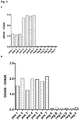
- CCHFV-NP HRP was co-incubated with a monoclonal anti-CCHFV-NP IgG antibody (Emmerich et al., 2010) on His 10 -3C- Hs Fc ⁇ R-Ig1-coated plates. While the respective IgG/antigen immune complexes readily bound to plates coated with the IgG immune complex binding protein CD32 (data not shown), no signal was detected on His 10 -3C- Hs Fc ⁇ R-Ig1-coated plates ( Fig. 5 ). Thus, His 10 -3C- Hs Fc ⁇ R-Ig1 does not bind to IgG/antigen immune complexes.
- the immunoglobulin-like domain of HsFc ⁇ R preferentially binds nascent IgM / antigen immune complexes.
- IgM/antigen immune complexes can be bound by His 10 -3C- Hs Fc ⁇ R-Ig1
- CCHFV-NP HRP and a anti-CCHFV-NP IgM positive serum were either co-incubated directly on the His 10 -3C- Hs Fc ⁇ R-Ig1-coated plate for 24 h at 4°C or co-incubated for 19 h at 4°C in a test tube and then applied to a His 10 -3C- Hs Fc ⁇ R-Ig1-coated plate for 5 h at 4°C. After washing, bound IgM/antigen complexes were quantified by TMB conversion ( Fig. 5 ).
- His 10 -3C- Hs Fc ⁇ R-Ig1 preferentially binds nascent IgM/antigen complexes.
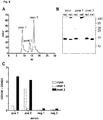
- His 10 -3C-HsFc ⁇ R-Igl was purified from E.coli inclusion bodies under denaturing conditions by Ni-NTA affinity chromatography, refolded by rapid dilution and concentrated.
- SEC Size exclusion Chromatography
- Peak 1 eluted earlier than expected for a protein of a calculated molecular weight of 15,4 kDa and SDS-PAGE under native conditions correspondingly revealed the presence of high molecular weight aggregates in this fraction ( Figure 6B ).
- Peak 2 eluted at the expected volume for a protein of a calculated molecular weight of 15.4 kDa, SDS-PAGE under native conditions was found to be indicative of a compactly folded monomer ( Figure 6B ).
- Peak 3 did not contain any His 10 -3C-HsFc ⁇ R-Igl protein (data not shown), the observed absorption at 280 nm most likely originates from low molecular weight degradation products/peptides and/or buffer substances.
- Functional, testing of peak fractions 1 (high molecular weight aggregates) and 2 (monomers) revealed that only the monomeric protein efficiently binds IgM/antigen immune complexes ( Figure 6C ).
Landscapes
- Health & Medical Sciences (AREA)
- Life Sciences & Earth Sciences (AREA)
- Engineering & Computer Science (AREA)
- Immunology (AREA)
- Molecular Biology (AREA)
- Urology & Nephrology (AREA)
- Biomedical Technology (AREA)
- Chemical & Material Sciences (AREA)
- Hematology (AREA)
- Food Science & Technology (AREA)
- Analytical Chemistry (AREA)
- Microbiology (AREA)
- Cell Biology (AREA)
- Biotechnology (AREA)
- Medicinal Chemistry (AREA)
- Physics & Mathematics (AREA)
- Pathology (AREA)
- Biochemistry (AREA)
- General Health & Medical Sciences (AREA)
- General Physics & Mathematics (AREA)
- Proteomics, Peptides & Aminoacids (AREA)
- Peptides Or Proteins (AREA)
- Preparation Of Compounds By Using Micro-Organisms (AREA)
Claims (14)
- Verwendung eines auf einem festen Träger immobilisierten FcµR zur In-vitro-Quantifizierung von IgM- oder IgA-Antikörpern in Form von Immunkomplexen, wobei die Immunkomplexe gebildet werden, indem ein Antigen mit einer Probe gemischt wird, die Antikörper umfasst.
- Verwendung nach Anspruch 1, wobei das Antigen ein detektierbares Antigen wie ein markiertes Antigen ist, das aus der Gruppe ausgewählt ist, die ein enzymmarkiertes Antigen und ein biotinmarkiertes Antigen umfasst, oder ein mehr als ein Epitop umfassendes Antigen.
- Verwendung nach einem der vorhergehenden Ansprüche, wobei die Zugabe des Antigens zu der Probe in Gegenwart von FcµR durchgeführt wird.
- Verwendung nach einem der vorhergehenden Ansprüche, wobei die Antikörper von einer biologischen Probe stammen.
- Verwendung nach einem der vorhergehenden Ansprüche, wobei die Antikörper von einer Spezies stammen, die ausgewählt ist aus der Gruppe umfassend Mensch, Ziege, Schaf, Pferd, Rind, Vogel, wie beispielsweise Hühner, und Nagetier, wie beispielsweis Maus, Ratte, Kaninchen, Meerschweinchen und Hamster.
- Verwendung nach einem der vorhergehenden Ansprüche, wobei die Antikörper IgM-Antikörper sind.
- Verwendung nach einem der vorhergehenden Ansprüche, wobei der FcµR Faim 3/Toso oder ein funktionelles Fragment davon, das imstande ist, an Immunkomplexe zu binden, oder CD351 oder ein funktionelles Fragment davon, das imstande ist, an Immunkomplexe zu binden, ist.
- Verwendung nach einem der vorhergehenden Ansprüche, wobei der FcµR ein funktionelles Fragment von Faim 3/Toso ist, das imstande ist, an Immunkomplexe zu binden und die Ig-ähnliche Domäne von Faim 3/Toso umfasst oder im Wesentlichen daraus besteht, optional in Form eines Fusionsproteins,
wobei der FcµR vorzugsweise eine monomere Ig-ähnliche Domäne von Faim 3/Toso, optional in Form eines Fusionsproteins, umfasst. - Verwendung nach einem der Ansprüche 7 oder 8, wobei Faim 3/Toso, CD351 oder das funktionelle Fragment davon eine Aminosäuresequenz umfasst, die wenigstens 80 % Sequenzidentität mit menschlichem Faim 3/Toso, CD351 oder dem jeweiligen Fragment aufweist.
- Verwendung nach einem der vorhergehenden Ansprüche, wobei der FcµR SEQ ID NO: 2 umfasst.
- Verfahren zur In-vitro-Quantifizierung von Antikörpern in Form von Immunkomplexen, umfassend:a) Beschichten eines festen Trägers mit einem FcµR unter Bedingungen, die geeignet für die Bindung des FcµR an den festen Träger sind,b) Inkubieren des festen Trägers mit einer Zusammensetzung, die einen IgM- oder IgA-Antikörper und ein Antigen umfasst, wobei der Antikörper und das Antigen imstande sind, einen Immunkomplex zu bilden, und wobei der Immunkomplex gebildet wird, indem ein Antigen zu einer Probe zugegeben wird, die Antikörper umfasst, undc) Waschen des festen Trägers, und anschließendd) Bestimmen der Menge des Immunkomplexes.
- Verfahren nach Anspruch 11, wobei das Antigen ein markiertes Antigen ist, das aus der Gruppe ausgewählt ist, die ein enzymmarkiertes Antigen und ein biotinmarkiertes Antigen umfasst.
- Verfahren nach einem der Ansprüche 11 bis 12, wobei der FcµR Faim 3/Toso oder ein funktionelles Fragment davon, das imstande ist, an Immunkomplexe zu binden, oder CD351 oder ein funktionelles Fragment davon, das imstande ist, an Immunkomplexe zu binden, ist.
- Verfahren zur Diagnostizierung einer Infektion mit einem Pathogen in einem Individuum, wobei das Verfahren nach einem der Ansprüche 11 bis 13 ausgeführt wird, wobei die Zusammensetzung, die einen Antikörper umfasst, eine Probe von dem Individuum ist, und wobei das Antigen, das spezifisch durch den Antikörper gebunden werden kann, ein Antigen ist, das von dem Pathogen ableitbar ist, wie ein Hüllprotein eines Virus.
Applications Claiming Priority (2)
| Application Number | Priority Date | Filing Date | Title |
|---|---|---|---|
| EP14188831.3A EP3009841A1 (de) | 2014-10-14 | 2014-10-14 | Nachweis von IgM-Antikörpern mit einem verbesserten Immunkomplex- (IC) ELISA |
| PCT/EP2015/073623 WO2016059018A1 (en) | 2014-10-14 | 2015-10-13 | DETECTION OF IgM ANTIBODIES USING AN IMMUNE COMPLEX (IC) ELISA |
Publications (2)
| Publication Number | Publication Date |
|---|---|
| EP3207375A1 EP3207375A1 (de) | 2017-08-23 |
| EP3207375B1 true EP3207375B1 (de) | 2020-01-08 |
Family
ID=51690296
Family Applications (2)
| Application Number | Title | Priority Date | Filing Date |
|---|---|---|---|
| EP14188831.3A Withdrawn EP3009841A1 (de) | 2014-10-14 | 2014-10-14 | Nachweis von IgM-Antikörpern mit einem verbesserten Immunkomplex- (IC) ELISA |
| EP15778343.2A Active EP3207375B1 (de) | 2014-10-14 | 2015-10-13 | Nachweis von igm antikörpern mit einem verbesserten immunkomplex (ic) elisa |
Family Applications Before (1)
| Application Number | Title | Priority Date | Filing Date |
|---|---|---|---|
| EP14188831.3A Withdrawn EP3009841A1 (de) | 2014-10-14 | 2014-10-14 | Nachweis von IgM-Antikörpern mit einem verbesserten Immunkomplex- (IC) ELISA |
Country Status (2)
| Country | Link |
|---|---|
| EP (2) | EP3009841A1 (de) |
| WO (1) | WO2016059018A1 (de) |
Families Citing this family (2)
| Publication number | Priority date | Publication date | Assignee | Title |
|---|---|---|---|---|
| EP3465206B1 (de) | 2016-06-06 | 2019-12-04 | Medizinische Universität Wien | Verfahren zur detektion eines für einen flavivirus in einer probe spezifischen igm-antikörpers |
| CN111518210B (zh) * | 2020-05-11 | 2022-07-19 | 上海米地生物医药有限公司 | 一种特异性识别faim3受体的全人源单抗 |
Family Cites Families (3)
| Publication number | Priority date | Publication date | Assignee | Title |
|---|---|---|---|---|
| US6727350B2 (en) * | 1997-11-17 | 2004-04-27 | The Board Of Trustees Of The Leland Stanford Junior University | Toso |
| EP2492689B8 (de) | 2011-02-22 | 2013-12-25 | Bernhard-Nocht-Institut für Tropenmedizin | Nachweis von Antikörpern mit einem verbesserten Immunkomplex- (IC) ELISA |
| AU2013234046B2 (en) * | 2012-03-16 | 2017-09-07 | University Health Network | Methods and compositions for modulating Toso activity |
-
2014
- 2014-10-14 EP EP14188831.3A patent/EP3009841A1/de not_active Withdrawn
-
2015
- 2015-10-13 EP EP15778343.2A patent/EP3207375B1/de active Active
- 2015-10-13 WO PCT/EP2015/073623 patent/WO2016059018A1/en not_active Ceased
Non-Patent Citations (1)
| Title |
|---|
| None * |
Also Published As
| Publication number | Publication date |
|---|---|
| WO2016059018A1 (en) | 2016-04-21 |
| EP3009841A1 (de) | 2016-04-20 |
| EP3207375A1 (de) | 2017-08-23 |
Similar Documents
| Publication | Publication Date | Title |
|---|---|---|
| US8828379B2 (en) | Disulphide bond-stabilized functional soluble MHC class II heterodimers | |
| KR102019008B1 (ko) | 메르스 코로나바이러스 뉴클레오캡시드 융합 단백질을 이용한 메르스 코로나바이러스 검출 방법 | |
| Blackburn et al. | Purification and biochemical characterization of the D6 chemokine receptor | |
| MX2010011598A (es) | Metodos para identificar compuestos anti-inflamatorios. | |
| CN108633305A (zh) | 用于诊断病毒感染的免疫测定法 | |
| US10907141B2 (en) | Rep protein for use in a diagnostic assay | |
| US8563704B2 (en) | Peptide aptamer for neutralizing the binding of platelet antigen specific antibodies and diagnostic and therapeutic applications containing the same | |
| US9945846B2 (en) | Detection of antibodies using an improved immune complex (IC) ELISA | |
| EP4375668A2 (de) | Neue, in säugetieren exprimierte hüllprotein-antigene des humanen immunschwächevirus | |
| KR102360851B1 (ko) | 신규 항프레세프신 항체 | |
| KR20210035249A (ko) | Ns1 단백질의 결합 단백질 | |
| Nogi et al. | Novel affinity tag system using structurally defined antibody‐tag interaction: application to single‐step protein purification | |
| EP3009841A1 (de) | Nachweis von IgM-Antikörpern mit einem verbesserten Immunkomplex- (IC) ELISA | |
| US20080305098A1 (en) | Recombinant Polypeptides and Methods for Detecting and/or Quantifying Autoantibodies Against Tsh Receptor | |
| US11008365B2 (en) | Polypeptide exhibiting affinity to antibodies forming non-native three-dimensional structure | |
| EP3147294B1 (de) | Unterscheidung von dengue-virusinfektionen von anderen flaviviralen infektionen mittels einem rekombinanten mutanten hüllprotein | |
| WO2023104933A1 (en) | Camelid antibodies for use in therapy and diagnosis | |
| JP7846612B2 (ja) | ヒノキ花粉抗原Cha o 2に対するモノクローナル抗体及びその用途 | |
| Crusius et al. | Tab2, a novel recombinant polypeptide tag offering sensitive and specific protein detection and reliable affinity purification | |
| JP2014125481A (ja) | オートタキシンアイソフォーム特異的抗体および検出方法 |
Legal Events
| Date | Code | Title | Description |
|---|---|---|---|
| STAA | Information on the status of an ep patent application or granted ep patent |
Free format text: STATUS: THE INTERNATIONAL PUBLICATION HAS BEEN MADE |
|
| PUAI | Public reference made under article 153(3) epc to a published international application that has entered the european phase |
Free format text: ORIGINAL CODE: 0009012 |
|
| STAA | Information on the status of an ep patent application or granted ep patent |
Free format text: STATUS: REQUEST FOR EXAMINATION WAS MADE |
|
| 17P | Request for examination filed |
Effective date: 20170323 |
|
| AK | Designated contracting states |
Kind code of ref document: A1 Designated state(s): AL AT BE BG CH CY CZ DE DK EE ES FI FR GB GR HR HU IE IS IT LI LT LU LV MC MK MT NL NO PL PT RO RS SE SI SK SM TR |
|
| AX | Request for extension of the european patent |
Extension state: BA ME |
|
| DAV | Request for validation of the european patent (deleted) | ||
| DAX | Request for extension of the european patent (deleted) | ||
| STAA | Information on the status of an ep patent application or granted ep patent |
Free format text: STATUS: EXAMINATION IS IN PROGRESS |
|
| 17Q | First examination report despatched |
Effective date: 20180129 |
|
| GRAP | Despatch of communication of intention to grant a patent |
Free format text: ORIGINAL CODE: EPIDOSNIGR1 |
|
| STAA | Information on the status of an ep patent application or granted ep patent |
Free format text: STATUS: GRANT OF PATENT IS INTENDED |
|
| INTG | Intention to grant announced |
Effective date: 20190301 |
|
| GRAJ | Information related to disapproval of communication of intention to grant by the applicant or resumption of examination proceedings by the epo deleted |
Free format text: ORIGINAL CODE: EPIDOSDIGR1 |
|
| STAA | Information on the status of an ep patent application or granted ep patent |
Free format text: STATUS: EXAMINATION IS IN PROGRESS |
|
| GRAP | Despatch of communication of intention to grant a patent |
Free format text: ORIGINAL CODE: EPIDOSNIGR1 |
|
| STAA | Information on the status of an ep patent application or granted ep patent |
Free format text: STATUS: GRANT OF PATENT IS INTENDED |
|
| INTC | Intention to grant announced (deleted) | ||
| INTG | Intention to grant announced |
Effective date: 20190729 |
|
| GRAS | Grant fee paid |
Free format text: ORIGINAL CODE: EPIDOSNIGR3 |
|
| GRAA | (expected) grant |
Free format text: ORIGINAL CODE: 0009210 |
|
| STAA | Information on the status of an ep patent application or granted ep patent |
Free format text: STATUS: THE PATENT HAS BEEN GRANTED |
|
| AK | Designated contracting states |
Kind code of ref document: B1 Designated state(s): AL AT BE BG CH CY CZ DE DK EE ES FI FR GB GR HR HU IE IS IT LI LT LU LV MC MK MT NL NO PL PT RO RS SE SI SK SM TR |
|
| REG | Reference to a national code |
Ref country code: GB Ref legal event code: FG4D |
|
| REG | Reference to a national code |
Ref country code: CH Ref legal event code: EP |
|
| REG | Reference to a national code |
Ref country code: DE Ref legal event code: R096 Ref document number: 602015045215 Country of ref document: DE |
|
| REG | Reference to a national code |
Ref country code: IE Ref legal event code: FG4D |
|
| REG | Reference to a national code |
Ref country code: AT Ref legal event code: REF Ref document number: 1223337 Country of ref document: AT Kind code of ref document: T Effective date: 20200215 |
|
| REG | Reference to a national code |
Ref country code: NL Ref legal event code: MP Effective date: 20200108 |
|
| REG | Reference to a national code |
Ref country code: LT Ref legal event code: MG4D |
|
| PG25 | Lapsed in a contracting state [announced via postgrant information from national office to epo] |
Ref country code: PT Free format text: LAPSE BECAUSE OF FAILURE TO SUBMIT A TRANSLATION OF THE DESCRIPTION OR TO PAY THE FEE WITHIN THE PRESCRIBED TIME-LIMIT Effective date: 20200531 Ref country code: NL Free format text: LAPSE BECAUSE OF FAILURE TO SUBMIT A TRANSLATION OF THE DESCRIPTION OR TO PAY THE FEE WITHIN THE PRESCRIBED TIME-LIMIT Effective date: 20200108 Ref country code: LT Free format text: LAPSE BECAUSE OF FAILURE TO SUBMIT A TRANSLATION OF THE DESCRIPTION OR TO PAY THE FEE WITHIN THE PRESCRIBED TIME-LIMIT Effective date: 20200108 Ref country code: NO Free format text: LAPSE BECAUSE OF FAILURE TO SUBMIT A TRANSLATION OF THE DESCRIPTION OR TO PAY THE FEE WITHIN THE PRESCRIBED TIME-LIMIT Effective date: 20200408 Ref country code: FI Free format text: LAPSE BECAUSE OF FAILURE TO SUBMIT A TRANSLATION OF THE DESCRIPTION OR TO PAY THE FEE WITHIN THE PRESCRIBED TIME-LIMIT Effective date: 20200108 Ref country code: RS Free format text: LAPSE BECAUSE OF FAILURE TO SUBMIT A TRANSLATION OF THE DESCRIPTION OR TO PAY THE FEE WITHIN THE PRESCRIBED TIME-LIMIT Effective date: 20200108 |
|
| PG25 | Lapsed in a contracting state [announced via postgrant information from national office to epo] |
Ref country code: SE Free format text: LAPSE BECAUSE OF FAILURE TO SUBMIT A TRANSLATION OF THE DESCRIPTION OR TO PAY THE FEE WITHIN THE PRESCRIBED TIME-LIMIT Effective date: 20200108 Ref country code: IS Free format text: LAPSE BECAUSE OF FAILURE TO SUBMIT A TRANSLATION OF THE DESCRIPTION OR TO PAY THE FEE WITHIN THE PRESCRIBED TIME-LIMIT Effective date: 20200508 Ref country code: LV Free format text: LAPSE BECAUSE OF FAILURE TO SUBMIT A TRANSLATION OF THE DESCRIPTION OR TO PAY THE FEE WITHIN THE PRESCRIBED TIME-LIMIT Effective date: 20200108 Ref country code: HR Free format text: LAPSE BECAUSE OF FAILURE TO SUBMIT A TRANSLATION OF THE DESCRIPTION OR TO PAY THE FEE WITHIN THE PRESCRIBED TIME-LIMIT Effective date: 20200108 Ref country code: GR Free format text: LAPSE BECAUSE OF FAILURE TO SUBMIT A TRANSLATION OF THE DESCRIPTION OR TO PAY THE FEE WITHIN THE PRESCRIBED TIME-LIMIT Effective date: 20200409 Ref country code: BG Free format text: LAPSE BECAUSE OF FAILURE TO SUBMIT A TRANSLATION OF THE DESCRIPTION OR TO PAY THE FEE WITHIN THE PRESCRIBED TIME-LIMIT Effective date: 20200408 |
|
| REG | Reference to a national code |
Ref country code: DE Ref legal event code: R097 Ref document number: 602015045215 Country of ref document: DE |
|
| PG25 | Lapsed in a contracting state [announced via postgrant information from national office to epo] |
Ref country code: EE Free format text: LAPSE BECAUSE OF FAILURE TO SUBMIT A TRANSLATION OF THE DESCRIPTION OR TO PAY THE FEE WITHIN THE PRESCRIBED TIME-LIMIT Effective date: 20200108 Ref country code: SM Free format text: LAPSE BECAUSE OF FAILURE TO SUBMIT A TRANSLATION OF THE DESCRIPTION OR TO PAY THE FEE WITHIN THE PRESCRIBED TIME-LIMIT Effective date: 20200108 Ref country code: RO Free format text: LAPSE BECAUSE OF FAILURE TO SUBMIT A TRANSLATION OF THE DESCRIPTION OR TO PAY THE FEE WITHIN THE PRESCRIBED TIME-LIMIT Effective date: 20200108 Ref country code: CZ Free format text: LAPSE BECAUSE OF FAILURE TO SUBMIT A TRANSLATION OF THE DESCRIPTION OR TO PAY THE FEE WITHIN THE PRESCRIBED TIME-LIMIT Effective date: 20200108 Ref country code: ES Free format text: LAPSE BECAUSE OF FAILURE TO SUBMIT A TRANSLATION OF THE DESCRIPTION OR TO PAY THE FEE WITHIN THE PRESCRIBED TIME-LIMIT Effective date: 20200108 Ref country code: SK Free format text: LAPSE BECAUSE OF FAILURE TO SUBMIT A TRANSLATION OF THE DESCRIPTION OR TO PAY THE FEE WITHIN THE PRESCRIBED TIME-LIMIT Effective date: 20200108 Ref country code: DK Free format text: LAPSE BECAUSE OF FAILURE TO SUBMIT A TRANSLATION OF THE DESCRIPTION OR TO PAY THE FEE WITHIN THE PRESCRIBED TIME-LIMIT Effective date: 20200108 |
|
| PLBE | No opposition filed within time limit |
Free format text: ORIGINAL CODE: 0009261 |
|
| STAA | Information on the status of an ep patent application or granted ep patent |
Free format text: STATUS: NO OPPOSITION FILED WITHIN TIME LIMIT |
|
| REG | Reference to a national code |
Ref country code: AT Ref legal event code: MK05 Ref document number: 1223337 Country of ref document: AT Kind code of ref document: T Effective date: 20200108 |
|
| REG | Reference to a national code |
Ref country code: DE Ref legal event code: R082 Ref document number: 602015045215 Country of ref document: DE Representative=s name: RGTH PATENTANWAELTE PARTGMBB, DE Ref country code: DE Ref legal event code: R082 Ref document number: 602015045215 Country of ref document: DE Representative=s name: RGTH RICHTER GERBAULET THIELEMANN HOFMANN PATE, DE |
|
| 26N | No opposition filed |
Effective date: 20201009 |
|
| PG25 | Lapsed in a contracting state [announced via postgrant information from national office to epo] |
Ref country code: IT Free format text: LAPSE BECAUSE OF FAILURE TO SUBMIT A TRANSLATION OF THE DESCRIPTION OR TO PAY THE FEE WITHIN THE PRESCRIBED TIME-LIMIT Effective date: 20200108 Ref country code: AT Free format text: LAPSE BECAUSE OF FAILURE TO SUBMIT A TRANSLATION OF THE DESCRIPTION OR TO PAY THE FEE WITHIN THE PRESCRIBED TIME-LIMIT Effective date: 20200108 |
|
| PG25 | Lapsed in a contracting state [announced via postgrant information from national office to epo] |
Ref country code: SI Free format text: LAPSE BECAUSE OF FAILURE TO SUBMIT A TRANSLATION OF THE DESCRIPTION OR TO PAY THE FEE WITHIN THE PRESCRIBED TIME-LIMIT Effective date: 20200108 Ref country code: PL Free format text: LAPSE BECAUSE OF FAILURE TO SUBMIT A TRANSLATION OF THE DESCRIPTION OR TO PAY THE FEE WITHIN THE PRESCRIBED TIME-LIMIT Effective date: 20200108 |
|
| PG25 | Lapsed in a contracting state [announced via postgrant information from national office to epo] |
Ref country code: MC Free format text: LAPSE BECAUSE OF FAILURE TO SUBMIT A TRANSLATION OF THE DESCRIPTION OR TO PAY THE FEE WITHIN THE PRESCRIBED TIME-LIMIT Effective date: 20200108 Ref country code: LU Free format text: LAPSE BECAUSE OF NON-PAYMENT OF DUE FEES Effective date: 20201013 |
|
| REG | Reference to a national code |
Ref country code: BE Ref legal event code: MM Effective date: 20201031 |
|
| PG25 | Lapsed in a contracting state [announced via postgrant information from national office to epo] |
Ref country code: BE Free format text: LAPSE BECAUSE OF NON-PAYMENT OF DUE FEES Effective date: 20201031 |
|
| PG25 | Lapsed in a contracting state [announced via postgrant information from national office to epo] |
Ref country code: IE Free format text: LAPSE BECAUSE OF NON-PAYMENT OF DUE FEES Effective date: 20201013 |
|
| PG25 | Lapsed in a contracting state [announced via postgrant information from national office to epo] |
Ref country code: TR Free format text: LAPSE BECAUSE OF FAILURE TO SUBMIT A TRANSLATION OF THE DESCRIPTION OR TO PAY THE FEE WITHIN THE PRESCRIBED TIME-LIMIT Effective date: 20200108 Ref country code: MT Free format text: LAPSE BECAUSE OF FAILURE TO SUBMIT A TRANSLATION OF THE DESCRIPTION OR TO PAY THE FEE WITHIN THE PRESCRIBED TIME-LIMIT Effective date: 20200108 Ref country code: CY Free format text: LAPSE BECAUSE OF FAILURE TO SUBMIT A TRANSLATION OF THE DESCRIPTION OR TO PAY THE FEE WITHIN THE PRESCRIBED TIME-LIMIT Effective date: 20200108 |
|
| PG25 | Lapsed in a contracting state [announced via postgrant information from national office to epo] |
Ref country code: MK Free format text: LAPSE BECAUSE OF FAILURE TO SUBMIT A TRANSLATION OF THE DESCRIPTION OR TO PAY THE FEE WITHIN THE PRESCRIBED TIME-LIMIT Effective date: 20200108 Ref country code: AL Free format text: LAPSE BECAUSE OF FAILURE TO SUBMIT A TRANSLATION OF THE DESCRIPTION OR TO PAY THE FEE WITHIN THE PRESCRIBED TIME-LIMIT Effective date: 20200108 |
|
| P01 | Opt-out of the competence of the unified patent court (upc) registered |
Effective date: 20230515 |
|
| PGFP | Annual fee paid to national office [announced via postgrant information from national office to epo] |
Ref country code: GB Payment date: 20250916 Year of fee payment: 11 |
|
| PGFP | Annual fee paid to national office [announced via postgrant information from national office to epo] |
Ref country code: FR Payment date: 20250919 Year of fee payment: 11 |
|
| REG | Reference to a national code |
Ref country code: CH Ref legal event code: U11 Free format text: ST27 STATUS EVENT CODE: U-0-0-U10-U11 (AS PROVIDED BY THE NATIONAL OFFICE) Effective date: 20251101 |
|
| PGFP | Annual fee paid to national office [announced via postgrant information from national office to epo] |
Ref country code: DE Payment date: 20250919 Year of fee payment: 11 |
|
| PGFP | Annual fee paid to national office [announced via postgrant information from national office to epo] |
Ref country code: CH Payment date: 20251101 Year of fee payment: 11 |