EP3218721B1 - Composés servant d'agents d'imagerie - Google Patents
Composés servant d'agents d'imagerie Download PDFInfo
- Publication number
- EP3218721B1 EP3218721B1 EP14906044.4A EP14906044A EP3218721B1 EP 3218721 B1 EP3218721 B1 EP 3218721B1 EP 14906044 A EP14906044 A EP 14906044A EP 3218721 B1 EP3218721 B1 EP 3218721B1
- Authority
- EP
- European Patent Office
- Prior art keywords
- synuclein
- aggregates
- sample
- dihydromyricetin
- compound
- Prior art date
- Legal status (The legal status is an assumption and is not a legal conclusion. Google has not performed a legal analysis and makes no representation as to the accuracy of the status listed.)
- Active
Links
Images






Classifications
-
- G—PHYSICS
- G01—MEASURING; TESTING
- G01N—INVESTIGATING OR ANALYSING MATERIALS BY DETERMINING THEIR CHEMICAL OR PHYSICAL PROPERTIES
- G01N33/00—Investigating or analysing materials by specific methods not covered by groups G01N1/00 - G01N31/00
- G01N33/48—Biological material, e.g. blood, urine; Haemocytometers
- G01N33/50—Chemical analysis of biological material, e.g. blood, urine; Testing involving biospecific ligand binding methods; Immunological testing
- G01N33/58—Chemical analysis of biological material, e.g. blood, urine; Testing involving biospecific ligand binding methods; Immunological testing involving labelled substances
-
- A—HUMAN NECESSITIES
- A61—MEDICAL OR VETERINARY SCIENCE; HYGIENE
- A61K—PREPARATIONS FOR MEDICAL, DENTAL OR TOILETRY PURPOSES
- A61K31/00—Medicinal preparations containing organic active ingredients
- A61K31/21—Esters, e.g. nitroglycerine, selenocyanates
- A61K31/215—Esters, e.g. nitroglycerine, selenocyanates of carboxylic acids
- A61K31/216—Esters, e.g. nitroglycerine, selenocyanates of carboxylic acids of acids having aromatic rings, e.g. benactizyne, clofibrate
-
- A—HUMAN NECESSITIES
- A61—MEDICAL OR VETERINARY SCIENCE; HYGIENE
- A61K—PREPARATIONS FOR MEDICAL, DENTAL OR TOILETRY PURPOSES
- A61K31/00—Medicinal preparations containing organic active ingredients
- A61K31/33—Heterocyclic compounds
- A61K31/335—Heterocyclic compounds having oxygen as the only ring hetero atom, e.g. fungichromin
- A61K31/34—Heterocyclic compounds having oxygen as the only ring hetero atom, e.g. fungichromin having five-membered rings with one oxygen as the only ring hetero atom, e.g. isosorbide
- A61K31/343—Heterocyclic compounds having oxygen as the only ring hetero atom, e.g. fungichromin having five-membered rings with one oxygen as the only ring hetero atom, e.g. isosorbide condensed with a carbocyclic ring, e.g. coumaran, bufuralol, befunolol, clobenfurol, amiodarone
-
- A—HUMAN NECESSITIES
- A61—MEDICAL OR VETERINARY SCIENCE; HYGIENE
- A61K—PREPARATIONS FOR MEDICAL, DENTAL OR TOILETRY PURPOSES
- A61K31/00—Medicinal preparations containing organic active ingredients
- A61K31/70—Carbohydrates; Sugars; Derivatives thereof
- A61K31/7028—Compounds having saccharide radicals attached to non-saccharide compounds by glycosidic linkages
- A61K31/7034—Compounds having saccharide radicals attached to non-saccharide compounds by glycosidic linkages attached to a carbocyclic compound, e.g. phloridzin
- A61K31/704—Compounds having saccharide radicals attached to non-saccharide compounds by glycosidic linkages attached to a carbocyclic compound, e.g. phloridzin attached to a condensed carbocyclic ring system, e.g. sennosides, thiocolchicosides, escin, daunorubicin
-
- A—HUMAN NECESSITIES
- A61—MEDICAL OR VETERINARY SCIENCE; HYGIENE
- A61P—SPECIFIC THERAPEUTIC ACTIVITY OF CHEMICAL COMPOUNDS OR MEDICINAL PREPARATIONS
- A61P25/00—Drugs for disorders of the nervous system
-
- A—HUMAN NECESSITIES
- A61—MEDICAL OR VETERINARY SCIENCE; HYGIENE
- A61P—SPECIFIC THERAPEUTIC ACTIVITY OF CHEMICAL COMPOUNDS OR MEDICINAL PREPARATIONS
- A61P25/00—Drugs for disorders of the nervous system
- A61P25/14—Drugs for disorders of the nervous system for treating abnormal movements, e.g. chorea, dyskinesia
- A61P25/16—Anti-Parkinson drugs
-
- G—PHYSICS
- G01—MEASURING; TESTING
- G01N—INVESTIGATING OR ANALYSING MATERIALS BY DETERMINING THEIR CHEMICAL OR PHYSICAL PROPERTIES
- G01N2800/00—Detection or diagnosis of diseases
- G01N2800/28—Neurological disorders
-
- G—PHYSICS
- G01—MEASURING; TESTING
- G01N—INVESTIGATING OR ANALYSING MATERIALS BY DETERMINING THEIR CHEMICAL OR PHYSICAL PROPERTIES
- G01N2800/00—Detection or diagnosis of diseases
- G01N2800/28—Neurological disorders
- G01N2800/2814—Dementia; Cognitive disorders
-
- G—PHYSICS
- G01—MEASURING; TESTING
- G01N—INVESTIGATING OR ANALYSING MATERIALS BY DETERMINING THEIR CHEMICAL OR PHYSICAL PROPERTIES
- G01N2800/00—Detection or diagnosis of diseases
- G01N2800/28—Neurological disorders
- G01N2800/2835—Movement disorders, e.g. Parkinson, Huntington, Tourette
-
- G—PHYSICS
- G01—MEASURING; TESTING
- G01N—INVESTIGATING OR ANALYSING MATERIALS BY DETERMINING THEIR CHEMICAL OR PHYSICAL PROPERTIES
- G01N33/00—Investigating or analysing materials by specific methods not covered by groups G01N1/00 - G01N31/00
- G01N33/48—Biological material, e.g. blood, urine; Haemocytometers
- G01N33/50—Chemical analysis of biological material, e.g. blood, urine; Testing involving biospecific ligand binding methods; Immunological testing
- G01N33/52—Use of compounds or compositions for colorimetric, spectrophotometric or fluorometric investigation, e.g. use of reagent paper and including single- and multilayer analytical elements
-
- G—PHYSICS
- G01—MEASURING; TESTING
- G01N—INVESTIGATING OR ANALYSING MATERIALS BY DETERMINING THEIR CHEMICAL OR PHYSICAL PROPERTIES
- G01N33/00—Investigating or analysing materials by specific methods not covered by groups G01N1/00 - G01N31/00
- G01N33/48—Biological material, e.g. blood, urine; Haemocytometers
- G01N33/50—Chemical analysis of biological material, e.g. blood, urine; Testing involving biospecific ligand binding methods; Immunological testing
- G01N33/68—Chemical analysis of biological material, e.g. blood, urine; Testing involving biospecific ligand binding methods; Immunological testing involving proteins, peptides or amino acids
- G01N33/6893—Chemical analysis of biological material, e.g. blood, urine; Testing involving biospecific ligand binding methods; Immunological testing involving proteins, peptides or amino acids related to diseases not provided for elsewhere
- G01N33/6896—Neurological disorders, e.g. Alzheimer's disease
Definitions
- This invention relates to ginsenoside Rb1, which can be isolated from Chinese herbal medicines, and the use of this compound for detecting the presence of ⁇ -synuclein aggregates and in diagnosing and monitoring synucleinopathic disorders.
- the disclosure also relates to the use of compounds isolated from Chinese herbal medicines in the treatment of synucleinopathic disorders.
- synucleinopathies include the neurodegenerative conditions, Parkinson's disease (PD), dementia with Lewy bodies (DLB), and multiple system atrophy (MSA).
- PD Parkinson's disease
- DLB dementia with Lewy bodies
- MSA multiple system atrophy
- Synucleins are a family of small proteins, about 14kDa that are expressed at high levels in nervous tissues. Three members of the family are ⁇ -, ⁇ -, and ⁇ -synucleins. ⁇ -Synuclein is expressed mainly in brain tissues and is primarily located at the presynpatic terminal of neurons. ⁇ -Synuclein normally exists as a soluble monomeric protein but can adopt several different folded confirmations depending on its environment. Monomeric ⁇ -synuclein can also aggregate into oligomers and into higher molecular weight insoluble fibrils. ⁇ -Synuclein aggregation proceeds from monomeric ⁇ -synuclein through to the formation of soluble oligomers (early aggregates), which then converts into higher molecule weight ordered fibrils (late aggregates).
- ⁇ -synuclein oligomers in brain homogenates of PD and DLB are elevated compared to normal brains. While neuropathological studies of PD have revealed the presence of cytoplasmic inclusions found in the degenerating dopaminergic nerurons of the substantia nigra and other cortical and subcortical neurons. These lesions in the brain are known as Lewy bodies and Lewy neurities and have also been shown to constitute the main pathological features in the brain of DLB patients. These neuropathologic lesions often characterise the end stage of PD and DLB have largely been found to be composed of fibrillar ⁇ -synuclein deposits. Additional studies have shown that ⁇ -synuclein is also associated with pathological lesions in other neurodegenerative diseases sometimes involving non-neuronal cells, such as the cytoplasmic inclusions found in MSA.
- Ginseng is one Chinese medicinal composition that has been studied.
- the pharmacological properties of ginseng are mainly attributed to its biologically active ingredients, the ginsenosides.
- the ginsenosides can be extracted from many parts of the ginseng plant including the root, leaves and ginseng berries.
- Ginsenosides are derivatives of triterpenoid dammarane with a four-ring, steroidal structure bearing sugar moieties and an aliphatic side chain..
- Another Chinese medicinal composition is danshen. Pharmacological properties of danshen have been attributed to salvianolic acid B and its anti-oxidative ability.
- a further Chinese medicinal composition is the Japanese raisin tree. The pharmacological properties of the Japanese raisin tree have been attributed to dihydromyricetin, a flavonoid, and the compound has been used as a hangover cure.
- CN 102727508 A discloses the use of ginsenosides to treat Parkinsons disease.
- WO2009/027690 discloses peptides that bind ⁇ -synuclein and mentions their use in the diagnosis and monitoring of synucleinopathic diseases.
- WO03/69332 and WO99/50300 and US 2014/241984 disclose method of detecting ⁇ -synuclein oligomerisation using monoclonal antibodies.
- US 2012/251448 discloses the use of small molecules to detect alpha-synuclein aggregates.
- This invention aims to provide further compounds that can be used in the imaging and/or diagnosing of synucleinopathies in subjects.
- the invention is directed to the use of ginsenoside Rb1 to detect alpha-synuclein aggregates in vitro.
- the compounds, ginsenoside Rb1, dihydromyricetin and salvianolic acid B, found in plants used in traditional Chinese medicine have been found to bind and disaggregate ⁇ -synuclein aggregates. Accordingly the disclosure is directed to an imaging agent for use in detecting ⁇ -synuclein aggregates comprising a compound selected from ginsenoside Rb1, dihydromyricetin, or salvianolic acid B, and a detectable label.
- the method can be an in vitro method or an in vivo, i.e. in a subject.
- the method comprises administering the compound to a sample obtained from a subject.
- the invention further provides a method of imaging ⁇ -synuclein aggregates comprising detecting the binding of the compound ginsenoside Rb1 to ⁇ -synuclein aggregates.
- the method for imaging aggregates can comprise administering the compound ginsenoside Rb1; and detecting the binding of the compound to ⁇ -synuclein aggregates.
- the method is for imaging ⁇ -synuclein aggregates in a sample.
- the method can comprise administering the compound to a sample obtained from a subject.
- the sample can be a brain tissue sample.
- the compound being detect can comprise a detectable label.
- the presence of the binding of the compound to ⁇ -synuclein fibrils can be detected by autoradiography, positron emission tomography, magnetic resonance imaging, a gamma counter, or a scintillation counter.
- the invention further provides a method of diagnosing a synucleinopathic disease comprising detecting the presence or absence of ⁇ -synuclein aggregates.
- the presence of ⁇ -synuclein aggregates indicates that the subject has a synucleinopathic disease and the absence of ⁇ -synuclein aggregates indicates that the subject does not have the synucleinopathic disease.
- the method of diagnosing a synucleinopathic disease can comprise the steps of:
- the invention also provides an in vitro method of diagnosing a synucleinopathic disease involving ⁇ -synuclein and/or fragment(s) of ⁇ -synuclein, the method comprising the steps of:
- Also described is an alternative method of diagnosing a synucleinopathic disease in a subject comprising the steps of:
- the compound may be administered to the subject by a non-surgical method.
- the disclosure further provides a method for disaggregating ⁇ -synuclein aggregates in a subject having a synucleinopathic disorder, the method comprising administering a compound selected from ginsenoside Rb1, dihydromyricetin or salvianolic acid B to the subject.
- the ⁇ -synuclein aggregates can be Lewy bodies or Lewy neuritis.
- the disclosure also relates to a pharmaceutical composition
- a pharmaceutical composition comprising dihydromyricetin, ginsenoside Rb1 or salvianolic acid B, or a pharmaceutically acceptable salt or solvate thereof and a pharmaceutically acceptable diluents or carrier for disaggregating fibrils in Lewy bodies or Lewy neurites in a subject having a synucleinopathic disorder.
- the disclosure further provides a method of inhibiting the ⁇ -synuclein fibrillation in a subject having a synucleinopathic disorder, the method comprising administering a compound selected from ginsenoside Rb1, dihydromyricetin, or salvianolic acid B to the subject.
- the compounds ginsenoside Rb1, dihydromyricetin and salvianolic acid B, found in traditional Chinese medicinal compositions have been found to bind and disaggregate ⁇ -synuclein aggregates. Accordingly the invention is directed to the use of ginsenoside Rb1 in methods of detecting ⁇ -synuclein aggregates.
- the compounds can also be used as imaging agents in detecting ⁇ -synuclein aggregates.
- ⁇ -synuclein aggregates it is meant mature insoluble aggregates of ⁇ -synuclein fibrils, including but not limited to ⁇ -synuclein deposits such as Lewy bodies and Lewy neurites.
- the aggregates may also comprise other components.
- the compounds Due to the ability of the compounds to bind to and disaggregate performed ⁇ -synuclein fibrils the compounds are suitable for use in the imaging of ⁇ -synuclein aggregates and/or in diagnosing of diseases involving ⁇ -synuclein.
- the compounds can bind the fibrils in the aggregates and causes the pre-formed fibrils to disaggregate, which enables the fibril-compound complexes formed to be detected for imaging and diagnostic purposes.
- Suitable compounds for use in detecting and imaging ⁇ -synuclein deposits and for use in diagnosing synucleinopathic diseases include ginsenosides such as ginsenoside Rb1, having the structure as shown in formula (I): dihydromyricetin, having the structure as shown in formula (II): and salvianolic acid B, having the structure as shown in formula (III):
- the compounds can be labelled to facilitate imaging of ⁇ -synuclein aggregates.
- the compounds can for example include a detectable label.
- the detectable label is typically one which enables the detection of the compound when bound to the ⁇ -synuclein fibrils.
- Useful labels include fluorescent labels, radiolabels and contrast agents.
- Suitable radiolabels include 18 F, 123 I, 111 In, 131 I, 14 C, 3 H, 99m Tc, 32 P and 125 I.
- Suitable fluorescent labels include fluorescein and rhodamine.
- Suitable contrast agents include rare earth ions such as gadolinium (Gd), dysprosium and iron, and magnetic agents.
- Other labels include nuclear magnetic resonance active labels, positron emitting isotopes detectable by a positron emission tomography (PET) scanner, chemiluminescent and enzymatic markers.
- the detectable label may be attached to the compound directly or via linker. Alternatively the detectable label may be incorporated into the structure of the compound.
- the compounds, ginsenoside Rb1, dihydromyricetin or salvianolic acid B can be labelled with the above and other detectable labels using standard techniques.
- the compounds, ginsenoside Rb1, dihydromyricetin and salvianolic acid B comprising a detectable label are useful in methods of imaging ⁇ -synuclein aggregates.
- the presence or absence of the ⁇ -synuclein aggregates may be detected in the subject in vivo, for example in the brain, using any suitable imaging techniques.
- the subject is typically a mammal, preferably a human.
- the subject may be an experimental animal in particular an experimental animal model of synucleinopathic disease. Animal models of, for example Parkinson's Disease are known in the art and include transgenic mice.
- Suitable imaging techniques include PET, gamma-scintigraphy, magnetic resonance imaging (MRI), functional magnetic resonance imaging (FMRI), magnetoencephalography (MEG) and single photon emission computerized tomography (SPECT).
- MRI and Pet are preferred methods for in vivo imaging.
- the presence or absence of the ⁇ -synuclein aggregates may also be detected in vitro, for example to identify agents that inhibit ⁇ -synuclein aggregation or in biological samples taken from the subject.
- imaging methods may include microscopy, such as electron microscopy, confocal microscopy or light microscopy.
- the ⁇ -synuclein aggregates may be detected in vitro or in vivo, by administering ginsenoside Rb1, dihydromyricetin or salvianolic acid B, and detecting the binding of the compound to ⁇ -synuclein fibrils of the aggregates.
- the ⁇ -synuclein aggregates to be detected may be present in the body fluids or tissues or other samples to be tested, including but not limited to aggregates present in the brain of a living mammal or in a brain sample obtained from the subject to be tested.
- the method can also be carried out on other body tissues or body fluids, such as heart or gastrointestinal tissue.
- in vitro methods can be carried out on tissue or biological fluid samples.
- the sample is combined with the compound, ginsenoside Rb1, dihydromyricetin or salvianolic acid B, for a time and under conditions effective to allow binding of the compound to any ⁇ -synuclein aggregate present in the sample.
- the sample is combined with the compound for a time and under conditions effective to allow binding of the compound to the fibrils in a Lewy body.
- the sample may be processed prior to being assayed using standard methods.
- the in vitro methods of detecting ⁇ -synuclein aggregates also include the use of the compounds, ginsenoside Rb1, dihydromyricetin or salvianolic acid B, in detecting ⁇ -synuclein aggregates in samples to determine the effectiveness of other compounds to disaggregate ⁇ -synuclein and/or have an effect on the formation ⁇ -synuclein aggregates.
- the compounds ginsenoside Rb1, dihydromyricetin and salvianolic acid B can be used in methods of diagnosing synucleinopathies.
- a method of diagnosing a synucleinopathic disease can comprise detecting the presence or absence of ⁇ -synuclein aggregates. The presence of ⁇ -synuclein aggregates indicates that the subject has a synucleinopathic disease and the absence of ⁇ -synuclein aggregates indicates that the subject does not have the synucleinopathic disease.
- the presence of absence of ⁇ -synuclein aggregates can be detected by determining whether a complex between the fibrils of the aggregate and the compound are present.
- the presence and or absence of complexes between the fibrils and the compound can be determined by the imaging techniques described above.
- the diagnostic methods can be in vitro methods, using a tissue sample or a biological fluid from a subject. The sample is then contacted with the compound and the presence of absence of a complex between the fibrils of the aggregates and the compound is determined.
- the diagnostic methods can be in vivo methods, wherein the compound, ginsenoside Rb1, dihydromyricetin or salvianolic acid B is administered to the subject; and the presence or absence of aggregates of ⁇ -synuclein is detected in the subject.
- the compound may be administered to the subject by a non-surgical means.
- the subject may be undergoing therapy to treat a synucleinopathic disease and the method is for monitoring the effectiveness of the treatment.
- Levels of ⁇ -synuclein aggregates detected by the imaging methods may be compared with a standard to determine the status of the disease and/or whether a particular treatment has been successful.
- the number and/or size of ⁇ -synuclein aggregates present in the brain of the subject correlates with synucleinopathic disease progression. A decrease in the number and/or size of ⁇ -synuclein aggregates suggests the disease is regressing. An increase in the number and/or size of ⁇ -synuclein aggregates indicates the disease is progressing.
- the compounds used in the method of diagnosis can be labelled as discussed above. Imaging techniques as discussed above can be used in detecting the binding of the compounds to the fibrils, in the diagnostic methods.
- the formulation of the compounds for use in the imaging and diagnostic methods may depend on the method it will be used in.
- the compound may be administered to the subject by non-surgical means.
- Non-surgical means of administration include for examples, administration orally (e.g. as tablets, troches, lozenges, aqueous or oily suspensions, dispersible powders or granules), topically, transdermally or by infusion or inhalation techniques. The doctor will be able to determine the required route of administration for each particular subject.
- the compound may be administered directly to the site of an ⁇ -synuclein aggregate e.g. a Lewy body, typically by injection into a blood vessel supplying the brain or into the brain itself.
- an ⁇ -synuclein aggregate e.g. a Lewy body
- the compound will be formulated with a pharmaceutically acceptable carrier or diluent.
- a physician will be able to determine a suitable pharmaceutical form for delivery to the subject.
- Also described is a method for disaggregating fibrils in Lewy bodies or Lewy neurites in a subject having a synucleinopathic disorder comprising administering a compound selected from ginsenoside Rb1, dihydromyricetin, or salvianolic acid B to the subject.
- the compound may be administered in a therapeutically effective amount.
- the disclosure also relates to a pharmaceutical composition for disaggregating fibrils in Lewy bodies or Lewy neurites in a subject having a synucleinopathic disorder, the composition comprising a dihydromyricetin, ginsenoside Rb1 or salvianolic acid B, or a pharmaceutically acceptable salt or solvate thereof, and a pharmaceutically acceptable diluent or carrier.
- ginsenoside Rb1 has also been found to inhibit ⁇ -synuclein fibrillation. Without being bound by theory ginsenoside Rb1 is thought to bind and stabilize the structure of soluble oligomeric ⁇ -synuclein with no ⁇ -sheet content, and block ⁇ -synuclein induced toxicity. Whilst the compounds dihydromyricetin and salvianolic acid B have been found to inhibit ⁇ -synuclein oligomerization and fibrillation. Therefore these compounds are suitable in the treatment of synucleinopathic diseases.
- Also described is a method of inhibiting the ⁇ -synuclein fibrillation in a subject having a synucleinopathic disorder comprising administering a compound selected from ginsenoside Rb1, dihydromyricetin, or salvianolic acid B to the subject.
- the compound may be administered in a therapeutically effective amount.
- a synucleinopathic disease or disorder or synucleinopathy is a disease involving synucleins, in particular ⁇ -synuclein.
- Synucleinopathies include but are not limited to diseases selected from the group comprising Parkinson's disease, dementia with Lewy bodies, and multiple system atrophy.
- the subject has Parkinson's disease or dementia with Lewy bodies.
- a “therapeutically effective amount” of a compound is an amount of the compound, which when administered to a subject, is sufficient to confer the intended therapeutic effect.
- a therapeutically effective amount can be given in one or more administrations.
- compositions suitable for the delivery of the compounds of the present invention and methods of preparing the various pharmaceutical compositions will be readily apparent to those skilled in the art. Such compositions and methods for their preparations may be found, for example in Remington's Pharmaceutical Sciences, 19th Edition (Mack Publishing Company 1995 ).
- composition forms can include forms suitable for oral administration such as tablets, capsule, pills, powders, sustained release formulations, solutions, and suspension, for parental injection such as sterile saline solutions, suspensions or emulsion; or rectal administration such as suppositories.
- parental injection such as sterile saline solutions, suspensions or emulsion
- rectal administration such as suppositories.
- parenteral administration forms include suspensions or solutions in sterile aqueous solutions, for example aqueous propylene glycol or dextrose solutions.
- Such dosage forms can be suitably buffered, if desired.
- Suitable pharmaceutical carriers include inert diluents or fillers, water and various organic solvents. Compositions may also include additional ingredients such as flavouring, binders, and excipients. Tablets may include: disintegrates such as starch, alginic acid and complex silicates; binding agents such as sucrose, gelatine and acacia, and lubricating agents such as magnesium stearate, sodium lauryl sulphate and talc.
- Solid compositions may also include soft and hard gelatin capsules.
- Preferred materials include lactose, milk sugars and high molecular weight polyethylene glycols.
- Aqueous suspensions or elixirs may include sweetening or flavouring agents, colours and dyes, emulsifying agents, suspending agents as wells as diluents such as water, ethanol, propylene glycol, glycerin or combinations thereof.
- a GST- ⁇ -synuclein fusion construct in the pGEX-4T1 vector (provided by Dr. Hyangshuk Rhim of The Catholic University College of Medicine, Seoul, Korea) was inserted into BL21 E.coli bacteria by heat shock.
- the transformed bacteria were grown in LB medium supplemented with 0.1 mg/ml ampicillin at 37°C in an orbital shaker to an OD 600 of 0.5. Expression was then induced by adding 0.5 mM IPTG (Sigma-Aldrich Chemie GmbH, Germany), and the culture was incubated for 2 hours at 37°C.
- the cells were harvested by a 15 minute centrifugation at 9000 x g, and the resulting pellet was then resuspended in lysis buffer (50 mM Tris-HCI, pH 7.4, 150 mM NaCl, 2 mM EDTA, 1% NP-40, 0.1% DTT) and shaken for 10 minutes at room temperature.
- lysis buffer 50 mM Tris-HCI, pH 7.4, 150 mM NaCl, 2 mM EDTA, 1% NP-40, 0.1% DTT
- the resuspended pellet was subjected to 6 freeze-thaw cycles in liquid nitrogen and a 37°C water bath.
- the lysate was then centrifuged at 27,000 x g for 15 minutes, and the resulting supernatant was retained for purification by affinity chromatography using sepharose beads conjugated to glutathione, which has a high affinity for the GST tag.
- the cell lysate was mixed with glutathione sepharose beads and incubated for 1 hour at room temperature, followed by centrifugation at 500 x g at 4°C for 8 minutes.
- the beads were then washed twice with wash buffer (50 mM Tris-HCI, 150 mM NaCl, 10 mM EDTA, 1% Triton X-100, pH 8.0); twice with 50 mM Tris-HCI, pH 8.0; and once with 1X PBS.
- the washed beads were resuspended in 5 ml of 1X PBS, and the GST tag was cleaved by human plasma thrombin (1 unit/ ⁇ l), (Sigma-Aldrich, USA).
- the thrombin-catalyzed cleavage reaction was incubated overnight at room temperature with continuous mixing followed by a 5 minute incubation at 37°C.
- the reaction mixture was then centrifuged for 8 minutes at 500 x g at 4°C, and benzamidine sepharose beads (Amersham, Sweden) were used to 'fish out' thrombin. Pure ⁇ -synuclein was collected by centrifugation at 500 x g for 8 minutes at 4°C.
- the ⁇ -synuclein concentration was estimated using a BCA assay (Pierce Biotechnology, Rockford, IL).
- the protein purity was estimated to be >95% using an SDS gel.
- Ginsenoside, Rb1 stock solutions (10 mM) were prepared in 100% DMSO, and the final amount of DMSO in the sample solutions was 1%.
- Samples of 25 ⁇ M ⁇ -synuclein in PBS were aged alone or with Rb1 at various molar ratios (Rb1: ⁇ -synuclein molar ratios of 4:1, 2:1 and 1:1). The samples were placed in 1.5 ml sterile polypropylene tubes, drops of mineral oil were added to prevent sample evaporation, and the tubes were then sealed with parafilm and incubated at 37°C for 5 days with continuous shaking at 800 rpm in a Thermomixer (Eppendorf). Samples were collected at the indicated time points, and a Thioflavin-T assay was performed immediately at each time point, while the rest of the samples were stored at - 80°C until needed for further analyses.
- Th-T is a fluorescent dye that interacts with fibrils containing a ⁇ -sheet structure.
- a total of 10 ⁇ l of each sample was diluted in 40 ⁇ l of Th-T in PBS. Fluorescence was then measured in a 384-well, untreated black micro-well plate (Nunc, Denmark) using a microplate reader (Victor X3 2030, Perkin Elmer) with the excitation and emission wavelengths set at 450 and 486 nm, respectively. To allow for background fluorescence, the fluorescence intensity of a blank PBS solution was subtracted from all readings.
- Electron images were produced from ⁇ -synuclein aged alone or in the presence of the compounds.
- the samples (5 ⁇ L) were deposited on Formvar-coated 400-mesh copper grids (Agar Scientific, UK), fixed briefly with 0.5% glutaraldehyde (5 ⁇ l), negatively stained with 2% uranyl acetate (Sigma-Aldrich, USA) and examined with a Philips CM-10 TEM electron microscope.
- Samples of ⁇ -synuclein (20 ng) incubated alone or with the compounds were mixed with 1X sample buffer (250 mM Tris-HCI, pH 6.8, 30% glycerol, 0.02% bromophenol blue) and then separated in 15% 1 mm SDS-PAGE gels. The separated proteins were transferred to 0.45 ⁇ m nitrocellulose membranes (Whatman Gmbh-Germany) at 90 V for 80 min. The membranes were boiled for 5 min in PBS and then blocked for 1 hour with 5% non-fat milk prepared in PBS-Tween-20 (0.05%; PBST).
- 1X sample buffer 250 mM Tris-HCI, pH 6.8, 30% glycerol, 0.02% bromophenol blue
- the membranes were incubated overnight at 4°C with the primary mouse monoclonal anti- ⁇ -syn (211) antibody, which recognizes human ⁇ -synuclein (121-125) (Santa Cruz Biotechnology, USA), at a dilution of 1:1000.
- the membranes were then washed several times with PBST, followed by incubation with an HRP-conjugated goat anti-mouse antibody (Dako Ltd., Ely, UK) at a dilution of 1:70,000 for 60 minutes at room temperature with gentle agitation.
- the membranes were then extensively washed for 25 min, and immunoreactive bands were visualized with the Super Signal West Femto Chemiluminescent Substrate Kit (Pierce, Rockford, USA) according to the manufacturer's instructions.
- CR is a dye with high affinity for amyloid fibrils.
- Congo red (20 ⁇ M) was prepared in PBS (pH 7.4) and filtered through a 0.45 ⁇ m filter. Samples of ⁇ -synuclein (5 ⁇ M), aged alone or with the compounds at different molar ratios, were mixed with Congo red (final concentration 5 ⁇ M), and the reaction samples were thoroughly mixed. The UV absorbance spectrum was recorded between 400 and 600 nm in a spectrophotometer (DU-800, Beckman-Coulter) using 10-mm quartz cuvettes (Hellma Analytics-Germany). Congo red alone was used as blank.
- BE(2)-M17 human neuroblastoma cells were routinely cultured in Dulbecco's MEM/Nutrient Mix F-12 (1:1) (Gibco BRL, Rockville, MD) containing 15% fetal bovine serum and 1% penicillin-streptomycin (P/S; 100 U/ml penicillin, 100 mg/ml streptomycin). The cells were maintained at 37°C in a humidified incubator with 5% CO 2 /95% air.
- BE(2)-M17 cells suspended in DMEM medium were plated at a density of 15,000 cells (100 ⁇ l /well) in a 96-well plate. After 24 hours, the medium was replaced with 200 ⁇ l of OPTI-MEM (Gibco-USA) serum-free medium containing aged ⁇ -synuclein solutions with or without the compounds. Aged ⁇ -synuclein and compound-containing solutions were diluted in OPTI-MEM to obtain the desired concentration. Cells were then incubated at 37°C in 5% CO 2 for 48 hours.
- OPTI-MEM Gibco-USA
- MTT 3-(4, 5-dimethylthiazol-2-yl)-2,5-diphenyltetrazolium bromide
- PBS PBS
- MTT-containing medium 100 ⁇ l was carefully removed, and 100 ⁇ l of lysis buffer (15% SDS, 50% N,N-dimethylformamide, pH 4.7) was added to each well.
- lysis buffer 15% SDS, 50% N,N-dimethylformamide, pH 4.7
- ⁇ -Synuclein solution in PBS was aggregated at a concentration of 25 ⁇ M as indicated above.
- the resulting aggregated ⁇ -synuclein was incubated either alone or with Gn Rb1 or dihydromyricetin at various molar ratios (compound: ⁇ -synuclein molar ratios of 6:1, 4:1 and 2:1). It should be noted that for the purpose of the experiment, the concentration of ⁇ -synuclein taken into account was the concentration of fresh ⁇ -synuclein.
- the samples were incubated at 37°C for 48 hours on a thermomixer with continuous shaking at 800 rpm. Samples were collected at regular time points, and Th-T fluorescence was measured immediately.
- the aggregation of monomeric ⁇ -synuclein with or without seeding was performed as described elsewhere (17).
- the seeds were prepared by fragmenting the mature ⁇ -synuclein fibrils by sonication to obtain short fibrils, which were employed as 'seeds'. Briefly, monomeric ⁇ -synuclein at a concentration of 100 ⁇ M was seeded with 2 ⁇ M of seeds and incubated in the presence or absence of the compounds (10 ⁇ M or 50 ⁇ M) at 37°C for 6 hours with continuous shaking. The fibrillization was monitored by Th-T binding assay as described above.
- the column Prior to injecting 80% of the generated supernatant, the column was thoroughly equilibrated with SEC running buffer (1x PBS, pH 7.4) and the flow rate was set to 0.1 ml/min (0.5 ml/fraction). The elution of ⁇ -synuclein was monitored at absorbance wavelengths of 215 nm, 254 nm, and 280 nm.
- molecular weight standards (ferritin 440 kDa, aldolase 171 kDa, abmumin 68 kDa and chymotrypsinogenA 25 kDa) and monomeric ⁇ -synuclein were co-injected into the column and eluted at the same conditions mentioned above.
- fractions eluting between 7-8 ml CV were combined and labeled as oligomers (sample P1), and fractions eluting between 12-14 ml CV were combined and labeled as oligomers (sample P2), whereas the fractions eluting in the 14-16 ml CV were combined and labeled as monomers (sample P3).
- the P1, P2 and P3 fractions were further characterized by western blotting and TEM.
- the P1, P2 and P3 samples representing the oligomeric and monomeric fractions of SEC, were concentrated using a speed vac (CentriVap, Labconco). Their protein content concentration was estimated by the BCA assay.
- the UV absorbance spectrum was recorded from 200- 600 nm in a spectrophotometer (DU-800, Beckman-Coulter) using 10 mm quartz cuvettes (Hellma Analytics-Germany) and employing equal concentrations of the P1, P2 and P3. Fresh monomeric ⁇ -synuclein was used as negative control.
- ⁇ -Synuclein solution at a concentration of 25 ⁇ M was incubated at 37°C with continuous shaking for 5 days, leading to fibril formation, which was monitored by Th-T fluorescence at regular time intervals.
- ⁇ -Synuclein was incubated with each of Gn Rb1, salvianolic acid B and dihydromyricetin at molar ratios of 4:1, 2:1 and 1:1 (molar ratio Compound: ⁇ -synuclein) with a constant ⁇ -synuclein concentration of 25 ⁇ M.
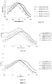
- Gn Rb1 exhibited a significant inhibitory effect on ⁇ -synuclein fibrillation as indicated by the reduced Th-T fluorescence at all tested concentrations ( Fig. 1A ). This effect was observed as early as 2 days of incubation, when the inhibition percentage was approximately 90%. After 5 days of incubation, the fibrillation of ⁇ -synuclein was reduced by approximately 80%.
- Salvianolic acid B and dihydromyricetin inhibited ⁇ -synuclein fibrillation as indicated by reduced Th-T fluorescence ( Fig. 1B and C ).
- concentrations of 100 and 50 ⁇ M salvianolic acid B exhibited a complete inhibitory effect, which was prominent beginning on the third day of incubation. After 5 days of incubation, salvianolic acid B at 100 ⁇ M completely abolished the fibrillation of ⁇ -synuclein, while at 50 ⁇ M, it inhibited fibrillation by almost 80%.
- a lower concentration i.e.
- salvianolic acid B also induced the inhibition of ⁇ -synuclein fibrillation, the percentage of which was reduced by approximately 35% after 5 days of incubation ( Fig. 1B ). These results indicate that salvianolic acid B inhibited the formation of fibrils in a concentration-dependent manner.
- Dihydromyricetin was observed as a good inhibitor of fibrillation. After 5 days of incubation, dihydromyricetin inhibited ⁇ -synuclein fibrillation by 80 and 40% at 100 and 50 ⁇ M, respectively, while at 25 ⁇ M, the compound failed to inhibit fibrillation ( Fig. 1C ).
- the amount of monomeric ⁇ -synuclein in the ⁇ -synuclein aged alone samples and in the presence of compounds was quantified against a fresh ⁇ -synuclein sample that contained only the monomeric species using ImageJ software.
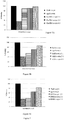
- Gn Rb1 was shown to be a potent inhibitor of ⁇ -synuclein oligomerization at all molar ratios ( Figs. 4A and 5A )
- Gn Rb1 inhibited the formation of larger aggregates (MW >250 kDa) and high MW oligomers, generating a prominent monomeric band and a weak dimeric band ( Fig. 4A ).
- Densitometric analysis of the monomeric bands revealed that Gn Rb1 hindered ⁇ -synuclein oligomerization to the same extent in all tested concentrations, ( Fig. 5A ). Even at the lowest concentration, Gn Rb1 was able to inhibit the formation of larger aggregates.
- Salvianolic acid B and dihydromyricetin were shown to be potent inhibitors of ⁇ -synuclein oligomerization. Salvianolic acid B and dihydromyricetin inhibited ⁇ -synuclein oligomerization in a dose-dependent fashion ( Figs. 4B -C and 5B-C ).
- Salvianolic acid B inhibited the formation of larger aggregates (MW >250 kDa) and high MW oligomers; bands corresponding to monomeric and dimeric ⁇ -synuclein were the most prominent species, and a band corresponding to trimeric ⁇ -synuclein was faint ( Fig. 4A ). With decreasing salvianolic acid B concentration, the dimeric, trimeric and oligomeric bands were increasingly stronger, indicating that salvianolic acid B inhibited ⁇ -synuclein oligomerization in a dose-dependent fashion ( Fig. 4A ). Even at the lowest concentration, salvianolic acid B still induced the inhibition of larger aggregates.
- Dihydromyricetin also suppressed the formation of oligomeric forms in a concentration-dependent manner; the highest concentration was the most potent of the three concentrations tested ( Fig. 4B ). At the highest concentration of dihydromyricetin, ⁇ -synuclein produced faint dimeric and trimeric bands ( Fig. 4B ). However, these bands were progressively stronger with decreasing CMC10 concentration, consistent with dose-dependent inhibition ( Fig. 4B ).
- BE(2)-M17 human neuroblastoma cells were treated with aged ⁇ -synuclein solutions at three different concentrations, 0.5 uM, 1 uM and 5 ⁇ M, either alone or in the presence of Gn Rb1, salvianolic acid B or dihydromyricetin.
- the viability of cells treated with ⁇ -synuclein aged in the presence or absence of the Gn Rb1, salvianolic acid B or dihydromyricetin was determined by the MTT assay. Prior to the experiments, the effect of the Gn Rb1, salvianolic acid B and dihydromyricetin alone on cell viability was assessed (data not shown), employing the same non-toxic Gn Rb1, salvianolic acid B and dihydromyricetin concentrations that were later employed for the experiments with aged ⁇ -synuclein.
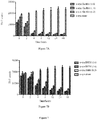
- the aged ⁇ -synuclein induced the decrease in the number of viable cells by almost 50%, whereas in the presence of all concentrations of Gn Rb1, the viability of the cells increased by approximately 30%, with approximately 80% of the cells surviving ( Fig. 6A ).
- Gn Rb1 improved the viability of the cells treated with 1 uM aged ⁇ -synuclein by approximately 30 % ( Fig. 6A ).
- salvianolic acid B In the presence of salvianolic acid B, approximately 95% of the cells survived for the molar ratios of 4:1 and 2:1 ( Fig. 6B ). At these molar ratios, salvianolic acid B had also proved to be a good inhibitor for fibril formation, as also indicated by the reduction in the Th-T counts ( Fig. 1B ). Dihydromyricetin also reduced the ⁇ -synuclein toxicity ( Fig. 6C ). Dihydromyricetin exhibited protection for cells at the highest concentration, which is represented by the molar ratio of 4:1, thus improving cell viability by approximately 10%. These findings are in accordance with the Th-T fluorescence measurements ( Fig. 1C ) and immunoblotting analyses ( Fig. 4 ).
- Gn Rb1 and dihydromyricetin were assessed for their effectiveness on reversing fibrillation.
- 25 ⁇ M of preformed ⁇ -synuclein fibrils were incubated at 37°C in the presence of Gn Rb1 or dihydromyricetin at varying molar ratios (compound: ⁇ -synuclein of 6:1, 4:1 and 2:1) for a period of 48 hours.
- the fibril content was estimated at different time points.
- the Th-T counts for ⁇ -synuclein incubating alone were much higher compared to the Th-T counts of the samples containing Gn Rb1, especially the one containing the highest concentration of the particular ginsenoside.
- the ⁇ -synuclein fibrils that were incubated in the absence of Gn Rb1 continued to aggregate further, as indicated by the increase in Th-T counts, whereas the fibrils incubating in the presence of the ginsenoside disaggregated in a dose-dependent fashion, given the decrease in Th-T counts ( Fig. 7A ). This trend was apparent after 4 hours of incubation, with the Th-T counts for the Gn Rb1 containing samples being approximately the 1/5 of the Th-T counts of the control.
- the Th-T counts were approximately 20,000 for ⁇ -synuclein incubated alone, while in the presence of dihydromyricetin, the Th-T count was less at that time point.
- the ⁇ -synuclein fibrils that were incubated alone continued to aggregate further, as indicated by the increase in Th-T counts, whereas ⁇ -synuclein fibrils incubated in the presence of dihydromyricetin disaggregated over time, in a dose dependant fashion, given the decrease in Th-T counts ( Figs. 7B ).
- dihydromyricetin disaggregated preformed ⁇ -synuclein fibrils in a dose-dependent fashion.
- Gn Rb1 and dihydromyricetin to disaggregate preformed fibrils makes them suitable compounds to be used in the imaging of ⁇ -synuclein aggregates such as Lewy bodies, in subjects.
- nucleation-dependent polymerization model It has been previously shown that the process of amyloid fibril formation follows a nucleation-dependent polymerization model. According to this model, soluble species generate via nucleation oligomeric species (nucleation or lag time phase), which in turn polymerize (polymerization or growth phase) to generate fibrils, thus reaching a final plateau known as the equilibrium phase. Small aggregates or seeds have been shown to accelerate the nucleation phase of amyloid formation in vitro and in vivo via a process known as seeding.
- the compounds were added at concentrations of 10 or 50 ⁇ M to 100 ⁇ M monomeric ⁇ -synuclein containing seeds at a final concentration of 2 ⁇ M. The mixture was then incubated with continuous mixing at 37°C for 6 hours.
- Salvianolic acid B and dihydromyricetin inhibited the seeding process by approximately 90%, as indicated by the low Th-S counts ( Fig. 8B and C ).
- a concentration of 50 ⁇ M inhibited the seeding process by approximately 90%, as indicated by the low Th-S counts ( Fig. 8B and C ).
- a lower concentration (10 ⁇ M) only dihydromyricetin showed an inhibitory effect on the seeding of ⁇ -synuclein monomers, but to a smaller extent in the case of salvianolic acid B.
- Gn Rb1 inhibition of ⁇ -synuclein fibrillation is mediated via binding to the intermediate species and forming stable oligomers
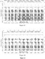
- ⁇ -synuclein species in the samples were characterized by western blotting and electron microscopy ( FIG. 10B , C, Fig. 11 B-C , Fig 12 B-C ). Electron microscopy of the same samples indicated the presence of different species of oligomers in P1 and P2 ( Fig. 10C , 11C , 12C ), in agreement with the immunoblotting results ( Fig. 10B , 11B , 12B ).
- Gn Rb1 To detect the incorporated Gn Rb1 in the P1, P2 and P3 samples, Gn Rb1's ability to produce UV absorbance spectra with three notable peaks was exploited. In the sample containing Gn Rb1: ⁇ -synuclein at 4:1 molar ratio, Gn Rb1 was detected only in the oligomeric P2 samples ( Fig. 10D ).
- dihydromyricetin To detect the incorporated dihydromyricetin in the P1, P2 and P3 samples, dihydromyricetin's ability to produce UV absorbance spectra with three notable peaks was exploited. In the sample containing DHM: ⁇ -synuclein at 4:1, dihydromyricetin was detected in both P2 and P3 which represent the oligomeric and monomeric species respectively ( Fig. 11D ).
- salvianolic acid B To detect the incorporated salvianolic acid B in the P1, P2 and P3 samples, salvianolic acid B's ability to produce UV absorbance spectra with three notable peaks was exploited. In the sample containing salvianolic acid B: ⁇ -synuclein at 4:1, salvianolic acid B was detected in the P2 sample which represents the oligomeric species ( Fig. 12D ).
- ginsenoside Rb1 and salvianolic acid B to bind oligomeric forms of ⁇ -synucleins, but not monomeric forms of ⁇ -synuclein, further suggest that Gn Rb1 and salvianolic acid B would be suitable for use in the diagnosis and imaging of aggregates, for disease involving ⁇ -synuclein.
Landscapes
- Health & Medical Sciences (AREA)
- Life Sciences & Earth Sciences (AREA)
- Chemical & Material Sciences (AREA)
- Medicinal Chemistry (AREA)
- General Health & Medical Sciences (AREA)
- Engineering & Computer Science (AREA)
- Veterinary Medicine (AREA)
- Pharmacology & Pharmacy (AREA)
- Public Health (AREA)
- Animal Behavior & Ethology (AREA)
- Biomedical Technology (AREA)
- Epidemiology (AREA)
- Molecular Biology (AREA)
- Immunology (AREA)
- Hematology (AREA)
- Urology & Nephrology (AREA)
- Neurosurgery (AREA)
- Neurology (AREA)
- Bioinformatics & Cheminformatics (AREA)
- Physics & Mathematics (AREA)
- Biochemistry (AREA)
- Biotechnology (AREA)
- Microbiology (AREA)
- Cell Biology (AREA)
- Pathology (AREA)
- Food Science & Technology (AREA)
- General Physics & Mathematics (AREA)
- Analytical Chemistry (AREA)
- Emergency Medicine (AREA)
- Nuclear Medicine, Radiotherapy & Molecular Imaging (AREA)
- Chemical Kinetics & Catalysis (AREA)
- General Chemical & Material Sciences (AREA)
- Organic Chemistry (AREA)
- Psychology (AREA)
- Pharmaceuticals Containing Other Organic And Inorganic Compounds (AREA)
- Medicines Containing Antibodies Or Antigens For Use As Internal Diagnostic Agents (AREA)
- Measuring Or Testing Involving Enzymes Or Micro-Organisms (AREA)
- Magnetic Resonance Imaging Apparatus (AREA)
- Nuclear Medicine (AREA)
Claims (7)
- Procédé in vitro de détection d'agrégats d'a-synucléine, le procédé comprenant :- l'administration de ginsenoside Rb1 à un échantillon de tissu et/ou à un fluide biologique provenant d'un sujet ; et- la détection de la liaison du composé aux agrégats d'a-synucléine par mise en contact de l'échantillon avec du rouge congo ou de la thioflavine-T.
- Procédé in vitro selon la revendication 1, le procédé étant destiné à imager les agrégats d'a-synucléine.
- Procédé in vitro selon la revendication 1, dans lequel l'échantillon est un échantillon de tissu cérébral.
- Procédé in vitro selon la revendication 1, dans lequel la présence de la liaison du composé à des fibrilles d'a-synucléine est détectée in vitro par autoradiographie, tomographie par émission de positons, imagerie par résonance magnétique, un compteur gamma ou un compteur à scintillation.
- Procédé in vitro selon la revendication 1, dans lequel les agrégats d'α-synucléine sont des corps de Lewy.
- Procédé in vitro de diagnostic d'une maladie synucléinopathique, le procédé comprenant les étapes de :(a) combinaison in vitro d'un échantillon de tissu et/ou de fluide biologique provenant du patient avec le composé ginsenoside Rb1 dans des conditions permettant la liaison du composé aux agrégats d'a-synucléine présents dans l'échantillon ; et(b) détection de la présence ou de l'absence d'agrégats d'α-synucléine dans ledit échantillon par mise en contact de l'échantillon avec du rouge congo ou de la thioflavine-T, la présence d'agrégats d'α-synucléine indiquant que le sujet présente une maladie synucléinopathique, et l'absence d'agrégats d'α-synucléine indiquant que le sujet ne présente pas pas ladite maladie synucléinopathique.
- Utilisation de ginsenoside Rbl en tant qu'agent d'imagerie pour détecter la présence d'agrégats d'α-synucléine dans un échantillon provenant d'un sujet, grâce aux spectres d'absorption UV produits.
Priority Applications (2)
| Application Number | Priority Date | Filing Date | Title |
|---|---|---|---|
| EP24151830.7A EP4356967A3 (fr) | 2014-11-14 | 2014-11-14 | Composés destinés à être utilisés en tant qu'agents d'imagerie |
| EP21185274.4A EP3961216B1 (fr) | 2014-11-14 | 2014-11-14 | Dihydromyricetine destinés à être utilisés comme agent d'imagerie |
Applications Claiming Priority (1)
| Application Number | Priority Date | Filing Date | Title |
|---|---|---|---|
| PCT/IB2014/066041 WO2016075516A1 (fr) | 2014-11-14 | 2014-11-14 | Composés servant d'agents d'imagerie |
Related Child Applications (3)
| Application Number | Title | Priority Date | Filing Date |
|---|---|---|---|
| EP24151830.7A Division EP4356967A3 (fr) | 2014-11-14 | 2014-11-14 | Composés destinés à être utilisés en tant qu'agents d'imagerie |
| EP21185274.4A Division EP3961216B1 (fr) | 2014-11-14 | 2014-11-14 | Dihydromyricetine destinés à être utilisés comme agent d'imagerie |
| EP21185274.4A Division-Into EP3961216B1 (fr) | 2014-11-14 | 2014-11-14 | Dihydromyricetine destinés à être utilisés comme agent d'imagerie |
Publications (3)
| Publication Number | Publication Date |
|---|---|
| EP3218721A1 EP3218721A1 (fr) | 2017-09-20 |
| EP3218721A4 EP3218721A4 (fr) | 2018-11-21 |
| EP3218721B1 true EP3218721B1 (fr) | 2021-08-18 |
Family
ID=55953787
Family Applications (3)
| Application Number | Title | Priority Date | Filing Date |
|---|---|---|---|
| EP21185274.4A Active EP3961216B1 (fr) | 2014-11-14 | 2014-11-14 | Dihydromyricetine destinés à être utilisés comme agent d'imagerie |
| EP14906044.4A Active EP3218721B1 (fr) | 2014-11-14 | 2014-11-14 | Composés servant d'agents d'imagerie |
| EP24151830.7A Pending EP4356967A3 (fr) | 2014-11-14 | 2014-11-14 | Composés destinés à être utilisés en tant qu'agents d'imagerie |
Family Applications Before (1)
| Application Number | Title | Priority Date | Filing Date |
|---|---|---|---|
| EP21185274.4A Active EP3961216B1 (fr) | 2014-11-14 | 2014-11-14 | Dihydromyricetine destinés à être utilisés comme agent d'imagerie |
Family Applications After (1)
| Application Number | Title | Priority Date | Filing Date |
|---|---|---|---|
| EP24151830.7A Pending EP4356967A3 (fr) | 2014-11-14 | 2014-11-14 | Composés destinés à être utilisés en tant qu'agents d'imagerie |
Country Status (4)
| Country | Link |
|---|---|
| US (1) | US10488417B2 (fr) |
| EP (3) | EP3961216B1 (fr) |
| JP (1) | JP6564032B2 (fr) |
| WO (1) | WO2016075516A1 (fr) |
Families Citing this family (5)
| Publication number | Priority date | Publication date | Assignee | Title |
|---|---|---|---|---|
| CN108398452A (zh) * | 2018-02-07 | 2018-08-14 | 山东省分析测试中心 | 一种基于氢核磁定量分析技术测定人参皂苷Re对照品含量的方法 |
| CN108152319A (zh) * | 2018-02-07 | 2018-06-12 | 山东省分析测试中心 | 一种基于氢核磁定量分析技术测定人参皂苷Rd对照品含量的方法 |
| CN108514546B (zh) * | 2018-05-14 | 2020-09-29 | 山东师范大学 | 一种二氢杨梅素的壳聚糖水凝胶制剂及制备方法 |
| ES2988770T3 (es) * | 2018-06-08 | 2024-11-21 | Ac Immune Sa | Nuevos compuestos para diagnóstico |
| US20230365665A1 (en) | 2020-09-17 | 2023-11-16 | Qatar Foundation For Education, Science And Community Development | ANTIBODY COMPOSITIONS TARGETING NON-PHOSPHORYLATED a-SYNUCLEIN AGGREGATES |
Family Cites Families (15)
| Publication number | Priority date | Publication date | Assignee | Title |
|---|---|---|---|---|
| AU3117799A (en) | 1998-03-30 | 1999-10-18 | Trustees Of The University Of Pennsylvania, The | Method of identifying, diagnosing and treating synuclein positive neurodegenerative disorders |
| JP2000191539A (ja) * | 1998-12-22 | 2000-07-11 | Japan Science & Technology Corp | ジンセノサイドRb1からなる脳細胞又は神経細胞保護剤 |
| GB0203446D0 (en) | 2002-02-14 | 2002-04-03 | Univ Lancaster | Detection and/or monitoring of synuclein-related diseases |
| DE602005026276D1 (de) * | 2004-05-18 | 2011-03-24 | Bright Future Pharmaceutical Lab Ltd | Myricitrin verbindungen zur behandlung von schlafstörungen |
| WO2007118363A1 (fr) * | 2006-04-19 | 2007-10-25 | Jianxun Liu | Composition médicinale chinoise, procédé de préparation et utilisation de celle-ci |
| CN1985836A (zh) | 2006-12-18 | 2007-06-27 | 贵州信邦远东药业有限公司 | 一种抗脑缺血作用的药物组合物 |
| CN101239057A (zh) * | 2007-02-06 | 2008-08-13 | 中国人民解放军军事医学科学院放射与辐射医学研究所 | 丹酚酸b及其盐类在治疗帕金森病中的用途 |
| GB0716885D0 (en) | 2007-08-30 | 2007-10-10 | United Arab Emirates Universit | Diagnostic agent |
| US20110159527A1 (en) * | 2008-06-16 | 2011-06-30 | Michael Gebhard Schlossmacher | Methods and kits for diagnosing neurodegenerative disease |
| US20120053235A1 (en) * | 2010-08-30 | 2012-03-01 | Hong Kong Baptist University | Dihydromyricetin as an IKK-beta inhibitor used for treatment of arthritis, cancer and autoimmune conditions, and other diseases or disorders |
| US20120251448A1 (en) * | 2011-03-03 | 2012-10-04 | Hefti Franz F | Compounds for Use in the Detection of Neurodegenerative Diseases |
| CN102727508B (zh) * | 2012-07-15 | 2014-09-24 | 浙江大学 | 人参二醇皂苷组分制备及在防治帕金森氏病中的药物用途 |
| CN103054847A (zh) * | 2013-01-04 | 2013-04-24 | 中国药科大学 | 丹酚酸b作为儿茶酚-o-甲基转移酶抑制剂在帕金森病治疗中的应用 |
| US9534044B2 (en) | 2013-02-28 | 2017-01-03 | United Arab Emirates University | Alpha-synuclein antibodies and uses thereof |
| CN104116730A (zh) * | 2013-04-26 | 2014-10-29 | 中国科学院大连化学物理研究所 | 二氢杨梅素作为活性成份在制备帕金森症治疗药物中的应用 |
-
2014
- 2014-11-14 JP JP2017525594A patent/JP6564032B2/ja active Active
- 2014-11-14 WO PCT/IB2014/066041 patent/WO2016075516A1/fr not_active Ceased
- 2014-11-14 EP EP21185274.4A patent/EP3961216B1/fr active Active
- 2014-11-14 EP EP14906044.4A patent/EP3218721B1/fr active Active
- 2014-11-14 US US15/526,525 patent/US10488417B2/en active Active
- 2014-11-14 EP EP24151830.7A patent/EP4356967A3/fr active Pending
Non-Patent Citations (1)
| Title |
|---|
| None * |
Also Published As
| Publication number | Publication date |
|---|---|
| EP4356967A2 (fr) | 2024-04-24 |
| EP3961216B1 (fr) | 2024-02-14 |
| EP3218721A4 (fr) | 2018-11-21 |
| JP2018505845A (ja) | 2018-03-01 |
| US10488417B2 (en) | 2019-11-26 |
| EP3218721A1 (fr) | 2017-09-20 |
| JP6564032B2 (ja) | 2019-08-21 |
| EP3961216A1 (fr) | 2022-03-02 |
| US20170307623A1 (en) | 2017-10-26 |
| EP4356967A3 (fr) | 2024-07-24 |
| WO2016075516A1 (fr) | 2016-05-19 |
Similar Documents
| Publication | Publication Date | Title |
|---|---|---|
| Ono et al. | Phenolic compounds prevent amyloid β-protein oligomerization and synaptic dysfunction by site-specific binding | |
| Li et al. | ROS and iron homeostasis dependent ferroptosis play a vital role in 5-Fluorouracil induced cardiotoxicity in vitro and in vivo | |
| Chern et al. | Melatonin ameliorates neural function by promoting endogenous neurogenesis through the MT2 melatonin receptor in ischemic-stroke mice | |
| Kai et al. | Tabersonine inhibits amyloid fibril formation and cytotoxicity of Aβ (1–42) | |
| EP3961216B1 (fr) | Dihydromyricetine destinés à être utilisés comme agent d'imagerie | |
| WO2023051088A1 (fr) | Utilisation de la dihydrotanshinone i dans la préparation d'un inhibiteur de nrf2 | |
| Xu et al. | Advances in the development of imaging probes and aggregation inhibitors for alpha-synuclein | |
| Dai et al. | Astragalus polysaccharide inhibits isoprenaline-induced cardiac hypertrophy via suppressing Ca2+-mediated calcineurin/NFATc3 and CaMKII signaling cascades | |
| Ardah et al. | RETRACTED ARTICLE: Inhibition of alpha-synuclein seeded fibril formation and toxicity by herbal medicinal extracts | |
| CN102821777B (zh) | 神经丝肽用于神经胶质瘤的治疗的用途 | |
| US6346280B1 (en) | Composition and methods for inhibiting the formation of brain amyloid deposits | |
| Wang et al. | S 24795 limits β-amyloid–α7 nicotinic receptor interaction and reduces Alzheimer's disease-like pathologies | |
| CN101854953A (zh) | 阶段ⅱ解毒和抗氧化剂活性 | |
| Wang et al. | Inhibition of amyloid peptide fibril formation by gold–sulfur complexes | |
| Zeng et al. | Ethyl ferulate suppresses post-myocardial infarction myocardial fibrosis by inhibiting transforming growth factor receptor 1 | |
| Li et al. | Protective effect of compound tongluo decoction on brain vascular endothelial cells after ischemia‐reperfusion by inhibition of ferroptosis through regulating Nrf2/ARE/SLC7A11 signaling pathway | |
| KR20190098649A (ko) | 칸나비디올 추출물을 함유하는 대장암 예방 또는 치료용 약학 조성물 | |
| Wang et al. | Ginsenoside Rg1 alleviates chronic stress-induced depression in rats by targeting Cx43-YAP axis | |
| EP3970710A1 (fr) | Composition pharmaceutique pour la prévention ou le traitement de maladies cérébrales dégénératives par activation de l'autophagie | |
| Nie et al. | A-type EGCG dimer, a new proanthocyanidins dimer from persimmon fruits, interacts with the amino acid residues of Aβ40 which possessed high aggregation-propensity and strongly inhibits its amyloid fibrils formation | |
| Szegedi et al. | Endomorphin‐2, an endogenous tetrapeptide, protects against Aβ1–42 in vitro and in vivo | |
| Wang et al. | The roles and mechanisms of CDGSH iron-sulfur domain 1 in kainic acid-induced mitochondrial iron overload, dysfunction and neuronal damage | |
| CN119818652A (zh) | 一种预防/治疗阿尔茨海默症的药物组合物 | |
| Leri et al. | Maysin plays a protective role against α-Synuclein oligomers cytotoxicity by triggering autophagy activation | |
| HK1236052B (en) | Compounds for use as imaging agents |
Legal Events
| Date | Code | Title | Description |
|---|---|---|---|
| STAA | Information on the status of an ep patent application or granted ep patent |
Free format text: STATUS: THE INTERNATIONAL PUBLICATION HAS BEEN MADE |
|
| PUAI | Public reference made under article 153(3) epc to a published international application that has entered the european phase |
Free format text: ORIGINAL CODE: 0009012 |
|
| STAA | Information on the status of an ep patent application or granted ep patent |
Free format text: STATUS: REQUEST FOR EXAMINATION WAS MADE |
|
| 17P | Request for examination filed |
Effective date: 20170523 |
|
| AK | Designated contracting states |
Kind code of ref document: A1 Designated state(s): AL AT BE BG CH CY CZ DE DK EE ES FI FR GB GR HR HU IE IS IT LI LT LU LV MC MK MT NL NO PL PT RO RS SE SI SK SM TR |
|
| AX | Request for extension of the european patent |
Extension state: BA ME |
|
| DAX | Request for extension of the european patent (deleted) | ||
| REG | Reference to a national code |
Ref country code: HK Ref legal event code: DE Ref document number: 1236052 Country of ref document: HK |
|
| RIC1 | Information provided on ipc code assigned before grant |
Ipc: G01N 33/58 20060101AFI20180607BHEP Ipc: A61K 31/352 20060101ALI20180607BHEP Ipc: A61K 31/343 20060101ALI20180607BHEP Ipc: A61P 25/28 20060101ALI20180607BHEP Ipc: A61K 31/704 20060101ALI20180607BHEP |
|
| A4 | Supplementary search report drawn up and despatched |
Effective date: 20181018 |
|
| RIC1 | Information provided on ipc code assigned before grant |
Ipc: A61K 31/343 20060101ALI20181012BHEP Ipc: A61K 31/352 20060101ALI20181012BHEP Ipc: A61K 31/704 20060101ALI20181012BHEP Ipc: G01N 33/58 20060101AFI20181012BHEP Ipc: A61P 25/28 20060101ALI20181012BHEP |
|
| STAA | Information on the status of an ep patent application or granted ep patent |
Free format text: STATUS: EXAMINATION IS IN PROGRESS |
|
| 17Q | First examination report despatched |
Effective date: 20200331 |
|
| GRAP | Despatch of communication of intention to grant a patent |
Free format text: ORIGINAL CODE: EPIDOSNIGR1 |
|
| STAA | Information on the status of an ep patent application or granted ep patent |
Free format text: STATUS: GRANT OF PATENT IS INTENDED |
|
| RIC1 | Information provided on ipc code assigned before grant |
Ipc: G01N 33/58 20060101AFI20210212BHEP Ipc: A61P 25/28 20060101ALI20210212BHEP Ipc: A61K 31/704 20060101ALI20210212BHEP |
|
| INTG | Intention to grant announced |
Effective date: 20210301 |
|
| GRAS | Grant fee paid |
Free format text: ORIGINAL CODE: EPIDOSNIGR3 |
|
| GRAA | (expected) grant |
Free format text: ORIGINAL CODE: 0009210 |
|
| STAA | Information on the status of an ep patent application or granted ep patent |
Free format text: STATUS: THE PATENT HAS BEEN GRANTED |
|
| AK | Designated contracting states |
Kind code of ref document: B1 Designated state(s): AL AT BE BG CH CY CZ DE DK EE ES FI FR GB GR HR HU IE IS IT LI LT LU LV MC MK MT NL NO PL PT RO RS SE SI SK SM TR |
|
| REG | Reference to a national code |
Ref country code: GB Ref legal event code: FG4D |
|
| REG | Reference to a national code |
Ref country code: CH Ref legal event code: EP |
|
| REG | Reference to a national code |
Ref country code: DE Ref legal event code: R096 Ref document number: 602014079607 Country of ref document: DE |
|
| REG | Reference to a national code |
Ref country code: IE Ref legal event code: FG4D Ref country code: AT Ref legal event code: REF Ref document number: 1422101 Country of ref document: AT Kind code of ref document: T Effective date: 20210915 |
|
| REG | Reference to a national code |
Ref country code: LT Ref legal event code: MG9D |
|
| REG | Reference to a national code |
Ref country code: NL Ref legal event code: MP Effective date: 20210818 |
|
| REG | Reference to a national code |
Ref country code: AT Ref legal event code: MK05 Ref document number: 1422101 Country of ref document: AT Kind code of ref document: T Effective date: 20210818 |
|
| PG25 | Lapsed in a contracting state [announced via postgrant information from national office to epo] |
Ref country code: HR Free format text: LAPSE BECAUSE OF FAILURE TO SUBMIT A TRANSLATION OF THE DESCRIPTION OR TO PAY THE FEE WITHIN THE PRESCRIBED TIME-LIMIT Effective date: 20210818 Ref country code: SE Free format text: LAPSE BECAUSE OF FAILURE TO SUBMIT A TRANSLATION OF THE DESCRIPTION OR TO PAY THE FEE WITHIN THE PRESCRIBED TIME-LIMIT Effective date: 20210818 Ref country code: RS Free format text: LAPSE BECAUSE OF FAILURE TO SUBMIT A TRANSLATION OF THE DESCRIPTION OR TO PAY THE FEE WITHIN THE PRESCRIBED TIME-LIMIT Effective date: 20210818 Ref country code: LT Free format text: LAPSE BECAUSE OF FAILURE TO SUBMIT A TRANSLATION OF THE DESCRIPTION OR TO PAY THE FEE WITHIN THE PRESCRIBED TIME-LIMIT Effective date: 20210818 Ref country code: BG Free format text: LAPSE BECAUSE OF FAILURE TO SUBMIT A TRANSLATION OF THE DESCRIPTION OR TO PAY THE FEE WITHIN THE PRESCRIBED TIME-LIMIT Effective date: 20211118 Ref country code: AT Free format text: LAPSE BECAUSE OF FAILURE TO SUBMIT A TRANSLATION OF THE DESCRIPTION OR TO PAY THE FEE WITHIN THE PRESCRIBED TIME-LIMIT Effective date: 20210818 Ref country code: NO Free format text: LAPSE BECAUSE OF FAILURE TO SUBMIT A TRANSLATION OF THE DESCRIPTION OR TO PAY THE FEE WITHIN THE PRESCRIBED TIME-LIMIT Effective date: 20211118 Ref country code: PT Free format text: LAPSE BECAUSE OF FAILURE TO SUBMIT A TRANSLATION OF THE DESCRIPTION OR TO PAY THE FEE WITHIN THE PRESCRIBED TIME-LIMIT Effective date: 20211220 Ref country code: ES Free format text: LAPSE BECAUSE OF FAILURE TO SUBMIT A TRANSLATION OF THE DESCRIPTION OR TO PAY THE FEE WITHIN THE PRESCRIBED TIME-LIMIT Effective date: 20210818 Ref country code: FI Free format text: LAPSE BECAUSE OF FAILURE TO SUBMIT A TRANSLATION OF THE DESCRIPTION OR TO PAY THE FEE WITHIN THE PRESCRIBED TIME-LIMIT Effective date: 20210818 |
|
| PG25 | Lapsed in a contracting state [announced via postgrant information from national office to epo] |
Ref country code: PL Free format text: LAPSE BECAUSE OF FAILURE TO SUBMIT A TRANSLATION OF THE DESCRIPTION OR TO PAY THE FEE WITHIN THE PRESCRIBED TIME-LIMIT Effective date: 20210818 Ref country code: LV Free format text: LAPSE BECAUSE OF FAILURE TO SUBMIT A TRANSLATION OF THE DESCRIPTION OR TO PAY THE FEE WITHIN THE PRESCRIBED TIME-LIMIT Effective date: 20210818 Ref country code: GR Free format text: LAPSE BECAUSE OF FAILURE TO SUBMIT A TRANSLATION OF THE DESCRIPTION OR TO PAY THE FEE WITHIN THE PRESCRIBED TIME-LIMIT Effective date: 20211119 |
|
| PG25 | Lapsed in a contracting state [announced via postgrant information from national office to epo] |
Ref country code: NL Free format text: LAPSE BECAUSE OF FAILURE TO SUBMIT A TRANSLATION OF THE DESCRIPTION OR TO PAY THE FEE WITHIN THE PRESCRIBED TIME-LIMIT Effective date: 20210818 |
|
| PG25 | Lapsed in a contracting state [announced via postgrant information from national office to epo] |
Ref country code: DK Free format text: LAPSE BECAUSE OF FAILURE TO SUBMIT A TRANSLATION OF THE DESCRIPTION OR TO PAY THE FEE WITHIN THE PRESCRIBED TIME-LIMIT Effective date: 20210818 |
|
| REG | Reference to a national code |
Ref country code: DE Ref legal event code: R097 Ref document number: 602014079607 Country of ref document: DE |
|
| PG25 | Lapsed in a contracting state [announced via postgrant information from national office to epo] |
Ref country code: SM Free format text: LAPSE BECAUSE OF FAILURE TO SUBMIT A TRANSLATION OF THE DESCRIPTION OR TO PAY THE FEE WITHIN THE PRESCRIBED TIME-LIMIT Effective date: 20210818 Ref country code: SK Free format text: LAPSE BECAUSE OF FAILURE TO SUBMIT A TRANSLATION OF THE DESCRIPTION OR TO PAY THE FEE WITHIN THE PRESCRIBED TIME-LIMIT Effective date: 20210818 Ref country code: RO Free format text: LAPSE BECAUSE OF FAILURE TO SUBMIT A TRANSLATION OF THE DESCRIPTION OR TO PAY THE FEE WITHIN THE PRESCRIBED TIME-LIMIT Effective date: 20210818 Ref country code: EE Free format text: LAPSE BECAUSE OF FAILURE TO SUBMIT A TRANSLATION OF THE DESCRIPTION OR TO PAY THE FEE WITHIN THE PRESCRIBED TIME-LIMIT Effective date: 20210818 Ref country code: CZ Free format text: LAPSE BECAUSE OF FAILURE TO SUBMIT A TRANSLATION OF THE DESCRIPTION OR TO PAY THE FEE WITHIN THE PRESCRIBED TIME-LIMIT Effective date: 20210818 Ref country code: AL Free format text: LAPSE BECAUSE OF FAILURE TO SUBMIT A TRANSLATION OF THE DESCRIPTION OR TO PAY THE FEE WITHIN THE PRESCRIBED TIME-LIMIT Effective date: 20210818 |
|
| PLBE | No opposition filed within time limit |
Free format text: ORIGINAL CODE: 0009261 |
|
| STAA | Information on the status of an ep patent application or granted ep patent |
Free format text: STATUS: NO OPPOSITION FILED WITHIN TIME LIMIT |
|
| PG25 | Lapsed in a contracting state [announced via postgrant information from national office to epo] |
Ref country code: MC Free format text: LAPSE BECAUSE OF FAILURE TO SUBMIT A TRANSLATION OF THE DESCRIPTION OR TO PAY THE FEE WITHIN THE PRESCRIBED TIME-LIMIT Effective date: 20210818 |
|
| REG | Reference to a national code |
Ref country code: CH Ref legal event code: PL |
|
| 26N | No opposition filed |
Effective date: 20220519 |
|
| PG25 | Lapsed in a contracting state [announced via postgrant information from national office to epo] |
Ref country code: LU Free format text: LAPSE BECAUSE OF NON-PAYMENT OF DUE FEES Effective date: 20211114 Ref country code: IT Free format text: LAPSE BECAUSE OF FAILURE TO SUBMIT A TRANSLATION OF THE DESCRIPTION OR TO PAY THE FEE WITHIN THE PRESCRIBED TIME-LIMIT Effective date: 20210818 Ref country code: BE Free format text: LAPSE BECAUSE OF NON-PAYMENT OF DUE FEES Effective date: 20211130 |
|
| REG | Reference to a national code |
Ref country code: BE Ref legal event code: MM Effective date: 20211130 |
|
| PG25 | Lapsed in a contracting state [announced via postgrant information from national office to epo] |
Ref country code: SI Free format text: LAPSE BECAUSE OF FAILURE TO SUBMIT A TRANSLATION OF THE DESCRIPTION OR TO PAY THE FEE WITHIN THE PRESCRIBED TIME-LIMIT Effective date: 20210818 |
|
| PG25 | Lapsed in a contracting state [announced via postgrant information from national office to epo] |
Ref country code: IE Free format text: LAPSE BECAUSE OF NON-PAYMENT OF DUE FEES Effective date: 20211114 |
|
| PG25 | Lapsed in a contracting state [announced via postgrant information from national office to epo] |
Ref country code: FR Free format text: LAPSE BECAUSE OF NON-PAYMENT OF DUE FEES Effective date: 20211130 |
|
| PG25 | Lapsed in a contracting state [announced via postgrant information from national office to epo] |
Ref country code: HU Free format text: LAPSE BECAUSE OF FAILURE TO SUBMIT A TRANSLATION OF THE DESCRIPTION OR TO PAY THE FEE WITHIN THE PRESCRIBED TIME-LIMIT; INVALID AB INITIO Effective date: 20141114 |
|
| PG25 | Lapsed in a contracting state [announced via postgrant information from national office to epo] |
Ref country code: CY Free format text: LAPSE BECAUSE OF FAILURE TO SUBMIT A TRANSLATION OF THE DESCRIPTION OR TO PAY THE FEE WITHIN THE PRESCRIBED TIME-LIMIT Effective date: 20210818 |
|
| PG25 | Lapsed in a contracting state [announced via postgrant information from national office to epo] |
Ref country code: LI Free format text: LAPSE BECAUSE OF NON-PAYMENT OF DUE FEES Effective date: 20220701 Ref country code: CH Free format text: LAPSE BECAUSE OF NON-PAYMENT OF DUE FEES Effective date: 20220701 |
|
| PG25 | Lapsed in a contracting state [announced via postgrant information from national office to epo] |
Ref country code: MK Free format text: LAPSE BECAUSE OF FAILURE TO SUBMIT A TRANSLATION OF THE DESCRIPTION OR TO PAY THE FEE WITHIN THE PRESCRIBED TIME-LIMIT Effective date: 20210818 |
|
| PG25 | Lapsed in a contracting state [announced via postgrant information from national office to epo] |
Ref country code: MT Free format text: LAPSE BECAUSE OF FAILURE TO SUBMIT A TRANSLATION OF THE DESCRIPTION OR TO PAY THE FEE WITHIN THE PRESCRIBED TIME-LIMIT Effective date: 20210818 |
|
| PGFP | Annual fee paid to national office [announced via postgrant information from national office to epo] |
Ref country code: GB Payment date: 20250916 Year of fee payment: 12 |
|
| PG25 | Lapsed in a contracting state [announced via postgrant information from national office to epo] |
Ref country code: TR Free format text: LAPSE BECAUSE OF FAILURE TO SUBMIT A TRANSLATION OF THE DESCRIPTION OR TO PAY THE FEE WITHIN THE PRESCRIBED TIME-LIMIT Effective date: 20210818 |
|
| PGFP | Annual fee paid to national office [announced via postgrant information from national office to epo] |
Ref country code: DE Payment date: 20251126 Year of fee payment: 12 |