EP3243076B1 - Procédés pour la détection d'un marqueur pour la tuberculose active - Google Patents
Procédés pour la détection d'un marqueur pour la tuberculose active Download PDFInfo
- Publication number
- EP3243076B1 EP3243076B1 EP16720577.2A EP16720577A EP3243076B1 EP 3243076 B1 EP3243076 B1 EP 3243076B1 EP 16720577 A EP16720577 A EP 16720577A EP 3243076 B1 EP3243076 B1 EP 3243076B1
- Authority
- EP
- European Patent Office
- Prior art keywords
- sample
- substrate
- mycolic acid
- sterol lipid
- acid derived
- Prior art date
- Legal status (The legal status is an assumption and is not a legal conclusion. Google has not performed a legal analysis and makes no representation as to the accuracy of the status listed.)
- Active
Links
Images

Classifications
-
- G—PHYSICS
- G01—MEASURING; TESTING
- G01N—INVESTIGATING OR ANALYSING MATERIALS BY DETERMINING THEIR CHEMICAL OR PHYSICAL PROPERTIES
- G01N1/00—Sampling; Preparing specimens for investigation
- G01N1/28—Preparing specimens for investigation including physical details of (bio-)chemical methods covered elsewhere, e.g. G01N33/50, C12Q
-
- G—PHYSICS
- G01—MEASURING; TESTING
- G01N—INVESTIGATING OR ANALYSING MATERIALS BY DETERMINING THEIR CHEMICAL OR PHYSICAL PROPERTIES
- G01N33/00—Investigating or analysing materials by specific methods not covered by groups G01N1/00 - G01N31/00
- G01N33/48—Biological material, e.g. blood, urine; Haemocytometers
- G01N33/50—Chemical analysis of biological material, e.g. blood, urine; Testing involving biospecific ligand binding methods; Immunological testing
- G01N33/53—Immunoassay; Biospecific binding assay; Materials therefor
- G01N33/569—Immunoassay; Biospecific binding assay; Materials therefor for microorganisms, e.g. protozoa, bacteria, viruses
- G01N33/56911—Bacteria
- G01N33/5695—Mycobacteria
-
- G—PHYSICS
- G01—MEASURING; TESTING
- G01N—INVESTIGATING OR ANALYSING MATERIALS BY DETERMINING THEIR CHEMICAL OR PHYSICAL PROPERTIES
- G01N33/00—Investigating or analysing materials by specific methods not covered by groups G01N1/00 - G01N31/00
- G01N33/48—Biological material, e.g. blood, urine; Haemocytometers
- G01N33/50—Chemical analysis of biological material, e.g. blood, urine; Testing involving biospecific ligand binding methods; Immunological testing
- G01N33/53—Immunoassay; Biospecific binding assay; Materials therefor
- G01N33/569—Immunoassay; Biospecific binding assay; Materials therefor for microorganisms, e.g. protozoa, bacteria, viruses
- G01N33/56911—Bacteria
- G01N33/56933—Mycoplasma
-
- A—HUMAN NECESSITIES
- A61—MEDICAL OR VETERINARY SCIENCE; HYGIENE
- A61B—DIAGNOSIS; SURGERY; IDENTIFICATION
- A61B5/00—Measuring for diagnostic purposes; Identification of persons
- A61B5/15—Devices for taking samples of blood
- A61B5/150007—Details
- A61B5/150015—Source of blood
- A61B5/150022—Source of blood for capillary blood or interstitial fluid
-
- A—HUMAN NECESSITIES
- A61—MEDICAL OR VETERINARY SCIENCE; HYGIENE
- A61B—DIAGNOSIS; SURGERY; IDENTIFICATION
- A61B5/00—Measuring for diagnostic purposes; Identification of persons
- A61B5/15—Devices for taking samples of blood
- A61B5/150007—Details
- A61B5/150206—Construction or design features not otherwise provided for; manufacturing or production; packages; sterilisation of piercing element, piercing device or sampling device
- A61B5/150251—Collection chamber divided into at least two compartments, e.g. for division of samples
-
- A—HUMAN NECESSITIES
- A61—MEDICAL OR VETERINARY SCIENCE; HYGIENE
- A61B—DIAGNOSIS; SURGERY; IDENTIFICATION
- A61B5/00—Measuring for diagnostic purposes; Identification of persons
- A61B5/15—Devices for taking samples of blood
- A61B5/150007—Details
- A61B5/150755—Blood sample preparation for further analysis, e.g. by separating blood components or by mixing
-
- A—HUMAN NECESSITIES
- A61—MEDICAL OR VETERINARY SCIENCE; HYGIENE
- A61B—DIAGNOSIS; SURGERY; IDENTIFICATION
- A61B5/00—Measuring for diagnostic purposes; Identification of persons
- A61B5/15—Devices for taking samples of blood
- A61B5/157—Devices characterised by integrated means for measuring characteristics of blood
-
- B—PERFORMING OPERATIONS; TRANSPORTING
- B01—PHYSICAL OR CHEMICAL PROCESSES OR APPARATUS IN GENERAL
- B01L—CHEMICAL OR PHYSICAL LABORATORY APPARATUS FOR GENERAL USE
- B01L3/00—Containers or dishes for laboratory use, e.g. laboratory glassware; Droppers
- B01L3/50—Containers for the purpose of retaining a material to be analysed, e.g. test tubes
- B01L3/502—Containers for the purpose of retaining a material to be analysed, e.g. test tubes with fluid transport, e.g. in multi-compartment structures
-
- G—PHYSICS
- G01—MEASURING; TESTING
- G01N—INVESTIGATING OR ANALYSING MATERIALS BY DETERMINING THEIR CHEMICAL OR PHYSICAL PROPERTIES
- G01N33/00—Investigating or analysing materials by specific methods not covered by groups G01N1/00 - G01N31/00
- G01N33/48—Biological material, e.g. blood, urine; Haemocytometers
- G01N33/50—Chemical analysis of biological material, e.g. blood, urine; Testing involving biospecific ligand binding methods; Immunological testing
-
- G—PHYSICS
- G01—MEASURING; TESTING
- G01N—INVESTIGATING OR ANALYSING MATERIALS BY DETERMINING THEIR CHEMICAL OR PHYSICAL PROPERTIES
- G01N33/00—Investigating or analysing materials by specific methods not covered by groups G01N1/00 - G01N31/00
- G01N33/48—Biological material, e.g. blood, urine; Haemocytometers
- G01N33/50—Chemical analysis of biological material, e.g. blood, urine; Testing involving biospecific ligand binding methods; Immunological testing
- G01N33/5005—Chemical analysis of biological material, e.g. blood, urine; Testing involving biospecific ligand binding methods; Immunological testing involving human or animal cells
- G01N33/5008—Chemical analysis of biological material, e.g. blood, urine; Testing involving biospecific ligand binding methods; Immunological testing involving human or animal cells for testing or evaluating the effect of chemical or biological compounds, e.g. drugs, cosmetics
- G01N33/502—Chemical analysis of biological material, e.g. blood, urine; Testing involving biospecific ligand binding methods; Immunological testing involving human or animal cells for testing or evaluating the effect of chemical or biological compounds, e.g. drugs, cosmetics for testing non-proliferative effects
-
- G—PHYSICS
- G01—MEASURING; TESTING
- G01N—INVESTIGATING OR ANALYSING MATERIALS BY DETERMINING THEIR CHEMICAL OR PHYSICAL PROPERTIES
- G01N33/00—Investigating or analysing materials by specific methods not covered by groups G01N1/00 - G01N31/00
- G01N33/48—Biological material, e.g. blood, urine; Haemocytometers
- G01N33/50—Chemical analysis of biological material, e.g. blood, urine; Testing involving biospecific ligand binding methods; Immunological testing
- G01N33/53—Immunoassay; Biospecific binding assay; Materials therefor
- G01N33/543—Immunoassay; Biospecific binding assay; Materials therefor with an insoluble carrier for immobilising immunochemicals
-
- G—PHYSICS
- G01—MEASURING; TESTING
- G01N—INVESTIGATING OR ANALYSING MATERIALS BY DETERMINING THEIR CHEMICAL OR PHYSICAL PROPERTIES
- G01N33/00—Investigating or analysing materials by specific methods not covered by groups G01N1/00 - G01N31/00
- G01N33/48—Biological material, e.g. blood, urine; Haemocytometers
- G01N33/50—Chemical analysis of biological material, e.g. blood, urine; Testing involving biospecific ligand binding methods; Immunological testing
- G01N33/53—Immunoassay; Biospecific binding assay; Materials therefor
- G01N33/543—Immunoassay; Biospecific binding assay; Materials therefor with an insoluble carrier for immobilising immunochemicals
- G01N33/54306—Solid-phase reaction mechanisms
-
- G—PHYSICS
- G01—MEASURING; TESTING
- G01N—INVESTIGATING OR ANALYSING MATERIALS BY DETERMINING THEIR CHEMICAL OR PHYSICAL PROPERTIES
- G01N33/00—Investigating or analysing materials by specific methods not covered by groups G01N1/00 - G01N31/00
- G01N33/48—Biological material, e.g. blood, urine; Haemocytometers
- G01N33/50—Chemical analysis of biological material, e.g. blood, urine; Testing involving biospecific ligand binding methods; Immunological testing
- G01N33/53—Immunoassay; Biospecific binding assay; Materials therefor
- G01N33/543—Immunoassay; Biospecific binding assay; Materials therefor with an insoluble carrier for immobilising immunochemicals
- G01N33/54366—Apparatus specially adapted for solid-phase testing
- G01N33/54386—Analytical elements
-
- G—PHYSICS
- G01—MEASURING; TESTING
- G01N—INVESTIGATING OR ANALYSING MATERIALS BY DETERMINING THEIR CHEMICAL OR PHYSICAL PROPERTIES
- G01N33/00—Investigating or analysing materials by specific methods not covered by groups G01N1/00 - G01N31/00
- G01N33/48—Biological material, e.g. blood, urine; Haemocytometers
- G01N33/50—Chemical analysis of biological material, e.g. blood, urine; Testing involving biospecific ligand binding methods; Immunological testing
- G01N33/53—Immunoassay; Biospecific binding assay; Materials therefor
- G01N33/569—Immunoassay; Biospecific binding assay; Materials therefor for microorganisms, e.g. protozoa, bacteria, viruses
-
- G—PHYSICS
- G01—MEASURING; TESTING
- G01N—INVESTIGATING OR ANALYSING MATERIALS BY DETERMINING THEIR CHEMICAL OR PHYSICAL PROPERTIES
- G01N33/00—Investigating or analysing materials by specific methods not covered by groups G01N1/00 - G01N31/00
- G01N33/48—Biological material, e.g. blood, urine; Haemocytometers
- G01N33/50—Chemical analysis of biological material, e.g. blood, urine; Testing involving biospecific ligand binding methods; Immunological testing
- G01N33/92—Chemical analysis of biological material, e.g. blood, urine; Testing involving biospecific ligand binding methods; Immunological testing involving lipids, e.g. cholesterol, lipoproteins, or their receptors
-
- B—PERFORMING OPERATIONS; TRANSPORTING
- B01—PHYSICAL OR CHEMICAL PROCESSES OR APPARATUS IN GENERAL
- B01L—CHEMICAL OR PHYSICAL LABORATORY APPARATUS FOR GENERAL USE
- B01L2200/00—Solutions for specific problems relating to chemical or physical laboratory apparatus
- B01L2200/10—Integrating sample preparation and analysis in single entity, e.g. lab-on-a-chip concept
-
- B—PERFORMING OPERATIONS; TRANSPORTING
- B01—PHYSICAL OR CHEMICAL PROCESSES OR APPARATUS IN GENERAL
- B01L—CHEMICAL OR PHYSICAL LABORATORY APPARATUS FOR GENERAL USE
- B01L2300/00—Additional constructional details
- B01L2300/06—Auxiliary integrated devices, integrated components
- B01L2300/0627—Sensor or part of a sensor is integrated
- B01L2300/0636—Integrated biosensor, microarrays
-
- B—PERFORMING OPERATIONS; TRANSPORTING
- B01—PHYSICAL OR CHEMICAL PROCESSES OR APPARATUS IN GENERAL
- B01L—CHEMICAL OR PHYSICAL LABORATORY APPARATUS FOR GENERAL USE
- B01L2300/00—Additional constructional details
- B01L2300/06—Auxiliary integrated devices, integrated components
- B01L2300/0672—Integrated piercing tool
-
- B—PERFORMING OPERATIONS; TRANSPORTING
- B01—PHYSICAL OR CHEMICAL PROCESSES OR APPARATUS IN GENERAL
- B01L—CHEMICAL OR PHYSICAL LABORATORY APPARATUS FOR GENERAL USE
- B01L2300/00—Additional constructional details
- B01L2300/06—Auxiliary integrated devices, integrated components
- B01L2300/0681—Filter
-
- B—PERFORMING OPERATIONS; TRANSPORTING
- B01—PHYSICAL OR CHEMICAL PROCESSES OR APPARATUS IN GENERAL
- B01L—CHEMICAL OR PHYSICAL LABORATORY APPARATUS FOR GENERAL USE
- B01L2300/00—Additional constructional details
- B01L2300/08—Geometry, shape and general structure
- B01L2300/0861—Configuration of multiple channels and/or chambers in a single devices
- B01L2300/0864—Configuration of multiple channels and/or chambers in a single devices comprising only one inlet and multiple receiving wells, e.g. for separation, splitting
-
- B—PERFORMING OPERATIONS; TRANSPORTING
- B01—PHYSICAL OR CHEMICAL PROCESSES OR APPARATUS IN GENERAL
- B01L—CHEMICAL OR PHYSICAL LABORATORY APPARATUS FOR GENERAL USE
- B01L2300/00—Additional constructional details
- B01L2300/08—Geometry, shape and general structure
- B01L2300/0861—Configuration of multiple channels and/or chambers in a single devices
- B01L2300/087—Multiple sequential chambers
-
- G—PHYSICS
- G01—MEASURING; TESTING
- G01N—INVESTIGATING OR ANALYSING MATERIALS BY DETERMINING THEIR CHEMICAL OR PHYSICAL PROPERTIES
- G01N2333/00—Assays involving biological materials from specific organisms or of a specific nature
- G01N2333/195—Assays involving biological materials from specific organisms or of a specific nature from bacteria
- G01N2333/35—Assays involving biological materials from specific organisms or of a specific nature from bacteria from Mycobacteriaceae (F)
-
- G—PHYSICS
- G01—MEASURING; TESTING
- G01N—INVESTIGATING OR ANALYSING MATERIALS BY DETERMINING THEIR CHEMICAL OR PHYSICAL PROPERTIES
- G01N2469/00—Immunoassays for the detection of microorganisms
- G01N2469/20—Detection of antibodies in sample from host which are directed against antigens from microorganisms
-
- G—PHYSICS
- G01—MEASURING; TESTING
- G01N—INVESTIGATING OR ANALYSING MATERIALS BY DETERMINING THEIR CHEMICAL OR PHYSICAL PROPERTIES
- G01N2800/00—Detection or diagnosis of diseases
- G01N2800/12—Pulmonary diseases
Definitions
- the present invention relates to methods of detecting a marker for active tuberculosis, a portable device for carrying out a method of detecting a marker for active tuberculosis.
- the present invention further a system for carrying out a method of detecting a marker for active tuberculosis, a method for pre-treating a sample stream from a human or animal suspected of having active tuberculosis, a system for pre-treating a sample stream from a human or animal suspected of having active tuberculosis and kits for use in said methods.
- Mycobacterium tuberculosis is a pathogenic bacterial species in the family Mycobacteriaceae and the causative agent of most cases of tuberculosis (TB).
- TB skin test also called the Mantoux tuberculin skin test
- TB blood tests also called interferon-gamma release assays or IGRAs
- chest radiography X-ray
- a definitive diagnosis of TB is made by identifying M. tuberculosis in a clinical sample (e.g. sputum, pus, or a tissue biopsy).
- a clinical sample e.g. sputum, pus, or a tissue biopsy.
- the difficult culture process for this slow-growing organism can take two to six weeks for blood or sputum culture.
- WO 2005/116654 describes a method based on this principle and discloses detecting a marker for active tuberculosis. The method of WO 2005/116654 involves obtaining first, second and third samples from a subject suspected of having active tuberculosis, diluting the first sample and exposing part of it to an immobilized mycolic acid antigen in a test vessel and part of it to an immobilized mycolic antigen in a control vessel.
- the second sample is exposed to mycolic acid antigen-containing liposomes and the third sample is exposed to liposomes not containing mycolic acid antigens.
- the second sample is added to the test vessel and the third to the control vessel and binding of antibodies to the mycolic acid and antigen in both the test and control vessel is detected.
- the degree of binding between the test and control vessels is compared and lesser binding in the test vessel is an indicator of the presence of antibodies to the mycolic acid antigen.
- WO 2013/186679 discloses a method of detecting antigen specific biomarker antibodies for the diagnosis of active tuberculosis, the method including the steps of: providing a lipid antigen-presenting liposomal composition comprising liposomes comprising a sterol-modified lipid and a purified mycobacterial lipid cell wall component or analogue or derivative thereof; immobilizing the liposomes to produce immobilized mycolic acid antigens comprising the purified mycobacterial lipid cell wall component or analogue or derivative thereof; obtaining a first, a second and a third sample from a human or animal suspected of having active tuberculosis, wherein each sample may contain antibodies to the antigen, the first sample having a lower concentration by dilution than the second and third samples; exposing part of the first sample to the immobilized mycolic acid antigens in a test vessel; exposing part of the first sample to the immobilized mycolic acid antigens in a control vessel
- WO 2005/116654 and WO 2013/186679 provide reliable methods for diagnosis of tuberculosis, but have several disadvantages.
- the total time from obtaining a sample from a subject suspected of having active tuberculosis to determining whether or not an individual is infected with active tuberculosis using the methods disclosed in WO 2005/116654 and WO 2013/186679 takes an estimated time of at least 2 hours per sample.
- said methods may be useful in a hospital environment, but not in areas which are deprived from hospitals and well developed healthcare, for instance in developing countries. It is in particular in these countries where tuberculosis is most prevalent and where reliable and fast diagnosis is most desired.
- the invention aims to provide a way to determine whether or not an individual is infected with active tuberculosis (e.g. pulmonary or extra-pulmonary tuberculosis) which is fast and reliable, and which can be carried out outside of a professional medical environment, i.e. outside of a hospital, for instance on the streets.
- active tuberculosis e.g. pulmonary or extra-pulmonary tuberculosis
- the invention relates to a method of detecting a marker for active tuberculosis in real time as defined in claim 1.
- the invention in a second aspect relates to a method of detecting a marker for active tuberculosis in an end-point assay as defined in claim 5.
- the invention relates to a method for pre-treating a sample from a human or animal suspected of having active tuberculosis as defined in claim 11.
- the invention relates to a portable device as defined in claim 20.
- the invention in a fifth aspect relates to a system for carrying out a method of detecting a marker for active tuberculosis as defined in claim 29 and claim 35.
- the invention in a sixth aspect relates to a system for pre-treating a sample from a human or animal suspected of having active tuberculosis as defined in claims 32 and 34.
- kits as defined in claims 39 and 40.
- the principle of the invention is based on the observation of the inventor that antibodies against mycolic acid derivatives in a sample can be detected fast and reliably if the sample is pretreated in accordance with steps i)-v) of the methods described below.
- the present invention relates in one aspect to a method for detecting antibodies against mycolic acid derivatives, such as cord factors antibodies or mycolic acid (MA) antibodies in a sample, wherein detection takes place in real time.
- mycolic acid derivatives such as cord factors antibodies or mycolic acid (MA) antibodies
- This method comprises the steps of:
- the real time method enables samples, preferably blood samples to be analyzed in a reliable and fast way without the need to transfer samples from and into several vessels.
- the subsequent pre-incubation steps of the sample with a sterol lipid, followed by subjecting a fraction of the sample to a pre-incubation step with a mycolic acid derived antigen to obtain a test sample and another fraction of the sample to a pre-incubation without a mycolic acid derived antigen to obtain a control sample render the sample suitable for direct application in the eventual detection step which entails binding of mycolic acid antigens in the pretreated sample to a biosensor substrate carrying a mycolic acid.
- the biosensor substrate therefore does not require pre-incubation or pretreatment of the sensor with a dilution of the sample. Accordingly, the method of the invention does not require the several dilution and transfer steps required in methods of the prior art.
- the real time method of the invention is advantageously suitable for carrying out by means of a portable device, in particular by means of the device defined below.
- This makes it possible to determine whether or not an individual is infected with active tuberculosis (e.g. pulmonary or extra-pulmonary tuberculosis) in fast and reliable way, allowing detection of tuberculosis in less than 15 to 25 minutes.
- active tuberculosis e.g. pulmonary or extra-pulmonary tuberculosis
- the method can be carried out outside of a professional medical environment.
- the real time method provides a quick method for determining whether or not an individual is infected with active tuberculosis as a point of care test i.e. as a test at or near the site of patient care.
- the test of the present invention provides simple medical tests which can be performed at the bedside.
- the real time method only requires taking one sample from a subject, which sample can be used directly in the method of the invention.
- Step i real time method
- the real time method of the invention is based on diagnostic testing of samples, in particular blood samples of a human or animal suspected of having active tuberculosis.
- a sample from a human or animal suspected of having active tuberculosis is provided.
- the sample is preferably a whole blood sample.
- the sample may be obtained by any regular means of obtaining blood from a subject.
- samples may be used that have been collected at an earlier stage, stored until use under suitable conditions and provided at a suitable moment.
- a sample may be used in the method of the invention on the spot, i.e. as a point of care test. In the latter situation the portable device of the invention will be particularly suitable to use.
- the sample may depending on the way of detection of binding of antibodies to the antigen be filtered or separated to plasma or serum before step ii).
- a filtering step may be preferred to filter out high mass components such as blood cells.
- binding is detected by fluorescence, one may choose not to filter the sample. This would even shorten the time required for diagnosis.
- the sample is preferably a blood derived sample.
- the sample may be a whole blood sample, a plasma sample or a serum sample.
- Blood serum is blood plasma without clotting factors and is preferred as plasma.
- the word plasma in this application may therefore as well refer to (blood) serum.
- the choice of blood plasma or blood serum depends on whether the device of the invention is designed to separate the whole blood into plasma or serum. Serum is preferred because it contains less different materials than blood plasma which may lead to aspecific interactions or unwanted biological activity. In addition serum may have a lower viscosity than blood plasma. Using serum therefore may circumvent the need for diluting a sample, which saves time and materials.
- Plasma or serum in this application may therefore also refer to diluted plasma or serum.
- a dilution of the blood or plasma may therefore be implemented in the real time method of the invention, such as a 250 to 5000 x dilution, a 750 to 1250 x dilution, such as for instance a 4000, 2000 or 1000 x dilution.
- such dilution may take place before the separating the plasma from the blood step or after the separating step or alternative to the separating step.
- the dilution step may take place after the separating step but before step iv) or v) or after step v)/vi) and before step vii). It is preferred to dilute the sample after step v)/vi) and before step vii) i.e. just before the samples enters the biosensor. This way the volume of the sample is kept as low as possible during most of the steps of the method, which is beneficial to the speed of the process and the compactness of the device used therein.
- Dilution may be performed with any suitable diluent, for example a PBS based buffer.
- buffer may for example be a PBS/AE buffer comprising NaCl, KCl, KH 2 PO 4 , Na 2 HPO 4 and EDTA in water at physiological pH.
- buffer may be a PBS based buffer consisting of 8.0 g NaCl, 0.2 g KCl, 0.2 g KH 2 PO 4 , and 1.05 g Na 2 HPO 4 per liter of double distilled, deionized water containing 1 mM EDTA and 0.025% (m/v) sodium azide which is adjusted to pH 7.4.
- the whole blood sample or plasma or serum may be further diluted with agents that prevent blood clotting, such as EDTA, heparine or citrate.
- a detergent may be added in low concentration to the blood/plasma/serum to avoid sticking of components to walls of tubings, vessels or containers.
- Step ii real time method
- step ii the sample is exposed to, i.e. contacted with a sterol lipid.
- the exposure time of the sample to the sterol lipid is preferably less than 10 minutes, such as 2 to 8, 3 to 7, 4 to 6 or about 5 minutes.
- the exposure time depends on the way the sample is brought into contact with the sterol lipid.
- One exemplary way of exposing the sample to the sterol lipid is to lead the sample into a long spiral channel and pass it along the length of the channel, which is pre-coated with a sterol lipid, which is preferably cholesterol.
- the lipid sterol exposed sample may pass a means for dividing the sample stream such as a passive valved branch point that leads the divided sample streams to the next substrate carrying an immobilised mycolic acid derived antigen of step v) and the substrate not carrying an immobilised mycolic acid derived antigen of step vi), i.e. a container comprising a substrate carrying an immobilised mycolic acid derived antigen, and a container comprising a substrate not carrying an immobilised mycolic acid derived antigen respectively.
- An alternative exemplary way of exposing the sample to the sterol lipid is to inject it into a container coated with cholesterol, followed by incubation therein for less than 10 minutes.
- substrate material that is inert for non-specific binding of molecules of this sample.
- substrate should be understood to be a support material.
- step iv) a fraction of the sterol lipid exposed sample is exposed to a substrate carrying an immobilised mycolic acid derived antigen
- step v) another fraction of the sterol lipid exposed sample to a substrate not carrying an immobilised mycolic acid derived antigen
- the blood/plasma stream has to be divided (step iii of the real time method).
- the stream is divided after exposure to the sterol lipid, i.e. that the sterol lipid exposed sample is divided into at least two fractions to provide a test stream and a control stream. It is preferred that the sample is divided in two equal or substantially equal fractions, because this enables a simple comparison of both fractions without the need for correction calculations. Dividing the sample stream may be carried out by any means for dividing the sample stream such as a passive valved branch point.
- sample stream (which is filtered to plasma or serum) is divided into at least two fractions after the filtering step, but before exposure to the sterol lipid.
- blood stream is divided before filtering. This is less preferred because in this case two filters would be required.
- the substrate carrying an immobilised mycolic acid derived antigen and the substrate not carrying an immobilised mycolic acid derived antigen of steps iv) and v) are preferably of the same material.
- the substrates for exposure in steps iv) and v) will be of the same material as the substrate that may be used to in step ii). It will be understood that suitable substrate material is inert for non-specific binding of molecules of this sample.
- the exposure time of the sample to the sterol lipid is preferably less than 10 minutes, such as 2 to 8, 3 to 7, 4 to 6 or about 5 minutes.
- One exemplary way of exposing the sample to the substrates of step iv) and v) is to lead the divided sample streams into long spiral channels, preferably implemented in micro-chips, pre-coated either with a mycolic acid derivative (step iv) or without a mycolic acid derivative (step v) and pass it along the length of these channels.
- the distance of both channels to the biosensor will need to be of the same length. It is important that there is as less as possible non-specific binding the substrate material.
- An alternative exemplary way of exposing the divided lipid sterol exposed samples to a substrate coated either with a mycolic acid derivative or without a mycolic acid derivative is to inject the a first stream into a container comprising a substrate carrying an immobilised mycolic acid derived antigen (step iv), and a second sample stream into a container comprising a second substrate not carrying an immobilised mycolic acid derived antigen (step v) and incubate the samples for less than 10 minutes, such as 2 to 8, 3 to 7, 4 to 6 or about 5 minutes.
- step iv the divided sterol lipid exposed sample streams, either exposed to mycolic acid derivatives in step iv(test stream) or not in step v (control stream) are passed to further substrates, test and control substrates respectively, carrying mycolic acid derivatives, preferably the same derivatives as in step iv.
- test and control substrates are contained in a biosensor comprising a first chamber (test chamber) with a test substrate for receiving the test stream exposed to mycolic acid derivatives in step iv), and in a second chamber (control chamber) with a control substrate for receiving the control stream not exposed to mycolic acid derivatives in step v).
- the substrates in both chambers are preferably of the same material and carry the same mycolic acid derivative.
- a biosensor is in this context can be any means capable to generate a measurable signal when antibodies contact the mycolic acid derivatives on the test and control substrates.
- test and control substrates of step vi) are silica based, such as substrates based on silicium dioxide.
- Silica based substrates are particularly useful when ring resonance technology is used to detect binding of antibodies to the immobilised mycolic acid antigens.
- the detection is carried out using a biosensor chip using Si-based ring resonator. This enables the method of the invention to be carried out with a very compact device.
- the substrates of step vi) are gold based.
- Gold based substrates are particularly useful when surface plasmon resonance or electrochemical impedance spectroscopy are used to detect binding of antibodies to the immobilised mycolic acid antigens.
- Steps vii and viii (real time method)
- step vii the binding of antibodies to the immobilised mycolic acid antigens is detected. Because detection takes place in real time, binding of antibodies on the substrates of step vi is directly detected during the binding process. It is therefore to be understood that steps vi and vii are not steps that need to take place at separate times. This also applies for step viii, i.e. comparison of binding to test and control substrates and detection may take place while the binding process of step vii is still going on.
- label free analysis techniques such as surface plasmon resonance or electrochemical impedance spectroscopy isothermal titration calorimetry, bio-layer interferometry, optical gratings, photonic crystal, acoustic resonant profiling, quartz crystal microbalances.
- the detection of binding of antibodies and/or other material to the mycolic acid antigen may be carried out in an automated device.
- Various automated devices will be known to the person skilled in the art and the skilled person will be able to select suitable software means to make the comparison of step viii) of the degree or extent of binding between the test and control substrates, wherein any observed lesser binding to the test substrate is an indicator of the presence of antibodies to the antigen in the samples that relates to active tuberculosis in the human or animal from which the samples originated.
- lesser can be interpreted qualitatively and quantitatively, i.e. lesser binding may be interpreted as having less binding events as well as having weaker bindings.
- the invention in another aspect relates a portable device for carrying out a real time method of detecting a marker for active tuberculosis, comprising at least one container comprising a sterol lipid arranged and configured to receive a sample stream from a human or animal suspected of having active tuberculosis; means for dividing said sample stream into at least a first and a second sample stream; said means either connected upstream or downstream of said at least one container comprising a sterol lipid; a further container for receiving said first sample stream in downstream connection with a said container comprising a sterol lipid, and comprising a first substrate carrying an immobilised mycolic acid derived antigen; a still further container for receiving the second sample stream in parallel arrangement to said further container and in downstream connection with a said container comprising a sterol lipid; and comprising a second substrate not carrying an immobilised mycolic acid derived antigen, and a biosensor comprising a first chamber for receiving the first sample stream in connection with said further container, comprising
- the device of the invention comprises a skin penetrating end connected to a first tubing, which is arranged and configured receive a blood sample from the skin penetrating end and to carry said blood sample away from the skin site to said container comprising a sterol lipid.
- the device comprises a filter unit in connection with said first tubing for receiving said blood sample and configured to separate plasma from said whole blood sample.
- the portable device comprises a skin penetrating end connected to a first tubing, which is arranged and configured receive a blood sample from the skin penetrating end and to carry said blood sample away from the skin site; a filter unit in connection with said first tubing for receiving said blood sample and configured to separate plasma from said whole blood sample; a first container for receiving said sample, said first container comprising a sterol lipid; means for dividing said sample into at least a first and a second sample stream; a second container for receiving said first sample stream, said second container comprising a first substrate carrying an immobilised mycolic acid derived antigen; a third container for receiving the second sample stream, said third container comprising a second substrate not carrying an immobilised mycolic acid derived antigen; and a biosensor comprising a first chamber for receiving the first sample stream in connection with the second container, comprising a substrate carrying an immobilised mycolic acid derived antigen and a second chamber for receiving the second sample stream in connection with the third container comprising
- the stream through the apparatus is preferably driven by a pump, preferably a continuous flow pump, for example a peristaltic pump or a diaphragm pump.
- the pump is preferably located downstream of the biosensor, in order to be able to suck the sample through the apparatus and to prevent contamination of the sample before analysis.
- the skin penetrating means may be any suitable means to obtain a blood sample from a human or animal, such as a needle syringe or the like.
- the first tubing has dimensions that are suitable for purpose of the device, i.e. it needs to be well adaptable to the skin penetrating means and the first container.
- the components of the device i.e. the containers, means for diluting, filter unit and biosensor may be interconnected by suitable interlinking tubings.
- the components of the device may be designed such that they can be connected directly to each other.
- the material of the tubings used in the invention can be any suitable material which is known to the person skilled in the field of testing blood samples. Suitable materials are inert to blood/plasma/serum components and include polytetrafluorethylene (e.g. Teflon®), polypropylene, polyetherketone (PEEK) and polyethylene.
- Further components may be connected between the components of the device. Such further components may be connected in tubings interlinking the components are directly attached to the components.
- the device of the invention may also comprise a means for dilution of the blood or plasma.
- a means for dilution of the blood or plasma may be implemented before the sterol lipid containing container, before the filter unit, between the filter unit and the sterol lipid containing container, between the sterol containing container and the other (further and still further or (second and third) containers or between the further/still further or second /third containers and the respective chambers of the biosensor.
- the means for diluting the sample are connected between the further or second container and the biosensor and between the still further or third container and the biosensor.
- a 10 ml container may be implemented at the outlet of the further or second and still further or third containers.
- a suitable buffer may already be present in the container or be added into this container to provide the desired dilution. This way the volume of the sample is kept as low as possible during most of the steps of the method of the invention, which is beneficial to the speed of the process and the compactness of the device of the invention.
- the containers may be vessels or a channel or tubing, such as a spiral channel or spiral tube.
- a spiral channel or tube is preferred because such structure takes little space whilst maintaining a long flow path.
- a spiral channel or tube may be advantageously implemented on a micro-chip. This contributes to the compactness of the device of the invention.
- the filter unit may comprise a filter matrix.
- the filter is implemented on a filter microchip for the sake of compactness.
- the material of the containers is preferably the same and suitable materials may be bk-7 glass, polytetrafluorethyleen, polypropylene, polyether ketone or polyethylene.
- the substrates of the containers have to be material that is inert for non-specific binding of molecules of the sample, for instance bk-7 glass, polytetrafluorethyleen, polypropylene, polyether ketone or polyethylene.
- the device comprises a container comprising a substrate carrying an immobilised mycolic acid derived antigen (second container) and a container comprising a substrate not carrying an immobilised mycolic acid derived antigen.
- second container a container comprising a substrate carrying an immobilised mycolic acid derived antigen
- the latter container may an inert coating or no coating, as long as no aspecific binding takes place.
- test and control substrates of the chambers of the biosensor are silica based, such as substrates based on silicium dioxide.
- Silica based substrates are particularly useful when ring resonance technology is used to detect binding of antibodies to the immobilised mycolic acid antigens.
- the biosensor comprises a first and a second chamber. It is to be understood that the two chambers do not need to be in one compartment or housing.
- the biosensor can comprise two separate sensor units each comprising a chamber with a substrate carrying mycolic acid derivative antigens, one sensor unit being in connection via a tubing with a container comprising a substrate carrying an immobilised mycolic acid derived antigen, the other sensor unit being in connection via a tubing with a container comprising a substrate not carrying an immobilised mycolic acid derived antigen.
- the biosensor comprises a Si-based ring resonator. This enables the device of the invention to be very compact.
- the invention also relates to a biosensor, comprising at least two chambers comprising a silica based of substrate with an immobilised mycolic acid derived antigen and a Si ring resonator.
- the substrates of the chambers of the biosensor are gold based.
- Gold based substrates are particularly useful when surface plasmon resonance or electrochemical impedance spectroscopy are used to detect binding of antibodies to the immobilised mycolic acid antigens.
- the device of the invention may be connected to any suitable automated analysis means, such as a computer with suitable software programs to carry out the comparison of binding of mycolic acid antibodies to the immobilised antigens in the chambers of the biosensor.
- suitable automated analysis means such as a computer with suitable software programs to carry out the comparison of binding of mycolic acid antibodies to the immobilised antigens in the chambers of the biosensor.
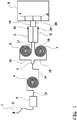
- FIG. 1 An exemplary embodiment of the device of the invention is shown in Fig. 1 .
- Fig. 1 shows a skin penetrating needle 1 connected to a first tubing 2, which is arranged and configured receive blood from the needle 1 and to carry the amount of blood away from the skin site.
- Tubing 1 is connected to a filter unit 3.
- This filter unit serves to separating plasma from said whole blood sample.
- Filter unit 3 is connected via tubing 13 to a first container 4, which is in the form of a spiral channel of which the inner wall is coated with a sterol lipid.
- the spiral channel is connected via tubing 14 with a means for dividing plasma 5 in the form of a passive valved branch point.
- One branch of the branch point is connected via tubing 16 with a second container 6 for receiving a first plasma stream, which comprises a first substrate carrying an immobilised mycolic acid derived antigen.
- the other branch of the branch point is connected via tubing 15 with a third container 7 for receiving a second plasma stream, which comprises a second substrate not carrying an immobilised mycolic acid derived antigen.
- Containers 6 and 7 in this embodiment are in the of a spiral channel of which the inner wall is either coated with a mycolic acid derived antigen (container 6) or not coated with a mycolic acid derived antigen (container 7).
- Containers 6 and 7 are connecter via respectively tubings 17 and 18 with dilution containers 11 and 12 respectively. These containers may be prefilled with a suitable dilution buffer of comprise inlets 21, 22 for introducing a dilution buffer.
- the dilution containers 21 and 22 are connected via respective tubings 19 and 20 to a test chamber 9 of a biosensor 8 and to a control chamber 10 of biosensor 8 respectively.
- the test and control chambers 9 and 10 both comprise the same substrate carrying an immobilised mycolic acid derived antigen.
- the invention in another aspect relates to a kit for use in diagnosing tuberculosis using a real time detection method, comprising one or more skin penetrating means; one or more tubings; one or more containers coated with a sterol lipid and optionally phosphatidyl choline, pectin, amphothericin B or ⁇ -cyclodextrin; one or more containers comprising a substrate with an immobilised mycolic acid derived antigen; one or more containers comprising a substrate without an immobilised mycolic acid derived antigen; and one or more biosensors comprising at least two chambers comprising a substrate with an immobilised mycolic acid derived antigen; and optionally means for diluting blood or plasma and/or a filter unit.
- the kit may also comprise tools to assemble the components, such as screws, clamps, glue, tape, screwdrivers etc.
- the kit may also comprise means to dilute the blood or plasma during blood analysis.
- means to dilute the blood or plasma during blood analysis Such as container (for instance of 10 ml) to be implemented at the outlet of the second and third containers.
- a suitable buffer may already be present in the container or be added into this container to provide the desired dilution.
- the components of the kit enable the person that is to perform a tuberculosis diagnosis test to assemble the device of the invention on the spot, i.e. at the point of care. It is therefore to be understood that the components of the kit have the same characteristics and preferred properties as explained above for the device of the invention.
- the device of the invention is easy to be assembled by means of the components of the kit of the invention, i.e. no specialist technical background is required.
- the kit may be provided conveniently with instructions for assembly and use.
- the present invention relates in a further aspect to a detection method for detecting antibodies against mycolic acid derivatives, such as cord factors antibodies or mycolic acid (MA) antibodies in a sample, wherein detection takes place by means of an end-point assay.
- mycolic acid derivatives such as cord factors antibodies or mycolic acid (MA) antibodies
- This method comprises the steps of:
- steps i)-v) do not necessarily have to be followed directly by the detection steps vi-viii).
- the samples fractions obtained after step v) can be stored until further use or for transport to the detection substrate.
- steps i)-iv) do not necessarily have to be followed by steps vi)-viii) allows pre-treated samples to be collected, so that they can be tested all at once on one substrate material, which improves reliability and reproducibility. This also makes it easier to test samples from multiple patients under the same conditions.
- the invention also relates to a method for pre-treating a sample from a human or animal suspected of having active tuberculosis for detection:
- the end-point method of the invention enables samples, preferably blood samples to be analyzed in a reliable and fast way.
- the subsequent pre-incubation steps i)-v) of the sample with a sterol lipid, followed by subjecting a fraction of the sample to a pre-incubation step with a mycolic acid derived antigen to obtain a test sample and another fraction of the sample to a pre-incubation without a mycolic acid derived antigen to obtain a control sample render the sample suitable for direct application in the eventual end-point detection step which entails binding of mycolic acid antigens in the pretreated sample to a substrate carrying a mycolic acid.
- the substrate therefore does not require pre-incubation or pretreatment with a dilution of the sample. Accordingly, the several dilution and transfer steps required in methods of the prior art are not required.
- the end-point method makes it possible to determine whether or not an individual is infected with active tuberculosis (e.g. pulmonary or extra-pulmonary tuberculosis) in fast and reliable way, allowing detection of tuberculosis in less than 15 to 25 minutes. This is in particular due to the pretreatment steps i)-v) of the end point method and pre-treatment method.
- the end-point method can be carried out outside of a professional medical environment.
- a quick method is provided for determining whether or not an individual is infected with active tuberculosis as a point of care test i.e. as a test at or near the site of patient care.
- the test of the present invention provides simple medical tests which can be performed at the bedside.
- Step i end-point method / pre-treatment method
- the end-point detection method of the invention is based on diagnostic testing of samples, in particular blood samples of a human or animal suspected of having active tuberculosis.
- a sample from a human or animal suspected of having active tuberculosis is provided.
- the sample is preferably a whole blood sample.
- the sample may be obtained by any regular means of obtaining blood from a subject.
- samples may be used that have been collected at an earlier stage, stored until use under suitable conditions and provided at a suitable moment.
- a sample may be used in the detection method of the invention on the spot, i.e. as a point of care test.
- Another alternative is that the sample is pretreated on the spot in accordance with abovementioned steps i)-v), and that the resulting pre-treated samples are stored until further use and/or transferred to another location where detection takes place.
- the sample may, depending on the way of detection of binding of antibodies to the antigen, be filtered or separated to plasma or serum before step ii).
- a filtering step may be preferred to filter out high mass components such as blood cells.
- binding is detected by fluorescence, one may choose not to filter the sample. This would even shorten the time required for pre-treatment and diagnosis.
- the sample is preferably a blood derived sample.
- the sample may be a whole blood sample, a plasma sample or a serum sample.
- Blood serum is blood plasma without clotting factors and is preferred as plasma.
- the word "plasma” in this application may therefore as well refer to (blood) serum.
- the choice of blood plasma or blood serum depends on whether the device of the invention is designed to separate the whole blood into plasma or serum. Serum is preferred because it contains less different materials than blood plasma which may lead to aspecific interactions or unwanted biological activity. In addition, serum may have a lower viscosity than blood plasma. Using serum therefore may circumvent the need for diluting a sample, which saves time and materials.
- Plasma or serum in this application may therefore also refer to diluted plasma or serum.
- a dilution of the blood or plasma may be implemented in the methods of the invention.
- a dilution of 10 x to 250 x is preferred, in particular when the end-point assay applied is an immunogold filtration assay, such as a dot immunogold filtration assay.
- an immunogold filtration assay such as a dot immunogold filtration assay.
- such dilution may take place before the separating the plasma from the blood step or after the separating step or alternative to the separating step.
- the dilution step may take place after the separating step but before step iv) or v) or after step v)/vi) and before step vii).
- Dilution may be performed with any suitable diluent, for example a PBS based buffer.
- buffer may for example be a PBS/AE buffer comprising NaCl, KCl, KH 2 PO 4 , Na 2 HPO 4 and EDTA in water at physiological pH.
- buffer may be a PBS based buffer consisting of 8.0 g NaCl, 0.2 g KCl, 0.2 g KH 2 PO 4 , and 1.05 g Na 2 HPO 4 per liter of double distilled, deionized water containing 1 mM EDTA and 0.025% (m/v) sodium azide which is adjusted to pH 7.4.
- the whole blood sample or plasma or serum may be further diluted with agents that prevent blood clotting, such as EDTA, heparine or citrate.
- a detergent may be added in low concentration to the blood/plasma/serum to avoid sticking of components to walls of tubings, vessels or containers.
- Step ii end-point method / pre-treatment method
- step ii the sample is exposed to, i.e. contacted with a sterol lipid.
- the exposure time of the sample to the sterol lipid is preferably less than 10 minutes, such as 2 to 8, 3 to 7, 4 to 6 or about 5 minutes.
- the exposure time depends on the way the sample is brought into contact with the sterol lipid.
- One exemplary way of exposing the sample to the sterol lipid is to lead the sample into a long spiral channel and pass it along the length of the channel, which is pre-coated with a sterol lipid, which is preferably cholesterol.
- the lipid sterol exposed sample may pass a means for dividing the sample stream such as a passive valved branch point that leads the divided sample streams to the next substrate carrying an immobilised mycolic acid derived antigen of step v) and the substrate not carrying an immobilised mycolic acid derived antigen of step vi), i.e. a container comprising a substrate carrying an immobilised mycolic acid derived antigen, and a container comprising a substrate not carrying an immobilised mycolic acid derived antigen respectively.
- An alternative exemplary way of exposing the sample to the sterol lipid is to inject it into a container coated with cholesterol, followed by incubation therein for less than 10 minutes.
- Another alternative exemplary way is to expose the sample to the sterol lipid, wherein the sterol lipid is contained in a compartment of a column. In such a compartment the sterol lipid is preferably coated on beads.
- substrate material that is inert for non-specific binding of molecules of this sample.
- substrate should be understood to be a support material.
- Steps iv and v end-point method / pre-treatment method
- step iv) a fraction of the sterol lipid exposed sample is exposed to a substrate carrying an immobilised mycolic acid derived antigen
- step v) another fraction of the sterol lipid exposed sample to a substrate not carrying an immobilised mycolic acid derived antigen
- the blood/plasma stream has to be divided (step iii).
- the stream may be divided after exposure to the sterol lipid, i.e. that the sterol lipid exposed sample is divided into at least two fractions to provide a test stream and a control stream. It is preferred that the sample is divided in two equal or substantially equal fractions, because this enables a simple comparison of both fractions without the need for correction calculations. Dividing the sample stream may be carried out by any means for dividing the sample stream such as a passive valved branch point.
- sample stream (which is filtered to plasma or serum) is divided into at least two fractions after the filtering step, but before exposure to the sterol lipid.
- blood stream is divided before filtering. This is less preferred because in this case two filters would be required.
- step ii) the stream is in this case divided before exposure to the sterol lipid in step ii), so that steps ii) and iv) can take place in the same column and step ii) and v) can take place in another column.
- the substrate carrying an immobilised mycolic acid derived antigen and the substrate not carrying an immobilised mycolic acid derived antigen of steps iv) and v) are preferably of the same material.
- the substrates for exposure in steps iv) and v) may be of the same material as the substrate that may be used to in step ii). It will be understood that suitable substrate material is inert for non-specific binding of molecules of this sample.
- the exposure time of the sample to the sterol lipid is preferably less than 10 minutes, such as 2 to 8, 3 to 7, 4 to 6 or about 5 minutes.
- One exemplary way of exposing the sample to the substrates of step iv) and v) is to lead the divided sample streams into long spiral channels, preferably implemented in micro-chips, pre-coated either with a mycolic acid derivative (step iv) or without a mycolic acid derivative (step v) and pass it along the length of these channels.
- An alternative exemplary way of exposing the divided lipid sterol exposed samples to a substrate coated either with a mycolic acid derivative or without a mycolic acid derivative is to inject the a first stream into a container comprising a substrate carrying an immobilised mycolic acid derived antigen (step iv), and a second sample stream into a container comprising a second substrate not carrying an immobilised mycolic acid derived antigen (step v) and incubate the samples for less than 10 minutes, such as 2 to 8, 3 to 7, 4 to 6 or about 5 minutes.
- steps iv) and v) take place in a column.
- the stream is in this case divided before exposure to the sterol lipid in step ii), so that step ii) and iv) can take place in one column and step ii) and v) can take place in another column.
- step v) said another fraction of the sterol lipid exposed sample is exposed to a substrate not carrying an immobilised mycolic acid derived antigen
- step vi) the step of exposing said fraction to a substrate not carrying an immobilised mycolic acid derived antigen is skipped and that said fraction is stored until further use, such as in step vi). This may for instance take place in the same column or container wherein the incubation with the sterol lipid took place.
- the invention also relates to a method which comprises the steps of:
- the invention also relates to a method for pre-treating a sample from a human or animal suspected of having active tuberculosis for detection:
- step iv the divided sterol lipid exposed sample streams, either exposed to mycolic acid derivatives in step iv(test stream) or not in step v (control stream) are passed to further substrates, test and control substrates respectively, carrying mycolic acid derivatives, preferably the same derivatives as in step iv.
- the test and control substrates may be any substrate which can carry an immobilized mycolic acid derivative antigen and from which binding between antibodies contained in the sample to said antigens can be detected.
- test and control substrate are a microporous membrane, preferably a nitrocellulose membrane, which is coated an immobilised mycolic acid derived antigen.
- test substrate and the control substrate may be separate entities.
- the test substrate and the control substrate may be realized as different positions on one substrate entity.
- the test substrate and the control substrate may be formed by a first microporous (e.g. nitrocellulose) membrane and a second microporous (e.g. nitrocellulose) membrane.
- the test substrate and control substrate are contained on one membrane. In such a case there is a test area where antibody/antigen interaction can take place (e.g. a spot) on one position of the membrane and a control area where antibody/antigen interaction can take place on another position of the membrane.
- step vii) the binding of antibodies to the immobilised mycolic acid antigens is detected in an end-point assay. At least part of the obtained pre-treated fractions is subjected to said end-point detection assay. It is possible to use the whole fractions for the end-point assay. Alternatively part of the fractions are used to detect binding of antibodies to the immobilised mycolic acid antigens in the end-point assay and another part is stored for further testing depending on the results of the first part of the fractions.
- end-point assay is to be understood as an assay wherein the outcome of interest is the end result after a fixed assay incubation period, in contrast to a so-called real-time assay.
- detection method of the invention that means that the treated samples obtained after steps iv) and v) are taken and subjected to a test that provides an indication of the amount of antibodies that are present in the treated samples after steps iv) and v).
- An end-point detection assay may for instance detect changes to levels of color, fluorescence, absorbance or luminescence at the end of a test.
- detection may suitably be performed by means of techniques such as photospectroscopy, fluorescence microscopy, chemo-luminescence or electrochemiluminescence detection techniques. This may involve use of for instance spectrophotometric/colorimetric plate readers, fluorescence plate readers or chemiluminescence readers.
- Suitable end-point assays may involve enzyme-linked immunosorbent assay (ELISA), Western blotting, radioactive labelling assay, photospectrometric assay, immunofluorescence, immunoprecipitation, immunocytochemistry, immunohistochemistry, electrochemical impedance spectroscopy (EIS) etc.
- ELISA for instance involves at least one antibody with specificity for a particular antigen.
- the antigen is a mycolic acid derived antigen and the antibody is an antibody against mycolic acid derivative antigens.
- an end-point assay interaction of antibodies with the mycolic acid derived antigens may be carried out using secondary antibodies that bind the heavy chain of the antibodies against the mycolic acid derivatives.
- secondary antibodies are commercially available.
- the secondary antibody may be coupled to beads, for instance gold beads, or associated with liposomes.
- Examples of secondary antibodies may be protein A or G, possibly conjugated with an enzyme that enables detection.
- a particular suitable technique or detecting the binding of antibodies to the immobilised mycolic acid antigens in step vii) is the so-called immunogold filtration assay (IGFA), and in particular the dot immunogold filtration assay (DIGFA).
- IGFA immunogold filtration assay
- DIGFA dot immunogold filtration assay
- Immunogold filtration assays are methods combining ELISA and immunogold technique and are methods in which a sample to be assayed is allowed to filtrate through a microporous membrane, preferably a nitrocellulose membrane, and is captured by a capture probe coated on the membrane.
- a colloidal gold labelled probe is allowed to filtrate through the microporous membrane in the same manner.
- a microporous membrane as the carrier for the capture probe and employing the capillary action and permeability of the membrane antigens and antibodies can easily react and may conveniently be subjected to optional washing and/or blocking steps.
- the colloidal gold labelled probe binds to the capture probe the colloidal gold particles aggregate and a red dot appears which is visible with the naked eye.
- Immunogold filtration assays are simple and rapid detection methods because no instruments are required except a membrane and the reagents and the results can be observed by the naked eye within a few minutes.
- the microporous membrane may be for example a nitrocellulose membrane, a cellulose acetate membrane or a PVDF membrane with a suitable pore diameter.
- nitrocellulose is used.
- a suitable pore diameter is 0,2 to 5 ⁇ m.
- the substrate mentioned in steps vi) and viii) of the detection method of the invention is a microporous membrane, preferably a nitrocellulose membrane, which is coated an immobilised mycolic acid derived antigen. After immobilizing the mycolic acid derived antigen onto the microporous membrane, the sample fractions pre-treated in steps i) to v) can be applied to the membrane.
- colloidal gold-labeled second antibodies are added onto the membrane to have gold particle aggregation in the antigen-antibody reaction place.
- the intensity of the spot is proportional to the amount of reactions between antigen and antibody, i.e. to the amount of antibodies in the pre-treated sample.
- the control substrate shows a more intense signal than the test substrate this is indicative for active tuberculosis in the person from which the sample was derived.
- the control substrate shows no (or only insignificant) difference in color intensity signal compared the test substrate this indicates that the human or animal from which the sample was derived does not have active tuberculosis.
- the membrane may be washed with a suitable buffer, for example a PBS based buffer.
- a PBS based buffer may for example be a PBS/AE buffer comprising NaCl, KCl, KH 2 PO 4 , Na 2 HPO 4 and EDTA in water at physiological pH.
- Such buffer may be a PBS based buffer consisting of 8.0 g NaCl, 0.2 g KCl, 0.2 g KH 2 PO 4 , and 1.05 g Na 2 HPO 4 per liter of double distilled, deionized water containing 1 mM EDTA and 0.025% (m/v) sodium azide which is adjusted to pH 7.4.
- the microporous membrane may be coated with said mycolic acid derived antigen in a dot wise manner.
- the samples pre-treated in steps i) to vi) are applied to the membrane in the form of dots.
- the colloidal gold-labeled second antibodies are added in the form of dots.
- the control and test substrate are preferably the same microporous membrane with dots of mycolic acid derived antigens immobilised thereon. The detection method of the invention makes it possible to detect both the control and test fractions on the same membrane. This improves reliability of the test and is cost effective.
- a DIGFA assay is particularly preferred because at different spots on several membranes various antigens deriving from various mycobacterial strains may be immobilized. This way it becomes possible to provide information on which mycobacterial strain a patient is infected with.
- Another advantage of using DIGFA is that samples derived from different persons suspected of having active tuberculosis can be compared in one test, because DIGFA enables fast and reliable detection of antibody-antigen interaction in an unlimited amount of spots, depending on the size of the membrane.
- the detection of binding of antibodies and/or other material to the mycolic acid antigen may be carried out in an automated device.
- Various automated devices will be known to the person skilled in the art and the skilled person will be able to select suitable software means to make the comparison of step viii) of the degree or extent of binding between the test and control substrates, wherein any observed lesser binding to the test substrate is an indicator of the presence of antibodies to the antigen in the samples that relates to active tuberculosis in the human or animal from which the samples originated.
- lesser can be interpreted qualitatively and quantitatively, i.e. lesser binding may be interpreted as having less binding events as well as having weaker bindings.
- the detection of binding of antibodies and/or other material to the mycolic acid antigen may be performed by a visual detection technique or any other suitable detection technique.
- detection by means of the end-point assay takes place visually, preferably with the naked eye. This has the advantage of easy detection without the need for expensive and complicated detection technology.
- binding of antibody antibodies and/or other material to the mycolic acid antigen may be assessed by means of the naked eye.
- a visual signal e.g. the red staining in case a DIGFA assay is applied as end-point assay, may also be detected with help of a mobile app, i.e. a computer program designed to run on mobile devices such as tablet computers or smart phones.
- a mobile app i.e. a computer program designed to run on mobile devices such as tablet computers or smart phones.
- an app can be used that is designed to compare the binding signal of the test and control substrate and which indicates whether the human or animal from which the sample originated has active tuberculosis.
- the invention in another aspect relates to a system or device, preferably a portable system or device, for carrying out an end-point method of detecting a marker for active tuberculosis.
- This system can be subdivided in 1) a system or device for pre-treating a sample stream from a human or animal suspected of having active tuberculosis in order to make the sample suitable for detection with the method of the invention; and 2) a detection means to enable detection of interaction of antibodies contained in the pre-treated sample.
- the system comprises at least one container comprising a sterol lipid arranged and configured to receive a sample stream from a human or animal suspected of having active tuberculosis; means for dividing said sample stream into at least a first and a second sample stream; said means either connected upstream or downstream of said at least one container comprising a sterol lipid; a further container for receiving said first sample stream in downstream connection with a said container comprising a sterol lipid, and comprising a first substrate carrying an immobilised mycolic acid derived antigen; a still further container for receiving the second sample stream in parallel arrangement to said further container and in downstream connection with a said container comprising a sterol lipid; and comprising a second substrate not carrying an immobilised mycolic acid derived antigen, and a first detection substrate carrying an immobilised mycolic acid derived antigen to receive at least part of the first sample stream and a second detection substrate carrying an immobilised mycolic acid derived antigen to receive at least part of the second sample
- the system for carrying out a method of detecting a marker for active tuberculosis comprises at least one container comprising a sterol lipid arranged and configured to receive a sample stream from a human or animal suspected of having active tuberculosis; means for dividing said sample stream into at least a first and a second sample stream; said means either connected upstream or downstream of said at least one container comprising a sterol lipid; a further container for receiving said first sample stream in downstream connection with a said container comprising a sterol lipid, and comprising a first substrate carrying an immobilised mycolic acid derived antigen; a still further container for receiving the second sample stream in parallel arrangement to said further container and in downstream connection with a said container comprising a sterol lipid; and comprising a second substrate not carrying an immobilised mycolic acid derived antigen, and a first detection substrate carrying an immobilised mycolic acid derived antigen to receive at least part of the first sample stream from said further container and
- the first and second detection substrates are preferably a microporous membrane, preferably a nitrocellulose membrane.
- the first and second detection substrates may in this case have separate spots, i.e. first and second detection spots, on the same membrane. This way one and the same membrane can be used for detection of antibody/antigen interaction. This increases reliability of the detection and makes it easier to compare the interaction detected on the test and control substrate. Moreover, this enables multiple samples to be tested at the same time.
- the detection substrates do not have to be in physical connection with the other components of the system.
- the samples derived after passing said further and still further containers may be stored until further use or for transport to the detection substrate.
- This way the practicer has great flexibility in planning his test. For instance, this allows pre-treated samples from a number of subjects to be collected, so that they can be tested all at once on one substrate material. This also makes it easier to test samples from multiple patients under the same conditions.
- the invention relates to a system or device, preferably a portable system or device, for pretreating a sample stream from a human or animal suspected of having active tuberculosis in order to make the sample suitable for detection in a detection method, comprising at least one container comprising a sterol lipid arranged and configured to receive a sample stream from a human or animal suspected of having active tuberculosis; means for dividing said sample stream into at least a first and a second sample stream; said means either connected upstream or downstream of said at least one container comprising a sterol lipid; a further container for receiving said first sample stream in downstream connection with a said container comprising a sterol lipid, and comprising a first substrate carrying an immobilised mycolic acid derived antigen; a still further container for receiving the second sample stream in parallel arrangement to said further container and in downstream connection with a said container comprising a sterol lipid; and comprising a second substrate not carrying an immobilised mycolic acid derived
- the system or device for pre-treating a sample stream from a human or animal suspected of having active tuberculosis in order to make the sample suitable for detection with the method of the invention may comprise two columns.
- Each column comprises two compartments or containers.
- An upper compartment or container comprises a substrate coated with a sterol lipid, for instance beads coated with cholesterol.
- a lower compartment of one of the columns comprises a first substrate carrying an immobilised mycolic acid derived antigen and a lower compartment of another column comprises a substrate not carrying an immobilised mycolic acid derived antigen.
- the invention also relates to a system for pre-treating a sample stream derived from a human or animal suspected of having active tuberculosis, which comprises means for dividing said sample stream into at least a first and a second sample stream; at least a first and a second column, configured to receive said first and a second sample stream respectively; said first column comprising a compartment comprising a sterol lipid and a further compartment comprising a first substrate carrying an immobilised mycolic acid derived antigen downstream of said compartment comprising a sterol lipid; and said second column comprising a compartment comprising a sterol lipid and a still further compartment comprising a second substrate not carrying an immobilised mycolic acid derived antigen downstream of said compartment comprising a sterol lipid; and means for storage or transport of the treated samples passed through said columns.
- This or these means for storage and transport do not have to be in physical connection with the containers comprising said first and second substrates respectively and are located downstream of the columns when the system is in use
- the invention also relates to a system for carrying out a method of detecting a marker for active tuberculosis in a sample derived from a human or animal suspected of having active tuberculosis, which comprises means for dividing said sample stream into at least a first and a second sample stream; at least a first and a second column, configured to receive said first and a second sample stream respectively; said first column comprising a compartment comprising a sterol lipid and a further compartment comprising a first substrate carrying an immobilised mycolic acid derived antigen downstream of said compartment comprising a sterol lipid; and said second column comprising a compartment comprising a sterol lipid and a still further compartment comprising a second substrate not carrying an immobilised mycolic acid derived antigen downstream of said compartment comprising a sterol lipid and a first detection substrate carrying an im
- the system or device comprises a skin penetrating end connected to a first tubing, which is arranged and configured receive a blood sample from the skin penetrating end and to carry said blood sample away from the skin site to said container/compartment comprising a sterol lipid.
- system or device comprises a filter unit in connection with said first tubing for receiving said blood sample and configured to separate plasma from said whole blood sample.
- the system or device comprises a skin penetrating end connected to a first tubing, which is arranged and configured receive a blood sample from the skin penetrating end and to carry said blood sample away from the skin site; a filter unit in connection with said first tubing for receiving said blood sample and configured to separate plasma from said whole blood sample; a first container for receiving said sample, said first container comprising a sterol lipid; means for dividing said sample into at least a first and a second sample stream; a second container for receiving said first sample stream, said second container comprising a first substrate carrying an immobilised mycolic acid derived antigen; a third container for receiving the second sample stream, said third container comprising a second substrate not carrying an immobilised mycolic acid derived antigen; and a means for storage or transport of the treated samples.
- the stream through the system or device is preferably driven by a pump, preferably a continuous flow pump, for example a peristaltic pump or a diaphragm pump.
- a pump preferably a continuous flow pump, for example a peristaltic pump or a diaphragm pump.
- the stream may be driven by gravity or capillary action
- the skin penetrating means may be any suitable means to obtain a blood sample from a human or animal, such as a needle syringe or the like.
- the first tubing has dimensions that are suitable for purpose of the device, i.e. it needs to be well adaptable to the skin penetrating means and the first container.
- the components of the system or device i.e. the containers, means for diluting and filter unit may be interconnected by suitable interlinking tubings.
- the components of the device may be designed such that they can be connected directly to each other, and separated by for instance filters through which the sample but not the substrate can selectively pass.
- the material of the tubings used in the invention can be any suitable material which is known to the person skilled in the field of testing blood samples. Suitable materials are inert to blood/plasma/serum components and include polytetrafluorethylene (e.g. Teflon®), polypropylene, polyetherketone (PEEK) and polyethylene.
- Further components may be connected between the components of the device. Such further components may be connected in tubings interlinking the components are directly attached to the components.
- the system or sub-device for performing steps i-v of the methods of the invention may also comprise a means for dilution of the blood or plasma.
- a means for dilution of the blood or plasma may be implemented before the sterol lipid containing container, before the filter unit, between the filter unit and the sterol lipid containing container, between the sterol containing container and the other (further and still further or (second and third) containers or between the further/still further or second /third containers and the means for storage and transport.
- a 10 ml container may be implemented at the outlet of the further or second and still further or third containers.
- a suitable buffer may already be present in the container or be added into this container to provide the desired dilution. This way the volume of the sample is kept as low as possible during most of the steps of the methods of the invention, which is beneficial to the speed of the process and the compactness of the device of the invention.
- the filter unit may comprise a filter matrix.
- the filter is implemented on a filter microchip for the sake of compactness.
- the containers may be vessels or a channel or tubing, such as a spiral channel or spiral tube.
- a spiral channel or tube is preferred because such structure takes little space whilst maintaining a long flow path.
- a spiral channel or tube may be advantageously implemented on a micro-chip. This contributes to the compactness of the device of the invention.
- the term “container” is to be interpreted in the broad sense, i.e. it can be seen as defined area in another unit, i.e. a compartment, for instance a compartment of a column.
- the terms “container” and “compartment” may thus be used interchangeably in the context of the present invention. What matters is that sterol lipid containing container and the other (further and still further or (second and third) containers are spatially separated.
- a sterol containing container may be included in a column together with said further (or second) container while another sterol containing container is included in a column together with a still further (or third) container.
- a column for preparing a stream to be tested on a test substrate may comprise an upper (or upstream) substrate coated with a sterol lipid which is separated by a barrier from, but in fluid connection with a lower (or downstream) substrate coated with mycolic acid derived antigens.
- a column for preparing a stream to be tested on a control substrate may comprise an upper (or upstream) substrate coated with a sterol lipid which is separated by a barrier from, but in fluid connection with a lower (or downstream substrate not coated with mycolic acid derived antigens. It is to be understood that in connection in this context means that the sample to be tested can pass through said barrier, while the barrier does not allow passing of the substrates. Suitable substrates for such columns are beads.
- an embodiment of the system of the invention for pre-treating a sample stream from a human or animal suspected of having active tuberculosis in order to make the sample suitable for detection with the method of the invention comprises at least a first and a second column, configured to receive said first and a second sample stream respectively, said first column comprising a container comprising a sterol lipid and said further container comprising a first substrate carrying an immobilised mycolic acid derived antigen, and said second column comprising a container comprising a sterol lipid and said still further container comprising a second substrate not carrying an immobilised mycolic acid derived antigen.
- the material of the containers is preferably the same and suitable materials may be bk-7 glass, polytetrafluorethylene, polypropylene, polyether ketone or polyethylene.
- a boundary between two containers may be a filter that does not allow substrate material to pass, but which does allow passage of a sample stream.
- the substrates of the containers have to be material that is inert for non-specific binding of molecules of the sample, for instance bk-7 glass, polytetrafluorethylene, polypropylene, polystyrene polyether ketone or polyethylene.
- the substrates may be in the form of beads, optionally crosslinked beads, for instance polystyrene beads.
- the systems of the invention comprise a container comprising a substrate carrying an immobilised mycolic acid derived antigen (second container) and a container comprising a substrate not carrying an immobilised mycolic acid derived antigen.
- second container a container comprising a substrate carrying an immobilised mycolic acid derived antigen
- the latter container may an inert coating or no coating, as long as no aspecific binding takes place.
- the means for storage or transport of the treated samples may be small vials wherein the samples are stored for further analysis, for example by means of a dot immunogold filtration assay.
- the means may be formed by a pipette which can be applied to transfer the sample fractions treated in steps i)-v) of the methods of the invention to the substrates with immobilised mycolic acid derived antigens of the end-point assay, for instance to spot fractions as dots in a dot immunogold filtration assay.
- the device/system may be connected to any suitable automated analysis means, such as a computer with suitable software programs to carry out the comparison of binding of mycolic acid antibodies to the immobilised antigens on the substrate.
- suitable automated analysis means such as a computer with suitable software programs to carry out the comparison of binding of mycolic acid antibodies to the immobilised antigens on the substrate.
- FIG. 2 An exemplary embodiment of the system according to claim 32 is shown in Fig. 2 .
- Fig. 2 shows a skin penetrating needle 1 connected to a first tubing 102, which is arranged and configured receive blood from the needle 101 and to carry the amount of blood away from the skin site.
- Tubing 101 is connected to a filter unit 103.
- This filter unit serves to separating plasma from said whole blood sample.
- Filter unit 103 is connected via tubing 113 to a first container 14, which is in the form of a spiral channel of which the inner wall is coated with a sterol lipid.
- the spiral channel is connected via tubing 114 with a means for dividing plasma 105 in the form of a passive valved branch point.
- One branch of the branch point is connected via tubing 116 with a second container 106 for receiving a first plasma stream, which comprises a first substrate carrying an immobilised mycolic acid derived antigen.
- the other branch of the branch point is connected via tubing 115 with a third container 107 for receiving a second plasma stream, which comprises a second substrate not carrying an immobilised mycolic acid derived antigen.
- Containers 106 and 107 in this embodiment are in the of a spiral channel of which the inner wall is either coated with a mycolic acid derived antigen (container 106) or not coated with a mycolic acid derived antigen (container 107).
- Containers 106 and 107 are connected via respectively tubings 117 and 118 with dilution containers 111 and 112 respectively. These containers may be prefilled with a suitable dilution buffer of comprise inlets 121, 122 for introducing a dilution buffer.
- the dilution containers 121 and 122 can be connected via respective tubings 119 and 120 to means for storage or transport 108 and 109 respectively.
- the dilution containers 121 and 122 can be connected via respective tubings 119 and 120 to test and control substrates with immobilized mycolic acid derived antigens that are tested in an end-point assay.
- the invention in another aspect relates to a kit for use in diagnosing tuberculosis or pre-treatment of samples for diagnosing tuberculosis, comprising one or more skin penetrating means; one or more tubings; one or more containers coated with a sterol lipid and optionally phosphatidyl choline, pectin, amphothericin B or ⁇ -cyclodextrin; one or more containers comprising a substrate with an immobilised mycolic acid derived antigen; one or more containers comprising a substrate without an immobilised mycolic acid derived antigen; optionally one or more containers for transport and storage of samples; optionally means for diluting blood or plasma and/or a filter unit; at least one microporous membrane with mycolic acid derived antigen immobilised thereon, and optionally secondary antibodies binding to the heavy chain of antibodies against mycolic acid derived antigens contained in a sample derived from a human or animal suspected of having active tuberculosis, preferably
- the kit comprises: one or more skin penetrating means; one or more tubings; one or more columns, comprising a compartment comprising a substrate coated with a sterol lipid and optionally phosphatidyl choline, pectin, amphothericin B or ⁇ -cyclodextrin; and a compartment comprising a substrate coated with an immobilised mycolic acid derived antigen below said compartment comprising a substrate coated with a sterol lipid; one or more columns, comprising a compartment comprising a substrate coated with a sterol lipid and optionally phosphatidyl choline, pectin, amphothericin B or ⁇ -cyclodextrin; and a compartment comprising a substrate coated without an immobilised mycolic acid derived antigen below said compartment comprising a substrate coated with a sterol lipid, optionally one or more containers for transport and storage of samples; optionally means for diluting blood or plasma and/or
- the kit may also comprise one or more means to transfer sample fractions pre-treated in accordance with steps i) to v) of the methods of the invention to the microporous membrane, for instance a pipette and pipette accessories, such as pipette tips.
- a pipette and pipette accessories such as pipette tips.
- the kit may also comprise one or more means to store sample fractions pre-treated in accordance with steps i) to v) for further use, such as vials or tubes.
- the kit may also comprise tools to assemble the components, such as screws, clamps, glue, tape, screwdrivers etc.
- the kit may also comprise means to dilute the blood or plasma during blood analysis, such as containers (for instance of 10 ml) which can be implemented at the outlet of the second and third containers.
- containers for instance of 10 ml
- a suitable buffer may already be present in the container or be added into this container to provide the desired dilution.
- the components of the kit enable the person that is to perform a tuberculosis diagnosis test to assemble the systems of the invention on the spot, i.e. at the point of care. It is therefore to be understood that the components of the kit have the same characteristics and preferred properties as explained above for the systems of the invention.
- the systems of the invention are easy to be assembled by means of the components of the kit of the invention, i.e. no specialist technical background is required.
- the kit may be provided conveniently with instructions for assembly and use.
- the sterol lipid used in the context of the invention preferably is cholesterol or a derivative thereof.
- the sterol lipid may also be a sterol modified phospholipid.
- Such sterol-modified lipid may a sterol-modified phospholipid, for instance a sterol-modified phosphatidylcholine lipid or glycerophospholipid.
- the sterol is preferably cholesterol.
- a good example of a sterol-modified lipid suitable for the purposes of the invention is 1-palmitoyl-2-cholesteryl carbonoyl-sn-glycero-3-phosphocholine.
- the sterol lipid is preferably immobilized on a surface.
- An example is a substrate having a coating containing cholesterol or cholesterol ester wherein the cholesterol ester is cholesterol linoleate, wherein a weight ratio of linoleic acid to cholesterol is in the range from 1:3 to 1:20.
- a substrate may also be coated with a sterol lipid, preferably cholesterol, in combination with other molecules.
- said sterol lipid is cholesterol immobilized on a substrate together with phosphatidyl choline.
- the sterol lipid scavenges away the anti-cholesterol antibodies from the blood/plasma/serum that would otherwise cross react with the mycolic acid antigens on the substrate and lead to false positive diagnosis of tuberculosis.
- Phosphatidyl choline will bind to hydrophobic materials in the blood sample, rendering the sample more hydrophilic after exposure. The resulting hydrophilic sample will easier to be handled in the subsequent method steps and be less prone to clotting.
- a sterol lipid preferably cholesterol is immobilised in together with pectin on a substrate, e.g. the inner wall of a tubing.
- the sterol lipid scavenges away the anti-cholesterol antibodies from the blood/plasma/serum that would otherwise cross react with the mycolic acid antigens on the substrate and lead to false positive diagnosis of tuberculosis.
- pectin scavenges away cholesterol in the sample and therewith also cholesterol antibodies. This leads to an even higher reliability of detection.
- a sterol lipid preferably cholesterol is immobilised together with a compound binding to cholesterol in the blood derived sample, such as cholesterol binding heteropolysaccharide, such as ⁇ -cyclodextrin, pectin, amphotericin B or dextrin.
- a compound binding to cholesterol in the blood derived sample such as cholesterol binding heteropolysaccharide, such as ⁇ -cyclodextrin, pectin, amphotericin B or dextrin.
- cholesterol binding heteropolysaccharide such as ⁇ -cyclodextrin, pectin, amphotericin B or dextrin.
- a sterol lipid preferably cholesterol is immobilised with ⁇ -cyclodextrin or pectin, amphotericin B on a substrate, such as hollow fiber polypropylene membranes or glass.
- a coating may be applied by initially dissolving cholesterol in an organic solvent and further diluting the dissolved cholesterol in an ethanol solution, permitting the solution to evaporate in place within a container, rinsing the coated container with buffered saline, air-drying the container, and sealing the container in vapor-proof pouches with desiccant.
- the mycolic acid derived antigen may be derived from mycobacteria selected from virulent and pathogenic mycobacteria.
- the mycolic acid antigen is derived from Mycobacterium tuberculosis.
- Said mycolic acid derived antigen is at least one selected from the group of mycolic acid, cord factor, chemically modified mycolic acid, chemically modified cord factor, a synthetic mycolic acid derivative, a synthetic cord factor derivative.
- Natural sources of mycolic acid derivatives include the cell walls of mycobacteria such as Mycobacterium tuberculosis include mixtures of different classes of compounds and different mycolic acid homologues, often as derivatives in which they are bonded to the wall of the cell.
- the cells of mycobacteria also contain compounds derived from the acid, such as sugar esters of mycolic acids.
- Naturally occurring sugar esters comprise for instance trehalose-6,6'-dimycolate, commonly referred to as TDM, also known as “cord factors”; and trehalose monomycolates (often referred to as TMM). These sugar esters occur in nature as complex mixtures of different classes of mycolic acids and of different homologues within each class.
- mycolic acid derivatives Because it is difficult to establish the identity of cord factors present in natural products and to separate individual molecular species it is preferred to use semi-synthetic or more preferably synthetic mycolic acid derivatives for the purposes of the invention. Further, it is known that the structure of the mycolic acid unit affects the biological activity of the cord factor. Therefore when natural mycolic acid derivatives would be used in the present invention the difference in biological activity between these derivatives and thus the detection of binding of antibodies of the antigen in the sample may impart a factor of unpredictability and uncertainty to the outcome of the detection. In addition, deviations in the preparation of the natural mycolic acid derivatives may result in problems regarding reproducibility of different test batches.
- Suitable semi-synthetic derivatives include semi-synthetic cord-factors which may be prepared by attaching mycolic acids to the sugar group. These semi-synthetic factors however still contain mixtures of different homologue.
- mycolic acid derivatives for use in the context of the present invention are synthetic cord factors, for example the synthetic cord factors described in WO 2010/08667 , i.e. compounds of formula (M) x (S) y (M') z , wherein x is from 1 to 6, y is from 1 to 12, z is from 0 to 10, each M and each M' is independently a mycolic acid residue including a ⁇ -hydroxy acid moiety and each S is a monosaccharide unit.
- synthetic cord factors for example the synthetic cord factors described in WO 2010/08667 , i.e. compounds of formula (M) x (S) y (M') z , wherein x is from 1 to 6, y is from 1 to 12, z is from 0 to 10, each M and each M' is independently a mycolic acid residue including a ⁇ -hydroxy acid moiety and each S is a monosaccharide unit.
- the mycolic acid antigen may be in a form selected from homogenous and heterogenous compound mixtures.
- the mycolic acid derived antigen may for instance be used in combination with a phospholipid such as phosphatidylcholine.
- the mycolic acid antigen may be immobilised on the substrates in various ways that are known to the skilled person. Synthetic mycolic acid derived antigens may be synthesised with particular active groups that enable immobilisation to a substrate material.
- silane coupling chemistry may be applied for immobilisation on silica.
- the mycolic acid derived antigen may be suitably immobilised as follows.
- Mycolic acid derived antigens may be obtained in lyophilized form and be reconstituted in a solvent mixture, for instance a chloroform: methanol: water mixture, and diluted to a concentration in the order of several nanomolars, for instance 1 nM. This dilution can then be spotted on a nitrocellulose membrane, wherein each spot is separated at a predetermined distance, e.g. 1 cm. After drying of the spots the mycolic acid derived antigens are immobilized on the membrane.
- Alternative immobilisation methods may for instance involve dissolving the antigens in hexane or hot PBS to form an antigen coating solution before spotting the solution on a membrane.
- beads were used as a substrate representative of the substrate mentioned in steps ii), iv and v) of the methods of the invention.
- Toyopearl AF-Amino-650M beads (Tosoh Bioscience) were used. Beads were prepared in accordance with the manufacturer's instructions. As cholesterol 7-keto cholesterol was conjugated to the beads basically as described in Abdel-Magid AF et al. (Reductive Amination of Aldehydes and Ketones with Sodium Triacetoxyborohydride. Studies on Direct and Indirect Reductive Amination Procedures. J. Org. Chem.
- mycolic acids a mixture of natural occurring isoforms of mycolic acid (Mw ⁇ 1100-1300) was used to be coupled to the beads basically as described in Law B, Jenner WN. Immunoassay: A Practical Guide. London: Taylor & Francis. 2005: p 11-31 and Hermanson GT. Bioconjugate Techniques. 3th ed. London; Elsevier. 2013: p 535-545 .
- N-(3-Dimethylaminopropyl)-N'-ethylcarbodiimide hydrochloride EDC.HCl
- DIPEA N, N- diisopropylethylamine
- amine Toyopearl amino-activated, 2,5 mL, 0,25 mmol reactive groups
- acid isolithocholic acid: 93,8 mg, 0,25 mmol (1 equiv.)/ Mycolic acids: 300 mg, 0,25 mmol (1 equiv.)
- HOBt 1-Hydroxybenzotriazole hydrate
- the solvent was removed by evaporation and the beads were washed used water and 1M NaCl and water. Excess (unbound) amino groups were blocked using acetylation. Beads were washed using water, 1M NaCl and water. First an inert atmosphere with nitrogen or argon is created. The order of the reagents: Mycolic acids with DMF (at 0°C), then (HOBt), 10 min later beads, 10 min later base (DIPEA) and finally 15 min later EDC.
- Serum derived from a human that were known to be suffering of tuberculosis was diluted 1:5 in blocking buffer.
- the samples used were derived from both smear positive and smear negative patients. Two fractions of 0,5 ml were obtained and transferred each to a 0,2 micron spin filter and centrifuged at 10000g. The flow-through was pooled from 1:5 to 1:20 in blocking buffer.
- the total pre-treatment of the samples with beads (in line with steps i)-v) of the method (s) of the invention) in this particular set-up can take place in less than 10 minutes, after which analysis can directly follow.
- the time that is required for analysis is dependent on the detection assay that is applied.
- detection assays instead of analysis with ELISA, other detection assays are possible.
- the samples pretreated as described above can be analysed using any suitable label free technology.
- the inventor has found in particular that when electrochemical impedance spectroscopy (EIS) is applied detection of binding of antibodies present in the samples to the detection substrates can take place within seconds. This makes it possible to determine whether or not an individual is infected with active tuberculosis in a fast and reliable way, allowing detection of tuberculosis in less than 10 minutes.
- EIS electrochemical impedance spectroscopy
Landscapes
- Health & Medical Sciences (AREA)
- Life Sciences & Earth Sciences (AREA)
- Engineering & Computer Science (AREA)
- Immunology (AREA)
- Molecular Biology (AREA)
- Biomedical Technology (AREA)
- Hematology (AREA)
- Chemical & Material Sciences (AREA)
- Urology & Nephrology (AREA)
- General Health & Medical Sciences (AREA)
- Physics & Mathematics (AREA)
- Pathology (AREA)
- Analytical Chemistry (AREA)
- General Physics & Mathematics (AREA)
- Biochemistry (AREA)
- Food Science & Technology (AREA)
- Microbiology (AREA)
- Cell Biology (AREA)
- Biotechnology (AREA)
- Medicinal Chemistry (AREA)
- Tropical Medicine & Parasitology (AREA)
- Biophysics (AREA)
- Virology (AREA)
- Surgery (AREA)
- Heart & Thoracic Surgery (AREA)
- Medical Informatics (AREA)
- Animal Behavior & Ethology (AREA)
- Public Health (AREA)
- Veterinary Medicine (AREA)
- Endocrinology (AREA)
- Toxicology (AREA)
- Bioinformatics & Cheminformatics (AREA)
- Chemical Kinetics & Catalysis (AREA)
- Manufacturing & Machinery (AREA)
- Clinical Laboratory Science (AREA)
- Investigating Or Analysing Biological Materials (AREA)
Claims (17)
- Procédé pour détecter un marqueur d'une tuberculose active dans un échantillon obtenu auprès d'un humain ou animal suspecté d'avoir une tuberculose active , comprenant les étapes consistant à :ii) exposer ledit échantillon à un lipide de type stérol, où le lipide de type stérol est immobilisé sur un substrat, où le lipide de type stérol est le cholestérol ou un dérivé de celui-ci, et où le lipide de type stérol piège les anticorps anti-cholestérol hors de l'échantillon ;iii) obtenir au moins deux fractions dudit échantillon soit avant soit après son exposition audit lipide de type stérol ;iv) exposer la première desdites fractions exposées à un lipide de type stérol à un substrat portant un antigène dérivé d'acide mycolique immobilisé ;v) exposer la deuxième desdites fractions exposées à un lipide de type stérol à un substrat ne portant pas un antigène dérivé d'acide mycolique immobilisé ;vi) exposer la fraction d'échantillon exposée dans l'étape iv) à un substrat de test portant un antigène dérivé d'acide mycolique immobilisé et soumettre la fraction d'échantillon exposée dans l'étape v) à un substrat témoin portant un antigène dérivé d'acide mycolique immobilisé ;vii) détecter la liaison d'anticorps à l'antigène de l'étape vi) en temps réel ; etviii) comparer le degré ou l'ampleur de liaison entre les substrats de test et témoin, toute liaison réduite au substrat de test observée étant un indicateur de la présence d'anticorps dirigés contre l'antigène dans l'échantillon, qui indique une tuberculose active chez l'humain ou l'animal dont provient l'échantillon.
- Procédé selon la revendication 1, dans lequel la détection est effectuée par utilisation d'une résonance plasmonique de surface ou d'une spectroscopie par impédance électrochimique, d'une calorimétrie par titrage isotherme, d'une interférométrie de biocouche, de réseaux optiques, d'un cristal photonique, d'un profilage de résonance acoustique, de microbalances de cristal de quartz ou par utilisation d'une puce de biocapteurs utilisant un résonateur annulaire au Si.
- Procédé de détection d'un marqueur d'une tuberculose active dans un échantillon obtenu auprès d'un humain ou animal suspecté d'avoir une tuberculose active , comprenant les étapes consistant à :ii) exposer ledit échantillon à un lipide de type stérol, où le lipide de type stérol est immobilisé sur un substrat, où le lipide de type stérol est le cholestérol ou un dérivé de celui-ci, et où le lipide de type stérol piège les anticorps anti-cholestérol hors de l'échantillon ;iii) obtenir au moins deux fractions dudit échantillon soit avant soit après son exposition audit lipide de type stérol ;iv) exposer la première desdites fractions exposées à un lipide de type stérol à un substrat portant un antigène dérivé d'acide mycolique immobilisé ;v) exposer la deuxième desdites fractions exposées à un lipide de type stérol à un substrat ne portant pas un antigène dérivé d'acide mycolique immobilisé ; ou stocker au moins une partie de la deuxième desdites fractions jusqu'à l'étape vi), en sautant l'étape d'exposition de la deuxième desdites fractions à un substrat ne portant pas un antigène dérivé d'acide mycolique immobilisé ;vi) exposer au moins une partie de la fraction d'échantillon exposée dans l'étape iv) à un substrat de test portant un antigène dérivé d'acide mycolique immobilisé et exposer au moins une partie de la fraction d'échantillon exposée ou stockée dans l'étape v) à un substrat témoin portant un antigène dérivé d'acide mycolique immobilisé ;vii) détecter la liaison d'anticorps à l'antigène de l'étape vi) dans un dosage de point limite ; etviii) comparer le degré ou l'ampleur de liaison entre les substrats de test et témoin, toute liaison réduite au substrat de test observée étant un indicateur de la présence d'anticorps dirigés contre l'antigène dans l'échantillon, qui indique une tuberculose active chez l'humain ou l'animal dont provient l'échantillon.
- Procédé selon la revendication 3, dans lequel le dosage de point limite est choisi dans le groupe constitué par un immunodosage enzymatique (ELISA), un transfert Western, un dosage par marquage radioactif, un dosage photospectrométrique, une immunofluorescence, une immuno-précipitation, une immunocytochimie, une immunohistochimie, une spectroscopie par impédance électrochimique, un dosage de filtration à l'or colloïdal, ou un dosage à l'or colloïdal par points (DIGFA) ; et/ou
dans lequel la détection conduit à un signal visuel qui est analysé au moyen d'une application mobile qui est conçue pour comparer le signal de liaison des substrats de test et témoin et qui indique si l'humain ou l'animal dont provient l'échantillon a une tuberculose active. - Procédé pour prétraiter un échantillon obtenu auprès d'un humain ou animal suspecté d'avoir une tuberculose active à des fins de détection d'un marqueur d'une tuberculose active dans ledit échantillon, comprenant les étapes consistant à :ii) exposer ledit échantillon à un lipide de type stérol, où le lipide de type stérol est immobilisé sur un substrat, où le lipide de type stérol est le cholestérol ou un dérivé de celui-ci, et où le lipide de type stérol piège les anticorps anti-cholestérol hors de l'échantillon ;iii) obtenir au moins deux fractions dudit échantillon soit avant soit après son exposition audit lipide de type stérol ;iv) exposer la première desdites fractions exposées à un lipide de type stérol à un substrat portant un antigène dérivé d'acide mycolique immobilisé ;v) exposer la deuxième desdites fractions exposées à un lipide de type stérol à un substrat ne portant pas un antigène dérivé d'acide mycolique immobilisé ; ou stocker au moins une partie de la deuxième desdites fractions pour un usage ultérieur, en sautant l'étape d'exposition de la deuxième desdites fractions à un substrat ne portant pas un antigène dérivé d'acide mycolique immobilisé ;
dans lequel, après l'exposition dans les étapes iv) et v), au moins une partie des fractions d'échantillon exposées est stockée pour un usage ultérieur. - Procédé selon l'une quelconque des revendications 1 à 5, dans lequel ledit antigène dérivé d'acide mycolique est au moins l'un choisi dans le groupe constitué par l'acide mycolique, le 6,6'-dimycolate de tréhalose, l'acide mycolique chimiquement modifié, le 6,6'-dimycolate de tréhalose chimiquement modifié, un dérivé d'acide mycolique synthétique, un dérivé de 6,6'-dimycolate de tréhalose synthétique ; et/ou
dans lequel ledit lipide de type stérol est immobilisé sur un substrat comprenant ledit lipide de type stérol et un composé se liant au cholestérol dans l'échantillon, tel que la pectine, l'amphotéricine B ou la β-cyclodextrine, ou sur un substrat comprenant ledit lipide de type stérol et de la phosphatidylcholine. - Système ou dispositif pour mettre en oeuvre un procédé de détection d'un marqueur d'une tuberculose active, comprenant au moins un récipient comprenant un lipide de type stérol, dans lequel ledit lipide de type stérol est immobilisé sur un substrat, dans lequel ledit lipide de type stérol est le cholestérol ou un dérivé de celui-ci, et dans lequel ledit lipide de type stérol piège les anticorps anti-cholestérol hors de l'échantillon, conçu et configuré de manière à recevoir un flux d'échantillon provenant d'un humain ou d'un animal suspecté d'avoir une tuberculose active ; des moyens pour diviser ledit flux d'échantillon en au moins un premier et un deuxième flux d'échantillon ; lesdits moyens étant connectés soit en amont soit en aval dudit au moins un récipient comprenant un lipide de type stérol ; un autre récipient pour recevoir ledit premier flux d'échantillon en connexion aval avec undit récipient comprenant un lipide de type stérol, et comprenant un premier substrat portant un antigène dérivé d'acide mycolique immobilisé ; encore un autre récipient pour recevoir ledit deuxième flux d'échantillon en agencement parallèle avec ledit autre récipient et en connexion aval avec un dit récipient comprenant un lipide de type stérol ; et comprenant un deuxième substrat ne portant pas un antigène dérivé d'acide mycolique immobilisé, et un premier substrat de détection portant un antigène dérivé d'acide mycolique immobilisé pour recevoir au moins une partie du premier flux d'échantillon provenant dudit autre récipient et un deuxième substrat de détection portant un antigène dérivé d'acide mycolique immobilisé pour recevoir au moins une partie du deuxième flux d'échantillon provenant dudit encore autre récipient.
- Système ou dispositif selon la revendication 7, qui est un dispositif portable pour mettre en oeuvre un procédé de détection d'un marqueur d'une tuberculose active, comprenant :un biocapteur (8) comprenant une première chambre (9) pour recevoir le premier flux d'échantillon en connexion avec ledit autre récipient (6), comprenant un substrat portant un antigène dérivé d'acide mycolique immobilisé, et une deuxième chambre (10) pour recevoir le deuxième flux d'échantillon en connexion avec ledit encore autre récipient (7) et comprenant le même substrat portant un antigène dérivé d'acide mycolique immobilisé que la première chambre (9) ; et/oucomprenant en outre une unité formant filtre (3) en connexion avec ladite première tubulure (2) et ledit premier récipient (4) et configurée pour séparer le plasma du sang entier, de préférencedans lequel l'unité formant filtre (3) comprend une matrice formant filtre.
- Système ou dispositif selon l'une quelconque des revendications 7 et 8, comprenant :- une extrémité de pénétration dans la peau (1) connectée à une première tubulure (2), qui est conçue et configurée de manière à recevoir un échantillon de sang depuis l'extrémité de pénétration dans la peau (1) et à transporter ledit échantillon de sang à distance du site cutané ;- une unité formant filtre (3) en connexion avec ladite première tubulure (2) pour recevoir ledit échantillon de sang et configurée de manière à séparer le plasma dudit échantillon de sang entier ;- un premier récipient (4) pour recevoir ledit échantillon, ledit premier échantillon (4) comprenant un lipide de type stérol ;- des moyens (5) pour diviser ledit échantillon en au moins un premier et un deuxième flux d'échantillon ;- un deuxième récipient (6) pour recevoir ledit premier flux d'échantillon, ledit deuxième récipient comprenant un premier substrat portant un antigène dérivé d'acide mycolique immobilisé ;- un troisième récipient (7) pour recevoir le deuxième flux d'échantillon, le troisième récipient comprenant un deuxième substrat ne portant pas un antigène dérivé d'acide mycolique immobilisé ; et- un biocapteur (8) comprenant une première chambre (9) pour recevoir le premier flux d'échantillon en connexion avec le deuxième récipient (6), comprenant un substrat portant un antigène dérivé d'acide mycolique immobilisé, et une deuxième chambre (10) pour recevoir le deuxième flux d'échantillon en connexion avec le troisième récipient (7) et comprenant le même substrat portant un dérivé d'antigène dérivé d'acide mycolique que la première chambre (9).
- Système ou dispositif selon l'une quelconque des revendications 8 et 9, dans lequel le substrat des première et deuxième chambres (9, 10) est à base de silice, de préférence dans lequel ledit biocapteur comprend un résonateur annulaire au Si.
- Système ou dispositif pour prétraiter un flux d'échantillon provenant d'un humain ou d'un animal suspecté d'avoir une tuberculose active, comprenant au moins un récipient comprenant un lipide de type stérol immobilisé sur un substrat, dans lequel ledit lipide de type stérol est le cholestérol ou un dérivé de celui-ci, et dans lequel ledit lipide de type stérol piège les anticorps anti-cholestérol hors de l'échantillon, conçu et configuré de manière à recevoir un flux d'échantillon provenant d'un humain ou d'un animal suspecté d'avoir une tuberculose active ; des moyens pour diviser ledit flux d'échantillon en au moins un premier et un deuxième flux d'échantillon ; lesdits moyens étant connectés soit en amont soit en aval dudit au moins un récipient comprenant un lipide de type stérol ; un autre récipient pour recevoir ledit premier flux d'échantillon en connexion aval avec undit récipient comprenant un lipide de type stérol, et comprenant un premier substrat portant un antigène dérivé d'acide mycolique immobilisé ; encore un autre récipient pour recevoir ledit deuxième flux d'échantillon en agencement parallèle avec ledit autre récipient et en connexion aval avec undit récipient comprenant un lipide de type stérol ; et comprenant un deuxième substrat ne portant pas un antigène dérivé d'acide mycolique immobilisé, et un moyen de stockage ou de transport des échantillons traités.
- Système ou dispositif pour prétraiter un flux d'échantillon provenant d'un humain ou d'un animal suspecté d'avoir une tuberculose active, qui comprend des moyens pour diviser ledit flux d'échantillon en au moins un premier et un deuxième flux d'échantillon ; au moins une première et une deuxième colonnes, configurées de manière à recevoir ledit premier et ledit deuxième flux d'échantillon respectivement ; ladite première colonne comprenant un compartiment comprenant un lipide de type stérol immobilisé sur un substrat, où le lipide de type stérol est le cholestérol ou un dérivé de celui-ci, et où le lipide de type stérol piège les anticorps anti-cholestérol hors de l'échantillon, et un autre compartiment comprenant un premier substrat portant un antigène dérivé d'acide mycolique immobilisé en aval dudit compartiment comprenant un lipide de type stérol ; et ladite deuxième colonne comprenant un compartiment comprenant un lipide de type stérol et encore un autre compartiment comprenant un deuxième substrat ne portant pas un antigène dérivé d'acide mycolique immobilisé en aval dudit compartiment comprenant un lipide de type stérol ; et des moyens pour le stockage ou le transport des échantillons traités qui sont passés dans lesdites colonnes.
- Système ou dispositif pour mettre en oeuvre un procédé de détection d'un marqueur d'une tuberculose active dans un échantillon provenant d'un humain ou d'un animal suspecté d'avoir une tuberculose active, qui comprend des moyens pour diviser ledit flux d'échantillon en au moins un premier et un deuxième flux d'échantillon ; au moins une première et une deuxième colonnes, configurées de manière à recevoir ledit premier et ledit deuxième flux d'échantillon respectivement ; ladite première colonne comprenant un compartiment comprenant un lipide de type stérol immobilisé sur un substrat, où le lipide de type stérol est le cholestérol ou un dérivé de celui-ci, et où le lipide de type stérol piège les anticorps anti-cholestérol hors de l'échantillon, et un autre compartiment comprenant un premier substrat portant un antigène dérivé d'acide mycolique immobilisé en aval dudit compartiment comprenant un lipide de type stérol ; et ladite deuxième colonne comprenant un compartiment comprenant un lipide de type stérol et encore un autre compartiment comprenant un deuxième substrat ne portant pas un antigène dérivé d'acide mycolique immobilisé en aval dudit compartiment comprenant un lipide de type stérol et un premier substrat de détection portant un antigène dérivé d'acide mycolique immobilisé pour recevoir au moins une partie du premier flux d'échantillon et un deuxième substrat de détection portant un antigène dérivé d'acide mycolique immobilisé pour recevoir au moins une partie du deuxième flux d'échantillon.
- Système ou dispositif selon la revendication 12 ou 13, dans lequel ledit lipide de type stérol est immobilisé sur des billes, et lesdits premier et deuxième substrats sont des billes.
- Système ou dispositif selon l'une quelconque des revendications 10, 13 et 14, dans lequel les premier et deuxième moyens de détection sont une membrane microporeuse, de préférence une membrane en nitrocellulose, et/ou
dans lequel les premier et deuxième substrats de détection sont formés par des points séparés sur la même membrane. - Système ou dispositif selon l'une quelconque des revendications 7 à 15, dans lequel ledit lipide de type stérol est immobilisé sur un substrat comprenant ledit lipide de type stérol et un composé se liant au cholestérol dans l'échantillon, tel que la pectine, l'amphothéricine B ou la β-cyclodextrine.
- Kit pour utilisation dans le diagnostic d'une tuberculose ou le prétraitement d'échantillons pour le diagnostic d'une tuberculose, comprenant :- un ou plusieurs moyens de pénétration dans la peau ;- une ou plusieurs tubulures ;- un ou plusieurs récipients revêtus d'un lipide de type stérol, où le lipide de type stérol est le cholestérol ou un dérivé de celui-ci, et éventuellement de phosphatidylcholine, de pectine, d'amphothéricine B ou de β-cyclodextrine ;- un ou plusieurs récipients comprenant un substrat avec et sans un antigène dérivé d'acide mycolique immobilisé ;- un ou plusieurs récipients pour le transport et le stockage d'échantillons et/ou au moins une membrane microporeuse avec un antigène dérivé d'acide mycolique immobilisé dessus, et éventuellement des anticorps secondaires se liant à la chaîne lourde d'anticorps dirigés contre des antigènes dérivés d'acide mycolique contenus dans un échantillon dérivé d'un humain ou d'un animal suspecté d'avoir une tuberculose active ; un ou plusieurs biocapteurs comprenant au moins deux chambres comprenant un substrat avec un antigène dérivé d'acide mycolique immobilisé ; et- éventuellement des moyens pour diluer le sang ou le plasma et/ou une unité formant filtre ; oucomprenant :- un ou plusieurs moyens de pénétration dans la peau ;- une ou plusieurs tubulures ;- une ou plusieurs colonnes, comprenant un compartiment comprenant un substrat revêtu d'un lipide de type stérol et éventuellement de la phosphatidylcholine, de la pectine, de l'amphothéricine B ou de la β-cyclodextrine ; et un compartiment comprenant un substrat revêtu avec un antigène dérivé d'acide mycolique immobilisé sous ledit compartiment comprenant un substrat revêtu d'un lipide de type stérol ;- une ou plusieurs colonnes, comprenant un compartiment comprenant un substrat revêtu d'un lipide de type stérol et éventuellement de la phosphatidylcholine, de la pectine, de l'amphothéricine B ou de la β-cyclodextrine ; et un compartiment comprenant un substrat revêtu sans antigène dérivé d'acide mycolique immobilisé sous ledit compartiment comprenant un substrat revêtu d'un lipide de type stérol ;- éventuellement un ou plusieurs récipients pour le transport et le stockage d'échantillons ;- éventuellement des moyens pour diluer le sang ou le plasma et/ou une unité formant filtre ;- éventuellement au moins une membrane microporeuse avec un antigène dérivé d'acide mycolique immobilisé dessus ;- et éventuellement des anticorps secondaires se liant à la chaîne lourde d'anticorps dirigés contre des antigènes dérivés d'acide mycolique contenus dans un échantillon dérivé d'un humain ou d'un animal suspecté d'avoir une tuberculose active.
Priority Applications (1)
| Application Number | Priority Date | Filing Date | Title |
|---|---|---|---|
| PL16720577T PL3243076T3 (pl) | 2015-01-05 | 2016-01-04 | Sposoby wykrywania markera aktywnej gruźlicy |
Applications Claiming Priority (3)
| Application Number | Priority Date | Filing Date | Title |
|---|---|---|---|
| NL2014085A NL2014085B1 (en) | 2015-01-05 | 2015-01-05 | A method, portable device, kit and biosensor for detecting a marker for active tuberculosis. |
| NL2015553A NL2015553B1 (en) | 2015-10-02 | 2015-10-02 | A method for detecting a marker for active tuberculosis. |
| PCT/NL2016/050002 WO2016111619A1 (fr) | 2015-01-05 | 2016-01-04 | Procédés pour la détection d'un marqueur pour la tuberculose active |
Publications (2)
| Publication Number | Publication Date |
|---|---|
| EP3243076A1 EP3243076A1 (fr) | 2017-11-15 |
| EP3243076B1 true EP3243076B1 (fr) | 2019-08-14 |
Family
ID=55911026
Family Applications (1)
| Application Number | Title | Priority Date | Filing Date |
|---|---|---|---|
| EP16720577.2A Active EP3243076B1 (fr) | 2015-01-05 | 2016-01-04 | Procédés pour la détection d'un marqueur pour la tuberculose active |
Country Status (10)
| Country | Link |
|---|---|
| US (1) | US10921322B2 (fr) |
| EP (1) | EP3243076B1 (fr) |
| KR (1) | KR20170102344A (fr) |
| CN (1) | CN107438767B (fr) |
| ES (1) | ES2755679T3 (fr) |
| PH (1) | PH12017501240B1 (fr) |
| PL (1) | PL3243076T3 (fr) |
| RU (1) | RU2712270C2 (fr) |
| WO (1) | WO2016111619A1 (fr) |
| ZA (1) | ZA201705206B (fr) |
Families Citing this family (6)
| Publication number | Priority date | Publication date | Assignee | Title |
|---|---|---|---|---|
| NL2017204B1 (en) | 2016-06-08 | 2017-12-18 | Kei International Ltd | Solid substrate comprising antigens immobilised thereto, biosensor comprising said solid substrate and method for detecting the presence of mycobacterial material in a sample |
| NL2021443B1 (en) | 2018-08-08 | 2020-02-20 | Kei International Ltd | Synthetic antigens for tuberculosis detection |
| NL2022166B1 (en) * | 2018-12-10 | 2020-07-02 | Kei International Ltd | Nitrocellulose sheet comprising immobilized immunoglobulins and lipid based antigens and use thereof |
| EP4126358B1 (fr) * | 2020-04-03 | 2026-05-27 | Revosketch Inc. | Cartouche pour test elisa en sandwich pré-chargée avec un réactif de détection personnalisé par un antigène et dispositif elisa en sandwich utilisant la cartouche |
| KR102486474B1 (ko) * | 2021-02-04 | 2023-01-06 | 안영관 | 자가진단 패치형 aldh2 유전변이체질 검사 키트 및 aldh2 유전변이체질 관리 시스템 |
| NL2027721B1 (en) | 2021-03-08 | 2022-09-26 | Tomorrows Ip Ltd | Method of preparing a detection substrate, detection substrate, and uses thereof |
Family Cites Families (6)
| Publication number | Priority date | Publication date | Assignee | Title |
|---|---|---|---|---|
| WO2005116654A1 (fr) | 2004-05-13 | 2005-12-08 | University Of Pretoria | Methode de detection d'une infection mycobacterienne |
| US8277826B2 (en) | 2008-06-25 | 2012-10-02 | Baxter International Inc. | Methods for making antimicrobial resins |
| WO2012119128A1 (fr) | 2011-03-02 | 2012-09-07 | Massachusetts Institute Of Technology | Systèmes de diagnostic multiplexés |
| RU2470801C1 (ru) | 2011-06-29 | 2012-12-27 | Открытое акционерное общество "Инженерно-маркетинговый центр Концерна "Вега" (ОАО "ИМЦ Концерна "Вега") | Мобильный комплекс забора и заготовки крови |
| CN104685358B (zh) | 2012-06-11 | 2017-09-22 | 比勒陀利亚大学 | 包含固醇修饰的脂质和纯化的分枝杆菌脂质细胞壁组分的脂质体组合物及其在肺结核诊断中的用途 |
| US10551391B2 (en) * | 2013-05-16 | 2020-02-04 | University Of Pretoria | Method of diagnosing tuberculosis |
-
2016
- 2016-01-04 EP EP16720577.2A patent/EP3243076B1/fr active Active
- 2016-01-04 CN CN201680012300.9A patent/CN107438767B/zh not_active Expired - Fee Related
- 2016-01-04 PL PL16720577T patent/PL3243076T3/pl unknown
- 2016-01-04 ES ES16720577T patent/ES2755679T3/es active Active
- 2016-01-04 WO PCT/NL2016/050002 patent/WO2016111619A1/fr not_active Ceased
- 2016-01-04 US US15/541,683 patent/US10921322B2/en active Active
- 2016-01-04 RU RU2017127905A patent/RU2712270C2/ru active
- 2016-01-04 KR KR1020177021903A patent/KR20170102344A/ko not_active Ceased
-
2017
- 2017-07-04 PH PH12017501240A patent/PH12017501240B1/en unknown
- 2017-08-01 ZA ZA2017/05206A patent/ZA201705206B/en unknown
Non-Patent Citations (1)
| Title |
|---|
| None * |
Also Published As
| Publication number | Publication date |
|---|---|
| PH12017501240A1 (en) | 2017-10-30 |
| RU2017127905A (ru) | 2019-02-07 |
| ZA201705206B (en) | 2018-12-19 |
| CN107438767B (zh) | 2020-05-15 |
| PL3243076T3 (pl) | 2020-03-31 |
| KR20170102344A (ko) | 2017-09-08 |
| ES2755679T3 (es) | 2020-04-23 |
| US10921322B2 (en) | 2021-02-16 |
| BR112017014491A2 (en) | 2018-02-06 |
| CN107438767A (zh) | 2017-12-05 |
| PH12017501240B1 (en) | 2017-10-30 |
| RU2017127905A3 (fr) | 2019-06-24 |
| RU2712270C2 (ru) | 2020-01-28 |
| US20170328901A1 (en) | 2017-11-16 |
| EP3243076A1 (fr) | 2017-11-15 |
| HK1246393A1 (en) | 2018-09-07 |
| WO2016111619A1 (fr) | 2016-07-14 |
Similar Documents
| Publication | Publication Date | Title |
|---|---|---|
| Zhang et al. | Multiplex quantitative detection of SARS-CoV-2 specific IgG and IgM antibodies based on DNA-assisted nanopore sensing | |
| US10921322B2 (en) | Methods for detecting a marker for active tuberculosis | |
| Wang et al. | Bridging exosome and liposome through zirconium–phosphate coordination chemistry: A new method for exosome detection | |
| CN100420947C (zh) | 用单一捕获剂定量检测特异性分析物的方法及其试剂盒 | |
| CN102859359A (zh) | 固定有抗原的免疫荧光载片的制备方法和由该方法制备的免疫荧光载片 | |
| US11768202B2 (en) | Method of detecting anti-Ri in a subject with a previous streptococcal infection | |
| US20160245811A1 (en) | Systems and methods for monitoring biological fluids | |
| WO2017211316A1 (fr) | Procédé de détection de la présence d'une substance mycobactérienne dans un échantillon au moyen d'au moins deux antigènes | |
| US11243204B2 (en) | Method for detecting the presence of mycobacterial material in a sample using at least two antigens | |
| JP2004233127A (ja) | 免疫学的測定方法および免疫クロマトグラフィー法測定キット。 | |
| CN102305866B (zh) | 快速诊断急性心肌梗死的检测装置 | |
| US20180321202A1 (en) | Methods and devices for detecting methanol poisoning using formate oxidase | |
| HK1246393B (en) | Methods for detecting a marker for active tuberculosis | |
| WO2017143985A1 (fr) | Procédé de détection d'un marqueur de la tuberculose active | |
| NL2014085B1 (en) | A method, portable device, kit and biosensor for detecting a marker for active tuberculosis. | |
| NL2015553B1 (en) | A method for detecting a marker for active tuberculosis. | |
| RU2332666C1 (ru) | Способ диагностики отравления наркотическими и/или сильнодействующими веществами | |
| CN105652006A (zh) | 梅毒螺旋体总抗体Dot-ELISA检测方法 | |
| BR112017014491B1 (pt) | Métodos para detectar um marcador para tuberculose ativa | |
| NL2016913B1 (en) | Solid substrate comprising antigens immobilised thereto and use thereof in a method for detecting the presence of mycobacterial material in a sample | |
| Vh | Current awareness in biosensors & bioefectronics | |
| CN105738625A (zh) | 梅毒螺旋体IgG抗体Dot-ELISA检测方法 | |
| CN108872571B (zh) | 一种基于硫磺素t的免疫分析方法 | |
| US20200408758A1 (en) | Compositions and methods for detecting mycobacterium tuberculosis | |
| JP2008233011A (ja) | 腸管出血性大腸菌o−157:h7産生ベロ毒素の検出法、検出用器具、及び検出用キット |
Legal Events
| Date | Code | Title | Description |
|---|---|---|---|
| STAA | Information on the status of an ep patent application or granted ep patent |
Free format text: STATUS: THE INTERNATIONAL PUBLICATION HAS BEEN MADE |
|
| PUAI | Public reference made under article 153(3) epc to a published international application that has entered the european phase |
Free format text: ORIGINAL CODE: 0009012 |
|
| STAA | Information on the status of an ep patent application or granted ep patent |
Free format text: STATUS: REQUEST FOR EXAMINATION WAS MADE |
|
| 17P | Request for examination filed |
Effective date: 20170720 |
|
| AK | Designated contracting states |
Kind code of ref document: A1 Designated state(s): AL AT BE BG CH CY CZ DE DK EE ES FI FR GB GR HR HU IE IS IT LI LT LU LV MC MK MT NL NO PL PT RO RS SE SI SK SM TR |
|
| AX | Request for extension of the european patent |
Extension state: BA ME |
|
| REG | Reference to a national code |
Ref country code: HK Ref legal event code: DE Ref document number: 1246393 Country of ref document: HK |
|
| GRAP | Despatch of communication of intention to grant a patent |
Free format text: ORIGINAL CODE: EPIDOSNIGR1 |
|
| STAA | Information on the status of an ep patent application or granted ep patent |
Free format text: STATUS: GRANT OF PATENT IS INTENDED |
|
| RIC1 | Information provided on ipc code assigned before grant |
Ipc: A61B 5/157 20060101ALI20190115BHEP Ipc: G01N 33/543 20060101ALI20190115BHEP Ipc: B01L 3/00 20060101ALI20190115BHEP Ipc: A61B 5/15 20060101ALI20190115BHEP Ipc: G01N 33/92 20060101ALI20190115BHEP Ipc: G01N 33/50 20060101AFI20190115BHEP Ipc: G01N 33/569 20060101ALI20190115BHEP |
|
| INTG | Intention to grant announced |
Effective date: 20190215 |
|
| GRAJ | Information related to disapproval of communication of intention to grant by the applicant or resumption of examination proceedings by the epo deleted |
Free format text: ORIGINAL CODE: EPIDOSDIGR1 |
|
| STAA | Information on the status of an ep patent application or granted ep patent |
Free format text: STATUS: REQUEST FOR EXAMINATION WAS MADE |
|
| GRAR | Information related to intention to grant a patent recorded |
Free format text: ORIGINAL CODE: EPIDOSNIGR71 |
|
| GRAS | Grant fee paid |
Free format text: ORIGINAL CODE: EPIDOSNIGR3 |
|
| STAA | Information on the status of an ep patent application or granted ep patent |
Free format text: STATUS: GRANT OF PATENT IS INTENDED |
|
| GRAA | (expected) grant |
Free format text: ORIGINAL CODE: 0009210 |
|
| STAA | Information on the status of an ep patent application or granted ep patent |
Free format text: STATUS: THE PATENT HAS BEEN GRANTED |
|
| INTC | Intention to grant announced (deleted) | ||
| INTG | Intention to grant announced |
Effective date: 20190704 |
|
| AK | Designated contracting states |
Kind code of ref document: B1 Designated state(s): AL AT BE BG CH CY CZ DE DK EE ES FI FR GB GR HR HU IE IS IT LI LT LU LV MC MK MT NL NO PL PT RO RS SE SI SK SM TR |
|
| AX | Request for extension of the european patent |
Extension state: BA ME |
|
| REG | Reference to a national code |
Ref country code: GB Ref legal event code: FG4D |
|
| REG | Reference to a national code |
Ref country code: AT Ref legal event code: REF Ref document number: 1167642 Country of ref document: AT Kind code of ref document: T Effective date: 20190815 Ref country code: CH Ref legal event code: EP |
|
| REG | Reference to a national code |
Ref country code: DE Ref legal event code: R096 Ref document number: 602016018591 Country of ref document: DE |
|
| REG | Reference to a national code |
Ref country code: IE Ref legal event code: FG4D |
|
| REG | Reference to a national code |
Ref country code: RO Ref legal event code: EPE |
|
| REG | Reference to a national code |
Ref country code: NL Ref legal event code: MP Effective date: 20190814 |
|
| REG | Reference to a national code |
Ref country code: LT Ref legal event code: MG4D |
|
| PG25 | Lapsed in a contracting state [announced via postgrant information from national office to epo] |
Ref country code: NO Free format text: LAPSE BECAUSE OF FAILURE TO SUBMIT A TRANSLATION OF THE DESCRIPTION OR TO PAY THE FEE WITHIN THE PRESCRIBED TIME-LIMIT Effective date: 20191114 Ref country code: LT Free format text: LAPSE BECAUSE OF FAILURE TO SUBMIT A TRANSLATION OF THE DESCRIPTION OR TO PAY THE FEE WITHIN THE PRESCRIBED TIME-LIMIT Effective date: 20190814 Ref country code: NL Free format text: LAPSE BECAUSE OF FAILURE TO SUBMIT A TRANSLATION OF THE DESCRIPTION OR TO PAY THE FEE WITHIN THE PRESCRIBED TIME-LIMIT Effective date: 20190814 Ref country code: BG Free format text: LAPSE BECAUSE OF FAILURE TO SUBMIT A TRANSLATION OF THE DESCRIPTION OR TO PAY THE FEE WITHIN THE PRESCRIBED TIME-LIMIT Effective date: 20191114 Ref country code: SE Free format text: LAPSE BECAUSE OF FAILURE TO SUBMIT A TRANSLATION OF THE DESCRIPTION OR TO PAY THE FEE WITHIN THE PRESCRIBED TIME-LIMIT Effective date: 20190814 Ref country code: HR Free format text: LAPSE BECAUSE OF FAILURE TO SUBMIT A TRANSLATION OF THE DESCRIPTION OR TO PAY THE FEE WITHIN THE PRESCRIBED TIME-LIMIT Effective date: 20190814 Ref country code: PT Free format text: LAPSE BECAUSE OF FAILURE TO SUBMIT A TRANSLATION OF THE DESCRIPTION OR TO PAY THE FEE WITHIN THE PRESCRIBED TIME-LIMIT Effective date: 20191216 Ref country code: FI Free format text: LAPSE BECAUSE OF FAILURE TO SUBMIT A TRANSLATION OF THE DESCRIPTION OR TO PAY THE FEE WITHIN THE PRESCRIBED TIME-LIMIT Effective date: 20190814 |
|
| REG | Reference to a national code |
Ref country code: AT Ref legal event code: MK05 Ref document number: 1167642 Country of ref document: AT Kind code of ref document: T Effective date: 20190814 |
|
| PG25 | Lapsed in a contracting state [announced via postgrant information from national office to epo] |
Ref country code: RS Free format text: LAPSE BECAUSE OF FAILURE TO SUBMIT A TRANSLATION OF THE DESCRIPTION OR TO PAY THE FEE WITHIN THE PRESCRIBED TIME-LIMIT Effective date: 20190814 Ref country code: GR Free format text: LAPSE BECAUSE OF FAILURE TO SUBMIT A TRANSLATION OF THE DESCRIPTION OR TO PAY THE FEE WITHIN THE PRESCRIBED TIME-LIMIT Effective date: 20191115 Ref country code: AL Free format text: LAPSE BECAUSE OF FAILURE TO SUBMIT A TRANSLATION OF THE DESCRIPTION OR TO PAY THE FEE WITHIN THE PRESCRIBED TIME-LIMIT Effective date: 20190814 Ref country code: LV Free format text: LAPSE BECAUSE OF FAILURE TO SUBMIT A TRANSLATION OF THE DESCRIPTION OR TO PAY THE FEE WITHIN THE PRESCRIBED TIME-LIMIT Effective date: 20190814 Ref country code: IS Free format text: LAPSE BECAUSE OF FAILURE TO SUBMIT A TRANSLATION OF THE DESCRIPTION OR TO PAY THE FEE WITHIN THE PRESCRIBED TIME-LIMIT Effective date: 20191214 |
|
| REG | Reference to a national code |
Ref country code: ES Ref legal event code: FG2A Ref document number: 2755679 Country of ref document: ES Kind code of ref document: T3 Effective date: 20200423 |
|
| PG25 | Lapsed in a contracting state [announced via postgrant information from national office to epo] |
Ref country code: AT Free format text: LAPSE BECAUSE OF FAILURE TO SUBMIT A TRANSLATION OF THE DESCRIPTION OR TO PAY THE FEE WITHIN THE PRESCRIBED TIME-LIMIT Effective date: 20190814 Ref country code: DK Free format text: LAPSE BECAUSE OF FAILURE TO SUBMIT A TRANSLATION OF THE DESCRIPTION OR TO PAY THE FEE WITHIN THE PRESCRIBED TIME-LIMIT Effective date: 20190814 Ref country code: EE Free format text: LAPSE BECAUSE OF FAILURE TO SUBMIT A TRANSLATION OF THE DESCRIPTION OR TO PAY THE FEE WITHIN THE PRESCRIBED TIME-LIMIT Effective date: 20190814 Ref country code: IT Free format text: LAPSE BECAUSE OF FAILURE TO SUBMIT A TRANSLATION OF THE DESCRIPTION OR TO PAY THE FEE WITHIN THE PRESCRIBED TIME-LIMIT Effective date: 20190814 |
|
| VS25 | Lapsed in a validation state [announced via postgrant information from nat. office to epo] |
Ref country code: MD Free format text: LAPSE BECAUSE OF FAILURE TO SUBMIT A TRANSLATION OF THE DESCRIPTION OR TO PAY THE FEE WITHIN THE PRESCRIBED TIME-LIMIT Effective date: 20190814 |
|
| PG25 | Lapsed in a contracting state [announced via postgrant information from national office to epo] |
Ref country code: IS Free format text: LAPSE BECAUSE OF FAILURE TO SUBMIT A TRANSLATION OF THE DESCRIPTION OR TO PAY THE FEE WITHIN THE PRESCRIBED TIME-LIMIT Effective date: 20200224 Ref country code: SM Free format text: LAPSE BECAUSE OF FAILURE TO SUBMIT A TRANSLATION OF THE DESCRIPTION OR TO PAY THE FEE WITHIN THE PRESCRIBED TIME-LIMIT Effective date: 20190814 Ref country code: SK Free format text: LAPSE BECAUSE OF FAILURE TO SUBMIT A TRANSLATION OF THE DESCRIPTION OR TO PAY THE FEE WITHIN THE PRESCRIBED TIME-LIMIT Effective date: 20190814 Ref country code: CZ Free format text: LAPSE BECAUSE OF FAILURE TO SUBMIT A TRANSLATION OF THE DESCRIPTION OR TO PAY THE FEE WITHIN THE PRESCRIBED TIME-LIMIT Effective date: 20190814 |
|
| REG | Reference to a national code |
Ref country code: DE Ref legal event code: R097 Ref document number: 602016018591 Country of ref document: DE |
|
| PLBE | No opposition filed within time limit |
Free format text: ORIGINAL CODE: 0009261 |
|
| STAA | Information on the status of an ep patent application or granted ep patent |
Free format text: STATUS: NO OPPOSITION FILED WITHIN TIME LIMIT |
|
| PG2D | Information on lapse in contracting state deleted |
Ref country code: IS |
|
| 26N | No opposition filed |
Effective date: 20200603 |
|
| PG25 | Lapsed in a contracting state [announced via postgrant information from national office to epo] |
Ref country code: SI Free format text: LAPSE BECAUSE OF FAILURE TO SUBMIT A TRANSLATION OF THE DESCRIPTION OR TO PAY THE FEE WITHIN THE PRESCRIBED TIME-LIMIT Effective date: 20190814 Ref country code: MC Free format text: LAPSE BECAUSE OF FAILURE TO SUBMIT A TRANSLATION OF THE DESCRIPTION OR TO PAY THE FEE WITHIN THE PRESCRIBED TIME-LIMIT Effective date: 20190814 |
|
| REG | Reference to a national code |
Ref country code: CH Ref legal event code: PL |
|
| REG | Reference to a national code |
Ref country code: BE Ref legal event code: MM Effective date: 20200131 |
|
| PG25 | Lapsed in a contracting state [announced via postgrant information from national office to epo] |
Ref country code: LU Free format text: LAPSE BECAUSE OF NON-PAYMENT OF DUE FEES Effective date: 20200104 |
|
| PG25 | Lapsed in a contracting state [announced via postgrant information from national office to epo] |
Ref country code: BE Free format text: LAPSE BECAUSE OF NON-PAYMENT OF DUE FEES Effective date: 20200131 Ref country code: LI Free format text: LAPSE BECAUSE OF NON-PAYMENT OF DUE FEES Effective date: 20200131 Ref country code: CH Free format text: LAPSE BECAUSE OF NON-PAYMENT OF DUE FEES Effective date: 20200131 |
|
| PG25 | Lapsed in a contracting state [announced via postgrant information from national office to epo] |
Ref country code: IE Free format text: LAPSE BECAUSE OF NON-PAYMENT OF DUE FEES Effective date: 20200104 |
|
| VS25 | Lapsed in a validation state [announced via postgrant information from nat. office to epo] |
Ref country code: MA Free format text: LAPSE BECAUSE OF FAILURE TO SUBMIT A TRANSLATION OF THE DESCRIPTION OR TO PAY THE FEE WITHIN THE PRESCRIBED TIME-LIMIT Effective date: 20190814 |
|
| PG25 | Lapsed in a contracting state [announced via postgrant information from national office to epo] |
Ref country code: MT Free format text: LAPSE BECAUSE OF FAILURE TO SUBMIT A TRANSLATION OF THE DESCRIPTION OR TO PAY THE FEE WITHIN THE PRESCRIBED TIME-LIMIT Effective date: 20190814 Ref country code: CY Free format text: LAPSE BECAUSE OF FAILURE TO SUBMIT A TRANSLATION OF THE DESCRIPTION OR TO PAY THE FEE WITHIN THE PRESCRIBED TIME-LIMIT Effective date: 20190814 |
|
| PG25 | Lapsed in a contracting state [announced via postgrant information from national office to epo] |
Ref country code: MK Free format text: LAPSE BECAUSE OF FAILURE TO SUBMIT A TRANSLATION OF THE DESCRIPTION OR TO PAY THE FEE WITHIN THE PRESCRIBED TIME-LIMIT Effective date: 20190814 |
|
| PGFP | Annual fee paid to national office [announced via postgrant information from national office to epo] |
Ref country code: ES Payment date: 20240227 Year of fee payment: 9 |
|
| PGFP | Annual fee paid to national office [announced via postgrant information from national office to epo] |
Ref country code: RO Payment date: 20240104 Year of fee payment: 9 Ref country code: DE Payment date: 20240119 Year of fee payment: 9 Ref country code: GB Payment date: 20240119 Year of fee payment: 9 |
|
| PGFP | Annual fee paid to national office [announced via postgrant information from national office to epo] |
Ref country code: TR Payment date: 20240103 Year of fee payment: 9 Ref country code: PL Payment date: 20240104 Year of fee payment: 9 Ref country code: FR Payment date: 20240124 Year of fee payment: 9 |
|
| REG | Reference to a national code |
Ref country code: DE Ref legal event code: R119 Ref document number: 602016018591 Country of ref document: DE |
|
| GBPC | Gb: european patent ceased through non-payment of renewal fee |
Effective date: 20250104 |
|
| PG25 | Lapsed in a contracting state [announced via postgrant information from national office to epo] |
Ref country code: DE Free format text: LAPSE BECAUSE OF NON-PAYMENT OF DUE FEES Effective date: 20250801 |
|
| PG25 | Lapsed in a contracting state [announced via postgrant information from national office to epo] |
Ref country code: GB Free format text: LAPSE BECAUSE OF NON-PAYMENT OF DUE FEES Effective date: 20250104 |
|
| PG25 | Lapsed in a contracting state [announced via postgrant information from national office to epo] |
Ref country code: FR Free format text: LAPSE BECAUSE OF NON-PAYMENT OF DUE FEES Effective date: 20250131 |
|
| PG25 | Lapsed in a contracting state [announced via postgrant information from national office to epo] |
Ref country code: RO Free format text: LAPSE BECAUSE OF NON-PAYMENT OF DUE FEES Effective date: 20250104 |
|
| REG | Reference to a national code |
Ref country code: ES Ref legal event code: FD2A Effective date: 20260226 |
|
| PG25 | Lapsed in a contracting state [announced via postgrant information from national office to epo] |
Ref country code: ES Free format text: LAPSE BECAUSE OF NON-PAYMENT OF DUE FEES Effective date: 20250105 |
|
| PG25 | Lapsed in a contracting state [announced via postgrant information from national office to epo] |
Ref country code: TR Free format text: LAPSE BECAUSE OF NON-PAYMENT OF DUE FEES Effective date: 20250104 |
|
| PG25 | Lapsed in a contracting state [announced via postgrant information from national office to epo] |
Ref country code: PL Free format text: LAPSE BECAUSE OF NON-PAYMENT OF DUE FEES Effective date: 20250104 |