EP3247270B1 - Systèmes d'administration d'agents de scellement bronchique - Google Patents
Systèmes d'administration d'agents de scellement bronchique Download PDFInfo
- Publication number
- EP3247270B1 EP3247270B1 EP16740705.5A EP16740705A EP3247270B1 EP 3247270 B1 EP3247270 B1 EP 3247270B1 EP 16740705 A EP16740705 A EP 16740705A EP 3247270 B1 EP3247270 B1 EP 3247270B1
- Authority
- EP
- European Patent Office
- Prior art keywords
- agent
- collateral
- injection
- lung
- flow
- Prior art date
- Legal status (The legal status is an assumption and is not a legal conclusion. Google has not performed a legal analysis and makes no representation as to the accuracy of the status listed.)
- Active
Links
Images





Classifications
-
- A—HUMAN NECESSITIES
- A61—MEDICAL OR VETERINARY SCIENCE; HYGIENE
- A61M—DEVICES FOR INTRODUCING MEDIA INTO, OR ONTO, THE BODY; DEVICES FOR TRANSDUCING BODY MEDIA OR FOR TAKING MEDIA FROM THE BODY; DEVICES FOR PRODUCING OR ENDING SLEEP OR STUPOR
- A61M25/00—Catheters; Hollow probes
- A61M25/10—Balloon catheters
-
- A—HUMAN NECESSITIES
- A61—MEDICAL OR VETERINARY SCIENCE; HYGIENE
- A61B—DIAGNOSIS; SURGERY; IDENTIFICATION
- A61B17/00—Surgical instruments, devices or methods
- A61B17/12—Surgical instruments, devices or methods for ligaturing or otherwise compressing tubular parts of the body, e.g. blood vessels or umbilical cord
- A61B17/12022—Occluding by internal devices, e.g. balloons or releasable wires
- A61B17/12099—Occluding by internal devices, e.g. balloons or releasable wires characterised by the location of the occluder
- A61B17/12104—Occluding by internal devices, e.g. balloons or releasable wires characterised by the location of the occluder in an air passage
-
- A—HUMAN NECESSITIES
- A61—MEDICAL OR VETERINARY SCIENCE; HYGIENE
- A61B—DIAGNOSIS; SURGERY; IDENTIFICATION
- A61B17/00—Surgical instruments, devices or methods
- A61B17/12—Surgical instruments, devices or methods for ligaturing or otherwise compressing tubular parts of the body, e.g. blood vessels or umbilical cord
- A61B17/12022—Occluding by internal devices, e.g. balloons or releasable wires
- A61B17/12131—Occluding by internal devices, e.g. balloons or releasable wires characterised by the type of occluding device
- A61B17/12181—Occluding by internal devices, e.g. balloons or releasable wires characterised by the type of occluding device formed by fluidized, gelatinous or cellular remodelable materials, e.g. embolic liquids, foams or extracellular matrices
- A61B17/12186—Occluding by internal devices, e.g. balloons or releasable wires characterised by the type of occluding device formed by fluidized, gelatinous or cellular remodelable materials, e.g. embolic liquids, foams or extracellular matrices liquid materials adapted to be injected
-
- A—HUMAN NECESSITIES
- A61—MEDICAL OR VETERINARY SCIENCE; HYGIENE
- A61M—DEVICES FOR INTRODUCING MEDIA INTO, OR ONTO, THE BODY; DEVICES FOR TRANSDUCING BODY MEDIA OR FOR TAKING MEDIA FROM THE BODY; DEVICES FOR PRODUCING OR ENDING SLEEP OR STUPOR
- A61M16/00—Devices for influencing the respiratory system of patients by gas treatment, e.g. ventilators; Tracheal tubes
- A61M16/04—Tracheal tubes
-
- A—HUMAN NECESSITIES
- A61—MEDICAL OR VETERINARY SCIENCE; HYGIENE
- A61M—DEVICES FOR INTRODUCING MEDIA INTO, OR ONTO, THE BODY; DEVICES FOR TRANSDUCING BODY MEDIA OR FOR TAKING MEDIA FROM THE BODY; DEVICES FOR PRODUCING OR ENDING SLEEP OR STUPOR
- A61M16/00—Devices for influencing the respiratory system of patients by gas treatment, e.g. ventilators; Tracheal tubes
- A61M16/04—Tracheal tubes
- A61M16/0402—Special features for tracheal tubes not otherwise provided for
- A61M16/0404—Special features for tracheal tubes not otherwise provided for with means for selective or partial lung respiration
-
- A—HUMAN NECESSITIES
- A61—MEDICAL OR VETERINARY SCIENCE; HYGIENE
- A61M—DEVICES FOR INTRODUCING MEDIA INTO, OR ONTO, THE BODY; DEVICES FOR TRANSDUCING BODY MEDIA OR FOR TAKING MEDIA FROM THE BODY; DEVICES FOR PRODUCING OR ENDING SLEEP OR STUPOR
- A61M16/00—Devices for influencing the respiratory system of patients by gas treatment, e.g. ventilators; Tracheal tubes
- A61M16/04—Tracheal tubes
- A61M16/0434—Cuffs
- A61M16/0454—Redundant cuffs
- A61M16/0459—Redundant cuffs one cuff behind another
-
- A—HUMAN NECESSITIES
- A61—MEDICAL OR VETERINARY SCIENCE; HYGIENE
- A61M—DEVICES FOR INTRODUCING MEDIA INTO, OR ONTO, THE BODY; DEVICES FOR TRANSDUCING BODY MEDIA OR FOR TAKING MEDIA FROM THE BODY; DEVICES FOR PRODUCING OR ENDING SLEEP OR STUPOR
- A61M16/00—Devices for influencing the respiratory system of patients by gas treatment, e.g. ventilators; Tracheal tubes
- A61M16/04—Tracheal tubes
- A61M16/0475—Tracheal tubes having openings in the tube
- A61M16/0477—Tracheal tubes having openings in the tube with incorporated means for delivering or removing fluids
- A61M16/0479—Tracheal tubes having openings in the tube with incorporated means for delivering or removing fluids above the cuff, e.g. giving access to the upper trachea
-
- A—HUMAN NECESSITIES
- A61—MEDICAL OR VETERINARY SCIENCE; HYGIENE
- A61M—DEVICES FOR INTRODUCING MEDIA INTO, OR ONTO, THE BODY; DEVICES FOR TRANSDUCING BODY MEDIA OR FOR TAKING MEDIA FROM THE BODY; DEVICES FOR PRODUCING OR ENDING SLEEP OR STUPOR
- A61M16/00—Devices for influencing the respiratory system of patients by gas treatment, e.g. ventilators; Tracheal tubes
- A61M16/04—Tracheal tubes
- A61M16/0486—Multi-lumen tracheal tubes
-
- A—HUMAN NECESSITIES
- A61—MEDICAL OR VETERINARY SCIENCE; HYGIENE
- A61M—DEVICES FOR INTRODUCING MEDIA INTO, OR ONTO, THE BODY; DEVICES FOR TRANSDUCING BODY MEDIA OR FOR TAKING MEDIA FROM THE BODY; DEVICES FOR PRODUCING OR ENDING SLEEP OR STUPOR
- A61M16/00—Devices for influencing the respiratory system of patients by gas treatment, e.g. ventilators; Tracheal tubes
- A61M16/04—Tracheal tubes
- A61M16/0434—Cuffs
-
- A—HUMAN NECESSITIES
- A61—MEDICAL OR VETERINARY SCIENCE; HYGIENE
- A61M—DEVICES FOR INTRODUCING MEDIA INTO, OR ONTO, THE BODY; DEVICES FOR TRANSDUCING BODY MEDIA OR FOR TAKING MEDIA FROM THE BODY; DEVICES FOR PRODUCING OR ENDING SLEEP OR STUPOR
- A61M16/00—Devices for influencing the respiratory system of patients by gas treatment, e.g. ventilators; Tracheal tubes
- A61M16/0003—Accessories therefor, e.g. sensors, vibrators, negative pressure
- A61M2016/0027—Accessories therefor, e.g. sensors, vibrators, negative pressure pressure meter
-
- A—HUMAN NECESSITIES
- A61—MEDICAL OR VETERINARY SCIENCE; HYGIENE
- A61M—DEVICES FOR INTRODUCING MEDIA INTO, OR ONTO, THE BODY; DEVICES FOR TRANSDUCING BODY MEDIA OR FOR TAKING MEDIA FROM THE BODY; DEVICES FOR PRODUCING OR ENDING SLEEP OR STUPOR
- A61M25/00—Catheters; Hollow probes
- A61M25/10—Balloon catheters
- A61M2025/1043—Balloon catheters with special features or adapted for special applications
- A61M2025/1052—Balloon catheters with special features or adapted for special applications for temporarily occluding a vessel for isolating a sector
-
- A—HUMAN NECESSITIES
- A61—MEDICAL OR VETERINARY SCIENCE; HYGIENE
- A61M—DEVICES FOR INTRODUCING MEDIA INTO, OR ONTO, THE BODY; DEVICES FOR TRANSDUCING BODY MEDIA OR FOR TAKING MEDIA FROM THE BODY; DEVICES FOR PRODUCING OR ENDING SLEEP OR STUPOR
- A61M2202/00—Special media to be introduced, removed or treated
- A61M2202/02—Gases
- A61M2202/0208—Oxygen
-
- A—HUMAN NECESSITIES
- A61—MEDICAL OR VETERINARY SCIENCE; HYGIENE
- A61M—DEVICES FOR INTRODUCING MEDIA INTO, OR ONTO, THE BODY; DEVICES FOR TRANSDUCING BODY MEDIA OR FOR TAKING MEDIA FROM THE BODY; DEVICES FOR PRODUCING OR ENDING SLEEP OR STUPOR
- A61M2202/00—Special media to be introduced, removed or treated
- A61M2202/02—Gases
- A61M2202/025—Helium
-
- A—HUMAN NECESSITIES
- A61—MEDICAL OR VETERINARY SCIENCE; HYGIENE
- A61M—DEVICES FOR INTRODUCING MEDIA INTO, OR ONTO, THE BODY; DEVICES FOR TRANSDUCING BODY MEDIA OR FOR TAKING MEDIA FROM THE BODY; DEVICES FOR PRODUCING OR ENDING SLEEP OR STUPOR
- A61M2205/00—General characteristics of the apparatus
- A61M2205/33—Controlling, regulating or measuring
- A61M2205/3331—Pressure; Flow
- A61M2205/3334—Measuring or controlling the flow rate
-
- A—HUMAN NECESSITIES
- A61—MEDICAL OR VETERINARY SCIENCE; HYGIENE
- A61M—DEVICES FOR INTRODUCING MEDIA INTO, OR ONTO, THE BODY; DEVICES FOR TRANSDUCING BODY MEDIA OR FOR TAKING MEDIA FROM THE BODY; DEVICES FOR PRODUCING OR ENDING SLEEP OR STUPOR
- A61M2205/00—General characteristics of the apparatus
- A61M2205/33—Controlling, regulating or measuring
- A61M2205/3331—Pressure; Flow
- A61M2205/3344—Measuring or controlling pressure at the body treatment site
-
- A—HUMAN NECESSITIES
- A61—MEDICAL OR VETERINARY SCIENCE; HYGIENE
- A61M—DEVICES FOR INTRODUCING MEDIA INTO, OR ONTO, THE BODY; DEVICES FOR TRANSDUCING BODY MEDIA OR FOR TAKING MEDIA FROM THE BODY; DEVICES FOR PRODUCING OR ENDING SLEEP OR STUPOR
- A61M2205/00—General characteristics of the apparatus
- A61M2205/50—General characteristics of the apparatus with microprocessors or computers
- A61M2205/502—User interfaces, e.g. screens or keyboards
- A61M2205/505—Touch-screens; Virtual keyboard or keypads; Virtual buttons; Soft keys; Mouse touches
Definitions
- Present disclosure relates generally to devices, methods, and systems for delivering an agent to a lung region.
- COPD chronic obstructive pulmonary disease
- emphysema and other pulmonary diseases reduce the ability of one or both lungs to fully expel air during the exhalation phase of the breathing cycle.
- One of the effects of such diseases is that the diseased lung tissue is less elastic than healthy lung tissue, which is one factor that prevents full exhalation of air.
- the diseased portion of the lung does not fully recoil due to the diseased (e.g., emphysematic) lung tissue being less elastic than healthy tissue. Consequently, the diseased lung tissue exerts a relatively low driving force, which results in the diseased lung expelling less air volume than a healthy lung.
- the reduced air volume exerts less force on the airway, which allows the airway to close before all air has been expelled, another factor that prevents full exhalation.
- the problem is further compounded by the diseased, less elastic tissue that surrounds the very narrow airways that lead to the alveoli, which are the air sacs where oxygen-carbon dioxide exchange occurs.
- the diseased tissue has less tone than healthy tissue and is typically unable to maintain the narrow airways open until the end of the exhalation cycle. This traps air in the lungs and exacerbates the already-inefficient breathing cycle. The trapped air causes the tissue to become hyper-expanded and no longer able to effect efficient oxygen-carbon dioxide exchange.
- hyper-expanded, diseased lung tissue occupies more of the pleural space than healthy lung tissue. In most cases, a portion of the lung is diseased while the remaining part is relatively healthy and, therefore, still able to efficiently carry out oxygen exchange.
- the hyper-expanded lung tissue reduces the amount of space available to accommodate the healthy, functioning lung tissue. As a result, the hyper-expanded lung tissue causes inefficient breathing due to its own reduced functionality and because it adversely affects the functionality of adjacent healthy tissue.
- Some recent treatments include the use of devices that isolate a diseased region of the lung in order to reduce the volume of the diseased region, such as by collapsing the diseased lung region.
- a delivery catheter is used to implant one or more implantable devices in airways feeding a diseased region of the lung to regulate fluid flow to the diseased lung region in order to fluidly isolate the region of the lung.
- implantable devices can be, for example, one-way valves that allow flow in the exhalation direction only, occluders or plugs that prevent flow in either direction, or two-way valves that control flow in both directions.
- Patent document US2007/0186932 discloses a collateral pathway treatment using agent entrained by aspiration flow current.
- sealing collateral flow channels between adjacent lung segments. Such sealing methods may be particularly useful for treating patients prior to endobronchial or other lung volume reduction procedures.
- methods and apparatus for sealing collateral flow channels should be compatible with known protocols for occluding diseased lung segments and regions for performing lung volume reduction, including the placement of plugs and occluding members within the airways leading to such diseased lung segments and regions.
- One such sealing method and system has been described in U.S. Patent No. 8,137,302 .
- sealing agents may be provided, though they are not equipped with the systems or methods for delivery for collateral channels.
- Other such sealing agents have been used in U.S. Patent Nos. 7,819,908 and 8,445,589 . The objective thus remains to provide additional methods for sealing collateral channels. At least some of these objectives will be met by the disclosures described herein below.
- US 2006/0264772 describes minimally invasive methods, systems and devices are provided for qualitatively and quantitatively assessing collateral ventilation in the lungs.
- collateral ventilation of a target compartment within a lung of a patient is assessed by advancement of a catheter through the tracheobronchial tree to a feeding bronchus of the target compartment.
- the feeding bronchus is occluded by the catheter and a variety of measurements are taken with the use of the catheter in a manner which is of low risk to the patient. Examples of such measurements include but are not limited to flow rate, volume and pressure. These measurements are used to determine the presence of collateral ventilation and to quantify such collateral ventilation.
- US20110220104 describes methods and devices for partially or completely closing collateral pathways in lungs, including between and within individual lobes.
- the methods comprise isolating a target lung region (TLR) with an isolation catheter, applying vacuum to the target area via the catheter to establish a convective flow current from a neighboring lung compartment (NLC) through the collateral pathway (if any) into the isolated target area and the catheter and out the body.
- NLC neighboring lung compartment
- an agent is delivered into the neighboring lung compartment, typically using a catheter or other delivery device introduced through the bronchial tree and into the neighboring lung compartment.
- the agent is entrained into the convective flow current through the collateral pathway(s) established by the vacuum.
- the entrained substance enters and lodges in the collateral pathway(s), while excess agent remains entrained with the convective flow current through the collateral pathway and is typically conducted out of the body through the aspiration catheter
- a method for occluding a collateral flow channel between a target lung compartment and an adjacent lung compartment comprises accessing the target lung compartment through an isolation catheter, isolating the target lung compartment, delivering a fluid into the target lung compartment such that the target compartment is pressurized and the fluid flows through the collateral flow channel, and injecting an agent into the isolated lung compartment such that the agent is carried by the fluid to the collateral flow channel, thereby sealing the collateral flow channel.
- the agent is may be a sealant.
- the above method may further comprise measuring pressure within the target lung compartment after the compartment is pressurized and determining that the target lung compartment comprises a collateral flow channel based on a measured drop in pressure. Further, pressure within the target lung compartment may be measured and the timing of the injection of the agent may be based on the measured pressure. Timing the injection of the agent may comprise injecting the agent after the measured pressure drops below a predetermined threshold value. Timing the injection of the agent may comprise waiting a predetermined period of time after measuring a drop in pressure before injecting the agent. The injection of the agent may comprise determining that the collateral channel has been sealed based on the measured pressure and stopping the injection of the agent after the collateral channel has been determined to be sealed.
- a system for occluding a collateral flow channel between a target lung compartment and an adjacent lung compartment comprises an isolation catheter configured to access and isolate the lung compartment, a fluid delivery mechanism configured to deliver a fluid into the target lung compartment such that the target compartment is pressurized and the fluid flows through the collateral flow channel, an agent delivery mechanism configured to inject the agent into the isolated lung compartment such that the agent is carried by the fluid to the collateral flow channel, thereby sealing the collateral flow channel and a processor configured to receive input from a sensor associated with the isolation catheter, and time the injection of the agent based on the input.
- the agent within this system is a sealant.
- the input received by the processor is a measure of flow within the target lung compartment and the timing of the injection of the agent is based on the measured flow.
- Timing the injection of the agent may comprise injecting the agent after the measured flow reaches a predetermined threshold value.
- Timing the injection of the agent comprises determining that the collateral channel has been sealed based on the measured flow and stopping the injection of the agent after the collateral channel has been determined to be sealed.
- the processor is further configured to receive input from a user regarding the injection of the agent.
- the system further comprises a display unit to display values obtained from the sensor or display calculations derived from input from the sensor.
- agent refers to a sealing agent.
- agent is interchangeable with “sealant” and “sealing agent.”
- lung region refers to a defined division or portion of a lung.
- lung regions are described herein with reference to human lungs, wherein some exemplary lung regions include lung lobes and lung segments.
- the term “lung region” as used herein can refer, for example, to a lung lobe or a lung segment. Such nomenclature conforms to nomenclature for portions of the lungs that are known to those skilled in the art.
- lung region does not necessarily refer to a lung lobe or a lung segment, but can refer to some other defined division or portion of a human or non-human lung.
- the present disclosure describes a method for sealing a collateral flow channel between a target lung compartment and an adjacent lung compartment.
- the method comprises accessing the target lung compartment through an isolation catheter, isolating the target lung compartment; delivering a fluid, such as a gas, into the target lung compartment such that the target compartment is pressurized and the gas flows through the collateral flow channel. Thereafter, an agent, such as a sealant, is injected into the isolated lung compartment such that the agent is carried by the fluid to the collateral flow channel, thereby sealing the collateral flow channel.
- the method comprises measuring pressure within the target lung compartment after the compartment is pressurized. If it is determined that the target lung compartment comprises a collateral flow channel based on a measured drop in pressure, the target lung compartment may be sealed.
- delivery of the agent may be timed based on the measured pressure, to be delivered, for example, when the pressure reaches a predetermined threshold value.
- the method comprises measuring flow within the target lung compartment after the compartment, and delivery of the agent is synchronized with the measured flow reaching a predetermined threshold value. Additionally, systems comprising the application of these methods are also described.
- FIG. 1 describes a catheter for use in conjunction with the present invention. Similar delivery catheters have been described in U.S. Patent Nos. 8,137,302 and 7,883,471 .
- Delivery catheter 10 comprises an elongate catheter body 12 having a proximal end 16, and a distal end 14 that comprises an occlusion element 15, which in this case is an inflatable balloon.
- Catheter body 12 includes at least one central lumen or passage 18 with a distal opening 19 (shown in greater detail in FIGS. 2 and 3 ).
- a hub 20 is disposed at the proximal end 16 of the catheter body 12 and includes at least one port 17 for connection to an inflation lumen 21 which feeds an inflation medium to the expandable element 15, for sealing the distal end of the catheter within a lung airway.
- catheter 10 comprises a single central lumen or passage 18 for delivery of fluid or agents.
- the balloon inflation lumen 21 opens through a port 22 to deliver the inflation medium to the expandable member 15.
- a second embodiment of the catheter 10, as shown in FIG. 3 has the central lumen or passageway 18 and a second lumen or passageway 24 which terminates in at least one side port 26 for delivering the agent, as described in more detail below.
- catheter 10 may be provided with other features, such as sensors disposed within or in-line with the catheter. Additionally, the catheter may be provided with pull wires or other mechanisms for steering the distal ends of the catheters in order to facilitate advancement through the branching airways of the lung. Still further additionally, the catheters 10 may be provided with optical fibers, small CCD's or other cameras, or other means at their distal ends for visualizing advancement of the catheters through the airways.
- the catheter body may be composed of conventional catheter materials to provide the desired flexibility and biocompatibility. Suitable materials include PTFE, PVC, polyurethane, PET, polypropelene or other polymer alloys or interpenetrating network polymers (IPNs) with or without metallic and/or ceramic braid or support. Using such materials, the catheters may be formed by conventional extrusion techniques.
- the catheter 10 terminates at or is in-line with unit 20 that may include components such as a display unit, a user feedback mechanism and a processor.
- the display unit comprises a screen 21 showing input from the one or more sensors within or in-line with the catheter.
- the user feedback mechanism comprises a mechanism for user input, such as a touch-screen 21.
- Other user feedback mechanisms may include knobs, dials, buttons, or any other such mechanism.
- the processor (not shown) is internal or otherwise associated with unit 20 and is configured to perform functions such as receive, process, calculate or relay input from the sensor.
- the unit 20 may comprise or may be associated with a fluid delivery mechanism (not shown) configured to deliver a fluid (e.g., a gas) via the catheter into the target lung compartment such that the target compartment is pressurized.
- a fluid e.g., a gas
- the fluid may flow through the collateral flow channel.
- the catheter may also be associated with an agent delivery mechanism (not shown) configured to inject the agent into the isolated lung compartment such that the agent is carried by the fluid to the collateral flow channel, thereby sealing the collateral flow channel.
- An exemplary agent delivery mechanism maybe a syringe configured to introduce the sealant into the distal end of the catheter such that the sealant moves through the catheter and into the target lung compartment.
- Such fluid delivery and agent delivery mechanisms may be components held within unit 20, or they may be separate components associated with the unit or the catheter directly.
- the processor of the unit is equipped to execute various functions. Such functions may include releasing fluid, releasing an agent, timing the release of the fluid or the agent to a predetermined event or user input, measuring input from a sensor, calculating input from a sensor and relaying input or calculations to a display.
- the respiratory system of the patient starts at the mouth and extends through the vocal cords and into the trachea where it then joins the main stem bronchi B which leads into the lungs, which are comprised of various segments.
- Each lung segment also referred to as a bronchopulmonary segment, is an anatomically distinct unit or compartment of the lung which is fed air by a tertiary bronchus and which oxygenates blood through a tertiary artery.
- the lung segment and its surrounding fibrous septum (lung walls) are intact units which can be surgically removed or separated from the remainder of the lung without interrupting the function of the surrounding lung segments.
- collateral flow channels in the fibrous septum or wall of a diseased lung segment is problematic since the diseased segment cannot be removed or even isolated successfully with the collateral channels intact.
- the presence of the collateral channels will permit the reentry of air as the patient breathes.
- the present disclosure by occluding the collateral passages, returns a perforate or porous lung wall into a functionally intact lung wall which permits subsequent treatment of diseased regions using endobronchial or other treatment protocols.
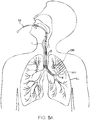
- Use of the agent delivery catheter 10 for treating collateral flow between adjacent lung segments begins with the endotracheal introduction of the catheter 10, as shown generally in FIG. 5A .
- the catheter 10 is advanced through the mouth, down through the trachea T, and through the main bronchus B into the left lung LL.
- the distal end 14 of catheter 10 is advanced into the left lung LL, and further advanced via airway AW to a target lung segment DR.
- the catheter 10 may be introduced through the main bronchus B and into the left lung LL without the use of a bronchoscope or other primary introducing catheter, as exemplarily shown in FIG. 5A .
- catheter 10 may be introduced through a conventional bronchoscope BS.
- a scope such as a bronchoscope which is capable of advancing into the lung is advantageous in that it facilitates visually-based positioning of the catheter 10 at the desired airway leading to a target lung segment.
- the catheter 10 may be introduced into the lung through any other scope, such as a visualizing endotracheal tube ET or bronchoscope with an inflatable cuff (not shown) which isolates an area of the lungs and permits local control of factors such as lung pressurization.
- catheter 10 may have an occlusion cuff or balloon 11 near its distal end to anchor the catheter.
- Construction and use of a visualizing endotracheal tube is taught, for example, in U.S. Pat. No. 5,285,778 . It would be possible, of course, to utilize both the bronchoscope BS and the endotracheal tube in combination for positioning the catheter 10 in the desired lung segment airway.
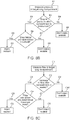
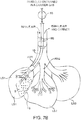
- FIGS. 6A , 6B and 6C are flow charts that describe the method of using the above embodiments, while FIGS. 7A and 7B are visual representations of the methods captured in the flow charts.
- the catheter is placed in a target lung compartment, and one or more variables within the target lung compartment are measured.
- a variable of air flow is measured within the target lung compartment (step A1 ), and as in step A2, the processor or a user determines whether the measured variable detects collateral flow, such as would occur in the presence of a collateral flow channel. If, as in Step A3, collateral flow is detected, then, as in Step A4, sealant is injected into the target lung compartment to stop collateral flow.
- Step A6 the sealant is injected into the target lung compartment.
- the sealant may be injected directly, or may be injected in conjunction with a carrier, for example a fluid such as a gas.
- FIGS. 6B and 6C show how specific variables may be measured and how such measurements may be used to determine when the sealant is injected into the lung compartment.
- the measured variable is pressure.
- the balloon is inflated and the target lung compartment is isolated.
- the compartment may be pressurized via methods described in U.S. Pub. No. 20030051733 .
- a fluid may be delivered to the target lung compartment such that the target compartment is pressurized.
- Step B1 the pressure of the pressurized lung compartment is measured, and, as shown in B2, it is determined whether there is a drop in pressure.
- Step B3 If, as shown in Step B3, the pressure drops, this is an indication of collateral flow to an adjacent channel, since the delivered fluid will flow from the target lung compartment through the collateral flow channels into the adjacent compartment.
- An agent may then be injected into the lung compartment in order to seal the collateral flow channels.
- the agent may be selectively directed to the collateral flow channels while little or no agent is delivered to other portions of the target lung compartment.
- the agent is injected into the isolated lung compartment such that the agent is carried by the fluid.
- the fluid flow guides the agent into the collateral flow path thereby sealing the collateral flow channel and the pressure in the other areas prevent the agent from going that direction.
- the injection of the agent may be timed in order to optimize selective delivery to the collateral flow channels.
- sealant is injected into the compartment, either alone or in conjunction with a carrier fluid. As shown in B7 and B8, if there is no decrease in pressure, then the sealant is not injected into the target lung compartment.
- the sealant may be injected instantaneously upon measuring a drop in pressure. Additionally, the sealant may be injected after waiting for a period of time at any point after a drop in pressure is detected, and such period of time may be either a preset or predetermined period of time, or it may be a period of time determined by a user.
- a feedback mechanism may exist where it may be determined based on measured pressure that any collateral channels have been sealed. In such an instance, the flow of the sealant is terminated when it is determined that the collateral channels have been sealed.
- the measured variable is flow rate.
- the balloon is inflated and the target lung compartment is isolated.
- the compartment may then be pressurized.
- An agent may be selectively directed to the collateral flow channels while little or no agent is delivered to other portions of the target lung compartment.
- the agent is injected into the isolated lung compartment such that the agent is carried by the fluid.
- the fluid flow guides the agent into the collateral flow path thereby sealing the collateral flow channel and the pressure in the other areas prevent the agent from going that direction.
- the injection of the agent may be timed in order to optimize selective delivery to the collateral flow channels.
- the flow rate is measured in the target lung compartment, and in C2, the processor or a user determines whether there is an increase in flow rate.
- sealant is injected into the compartment, either alone or in conjunction with a carrier fluid.
- sealant is not injected into the machine.
- the sealant may be injected instantaneously upon measuring a rise in flow rate.
- the sealant may be injected after waiting for a period of time at any point after a rise in flow rate is detected, and such period of time may be either a preset or predetermined period of time, or it may be a period of time determined by a user.
- a feedback mechanism may exist where it may be determined based on measured flow that any collateral channels have been sealed. In such an instance, the flow rate of the sealant is terminated when it is determined that the collateral channels have been sealed.
- the sealant may be injected into the target lung region independent of any feedback relating to a variable within the target lung compartment.
- the user would wait a predetermined period of time, or for the occurrence of a predetermined sequence of events, prior to introducing the sealant.
- the sealant may take any form such as a gel, particles, aerosol, liquid, or autologous blood.
- the sealant may be injected via a carrier fluid, such as a gas introducible into a human lung.
- FIGS. 7A and 7B show the practice of the methods described in FIGS. 6A , 6B, 6C .
- the individual lobes of a lung each comprise a plurality of lung segments LS which are fed by individual branches of the bronchi or airways AW.
- a first lung segment LS1, a second lung segment LS2, and a third lung segment LS3 may be fed from a single airway AW which divides into three branches AW1, AW2, and AW3, as illustrated in FIG. 7A .
- the fibrous septum may be perforate or porous to provide collateral flow channels therebetween, as illustrated at FS.
- the catheter 10 is positioned in airway AW1 leading into lung segment LS1, which may be a diseased lung segment.
- lung segment LS1 which may be a diseased lung segment.
- the collateral flow channels in the wall FS between the first lung segment LS1 and the second lung segment LS2 will permit gas flow in either direction prior to the treatments of the present disclosure.
- the first lung segment LS1 is isolated, and this isolation is compromised only by any collateral flow channels that allow air to leak in from adjacent lung compartments. If there is no collateral flow, then the pressure in the compartment will increase, while the flow will decrease.
- sealant S is released into the target lung compartment. Sealant S will thereafter permeate into and seal collateral channels.
- the catheter is thereafter retracted (not shown), leaving the lung segment LS1 with fewer or no collateral channels.
Landscapes
- Health & Medical Sciences (AREA)
- Life Sciences & Earth Sciences (AREA)
- Pulmonology (AREA)
- Heart & Thoracic Surgery (AREA)
- General Health & Medical Sciences (AREA)
- Engineering & Computer Science (AREA)
- Animal Behavior & Ethology (AREA)
- Public Health (AREA)
- Veterinary Medicine (AREA)
- Biomedical Technology (AREA)
- Anesthesiology (AREA)
- Hematology (AREA)
- Emergency Medicine (AREA)
- Surgery (AREA)
- Medical Informatics (AREA)
- Nuclear Medicine, Radiotherapy & Molecular Imaging (AREA)
- Molecular Biology (AREA)
- Vascular Medicine (AREA)
- Reproductive Health (AREA)
- Child & Adolescent Psychology (AREA)
- Biophysics (AREA)
- Surgical Instruments (AREA)
Claims (12)
- Système destiné à occlure un canal d'écoulement collatéral entre un compartiment de poumon cible et un compartiment de poumon adjacent, ledit système comprenant :un cathéter d'isolation (10) configuré pour accéder au compartiment de poumon cible et l'isoler ;un mécanisme de délivrance de fluide (18) configuré pour délivrer un fluide jusque dans le compartiment de poumon cible de sorte que le compartiment cible est pressurisé et le fluide s'écoule à travers le canal d'écoulement collatéral ;un mécanisme de délivrance d'agent (18 ; 24) configuré pour injecter l'agent dans le compartiment de poumon cible isolé de sorte que l'agent est transporté par le fluide jusqu'au canal d'écoulement collatéral, scellant ainsi le canal d'écoulement collatéral ; etun processeur (20) configuré pour libérer le fluide, pour recevoir une entrée d'un détecteur associé au cathéter d'isolation, et choisir le moment de l'injection de l'agent jusque dans le compartiment de poumon cible sur la base de l'entrée, dans lequel l'entrée reçue par le processeur (20) est une mesure de pression ou d'écoulement au sein du compartiment de poumon cible et le choix du moment de l'injection de l'agent jusque dans le compartiment de poumon cible est basé sur la pression ou l'écoulement mesuré.
- Système selon la revendication 1, dans lequel l'agent est un agent de scellement.
- Système selon la revendication 1, dans lequel l'entrée reçue par le processeur (20) est une mesure de pression au sein du compartiment de poumon cible et le choix du moment de l'injection de l'agent est basé sur la pression mesurée.
- Système selon la revendication 3, dans lequel le choix du moment de l'injection de l'agent comprend l'injection de l'agent après que la pression mesurée chute au-dessous d'une valeur seuil prédéterminée.
- Système selon la revendication 3, dans lequel le choix du moment de l'injection de l'agent comprend le fait d'attendre une période de temps prédéterminée après la mesure d'une chute de pression avant d'injecter l'agent.
- Système selon la revendication 3, dans lequel le choix du moment de l'injection de l'agent comprend le fait de déterminer que le canal collatéral est scellé sur la base de la pression mesurée et l'arrêt de l'injection de l'agent après qu'il est déterminé que le canal collatéral est scellé.
- Système selon la revendication 1, dans lequel l'entrée reçue par le processeur (20) est une mesure de l'écoulement au sein du compartiment de poumon cible et le choix du moment de l'injection est basé sur l'écoulement mesuré.
- Système selon la revendication 7, dans lequel le choix du moment de l'injection de l'agent comprend l'injection de l'agent après qu'il est déterminé que l'écoulement mesuré atteint une valeur seuil prédéterminée.
- Système selon la revendication 7, dans lequel le choix du moment de l'injection de l'agent comprend le fait de déterminer que le canal collatéral est scellé sur la base de l'écoulement mesuré et l'arrêt de l'injection de l'agent après qu'il est été déterminé que le canal collatéral est scellé.
- Système selon la revendication 1, dans lequel le processeur (20) est configuré en outre pour recevoir une entrée d'un utilisateur concernant l'injection de l'agent.
- Système selon la revendication 1, dans lequel le système comprend en outre une unité d'affichage (21) pour afficher des valeurs obtenues du détecteur.
- Système selon la revendication 1, dans lequel le système comprend en outre une unité d'affichage pour afficher des calculs dérivés de l'entrée du détecteur.
Applications Claiming Priority (2)
| Application Number | Priority Date | Filing Date | Title |
|---|---|---|---|
| US201562105649P | 2015-01-20 | 2015-01-20 | |
| PCT/US2016/014183 WO2016118663A1 (fr) | 2015-01-20 | 2016-01-20 | Méthodes et systèmes d'administration d'agents de scellement bronchique |
Publications (3)
| Publication Number | Publication Date |
|---|---|
| EP3247270A1 EP3247270A1 (fr) | 2017-11-29 |
| EP3247270A4 EP3247270A4 (fr) | 2018-08-22 |
| EP3247270B1 true EP3247270B1 (fr) | 2020-03-04 |
Family
ID=56406919
Family Applications (1)
| Application Number | Title | Priority Date | Filing Date |
|---|---|---|---|
| EP16740705.5A Active EP3247270B1 (fr) | 2015-01-20 | 2016-01-20 | Systèmes d'administration d'agents de scellement bronchique |
Country Status (6)
| Country | Link |
|---|---|
| US (1) | US10456562B2 (fr) |
| EP (1) | EP3247270B1 (fr) |
| JP (1) | JP6746592B2 (fr) |
| AU (1) | AU2016209346B2 (fr) |
| ES (1) | ES2777547T3 (fr) |
| WO (1) | WO2016118663A1 (fr) |
Families Citing this family (3)
| Publication number | Priority date | Publication date | Assignee | Title |
|---|---|---|---|---|
| ES2815423T3 (es) | 2016-02-05 | 2021-03-30 | Pulmonx Corp | Procedimientos, sistemas y dispositivos para analizar datos de imágenes pulmonares |
| US11419490B2 (en) * | 2016-08-02 | 2022-08-23 | Covidien Lp | System and method of using an endoscopic catheter as a port in laparoscopic surgery |
| CA3238644A1 (fr) * | 2021-11-18 | 2023-05-25 | Luiz MARACAJA | Administration selective au niveau des lobes d'agents therapeutiques et appareil pour une bronchoscopie interventionnelle |
Citations (1)
| Publication number | Priority date | Publication date | Assignee | Title |
|---|---|---|---|---|
| US20070186932A1 (en) * | 2006-01-06 | 2007-08-16 | Pulmonx | Collateral pathway treatment using agent entrained by aspiration flow current |
Family Cites Families (13)
| Publication number | Priority date | Publication date | Assignee | Title |
|---|---|---|---|---|
| US6770070B1 (en) * | 2000-03-17 | 2004-08-03 | Rita Medical Systems, Inc. | Lung treatment apparatus and method |
| US7883471B2 (en) | 2001-09-10 | 2011-02-08 | Pulmonx Corporation | Minimally invasive determination of collateral ventilation in lungs |
| US20030051733A1 (en) | 2001-09-10 | 2003-03-20 | Pulmonx | Method and apparatus for endobronchial diagnosis |
| US7412977B2 (en) * | 2002-03-08 | 2008-08-19 | Emphasys Medical, Inc. | Methods and devices for inducing collapse in lung regions fed by collateral pathways |
| US7819908B2 (en) | 2002-06-17 | 2010-10-26 | Aeris Therapeutics, Inc. | Compositions and methods for reducing lung volume |
| JP2008503254A (ja) * | 2004-06-16 | 2008-02-07 | ヌームアールエックス・インコーポレーテッド | 気管支内肺容量減少システム |
| TWI243538B (en) * | 2004-07-30 | 2005-11-11 | Realtek Semiconductor Corp | Apparatus for generating testing signals |
| US8876791B2 (en) | 2005-02-25 | 2014-11-04 | Pulmonx Corporation | Collateral pathway treatment using agent entrained by aspiration flow current |
| JP2009501568A (ja) * | 2005-07-13 | 2009-01-22 | プルモンクス | 肺の区域診断のための方法及びシステム |
| US8137302B2 (en) * | 2007-03-12 | 2012-03-20 | Pulmonx Corporation | Methods and systems for occluding collateral flow channels in the lung |
| CA2686563A1 (fr) | 2007-05-11 | 2008-11-20 | Aeris Therapeutics, Llc | Therapie de reduction de volume de poumon utilisant des polymeres non naturels reticules |
| US8808194B2 (en) | 2010-07-01 | 2014-08-19 | Pulmonx Corporation | Methods and systems for endobronchial diagnostics |
| JP2012045358A (ja) * | 2010-08-25 | 2012-03-08 | Terumo Corp | 肺気腫の治療剤 |
-
2016
- 2016-01-20 WO PCT/US2016/014183 patent/WO2016118663A1/fr not_active Ceased
- 2016-01-20 EP EP16740705.5A patent/EP3247270B1/fr active Active
- 2016-01-20 JP JP2017539343A patent/JP6746592B2/ja active Active
- 2016-01-20 ES ES16740705T patent/ES2777547T3/es active Active
- 2016-01-20 AU AU2016209346A patent/AU2016209346B2/en active Active
- 2016-01-20 US US15/001,598 patent/US10456562B2/en active Active
Patent Citations (1)
| Publication number | Priority date | Publication date | Assignee | Title |
|---|---|---|---|---|
| US20070186932A1 (en) * | 2006-01-06 | 2007-08-16 | Pulmonx | Collateral pathway treatment using agent entrained by aspiration flow current |
Also Published As
| Publication number | Publication date |
|---|---|
| ES2777547T3 (es) | 2020-08-05 |
| JP6746592B2 (ja) | 2020-08-26 |
| WO2016118663A1 (fr) | 2016-07-28 |
| EP3247270A4 (fr) | 2018-08-22 |
| AU2016209346A1 (en) | 2017-08-03 |
| US10456562B2 (en) | 2019-10-29 |
| JP2018502671A (ja) | 2018-02-01 |
| EP3247270A1 (fr) | 2017-11-29 |
| US20160206301A1 (en) | 2016-07-21 |
| AU2016209346B2 (en) | 2020-05-14 |
Similar Documents
| Publication | Publication Date | Title |
|---|---|---|
| US12350428B2 (en) | Methods and devices for passive residual lung volume reduction and functional lung volume expansion | |
| US11931170B2 (en) | Functional assessment and treatment catheters and methods for their use in the lung | |
| AU2004263132B2 (en) | Bronchoscopic repair of air leaks in a lung | |
| EP1339445B1 (fr) | Dispositifs destines a occlure et a aspirer des segments de tissu pulmonaire | |
| US8137302B2 (en) | Methods and systems for occluding collateral flow channels in the lung | |
| US12310594B2 (en) | Collateral flow channel sealant delivery methods and systems | |
| EP3247270B1 (fr) | Systèmes d'administration d'agents de scellement bronchique | |
| EP2903514B1 (fr) | Systèmes pour diagnostic pulmonaire |
Legal Events
| Date | Code | Title | Description |
|---|---|---|---|
| STAA | Information on the status of an ep patent application or granted ep patent |
Free format text: STATUS: THE INTERNATIONAL PUBLICATION HAS BEEN MADE |
|
| PUAI | Public reference made under article 153(3) epc to a published international application that has entered the european phase |
Free format text: ORIGINAL CODE: 0009012 |
|
| STAA | Information on the status of an ep patent application or granted ep patent |
Free format text: STATUS: REQUEST FOR EXAMINATION WAS MADE |
|
| 17P | Request for examination filed |
Effective date: 20170726 |
|
| AK | Designated contracting states |
Kind code of ref document: A1 Designated state(s): AL AT BE BG CH CY CZ DE DK EE ES FI FR GB GR HR HU IE IS IT LI LT LU LV MC MK MT NL NO PL PT RO RS SE SI SK SM TR |
|
| AX | Request for extension of the european patent |
Extension state: BA ME |
|
| DAV | Request for validation of the european patent (deleted) | ||
| DAX | Request for extension of the european patent (deleted) | ||
| A4 | Supplementary search report drawn up and despatched |
Effective date: 20180720 |
|
| RIC1 | Information provided on ipc code assigned before grant |
Ipc: A61M 16/00 20060101ALI20180716BHEP Ipc: A61M 29/02 20060101ALI20180716BHEP Ipc: A61B 17/12 20060101ALI20180716BHEP Ipc: A61M 16/04 20060101ALI20180716BHEP Ipc: A61B 5/08 20060101AFI20180716BHEP Ipc: A61M 25/10 20130101ALI20180716BHEP |
|
| GRAP | Despatch of communication of intention to grant a patent |
Free format text: ORIGINAL CODE: EPIDOSNIGR1 |
|
| STAA | Information on the status of an ep patent application or granted ep patent |
Free format text: STATUS: GRANT OF PATENT IS INTENDED |
|
| INTG | Intention to grant announced |
Effective date: 20190813 |
|
| GRAS | Grant fee paid |
Free format text: ORIGINAL CODE: EPIDOSNIGR3 |
|
| GRAA | (expected) grant |
Free format text: ORIGINAL CODE: 0009210 |
|
| STAA | Information on the status of an ep patent application or granted ep patent |
Free format text: STATUS: THE PATENT HAS BEEN GRANTED |
|
| AK | Designated contracting states |
Kind code of ref document: B1 Designated state(s): AL AT BE BG CH CY CZ DE DK EE ES FI FR GB GR HR HU IE IS IT LI LT LU LV MC MK MT NL NO PL PT RO RS SE SI SK SM TR |
|
| REG | Reference to a national code |
Ref country code: GB Ref legal event code: FG4D |
|
| REG | Reference to a national code |
Ref country code: CH Ref legal event code: NV Representative=s name: SERVOPATENT GMBH, CH Ref country code: CH Ref legal event code: EP |
|
| REG | Reference to a national code |
Ref country code: AT Ref legal event code: REF Ref document number: 1239452 Country of ref document: AT Kind code of ref document: T Effective date: 20200315 |
|
| REG | Reference to a national code |
Ref country code: DE Ref legal event code: R096 Ref document number: 602016031074 Country of ref document: DE |
|
| REG | Reference to a national code |
Ref country code: IE Ref legal event code: FG4D |
|
| REG | Reference to a national code |
Ref country code: NL Ref legal event code: FP |
|
| REG | Reference to a national code |
Ref country code: CH Ref legal event code: PCAR Free format text: NEW ADDRESS: WANNERSTRASSE 9/1, 8045 ZUERICH (CH) |
|
| REG | Reference to a national code |
Ref country code: SE Ref legal event code: TRGR |
|
| PG25 | Lapsed in a contracting state [announced via postgrant information from national office to epo] |
Ref country code: FI Free format text: LAPSE BECAUSE OF FAILURE TO SUBMIT A TRANSLATION OF THE DESCRIPTION OR TO PAY THE FEE WITHIN THE PRESCRIBED TIME-LIMIT Effective date: 20200304 Ref country code: NO Free format text: LAPSE BECAUSE OF FAILURE TO SUBMIT A TRANSLATION OF THE DESCRIPTION OR TO PAY THE FEE WITHIN THE PRESCRIBED TIME-LIMIT Effective date: 20200604 Ref country code: RS Free format text: LAPSE BECAUSE OF FAILURE TO SUBMIT A TRANSLATION OF THE DESCRIPTION OR TO PAY THE FEE WITHIN THE PRESCRIBED TIME-LIMIT Effective date: 20200304 |
|
| REG | Reference to a national code |
Ref country code: ES Ref legal event code: FG2A Ref document number: 2777547 Country of ref document: ES Kind code of ref document: T3 Effective date: 20200805 |
|
| PG25 | Lapsed in a contracting state [announced via postgrant information from national office to epo] |
Ref country code: BG Free format text: LAPSE BECAUSE OF FAILURE TO SUBMIT A TRANSLATION OF THE DESCRIPTION OR TO PAY THE FEE WITHIN THE PRESCRIBED TIME-LIMIT Effective date: 20200604 Ref country code: GR Free format text: LAPSE BECAUSE OF FAILURE TO SUBMIT A TRANSLATION OF THE DESCRIPTION OR TO PAY THE FEE WITHIN THE PRESCRIBED TIME-LIMIT Effective date: 20200605 Ref country code: LV Free format text: LAPSE BECAUSE OF FAILURE TO SUBMIT A TRANSLATION OF THE DESCRIPTION OR TO PAY THE FEE WITHIN THE PRESCRIBED TIME-LIMIT Effective date: 20200304 Ref country code: HR Free format text: LAPSE BECAUSE OF FAILURE TO SUBMIT A TRANSLATION OF THE DESCRIPTION OR TO PAY THE FEE WITHIN THE PRESCRIBED TIME-LIMIT Effective date: 20200304 |
|
| REG | Reference to a national code |
Ref country code: LT Ref legal event code: MG4D |
|
| PG25 | Lapsed in a contracting state [announced via postgrant information from national office to epo] |
Ref country code: IS Free format text: LAPSE BECAUSE OF FAILURE TO SUBMIT A TRANSLATION OF THE DESCRIPTION OR TO PAY THE FEE WITHIN THE PRESCRIBED TIME-LIMIT Effective date: 20200704 Ref country code: PT Free format text: LAPSE BECAUSE OF FAILURE TO SUBMIT A TRANSLATION OF THE DESCRIPTION OR TO PAY THE FEE WITHIN THE PRESCRIBED TIME-LIMIT Effective date: 20200729 Ref country code: RO Free format text: LAPSE BECAUSE OF FAILURE TO SUBMIT A TRANSLATION OF THE DESCRIPTION OR TO PAY THE FEE WITHIN THE PRESCRIBED TIME-LIMIT Effective date: 20200304 Ref country code: CZ Free format text: LAPSE BECAUSE OF FAILURE TO SUBMIT A TRANSLATION OF THE DESCRIPTION OR TO PAY THE FEE WITHIN THE PRESCRIBED TIME-LIMIT Effective date: 20200304 Ref country code: LT Free format text: LAPSE BECAUSE OF FAILURE TO SUBMIT A TRANSLATION OF THE DESCRIPTION OR TO PAY THE FEE WITHIN THE PRESCRIBED TIME-LIMIT Effective date: 20200304 Ref country code: SM Free format text: LAPSE BECAUSE OF FAILURE TO SUBMIT A TRANSLATION OF THE DESCRIPTION OR TO PAY THE FEE WITHIN THE PRESCRIBED TIME-LIMIT Effective date: 20200304 Ref country code: EE Free format text: LAPSE BECAUSE OF FAILURE TO SUBMIT A TRANSLATION OF THE DESCRIPTION OR TO PAY THE FEE WITHIN THE PRESCRIBED TIME-LIMIT Effective date: 20200304 Ref country code: SK Free format text: LAPSE BECAUSE OF FAILURE TO SUBMIT A TRANSLATION OF THE DESCRIPTION OR TO PAY THE FEE WITHIN THE PRESCRIBED TIME-LIMIT Effective date: 20200304 |
|
| REG | Reference to a national code |
Ref country code: AT Ref legal event code: MK05 Ref document number: 1239452 Country of ref document: AT Kind code of ref document: T Effective date: 20200304 |
|
| REG | Reference to a national code |
Ref country code: DE Ref legal event code: R097 Ref document number: 602016031074 Country of ref document: DE |
|
| PLBE | No opposition filed within time limit |
Free format text: ORIGINAL CODE: 0009261 |
|
| STAA | Information on the status of an ep patent application or granted ep patent |
Free format text: STATUS: NO OPPOSITION FILED WITHIN TIME LIMIT |
|
| PG25 | Lapsed in a contracting state [announced via postgrant information from national office to epo] |
Ref country code: AT Free format text: LAPSE BECAUSE OF FAILURE TO SUBMIT A TRANSLATION OF THE DESCRIPTION OR TO PAY THE FEE WITHIN THE PRESCRIBED TIME-LIMIT Effective date: 20200304 Ref country code: DK Free format text: LAPSE BECAUSE OF FAILURE TO SUBMIT A TRANSLATION OF THE DESCRIPTION OR TO PAY THE FEE WITHIN THE PRESCRIBED TIME-LIMIT Effective date: 20200304 |
|
| 26N | No opposition filed |
Effective date: 20201207 |
|
| PG25 | Lapsed in a contracting state [announced via postgrant information from national office to epo] |
Ref country code: PL Free format text: LAPSE BECAUSE OF FAILURE TO SUBMIT A TRANSLATION OF THE DESCRIPTION OR TO PAY THE FEE WITHIN THE PRESCRIBED TIME-LIMIT Effective date: 20200304 Ref country code: SI Free format text: LAPSE BECAUSE OF FAILURE TO SUBMIT A TRANSLATION OF THE DESCRIPTION OR TO PAY THE FEE WITHIN THE PRESCRIBED TIME-LIMIT Effective date: 20200304 |
|
| PG25 | Lapsed in a contracting state [announced via postgrant information from national office to epo] |
Ref country code: MC Free format text: LAPSE BECAUSE OF FAILURE TO SUBMIT A TRANSLATION OF THE DESCRIPTION OR TO PAY THE FEE WITHIN THE PRESCRIBED TIME-LIMIT Effective date: 20200304 |
|
| PG25 | Lapsed in a contracting state [announced via postgrant information from national office to epo] |
Ref country code: LU Free format text: LAPSE BECAUSE OF NON-PAYMENT OF DUE FEES Effective date: 20210120 |
|
| REG | Reference to a national code |
Ref country code: BE Ref legal event code: MM Effective date: 20210131 |
|
| PG25 | Lapsed in a contracting state [announced via postgrant information from national office to epo] |
Ref country code: BE Free format text: LAPSE BECAUSE OF NON-PAYMENT OF DUE FEES Effective date: 20210131 |
|
| PG25 | Lapsed in a contracting state [announced via postgrant information from national office to epo] |
Ref country code: HU Free format text: LAPSE BECAUSE OF FAILURE TO SUBMIT A TRANSLATION OF THE DESCRIPTION OR TO PAY THE FEE WITHIN THE PRESCRIBED TIME-LIMIT; INVALID AB INITIO Effective date: 20160120 |
|
| PG25 | Lapsed in a contracting state [announced via postgrant information from national office to epo] |
Ref country code: CY Free format text: LAPSE BECAUSE OF FAILURE TO SUBMIT A TRANSLATION OF THE DESCRIPTION OR TO PAY THE FEE WITHIN THE PRESCRIBED TIME-LIMIT Effective date: 20200304 |
|
| P01 | Opt-out of the competence of the unified patent court (upc) registered |
Effective date: 20230912 |
|
| PG25 | Lapsed in a contracting state [announced via postgrant information from national office to epo] |
Ref country code: MK Free format text: LAPSE BECAUSE OF FAILURE TO SUBMIT A TRANSLATION OF THE DESCRIPTION OR TO PAY THE FEE WITHIN THE PRESCRIBED TIME-LIMIT Effective date: 20200304 |
|
| PG25 | Lapsed in a contracting state [announced via postgrant information from national office to epo] |
Ref country code: TR Free format text: LAPSE BECAUSE OF FAILURE TO SUBMIT A TRANSLATION OF THE DESCRIPTION OR TO PAY THE FEE WITHIN THE PRESCRIBED TIME-LIMIT Effective date: 20200304 |
|
| PG25 | Lapsed in a contracting state [announced via postgrant information from national office to epo] |
Ref country code: MT Free format text: LAPSE BECAUSE OF FAILURE TO SUBMIT A TRANSLATION OF THE DESCRIPTION OR TO PAY THE FEE WITHIN THE PRESCRIBED TIME-LIMIT Effective date: 20200304 |
|
| PGFP | Annual fee paid to national office [announced via postgrant information from national office to epo] |
Ref country code: NL Payment date: 20251211 Year of fee payment: 11 |
|
| REG | Reference to a national code |
Ref country code: CH Ref legal event code: U11 Free format text: ST27 STATUS EVENT CODE: U-0-0-U10-U11 (AS PROVIDED BY THE NATIONAL OFFICE) Effective date: 20260201 |
|
| PGFP | Annual fee paid to national office [announced via postgrant information from national office to epo] |
Ref country code: SE Payment date: 20260121 Year of fee payment: 11 |
|
| PGFP | Annual fee paid to national office [announced via postgrant information from national office to epo] |
Ref country code: GB Payment date: 20260127 Year of fee payment: 11 |
|
| PGFP | Annual fee paid to national office [announced via postgrant information from national office to epo] |
Ref country code: ES Payment date: 20260218 Year of fee payment: 11 |
|
| PGFP | Annual fee paid to national office [announced via postgrant information from national office to epo] |
Ref country code: DE Payment date: 20251211 Year of fee payment: 11 Ref country code: IE Payment date: 20260121 Year of fee payment: 11 |
|
| PGFP | Annual fee paid to national office [announced via postgrant information from national office to epo] |
Ref country code: IT Payment date: 20251211 Year of fee payment: 11 |
|
| PGFP | Annual fee paid to national office [announced via postgrant information from national office to epo] |
Ref country code: FR Payment date: 20260122 Year of fee payment: 11 |
|
| PGFP | Annual fee paid to national office [announced via postgrant information from national office to epo] |
Ref country code: CH Payment date: 20260201 Year of fee payment: 11 |