EP3248006B1 - Procédé de préparation d'un échantillon biologique destiné à être utilisé dans un procédé d'immunomarquage - Google Patents
Procédé de préparation d'un échantillon biologique destiné à être utilisé dans un procédé d'immunomarquage Download PDFInfo
- Publication number
- EP3248006B1 EP3248006B1 EP16740471.4A EP16740471A EP3248006B1 EP 3248006 B1 EP3248006 B1 EP 3248006B1 EP 16740471 A EP16740471 A EP 16740471A EP 3248006 B1 EP3248006 B1 EP 3248006B1
- Authority
- EP
- European Patent Office
- Prior art keywords
- enhancer
- antibody
- antigen
- immunolabeling
- present
- Prior art date
- Legal status (The legal status is an assumption and is not a legal conclusion. Google has not performed a legal analysis and makes no representation as to the accuracy of the status listed.)
- Active
Links
Images

Classifications
-
- G—PHYSICS
- G01—MEASURING; TESTING
- G01N—INVESTIGATING OR ANALYSING MATERIALS BY DETERMINING THEIR CHEMICAL OR PHYSICAL PROPERTIES
- G01N33/00—Investigating or analysing materials by specific methods not covered by groups G01N1/00 - G01N31/00
- G01N33/48—Biological material, e.g. blood, urine; Haemocytometers
- G01N33/50—Chemical analysis of biological material, e.g. blood, urine; Testing involving biospecific ligand binding methods; Immunological testing
- G01N33/53—Immunoassay; Biospecific binding assay; Materials therefor
-
- G—PHYSICS
- G01—MEASURING; TESTING
- G01N—INVESTIGATING OR ANALYSING MATERIALS BY DETERMINING THEIR CHEMICAL OR PHYSICAL PROPERTIES
- G01N33/00—Investigating or analysing materials by specific methods not covered by groups G01N1/00 - G01N31/00
- G01N33/48—Biological material, e.g. blood, urine; Haemocytometers
- G01N33/50—Chemical analysis of biological material, e.g. blood, urine; Testing involving biospecific ligand binding methods; Immunological testing
- G01N33/68—Chemical analysis of biological material, e.g. blood, urine; Testing involving biospecific ligand binding methods; Immunological testing involving proteins, peptides or amino acids
- G01N33/6854—Immunoglobulins
-
- G—PHYSICS
- G01—MEASURING; TESTING
- G01N—INVESTIGATING OR ANALYSING MATERIALS BY DETERMINING THEIR CHEMICAL OR PHYSICAL PROPERTIES
- G01N33/00—Investigating or analysing materials by specific methods not covered by groups G01N1/00 - G01N31/00
- G01N33/48—Biological material, e.g. blood, urine; Haemocytometers
- G01N33/50—Chemical analysis of biological material, e.g. blood, urine; Testing involving biospecific ligand binding methods; Immunological testing
- G01N33/53—Immunoassay; Biospecific binding assay; Materials therefor
- G01N33/5306—Improving reaction conditions, e.g. reduction of non-specific binding, promotion of specific binding
-
- G—PHYSICS
- G01—MEASURING; TESTING
- G01N—INVESTIGATING OR ANALYSING MATERIALS BY DETERMINING THEIR CHEMICAL OR PHYSICAL PROPERTIES
- G01N33/00—Investigating or analysing materials by specific methods not covered by groups G01N1/00 - G01N31/00
- G01N33/48—Biological material, e.g. blood, urine; Haemocytometers
- G01N33/50—Chemical analysis of biological material, e.g. blood, urine; Testing involving biospecific ligand binding methods; Immunological testing
- G01N33/53—Immunoassay; Biospecific binding assay; Materials therefor
- G01N33/536—Immunoassay; Biospecific binding assay; Materials therefor with immune complex formed in liquid phase
- G01N33/542—Immunoassay; Biospecific binding assay; Materials therefor with immune complex formed in liquid phase with steric inhibition or signal modification, e.g. fluorescent quenching
-
- G—PHYSICS
- G01—MEASURING; TESTING
- G01N—INVESTIGATING OR ANALYSING MATERIALS BY DETERMINING THEIR CHEMICAL OR PHYSICAL PROPERTIES
- G01N33/00—Investigating or analysing materials by specific methods not covered by groups G01N1/00 - G01N31/00
- G01N33/48—Biological material, e.g. blood, urine; Haemocytometers
- G01N33/50—Chemical analysis of biological material, e.g. blood, urine; Testing involving biospecific ligand binding methods; Immunological testing
- G01N33/58—Chemical analysis of biological material, e.g. blood, urine; Testing involving biospecific ligand binding methods; Immunological testing involving labelled substances
-
- G—PHYSICS
- G01—MEASURING; TESTING
- G01N—INVESTIGATING OR ANALYSING MATERIALS BY DETERMINING THEIR CHEMICAL OR PHYSICAL PROPERTIES
- G01N2474/00—Immunochemical assays or immunoassays characterised by detection mode or means of detection
- G01N2474/20—Immunohistochemistry assay
Definitions
- the present invention generally relates to method for preparing a biological sample for use in an immunolabeling process.
- Antibodies are used for detection of molecules in biological and non-biological samples.
- Antibodies are immunoglobulin (Ig) proteins that bind with high specificity through its antigen-binding site to an antigen (target molecule).
- the antigen is a protein, but can be any immunogenic agent such as a shorter amino acid sequence (peptide), polysacharide, lipid, toxin etc.
- the part of the target molecule to which the antibody binds is called epitope.
- Antibodies used for immunolabeling can be polyclonal or monoclonal. Polyclonal antibodies are a heterogeneous mix of antibodies that recognize several epitopes of one target molecule, while monoclonal antibodies show specificity for a single epitope. In general, monoclonal antibodies gender more specific immunolabeling signals than polyclonal antibodies.
- the final step in immunolabeling is detection of a signal from the antibodies that has bound to the antigens in the sample.
- the signal is generated from some kind of reporter molecule.
- the reporter molecule can either be directly attached to the primary antibody, or attached to a secondary antibody that recognizes the primary antibody. Often several reporter molecules are attached to each antibody molecule.
- the reporter molecules used in immunolabeling vary depending on the nature of the detection method. The most common reporter molecules are enzymes for chromogenic detection or fluorochromes for fluorescence signals. Other examples are particles (e.g. gold particles, quantum dots), phosphorescent compunds (e.g. carbocyanide dyes), radioactive compounds (e.g. 3H or 32P labeled molecules) and transition metals (for mass spectrometry).
- Immunolabeling can either be direct or indirect.
- the direct method is a one-step immunolabeling method and involves a primary antibody that is labeled with a reporter molecule. When the labeled primary antibody is added to a sample it binds to its corresponding target antigen in the sample and reveals the location and/or amount of the target molecule. Since the direct method utilizes only one step it is simple and rapid. However, in some applications, for example microscopy, the signal is often too weak and needs to be amplified.
- the indirect method is a two-step labeling method that results in signal amplification. It involves a primary antibody (first step) that binds to the target molecule in the sample and a labeled secondary antibody (second step) that binds to the primary antibody. Since several secondary antibody molecules bind to each primary antibody molecule, the signal is amplified.
- the secondary antibody is usually raised against the immunoglubolin class of the animal species in which the primary antibody has been raised. For example, if the primary antibody is a mouse IgG antibody, the secondary antibody is an anti-mouse IgG antibody that recognizes all mouse antibodies of the IgG class.
- the indirect method is beneficial when it comes to signal amplification, it gives rise to unspecific signals due to unspecific binding of the secondary antibody to endogenous antibodies present in the sample.
- One last amplification step can be introduced by using a biotinylated secondary antibody and labeled streptavidin. Streptavidin binds tightly to biotin and since several biotin molecules are conjugated to each biotinylated antibody, amplification is achieved.
- biotin is also naturally present in biological samples, which causes unspecific binding of streptavidin to the sample, unless the endogenous biotin is blocked.
- an alternative signal amplifying system is desired that (1) does not cause background signal from endogenous antibodies/biotin, and that (2) enables more amplification steps.
- each primary antibody can be detected with a corresponding secondary antibody that recognizes the Ig class of the animal species of the primary antibody. For example, if one primary antibody is made in rat and the other is made in rabbit, these two primary antibodies can be detected with one anti-rat and one anti-rabbit secondary antibody that are labeled with two different reporter molecules, for example two different fluorochromes.
- compositions comprises a first antibody having an affinity for an antigen and a second antibody having an affinity for the first antibody, wherein at least one antibody is conjugated to a marker, and wherein the antigen is not present in the composition.
- EP0389301 disclose methods and a reagent complex suitable for attaching detectable signals to specific-binding reagents, the complex comprising a first unlabelled antibody, wherein the first antibody has free specific--binding affinity for an analyte or an analyte-specific binding reagent; and a second antibody having specific--binding affinity for an Fc region of the first antibody, the second antibody being bound to the Fc region to form the complex and being labelled with a detectable signal.
- WO2014163557 presents a method for detection of at least two cytokeratins selected from the group consisting of cytokeratin 8, 18 and 19, and/or soluble fragments thereof, in a sample.
- WO2009032128 shows compositions and methods comprising proteins that bind specifically to adalimumab.
- US2006286546 discloses a method and kit for quantitatively and/or qualitatively detecting one or more components in samples, including the use of metal-particle labelled reagents and an antibody conjugate.
- GB2098730 presents a method of detecting an antigenic substance, in animal tissue, in which a primary antibody to the substance is raised, labelled with a selected hapten such as DNP and contacted with the substance, after unwanted binding sites on the substance have been blocked by applying a wide spectrum of antibodies obtained from a different animal.
- the above is at least partly alleviated by a method for preparing a biological sample for use in an immunolabeling process, as defined by claim 1.
- a signal enhancer system for immunolabeling enables an unlimited number of amplification steps on top of a labeling component, for example being a primary antibody, without any antibody cross-binding.
- the absence of antibody cross-binding also enables any number of different labeling components (e.g. the labeling component and a plurality of additional labeling components, for example being different primary antibodies) to be combined for multi-immunolabeling, regardless of what animal species the primary antibodies are made from.
- the invention is based on carefully chosen antigens that are used as unique tags and corresponding antibodies that are used for detection of the tags. The antigens are chosen so that the antigen is non-present in immunolabeling process, i.e. not present in the biological sample and not present in reagents that are used in sample processing or staining reagents.
- the first enhancer antigen when introducing the labeling component provided with the first enhancer antigen, the first enhancer antigen has not been previously introduced (or comprised) in the immunolabeling process. Neither is a similar (identical, corresponding) antigen to be introduced subsequently in the immunolabeling process.
- the antigen should not be present in any further components used in the immunolabeling process, for example including reporter molecule used in the immunolabeling process, such as fluorochromes, chromogens, enzymes, etc.
- the antigen should furthermore not be present in any antibody comprised with the sample or used with the immunolabeling process, and the antigen should not be present in biotin or streptavidin.
- the labeling component is a primary antibody conjugated with the first enhancer antigen.
- the concept of the invention relies on the fact that the first enhancer antigen is not comprised with the sample or any reagents used in the immunolabeling process.
- the antigen may as such in accordance to some embodiments be seen as non-functional in relation to the sample, or in relation to immunolabeling process.
- the antigens are formed from artificially formulated peptide sequences that are not present in any proteins in nature, and thereby the artificial peptides may serve as unique antigens that are not present in any biological sample.
- Such non-biological peptides can be designed using protein sequence databases, such as the universal proteome database.
- the artificially formulated peptide sequence may then be used for forming the first enhancer antigen and for subsequent generation of a corresponding first enhancer antibody for use in relation to the present invention.
- the inventive concept is used for amplification, and thus the first enhancer antibody is conjugated with a second enhancer antigen, wherein the second enhancer antigen is non-present in the immunolabeling process and different from the first enhancer antigen, the method further comprising the step of providing a second enhancer antibody, the second enhancer antibody selected to solely bind to the second enhancer antigen.
- the unlimited amplification will, as understood based on the above, allow for the second enhancer antibody to possibly be conjugated with a third enhancer antigen, the third enhancer antigen being non-present in the immunolabeling process and different from either of the first and the second enhancer antigen.
- the process may of course continue with a "chain" of further antigens/antibodies.
- the "last" enhancer antibody in the chain is labeled with a reporter molecule.
- the reporter molecule is typically selected from a group comprising a fluorochrome, an enzyme, a peptide, quantum dots, and a transition metal. Other known or future reporter molecules are possible and within the scope of the invention, such as for example an oligonucleotide.
- the reporter molecule(s) are typically used in a subsequent detection/analysis process, such as for example by illumination of the biological sample under a microscope to detect a light from a fluorochrome.
- the reporter element is preferably a fluorochrome.
- inventive concept has been described in relation to the use of a single labeling component used for labeling the biological sample.
- first enhancer antigen as selected in accordance to the inventive concept does not bind any labeling components, including antibodies, streptavidin or proteins used for labeling
- the inventive concept may also be used in a multi-immunolabeling process, where more than one labeling component is used for labeling the biological sample.
- the inventive concept will essentially allow for the immunolabeling of a biological sample with an unlimited number of labeling components.
- the inventive concept allows for the use of a single first enhancer antibody for each of the different labeling components.
- the inventive concept also allows for the use of a chain of enhancer antibodies as discussed above. In any case, it is preferred, as above, that the last antibody in the chain is provided with a reporter molecule. In the present embodiment provided in relation to a multi color immunolabeling process, it is of course preferred that the reporter molecules are selected to generate different signals that can be separated in a subsequent analysis process.
- a first enhancer antigen is selected in accordance to the criteria defined in accordance to the invention. That is, the first enhancer antigen should be previously (or subsequently) non-present in the immunolabeling process. Accordingly, the first enhancer antigen should not be present in the biological sample and not present in reagents that are used in sample processing or staining reagents.
- the antigen should not be present in any further components used in the immunolabeling process, for example including reporter molecule used in the immunolabeling process, such as fluorochromes, chromogens, enzymes, etc.
- the antigen should furthermore not be present in any antibody comprised with the sample or used with the immunolabeling process, and the antigen should not be present in biotin or streptavidin.
- the selection process for the first enhancer antigen is such that it is formed, S 1, from a preselected peptide sequence 104, for example artificially formulated in a computerized process.
- the process for selecting the peptide sequence 104 as well as the formation of the first enhancer antigen from such a preselected peptide sequence involves numerous steps being well known to the skilled addressee and are therefore not further discussed.
- the antigen may also be non-peptide molecules.
- first enhancer antigen successfully has been formed, two separate steps are taken, including generating, S2, of a first enhancer antibody based on the first enhancer antigen, and providing, S3, a labeling component that is tagged with the first enhancer antigen.
- the generation process for the first enhancer antigen and first enhancer antibody also includes a plurality of steps known to the skilled addressee, including for example choice of immunogenic antigen, adjuvants, host animal, immunization, antibody selection, antibody purification, etc.
- the labeling component is a primary antibody, where the first enhancer antigen has been conjugated with the primary antibody.
- the primary antibody binds directly to a target antigen comprised with the biological sample 102, once being introduced with the biological sample 102.
- the primary antibody is selected dependent on what type of target antigen comprised with the biological sample 102 that subsequently is to be detected/analyzed in e.g. an immunofluorescence process.
- the primary antibody has been conjugated with the first enhancer antigen, and the first enhancer antibody has been generated based on the same first enhancer antigen.
- the first enhancer antibody will solely bind to the first enhancer antigen provided with the first enhancer antigen.
- the first enhancer antibody may be utilized in different ways dependent on the application at hand, as will be exemplified in Figs. 2 and 3 .
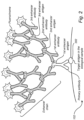
- it may in accordance to the invention be possible to form an enhancer chain for "amplifying" the detection of a target antigen 202 in the biological sample 102.
- the enhancer chain may comprise a plurality of enhancer steps, i.e. where the first enhancer antibody has been provided (conjugated) with a second enhancer antigen selected and formed in a similar process as discussed above, as well as again taking into account the criteria set for the selection of antigen.
- the enhancer chain could thus be arranged to include an in essence unlimited number of enhancer steps, e.g. second, third, fourth, etc., enhancer antibody/antigen forming an expanding "three structure".
- the last enhancer antibody in the chain (in Fig. 2 being the third enhancer antibody) with a reporter molecule, such as for example a fluorochrome.
- reporter molecules such as for example a fluorochrome.
- Other reporter molecules are possible, including for example an enzyme, a peptide, quantum dots, or a transition metal.
- Providing an antibody with a reporter molecule such as a fluorochrome is process known to the skilled addressee.
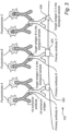
- Fig. 3 where the biological sample 102 has been prepared in accordance to a multi-immunolabeling process, where a first 302, a second 304 and a third 306 target antigen is to be subsequently detected/analyzed.
- a primary antibody is selected for each of the target antigens 302, 304, 306, in Fig. 3 denoted as primary antibodies A, B and C.
- a first enhancer antigen 1A is formed and provided with the primary antibody A
- a first enhancer antigen 1B is formed and provided for the primary antibody B, etc.
- corresponding first enhancer antibodies are generated for each of the first enhancer antigens 1A, 1B, 1C.
- Each of the first enhancer antibodies are provided with a different reporter molecule, such as with different fluorochromes generating lighting within different wavelength ranges, thus making detection and analysis of each of the target antigens 302, 304, 306 possible. It would of course be possible, and within the scope of the invention, to form enhancer chains for each of the target antibodies 302, 304, 306, in a similar manner as shown in Fig. 2 . Also, the concept discussed above e.g. in relation to Figs. 2 and 3 could of course be combined with known multi-immunolabeling processes, e.g. where the reporter molecules are on one or a plurality of primary antibodies, secondary antibodies or streptavidin (i.e.
Landscapes
- Health & Medical Sciences (AREA)
- Life Sciences & Earth Sciences (AREA)
- Immunology (AREA)
- Engineering & Computer Science (AREA)
- Chemical & Material Sciences (AREA)
- Molecular Biology (AREA)
- Biomedical Technology (AREA)
- Urology & Nephrology (AREA)
- Hematology (AREA)
- Biotechnology (AREA)
- Analytical Chemistry (AREA)
- Cell Biology (AREA)
- Pathology (AREA)
- Food Science & Technology (AREA)
- Medicinal Chemistry (AREA)
- Physics & Mathematics (AREA)
- Microbiology (AREA)
- Biochemistry (AREA)
- General Health & Medical Sciences (AREA)
- General Physics & Mathematics (AREA)
- Proteomics, Peptides & Aminoacids (AREA)
- Chemical Kinetics & Catalysis (AREA)
- Investigating Or Analysing Biological Materials (AREA)
- Peptides Or Proteins (AREA)
Claims (4)
- Procédé de préparation d'un échantillon biologique destiné à être utilisé dans un procédé d'immunomarquage, le procédé comprenant les étapes consistant à :- marquer l'échantillon biologique avec un composant de marquage, le composant de marquage étant fourni avec un premier antigène amplificateur et- fournir un premier anticorps amplificateur, le premier anticorps amplificateur étant choisi pour se lier seulement au premier antigène amplificateur,- le premier antigène amplificateur n'étant pas présent dans le procédé d'immunomarquage,- le composant de marquage étant un anticorps primaire conjugué avec le premier antigène amplificateur et- le premier anticorps amplificateur étant conjugué avec un second antigène amplificateur, le second antigène amplificateur n'étant pas présent dans le procédé d'immunomarquage et différent du premier antigène amplificateur,- les antigènes amplificateurs étant formés à partir de séquences peptidiques formulées artificiellement non présentes dans une quelconque protéine dans la nature,le procédé comprenant en outre :- la fourniture d'un second anticorps amplificateur, le second anticorps amplificateur étant choisi pour se lier seulement au second antigène amplificateur.
- Procédé selon la revendication 1, dans lequel le second anticorps amplificateur est marqué avec une molécule rapporteur ou conjugué avec un troisième antigène amplificateur, le troisième antigène amplificateur n'étant pas présent dans le procédé d'immunomarquage et différent du premier et du deuxième antigènes amplificateurs.
- Procédé selon la revendication 2, dans lequel la molécule rapporteur est choisie dans un groupe comprenant un fluorochrome, une enzyme, un peptide, des points quantiques et un métal de transition.
- Procédé selon la revendication 2, dans lequel la molécule rapporteur est un oligonucléotide.
Applications Claiming Priority (2)
| Application Number | Priority Date | Filing Date | Title |
|---|---|---|---|
| SE1550041A SE538541C2 (en) | 2015-01-19 | 2015-01-19 | Method for preparing a biological sample for use in an immunolabeling process |
| PCT/SE2016/050027 WO2016118065A1 (fr) | 2015-01-19 | 2016-01-18 | Procédé de préparation d'un échantillon biologique destiné à être utilisé dans un procédé d'immunomarquage |
Publications (4)
| Publication Number | Publication Date |
|---|---|
| EP3248006A1 EP3248006A1 (fr) | 2017-11-29 |
| EP3248006A4 EP3248006A4 (fr) | 2018-06-06 |
| EP3248006B1 true EP3248006B1 (fr) | 2024-10-23 |
| EP3248006C0 EP3248006C0 (fr) | 2024-10-23 |
Family
ID=56417465
Family Applications (1)
| Application Number | Title | Priority Date | Filing Date |
|---|---|---|---|
| EP16740471.4A Active EP3248006B1 (fr) | 2015-01-19 | 2016-01-18 | Procédé de préparation d'un échantillon biologique destiné à être utilisé dans un procédé d'immunomarquage |
Country Status (4)
| Country | Link |
|---|---|
| US (1) | US11913961B2 (fr) |
| EP (1) | EP3248006B1 (fr) |
| SE (1) | SE538541C2 (fr) |
| WO (1) | WO2016118065A1 (fr) |
Families Citing this family (6)
| Publication number | Priority date | Publication date | Assignee | Title |
|---|---|---|---|---|
| SE538541C2 (en) | 2015-01-19 | 2016-09-13 | Fogelstrand Per | Method for preparing a biological sample for use in an immunolabeling process |
| CN107209176B (zh) | 2015-01-21 | 2020-08-14 | 克罗姆尼贡公司 | 免疫标记复合物的形成方法及用途 |
| US11867696B2 (en) | 2015-02-06 | 2024-01-09 | Cell Idx, Inc. | Antigen-coupled immunoreagents |
| WO2017204729A1 (fr) * | 2016-05-25 | 2017-11-30 | Fogelstrand Per | Procédé de préparation d'un échantillon biologique destiné à être utilisé dans un procédé d'immunomarquage |
| US20220113314A1 (en) * | 2020-10-08 | 2022-04-14 | Korea Advanced Institute Of Science And Technology | Method of amplifying fluorescent signal through cyclic staining of target molecules via fluorophore-conjugated complementary antibodies |
| EP4555322B1 (fr) * | 2022-09-27 | 2025-10-22 | Pixelgen Technologies Ab | Procédé de fixation d'anticorps primaires à un échantillon biologique |
Citations (2)
| Publication number | Priority date | Publication date | Assignee | Title |
|---|---|---|---|---|
| GB2098730A (en) * | 1981-04-21 | 1982-11-24 | Welsh Nat School Med | Immunolocalisation |
| US20060286546A1 (en) * | 2003-08-25 | 2006-12-21 | Marc Ramael | Method and kit for the quantitative and/or qualitative detection of components in a sample |
Family Cites Families (18)
| Publication number | Priority date | Publication date | Assignee | Title |
|---|---|---|---|---|
| CA2013006A1 (fr) | 1989-03-24 | 1990-09-24 | Marc E. Key | Complexe de reactifs pour des dosages immunologiques |
| US6015662A (en) * | 1996-01-23 | 2000-01-18 | Abbott Laboratories | Reagents for use as calibrators and controls |
| WO2002085923A2 (fr) * | 2001-04-19 | 2002-10-31 | The Scripps Research Institute | Incorporation in vivo d'acides amines non naturels |
| US20030175828A1 (en) * | 2002-03-15 | 2003-09-18 | Lazar James G. | Signal amplification by Hybrid Capture |
| US8586708B2 (en) | 2005-10-28 | 2013-11-19 | Massachusetts Institute Of Technology | Monovalent streptavidin compositions |
| GB2435511A (en) * | 2006-02-23 | 2007-08-29 | Mologic Ltd | Protease detection |
| US20100285490A1 (en) * | 2006-12-29 | 2010-11-11 | Invitrogen Corporation | Detection apparatus |
| US20080286881A1 (en) | 2007-05-14 | 2008-11-20 | Apel William A | Compositions and methods for combining report antibodies |
| US8969024B2 (en) | 2007-08-28 | 2015-03-03 | Abbvie Biotechnology Ltd | Compositions and methods comprising binding proteins for adalimumab |
| JP5686098B2 (ja) | 2009-09-17 | 2015-03-18 | Jsr株式会社 | アビジンとビオチン誘導体の解離方法及び解離剤 |
| US20120214187A1 (en) * | 2009-11-02 | 2012-08-23 | Ffina Biolutions, Llc | Method for Enhancing the Sensitivity of Antibody Based Assays |
| JP5821198B2 (ja) | 2011-01-27 | 2015-11-24 | 富士レビオ株式会社 | 抗il28b抗体及びこれを用いたil28bの測定方法 |
| US9353161B2 (en) | 2011-09-13 | 2016-05-31 | Uti Limited Partnership | Streptavidin mutein exhibiting reversible binding for biotin and streptavidin binding peptide tagged proteins |
| SE538211C2 (sv) | 2013-04-05 | 2016-04-05 | Idl Biotech Ab | Metod för detektering av cytokeratin 8, 18 och/eller 19 och/eller lösliga fragment därav |
| US9753042B2 (en) | 2013-04-23 | 2017-09-05 | Rosalind Franklin University Of Medicine And Science | Kits for determining male fertility by measuring levels of a2V-ATPase, G-CSF, MIP 1 alpha, MCP-1, and methods and kits for improving reproductive outcomes in artificial insemination procedures |
| EP3136096B1 (fr) * | 2014-04-23 | 2021-12-01 | Nichirei Biosciences Inc. | Combinaison pour détection de marqueur cible |
| SE538541C2 (en) | 2015-01-19 | 2016-09-13 | Fogelstrand Per | Method for preparing a biological sample for use in an immunolabeling process |
| US11867696B2 (en) * | 2015-02-06 | 2024-01-09 | Cell Idx, Inc. | Antigen-coupled immunoreagents |
-
2015
- 2015-01-19 SE SE1550041A patent/SE538541C2/en not_active IP Right Cessation
-
2016
- 2016-01-18 US US15/543,465 patent/US11913961B2/en active Active
- 2016-01-18 WO PCT/SE2016/050027 patent/WO2016118065A1/fr not_active Ceased
- 2016-01-18 EP EP16740471.4A patent/EP3248006B1/fr active Active
Patent Citations (2)
| Publication number | Priority date | Publication date | Assignee | Title |
|---|---|---|---|---|
| GB2098730A (en) * | 1981-04-21 | 1982-11-24 | Welsh Nat School Med | Immunolocalisation |
| US20060286546A1 (en) * | 2003-08-25 | 2006-12-21 | Marc Ramael | Method and kit for the quantitative and/or qualitative detection of components in a sample |
Also Published As
| Publication number | Publication date |
|---|---|
| US11913961B2 (en) | 2024-02-27 |
| SE538541C2 (en) | 2016-09-13 |
| EP3248006A1 (fr) | 2017-11-29 |
| EP3248006C0 (fr) | 2024-10-23 |
| WO2016118065A1 (fr) | 2016-07-28 |
| EP3248006A4 (fr) | 2018-06-06 |
| US20180003718A1 (en) | 2018-01-04 |
| SE1550041A1 (sv) | 2016-07-20 |
Similar Documents
| Publication | Publication Date | Title |
|---|---|---|
| EP3248006B1 (fr) | Procédé de préparation d'un échantillon biologique destiné à être utilisé dans un procédé d'immunomarquage | |
| EP2297585B1 (fr) | Détection de l' usage de cannabis | |
| JP2022519641A (ja) | 生物学的試料の選択的標識による分析物検出 | |
| Warford et al. | Expression profiling by high-throughput immunohistochemistry | |
| ATE436022T1 (de) | Antikörperkomplexe sowie immunmarkierungsverfahren | |
| DK1232392T3 (da) | Forbedret fremgangsmåde til påvisning af syreresistente bakterier af slægten Helicobacter i afføring | |
| US20190112356A1 (en) | High-affinity immunopolymers | |
| Xing et al. | Development of a fluorescent immunoassay strip for the rapid quantitative detection of cadmium in rice | |
| US12596119B2 (en) | Analyte detection immunoassay | |
| KR20210118808A (ko) | 항 범종 특이적 말라리아 원충 젖산 탈수소효소의 항체 | |
| US6255060B1 (en) | Method of detecting protein by immuno RNA | |
| CN107209176B (zh) | 免疫标记复合物的形成方法及用途 | |
| Eyford et al. | Identification of trypanosome proteins in plasma from African sleeping sickness patients infected with T. b. rhodesiense | |
| Lundberg et al. | Creation of an antibody‐based subcellular protein atlas | |
| EP3465204B1 (fr) | Procédé de préparation d'un échantillon biologique destiné à être utilisé dans un procédé d'immunomarquage | |
| JP2020506395A5 (fr) | ||
| JP2002501176A (ja) | 蛋白質親和性リガンド同定へのマスフィンガープリンティングの利用 | |
| CN109313190A (zh) | 通过流体样品再利用而使流体样品多重化的设备、方法和试剂盒 | |
| Jordan | Enzyme-linked immunosorbent assay | |
| SE1550048A1 (sv) | Method for the formation and use of an immunolabeling complex | |
| US20090136966A1 (en) | Normalization of Complex Analyte Mixtures | |
| Coons | Introduction to Immunohistochemistry | |
| JPH06235727A (ja) | バイオ情報変換素子及びそれを用いた生物種検出方法 |
Legal Events
| Date | Code | Title | Description |
|---|---|---|---|
| STAA | Information on the status of an ep patent application or granted ep patent |
Free format text: STATUS: THE INTERNATIONAL PUBLICATION HAS BEEN MADE |
|
| PUAI | Public reference made under article 153(3) epc to a published international application that has entered the european phase |
Free format text: ORIGINAL CODE: 0009012 |
|
| STAA | Information on the status of an ep patent application or granted ep patent |
Free format text: STATUS: REQUEST FOR EXAMINATION WAS MADE |
|
| 17P | Request for examination filed |
Effective date: 20170704 |
|
| AK | Designated contracting states |
Kind code of ref document: A1 Designated state(s): AL AT BE BG CH CY CZ DE DK EE ES FI FR GB GR HR HU IE IS IT LI LT LU LV MC MK MT NL NO PL PT RO RS SE SI SK SM TR |
|
| AX | Request for extension of the european patent |
Extension state: BA ME |
|
| DAV | Request for validation of the european patent (deleted) | ||
| DAX | Request for extension of the european patent (deleted) | ||
| A4 | Supplementary search report drawn up and despatched |
Effective date: 20180507 |
|
| RIC1 | Information provided on ipc code assigned before grant |
Ipc: G01N 33/53 20060101AFI20180430BHEP |
|
| RAP1 | Party data changed (applicant data changed or rights of an application transferred) |
Owner name: KROMNIGON AB |
|
| STAA | Information on the status of an ep patent application or granted ep patent |
Free format text: STATUS: EXAMINATION IS IN PROGRESS |
|
| 17Q | First examination report despatched |
Effective date: 20191209 |
|
| GRAP | Despatch of communication of intention to grant a patent |
Free format text: ORIGINAL CODE: EPIDOSNIGR1 |
|
| STAA | Information on the status of an ep patent application or granted ep patent |
Free format text: STATUS: GRANT OF PATENT IS INTENDED |
|
| INTG | Intention to grant announced |
Effective date: 20240516 |
|
| GRAS | Grant fee paid |
Free format text: ORIGINAL CODE: EPIDOSNIGR3 |
|
| GRAA | (expected) grant |
Free format text: ORIGINAL CODE: 0009210 |
|
| STAA | Information on the status of an ep patent application or granted ep patent |
Free format text: STATUS: THE PATENT HAS BEEN GRANTED |
|
| AK | Designated contracting states |
Kind code of ref document: B1 Designated state(s): AL AT BE BG CH CY CZ DE DK EE ES FI FR GB GR HR HU IE IS IT LI LT LU LV MC MK MT NL NO PL PT RO RS SE SI SK SM TR |
|
| RAP3 | Party data changed (applicant data changed or rights of an application transferred) |
Owner name: KROMNIGON AB |
|
| REG | Reference to a national code |
Ref country code: GB Ref legal event code: FG4D |
|
| REG | Reference to a national code |
Ref country code: CH Ref legal event code: EP |
|
| REG | Reference to a national code |
Ref country code: DE Ref legal event code: R096 Ref document number: 602016089936 Country of ref document: DE |
|
| REG | Reference to a national code |
Ref country code: IE Ref legal event code: FG4D |
|
| U01 | Request for unitary effect filed |
Effective date: 20241119 |
|
| U07 | Unitary effect registered |
Designated state(s): AT BE BG DE DK EE FI FR IT LT LU LV MT NL PT RO SE SI Effective date: 20241125 |
|
| U20 | Renewal fee for the european patent with unitary effect paid |
Year of fee payment: 10 Effective date: 20250218 |
|
| PG25 | Lapsed in a contracting state [announced via postgrant information from national office to epo] |
Ref country code: IS Free format text: LAPSE BECAUSE OF FAILURE TO SUBMIT A TRANSLATION OF THE DESCRIPTION OR TO PAY THE FEE WITHIN THE PRESCRIBED TIME-LIMIT Effective date: 20250223 Ref country code: HR Free format text: LAPSE BECAUSE OF FAILURE TO SUBMIT A TRANSLATION OF THE DESCRIPTION OR TO PAY THE FEE WITHIN THE PRESCRIBED TIME-LIMIT Effective date: 20241023 |
|
| PG25 | Lapsed in a contracting state [announced via postgrant information from national office to epo] |
Ref country code: ES Free format text: LAPSE BECAUSE OF FAILURE TO SUBMIT A TRANSLATION OF THE DESCRIPTION OR TO PAY THE FEE WITHIN THE PRESCRIBED TIME-LIMIT Effective date: 20241023 |
|
| PG25 | Lapsed in a contracting state [announced via postgrant information from national office to epo] |
Ref country code: NO Free format text: LAPSE BECAUSE OF FAILURE TO SUBMIT A TRANSLATION OF THE DESCRIPTION OR TO PAY THE FEE WITHIN THE PRESCRIBED TIME-LIMIT Effective date: 20250123 |
|
| PG25 | Lapsed in a contracting state [announced via postgrant information from national office to epo] |
Ref country code: GR Free format text: LAPSE BECAUSE OF FAILURE TO SUBMIT A TRANSLATION OF THE DESCRIPTION OR TO PAY THE FEE WITHIN THE PRESCRIBED TIME-LIMIT Effective date: 20250124 |
|
| PG25 | Lapsed in a contracting state [announced via postgrant information from national office to epo] |
Ref country code: PL Free format text: LAPSE BECAUSE OF FAILURE TO SUBMIT A TRANSLATION OF THE DESCRIPTION OR TO PAY THE FEE WITHIN THE PRESCRIBED TIME-LIMIT Effective date: 20241023 |
|
| PG25 | Lapsed in a contracting state [announced via postgrant information from national office to epo] |
Ref country code: RS Free format text: LAPSE BECAUSE OF FAILURE TO SUBMIT A TRANSLATION OF THE DESCRIPTION OR TO PAY THE FEE WITHIN THE PRESCRIBED TIME-LIMIT Effective date: 20250123 |
|
| PG25 | Lapsed in a contracting state [announced via postgrant information from national office to epo] |
Ref country code: SM Free format text: LAPSE BECAUSE OF FAILURE TO SUBMIT A TRANSLATION OF THE DESCRIPTION OR TO PAY THE FEE WITHIN THE PRESCRIBED TIME-LIMIT Effective date: 20241023 |
|
| PG25 | Lapsed in a contracting state [announced via postgrant information from national office to epo] |
Ref country code: SK Free format text: LAPSE BECAUSE OF FAILURE TO SUBMIT A TRANSLATION OF THE DESCRIPTION OR TO PAY THE FEE WITHIN THE PRESCRIBED TIME-LIMIT Effective date: 20241023 |
|
| PG25 | Lapsed in a contracting state [announced via postgrant information from national office to epo] |
Ref country code: CZ Free format text: LAPSE BECAUSE OF FAILURE TO SUBMIT A TRANSLATION OF THE DESCRIPTION OR TO PAY THE FEE WITHIN THE PRESCRIBED TIME-LIMIT Effective date: 20241023 |
|
| PLBE | No opposition filed within time limit |
Free format text: ORIGINAL CODE: 0009261 |
|
| REG | Reference to a national code |
Ref country code: CH Ref legal event code: PL |
|
| STAA | Information on the status of an ep patent application or granted ep patent |
Free format text: STATUS: NO OPPOSITION FILED WITHIN TIME LIMIT |
|
| PG25 | Lapsed in a contracting state [announced via postgrant information from national office to epo] |
Ref country code: MC Free format text: LAPSE BECAUSE OF FAILURE TO SUBMIT A TRANSLATION OF THE DESCRIPTION OR TO PAY THE FEE WITHIN THE PRESCRIBED TIME-LIMIT Effective date: 20241023 |
|
| GBPC | Gb: european patent ceased through non-payment of renewal fee |
Effective date: 20250123 |
|
| 26N | No opposition filed |
Effective date: 20250724 |
|
| PG25 | Lapsed in a contracting state [announced via postgrant information from national office to epo] |
Ref country code: GB Free format text: LAPSE BECAUSE OF NON-PAYMENT OF DUE FEES Effective date: 20250123 |
|
| PG25 | Lapsed in a contracting state [announced via postgrant information from national office to epo] |
Ref country code: CH Free format text: LAPSE BECAUSE OF NON-PAYMENT OF DUE FEES Effective date: 20250131 |
|
| PG25 | Lapsed in a contracting state [announced via postgrant information from national office to epo] |
Ref country code: IE Free format text: LAPSE BECAUSE OF NON-PAYMENT OF DUE FEES Effective date: 20250118 |
|
| U20 | Renewal fee for the european patent with unitary effect paid |
Year of fee payment: 11 Effective date: 20260116 |