EP3251638A2 - Dispositif de planification pour un dispositif de traitement pour la chirurgie ophtalmologique - Google Patents
Dispositif de planification pour un dispositif de traitement pour la chirurgie ophtalmologique Download PDFInfo
- Publication number
- EP3251638A2 EP3251638A2 EP17173237.3A EP17173237A EP3251638A2 EP 3251638 A2 EP3251638 A2 EP 3251638A2 EP 17173237 A EP17173237 A EP 17173237A EP 3251638 A2 EP3251638 A2 EP 3251638A2
- Authority
- EP
- European Patent Office
- Prior art keywords
- corneal
- cornea
- cut surface
- data
- laser
- Prior art date
- Legal status (The legal status is an assumption and is not a legal conclusion. Google has not performed a legal analysis and makes no representation as to the accuracy of the status listed.)
- Granted
Links
Images


Classifications
-
- A—HUMAN NECESSITIES
- A61—MEDICAL OR VETERINARY SCIENCE; HYGIENE
- A61F—FILTERS IMPLANTABLE INTO BLOOD VESSELS; PROSTHESES; DEVICES PROVIDING PATENCY TO, OR PREVENTING COLLAPSING OF, TUBULAR STRUCTURES OF THE BODY, e.g. STENTS; ORTHOPAEDIC, NURSING OR CONTRACEPTIVE DEVICES; FOMENTATION; TREATMENT OR PROTECTION OF EYES OR EARS; BANDAGES, DRESSINGS OR ABSORBENT PADS; FIRST-AID KITS
- A61F9/00—Methods or devices for treatment of the eyes; Devices for putting in contact-lenses; Devices to correct squinting; Apparatus to guide the blind; Protective devices for the eyes, carried on the body or in the hand
- A61F9/007—Methods or devices for eye surgery
- A61F9/008—Methods or devices for eye surgery using laser
- A61F9/00825—Methods or devices for eye surgery using laser for photodisruption
- A61F9/00827—Refractive correction, e.g. lenticle
-
- A—HUMAN NECESSITIES
- A61—MEDICAL OR VETERINARY SCIENCE; HYGIENE
- A61F—FILTERS IMPLANTABLE INTO BLOOD VESSELS; PROSTHESES; DEVICES PROVIDING PATENCY TO, OR PREVENTING COLLAPSING OF, TUBULAR STRUCTURES OF THE BODY, e.g. STENTS; ORTHOPAEDIC, NURSING OR CONTRACEPTIVE DEVICES; FOMENTATION; TREATMENT OR PROTECTION OF EYES OR EARS; BANDAGES, DRESSINGS OR ABSORBENT PADS; FIRST-AID KITS
- A61F9/00—Methods or devices for treatment of the eyes; Devices for putting in contact-lenses; Devices to correct squinting; Apparatus to guide the blind; Protective devices for the eyes, carried on the body or in the hand
- A61F9/007—Methods or devices for eye surgery
- A61F9/008—Methods or devices for eye surgery using laser
- A61F9/00802—Methods or devices for eye surgery using laser for photoablation
- A61F9/00804—Refractive treatments
-
- A—HUMAN NECESSITIES
- A61—MEDICAL OR VETERINARY SCIENCE; HYGIENE
- A61B—DIAGNOSIS; SURGERY; IDENTIFICATION
- A61B18/00—Surgical instruments, devices or methods for transferring non-mechanical forms of energy to or from the body
- A61B18/18—Surgical instruments, devices or methods for transferring non-mechanical forms of energy to or from the body by applying electromagnetic radiation, e.g. microwaves
- A61B18/20—Surgical instruments, devices or methods for transferring non-mechanical forms of energy to or from the body by applying electromagnetic radiation, e.g. microwaves using laser
-
- A—HUMAN NECESSITIES
- A61—MEDICAL OR VETERINARY SCIENCE; HYGIENE
- A61F—FILTERS IMPLANTABLE INTO BLOOD VESSELS; PROSTHESES; DEVICES PROVIDING PATENCY TO, OR PREVENTING COLLAPSING OF, TUBULAR STRUCTURES OF THE BODY, e.g. STENTS; ORTHOPAEDIC, NURSING OR CONTRACEPTIVE DEVICES; FOMENTATION; TREATMENT OR PROTECTION OF EYES OR EARS; BANDAGES, DRESSINGS OR ABSORBENT PADS; FIRST-AID KITS
- A61F9/00—Methods or devices for treatment of the eyes; Devices for putting in contact-lenses; Devices to correct squinting; Apparatus to guide the blind; Protective devices for the eyes, carried on the body or in the hand
- A61F9/007—Methods or devices for eye surgery
- A61F9/00736—Instruments for removal of intra-ocular material or intra-ocular injection, e.g. cataract instruments
-
- A—HUMAN NECESSITIES
- A61—MEDICAL OR VETERINARY SCIENCE; HYGIENE
- A61F—FILTERS IMPLANTABLE INTO BLOOD VESSELS; PROSTHESES; DEVICES PROVIDING PATENCY TO, OR PREVENTING COLLAPSING OF, TUBULAR STRUCTURES OF THE BODY, e.g. STENTS; ORTHOPAEDIC, NURSING OR CONTRACEPTIVE DEVICES; FOMENTATION; TREATMENT OR PROTECTION OF EYES OR EARS; BANDAGES, DRESSINGS OR ABSORBENT PADS; FIRST-AID KITS
- A61F9/00—Methods or devices for treatment of the eyes; Devices for putting in contact-lenses; Devices to correct squinting; Apparatus to guide the blind; Protective devices for the eyes, carried on the body or in the hand
- A61F9/007—Methods or devices for eye surgery
- A61F9/008—Methods or devices for eye surgery using laser
-
- A—HUMAN NECESSITIES
- A61—MEDICAL OR VETERINARY SCIENCE; HYGIENE
- A61F—FILTERS IMPLANTABLE INTO BLOOD VESSELS; PROSTHESES; DEVICES PROVIDING PATENCY TO, OR PREVENTING COLLAPSING OF, TUBULAR STRUCTURES OF THE BODY, e.g. STENTS; ORTHOPAEDIC, NURSING OR CONTRACEPTIVE DEVICES; FOMENTATION; TREATMENT OR PROTECTION OF EYES OR EARS; BANDAGES, DRESSINGS OR ABSORBENT PADS; FIRST-AID KITS
- A61F9/00—Methods or devices for treatment of the eyes; Devices for putting in contact-lenses; Devices to correct squinting; Apparatus to guide the blind; Protective devices for the eyes, carried on the body or in the hand
- A61F9/007—Methods or devices for eye surgery
- A61F9/013—Instruments for compensation of ocular refraction ; Instruments for use in cornea removal, for reshaping or performing incisions in the cornea
-
- A—HUMAN NECESSITIES
- A61—MEDICAL OR VETERINARY SCIENCE; HYGIENE
- A61F—FILTERS IMPLANTABLE INTO BLOOD VESSELS; PROSTHESES; DEVICES PROVIDING PATENCY TO, OR PREVENTING COLLAPSING OF, TUBULAR STRUCTURES OF THE BODY, e.g. STENTS; ORTHOPAEDIC, NURSING OR CONTRACEPTIVE DEVICES; FOMENTATION; TREATMENT OR PROTECTION OF EYES OR EARS; BANDAGES, DRESSINGS OR ABSORBENT PADS; FIRST-AID KITS
- A61F9/00—Methods or devices for treatment of the eyes; Devices for putting in contact-lenses; Devices to correct squinting; Apparatus to guide the blind; Protective devices for the eyes, carried on the body or in the hand
- A61F9/007—Methods or devices for eye surgery
- A61F9/008—Methods or devices for eye surgery using laser
- A61F2009/00861—Methods or devices for eye surgery using laser adapted for treatment at a particular location
- A61F2009/00872—Cornea
-
- A—HUMAN NECESSITIES
- A61—MEDICAL OR VETERINARY SCIENCE; HYGIENE
- A61F—FILTERS IMPLANTABLE INTO BLOOD VESSELS; PROSTHESES; DEVICES PROVIDING PATENCY TO, OR PREVENTING COLLAPSING OF, TUBULAR STRUCTURES OF THE BODY, e.g. STENTS; ORTHOPAEDIC, NURSING OR CONTRACEPTIVE DEVICES; FOMENTATION; TREATMENT OR PROTECTION OF EYES OR EARS; BANDAGES, DRESSINGS OR ABSORBENT PADS; FIRST-AID KITS
- A61F9/00—Methods or devices for treatment of the eyes; Devices for putting in contact-lenses; Devices to correct squinting; Apparatus to guide the blind; Protective devices for the eyes, carried on the body or in the hand
- A61F9/007—Methods or devices for eye surgery
- A61F9/008—Methods or devices for eye surgery using laser
- A61F2009/00878—Planning
- A61F2009/0088—Planning based on wavefront
-
- A—HUMAN NECESSITIES
- A61—MEDICAL OR VETERINARY SCIENCE; HYGIENE
- A61F—FILTERS IMPLANTABLE INTO BLOOD VESSELS; PROSTHESES; DEVICES PROVIDING PATENCY TO, OR PREVENTING COLLAPSING OF, TUBULAR STRUCTURES OF THE BODY, e.g. STENTS; ORTHOPAEDIC, NURSING OR CONTRACEPTIVE DEVICES; FOMENTATION; TREATMENT OR PROTECTION OF EYES OR EARS; BANDAGES, DRESSINGS OR ABSORBENT PADS; FIRST-AID KITS
- A61F9/00—Methods or devices for treatment of the eyes; Devices for putting in contact-lenses; Devices to correct squinting; Apparatus to guide the blind; Protective devices for the eyes, carried on the body or in the hand
- A61F9/007—Methods or devices for eye surgery
- A61F9/008—Methods or devices for eye surgery using laser
- A61F2009/00878—Planning
- A61F2009/00882—Planning based on topography
-
- A—HUMAN NECESSITIES
- A61—MEDICAL OR VETERINARY SCIENCE; HYGIENE
- A61F—FILTERS IMPLANTABLE INTO BLOOD VESSELS; PROSTHESES; DEVICES PROVIDING PATENCY TO, OR PREVENTING COLLAPSING OF, TUBULAR STRUCTURES OF THE BODY, e.g. STENTS; ORTHOPAEDIC, NURSING OR CONTRACEPTIVE DEVICES; FOMENTATION; TREATMENT OR PROTECTION OF EYES OR EARS; BANDAGES, DRESSINGS OR ABSORBENT PADS; FIRST-AID KITS
- A61F9/00—Methods or devices for treatment of the eyes; Devices for putting in contact-lenses; Devices to correct squinting; Apparatus to guide the blind; Protective devices for the eyes, carried on the body or in the hand
- A61F9/007—Methods or devices for eye surgery
- A61F9/008—Methods or devices for eye surgery using laser
- A61F9/00825—Methods or devices for eye surgery using laser for photodisruption
- A61F9/00836—Flap cutting
-
- A—HUMAN NECESSITIES
- A61—MEDICAL OR VETERINARY SCIENCE; HYGIENE
- A61K—PREPARATIONS FOR MEDICAL, DENTAL OR TOILETRY PURPOSES
- A61K9/00—Medicinal preparations characterised by special physical form
- A61K9/0012—Galenical forms characterised by the site of application
- A61K9/0048—Eye, e.g. artificial tears
- A61K9/0051—Ocular inserts or implants
Definitions
- the invention relates to a planning device for generating control data for a treatment device for refractive correcting eye surgery, which separates by means of a laser device through at least one cutting surface in the cornea to be removed for correction corneal volume of the surrounding cornea.
- the invention further relates to a treatment device for refractive correcting eye surgery, having a planning device of the type mentioned.
- the invention further relates to a method for generating control data for a treatment device for refractive correcting eye surgery, which separates by means of a laser device through at least one cut surface in the cornea to be removed for correction corneal volume of the surrounding cornea.
- the invention also relates to a method for refractive correcting eye surgery, wherein a cornea volume to be removed for correction is separated from the surrounding cornea by means of a treatment device with a laser device through at least one cut surface in the cornea.
- a variety of treatment methods with the aim of refraction correction in the human eye are known.
- the aim of the surgical methods is to modify the cornea in a targeted manner in order to influence the refraction of light.
- several surgical methods are used.
- the most widespread currently is the so-called laser in situ keratomileusis, which is also abbreviated LASIK.
- LASIK laser in situ keratomileusis
- a corneal flap is unilaterally detached from the corneal surface and folded to the side.
- the dissolution of this lamella can be effected by means of a mechanical microkeratome, or else by means of a so-called laser keratome, as it is marketed, for example, by Intralase Corp., Irvine, USA.
- the LASIK operation involves the use of an excimer laser, which ablates the corneal tissue thus exposed beneath the lamella. After vaporizing beneath the corneal surface in this manner, the corneal flap is folded back to its original location.
- the use of a laser keratome to expose the blade is advantageous over a mechanical knife, since the risk of infection is reduced and at the same time the quality of cut is improved.
- the blade can be made with a much more constant thickness when laser radiation is used.
- the cut is usually smoother, which reduces the risk of subsequent optical interference by this remaining after the operation interface.
- a disadvantage of this method is that two different treatment devices must be used, on the one hand, the laser keratome to expose the lamella and on the other hand, the corneal tissue evaporating laser.
- FLEX Carl Zeiss Meditec AG
- a cut geometry is formed by means of a femtosecond laser in the cornea, which separates a corneal volume (so-called lenticle) in the cornea. This is then manually removed by the surgeon.
- the advantage of this method is, on the one hand, that the cut quality is further improved by the use of the femtosecond laser. On the other hand, only one treatment device is required; the excimer laser is no longer used.
- the need for after-treatment may arise if either the result of the previous refractive correction procedure is still unsatisfactory, or if for some reason the previous procedure could not be completed adequately (eg due to abortion of the procedure).
- the invention is therefore based on the object of specifying a planning device for generating control data, a treatment device for refraction correcting eye surgery and a method for generating control data for such a treatment device or a method for refractive correcting eye surgery, in which simply a post-treatment without ablation of Corneal tissue or the continuation of a terminated treatment is possible.
- the invention is defined in claim 1.
- a planning device of the type mentioned above which has an interface for supplying corneal data, which contain information on preoperative sections, which were generated in a previous ophthalmological surgery, and having calculation means for defining a corneal cut surface, the delimits the cornea volume to be taken, the calculation means determining the corneal cut surface based on the corneal data, and generating a control data set for driving the laser device for the corneal cut surface.
- a treatment apparatus for refractive correcting eye surgery which has an interface for supplying corneal data containing information on preoperative sections, which were generated in a previous ophthalmological surgery, having a laser device which by means of laser radiation according to control data at least one cut surface in the cornea separates a cornea volume to be removed from the surrounding cornea, and has a planning device of the type just mentioned for generating the control data.
- control data comprises: accessing corneal data containing information on preoperative incisions generated in a previous ophthalmological surgical procedure, defining a cornea incision surface, which limits the corneal volume to be extracted on the basis of the corneal data, and generating a control data record for the corneal cut surface for controlling the laser device.
- This concept provides a post-treatment with a device known for the FLEX method, since the inventors recognized that such a device can be applied surprisingly easily to cases in which cuts have already been made into the cornea before the operation.
- the invention therefore quite fundamentally provides that at least one further cutting surface is produced in the cornea, which isolates a cornea volume whose removal causes the desired refractive correction.
- This corneal volume is also referred to in the prior art as a lenticle, since it is usually lens-shaped.
- Aftertreatment is advantageously carried out in such a way that the corneal cut surface defined by the planning device or in the planning method or the cornea cut surface produced in the treatment device or during the treatment process does not cut the preoperative cuts.
- This has the advantage of not undesirably isolating volumes in the cornea which may be removed from the cornea with the removal of the intended volume and leading to an unpredictable change in the corneal surface.
- Next is also avoided that when folding the isolated during the treatment slat this undesirable weakened or unintentionally fold down further parts of the cornea due to a not sufficiently considered preoperative cut in a non-predetermined manner.
- Such negative events during the post-treatment can be avoided particularly reliably if the calculation means of the planning device or the appropriate scheduling techniques set the corneal incision surface so that the corneal volume to be removed is completely posterior to the pre-operative cuts produced, or lies completely anterior to the preoperative incisions, or includes the preoperative incisions produced.
- the first or second variant is particularly suitable for cases in which the previous procedure went according to plan, but still a residual defective vision has to be corrected.
- the third variant lends itself to cases in which the previous procedure - for whatever reason - was stopped, since the then remaining, possibly with regard to their location not to specify with final accuracy, preoperative sections with the removal of the isolated in the follow-up cornea Volume removed from the cornea.
- the corneal incision surface can be defined as a continuation of the existing preoperative incisions.
- the planning of the corneal incision surface naturally assumes particular importance, since the existing preoperative incisions have to be taken into account. This is made easier for the surgeon if a display device is provided for the visual display of the cornea and the existing preoperative sections, preferably a superimposed display.
- corneal data may be generated from a survey of the eye and delivered to the scheduler using a measuring device optionally having one or more of the following: autorefractor, refractometer, keratometer, aberrometer, wavefront surveyor, OCT, confocal corneal microscopy, pseudo-panning camera , Topograhpieflop.
- a particularly important indication is, of course, the defective vision to be corrected and / or an indication of the thickness and / or diameter of a pre-operatively produced, foldable corneal lamella.
- a laser surgical treatment device for refractive eye surgery has a device which records the progress of the sections produced during an intervention.
- the logging includes location and energy of each laser beam pulse focused in the cornea.
- the relative position of the cornea (or eye) and device is also logged.
- Such logging is not known in the prior art and also goes far beyond the usual level, which includes information about the patient, the refractive correction requirement and, at best, the cut surface geometry used.
- the considerable data storage effort proves to be advantageous if post-treatment is required, in particular if the previous intervention was not completed. Then, a continuation of the aborted sectional area generation is easily and precisely possible.
- the invention provides for aftertreatment that the defined or used cut surfaces are geometrically arranged so that either no cut at all with the pre-operative, already existing cut is given, or that this cut is supplemented or continued suitably.
- the further cuts may be arranged to lie at a lesser depth below the corneal surface than the preoperative incision which produces a flap of the corneal flap has been.
- the now to be taken lenticule is then removed from a corneal area, which lies below the preoperatively generated corneal lamella.
- the lenticle was in the corneal lamella.
- the pre-surgical cut already in place and to set the corneal cut surface to make that cut when isolating the cornea supplemented or used for post-treatment corneal volume to be taken.
- the lenticule is then delimited by the preoperative incision and the specified corneal cut surface. This achieves faster cutting surface production, but requires more precise knowledge of the pre-operative cut.
- the supplementation may be made above the preoperative incision, ie, through the corneal lamella, or below the preoperative incision, ie toward the corneal interior surface.
- the continuation may also involve the cut surface partially containing the pre-operative cut already created, i. is begun with the generation of the cut surface in the post-treatment in a range in which a pre-operative cut is already expected.
- An overlap ensures a continuous tissue separation in the interaction of pre-operative cut and cut surface produced in the aftertreatment. If one does not wish to continue the preoperative incisions, for example because their position is not sufficiently precise or because the quality of these incisions is unsatisfactory, it is expedient to fix the incision surface for isolating the cornea volume to be taken so that it is either consistently anterior to the preoperative Sections are consistently formed posterior to the preoperative incisions or pre-surgical incisions in the cornea volume to be extracted.
- the entprechende trajectory can be s ( t ) together with the associated set of disjoint intervals I j .
- a partial sectional area S j is completed when: ⁇ k ⁇ I j
- the time can be detected with a certain blurring, so for example at about 1% of T.
- the detection of the demolition time brings a simplification and increase the security of the logging. It is therefore advantageous to simplify the logging in such a way that not the position of each laser radiation pulse emitted into the cornea is logged, but only parameters of the laser radiation pulse output (eg frequency of the pulses), the deflection of the focus (eg deflection speed) and the exact Time indication of a possible interruption of the intervention and sectional geometry information.
- parameters of the laser radiation pulse output eg frequency of the pulses
- the deflection of the focus eg deflection speed
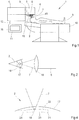
- a treatment device for the ophthalmic refractive correction is in Fig. 1 represented and provided with the general reference numeral 1.
- the treatment device 1 is designed for a post-treatment refraction correction on an eye 2 of a patient 3.
- the treatment device 1 has a laser device 4 which emits a laser beam 6 from a laser source 5, which is directed as a focused beam 7 into the eye 2 or the cornea of the eye.
- the laser beam 6 is a pulsed Laser beam with a wavelength between 400 nanometers and 10 micrometers.
- the pulse length of the laser beam 6 is in the range between 1 femtosecond and 10 picoseconds, with pulse repetition rates of 1 to 1000 kilohertz and pulse energies between 0.01 microjoule and 0.01 millijoule are possible.
- the treatment device 1 thus generates a cut surface in the cornea of the eye 2 by deflecting the pulsed laser radiation.
- a scanner 8 and a radiation intensity modulator 9 are provided in the laser device 4 or its laser source 5.
- the patient 3 is located on a couch 10, which is adjustable in three directions in space to align the eye 2 suitable for the incidence of the laser beam 6.
- the lounger 10 is adjustable by motor.
- the control can in particular be effected by a control unit 11, which basically controls the operation of the treatment apparatus 1 and is connected to the treatment apparatus via suitable data connections, for example connecting lines 12. Of course, this communication can also be done by other means, such as fiber optics or by radio.
- the control unit 11 performs the corresponding settings, time control on the treatment device 1, in particular of the laser device 4, and thus accomplishes corresponding functions of the treatment device 1.
- the treatment apparatus 1 further has a fixing device 15, which fixes the cornea of the eye 2 relative to the laser device 4.
- This fixing device 15 may comprise a known contact glass 45, to which the cornea is applied by negative pressure and which gives the cornea a desired geometric shape.
- Such contact lenses are known to those skilled in the art, for example from the DE 102005040338 A1 , The disclosure of this document is, insofar as the description of a design of the possible for the treatment device 1 contact glass 45 is concerned, fully incorporated herein.
- the control device 11 of the treatment device 1 further has a planning device 16, which will be explained in more detail later.
- Fig. 2 schematically shows the operation of the incident laser beam 6.
- the laser beam 6 is focused and falls as the focused laser beam 7 in the cornea 17 of the eye 2.
- a schematically drawn optics 18 is provided for focusing. It causes a focus in the cornea 17, in which the laser radiation energy density is so high that in combination with the pulse length of the pulsed laser radiation 6, a non-linear effect in the cornea 17 occurs.
- each pulse of the pulsed laser radiation 6 in focus 19 produce an optical breakthrough in the cornea 17, which in turn produces an in Fig. 2 only schematically indicated plasma bubble initiated.
- the tissue layer separation comprises a larger area than the focus 19, although the conditions for generating the optical breakthrough are achieved only in the focus 19.
- a tissue-separating effect can also be achieved by pulsed laser radiation by emitting a plurality of laser radiation pulses in one area, with the focus spots overlapping. It then act together several laser radiation pulses to achieve a tissue-separating effect.
- the type of tissue separation used by the treatment device 1 is not relevant to the following description; It is only essential that a cut surface generation takes place in the cornea 17 of the eye 2.
- a corneal volume is removed by means of the laser radiation 6 from an area within the cornea 17, where tissue layers are separated there, which isolate the corneal volume and then allow its removal.
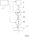
- the position of the focus 17 of the focused laser radiation 7 in the cornea 17 is adjusted, for example, in the case of pulsed laser radiation. This is schematically in Fig. 3 shown.
- the refractive properties of the cornea 17 are selectively changed by the removal of the volume, so as to achieve the refraction correction.
- the volume is therefore usually lenticular and is referred to as a lenticle.
- the removal of the corneal volume is done here as a post-treatment.
- Fig. 3 the elements of the treatment device 1 are registered only insofar as they are necessary for understanding the production of cut surfaces.
- the laser beam 6 is, as already mentioned, concentrated in a focus 19 in the cornea 17, and the position of the focus 19 in the cornea is adjusted so that focused energy from laser radiation pulses is introduced into the tissue of the cornea 17 for cutting surface generation at various points ,
- the laser radiation 6 is preferably provided by the laser source 5 as pulsed radiation.
- the scanner 8 is in the construction of the Fig. 3 constructed in two parts and consists of an xy scanner 8a, which in a variant by two substantially orthogonal deflecting Galvanometer mirror is realized.
- the scanner 8a deflects the coming of the laser source 5 laser beam 6 two-dimensionally, so that after the scanner 8, a deflected laser beam 20 is present.
- the scanner 8a thus effects an adjustment of the position of the focus 19 substantially perpendicular to the main direction of incidence of the laser beam 6 in the cornea 17.
- a z-scanner 8b is provided, for example as adjustable telescope is formed.
- the z-scanner 8b ensures that the z-position of the position of the focus 19, ie its position on the optical axis of the incidence is changed.
- the z-scanner 8b may be downstream of the xy-scanner 8a.
- the assignment of the individual coordinates to the spatial directions is not essential, just as little that the scanner 8a deflects about mutually perpendicular axes. Rather, any scanner can be used which is able to adjust the focus 19 in a plane in which the axis of incidence of the optical radiation is not. Further, any non-Cartesian coordinate systems for the deflection or control of the position of the focus 19 can be used. Examples of this are spherical coordinates or cylindrical coordinates.
- the control unit 11 ensures a suitable operation of the laser source 5 and the three-dimensional focus adjustment described here by way of example, so that ultimately a cut surface is formed which isolates a specific corneal volume which is to be removed for refractive correction.
- the control device 11 operates according to predetermined control data which, for example, are predetermined as target points for the focus adjustment in the case of the laser device 4 described here merely by way of example.
- the control data is usually summarized in a control data record. This makes geometrical specifications for the cut surface to be formed, for example the coordinates of the target points as a pattern.
- the control data set then also contains in this embodiment specific setting values for the focus position adjustment mechanism, e.g. for the scanner 8.
- FIG Fig. 4 The production of the cut surface with the treatment device 1 is exemplary in FIG Fig. 4 shown.
- a corneal volume 21 in the cornea 17 is isolated by adjusting the focus 19 in which the focused beam 7 is focused.
- cut surfaces are formed, which are here exemplarily designed as anterior flap cut surface 22 and as a posterior lenticule cut surface 23. These terms are to be understood as exemplary only and are intended to make reference to the conventional Lasik or Flex process for which the Treatment device 1, as already described, is formed. It is only essential here that the cut surfaces 22 and 23 as well as edge cuts, which further define the cut surfaces 22 and 23 at their edges, isolate the cornea volume 21.
- a corneal flap which delimits the corneal volume 21 anteriorly can also be folded down, so that the corneal volume 21 can be removed.
- Fig. 5 schematically shows the treatment device 1, and based on it, the importance of the planning device 16 will be explained in more detail.
- the treatment device 1 has at least two devices or modules in this variant.
- the already described laser device 4 emits the laser beam 6 on the eye 2.
- the operation of the laser device 4 takes place, as already described, fully automatically by the controller 11, ie the laser device 4 starts in response to a corresponding start signal to the generation and deflection of the laser beam 6 and thereby generates cut surfaces, which are constructed in the manner described to remove the corneal volume 21.
- the control signals required for the operation are received by the laser device 5 from the control unit 11, to which previously corresponding control data has been provided. This is done by means of the planning device 16, which in Fig.
- the planning device 16 may also be designed independently and communicate with the control device 11 by wire or wireless. It is only essential then that a corresponding data transmission channel between the planning device 16 and the control unit 11 is provided.
- the scheduler 16 generates a control record, which is provided to the controller 11 for performing the ophthalmic refractive correction.
- the planning device uses measured data about the cornea of the eye. In the embodiment described here, these data originate from a measuring device 28 which has previously measured the eye 2 of the patient 2. A similar measuring device 28 may also be provided for the transplant material. Of course, the measuring device 28 may be designed in any desired manner and transmit the corresponding data to the interface 29 of the planning device 16.
- the data on the transplant material may also come from other sources and need not be generated directly by a measurement beforehand.
- the planning device now supports the operator of the treatment device 1 in determining the cutting surface for isolating the corneal volume 21. This can go to a fully automatic determination of the cutting surfaces, which causes, for example, thereby can be that the planning device 16 from the measurement data determines the corneal volume to be taken 21, defines the boundary surfaces as cutting surfaces and generates corresponding control data for the control unit 11.
- the planning device 16 can provide input possibilities at which a user enters the cut surfaces in the form of geometric parameters, etc. Intermediate stages provide suggestions for the cut surfaces, which the planning device 16 automatically generates and which can then be modified by an engineer. In principle, all those concepts which have already been explained in the above more general description part can be used here in the planning device 16.
- FIG. 6 shows by way of example the possible position of the cut surfaces, whereby cut surfaces corresponding to those of FIG. 4 correspond, are provided with the same reference numerals.
- the main difference to the situation of FIG. 4 is now that in the cornea 17 already an older cut 30 is present, which comes from a previous intervention, for example, an intervention according to the Flex method.
- the older cut 30 is in FIG. 6 As shown in the following figures with a dash-dotted line. To distinguish it from the older section 30, the cut surfaces provided for the aftertreatment are shown in dashed lines.
- the control data are set so that the aftertreatment cut surfaces to correct the residual correction requirement are all below the older section 30.
- the cornea volume 21 to be removed is produced, for example, by a lenticule cut 23 and a flap cut 22 together with a lateral opening cut 24. An unwanted interference with the older section 30 is thus avoided.
- the cut surfaces provided or used for the aftertreatment may all be located in the corneal slat 31 formed between the older incision 30 and the front surface of the cornea 17.
- FIG. 7 shows a further variant, which is particularly suitable if the degree of training of the older cuts is not known with sufficient accuracy.
- the aftertreatment cut surface eg, with lenticule cut 23 and flap cut 22, is now defined so that the older cuts 30 (again drawn by dotted lines) lie completely within the cornea volume 21 which is removed for correction.
- This procedure has the advantage that the number of corneal boundary surfaces remaining after the procedure is small.
- FIG. 8 shows a possibility that is particularly useful when the situation of the older sections is particularly well known.
- the aftertreatment cut surfaces are now formed as a continuation of the older cut 30. This of course lends itself to when the previous intervention was discontinued undesirable.
- FIG. 9 Another way to use older cuts shows FIG. 9 in which the corneal volume 21 to be isolated is defined both by older cuts 30 and by cut surfaces made in the after-treatment.
- the use of the older cut 30 as a flap cut is shown, which is supplemented with a lenticule cut 23 provided in the aftertreatment.
- a cut extending into the lamella 31 may also be used to supplement the aftertreatment.
- treatment device 1 or the planning device 16 of course also realizes the implementation of the previously generally explained method.
- Another embodiment of the planning device is in the form of a computer program or a corresponding data carrier with a computer program that realizes the planning device on a corresponding computer, so that the input of the measured data or transplantation material data via suitable data transmission means to the computer and the control data of this computer are transmitted to the control unit 11, which in turn come to the expert known data transmission means in question.
Landscapes
- Health & Medical Sciences (AREA)
- Ophthalmology & Optometry (AREA)
- Life Sciences & Earth Sciences (AREA)
- Surgery (AREA)
- General Health & Medical Sciences (AREA)
- Public Health (AREA)
- Heart & Thoracic Surgery (AREA)
- Veterinary Medicine (AREA)
- Engineering & Computer Science (AREA)
- Animal Behavior & Ethology (AREA)
- Nuclear Medicine, Radiotherapy & Molecular Imaging (AREA)
- Biomedical Technology (AREA)
- Vascular Medicine (AREA)
- Physics & Mathematics (AREA)
- Optics & Photonics (AREA)
- Electromagnetism (AREA)
- Otolaryngology (AREA)
- Medical Informatics (AREA)
- Molecular Biology (AREA)
- Laser Surgery Devices (AREA)
Applications Claiming Priority (4)
| Application Number | Priority Date | Filing Date | Title |
|---|---|---|---|
| US91417907P | 2007-04-26 | 2007-04-26 | |
| DE102007019814.2A DE102007019814B4 (de) | 2007-04-26 | 2007-04-26 | Nachbehandlung bei augenchirurgischer Refraktionskorrektur |
| EP08735334.8A EP2136747B1 (fr) | 2007-04-26 | 2008-04-18 | Post-traitement lors d'une correction de la réfraction par chirurgie de l'oeil |
| PCT/EP2008/003161 WO2008131878A1 (fr) | 2007-04-26 | 2008-04-18 | Post-traitement lors d'une correction de la réfraction par chirurgie de l'oeil |
Related Parent Applications (1)
| Application Number | Title | Priority Date | Filing Date |
|---|---|---|---|
| EP08735334.8A Division EP2136747B1 (fr) | 2007-04-26 | 2008-04-18 | Post-traitement lors d'une correction de la réfraction par chirurgie de l'oeil |
Publications (3)
| Publication Number | Publication Date |
|---|---|
| EP3251638A2 true EP3251638A2 (fr) | 2017-12-06 |
| EP3251638A3 EP3251638A3 (fr) | 2017-12-20 |
| EP3251638B1 EP3251638B1 (fr) | 2022-06-01 |
Family
ID=39777478
Family Applications (2)
| Application Number | Title | Priority Date | Filing Date |
|---|---|---|---|
| EP17173237.3A Active EP3251638B1 (fr) | 2007-04-26 | 2008-04-18 | Dispositif de planification pour un dispositif de traitement pour la chirurgie ophtalmologique |
| EP08735334.8A Active EP2136747B1 (fr) | 2007-04-26 | 2008-04-18 | Post-traitement lors d'une correction de la réfraction par chirurgie de l'oeil |
Family Applications After (1)
| Application Number | Title | Priority Date | Filing Date |
|---|---|---|---|
| EP08735334.8A Active EP2136747B1 (fr) | 2007-04-26 | 2008-04-18 | Post-traitement lors d'une correction de la réfraction par chirurgie de l'oeil |
Country Status (7)
| Country | Link |
|---|---|
| US (1) | US12427065B2 (fr) |
| EP (2) | EP3251638B1 (fr) |
| KR (1) | KR101487895B1 (fr) |
| DE (1) | DE102007019814B4 (fr) |
| ES (1) | ES2634267T3 (fr) |
| PL (2) | PL2136747T3 (fr) |
| WO (1) | WO2008131878A1 (fr) |
Cited By (1)
| Publication number | Priority date | Publication date | Assignee | Title |
|---|---|---|---|---|
| DE102024114843A1 (de) * | 2024-05-27 | 2025-11-27 | Schwind Eye-Tech-Solutions Gmbh | Verfahren zum Bereitstellen von sekundären Steuerdaten für einen ophthalmologischen Laser einer Behandlungsvorrichtung, Verfahren zum Steuern einer Behandlungsvorrichtung, Steuereinrichtung, Behandlungsvorrichtung Computerprogramm, und computerlesbares Medium |
Families Citing this family (18)
| Publication number | Priority date | Publication date | Assignee | Title |
|---|---|---|---|---|
| DE102008062658A1 (de) | 2008-12-17 | 2010-06-24 | Carl Zeiss Meditec Ag | Ophthalmologisches Lasersystem und Betriebsverfahren |
| KR101315736B1 (ko) * | 2011-05-30 | 2013-10-14 | 주식회사 한빛나노바이오테크 | 펨토레이저를 이용한 안구 치료 장치 |
| DE102011108645A1 (de) | 2011-07-22 | 2013-01-24 | Carl Zeiss Meditec Ag | "Nachbehandlung bei augenchirurgischer Refraktionskorrektur" |
| DE102012014769A1 (de) | 2011-07-22 | 2013-01-24 | Carl Zeiss Meditec Ag | Fortsetzung von unterbrochenen augenchirurgischen Schnitten |
| KR101374292B1 (ko) | 2012-07-13 | 2014-03-17 | 주식회사 루트로닉 | 안과용 치료장치 |
| DE102012022081A1 (de) | 2012-11-08 | 2014-05-08 | Carl Zeiss Meditec Ag | Nachbehandlung bei augenchirurgischer Refrationskorrektur |
| DE102013204496A1 (de) | 2013-03-14 | 2014-09-18 | Carl Zeiss Meditec Ag | Erzeugung gekrümmter Schnitte im Inneren der Augenhornhaut |
| EP3007659B2 (fr) | 2013-06-14 | 2026-04-01 | Alcon Inc. | Réglages automatiques de machine pour une chirurgie réfractive personnalisée |
| DE102016208011A1 (de) * | 2016-05-10 | 2017-11-16 | Carl Zeiss Meditec Ag | Augenchirurgisches Verfahren |
| DE102017207529A1 (de) | 2017-05-04 | 2018-11-08 | Carl Zeiss Meditec Ag | Nachbehandlung bei augenchirurgischer Refraktionskorrektur |
| DE102019213869A1 (de) | 2018-09-20 | 2020-03-26 | Carl Zeiss Meditec Ag | Erzeugung von Schnitten im Inneren des Auges |
| DE102019103848B4 (de) | 2019-02-15 | 2023-03-09 | Schwind Eye-Tech-Solutions Gmbh | Verfahren zur Steuerung eines augenchirurgischen Lasers und Behandlungsvorrichtung |
| DE102019103851B4 (de) * | 2019-02-15 | 2023-03-09 | Schwind Eye-Tech-Solutions Gmbh | Verfahren zur Steuerung eines augenchirurgischen Lasers und Behandlungsvorrichtung |
| CN116249505A (zh) * | 2020-09-30 | 2023-06-09 | Amo开发有限责任公司 | 使用飞秒眼科激光系统的具有可再治疗选项的可再治疗角膜透镜切口 |
| DE102020134038A1 (de) | 2020-12-17 | 2022-06-23 | Schwind Eye-Tech-Solutions Gmbh | Verfahren und Steuereinrichtung zum Bereitstellen von Steuerdaten eines augenchirurgischen Lasers zum Korrigieren einer bereits erfolgten Hornhautkorrektur an einer Hornhaut sowie Behandlungsvorrichtung |
| DE102021125782A1 (de) * | 2021-10-05 | 2023-04-06 | Carl Zeiss Meditec Ag | Planungseinrichtung zum Erzeugen von Steuerdaten für eine Lasereinrichtung einer Behandlungsvorrichtung zur refraktiven Korrektur eines Auges, Behandlungsvorrichtung, Verfahren zum Erzeugen von Steuerdaten und Verfahren zur refraktiven Korrektur |
| DE102022117349B4 (de) | 2022-07-12 | 2026-05-07 | Schwind Eye-Tech-Solutions Gmbh | Verfahren zum Bereitstellen von Steuerdaten für einen augenchirurgischen Laser sowie Behandlungsvorrichtung mit zumindest einem entsprechenden augenchirurgischen Laser |
| DE102023123355A1 (de) | 2023-08-30 | 2025-03-06 | Schwind Eye-Tech-Solutions Gmbh | Verfahren zum Bereitstellen von Steuerdaten für einen ophthalmologischen Laser einer Behandlungsvorrichtung |
Family Cites Families (10)
| Publication number | Priority date | Publication date | Assignee | Title |
|---|---|---|---|---|
| US6296634B1 (en) * | 1991-03-08 | 2001-10-02 | Visx, Incorporated | Ophthalmological surgery technique with active patient data card |
| US6325792B1 (en) | 1991-11-06 | 2001-12-04 | Casimir A. Swinger | Ophthalmic surgical laser and method |
| US5656186A (en) | 1994-04-08 | 1997-08-12 | The Regents Of The University Of Michigan | Method for controlling configuration of laser induced breakdown and ablation |
| US6302877B1 (en) | 1994-06-29 | 2001-10-16 | Luis Antonio Ruiz | Apparatus and method for performing presbyopia corrective surgery |
| US6582078B2 (en) * | 2001-02-27 | 2003-06-24 | Barton L. Halpern | Method and system for planning corrective refractive surgery |
| ATE516786T1 (de) * | 2005-02-15 | 2011-08-15 | Zeiss Carl Meditec Ag | Verfahren zur herstellung eines ablationsprogramms, in abhängigkeit von der form eines laserstrahlprofils und von einer neigung der zu ablatierenden oberfläche ; mittel zur durchführung der verfahren |
| ES2371407T3 (es) * | 2005-05-02 | 2012-01-02 | Schwind Eye-Tech-Solutions Gmbh & Co. Kg | Procedimiento para el mando de un láser para la ablación de una capa de córnea. |
| US20070027438A1 (en) * | 2005-07-26 | 2007-02-01 | Frieder Loesel | System and method for compensating a corneal dissection |
| DE102005040338B4 (de) | 2005-08-25 | 2019-08-29 | Carl Zeiss Meditec Ag | Kontaktglas für die Augenchirurgie |
| US8685006B2 (en) * | 2006-11-10 | 2014-04-01 | Carl Zeiss Meditec Ag | Treatment apparatus for surgical correction of defective eyesight, method of generating control data therefore, and method for surgical correction of defective eyesight |
-
2007
- 2007-04-26 DE DE102007019814.2A patent/DE102007019814B4/de active Active
-
2008
- 2008-04-18 PL PL08735334T patent/PL2136747T3/pl unknown
- 2008-04-18 ES ES08735334.8T patent/ES2634267T3/es active Active
- 2008-04-18 WO PCT/EP2008/003161 patent/WO2008131878A1/fr not_active Ceased
- 2008-04-18 KR KR1020097021434A patent/KR101487895B1/ko active Active
- 2008-04-18 PL PL17173237.3T patent/PL3251638T3/pl unknown
- 2008-04-18 EP EP17173237.3A patent/EP3251638B1/fr active Active
- 2008-04-18 EP EP08735334.8A patent/EP2136747B1/fr active Active
-
2022
- 2022-10-07 US US17/938,703 patent/US12427065B2/en active Active
Cited By (1)
| Publication number | Priority date | Publication date | Assignee | Title |
|---|---|---|---|---|
| DE102024114843A1 (de) * | 2024-05-27 | 2025-11-27 | Schwind Eye-Tech-Solutions Gmbh | Verfahren zum Bereitstellen von sekundären Steuerdaten für einen ophthalmologischen Laser einer Behandlungsvorrichtung, Verfahren zum Steuern einer Behandlungsvorrichtung, Steuereinrichtung, Behandlungsvorrichtung Computerprogramm, und computerlesbares Medium |
Also Published As
| Publication number | Publication date |
|---|---|
| KR20100015565A (ko) | 2010-02-12 |
| ES2634267T3 (es) | 2017-09-27 |
| WO2008131878A1 (fr) | 2008-11-06 |
| DE102007019814A1 (de) | 2008-10-30 |
| US20230029999A1 (en) | 2023-02-02 |
| KR101487895B1 (ko) | 2015-02-03 |
| PL3251638T3 (pl) | 2022-10-03 |
| EP2136747B1 (fr) | 2017-05-31 |
| EP3251638A3 (fr) | 2017-12-20 |
| EP2136747A1 (fr) | 2009-12-30 |
| US12427065B2 (en) | 2025-09-30 |
| DE102007019814B4 (de) | 2024-11-07 |
| EP3251638B1 (fr) | 2022-06-01 |
| PL2136747T3 (pl) | 2017-10-31 |
Similar Documents
| Publication | Publication Date | Title |
|---|---|---|
| EP2136747B1 (fr) | Post-traitement lors d'une correction de la réfraction par chirurgie de l'oeil | |
| EP3925584B1 (fr) | Dispositif et procédé de production de données de commande pour la correction opératoire d'un défaut de vision d'un oeil | |
| EP2211803B1 (fr) | Dispositif de traitement permettant la correction chirurgicale de l'amétropie d'un oeil et procédé de génération de données de commande associées | |
| EP3618787B1 (fr) | Post-traitement lors d'une correction de la réfraction par chirurgie de l'oeil | |
| DE102016116267B4 (de) | Vorrichtung zur operativen Fehlsichtigkeitskorrektur eines Auges und Verfahren zum Erzeugen von Steuerdaten hierfür | |
| EP3454802B1 (fr) | Dispositif de planification et procede de generation de donnees de commande pour un dispositif de chirurgie ophtalmique | |
| DE102012018421A1 (de) | Augenchirurgische Refraktionskorrektur | |
| DE102012022080A1 (de) | Augenchirurgisches Verfahren | |
| DE102013218415A1 (de) | Augenchirurgisches Verfahren | |
| EP4424286B1 (fr) | Dispositif de planification et procédé de génération de données de commande pour un dispositif de traitement chirurgical ophtalmique | |
| DE102012022079A1 (de) | Augenchirurgisches Verfahren | |
| DE102016218564A1 (de) | Augenchirurgisches Verfahren | |
| WO2013014072A1 (fr) | Post-traitement lors d'une correction de la réfraction par chirurgie oculaire | |
| WO2019170669A1 (fr) | Équipement et procédé de planification pour la production de données de commande pour un appareil de thérapie laser ophtalmologique pour des structures de pontage d'allègement de pression de la cornée | |
| WO2013045564A1 (fr) | Dispositif de traitement permettant la correction chirurgicale de l'amétropie d'un œil, procédé permettant de générer des données de commande à cette fin, et procédé permettant la correction chirurgicale de l'amétropie d'un œil | |
| DE102012014769A1 (de) | Fortsetzung von unterbrochenen augenchirurgischen Schnitten | |
| WO2014140182A1 (fr) | Procédé de chirurgie ophtalmique | |
| WO2021048114A1 (fr) | Appareil de traitement chirurgical ophtalmique | |
| DE102015218909A1 (de) | Augenchirurgisches Verfahren | |
| DE102012022081A1 (de) | Nachbehandlung bei augenchirurgischer Refrationskorrektur | |
| DE102013219788A1 (de) | Intra-Cornealer Ring | |
| DE102007063962B4 (de) | Behandlungsvorrichtung zur operativen Fehlsichtigkeitskorrektur eines Auges und Verfahren zum Erzeugen von Steuerdaten dafür | |
| DE102023122201A1 (de) | Vorrichtung und Verfahren zum Erzeugen mindestens einer Schnittfläche, Vorrichtung und Verfahren zum Erzeugen von Steuerdaten | |
| DE102018203358A1 (de) | Planungseinrichtung und -verfahren zur Erzeugung von Steuerdaten für ein ophthalmologisches Lasertherapiegerät für eine Zugangsstruktur |
Legal Events
| Date | Code | Title | Description |
|---|---|---|---|
| PUAI | Public reference made under article 153(3) epc to a published international application that has entered the european phase |
Free format text: ORIGINAL CODE: 0009012 |
|
| STAA | Information on the status of an ep patent application or granted ep patent |
Free format text: STATUS: THE APPLICATION HAS BEEN PUBLISHED |
|
| PUAL | Search report despatched |
Free format text: ORIGINAL CODE: 0009013 |
|
| AC | Divisional application: reference to earlier application |
Ref document number: 2136747 Country of ref document: EP Kind code of ref document: P |
|
| AK | Designated contracting states |
Kind code of ref document: A2 Designated state(s): AT BE BG CH CY CZ DE DK EE ES FI FR GB GR HR HU IE IS IT LI LT LU LV MC MT NL NO PL PT RO SE SI SK TR |
|
| AK | Designated contracting states |
Kind code of ref document: A3 Designated state(s): AT BE BG CH CY CZ DE DK EE ES FI FR GB GR HR HU IE IS IT LI LT LU LV MC MT NL NO PL PT RO SE SI SK TR |
|
| RIC1 | Information provided on ipc code assigned before grant |
Ipc: A61F 9/008 20060101AFI20171110BHEP |
|
| STAA | Information on the status of an ep patent application or granted ep patent |
Free format text: STATUS: REQUEST FOR EXAMINATION WAS MADE |
|
| 17P | Request for examination filed |
Effective date: 20180620 |
|
| RBV | Designated contracting states (corrected) |
Designated state(s): AT BE BG CH CY CZ DE DK EE ES FI FR GB GR HR HU IE IS IT LI LT LU LV MC MT NL NO PL PT RO SE SI SK TR |
|
| STAA | Information on the status of an ep patent application or granted ep patent |
Free format text: STATUS: EXAMINATION IS IN PROGRESS |
|
| 17Q | First examination report despatched |
Effective date: 20201016 |
|
| GRAP | Despatch of communication of intention to grant a patent |
Free format text: ORIGINAL CODE: EPIDOSNIGR1 |
|
| STAA | Information on the status of an ep patent application or granted ep patent |
Free format text: STATUS: GRANT OF PATENT IS INTENDED |
|
| INTG | Intention to grant announced |
Effective date: 20211118 |
|
| GRAS | Grant fee paid |
Free format text: ORIGINAL CODE: EPIDOSNIGR3 |
|
| GRAA | (expected) grant |
Free format text: ORIGINAL CODE: 0009210 |
|
| STAA | Information on the status of an ep patent application or granted ep patent |
Free format text: STATUS: THE PATENT HAS BEEN GRANTED |
|
| AC | Divisional application: reference to earlier application |
Ref document number: 2136747 Country of ref document: EP Kind code of ref document: P |
|
| AK | Designated contracting states |
Kind code of ref document: B1 Designated state(s): AT BE BG CH CY CZ DE DK EE ES FI FR GB GR HR HU IE IS IT LI LT LU LV MC MT NL NO PL PT RO SE SI SK TR |
|
| REG | Reference to a national code |
Ref country code: GB Ref legal event code: FG4D Free format text: NOT ENGLISH |
|
| REG | Reference to a national code |
Ref country code: AT Ref legal event code: REF Ref document number: 1494894 Country of ref document: AT Kind code of ref document: T Effective date: 20220615 Ref country code: CH Ref legal event code: EP Ref country code: DE Ref legal event code: R096 Ref document number: 502008017250 Country of ref document: DE |
|
| REG | Reference to a national code |
Ref country code: IE Ref legal event code: FG4D Free format text: LANGUAGE OF EP DOCUMENT: GERMAN |
|
| REG | Reference to a national code |
Ref country code: LT Ref legal event code: MG9D |
|
| REG | Reference to a national code |
Ref country code: NL Ref legal event code: MP Effective date: 20220601 |
|
| PG25 | Lapsed in a contracting state [announced via postgrant information from national office to epo] |
Ref country code: SE Free format text: LAPSE BECAUSE OF FAILURE TO SUBMIT A TRANSLATION OF THE DESCRIPTION OR TO PAY THE FEE WITHIN THE PRESCRIBED TIME-LIMIT Effective date: 20220601 Ref country code: NO Free format text: LAPSE BECAUSE OF FAILURE TO SUBMIT A TRANSLATION OF THE DESCRIPTION OR TO PAY THE FEE WITHIN THE PRESCRIBED TIME-LIMIT Effective date: 20220901 Ref country code: LT Free format text: LAPSE BECAUSE OF FAILURE TO SUBMIT A TRANSLATION OF THE DESCRIPTION OR TO PAY THE FEE WITHIN THE PRESCRIBED TIME-LIMIT Effective date: 20220601 Ref country code: HR Free format text: LAPSE BECAUSE OF FAILURE TO SUBMIT A TRANSLATION OF THE DESCRIPTION OR TO PAY THE FEE WITHIN THE PRESCRIBED TIME-LIMIT Effective date: 20220601 Ref country code: GR Free format text: LAPSE BECAUSE OF FAILURE TO SUBMIT A TRANSLATION OF THE DESCRIPTION OR TO PAY THE FEE WITHIN THE PRESCRIBED TIME-LIMIT Effective date: 20220902 Ref country code: FI Free format text: LAPSE BECAUSE OF FAILURE TO SUBMIT A TRANSLATION OF THE DESCRIPTION OR TO PAY THE FEE WITHIN THE PRESCRIBED TIME-LIMIT Effective date: 20220601 Ref country code: ES Free format text: LAPSE BECAUSE OF FAILURE TO SUBMIT A TRANSLATION OF THE DESCRIPTION OR TO PAY THE FEE WITHIN THE PRESCRIBED TIME-LIMIT Effective date: 20220601 Ref country code: BG Free format text: LAPSE BECAUSE OF FAILURE TO SUBMIT A TRANSLATION OF THE DESCRIPTION OR TO PAY THE FEE WITHIN THE PRESCRIBED TIME-LIMIT Effective date: 20220901 |
|
| PG25 | Lapsed in a contracting state [announced via postgrant information from national office to epo] |
Ref country code: LV Free format text: LAPSE BECAUSE OF FAILURE TO SUBMIT A TRANSLATION OF THE DESCRIPTION OR TO PAY THE FEE WITHIN THE PRESCRIBED TIME-LIMIT Effective date: 20220601 |
|
| PG25 | Lapsed in a contracting state [announced via postgrant information from national office to epo] |
Ref country code: NL Free format text: LAPSE BECAUSE OF FAILURE TO SUBMIT A TRANSLATION OF THE DESCRIPTION OR TO PAY THE FEE WITHIN THE PRESCRIBED TIME-LIMIT Effective date: 20220601 |
|
| PG25 | Lapsed in a contracting state [announced via postgrant information from national office to epo] |
Ref country code: SK Free format text: LAPSE BECAUSE OF FAILURE TO SUBMIT A TRANSLATION OF THE DESCRIPTION OR TO PAY THE FEE WITHIN THE PRESCRIBED TIME-LIMIT Effective date: 20220601 Ref country code: RO Free format text: LAPSE BECAUSE OF FAILURE TO SUBMIT A TRANSLATION OF THE DESCRIPTION OR TO PAY THE FEE WITHIN THE PRESCRIBED TIME-LIMIT Effective date: 20220601 Ref country code: PT Free format text: LAPSE BECAUSE OF FAILURE TO SUBMIT A TRANSLATION OF THE DESCRIPTION OR TO PAY THE FEE WITHIN THE PRESCRIBED TIME-LIMIT Effective date: 20221003 Ref country code: EE Free format text: LAPSE BECAUSE OF FAILURE TO SUBMIT A TRANSLATION OF THE DESCRIPTION OR TO PAY THE FEE WITHIN THE PRESCRIBED TIME-LIMIT Effective date: 20220601 |
|
| PG25 | Lapsed in a contracting state [announced via postgrant information from national office to epo] |
Ref country code: IS Free format text: LAPSE BECAUSE OF FAILURE TO SUBMIT A TRANSLATION OF THE DESCRIPTION OR TO PAY THE FEE WITHIN THE PRESCRIBED TIME-LIMIT Effective date: 20221001 |
|
| REG | Reference to a national code |
Ref country code: DE Ref legal event code: R097 Ref document number: 502008017250 Country of ref document: DE |
|
| PLBE | No opposition filed within time limit |
Free format text: ORIGINAL CODE: 0009261 |
|
| STAA | Information on the status of an ep patent application or granted ep patent |
Free format text: STATUS: NO OPPOSITION FILED WITHIN TIME LIMIT |
|
| PG25 | Lapsed in a contracting state [announced via postgrant information from national office to epo] |
Ref country code: DK Free format text: LAPSE BECAUSE OF FAILURE TO SUBMIT A TRANSLATION OF THE DESCRIPTION OR TO PAY THE FEE WITHIN THE PRESCRIBED TIME-LIMIT Effective date: 20220601 |
|
| 26N | No opposition filed |
Effective date: 20230302 |
|
| PG25 | Lapsed in a contracting state [announced via postgrant information from national office to epo] |
Ref country code: SI Free format text: LAPSE BECAUSE OF FAILURE TO SUBMIT A TRANSLATION OF THE DESCRIPTION OR TO PAY THE FEE WITHIN THE PRESCRIBED TIME-LIMIT Effective date: 20220601 |
|
| P01 | Opt-out of the competence of the unified patent court (upc) registered |
Effective date: 20230525 |
|
| PG25 | Lapsed in a contracting state [announced via postgrant information from national office to epo] |
Ref country code: LU Free format text: LAPSE BECAUSE OF NON-PAYMENT OF DUE FEES Effective date: 20230418 |
|
| REG | Reference to a national code |
Ref country code: BE Ref legal event code: MM Effective date: 20230430 |
|
| PG25 | Lapsed in a contracting state [announced via postgrant information from national office to epo] |
Ref country code: MC Free format text: LAPSE BECAUSE OF FAILURE TO SUBMIT A TRANSLATION OF THE DESCRIPTION OR TO PAY THE FEE WITHIN THE PRESCRIBED TIME-LIMIT Effective date: 20220601 |
|
| PG25 | Lapsed in a contracting state [announced via postgrant information from national office to epo] |
Ref country code: MC Free format text: LAPSE BECAUSE OF FAILURE TO SUBMIT A TRANSLATION OF THE DESCRIPTION OR TO PAY THE FEE WITHIN THE PRESCRIBED TIME-LIMIT Effective date: 20220601 Ref country code: IT Free format text: LAPSE BECAUSE OF FAILURE TO SUBMIT A TRANSLATION OF THE DESCRIPTION OR TO PAY THE FEE WITHIN THE PRESCRIBED TIME-LIMIT Effective date: 20220601 |
|
| REG | Reference to a national code |
Ref country code: IE Ref legal event code: MM4A |
|
| PG25 | Lapsed in a contracting state [announced via postgrant information from national office to epo] |
Ref country code: BE Free format text: LAPSE BECAUSE OF NON-PAYMENT OF DUE FEES Effective date: 20230430 |
|
| PG25 | Lapsed in a contracting state [announced via postgrant information from national office to epo] |
Ref country code: IE Free format text: LAPSE BECAUSE OF NON-PAYMENT OF DUE FEES Effective date: 20230418 |
|
| PG25 | Lapsed in a contracting state [announced via postgrant information from national office to epo] |
Ref country code: IE Free format text: LAPSE BECAUSE OF NON-PAYMENT OF DUE FEES Effective date: 20230418 |
|
| REG | Reference to a national code |
Ref country code: AT Ref legal event code: MM01 Ref document number: 1494894 Country of ref document: AT Kind code of ref document: T Effective date: 20230418 |
|
| PG25 | Lapsed in a contracting state [announced via postgrant information from national office to epo] |
Ref country code: AT Free format text: LAPSE BECAUSE OF NON-PAYMENT OF DUE FEES Effective date: 20230418 |
|
| PG25 | Lapsed in a contracting state [announced via postgrant information from national office to epo] |
Ref country code: AT Free format text: LAPSE BECAUSE OF NON-PAYMENT OF DUE FEES Effective date: 20230418 |
|
| PG25 | Lapsed in a contracting state [announced via postgrant information from national office to epo] |
Ref country code: BG Free format text: LAPSE BECAUSE OF FAILURE TO SUBMIT A TRANSLATION OF THE DESCRIPTION OR TO PAY THE FEE WITHIN THE PRESCRIBED TIME-LIMIT Effective date: 20220601 |
|
| PG25 | Lapsed in a contracting state [announced via postgrant information from national office to epo] |
Ref country code: BG Free format text: LAPSE BECAUSE OF FAILURE TO SUBMIT A TRANSLATION OF THE DESCRIPTION OR TO PAY THE FEE WITHIN THE PRESCRIBED TIME-LIMIT Effective date: 20220601 |
|
| PGFP | Annual fee paid to national office [announced via postgrant information from national office to epo] |
Ref country code: PL Payment date: 20250410 Year of fee payment: 18 Ref country code: DE Payment date: 20250422 Year of fee payment: 18 |
|
| PGFP | Annual fee paid to national office [announced via postgrant information from national office to epo] |
Ref country code: GB Payment date: 20250423 Year of fee payment: 18 |
|
| PGFP | Annual fee paid to national office [announced via postgrant information from national office to epo] |
Ref country code: FR Payment date: 20250425 Year of fee payment: 18 |
|
| PGFP | Annual fee paid to national office [announced via postgrant information from national office to epo] |
Ref country code: CH Payment date: 20250501 Year of fee payment: 18 |
|
| PG25 | Lapsed in a contracting state [announced via postgrant information from national office to epo] |
Ref country code: CY Free format text: LAPSE BECAUSE OF FAILURE TO SUBMIT A TRANSLATION OF THE DESCRIPTION OR TO PAY THE FEE WITHIN THE PRESCRIBED TIME-LIMIT; INVALID AB INITIO Effective date: 20080418 |
|
| PGFP | Annual fee paid to national office [announced via postgrant information from national office to epo] |
Ref country code: CZ Payment date: 20250410 Year of fee payment: 18 |
|
| PG25 | Lapsed in a contracting state [announced via postgrant information from national office to epo] |
Ref country code: HU Free format text: LAPSE BECAUSE OF FAILURE TO SUBMIT A TRANSLATION OF THE DESCRIPTION OR TO PAY THE FEE WITHIN THE PRESCRIBED TIME-LIMIT; INVALID AB INITIO Effective date: 20080418 |
|
| PG25 | Lapsed in a contracting state [announced via postgrant information from national office to epo] |
Ref country code: TR Free format text: LAPSE BECAUSE OF FAILURE TO SUBMIT A TRANSLATION OF THE DESCRIPTION OR TO PAY THE FEE WITHIN THE PRESCRIBED TIME-LIMIT Effective date: 20220601 |