EP3255062B1 - Anti-nkp46 antibody for diganosis of a non-cutaneous peripheral t-cell lymphoma (ptcl) - Google Patents
Anti-nkp46 antibody for diganosis of a non-cutaneous peripheral t-cell lymphoma (ptcl) Download PDFInfo
- Publication number
- EP3255062B1 EP3255062B1 EP17177583.6A EP17177583A EP3255062B1 EP 3255062 B1 EP3255062 B1 EP 3255062B1 EP 17177583 A EP17177583 A EP 17177583A EP 3255062 B1 EP3255062 B1 EP 3255062B1
- Authority
- EP
- European Patent Office
- Prior art keywords
- ptcl
- nkp46
- antibody
- cells
- antibodies
- Prior art date
- Legal status (The legal status is an assumption and is not a legal conclusion. Google has not performed a legal analysis and makes no representation as to the accuracy of the status listed.)
- Active
Links
Images


Classifications
-
- C—CHEMISTRY; METALLURGY
- C07—ORGANIC CHEMISTRY
- C07K—PEPTIDES
- C07K16/00—Immunoglobulins [IG], e.g. monoclonal or polyclonal antibodies
- C07K16/18—Immunoglobulins [IG], e.g. monoclonal or polyclonal antibodies against material from animals or humans
- C07K16/28—Immunoglobulins [IG], e.g. monoclonal or polyclonal antibodies against material from animals or humans against receptors, cell surface antigens or cell surface determinants
-
- G—PHYSICS
- G01—MEASURING; TESTING
- G01N—INVESTIGATING OR ANALYSING MATERIALS BY DETERMINING THEIR CHEMICAL OR PHYSICAL PROPERTIES
- G01N33/00—Investigating or analysing materials by specific methods not covered by groups G01N1/00 - G01N31/00
- G01N33/48—Biological material, e.g. blood, urine; Haemocytometers
- G01N33/50—Chemical analysis of biological material, e.g. blood, urine; Testing involving biospecific ligand binding methods; Immunological testing
- G01N33/53—Immunoassay; Biospecific binding assay; Materials therefor
- G01N33/575—Immunoassay; Biospecific binding assay; Materials therefor for cancer
- G01N33/5758—Immunoassay; Biospecific binding assay; Materials therefor for cancer involving compounds serving as markers for tumours, cancers or neoplasias, e.g. cellular determinants, receptors, heat shock/stress proteins, A-protein, oligosaccharides or metabolites
-
- A—HUMAN NECESSITIES
- A61—MEDICAL OR VETERINARY SCIENCE; HYGIENE
- A61P—SPECIFIC THERAPEUTIC ACTIVITY OF CHEMICAL COMPOUNDS OR MEDICINAL PREPARATIONS
- A61P35/00—Antineoplastic agents
- A61P35/02—Antineoplastic agents specific for leukemia
-
- C—CHEMISTRY; METALLURGY
- C07—ORGANIC CHEMISTRY
- C07K—PEPTIDES
- C07K16/00—Immunoglobulins [IG], e.g. monoclonal or polyclonal antibodies
- C07K16/18—Immunoglobulins [IG], e.g. monoclonal or polyclonal antibodies against material from animals or humans
- C07K16/28—Immunoglobulins [IG], e.g. monoclonal or polyclonal antibodies against material from animals or humans against receptors, cell surface antigens or cell surface determinants
- C07K16/2803—Immunoglobulins [IG], e.g. monoclonal or polyclonal antibodies against material from animals or humans against receptors, cell surface antigens or cell surface determinants against the immunoglobulin superfamily
-
- C—CHEMISTRY; METALLURGY
- C07—ORGANIC CHEMISTRY
- C07K—PEPTIDES
- C07K16/00—Immunoglobulins [IG], e.g. monoclonal or polyclonal antibodies
- C07K16/18—Immunoglobulins [IG], e.g. monoclonal or polyclonal antibodies against material from animals or humans
- C07K16/28—Immunoglobulins [IG], e.g. monoclonal or polyclonal antibodies against material from animals or humans against receptors, cell surface antigens or cell surface determinants
- C07K16/30—Immunoglobulins [IG], e.g. monoclonal or polyclonal antibodies against material from animals or humans against receptors, cell surface antigens or cell surface determinants from tumour cells
- C07K16/3061—Blood cells
-
- G—PHYSICS
- G01—MEASURING; TESTING
- G01N—INVESTIGATING OR ANALYSING MATERIALS BY DETERMINING THEIR CHEMICAL OR PHYSICAL PROPERTIES
- G01N33/00—Investigating or analysing materials by specific methods not covered by groups G01N1/00 - G01N31/00
- G01N33/48—Biological material, e.g. blood, urine; Haemocytometers
- G01N33/50—Chemical analysis of biological material, e.g. blood, urine; Testing involving biospecific ligand binding methods; Immunological testing
- G01N33/53—Immunoassay; Biospecific binding assay; Materials therefor
- G01N33/575—Immunoassay; Biospecific binding assay; Materials therefor for cancer
- G01N33/57557—Immunoassay; Biospecific binding assay; Materials therefor for cancer of other specific parts of the body, e.g. brain
-
- C—CHEMISTRY; METALLURGY
- C07—ORGANIC CHEMISTRY
- C07K—PEPTIDES
- C07K2317/00—Immunoglobulins specific features
- C07K2317/70—Immunoglobulins specific features characterized by effect upon binding to a cell or to an antigen
- C07K2317/73—Inducing cell death, e.g. apoptosis, necrosis or inhibition of cell proliferation
- C07K2317/732—Antibody-dependent cellular cytotoxicity [ADCC]
-
- G—PHYSICS
- G01—MEASURING; TESTING
- G01N—INVESTIGATING OR ANALYSING MATERIALS BY DETERMINING THEIR CHEMICAL OR PHYSICAL PROPERTIES
- G01N2333/00—Assays involving biological materials from specific organisms or of a specific nature
- G01N2333/435—Assays involving biological materials from specific organisms or of a specific nature from animals; from humans
- G01N2333/705—Assays involving receptors, cell surface antigens or cell surface determinants
- G01N2333/70503—Immunoglobulin superfamily, e.g. VCAMs, PECAM, LFA-3
Definitions
- This invention relates to the use of NKp46-targeting agents for the diagnosis, prognosis or monitoring of peripheral T cell lymphomas.
- NCR Natural Cytotoxicity Receptors
- NKp30, NKp44, and NKp46 see, e.g., Lanier (2001) Nat Immunol 2:23-27 , Pende et al. (1999) J Exp Med. 190:1505-1516 , Cantoni et al. (1999) J Exp Med.
- Australian patent publication number 2012 202 895 A1 discloses in vivo depletion of NKp46-positive NK cells by anti-NKp46 antibodies Bab281 and 195314, but does not suggest depleting PTCL cells.
- Yamamoto et al. (2010) J. Clinical Oncology 28(9): 1591-1598 discloses defucosylated humanized anti-CCR4 mAb KW-0761 with increased ADCC used in a phase I study in PTCL patients, however does not suggest use of depleting anti-NKp46 antibodies.
- CS-1 (CD319) is expressed on nasal type NK/T lymphomas and may be a target of NK/T malignancy treatment by anti-CD-1 antibody elotuzumab, however does not suggest use of depleting anti-NKp46 antibodies.
- Weidmann et al. (2006) Blood 108(11, part 1): 769A discloses the treatment of PTCL by a combination of anti-CD52 antibody alemtuzumab and fludarabine, cyclophosphamide and doxorubicin, however does not suggest use of depleting anti-NKp46 antibodies.
- PTCLs Peripheral T-cell non-Hodgkin lymphomas
- NHS non-Hodgkin lymphomas
- NKp46 is expressed on peripheral T cell lymphoma (PTCL), particularly non-cutaneous PTCLs.
- PTCL peripheral T cell lymphoma
- NKp46-positive PTCLs NKp46 is expressed on the cell surface and at levels sufficient to permit targeting with NKp46-binding antibodies (e.g. as assessed by immunohistochemistry).
- NKp46 is expressed on few other tissues (only on a small fraction of NK cells), permitting NKp46 to serve as a marker and target for the detection and treatment of peripheral T cell lymphomas, particularly aggressive and/or advanced T cell lymphomas, e.g. aggressive and/or advanced nodal or extranodal peripheral T cell lymphomas.
- an antibody that binds a human NKp46 polypeptide for in vitro diagnosis, prognosis or monitoring of a non-cutaneous Peripheral T-Cell Lymphoma (PTCL) in an individual.
- PTCL Peripheral T-Cell Lymphoma
- an in vitro method for the diagnosis, prognosis or monitoring of a non-cutaneous PTCL in an individual comprising determining the level of expression of NKp46 polypeptide in a biological sample from the individual using an antibody that binds a human NKp46 polypeptide.
- NKp46 polypeptides at the surface of malignant PTCL cells permits the development of therapeutic agents that are able to directly and specifically target pathogenic cells, as well as diagnostic agents that can be used to diagnose PTCL.
- Disclosed are methods of using the antigen-binding compounds for example, provides a method for inhibiting PTCL cell proliferation or activity, for delivering a molecule to a PTCL cell (e.g. a toxic molecule, a detectable marker, etc.), for targeting, identifying or purifying a cell, for depleting, killing or eliminating a cell, for reducing cell proliferation, the method comprising exposing a cell, such as a PTCL cell which expresses a NKp46 polypeptide, to an antigen-binding compound that binds a NKp46 polypeptide.
- a cell such as a PTCL cell which expresses a NKp46 polypeptide
- cell proliferation can refer to any aspect of the growth or proliferation of cells, e.g., cell growth, cell division, or any aspect of the cell cycle.
- the cell may be in cell culture (in vitro) or in a mammal (in vivo), e.g. a mammal suffering from PTCL.
- a method for inducing the death of a cell or inhibiting the proliferation or activity of a PTCL cell which expresses a NKp46 polypeptide comprising exposing the cell to an antigen-binding compound that binds a NKp46 polypeptide in an amount effective to induce death and/or inhibit the proliferation of the cell.
- Antibodies specific for NKp46 can be used for a range of purposes for the diagnosis or treatment of PTCL, including purifying NKp46 or NKp46-expressing cells in patients having PTCL, suspected of having PTCL or susceptible to PTCL, targeting NKp46-expressing cells for destruction in vivo, or specifically labeling/binding NKp46 in vivo, ex vivo, or in vitro, cells in patients having PTCL, suspected of having PTCL or susceptible to PTCL, including in methods such as immunoblotting, IHC analysis (e.g. on frozen tissue samples from biopsies), FACS analysis, and immunoprecipitation.
- a or “an” may mean one or more.
- the words “a” or “an” when used in conjunction with the word “comprising”, the words “a” or “an” may mean one or more than one.
- another may mean at least a second or more.
- treatment of PTCL or the like is mentioned with reference to anti-NKp46 binding agent (e.g. antibody), there is meant: (a) method of treatment of PTCL, said method comprising the step of administering (for at least one treatment) an anti-NKp46 binding agent, (preferably in a pharmaceutically acceptable carrier material) to an individual, a mammal, especially a human, in need of such treatment, in a dose that allows for the treatment of PTCL, (a therapeutically effective amount), preferably in a dose (amount) as specified herein; (b) the use of an anti-NKp46 binding agent for the treatment of PTCL, or an anti-NKp46 binding agent, for use in said treatment (especially in a human); (c) the use of an anti-NKp46 binding agent for the manufacture of a pharmaceutical preparation for the treatment of PTCL, a method of using an anti-NKp46 binding agent for the manufacture of a pharmaceutical preparation for the treatment of PTCL, comprising admixing an anti-
- biopsy as used herein is defined as removal of a tissue for the purpose of examination, such as to establish diagnosis.
- types of biopsies include by application of suction, such as through a needle attached to a syringe; by instrumental removal of a fragment of tissue; by removal with appropriate instruments through an endoscope; by surgical excision, such as of the whole lesion; and the like.
- antibody refers to polyclonal and monoclonal antibodies. Depending on the type of constant domain in the heavy chains, antibodies are assigned to one of five major classes: IgA, IgD, IgE, IgG, and IgM. Several of these are further divided into subclasses or isotypes, such as IgG1, IgG2, IgG3, IgG4, and the like.
- An exemplary immunoglobulin (antibody) structural unit comprises a tetramer. Each tetramer is composed of two identical pairs of polypeptide chains, each pair having one "light” (about 25 kDa) and one "heavy" chain (about 50-70 kDa).
- each chain defines a variable region of about 100 to 110 or more amino acids that is primarily responsible for antigen recognition.
- the terms variable light chain (V L ) and variable heavy chain (V H ) refer to these light and heavy chains respectively.
- the heavy-chain constant domains that correspond to the different classes of immunoglobulins are termed "alpha,” “delta,” “epsilon,” “gamma” and “mu,” respectively.
- the subunit structures and three-dimensional configurations of different classes of immunoglobulins are well known.
- IgG are the exemplary classes of antibodies employed herein because they are the most common antibodies in the physiological situation and because they are most easily made in a laboratory setting.
- the antibody is a monoclonal antibody.
- Particular examples of antibodies are humanized, chimeric, human, or otherwise-human-suitable antibodies.
- Antibodies also includes any fragment or derivative of any of the herein described antibodies.
- the term "specifically binds to” means that an antibody can bind preferably in a competitive binding assay to the binding partner, e.g. NKp46, as assessed using either recombinant forms of the proteins, epitopes therein, or native proteins present on the surface of isolated target cells.
- a competitive binding assay to the binding partner, e.g. NKp46, as assessed using either recombinant forms of the proteins, epitopes therein, or native proteins present on the surface of isolated target cells.
- an antibody When an antibody is said to "compete with” a particular monoclonal antibody, it means that the antibody competes with the monoclonal antibody in a binding assay using either recombinant NKp46 molecules or surface expressed NKp46 molecules. For example, if a test antibody reduces the binding of Bab281, 9E2 or 195314 to a NKp46 polypeptide or NKp46-expressing cell in a binding assay, the antibody is said to "compete” respectively with Bab281, 9E2 or 195314.
- affinity means the strength of the binding of an antibody to an epitope.
- the affinity of an antibody is given by the dissociation constant Kd, defined as [Ab] x [Ag] / [Ab-Ag], where [Ab-Ag] is the molar concentration of the antibody-antigen complex, [Ab] is the molar concentration of the unbound antibody and [Ag] is the molar concentration of the unbound antigen.
- Kd dissociation constant
- a "determinant" designates a site of interaction or binding on a polypeptide.
- epitope refers to an antigenic determinant, and is the area or region on an antigen to which an antibody binds.
- a protein epitope may comprise amino acid residues directly involved in the binding as well as amino acid residues which are effectively blocked by the specific antigen binding antibody or peptide, i.e., amino acid residues within the "footprint” of the antibody. It is the simplest form or smallest structural area on a complex antigen molecule that can combine with e.g., an antibody or a receptor.
- Epitopes can be linear or conformational/structural.
- linear epitope is defined as an epitope composed of amino acid residues that are contiguous on the linear sequence of amino acids (primary structure).
- conformational or structural epitope is defined as an epitope composed of amino acid residues that are not all contiguous and thus represent separated parts of the linear sequence of amino acids that are brought into proximity to one another by folding of the molecule (secondary, tertiary and/or quaternary structures).
- a conformational epitope is dependent on the 3-dimensional structure.
- 'conformational' is therefore often used interchangeably with 'structural'.
- immunogenic fragment refers to any polypeptidic or peptidic fragment that is capable of eliciting an immune response such as (i) the generation of antibodies binding said fragment and/or binding any form of the molecule comprising said fragment, including the membrane-bound receptor and mutants derived therefrom, or (ii) the stimulation of a T-cell response involving T-cells reacting to the bi-molecular complex comprising any MHC molecule and a peptide derived from said fragment.
- an immunogenic fragment also refers to any construction capable of eliciting an immune response as defined above, such as a peptidic fragment conjugated to a carrier protein by covalent coupling, a chimeric recombinant polypeptide construct comprising said peptidic fragment in its amino acid sequence, and specifically includes cells transfected with a cDNA of which sequence comprises a portion encoding said fragment.
- NKp46-expressing cells means a process, method, or compound that can kill, eliminate, lyse or induce such killing, elimination or lysis, so as to negatively affect the number of NKp46-expressing cells present in a sample or in a subject.
- immunoconjugate refers to an antibody that is conjugated to another moiety (e.g. any non-antibody moiety, a therapeutic agent or a label).
- agent is used herein to denote a chemical compound, a mixture of chemical compounds, a biological macromolecule, or an extract made from biological materials.
- therapeutic agent refers to an agent that has biological activity.
- cytotoxic agent encompass any compound that can slow down, halt, or reverse the proliferation of cells, decrease their activity in any detectable way, or directly or indirectly kill them.
- cytotoxic agents cause cell death primarily by interfering directly with the cell's functioning, and include, but are not limited to, alkylating agents, tumor necrosis factor inhibitors, DNA intercalators, microtubule inhibitors, kinase inhibitors, proteasome inhibitors and topoisomerase inhibitors.
- a "toxic payload” as used herein refers to a sufficient amount of cytotoxic agent which, when delivered to a cell results in cell death.
- Delivery of a toxic payload may be accomplished by administration of a sufficient amount of immunoconjugate comprising an antibody or antigen binding fragment and a cytotoxic agent. Delivery of a toxic payload may also be accomplished by administration of a sufficient amount of an immunoconjugate comprising a cytotoxic agent, wherein the immunoconjugate comprises a secondary antibody or antigen binding fragment thereof which recognizes and binds an (anti-NKp46) antibody or antigen binding fragment.
- human-suitable refers to any antibody, derivatized antibody, or antibody fragment that can be safely used in humans for, e.g. the therapeutic methods described herein.
- Human-suitable antibodies include all types of humanized, chimeric, or fully human antibodies, or any antibodies in which at least a portion of the antibodies is derived from humans or otherwise modified so as to avoid the immune response that is generally provoked when native non-human antibodies are used.
- a “humanized” or “human” antibody refers to an antibody in which the constant and variable framework region of one or more human immunoglobulins is fused with the binding region, e.g. the CDR, of an animal immunoglobulin.
- Such antibodies are designed to maintain the binding specificity of the non-human antibody from which the binding regions are derived, but to avoid an immune reaction against the non-human antibody.
- Such antibodies can be obtained from transgenic mice or other animals that have been "engineered” to produce specific human antibodies in response to antigenic challenge (see, e.g., Green et al. (1994) Nature Genet 7:13 ; Lonberg et al. (1994) Nature 368:856 ; Taylor et al.
- a fully human antibody also can be constructed by genetic or chromosomal transfection methods, as well as phage display technology, all of which are known in the art (see, e.g., McCafferty et al. (1990) Nature 348:552-553 ). Human antibodies may also be generated by in vitro activated B cells (see, e.g., U.S. Pat. Nos. 5,567,610 and 5,229,275 ).
- a “chimeric antibody” is an antibody molecule in which (a) the constant region, or a portion thereof, is altered, replaced or exchanged so that the antigen binding site (variable region) is linked to a constant region of a different or altered class, effector function and/or species, or an entirely different molecule which confers new properties to the chimeric antibody, e.g., an enzyme, toxin, hormone, growth factor, drug, etc.; or (b) the variable region, or a portion thereof, is altered, replaced or exchanged with a variable region having a different or altered antigen specificity.
- Fc domain refers to a C-terminal fragment of an antibody heavy chain, e.g., from about amino acid (aa) 230 to about aa 450 of human ⁇ (gamma) heavy chain or its counterpart sequence in other types of antibody heavy chains (e.g., ⁇ , ⁇ , ⁇ and ⁇ for human antibodies), or a naturally occurring allotype thereof.
- aa amino acid
- gamma human ⁇
- ⁇ ⁇
- ⁇ and ⁇ for human antibodies e.g., ⁇ , ⁇ and ⁇ for human antibodies
- the commonly accepted Kabat amino acid numbering for immunoglobulins is used throughout this disclosure (see Kabat et al. (1991) Sequences of Protein of Immunological Interest, 5th ed., United States Public Health Service, National Institute of Health, Bethesda, MD ).
- ADCC antibody-dependent cell-mediated cytotoxicity
- NK natural killer
- isolated refers to material that is substantially or essentially free from components which normally accompany it as found in its native state. Purity and homogeneity are typically determined using analytical chemistry techniques such as polyacrylamide gel electrophoresis or high performance liquid chromatography. A protein that is the predominant species present in a preparation is substantially purified.
- polypeptide peptide
- protein protein
- amino acid polymers in which one or more amino acid residue is an artificial chemical mimetic of a corresponding naturally occurring amino acid, as well as to naturally occurring amino acid polymers and non-naturally occurring amino acid polymer.
- recombinant when used with reference, e.g., to a cell, or nucleic acid, protein, or vector, indicates that the cell, nucleic acid, protein or vector, has been modified by the introduction of a heterologous nucleic acid or protein or the alteration of a native nucleic acid or protein, or that the cell is derived from a cell so modified.
- recombinant cells express genes that are not found within the native (nonrecombinant) form of the cell or express native genes that are otherwise abnormally expressed, under expressed or not expressed at all.
- T cells refers to a sub-population of lymphocytes that mature in the thymus, and which display, among other molecules T cell receptors on their surface.
- T cells can be identified by virtue of certain characteristics and biological properties, such as the expression of specific surface antigens including the TCR, CD4 or CD8, optionally CD4 and IL-23R, the ability of certain T cells to kill tumor or infected cells, the ability of certain T cells to activate other cells of the immune system, and the ability to release protein molecules called cytokines that stimulate or inhibit the immune response. Any of these characteristics and activities can be used to identify T cells, using methods well known in the art.
- NKp46 polypeptide when referring to a NKp46 polypeptide, means that the NKp46 polypeptide is expressed in a substantial number of tumor cells (e.g. PTCL cells, malignant or over-proliferating T or NK cells) taken from a given patient. While the definition of the term “prominently expressed” is not bound by a precise percentage value, in most cases a receptor said to be “prominently expressed” will be present on at least 30%, 40%, preferably 50°%, 60%, 70%, 80%, or more of the PTCL cells taken from a patient.
- tumor cells e.g. PTCL cells, malignant or over-proliferating T or NK cells
- antibody that "binds" a polypeptide or epitope designates an antibody that binds said determinant with specificity and/or affinity.
- identity refers to the degree of sequence relatedness between polypeptides, as determined by the number of matches between strings of two or more amino acid residues. "Identity” measures the percent of identical matches between the smaller of two or more sequences with gap alignments (if any) addressed by a particular mathematical model or computer program (i.e., "algorithms”). Identity of related polypeptides can be readily calculated by known methods. Such methods include, but are not limited to, those described in Computational Molecular Biology, Lesk, A. M., ed., Oxford University Press, New York, 1988 ; Biocomputing: Informatics and Genome Projects, Smith, D.
- Methods for determining identity are designed to give the largest match between the sequences tested. Methods of determining identity are described in publicly available computer programs. Computer program methods for determining identity between two sequences include the GCG program package, including GAP ( Devereux et al., Nucl. Acid. Res. 12, 387 (1984 ); Genetics Computer Group, University of Wisconsin, Madison, Wis.), BLASTP, BLASTN, and FASTA ( Altschul et al., J. Mol. Biol. 215, 403-410 (1990 )). The BLASTX program is publicly available from the National Center for Biotechnology Information (NCBI) and other sources (BLAST Manual, Altschul et al. NCB/NLM/NIH Bethesda, Md. 20894; Altschul et al., supra). The well-known Smith Waterman algorithm may also be used to determine identity.
- NCBI National Center for Biotechnology Information
- NKp46 polypeptide and "NKp46 receptor” refer to a protein or polypeptide encoded by the Ncr1 gene or by a cDNA prepared from such a gene. Any naturally occurring isoform, allele or variant is encompassed by the term NKp46 polypeptide (e.g., an NKp46 polypeptide 90%, 95%, 98% or 99% identical to SEQ ID NO 1, or a contiguous sequence of at least 20, 30, 50, 100 or 200 amino acid residues thereof).
- the 304 amino acid residue sequence of human NKp46 (isoform a) is shown as follows: MSSTLPALLC VGLCLSQRIS AQQQTLPKPF IWAEPHFMVP KEKQVTICCQ GNY-GAVEYQL HFEGSLFAVD RPKPPERINK VKFYIPDMNS RMAGQYSCIY RVGELWSEPS NLLDLVVTEM YDTPTLSVHP GPEVISGEKV TFYCRLDTAT SMFLLLKEGR SSHVQR-GYGK VQAEFPLGPV TTAHRGTYRC FGSYNNHAWS FPSEPVKLLV TGDIENTSLA PEDPTFPADT WGTYLLTTET GLQKDHALWD HTAQNLLRMG LAFLVLVALV WFLVED-WLSR KRTRERASRA STWEGRRRLN TQTL (SEQ ID NO: 1).
- SEQ ID NO: 1 corresponds to NCBI accession number NP_004820.
- the human NKp46 mRNA sequence is described in NCBI accession number NM_004829.
- NKp46 examples include, e.g, Bab281, mlgG1, available commercially from Beckman Coulter, Inc. (Brea, CA, USA) (see Pessino et al, J. Exp. Med, 1998, 188 (5): 953-960 and Sivori et al, Eur J Immunol, 1999. 29:1656-1666 ) describing chromium release cytotoxicity assays).
- Another NKp46 binding antibody is 9E2, mlgG1, available commercially from Becton Dickinson (Franklin Lakes, NJ, USA) and Miltenyi Biotec (Bergisch Gladback, Germany) (see Brando et al, (2005) J. Leukoc.
- Another anti-NKp46 binding antibody is 195314, mlgG2b, available commercially from R&D Systems, Inc. (Minneapolis, USA) (see Nolte-'t Hoen et al, (2007) Blood 109:670-673 ). These antibodies all bind human NKp46, are of murine origin and have murine Fc domains. The antibodies all additionally inhibit the function of NKp46.
- the anti-NKp46 antibodies may include antibodies having variable region or CDR sequences from such Bab281, 9E2 or 195314 antibodies (e.g.
- the anti-NKp46 antibodies may be an antibody other than the antibodies having variable region or CDR sequences from a Bab281, 9E2 or 195314 antibody.
- an antibody competes with monoclonal antibody BAB281, 9E2 or 195314 and recognizes, binds to, or has immunospecificity for substantially or essentially the same, or the same, epitope or "epitopic site" on a NKp46 molecule as monoclonal antibody Bab281, 9E2 or 195314.
- the monoclonal antibody consists of, or is a derivative or fragment of, antibody Bab281, 9E2 or 195314.
- the antibodies may be produced by a variety of techniques known in the art. Typically, they are produced by immunization of a non-human animal, preferably a mouse, with an immunogen comprising a NKp46 polypeptide, preferably a human NKp46 polypeptide.
- the NKp46 polypeptide may comprise the full length sequence of a human NKp46 polypeptide, or a fragment or derivative thereof, typically an immunogenic fragment, i.e., a portion of the polypeptide comprising an epitope exposed on the surface of cells expressing a NKp46 polypeptide, preferably the epitope recognized by the Bab281, 9E2 or 195314 antibody.
- Such fragments typically contain at least about 7 consecutive amino acids of the mature polypeptide sequence, even more preferably at least about 10 consecutive amino acids thereof. Fragments typically are essentially derived from the extra-cellular domain of the receptor.
- the immunogen comprises a wild-type human NKp46 polypeptide in a lipid membrane, typically at the surface of a cell.
- the immunogen comprises intact cells, particularly intact human cells, optionally treated or lysed.
- the polypeptide is a recombinant NKp46 polypeptide.
- the step of immunizing a non-human mammal with an antigen may be carried out in any manner well known in the art for stimulating the production of antibodies in a mouse (see, for example, E. Harlow and D. Lane, Antibodies: A Laboratory Manual., Cold Spring Harbor Laboratory Press, Cold Spring Harbor, NY (1988 )).
- the immunogen is suspended or dissolved in a buffer, optionally with an adjuvant, such as complete or incomplete Freund's adjuvant.
- an adjuvant such as complete or incomplete Freund's adjuvant.
- the location and frequency of immunization sufficient to stimulate the production of antibodies is also well known in the art.
- the non-human animals are injected intraperitoneally with antigen on day 1 and again about a week later. This is followed by recall injections of the antigen around day 20, optionally with an adjuvant such as incomplete Freund's adjuvant.
- the recall injections are performed intravenously and may be repeated for several consecutive days. This is followed by a booster injection at day 40, either intravenously or intraperitoneally, typically without adjuvant.
- This protocol results in the production of antigen-specific antibody-producing B cells after about 40 days. Other protocols may also be used as long as they result in the production of B cells expressing an antibody directed to the antigen used in immunization.
- serum is obtained from an immunized non-human animal and the antibodies present therein isolated by well-known techniques.
- the serum may be affinity purified using any of the immunogens set forth above linked to a solid support so as to obtain antibodies that react with NKp46 polypeptides.
- lymphocytes from a non-immunized non-human mammal are isolated, grown in vitro, and then exposed to the immunogen in cell culture. The lymphocytes are then harvested and the fusion step described below is carried out.
- the next step is the isolation of splenocytes from the immunized non-human mammal and the subsequent fusion of those splenocytes with an immortalized cell in order to form an antibody-producing hybridoma.
- the isolation of splenocytes from a non-human mammal is well-known in the art and typically involves removing the spleen from an anesthetized non-human mammal, cutting it into small pieces and squeezing the splenocytes from the splenic capsule through a nylon mesh of a cell strainer into an appropriate buffer so as to produce a single cell suspension.
- the cells are washed, centrifuged and resuspended in a buffer that lyses any red blood cells.
- the solution is again centrifuged and remaining lymphocytes in the pellet are finally resuspended in fresh buffer.
- the lymphocytes can be fused to an immortal cell line.
- This is typically a mouse myeloma cell line, although many other immortal cell lines useful for creating hybridomas are known in the art.
- Murine myeloma lines include, but are not limited to, those derived from MOPC-21 and MPC-11 mouse tumors available from the Salk Institute Cell Distribution Center, San Diego, U. S. A., X63 Ag8653 and SP-2 cells available from the American Type Culture Collection, Rockville, Maryland U. S. A.
- the fusion is effected using polyethylene glycol or the like.
- the resulting hybridomas are then grown in selective media that contains one or more substances that inhibit the growth or survival of the unfused, parental myeloma cells.
- the culture medium for the hybridomas typically will include hypoxanthine, aminopterin, and thymidine (HAT medium), which substances prevent the growth of HGPRT-deficient cells.
- HGPRT hypoxanthine guanine phosphoribosyl transferase
- Hybridomas are typically grown on a feeder layer of macrophages.
- the macrophages are preferably from littermates of the non-human mammal used to isolate splenocytes and are typically primed with incomplete Freund's adjuvant or the like several days before plating the hybridomas. Fusion methods are described in Goding, "Monoclonal Antibodies: Principles and Practice,” pp. 59-103 (Academic Press, 1986 ).
- the cells are allowed to grow in the selection media for sufficient time for colony formation and antibody production. This is usually between about 7 and about 14 days.
- the hybridoma colonies are then assayed for the production of antibodies that specifically bind to NKp46 polypeptide gene products, optionally the epitope specifically recognized by antibody Bab281, 9E2 or 195314.
- the assay is typically a colorimetric ELISA-type assay, although any assay may be employed that can be adapted to the wells that the hybridomas are grown in. Other assays include radioimmunoassays or fluorescence activated cell sorting.
- the wells positive for the desired antibody production are examined to determine if one or more distinct colonies are present. If more than one colony is present, the cells may be re-cloned and grown to ensure that only a single cell has given rise to the colony producing the desired antibody.
- the antibodies will also be tested for the ability to bind to NKp46 polypeptides, e.g., NKp46-expressing cells.
- Hybridomas that are confirmed to produce a monoclonal antibody can be grown up in larger amounts in an appropriate medium, such as DMEM or RPMI-1640.
- an appropriate medium such as DMEM or RPMI-1640.
- the hybridoma cells can be grown in vivo as ascites tumors in an animal.
- the growth media containing monoclonal antibody (or the ascites fluid) is separated away from the cells and the monoclonal antibody present therein is purified. Purification is typically achieved by gel electrophoresis, dialysis, chromatography using protein A or protein G-Sepharose, or an antimouse Ig linked to a solid support such as agarose or Sepharose beads (all described, for example, in the Antibody Purification Handbook, Biosciences, publication No. 18-1037-46, Edition AC).
- the bound antibody is typically eluted from protein A/protein G columns by using low pH buffers (glycine or acetate buffers of pH 3.0 or less) with immediate neutralization of antibody-containing fractions. These fractions are pooled, dialyzed, and concentrated as needed.
- Positive wells with a single apparent colony are typically re-cloned and re-assayed to insure only one monoclonal antibody is being detected and produced.
- Antibodies may also be produced by selection of combinatorial libraries of immunoglobulins, as disclosed for instance in ( Ward et al. Nature, 341 (1989) p. 544 ).
- NKp46 bind(s) to NKp46, particularly substantially or essentially the same epitope as monoclonal antibody Bab281, 9E2 or 195314
- identification of one or more antibodies that bind(s) to NKp46, particularly substantially or essentially the same epitope as monoclonal antibody Bab281, 9E2 or 195314 can be readily determined using any one of a variety of immunological screening assays in which antibody competition can be assessed. Many such assays are routinely practiced and are well known in the art (see, e. g., U. S. Pat. No. 5,660,827, issued Aug. 26, 1997 ). It will be understood that actually determining the epitope to which an antibody described herein binds is not in any way required to identify an antibody that binds to the same or substantially the same epitope as the monoclonal antibody described herein.
- test antibodies to be examined are obtained from different source animals, or are even of a different Ig isotype
- a simple competition assay may be employed in which the control (Bab281, 9E2 or 195314, for example) and test antibodies are admixed (or pre-adsorbed) and applied to a sample containing NKp46 polypeptides. Protocols based upon western blotting and the use of BIACORE analysis are suitable for use in such competition studies.
- the control and varying amounts of test antibodies can simply be admixed during exposure to the NKp46 antigen sample. As long as one can distinguish bound from free antibodies (e. g., by using separation or washing techniques to eliminate unbound antibodies) and Bab281, 9E2 or 195314 from the test antibodies (e.
- the test antibodies reduce the binding of Bab281, 9E2 or 195314 to the antigens, indicating that the test antibody recognizes substantially the same epitope as Bab281, 9E2 or 195314.
- the binding of the (labeled) control antibodies in the absence of a completely irrelevant antibody can serve as the control high value.
- the control low value can be obtained by incubating the labeled (Bab281, 9E2 or 195314) antibodies with unlabelled antibodies of exactly the same type (Bab281, 9E2 or 195314), where competition would occur and reduce binding of the labeled antibodies.
- a significant reduction in labeled antibody reactivity in the presence of a test antibody is indicative of a test antibody that recognizes substantially the same epitope, i.e., one that "cross-reacts" or competes with the labeled (Bab281, 9E2 or 195314) antibody.
- test antibody between about 1:10 and about 1:100 is considered to be an antibody that binds to substantially the same epitope or determinant as Bab281, 9E2 or 195314.
- test antibody will reduce the binding of Bab281, 9E2 or 195314 to the NKp46 antigen by at least about 90% (e.g., about 95%).
- NKp46 polypeptide can be incubated first with Bab281, 9E2 or 195314, for example, and then with the test antibody labeled with a fluorochrome or biotin.
- the antibody is said to compete with Bab281, 9E2 or 195314 if the binding obtained upon preincubation with a saturating amount of Bab281, 9E2 or 195314 is about 80%, preferably about 50%, about 40% or less (e.g., about 30%, 20% or 10%) of the binding (as measured by mean of fluorescence) obtained by the antibody without pre-incubation with Bab281, 9E2 or 195314.
- an antibody is said to compete with Bab281, 9E2 or 195314 if the binding obtained with a labeled Bab281, 9E2 or 195314 antibody (by a fluorochrome or biotin) on cells preincubated with a saturating amount of test antibody is about 80%, preferably about 50%, about 40%, or less (e. g., about 30%, 20% or 10%) of the binding obtained without preincubation with the test antibody.
- a simple competition assay in which a test antibody is pre-adsorbed and applied at saturating concentration to a surface onto which a NKp46 antigen is immobilized may also be employed.
- the surface in the simple competition assay is preferably a BIACORE chip (or other media suitable for surface plasmon resonance analysis).
- the control antibody e.g., Bab281, 9E2 or 195314
- the control antibody is then brought into contact with the surface at a NKp46-saturating concentration and the NKp46 and surface binding of the control antibody is measured. This binding of the control antibody is compared with the binding of the control antibody to the NKp46-containing surface in the absence of test antibody.
- a significant reduction in binding of the NKp46-containing surface by the control antibody in the presence of a test antibody indicates that the test antibody recognizes substantially the same epitope as the control antibody such that the test antibody "cross-reacts" with the control antibody.
- Any test antibody that reduces the binding of control (such as Bab281, 9E2 or 195314) antibody to a NKp46 antigen by at least about 30% or more, preferably about 40%, can be considered to be an antibody that binds to substantially the same epitope or determinant as a control (e.g., Bab281, 9E2 or 195314).
- such a test antibody will reduce the binding of the control antibody (e.g., Bab281, 9E2 or 195314) to the NKp46 antigen by at least about 50% (e. g., at least about 60%, at least about 70%, or more).
- the order of control and test antibodies can be reversed: that is, the control antibody can be first bound to the surface and the test antibody is brought into contact with the surface thereafter in a competition assay.
- the antibody having higher affinity for the NKp46 antigen is bound to the surface first, as it will be expected that the decrease in binding seen for the second antibody (assuming the antibodies are cross-reacting) will be of greater magnitude. Further examples of such assays are provided in, e.g., Saunal (1995) J. Immunol. Methods 183: 33-41 .
- an epitope region for an anti-NKp46 antibody may be determined by epitope "foot-printing" using chemical modification of the exposed amines/carboxyls in the NKp46 protein.
- a foot-printing technique is the use of HXMS (hydrogen-deuterium exchange detected by mass spectrometry) wherein a hydrogen/deuterium exchange of receptor and ligand protein amide protons, binding, and back exchange occurs, wherein the backbone amide groups participating in protein binding are protected from back exchange and therefore will remain deuterated.
- NMR nuclear magnetic resonance epitope mapping
- the antigen typically is selectively isotopically labeled with 15N so that only signals corresponding to the antigen and no signals from the antigen binding peptide are seen in the NMR-spectrum.
- Antigen signals originating from amino acids involved in the interaction with the antigen binding peptide typically will shift position in the spectrum of the complex compared to the spectrum of the free antigen, and the amino acids involved in the binding can be identified that way. See, e. g., Ernst Schering Res Found Workshop. 2004; (44): 149-67 ; Huang et Journal of Molecular Biology, Vol. 281 (1) pp. 61-67 (1998 ); and Saito and Patterson, Methods. 1996 Jun; 9 (3): 516-24 .
- Epitope mapping/characterization also can be performed using mass spectrometry methods. See, e.g., Downard, J Mass Spectrom. 2000 Apr; 35 (4): 493-503 and Kiselar and Downard, Anal Chem. 1999 May 1; 71 (9): 1792-801 .
- Protease digestion techniques also can be useful in the context of epitope mapping and identification.
- Antigenic determinant-relevant regions/sequences can be determined by protease digestion, e.g. by using trypsin in a ratio of about 1:50 to NKp46 or o/n digestion at and pH 7-8, followed by mass spectrometry (MS) analysis for peptide identification.
- MS mass spectrometry
- the peptides protected from trypsin cleavage by the anti-NKp46 binder can subsequently be identified by comparison of samples subjected to trypsin digestion and samples incubated with antibody and then subjected to digestion by e.g. trypsin (thereby revealing a footprint for the binder).
- Other enzymes like chymotrypsin, pepsin, etc., also or alternatively can be used in similar epitope characterization methods.
- enzymatic digestion can provide a quick method for analyzing whether a potential antigenic determinant sequence is within a region of the NKp46 polypeptide that is not surface exposed and, accordingly, most likely not relevant in terms of immunogenicity/antigenicity. See, e. g., Manca, Ann Ist Super Sanita. 1991; 27: 15-9 for a discussion of similar techniques.
- Site-directed mutagenesis is another technique useful for elucidation of a binding epitope. For example, in “alanine-scanning", each residue within a protein segment is replaced with an alanine residue, and the consequences for binding affinity measured. If the mutation leads to a significant reduction in binding affinity, it is most likely involved in binding. Monoclonal antibodies specific for structural epitopes (i.e., antibodies which do not bind the unfolded protein) can be used to verify that the alanine-replacement does not influence overall fold of the protein. See, e.g., Clackson and Wells, Science 1995; 267:383-386 ; and Wells, Proc Natl Acad Sci USA 1996; 93:1-6 .
- Electron microscopy can also be used for epitope "foot-printing".
- Wang et al., Nature 1992; 355:275-278 used coordinated application of cryoelectron microscopy, three-dimensional image reconstruction, and X-ray crystallography to determine the physical footprint of a Fab-fragment on the capsid surface of native cowpea mosaic virus.
- label-free assay for epitope evaluation include surface plasmon resonance (SPR, BIACORE) and reflectometric interference spectroscopy (RifS).
- SPR surface plasmon resonance
- RifS reflectometric interference spectroscopy
- an antibody binding the same or substantially the same epitope as an antibody can be identified in one or more of the exemplary competition assays described herein.
- antibodies are identified that are capable of binding NKp46 and/or having other desired properties, they will also typically be assessed, using standard methods including those described herein, for their ability to bind to other polypeptides, including unrelated polypeptides. Ideally, the antibodies only bind with substantial affinity to NKp46, e.g., human NKp46, and do not bind at a significant level to unrelated polypeptides. However, it will be appreciated that, as long as the affinity for NKp46 is substantially greater (e.g., 5x, 10x, 50x, 100x, 500x, 1000x, 10,000x, or more) than it is for other, unrelated polypeptides), then the antibodies are suitable for use in the present methods.
- the binding of the antibodies to NKp46-expressing cells can also be assessed in non-human primates, e.g. cynomolgus monkeys, or other mammals such as mice.
- the disclosure therefore provides an antibody, as well as fragments and derivatives thereof, wherein said antibody, fragment or derivative specifically bind NKp46, and which furthermore bind NKp46 from non-human primates, e.g., cynomolgus monkeys.
- the disclosure also relates to methods of producing such antibodies, comprising: (a) immunizing a non-human mammal with an immunogen comprising a NKp46 polypeptide; and (b) preparing antibodies from said immunized animal; and (c) selecting antibodies from step (b) that are capable of binding NKp46.
- the antibodies prepared according to the present methods are monoclonal antibodies.
- the non-human animal used to produce antibodies according to the methods herein is a mammal, such as a rodent, bovine, porcine, fowl, horse, rabbit, goat, or sheep.
- the DNA encoding an antibody that binds an epitope present on NKp46 polypeptides is isolated from the hybridoma and placed in an appropriate expression vector for transfection into an appropriate host. The host is then used for the recombinant production of the antibody, or variants thereof, such as a humanized version of that monoclonal antibody, active fragments of the antibody, chimeric antibodies comprising the antigen recognition portion of the antibody, or versions comprising a detectable moiety.
- DNA encoding the monoclonal antibodies can be readily isolated and sequenced using conventional procedures (e. g., by using oligonucleotide probes that are capable of binding specifically to genes encoding the heavy and light chains of murine antibodies).
- the DNA can be placed into expression vectors, which are then transfected into host cells such as E. coli cells, simian COS cells, Chinese hamster ovary (CHO) cells, or myeloma cells that do not otherwise produce immunoglobulin protein, to obtain the synthesis of monoclonal antibodies in the recombinant host cells.
- DNA sequences can be modified for any of a large number of purposes, e.g., for humanizing antibodies, producing fragments or derivatives, or for modifying the sequence of the antibody, e.g., in the antigen binding site in order to optimize the binding specificity of the antibody.
- the methods comprise determining the level of expression of a NKp46 polypeptide in a biological sample from a patient, e.g. in tumor cells found in a biological sample (e.g. a biopsy). In one aspect the methods comprise determining the level of expression of a NKp46 polypeptide in a biological sample and comparing the level to a reference level (e.g. a value, weak cell surface staining, etc.) corresponding to a healthy individual.
- a reference level e.g. a value, weak cell surface staining, etc.
- a determination that a biological sample expresses a NKp46 polypeptide at a level that is increased compared to the reference level indicates that the patient has a peripheral T cell lymphoma, e.g. an NKp46-positive peripheral T cell lymphoma.
- detecting a NKp46 polypeptide in a biological sample comprises detecting NKp46 polypeptide expressed on the surface of a lymphocyte.
- the methods comprise: (a) determining whether an individual has a peripheral T cell lymphoma ; and (b) if the individual has a peripheral T cell lymphoma, determining whether an individual has peripheral T cell lymphoma cells that express a NKp46 polypeptide.
- a method for the assessment of the development level of a PTCL permitting the evaluation of the proportion (e.g. percentage) of malignant PTCL cells present within a certain body compartment of a patient.
- cells from a biological sample collected from said body compartment are brought into contact with an anti-NKp46 antibody and tumor (PTCL) cells (e.g. the proportion of cells) expressing a NKp46 polypeptide at their surface is measured.
- the cells may be, for example CD4+ cells, CD4-CD8+ cells.
- a finding that tumor cells express NKp46, e.g., predominantly express NKp46 may be used to indicate that the PTCL is an aggressive or advanced PTCL (e.g. stage IV, or more generally beyond stage II).
- Also described is a method for PTCL diagnosis comprising bringing cells from a biological sample from an individual into contact with an anti-NKp46 antibody and the proportion (e.g. percentage) of T cells expressing a NKp46 polypeptide at their surface is measured, and comparing such proportion to the average proportion (e.g. percentage) of T cells expressing a NKp46 polypeptide at their surface observed in non-PTCL humans (preferably in healthy humans), wherein a PTCL-positive diagnosis is made when said measured proportion is significantly higher than said average proportion.
- the PTCL is an aggressive T-cell neoplasm. In one disclosure the PTCL is an aggressive T-cell neoplasm. In one aspect the PTCL is aggressive non-cutaneous PTCL. In one disclosure the PTCL is aggressive cutaneous PTCL, optionally a primary cutaneous CD4+ small/medium T cell lymphoma or a primary CD8+ small/medium T cell lymphoma.
- the PTCL is a nodal (e.g. primarily or predominantly nodal) PTCL.
- Predominantly nodal PTCLs include PTCL-NOS (Peripheral T-cell lymphomas, not otherwise specified), anaplastic large cell lymphomas (ALCL) and angioimmunoblastic T-cell lymphomas (AITL).
- PTCL-NOS Peripheral T-cell lymphomas, not otherwise specified
- ALCL anaplastic large cell lymphomas
- AITL angioimmunoblastic T-cell lymphomas
- a PTCL may be an aggressive, non-cutaneous, predominantly nodal PCTL (the disease may additionally have extra-nodal presentation).
- the PTCL is an extranodal (e.g. primarily extranodal) PTCL.
- a PTCL may be an aggressive, non-cutaneous, extranodal PCTL.
- the PTCL is an adult T cell leukemia or lymphoma (ATL), e.g., an HTLV+ ATL.
- ATL adult T cell leukemia or lymphoma
- the PTCL is an orthovisceral extranodal PTCL.
- the PTCL is an extranodal NK-/T-cell lymphoma, nasal type.
- the PTCL is an enteropathy-associated T cell lymphoma.
- the PTCL is an anaplastic large cell lymphoma (ALCL), optionally an ALK+ ALCL, optionally an ALK- ALCL.
- ALK+ ALCL generally enjoys favorable prognostics using conventional therapy (93% 5 year survival) but ALK- ALCL has poor prognostics (37%).
- ALCL is generally characterized by uniform CD30 surface expression.
- Anti-NKp46 antibodies can therefore be used in combination with anti-CD30 antibodies (e.g. AdcetrisTM (brentuximab vedotin, Seattle Genetics, Inc.)), for the treatment of ALCL.
- ALCL is generally also CD4+, although with occasional CD4-CD8+ cases.
- Anti-NKp46 antibodies can therefore be used in combination with anti-CD4 antibodies to treat ALCL.
- the PTCL is an angioimmunoblastic T-cell lymphoma (AITL), optionally a cutaneous AITL, optionally a primary cutaneous CD4+ small/medium T cell lymphoma or a primary CD8+ small/medium T cell lymphoma, optionally a non-cutaneous AITL.
- AITL angioimmunoblastic T-cell lymphoma
- cutaneous AITL optionally a primary cutaneous CD4+ small/medium T cell lymphoma or a primary CD8+ small/medium T cell lymphoma
- non-cutaneous AITL optionally a non-cutaneous AITL.
- the PTCL is an intestinal lymphoma, e.g. an intestinal ALCL.
- the PTCL is a T-cell prolymphocytic leukemia.
- a PTCL is a PTCL-NOS (Peripheral T-cell lymphoma, not otherwise specified).
- PTCL-NOS also referred to as PCTL-U or PTCL-unspecified, are aggressive lymphomas, mainly of nodal type, but extranodal involvement is common. The majority of nodal cases are CD4 + and CD8 - , and CD30 can be expressed in large cell variants. Most patients with PTCL-NOS present with nodal involvement; however, a number of extranodal sites may also be involved (e.g., liver, bone marrow, gastrointestinal, skin. Studies generally report a 5-year overall survival of approximately 30%-35% using standard chemotherapy.
- T-cell neoplasm a number of definite entities corresponding to recognizable subtypes of T-cell neoplasm, such as Lennert lymphoma, T-zone lymphoma, pleomorphic T-cell lymphoma, small and medium-sized, and large-cell T-cell lymphoma, and T-immunoblastic lymphoma have been described, but evidence that these correspond to distinctive clinicopathologic entities is still lacking. For this reason the recent World Health Organization (WHO) classification of the hematopoietic and lymphoid neoplasms has collected these under the single broad category of PTCL-NOS/U.
- WHO World Health Organization
- PTCL-NOS may therefore be specified to exclude certain distinctive clinicopathologic entities such as T-cell prolymphocytic leukemia, Aggressive NK-cell leukemia, ATL/adult T cell leukemia, extranodal NK-/T-cell leukemia nasal type, EATL/enteropathy-type T cell lymphoma, hepatosplenic T -cell lymphoma, subcutaneous panniculitis-like T-cell lymphoma, ALCL/anaplastic large-cell lymphoma, and/or AITL/angioimmunoblastic T cell lymphoma.
- Anti-NKp46 antibodies can therefore be used in combination with anti-CD4 antibodies to treat PTCL-NOS.
- Anti-NKp46 antibodies can therefore be used in combination with anti-CD30 antibodies to treat PTCL-NOS that are CD30+.
- PTCL diagnosis criteria can be those of standard medical guidelines, for example, according to the World Health Organization (WHO) classification system (see, e.g., World Health Organization. WHO Classification of Tumoours of Haematopoietic and Lymphoid Tissues, 4th ed. Lyon, France: IARC Press, 2008 ). See also, e.g., Foss et al. (2011) Blood 117:6756-6767 ).
- WHO World Health Organization
- PTCL disease or cancer diagnosis and progression can be defined by standard criteria for the particular type of disease.
- PTCL e.g. PTCL-NOS
- PTCL-NOS is typically based on examination of peripheral blood or tissue biopsy for histological features supplemented by detailed immunohistochemistry, flow cytometry, cytogenetics and molecular genetics. Examination may include, for example, full blood count and differential, tests of renal and hepatic function, lactate dehydrogenase (LDH), Beta2 microglobulin, albumin, serum calcium, uric acid, bone marrow biopsy, chest X-ray and computerised tomography (CT) scan of chest, abdomen and pelvis. Progression is optionally determined by assessing the selective clonal expansion of initiated cells.
- LDH lactate dehydrogenase
- Beta2 microglobulin albumin
- serum calcium serum calcium
- uric acid bone marrow biopsy
- CT computerised tomography
- Methods for detecting cancers and cancer progression can be achieved by any suitable technique, several examples of which are known in the art.
- suitable techniques include PCR and RT-PCR (e.g., of cancer cell associated genes or "markers"), biopsy, imaging techniques, karyotyping and other chromosomal analysis, immunoassay/immunocytochemical detection techniques, histological and/or histopathology assays, cell kinetic studies and cell cycle analysis, flow cytometry, and physical examination techniques (e.g., for physical symptoms).
- diagnosing or assessing PTCL comprises chromosomal analysis. Cases of PTCL-NOS often show heterogeneous, variable morphology, with losses having been reported in 3q, 6q, 9p, 10q, 12q and/or 5q, and recurrent chromosomal gains, including in 8q, 9p and/or 19q.
- PTCL-NOS has shown frequent gains of 7q22-31, 1q, 3p, 5p and 8q24qter and losses of 6q22-24 and 10p13pter and cases with complex karyotypes had poor disease prognosis.
- diagnosing or assessing PTCL comprises biomarker analysis.
- a patient having a poor disease prognosis is identified by biomarker analysis wherein the presence or absence (e.g. level of) a nucleic acid or protein is detected in a biological sample from the patient (e.g. in tumor cells from a patient).
- a range of biomarkers are known in PTCL, including, e.g., p53, Ki-67, BCL-2, BCL-XL, CD26, EBV, MDR, CCND2, CCR4, NK-Kb, CCR3, CXCR3, PRDM1, and ALK-1.
- diagnosing or assessing PTCL comprises detecting chemokine receptors CXCR3 and/or CCR4 (e.g. detecting presence or absence of CXCR3 and/or CCR4 nucleic acid or proteins, levels of CXCR3 and/or CCR4 nucleic acid or proteins).
- CXCR3 and CCR4 have been found respectively in 63% and 34% of PTCL-NOS ( Percy et al. Int. Class Diseases for Oncol. (ICD-O-3). 3rd ed. Geneva, Switzerland: World Health Organization (2000 )).
- a determination that a patient has a PTCL e.g. PTCL cells
- CXCR3 positive and CCR4 negative indicates that the patient has a poor disease prognosis.
- enteropathy-type T cell lymphoma is diagnosed with an anti-NKp46 antibody composition.
- Enteropathy-associated T-cell lymphoma (EATL) is considered a complication of celiac disease (CD), see, e.g. Di Sabatino et al. (2012) Blood 119:2458-2468 .
- This tumor derives from the neoplastic transformation of aberrant intraepithelial T lymphocytes emerging in celiac patients unresponsive to a gluten-free diet. Poor adherence to a gluten-free diet, HLA-DQ2 homozygosity, and late diagnosis of CD are recognized as risk factors for malignant evolution of CD.
- Refractory CD (RCD) often progresses to EATL.
- Diagnosing or assessing EATL may thus comprises detecting a marker of EATL, or of CD or RCD that is susceptible to progressing to EATL, or identifying a patient having EATL, or CD or RCD susceptible to progressing to EATL.
- Example 1 Anti-NKp46 mAbs are capable of directing ADCC toward NKp46+ cells
- Antibody 195314 mlgG2b, available commercially from R&D Systems, Inc. (Minneapolis, USA) was tested for the ability to mediate ADCC towards the 721.221 EBV transfected B cell line transfected with human NKp46, in comparison with anti-CD20 antibody rituximab.
- cytolytic activity of human NK cell line KHYG-1 transfected with murine Fc ⁇ RIV was assessed in a classical 4-h 31 Cr-release assay in 96 well plates V from (Greiner). Briefly, 721.221 cells were labelled with 51 Cr (100 ⁇ Ci (3.7 MBq)/1 x 10 6 cells), then mixed with KHYG- transfected with murine Fc ⁇ RIV (to bind mlgG2b) at an effector/target ratio equal to 20, in the presence of antibody at indicated concentrations.
- Antibody 195314 (as well as positive control rituximab) induced specific lysis of NKp46-tranfected 721.221 cells by human KHYG-1 murine Fc ⁇ RIV NK cell lines thereby showing that these anti-NKp46 antibodies induce ADCC toward NKp46-expressing target cells ( Figure 1 ).
- Example 2 - NKp46 is expressed in PTCL
- Tumor biopsies were obtained and staining was performed on frozen samples.
- NKp46 was detected with anti-human NKp46 antibody clone "9E2" (mIgG1), Beckton Dickinson, Franklin Lakes, NJ, USA, product ref. 557911, by DAB chromogenic detection according to standard protocols, adapted for immunostaining with BenchMark XT Ventana Roche.
- control isotype mlgG1
- control DAB were performed.
- CD30 was additionally stained.
- Tumors 3, 4 and 5 were from the same patient.
- Tumors 1-5 are from patients having PTCL not otherwise specified.
- Tumors 6-8 are mycosis fungoides samples, a cutaneous T cell lymphoma (CTCL).
- CTCL cutaneous T cell lymphoma
- Tumor characteristics are shown in Table A. Results are shown in Table B, below.
- PTCL from each of the samples from patient from which tumor samples 3, 4 and 5 were obtained had strong membranar staining, with a high percentage of cells being NKp46 positive.
- the patient from which samples 3-5 were obtained had advanced (stage IV) disease.
- samples 1, 2, 6, 7 and 8 representing less advanced disease (stage I or II) all had either no staining or low percentages of NKp46+ tumor cells. Consequently, while some tumors are capable of expressing NKp46 at high levels and are thus suitable for targeting with a NKp46 binding agent, tumor cells may acquire the NK marker NKp46 at more advanced stages of disease, or more aggressive disease.
- NKp46 may therefore be a particularly suitable target for treatment of advanced disease, or for preventing progression of disease to advanced stages. Additionally, treatment of earlier stage disease with a NKp46 binding agent may benefit from diagnostic (e.g. theranostic) assays to identify patients having prominent expression of NKp46 on the surface of tumor cells.
- diagnostic e.g. theranostic
- the NKp46 positive tumors were CD30 negative; NKp46 may therefore furthermore represent a therapeutic target when anti-CD30 antibodies cannot be used (or when tumors resistant to anti-CD30 antibody).
- Example 3 - NKp46 is expressed in samples from ALCL and ortho visceral extranodal disease (NK/T lymphoma and EATL)
- NKp46 was stained with anti-NKp46 antibody linked to phycoerythrin (PE), Additional markers evaluated were hCD56 PE, hCD183/CXCR3 PE, hCD3 PE, hCD4 PE, hCD8 PE and CD54 / ICAM PE. Cells were harvested and stained using PE-labeled antibodies. After two washes, stainings were acquired on a BD FACS Canto II and analyzed using the FlowJo software.
- PE phycoerythrin
- Results are shown in Figure 2 .
- Anti-NKp46 antibody showed staining on the MEC04 and SNK6 cells, although with greater expression on SNK6.
- MEC04 and SNK6 cells were additionally strongly stained with CD183 (CXCR3), CD56 and CD54 (ICAM), but not CD3, CD4 or CD8 (the most common phenotype of extranodal NK/T lymphomas are surface CD3- and CD56+).
- NK/T lymphoma cells and in particular extranodal NK/Tcell lymphoma, nasal type, can express NKp46, thereby providing the possibility to treat NK/T lymphoma with anti- NKp46 antibodies.
- NKp46-positive NK/T lymphoma tumors were found to express CD183 (CXCR3), CD56 and CD54 (ICAM), which may permit administration of anti-NKp46 in poor prognosis patients, notably those having CXCR3 expression typically associated with poor disease prognosis.
- IHC immunohistochemistry
- EATL enteropathy-associated T cell lymphoma
- 5 samples were interpretable, of which in turn 2 were positively stained and 3 were negative for staining, confirming that EATL cells can express NKp46.
- samples from patients diagnosed with anaplastic large cell lymphoma (ALCL) of 4 interpretable patient samples, 3 of which were positively stained and 1 were negative for staining, confirming that ALCL cells can express NKp46.
- ALCL anaplastic large cell lymphoma
- 2 samples were ALK+ while one was ALK-.
Landscapes
- Health & Medical Sciences (AREA)
- Chemical & Material Sciences (AREA)
- Immunology (AREA)
- Life Sciences & Earth Sciences (AREA)
- Organic Chemistry (AREA)
- Molecular Biology (AREA)
- General Health & Medical Sciences (AREA)
- Medicinal Chemistry (AREA)
- Biochemistry (AREA)
- Hematology (AREA)
- Cell Biology (AREA)
- Engineering & Computer Science (AREA)
- Biophysics (AREA)
- Genetics & Genomics (AREA)
- Proteomics, Peptides & Aminoacids (AREA)
- Urology & Nephrology (AREA)
- Biomedical Technology (AREA)
- General Physics & Mathematics (AREA)
- Physics & Mathematics (AREA)
- Pathology (AREA)
- Biotechnology (AREA)
- Microbiology (AREA)
- Analytical Chemistry (AREA)
- Food Science & Technology (AREA)
- General Chemical & Material Sciences (AREA)
- Pharmacology & Pharmacy (AREA)
- Animal Behavior & Ethology (AREA)
- Public Health (AREA)
- Veterinary Medicine (AREA)
- Nuclear Medicine, Radiotherapy & Molecular Imaging (AREA)
- Chemical Kinetics & Catalysis (AREA)
- Oncology (AREA)
- Medicines Containing Antibodies Or Antigens For Use As Internal Diagnostic Agents (AREA)
- Peptides Or Proteins (AREA)
- Medicines That Contain Protein Lipid Enzymes And Other Medicines (AREA)
- Measuring Or Testing Involving Enzymes Or Micro-Organisms (AREA)
Description
- This invention relates to the use of NKp46-targeting agents for the diagnosis, prognosis or monitoring of peripheral T cell lymphomas.
- NK cell activity is regulated by a complex mechanism that involves both activating and inhibitory signals. Several distinct NK-specific receptors have been identified that play an important role in the NK cell mediated recognition and killing of HLA Class I deficient target cells. Natural Cytotoxicity Receptors (NCR) refers to a class of activating receptor proteins, and the genes expressing them, that are specifically expressed in NK cells. Examples of NCRs include NKp30, NKp44, and NKp46 (see, e.g., Lanier (2001) Nat Immunol 2:23-27, Pende et al. (1999) J Exp Med. 190:1505-1516, Cantoni et al. (1999) J Exp Med. 189:787-796, Sivori et al (1997) J. Exp. Med. 186:1129-1136, Pessino et al. (1998) J Exp Med. 188(5):953-60; Mandelboim et al. (2001) Nature 409:1055-1060, the entire disclosures of which are herein incorporated by reference). These receptors are members of the Ig super-family, and their cross-linking, induced by specific mAbs, leads to a strong NK cell activation resulting in increased intracellular Ca++ levels, triggering of cytotoxicity, and lymphokine release, and an activation of NK cytotoxicity against many types of target cells. The expression of NCRs, and NKp46 in particular, is reported as limited to NK cells.
- Australian patent publication number
2012 202 895 A1 - Consequently, there is a need in the art for improved benefit to patients having PTCL.
- The invention is defined as in the claims. The present inventors have discovered that NKp46 is expressed on peripheral T cell lymphoma (PTCL), particularly non-cutaneous PTCLs. In NKp46-positive PTCLs, NKp46 is expressed on the cell surface and at levels sufficient to permit targeting with NKp46-binding antibodies (e.g. as assessed by immunohistochemistry). NKp46 is expressed on few other tissues (only on a small fraction of NK cells), permitting NKp46 to serve as a marker and target for the detection and treatment of peripheral T cell lymphomas, particularly aggressive and/or advanced T cell lymphomas, e.g. aggressive and/or advanced nodal or extranodal peripheral T cell lymphomas. Accordingly, in one disclosure provided is the use of an antibody that binds a human NKp46 polypeptide, for in vitro diagnosis, prognosis or monitoring of a non-cutaneous Peripheral T-Cell Lymphoma (PTCL) in an individual. Also provided is an in vitro method for the diagnosis, prognosis or monitoring of a non-cutaneous PTCL in an individual, wherein the method comprises determining the level of expression of NKp46 polypeptide in a biological sample from the individual using an antibody that binds a human NKp46 polypeptide.
- These aspects are more fully described in, and additional aspects, features, and advantages will be apparent from, the description of the invention provided herein.
-
-
Figure 1 shows antibody 195314 (as well as positive control rituximab) induced specific lysis of NKp46-tranfected 721.221 cells in an ADCC assay, using human KHYG-1 murine FcγRIV NK cell lines. -
Figure 2 shows staining by anti-NKp46 antibody on NK-/T- lymphoma cells. The figure additionally shows that the NKp46-positive cells express CD183 (CXCR3), CD56 and CD54 (ICAM). - The identification expression of NKp46 polypeptides at the surface of malignant PTCL cells permits the development of therapeutic agents that are able to directly and specifically target pathogenic cells, as well as diagnostic agents that can be used to diagnose PTCL.
- Disclosed are methods of using the antigen-binding compounds; for example, provides a method for inhibiting PTCL cell proliferation or activity, for delivering a molecule to a PTCL cell (e.g. a toxic molecule, a detectable marker, etc.), for targeting, identifying or purifying a cell, for depleting, killing or eliminating a cell, for reducing cell proliferation, the method comprising exposing a cell, such as a PTCL cell which expresses a NKp46 polypeptide, to an antigen-binding compound that binds a NKp46 polypeptide. It will be appreciated that for the purposes herein, "cell proliferation" can refer to any aspect of the growth or proliferation of cells, e.g., cell growth, cell division, or any aspect of the cell cycle. The cell may be in cell culture (in vitro) or in a mammal (in vivo), e.g. a mammal suffering from PTCL. Also disclosed is a method for inducing the death of a cell or inhibiting the proliferation or activity of a PTCL cell which expresses a NKp46 polypeptide, comprising exposing the cell to an antigen-binding compound that binds a NKp46 polypeptide in an amount effective to induce death and/or inhibit the proliferation of the cell.
- Antibodies specific for NKp46 can be used for a range of purposes for the diagnosis or treatment of PTCL, including purifying NKp46 or NKp46-expressing cells in patients having PTCL, suspected of having PTCL or susceptible to PTCL, targeting NKp46-expressing cells for destruction in vivo, or specifically labeling/binding NKp46 in vivo, ex vivo, or in vitro, cells in patients having PTCL, suspected of having PTCL or susceptible to PTCL, including in methods such as immunoblotting, IHC analysis (e.g. on frozen tissue samples from biopsies), FACS analysis, and immunoprecipitation.
- As used in the specification, "a" or "an" may mean one or more. As used in the claim(s), when used in conjunction with the word "comprising", the words "a" or "an" may mean one or more than one. As used herein "another" may mean at least a second or more.
- Where "comprising" is used, this can optionally be replaced by "consisting essentially of" or by "consisting of".
- Whenever within this whole specification "treatment of PTCL" or the like is mentioned with reference to anti-NKp46 binding agent (e.g. antibody), there is meant: (a) method of treatment of PTCL, said method comprising the step of administering (for at least one treatment) an anti-NKp46 binding agent, (preferably in a pharmaceutically acceptable carrier material) to an individual, a mammal, especially a human, in need of such treatment, in a dose that allows for the treatment of PTCL, (a therapeutically effective amount), preferably in a dose (amount) as specified herein; (b) the use of an anti-NKp46 binding agent for the treatment of PTCL, or an anti-NKp46 binding agent, for use in said treatment (especially in a human); (c) the use of an anti-NKp46 binding agent for the manufacture of a pharmaceutical preparation for the treatment of PTCL, a method of using an anti-NKp46 binding agent for the manufacture of a pharmaceutical preparation for the treatment of PTCL, comprising admixing an anti-NKp46 binding agent with a pharmaceutically acceptable carrier, or a pharmaceutical preparation comprising an effective dose of an anti-NKp46 binding agent that is appropriate for the treatment of PTCL; or (d) any combination of a), b), and c), in accordance with the subject matter allowable for patenting in a country where this application is filed.
- The term "biopsy" as used herein is defined as removal of a tissue for the purpose of examination, such as to establish diagnosis. Examples of types of biopsies include by application of suction, such as through a needle attached to a syringe; by instrumental removal of a fragment of tissue; by removal with appropriate instruments through an endoscope; by surgical excision, such as of the whole lesion; and the like.
- The term "antibody," as used herein, refers to polyclonal and monoclonal antibodies. Depending on the type of constant domain in the heavy chains, antibodies are assigned to one of five major classes: IgA, IgD, IgE, IgG, and IgM. Several of these are further divided into subclasses or isotypes, such as IgG1, IgG2, IgG3, IgG4, and the like. An exemplary immunoglobulin (antibody) structural unit comprises a tetramer. Each tetramer is composed of two identical pairs of polypeptide chains, each pair having one "light" (about 25 kDa) and one "heavy" chain (about 50-70 kDa). The N-terminus of each chain defines a variable region of about 100 to 110 or more amino acids that is primarily responsible for antigen recognition. The terms variable light chain (VL) and variable heavy chain (VH) refer to these light and heavy chains respectively. The heavy-chain constant domains that correspond to the different classes of immunoglobulins are termed "alpha," "delta," "epsilon," "gamma" and "mu," respectively. The subunit structures and three-dimensional configurations of different classes of immunoglobulins are well known. IgG are the exemplary classes of antibodies employed herein because they are the most common antibodies in the physiological situation and because they are most easily made in a laboratory setting. Optionally the antibody is a monoclonal antibody. Particular examples of antibodies are humanized, chimeric, human, or otherwise-human-suitable antibodies. "Antibodies" also includes any fragment or derivative of any of the herein described antibodies.
- The term "specifically binds to" means that an antibody can bind preferably in a competitive binding assay to the binding partner, e.g. NKp46, as assessed using either recombinant forms of the proteins, epitopes therein, or native proteins present on the surface of isolated target cells. Competitive binding assays and other methods for determining specific binding are further described below and are well known in the art.
- When an antibody is said to "compete with" a particular monoclonal antibody, it means that the antibody competes with the monoclonal antibody in a binding assay using either recombinant NKp46 molecules or surface expressed NKp46 molecules. For example, if a test antibody reduces the binding of Bab281, 9E2 or 195314 to a NKp46 polypeptide or NKp46-expressing cell in a binding assay, the antibody is said to "compete" respectively with Bab281, 9E2 or 195314.
- The term "affinity", as used herein, means the strength of the binding of an antibody to an epitope. The affinity of an antibody is given by the dissociation constant Kd, defined as [Ab] x [Ag] / [Ab-Ag], where [Ab-Ag] is the molar concentration of the antibody-antigen complex, [Ab] is the molar concentration of the unbound antibody and [Ag] is the molar concentration of the unbound antigen. The affinity constant Ka is defined by 1/Kd. Methods for determining the affinity of mAbs can be found in Harlow, et al., Antibodies: A Laboratory Manual, Cold Spring Harbor Laboratory Press, Cold Spring Harbor, N.Y., 1988), Coligan et al., eds., Current Protocols in Immunology, Greene Publishing Assoc. and Wiley Interscience, N.Y., (1992, 1993), and Muller, Meth. Enzymol. 92:589-601 (1983). One standard method well known in the art for determining the affinity of mAbs is the use of surface plasmon resonance (SPR) screening (such as by analysis with a BIAcore™ SPR analytical device).
- Within the context herein a "determinant" designates a site of interaction or binding on a polypeptide.
- The term "epitope" refers to an antigenic determinant, and is the area or region on an antigen to which an antibody binds. A protein epitope may comprise amino acid residues directly involved in the binding as well as amino acid residues which are effectively blocked by the specific antigen binding antibody or peptide, i.e., amino acid residues within the "footprint" of the antibody. It is the simplest form or smallest structural area on a complex antigen molecule that can combine with e.g., an antibody or a receptor. Epitopes can be linear or conformational/structural. The term "linear epitope" is defined as an epitope composed of amino acid residues that are contiguous on the linear sequence of amino acids (primary structure). The term "conformational or structural epitope" is defined as an epitope composed of amino acid residues that are not all contiguous and thus represent separated parts of the linear sequence of amino acids that are brought into proximity to one another by folding of the molecule (secondary, tertiary and/or quaternary structures). A conformational epitope is dependent on the 3-dimensional structure. The term 'conformational' is therefore often used interchangeably with 'structural'.
- The term "immunogenic fragment," refers to any polypeptidic or peptidic fragment that is capable of eliciting an immune response such as (i) the generation of antibodies binding said fragment and/or binding any form of the molecule comprising said fragment, including the membrane-bound receptor and mutants derived therefrom, or (ii) the stimulation of a T-cell response involving T-cells reacting to the bi-molecular complex comprising any MHC molecule and a peptide derived from said fragment. Alternatively, an immunogenic fragment also refers to any construction capable of eliciting an immune response as defined above, such as a peptidic fragment conjugated to a carrier protein by covalent coupling, a chimeric recombinant polypeptide construct comprising said peptidic fragment in its amino acid sequence, and specifically includes cells transfected with a cDNA of which sequence comprises a portion encoding said fragment.
- The term "depleting", "deplete" or "depletion", with respect to NKp46-expressing cells, means a process, method, or compound that can kill, eliminate, lyse or induce such killing, elimination or lysis, so as to negatively affect the number of NKp46-expressing cells present in a sample or in a subject.
- The terms "immunoconjugate", "antibody conjugate", "antibody drug conjugate", and "ADC" are used interchangeably and refer to an antibody that is conjugated to another moiety (e.g. any non-antibody moiety, a therapeutic agent or a label).
- The term "agent" is used herein to denote a chemical compound, a mixture of chemical compounds, a biological macromolecule, or an extract made from biological materials. The term "therapeutic agent" refers to an agent that has biological activity.
- The terms "toxic agent", "toxic moiety" and "cytotoxic agent" encompass any compound that can slow down, halt, or reverse the proliferation of cells, decrease their activity in any detectable way, or directly or indirectly kill them. Preferably, cytotoxic agents cause cell death primarily by interfering directly with the cell's functioning, and include, but are not limited to, alkylating agents, tumor necrosis factor inhibitors, DNA intercalators, microtubule inhibitors, kinase inhibitors, proteasome inhibitors and topoisomerase inhibitors. A "toxic payload" as used herein refers to a sufficient amount of cytotoxic agent which, when delivered to a cell results in cell death. Delivery of a toxic payload may be accomplished by administration of a sufficient amount of immunoconjugate comprising an antibody or antigen binding fragment and a cytotoxic agent. Delivery of a toxic payload may also be accomplished by administration of a sufficient amount of an immunoconjugate comprising a cytotoxic agent, wherein the immunoconjugate comprises a secondary antibody or antigen binding fragment thereof which recognizes and binds an (anti-NKp46) antibody or antigen binding fragment.
- The term "human-suitable", with respect to an antibody, refers to any antibody, derivatized antibody, or antibody fragment that can be safely used in humans for, e.g. the therapeutic methods described herein. Human-suitable antibodies include all types of humanized, chimeric, or fully human antibodies, or any antibodies in which at least a portion of the antibodies is derived from humans or otherwise modified so as to avoid the immune response that is generally provoked when native non-human antibodies are used.
- For the purposes herein, a "humanized" or "human" antibody refers to an antibody in which the constant and variable framework region of one or more human immunoglobulins is fused with the binding region, e.g. the CDR, of an animal immunoglobulin. Such antibodies are designed to maintain the binding specificity of the non-human antibody from which the binding regions are derived, but to avoid an immune reaction against the non-human antibody. Such antibodies can be obtained from transgenic mice or other animals that have been "engineered" to produce specific human antibodies in response to antigenic challenge (see, e.g., Green et al. (1994) Nature Genet 7:13; Lonberg et al. (1994) Nature 368:856; Taylor et al. (1994) Int Immun 6:579). A fully human antibody also can be constructed by genetic or chromosomal transfection methods, as well as phage display technology, all of which are known in the art (see, e.g., McCafferty et al. (1990) Nature 348:552-553). Human antibodies may also be generated by in vitro activated B cells (see, e.g.,
U.S. Pat. Nos. 5,567,610 and5,229,275 ). - A "chimeric antibody" is an antibody molecule in which (a) the constant region, or a portion thereof, is altered, replaced or exchanged so that the antigen binding site (variable region) is linked to a constant region of a different or altered class, effector function and/or species, or an entirely different molecule which confers new properties to the chimeric antibody, e.g., an enzyme, toxin, hormone, growth factor, drug, etc.; or (b) the variable region, or a portion thereof, is altered, replaced or exchanged with a variable region having a different or altered antigen specificity.
- The terms "Fc domain," "Fc portion," and "Fc region" refer to a C-terminal fragment of an antibody heavy chain, e.g., from about amino acid (aa) 230 to about aa 450 of human γ (gamma) heavy chain or its counterpart sequence in other types of antibody heavy chains (e.g., α, δ, ε and µ for human antibodies), or a naturally occurring allotype thereof. Unless otherwise specified, the commonly accepted Kabat amino acid numbering for immunoglobulins is used throughout this disclosure (see Kabat et al. (1991) Sequences of Protein of Immunological Interest, 5th ed., United States Public Health Service, National Institute of Health, Bethesda, MD).
- The term "antibody-dependent cell-mediated cytotoxicity" or "ADCC" is a term well understood in the art, and refers to a cell-mediated reaction in which non- specific cytotoxic cells that express Fc receptors (FcRs) recognize bound antibody on a target cell and subsequently cause lysis of the target cell. Non-specific cytotoxic cells that mediate ADCC include natural killer (NK) cells, macrophages, monocytes, neutrophils, and eosinophils.
- The terms "isolated", "purified" or "biologically pure" refer to material that is substantially or essentially free from components which normally accompany it as found in its native state. Purity and homogeneity are typically determined using analytical chemistry techniques such as polyacrylamide gel electrophoresis or high performance liquid chromatography. A protein that is the predominant species present in a preparation is substantially purified.
- The terms "polypeptide," "peptide" and "protein" are used interchangeably herein to refer to a polymer of amino acid residues. The terms apply to amino acid polymers in which one or more amino acid residue is an artificial chemical mimetic of a corresponding naturally occurring amino acid, as well as to naturally occurring amino acid polymers and non-naturally occurring amino acid polymer.
- The term "recombinant" when used with reference, e.g., to a cell, or nucleic acid, protein, or vector, indicates that the cell, nucleic acid, protein or vector, has been modified by the introduction of a heterologous nucleic acid or protein or the alteration of a native nucleic acid or protein, or that the cell is derived from a cell so modified. Thus, for example, recombinant cells express genes that are not found within the native (nonrecombinant) form of the cell or express native genes that are otherwise abnormally expressed, under expressed or not expressed at all.
- As used herein, "T cells" refers to a sub-population of lymphocytes that mature in the thymus, and which display, among other molecules T cell receptors on their surface. T cells can be identified by virtue of certain characteristics and biological properties, such as the expression of specific surface antigens including the TCR, CD4 or CD8, optionally CD4 and IL-23R, the ability of certain T cells to kill tumor or infected cells, the ability of certain T cells to activate other cells of the immune system, and the ability to release protein molecules called cytokines that stimulate or inhibit the immune response. Any of these characteristics and activities can be used to identify T cells, using methods well known in the art.
- "Prominently expressed", when referring to a NKp46 polypeptide, means that the NKp46 polypeptide is expressed in a substantial number of tumor cells (e.g. PTCL cells, malignant or over-proliferating T or NK cells) taken from a given patient. While the definition of the term "prominently expressed" is not bound by a precise percentage value, in most cases a receptor said to be "prominently expressed" will be present on at least 30%, 40%, preferably 50°%, 60%, 70%, 80%, or more of the PTCL cells taken from a patient.
- Within the context herein, the term antibody that "binds" a polypeptide or epitope designates an antibody that binds said determinant with specificity and/or affinity.
- The term "identity" or "identical", when used in a relationship between the sequences of two or more polypeptides, refers to the degree of sequence relatedness between polypeptides, as determined by the number of matches between strings of two or more amino acid residues. "Identity" measures the percent of identical matches between the smaller of two or more sequences with gap alignments (if any) addressed by a particular mathematical model or computer program (i.e., "algorithms"). Identity of related polypeptides can be readily calculated by known methods. Such methods include, but are not limited to, those described in Computational Molecular Biology, Lesk, A. M., ed., Oxford University Press, New York, 1988; Biocomputing: Informatics and Genome Projects, Smith, D. W., ed., Academic Press, New York, 1993; Computer Analysis of Sequence Data, ; Sequence Analysis in Molecular Biology, von Heinje, G., Academic Press, 1987; Sequence Analysis Primer, Gribskov, M. and Devereux, J., eds., M. Stockton Press, New York, 1991; and Carillo et al., SIAM J. Applied Math. 48, 1073 (1988).
- Methods for determining identity are designed to give the largest match between the sequences tested. Methods of determining identity are described in publicly available computer programs. Computer program methods for determining identity between two sequences include the GCG program package, including GAP (Devereux et al., Nucl. Acid. Res. 12, 387 (1984); Genetics Computer Group, University of Wisconsin, Madison, Wis.), BLASTP, BLASTN, and FASTA (Altschul et al., J. Mol. Biol. 215, 403-410 (1990)). The BLASTX program is publicly available from the National Center for Biotechnology Information (NCBI) and other sources (BLAST Manual, Altschul et al. NCB/NLM/NIH Bethesda, Md. 20894; Altschul et al., supra). The well-known Smith Waterman algorithm may also be used to determine identity.
- "NKp46 polypeptide" and "NKp46 receptor" refer to a protein or polypeptide encoded by the Ncr1 gene or by a cDNA prepared from such a gene. Any naturally occurring isoform, allele or variant is encompassed by the term NKp46 polypeptide (e.g., an NKp46 polypeptide 90%, 95%, 98% or 99% identical to
SEQ ID NO 1, or a contiguous sequence of at least 20, 30, 50, 100 or 200 amino acid residues thereof). The 304 amino acid residue sequence of human NKp46 (isoform a) is shown as follows:
MSSTLPALLC VGLCLSQRIS AQQQTLPKPF IWAEPHFMVP KEKQVTICCQ GNY-GAVEYQL HFEGSLFAVD RPKPPERINK VKFYIPDMNS RMAGQYSCIY RVGELWSEPS NLLDLVVTEM YDTPTLSVHP GPEVISGEKV TFYCRLDTAT SMFLLLKEGR SSHVQR-GYGK VQAEFPLGPV TTAHRGTYRC FGSYNNHAWS FPSEPVKLLV TGDIENTSLA PEDPTFPADT WGTYLLTTET GLQKDHALWD HTAQNLLRMG LAFLVLVALV WFLVED-WLSR KRTRERASRA STWEGRRRLN TQTL (SEQ ID NO: 1). - SEQ ID NO: 1 corresponds to NCBI accession number NP_004820. The human NKp46 mRNA sequence is described in NCBI accession number NM_004829.
- Examples of antibodies that bind human NKp46 include, e.g, Bab281, mlgG1, available commercially from Beckman Coulter, Inc. (Brea, CA, USA) (see Pessino et al, J. Exp. Med, 1998, 188 (5): 953-960 and Sivori et al, Eur J Immunol, 1999. 29:1656-1666) describing chromium release cytotoxicity assays). Another NKp46 binding antibody is 9E2, mlgG1, available commercially from Becton Dickinson (Franklin Lakes, NJ, USA) and Miltenyi Biotec (Bergisch Gladback, Germany) (see Brando et al, (2005) J. Leukoc. Biol. 78:359-371; and EI-Sherbiny et al, (2007) Cancer Research 67(18):8444-9). Another anti-NKp46 binding antibody is 195314, mlgG2b, available commercially from R&D Systems, Inc. (Minneapolis, USA) (see Nolte-'t Hoen et al, (2007) Blood 109:670-673). These antibodies all bind human NKp46, are of murine origin and have murine Fc domains. The antibodies all additionally inhibit the function of NKp46. The anti-NKp46 antibodies may include antibodies having variable region or CDR sequences from such Bab281, 9E2 or 195314 antibodies (e.g. where such heavy and/or light chain variable region is fused to a human constant region; a heavy chain variable region fused to a human IgG1 heavy chain constant region); alternatively, the anti-NKp46 antibodies may be an antibody other than the antibodies having variable region or CDR sequences from a Bab281, 9E2 or 195314 antibody.
- In one aspect, an antibody competes with monoclonal antibody BAB281, 9E2 or 195314 and recognizes, binds to, or has immunospecificity for substantially or essentially the same, or the same, epitope or "epitopic site" on a NKp46 molecule as monoclonal antibody Bab281, 9E2 or 195314. In other disclosures the monoclonal antibody consists of, or is a derivative or fragment of, antibody Bab281, 9E2 or 195314.
- The antibodies may be produced by a variety of techniques known in the art. Typically, they are produced by immunization of a non-human animal, preferably a mouse, with an immunogen comprising a NKp46 polypeptide, preferably a human NKp46 polypeptide. The NKp46 polypeptide may comprise the full length sequence of a human NKp46 polypeptide, or a fragment or derivative thereof, typically an immunogenic fragment, i.e., a portion of the polypeptide comprising an epitope exposed on the surface of cells expressing a NKp46 polypeptide, preferably the epitope recognized by the Bab281, 9E2 or 195314 antibody. Such fragments typically contain at least about 7 consecutive amino acids of the mature polypeptide sequence, even more preferably at least about 10 consecutive amino acids thereof. Fragments typically are essentially derived from the extra-cellular domain of the receptor. In one disclosure the immunogen comprises a wild-type human NKp46 polypeptide in a lipid membrane, typically at the surface of a cell. In a specific disclosure the immunogen comprises intact cells, particularly intact human cells, optionally treated or lysed. In another disclosure the polypeptide is a recombinant NKp46 polypeptide.
- The step of immunizing a non-human mammal with an antigen may be carried out in any manner well known in the art for stimulating the production of antibodies in a mouse (see, for example, E. Harlow and D. Lane, Antibodies: A Laboratory Manual., Cold Spring Harbor Laboratory Press, Cold Spring Harbor, NY (1988)). The immunogen is suspended or dissolved in a buffer, optionally with an adjuvant, such as complete or incomplete Freund's adjuvant. Methods for determining the amount of immunogen, types of buffers and amounts of adjuvant are well known to those of skill in the art and are not limiting in any way. These parameters may be different for different immunogens, but are easily elucidated.
- Similarly, the location and frequency of immunization sufficient to stimulate the production of antibodies is also well known in the art. In a typical immunization protocol, the non-human animals are injected intraperitoneally with antigen on
day 1 and again about a week later. This is followed by recall injections of the antigen around day 20, optionally with an adjuvant such as incomplete Freund's adjuvant. The recall injections are performed intravenously and may be repeated for several consecutive days. This is followed by a booster injection at day 40, either intravenously or intraperitoneally, typically without adjuvant. This protocol results in the production of antigen-specific antibody-producing B cells after about 40 days. Other protocols may also be used as long as they result in the production of B cells expressing an antibody directed to the antigen used in immunization. - For polyclonal antibody preparation, serum is obtained from an immunized non-human animal and the antibodies present therein isolated by well-known techniques. The serum may be affinity purified using any of the immunogens set forth above linked to a solid support so as to obtain antibodies that react with NKp46 polypeptides.
- In an alternate disclosure lymphocytes from a non-immunized non-human mammal are isolated, grown in vitro, and then exposed to the immunogen in cell culture. The lymphocytes are then harvested and the fusion step described below is carried out.
- For monoclonal antibodies, the next step is the isolation of splenocytes from the immunized non-human mammal and the subsequent fusion of those splenocytes with an immortalized cell in order to form an antibody-producing hybridoma. The isolation of splenocytes from a non-human mammal is well-known in the art and typically involves removing the spleen from an anesthetized non-human mammal, cutting it into small pieces and squeezing the splenocytes from the splenic capsule through a nylon mesh of a cell strainer into an appropriate buffer so as to produce a single cell suspension. The cells are washed, centrifuged and resuspended in a buffer that lyses any red blood cells. The solution is again centrifuged and remaining lymphocytes in the pellet are finally resuspended in fresh buffer.
- Once isolated and present in single cell suspension, the lymphocytes can be fused to an immortal cell line. This is typically a mouse myeloma cell line, although many other immortal cell lines useful for creating hybridomas are known in the art. Murine myeloma lines include, but are not limited to, those derived from MOPC-21 and MPC-11 mouse tumors available from the Salk Institute Cell Distribution Center, San Diego, U. S. A., X63 Ag8653 and SP-2 cells available from the American Type Culture Collection, Rockville, Maryland U. S. A. The fusion is effected using polyethylene glycol or the like. The resulting hybridomas are then grown in selective media that contains one or more substances that inhibit the growth or survival of the unfused, parental myeloma cells. For example, if the parental myeloma cells lack the enzyme hypoxanthine guanine phosphoribosyl transferase (HGPRT or HPRT), the culture medium for the hybridomas typically will include hypoxanthine, aminopterin, and thymidine (HAT medium), which substances prevent the growth of HGPRT-deficient cells.
- Hybridomas are typically grown on a feeder layer of macrophages. The macrophages are preferably from littermates of the non-human mammal used to isolate splenocytes and are typically primed with incomplete Freund's adjuvant or the like several days before plating the hybridomas. Fusion methods are described in Goding, "Monoclonal Antibodies: Principles and Practice," pp. 59-103 (Academic Press, 1986).
- The cells are allowed to grow in the selection media for sufficient time for colony formation and antibody production. This is usually between about 7 and about 14 days.
- The hybridoma colonies are then assayed for the production of antibodies that specifically bind to NKp46 polypeptide gene products, optionally the epitope specifically recognized by antibody Bab281, 9E2 or 195314. The assay is typically a colorimetric ELISA-type assay, although any assay may be employed that can be adapted to the wells that the hybridomas are grown in. Other assays include radioimmunoassays or fluorescence activated cell sorting. The wells positive for the desired antibody production are examined to determine if one or more distinct colonies are present. If more than one colony is present, the cells may be re-cloned and grown to ensure that only a single cell has given rise to the colony producing the desired antibody. Typically, the antibodies will also be tested for the ability to bind to NKp46 polypeptides, e.g., NKp46-expressing cells.
- Hybridomas that are confirmed to produce a monoclonal antibody can be grown up in larger amounts in an appropriate medium, such as DMEM or RPMI-1640. Alternatively, the hybridoma cells can be grown in vivo as ascites tumors in an animal.
- After sufficient growth to produce the desired monoclonal antibody, the growth media containing monoclonal antibody (or the ascites fluid) is separated away from the cells and the monoclonal antibody present therein is purified. Purification is typically achieved by gel electrophoresis, dialysis, chromatography using protein A or protein G-Sepharose, or an antimouse Ig linked to a solid support such as agarose or Sepharose beads (all described, for example, in the Antibody Purification Handbook, Biosciences, publication No. 18-1037-46, Edition AC). The bound antibody is typically eluted from protein A/protein G columns by using low pH buffers (glycine or acetate buffers of pH 3.0 or less) with immediate neutralization of antibody-containing fractions. These fractions are pooled, dialyzed, and concentrated as needed.
- Positive wells with a single apparent colony are typically re-cloned and re-assayed to insure only one monoclonal antibody is being detected and produced.
- Antibodies may also be produced by selection of combinatorial libraries of immunoglobulins, as disclosed for instance in (Ward et al. Nature, 341 (1989) p. 544).
- The identification of one or more antibodies that bind(s) to NKp46, particularly substantially or essentially the same epitope as monoclonal antibody Bab281, 9E2 or 195314, can be readily determined using any one of a variety of immunological screening assays in which antibody competition can be assessed. Many such assays are routinely practiced and are well known in the art (see, e. g.,
U. S. Pat. No. 5,660,827, issued Aug. 26, 1997 ). It will be understood that actually determining the epitope to which an antibody described herein binds is not in any way required to identify an antibody that binds to the same or substantially the same epitope as the monoclonal antibody described herein. - For example, where the test antibodies to be examined are obtained from different source animals, or are even of a different Ig isotype, a simple competition assay may be employed in which the control (Bab281, 9E2 or 195314, for example) and test antibodies are admixed (or pre-adsorbed) and applied to a sample containing NKp46 polypeptides. Protocols based upon western blotting and the use of BIACORE analysis are suitable for use in such competition studies.
- In certain disclosures one pre-mixes the control antibodies (Bab281, 9E2 or 195314, for example) with varying amounts of the test antibodies (e.g., about 1:10 or about 1:100) for a period of time prior to applying to the NKp46 antigen sample. In other disclosures the control and varying amounts of test antibodies can simply be admixed during exposure to the NKp46 antigen sample. As long as one can distinguish bound from free antibodies (e. g., by using separation or washing techniques to eliminate unbound antibodies) and Bab281, 9E2 or 195314 from the test antibodies (e. g., by using species-specific or isotype-specific secondary antibodies or by specifically labeling Bab281, 9E2 or 195314 with a detectable label) one can determine if the test antibodies reduce the binding of Bab281, 9E2 or 195314 to the antigens, indicating that the test antibody recognizes substantially the same epitope as Bab281, 9E2 or 195314. The binding of the (labeled) control antibodies in the absence of a completely irrelevant antibody can serve as the control high value. The control low value can be obtained by incubating the labeled (Bab281, 9E2 or 195314) antibodies with unlabelled antibodies of exactly the same type (Bab281, 9E2 or 195314), where competition would occur and reduce binding of the labeled antibodies. In a test assay, a significant reduction in labeled antibody reactivity in the presence of a test antibody is indicative of a test antibody that recognizes substantially the same epitope, i.e., one that "cross-reacts" or competes with the labeled (Bab281, 9E2 or 195314) antibody. Any test antibody that reduces the binding of Bab281, 9E2 or 195314 to NKp46 antigens by at least about 50%, such as at least about 60%, or more preferably at least about 80% or 90% (e. g., about 65-100%), at any ratio of Bab281, 9E2 or 195314: test antibody between about 1:10 and about 1:100 is considered to be an antibody that binds to substantially the same epitope or determinant as Bab281, 9E2 or 195314. Preferably, such test antibody will reduce the binding of Bab281, 9E2 or 195314 to the NKp46 antigen by at least about 90% (e.g., about 95%).
- Competition can also be assessed by, for example, a flow cytometry test. In such a test, cells bearing a given NKp46 polypeptide can be incubated first with Bab281, 9E2 or 195314, for example, and then with the test antibody labeled with a fluorochrome or biotin. The antibody is said to compete with Bab281, 9E2 or 195314 if the binding obtained upon preincubation with a saturating amount of Bab281, 9E2 or 195314 is about 80%, preferably about 50%, about 40% or less (e.g., about 30%, 20% or 10%) of the binding (as measured by mean of fluorescence) obtained by the antibody without pre-incubation with Bab281, 9E2 or 195314. Alternatively, an antibody is said to compete with Bab281, 9E2 or 195314 if the binding obtained with a labeled Bab281, 9E2 or 195314 antibody (by a fluorochrome or biotin) on cells preincubated with a saturating amount of test antibody is about 80%, preferably about 50%, about 40%, or less (e. g., about 30%, 20% or 10%) of the binding obtained without preincubation with the test antibody.
- A simple competition assay in which a test antibody is pre-adsorbed and applied at saturating concentration to a surface onto which a NKp46 antigen is immobilized may also be employed. The surface in the simple competition assay is preferably a BIACORE chip (or other media suitable for surface plasmon resonance analysis). The control antibody (e.g., Bab281, 9E2 or 195314) is then brought into contact with the surface at a NKp46-saturating concentration and the NKp46 and surface binding of the control antibody is measured. This binding of the control antibody is compared with the binding of the control antibody to the NKp46-containing surface in the absence of test antibody. In a test assay, a significant reduction in binding of the NKp46-containing surface by the control antibody in the presence of a test antibody indicates that the test antibody recognizes substantially the same epitope as the control antibody such that the test antibody "cross-reacts" with the control antibody. Any test antibody that reduces the binding of control (such as Bab281, 9E2 or 195314) antibody to a NKp46 antigen by at least about 30% or more, preferably about 40%, can be considered to be an antibody that binds to substantially the same epitope or determinant as a control (e.g., Bab281, 9E2 or 195314). Preferably, such a test antibody will reduce the binding of the control antibody (e.g., Bab281, 9E2 or 195314) to the NKp46 antigen by at least about 50% (e. g., at least about 60%, at least about 70%, or more). It will be appreciated that the order of control and test antibodies can be reversed: that is, the control antibody can be first bound to the surface and the test antibody is brought into contact with the surface thereafter in a competition assay. Preferably, the antibody having higher affinity for the NKp46 antigen is bound to the surface first, as it will be expected that the decrease in binding seen for the second antibody (assuming the antibodies are cross-reacting) will be of greater magnitude. Further examples of such assays are provided in, e.g., Saunal (1995) J. Immunol. Methods 183: 33-41.
- Determination of whether an antibody binds within an epitope region can be carried out in ways known to the person skilled in the art. As one example of such mapping/characterization methods, an epitope region for an anti-NKp46 antibody may be determined by epitope "foot-printing" using chemical modification of the exposed amines/carboxyls in the NKp46 protein. One specific example of such a foot-printing technique is the use of HXMS (hydrogen-deuterium exchange detected by mass spectrometry) wherein a hydrogen/deuterium exchange of receptor and ligand protein amide protons, binding, and back exchange occurs, wherein the backbone amide groups participating in protein binding are protected from back exchange and therefore will remain deuterated. Relevant regions can be identified at this point by peptic proteolysis, fast microbore high-performance liquid chromatography separation, and/or electrospray ionization mass spectrometry. See, e. g., Ehring H, Analytical Biochemistry, Vol. 267 (2) pp. 252-259 (1999) Engen, J. R. and Smith, D. L. (2001) Anal. Chem. 73, 256A-265A. Another example of a suitable epitope identification technique is nuclear magnetic resonance epitope mapping (NMR), where typically the position of the signals in two-dimensional NMR spectra of the free antigen and the antigen complexed with the antigen binding peptide, such as an antibody, are compared. The antigen typically is selectively isotopically labeled with 15N so that only signals corresponding to the antigen and no signals from the antigen binding peptide are seen in the NMR-spectrum. Antigen signals originating from amino acids involved in the interaction with the antigen binding peptide typically will shift position in the spectrum of the complex compared to the spectrum of the free antigen, and the amino acids involved in the binding can be identified that way. See, e. g., Ernst Schering Res Found Workshop. 2004; (44): 149-67; Huang et Journal of Molecular Biology, Vol. 281 (1) pp. 61-67 (1998); and Saito and Patterson, Methods. 1996 Jun; 9 (3): 516-24.
- Epitope mapping/characterization also can be performed using mass spectrometry methods. See, e.g., Downard, J Mass Spectrom. 2000 Apr; 35 (4): 493-503 and Kiselar and Downard, Anal Chem. 1999 May 1; 71 (9): 1792-801. Protease digestion techniques also can be useful in the context of epitope mapping and identification. Antigenic determinant-relevant regions/sequences can be determined by protease digestion, e.g. by using trypsin in a ratio of about 1:50 to NKp46 or o/n digestion at and pH 7-8, followed by mass spectrometry (MS) analysis for peptide identification. The peptides protected from trypsin cleavage by the anti-NKp46 binder can subsequently be identified by comparison of samples subjected to trypsin digestion and samples incubated with antibody and then subjected to digestion by e.g. trypsin (thereby revealing a footprint for the binder). Other enzymes like chymotrypsin, pepsin, etc., also or alternatively can be used in similar epitope characterization methods. Moreover, enzymatic digestion can provide a quick method for analyzing whether a potential antigenic determinant sequence is within a region of the NKp46 polypeptide that is not surface exposed and, accordingly, most likely not relevant in terms of immunogenicity/antigenicity. See, e. g., Manca, Ann Ist Super Sanita. 1991; 27: 15-9 for a discussion of similar techniques.
- Site-directed mutagenesis is another technique useful for elucidation of a binding epitope. For example, in "alanine-scanning", each residue within a protein segment is replaced with an alanine residue, and the consequences for binding affinity measured. If the mutation leads to a significant reduction in binding affinity, it is most likely involved in binding. Monoclonal antibodies specific for structural epitopes (i.e., antibodies which do not bind the unfolded protein) can be used to verify that the alanine-replacement does not influence overall fold of the protein. See, e.g., Clackson and Wells, Science 1995; 267:383-386; and Wells, Proc Natl Acad Sci USA 1996; 93:1-6.
- Electron microscopy can also be used for epitope "foot-printing". For example, Wang et al., Nature 1992; 355:275-278 used coordinated application of cryoelectron microscopy, three-dimensional image reconstruction, and X-ray crystallography to determine the physical footprint of a Fab-fragment on the capsid surface of native cowpea mosaic virus.
- Other forms of "label-free" assay for epitope evaluation include surface plasmon resonance (SPR, BIACORE) and reflectometric interference spectroscopy (RifS). See, e.g., Fägerstam et al., Journal Of Molecular Recognition 1990;3:208-14; Nice et al., J. Chromatogr. 1993; 646:159-168; Leipert et al., Angew. Chem. Int. Ed. 1998; 37:3308-3311; Kröger et al., Biosensors and Bioelectronics 2002; 17:937-944.
- It should also be noted that an antibody binding the same or substantially the same epitope as an antibody can be identified in one or more of the exemplary competition assays described herein.
- Once antibodies are identified that are capable of binding NKp46 and/or having other desired properties, they will also typically be assessed, using standard methods including those described herein, for their ability to bind to other polypeptides, including unrelated polypeptides. Ideally, the antibodies only bind with substantial affinity to NKp46, e.g., human NKp46, and do not bind at a significant level to unrelated polypeptides. However, it will be appreciated that, as long as the affinity for NKp46 is substantially greater (e.g., 5x, 10x, 50x, 100x, 500x, 1000x, 10,000x, or more) than it is for other, unrelated polypeptides), then the antibodies are suitable for use in the present methods.
- The binding of the antibodies to NKp46-expressing cells can also be assessed in non-human primates, e.g. cynomolgus monkeys, or other mammals such as mice. The disclosure therefore provides an antibody, as well as fragments and derivatives thereof, wherein said antibody, fragment or derivative specifically bind NKp46, and which furthermore bind NKp46 from non-human primates, e.g., cynomolgus monkeys.
- Upon immunization and production of antibodies in a vertebrate or cell, particular selection steps may be performed to isolate antibodies as claimed. In this regard, the disclosure also relates to methods of producing such antibodies, comprising: (a) immunizing a non-human mammal with an immunogen comprising a NKp46 polypeptide; and (b) preparing antibodies from said immunized animal; and (c) selecting antibodies from step (b) that are capable of binding NKp46.
- In one aspect of any of the disclosure the antibodies prepared according to the present methods are monoclonal antibodies. In another aspect, the non-human animal used to produce antibodies according to the methods herein is a mammal, such as a rodent, bovine, porcine, fowl, horse, rabbit, goat, or sheep.
- According to an alternate disclosure the DNA encoding an antibody that binds an epitope present on NKp46 polypeptides is isolated from the hybridoma and placed in an appropriate expression vector for transfection into an appropriate host. The host is then used for the recombinant production of the antibody, or variants thereof, such as a humanized version of that monoclonal antibody, active fragments of the antibody, chimeric antibodies comprising the antigen recognition portion of the antibody, or versions comprising a detectable moiety.
- DNA encoding the monoclonal antibodies, e.g., antibody Bab281, 9E2 or 195314, can be readily isolated and sequenced using conventional procedures (e. g., by using oligonucleotide probes that are capable of binding specifically to genes encoding the heavy and light chains of murine antibodies). Once isolated, the DNA can be placed into expression vectors, which are then transfected into host cells such as E. coli cells, simian COS cells, Chinese hamster ovary (CHO) cells, or myeloma cells that do not otherwise produce immunoglobulin protein, to obtain the synthesis of monoclonal antibodies in the recombinant host cells. As described elsewhere in the present specification, such DNA sequences can be modified for any of a large number of purposes, e.g., for humanizing antibodies, producing fragments or derivatives, or for modifying the sequence of the antibody, e.g., in the antigen binding site in order to optimize the binding specificity of the antibody.
- Recombinant expression in bacteria of DNA encoding the antibody is well known in the art (see, for example, Skerra et al., Curr. Opinion in Immunol., 5, pp. 256 (1993); and Pluckthun, Immunol. 130, p. 151 (1992).
- Described are methods useful in the diagnosis, prognosis and monitoring of a non-cutaneous peripheral T cell lymphoma in an individual. In one disclosure the methods comprise determining the level of expression of a NKp46 polypeptide in a biological sample from a patient, e.g. in tumor cells found in a biological sample (e.g. a biopsy). In one aspect the methods comprise determining the level of expression of a NKp46 polypeptide in a biological sample and comparing the level to a reference level (e.g. a value, weak cell surface staining, etc.) corresponding to a healthy individual. A determination that a biological sample expresses a NKp46 polypeptide at a level that is increased compared to the reference level indicates that the patient has a peripheral T cell lymphoma, e.g. an NKp46-positive peripheral T cell lymphoma. Optionally, detecting a NKp46 polypeptide in a biological sample comprises detecting NKp46 polypeptide expressed on the surface of a lymphocyte.
- In one aspect the methods comprise: (a) determining whether an individual has a peripheral T cell lymphoma ; and (b) if the individual has a peripheral T cell lymphoma, determining whether an individual has peripheral T cell lymphoma cells that express a NKp46 polypeptide.
- Also described is a method for the assessment of the development level of a PTCL (staging disease) permitting the evaluation of the proportion (e.g. percentage) of malignant PTCL cells present within a certain body compartment of a patient. According to this method, cells from a biological sample collected from said body compartment are brought into contact with an anti-NKp46 antibody and tumor (PTCL) cells (e.g. the proportion of cells) expressing a NKp46 polypeptide at their surface is measured. The cells may be, for example CD4+ cells, CD4-CD8+ cells. A finding that tumor cells express NKp46, e.g., predominantly express NKp46, may be used to indicate that the PTCL is an aggressive or advanced PTCL (e.g. stage IV, or more generally beyond stage II).
- Also described is a method for PTCL diagnosis, comprising bringing cells from a biological sample from an individual into contact with an anti-NKp46 antibody and the proportion (e.g. percentage) of T cells expressing a NKp46 polypeptide at their surface is measured, and comparing such proportion to the average proportion (e.g. percentage) of T cells expressing a NKp46 polypeptide at their surface observed in non-PTCL humans (preferably in healthy humans), wherein a PTCL-positive diagnosis is made when said measured proportion is significantly higher than said average proportion.
- In one aspect the PTCL is an aggressive T-cell neoplasm. In one disclosure the PTCL is an aggressive T-cell neoplasm. In one aspect the PTCL is aggressive non-cutaneous PTCL. In one disclosure the PTCL is aggressive cutaneous PTCL, optionally a primary cutaneous CD4+ small/medium T cell lymphoma or a primary CD8+ small/medium T cell lymphoma. PTCL and PTCL-NOS as used herein excludes the cutaneous T cell lymphomas Sezary Syndrome and Mycosis fungiodes which are considered distinct pathologies.
- In one aspect the PTCL is a nodal (e.g. primarily or predominantly nodal) PTCL. Predominantly nodal PTCLs include PTCL-NOS (Peripheral T-cell lymphomas, not otherwise specified), anaplastic large cell lymphomas (ALCL) and angioimmunoblastic T-cell lymphomas (AITL). For example a PTCL may be an aggressive, non-cutaneous, predominantly nodal PCTL (the disease may additionally have extra-nodal presentation).
- In one aspect the PTCL is an extranodal (e.g. primarily extranodal) PTCL. For example a PTCL may be an aggressive, non-cutaneous, extranodal PCTL.
- In one embodiment, the PTCL is an adult T cell leukemia or lymphoma (ATL), e.g., an HTLV+ ATL.
- In one aspect the PTCL is an orthovisceral extranodal PTCL. In one embodiment, the PTCL is an extranodal NK-/T-cell lymphoma, nasal type. In one embodiment, the PTCL is an enteropathy-associated T cell lymphoma.
- In one aspect the PTCL is an anaplastic large cell lymphoma (ALCL), optionally an ALK+ ALCL, optionally an ALK- ALCL. ALK+ ALCL generally enjoys favorable prognostics using conventional therapy (93% 5 year survival) but ALK- ALCL has poor prognostics (37%). ALCL is generally characterized by uniform CD30 surface expression. Anti-NKp46 antibodies can therefore be used in combination with anti-CD30 antibodies (e.g. Adcetris™ (brentuximab vedotin, Seattle Genetics, Inc.)), for the treatment of ALCL. ALCL is generally also CD4+, although with occasional CD4-CD8+ cases. Anti-NKp46 antibodies can therefore be used in combination with anti-CD4 antibodies to treat ALCL.
- In one aspect the PTCL is an angioimmunoblastic T-cell lymphoma (AITL), optionally a cutaneous AITL, optionally a primary cutaneous CD4+ small/medium T cell lymphoma or a primary CD8+ small/medium T cell lymphoma, optionally a non-cutaneous AITL.
- In one aspect the PTCL is an intestinal lymphoma, e.g. an intestinal ALCL.
- In one aspect the PTCL is a T-cell prolymphocytic leukemia.
- In one aspect a PTCL is a PTCL-NOS (Peripheral T-cell lymphoma, not otherwise specified). PTCL-NOS, also referred to as PCTL-U or PTCL-unspecified, are aggressive lymphomas, mainly of nodal type, but extranodal involvement is common. The majority of nodal cases are CD4+ and CD8-, and CD30 can be expressed in large cell variants. Most patients with PTCL-NOS present with nodal involvement; however, a number of extranodal sites may also be involved (e.g., liver, bone marrow, gastrointestinal, skin. Studies generally report a 5-year overall survival of approximately 30%-35% using standard chemotherapy. In the past, a number of definite entities corresponding to recognizable subtypes of T-cell neoplasm, such as Lennert lymphoma, T-zone lymphoma, pleomorphic T-cell lymphoma, small and medium-sized, and large-cell T-cell lymphoma, and T-immunoblastic lymphoma have been described, but evidence that these correspond to distinctive clinicopathologic entities is still lacking. For this reason the recent World Health Organization (WHO) classification of the hematopoietic and lymphoid neoplasms has collected these under the single broad category of PTCL-NOS/U. PTCL-NOS may therefore be specified to exclude certain distinctive clinicopathologic entities such as T-cell prolymphocytic leukemia, Aggressive NK-cell leukemia, ATL/adult T cell leukemia, extranodal NK-/T-cell leukemia nasal type, EATL/enteropathy-type T cell lymphoma, hepatosplenic T -cell lymphoma, subcutaneous panniculitis-like T-cell lymphoma, ALCL/anaplastic large-cell lymphoma, and/or AITL/angioimmunoblastic T cell lymphoma. Anti-NKp46 antibodies can therefore be used in combination with anti-CD4 antibodies to treat PTCL-NOS. Anti-NKp46 antibodies can therefore be used in combination with anti-CD30 antibodies to treat PTCL-NOS that are CD30+.
- PTCL diagnosis criteria can be those of standard medical guidelines, for example, according to the World Health Organization (WHO) classification system (see, e.g., World Health Organization. WHO Classification of Tumoours of Haematopoietic and Lymphoid Tissues, 4th ed. Lyon, France: IARC Press, 2008). See also, e.g., Foss et al. (2011) Blood 117:6756-6767).
- Disease or cancer diagnosis and progression can be defined by standard criteria for the particular type of disease. PTCL (e.g. PTCL-NOS) is typically based on examination of peripheral blood or tissue biopsy for histological features supplemented by detailed immunohistochemistry, flow cytometry, cytogenetics and molecular genetics. Examination may include, for example, full blood count and differential, tests of renal and hepatic function, lactate dehydrogenase (LDH), Beta2 microglobulin, albumin, serum calcium, uric acid, bone marrow biopsy, chest X-ray and computerised tomography (CT) scan of chest, abdomen and pelvis. Progression is optionally determined by assessing the selective clonal expansion of initiated cells. Methods for detecting cancers and cancer progression can be achieved by any suitable technique, several examples of which are known in the art. Examples of suitable techniques include PCR and RT-PCR (e.g., of cancer cell associated genes or "markers"), biopsy, imaging techniques, karyotyping and other chromosomal analysis, immunoassay/immunocytochemical detection techniques, histological and/or histopathology assays, cell kinetic studies and cell cycle analysis, flow cytometry, and physical examination techniques (e.g., for physical symptoms).
- In one disclosure diagnosing or assessing PTCL (e.g. PTCL-NOS) comprises chromosomal analysis. Cases of PTCL-NOS often show heterogeneous, variable morphology, with losses having been reported in 3q, 6q, 9p, 10q, 12q and/or 5q, and recurrent chromosomal gains, including in 8q, 9p and/or 19q. One CGH study in PTCL-NOS has shown frequent gains of 7q22-31, 1q, 3p, 5p and 8q24qter and losses of 6q22-24 and 10p13pter and cases with complex karyotypes had poor disease prognosis.
- In one aspect diagnosing or assessing PTCL comprises biomarker analysis. In one aspect a patient having a poor disease prognosis is identified by biomarker analysis wherein the presence or absence (e.g. level of) a nucleic acid or protein is detected in a biological sample from the patient (e.g. in tumor cells from a patient). A range of biomarkers are known in PTCL, including, e.g., p53, Ki-67, BCL-2, BCL-XL, CD26, EBV, MDR, CCND2, CCR4, NK-Kb, CCR3, CXCR3, PRDM1, and ALK-1.
- In one aspect diagnosing or assessing PTCL comprises detecting chemokine receptors CXCR3 and/or CCR4 (e.g. detecting presence or absence of CXCR3 and/or CCR4 nucleic acid or proteins, levels of CXCR3 and/or CCR4 nucleic acid or proteins). CXCR3 and CCR4 have been found respectively in 63% and 34% of PTCL-NOS (Percy et al. Int. Class Diseases for Oncol. (ICD-O-3). 3rd ed. Geneva, Switzerland: World Health Organization (2000)). In one embodiment, a determination that a patient has a PTCL (e.g. PTCL cells) that is CXCR3 positive and CCR4 negative indicates that the patient has a poor disease prognosis.
- In one aspect enteropathy-type T cell lymphoma is diagnosed with an anti-NKp46 antibody composition. Enteropathy-associated T-cell lymphoma (EATL) is considered a complication of celiac disease (CD), see, e.g. Di Sabatino et al. (2012) Blood 119:2458-2468. This tumor derives from the neoplastic transformation of aberrant intraepithelial T lymphocytes emerging in celiac patients unresponsive to a gluten-free diet. Poor adherence to a gluten-free diet, HLA-DQ2 homozygosity, and late diagnosis of CD are recognized as risk factors for malignant evolution of CD. Refractory CD (RCD) often progresses to EATL. Diagnosing or assessing EATL may thus comprises detecting a marker of EATL, or of CD or RCD that is susceptible to progressing to EATL, or identifying a patient having EATL, or CD or RCD susceptible to progressing to EATL.
-
Antibody 195314, mlgG2b, available commercially from R&D Systems, Inc. (Minneapolis, USA) was tested for the ability to mediate ADCC towards the 721.221 EBV transfected B cell line transfected with human NKp46, in comparison with anti-CD20 antibody rituximab. - Briefly, the cytolytic activity of human NK cell line KHYG-1 transfected with murine FcγRIV was assessed in a classical 4-h 31Cr-release assay in 96 well plates V from (Greiner). Briefly, 721.221 cells were labelled with 51Cr (100 µCi (3.7 MBq)/1 x 106 cells), then mixed with KHYG- transfected with murine FcγRIV (to bind mlgG2b) at an effector/target ratio equal to 20, in the presence of antibody at indicated concentrations. After brief centrifugation and 4 hours of incubation at 37°C, 50µL supernatant were removed, and the 51Cr release was measured with a TopCount NXT beta detector (PerkinElmer Life Sciences, Boston, MA). All experimental groups were analyzed in triplicate, and the percentage of specific lysis was determined as follows: 100 x (mean cpm experimental release - mean cpm spontaneous release)/ (mean cpm total release - mean cpm spontaneous release). Percentage of total release obtained by lysis of target cells with 2% Triton X100 (Sigma).
- Antibody 195314 (as well as positive control rituximab) induced specific lysis of NKp46-tranfected 721.221 cells by human KHYG-1 murine FcγRIV NK cell lines thereby showing that these anti-NKp46 antibodies induce ADCC toward NKp46-expressing target cells (
Figure 1 ). We therefore show that antiNKp46 antibodies with constant regions that bind to activating Fc receptors can lead to depletion of NKp46-expressing tumor cells, and moreover despite KHYG-1 NK cells themselves expressing NKp46 at their surface. - Tumor biopsies were obtained and staining was performed on frozen samples. NKp46 was detected with anti-human NKp46 antibody clone "9E2" (mIgG1), Beckton Dickinson, Franklin Lakes, NJ, USA, product ref. 557911, by DAB chromogenic detection according to standard protocols, adapted for immunostaining with BenchMark XT Ventana Roche. For all staining control isotype (mlgG1) and control DAB were performed. CD30 was additionally stained. Tumors 3, 4 and 5 were from the same patient. Tumors 1-5 are from patients having PTCL not otherwise specified. Tumors 6-8 are mycosis fungoides samples, a cutaneous T cell lymphoma (CTCL).
- Tumor characteristics are shown in Table A. Results are shown in Table B, below. PTCL from each of the samples from patient from which tumor samples 3, 4 and 5 were obtained had strong membranar staining, with a high percentage of cells being NKp46 positive. The patient from which samples 3-5 were obtained had advanced (stage IV) disease. On the other hand,
samples 1, 2, 6, 7 and 8 representing less advanced disease (stage I or II) all had either no staining or low percentages of NKp46+ tumor cells. Consequently, while some tumors are capable of expressing NKp46 at high levels and are thus suitable for targeting with a NKp46 binding agent, tumor cells may acquire the NK marker NKp46 at more advanced stages of disease, or more aggressive disease. NKp46 may therefore be a particularly suitable target for treatment of advanced disease, or for preventing progression of disease to advanced stages. Additionally, treatment of earlier stage disease with a NKp46 binding agent may benefit from diagnostic (e.g. theranostic) assays to identify patients having prominent expression of NKp46 on the surface of tumor cells. The NKp46 positive tumors were CD30 negative; NKp46 may therefore furthermore represent a therapeutic target when anti-CD30 antibodies cannot be used (or when tumors resistant to anti-CD30 antibody). - MEC04 and SNK6 NK/T lymphoma cells were stained for NKp46 expression using flow cytometry (FACS), together with characterization of various cell surface markers. NKp46 was stained with anti-NKp46 antibody linked to phycoerythrin (PE), Additional markers evaluated were hCD56 PE, hCD183/CXCR3 PE, hCD3 PE, hCD4 PE, hCD8 PE and CD54 / ICAM PE. Cells were harvested and stained using PE-labeled antibodies. After two washes, stainings were acquired on a BD FACS Canto II and analyzed using the FlowJo software.
- Results are shown in
Figure 2 . Anti-NKp46 antibody showed staining on the MEC04 and SNK6 cells, although with greater expression on SNK6. MEC04 and SNK6 cells were additionally strongly stained with CD183 (CXCR3), CD56 and CD54 (ICAM), but not CD3, CD4 or CD8 (the most common phenotype of extranodal NK/T lymphomas are surface CD3- and CD56+). - We show that NK/T lymphoma cells, and in particular extranodal NK/Tcell lymphoma, nasal type, can express NKp46, thereby providing the possibility to treat NK/T lymphoma with anti- NKp46 antibodies. Additionally, NKp46-positive NK/T lymphoma tumors were found to express CD183 (CXCR3), CD56 and CD54 (ICAM), which may permit administration of anti-NKp46 in poor prognosis patients, notably those having CXCR3 expression typically associated with poor disease prognosis.
- Studies were then carried out by immunohistochemistry (IHC) to provide confirmation on patient samples and for different indications, by staining primary tumor cells from human patients in frozen tissue sections with labelled anti-NKp46 antibody. Briefly, cell lines known to be positive and negative for NKp46 expression were used as positive and negative controls, respectively. Next, frozen hematopoietic tissues sections from healthy donors were stained for NKp46 expression, all of which were negative for NKp46 expression. In NK/T lymphomas,nasal type, 6 patient samples were tested, of which 5 samples were interpretable. All 5 interpretable samples were positively stained, confirming that NK/T lymphomas express NKp46. In samples from patients diagnosed with enteropathy-associated T cell lymphoma (EATL), of 6 patient samples, 5 samples were interpretable, of which in turn 2 were positively stained and 3 were negative for staining, confirming that EATL cells can express NKp46. In samples from patients diagnosed with anaplastic large cell lymphoma (ALCL), of 4 interpretable patient samples, 3 of which were positively stained and 1 were negative for staining, confirming that ALCL cells can express NKp46. Of the ALCL that stained positive for NKp46, 2 samples were ALK+ while one was ALK-.
Table A Sample Tissue type Appearance Pathology diagnosed Tumor stage (minimum) % normal % lesion % tumor % nercosis 1 Lymphoid tissue/lymphatic ganglion Tumoral PTCL (unspecified) I 5 0 90 0 2 Lymphoid tissue/testicle Tumoral PTCL-NOS (unspecified) IE 10 0 60 0 3 Lymphoid tissue/spleen Tumoral PTCL-NOS (unspecified) IV 10 0 40 0 4 Lymphoid tissue/spleen Tumoral PTCL-NOS (unspecified) IV 5 0 90 0 5 Lymphoid tissue/spleen Tumoral PTCL-NOS (unspecified) IV 50 0 50 0 6 Lymphoid tissue/lymphatic ganglion Tumoral Mycosis fungoides II 0 0 70 20 7 Lymphoid tissue/lymphatic ganglion Tumoral Mycosis fungoides II 0 0 80 10 8 Lymphoid tissue/lymphatic ganglion Tumoral Mycosis fungoides II 0 0 80 10 
-
- <110> INNATE PHARMA
- <120> TREATMENT OF PERIPHERAL T CELL LYMPHOMA
- <130> NKp46-2
- <150>
US 61/764,639
<151> 2013-02-14 - <150>
US 61/831,792
<151> 2013-06-06 - <160> 1
- <170> PatentIn version 3.5
- <210> 1
<211> 304
<212> PRT
<213> homo sapiens - <400> 1

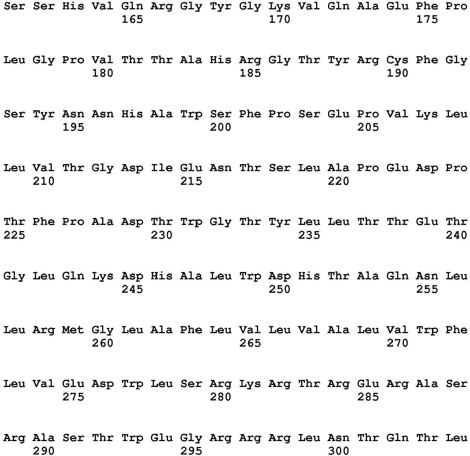
Claims (12)
- Use of an antibody that binds a human NKp46 polypeptide, for in vitro diagnosis, prognosis or monitoring of a non-cutaneous Peripheral T-Cell Lymphoma (PTCL) in an individual.
- The use of claim 1, wherein the individual has PTCL or is suspected of having PTCL or susceptible to PTCL.
- The use of claims 1 or 2, wherein the individual has an ortho visceral extranodal PTCL.
- The use of claims 1 to 3, wherein the PTCL is an intestinal lymphoma.
- The use of claims 1 to 4, wherein the PTCL is enteropathy associated T cell lymphoma (EATL).
- The use of claims 1 to 5, wherein the diagnosis comprises identifying a patient having EATL, or celiac disease (CD) or refractory celiac disease (RCD) susceptible to progressing to EATL.
- The use of claims 1 to 6, wherein the diagnosis comprises detecting a marker of EATL, or of CD or RCD that is susceptible to progressing to EATL.
- The use of claims 1 to 4, wherein PTCL is anaplastic large cell lymphoma (ALCL).
- The use of claims 1 to 3, wherein PTCL is NK/T- lymphoma
- The use of claims 1 to 9, wherein the diagnosis comprises:a) determining whether an individual has a peripheral T cell lymphoma ; andb) if the individual has a peripheral T cell lymphoma, determining whether an individual has peripheral T cell lymphoma cells that express a NKp46 polypeptide.
- An in vitro method for the diagnosis, prognosis or monitoring of a non-cutaneous PTCL in an individual, wherein the method comprises determining the level of expression of NKp46 polypeptide in a biological sample from the individual using an antibody that binds a human NKp46 polypeptide.
- The method of claim 11, wherein the PTCL is EATL, or celiac disease (CD) or refractory celiac disease (RCD) susceptible to progressing to EATL.
Applications Claiming Priority (4)
| Application Number | Priority Date | Filing Date | Title |
|---|---|---|---|
| US201361764639P | 2013-02-14 | 2013-02-14 | |
| US201361831792P | 2013-06-06 | 2013-06-06 | |
| EP14704576.9A EP2956482B1 (en) | 2013-02-14 | 2014-02-13 | Treatment of peripheral t cell lymphoma |
| PCT/EP2014/052849 WO2014125041A1 (en) | 2013-02-14 | 2014-02-13 | Treatment of peripheral t cell lymphoma |
Related Parent Applications (1)
| Application Number | Title | Priority Date | Filing Date |
|---|---|---|---|
| EP14704576.9A Division EP2956482B1 (en) | 2013-02-14 | 2014-02-13 | Treatment of peripheral t cell lymphoma |
Publications (2)
| Publication Number | Publication Date |
|---|---|
| EP3255062A1 EP3255062A1 (en) | 2017-12-13 |
| EP3255062B1 true EP3255062B1 (en) | 2019-07-03 |
Family
ID=50112904
Family Applications (2)
| Application Number | Title | Priority Date | Filing Date |
|---|---|---|---|
| EP14704576.9A Active EP2956482B1 (en) | 2013-02-14 | 2014-02-13 | Treatment of peripheral t cell lymphoma |
| EP17177583.6A Active EP3255062B1 (en) | 2013-02-14 | 2014-02-13 | Anti-nkp46 antibody for diganosis of a non-cutaneous peripheral t-cell lymphoma (ptcl) |
Family Applications Before (1)
| Application Number | Title | Priority Date | Filing Date |
|---|---|---|---|
| EP14704576.9A Active EP2956482B1 (en) | 2013-02-14 | 2014-02-13 | Treatment of peripheral t cell lymphoma |
Country Status (5)
| Country | Link |
|---|---|
| US (3) | US10344087B2 (en) |
| EP (2) | EP2956482B1 (en) |
| DK (2) | DK2956482T3 (en) |
| ES (2) | ES2747920T3 (en) |
| WO (1) | WO2014125041A1 (en) |
Families Citing this family (10)
| Publication number | Priority date | Publication date | Assignee | Title |
|---|---|---|---|---|
| DK2956482T3 (en) * | 2013-02-14 | 2017-10-16 | Innate Pharma | TREATMENT OF PERFECT T cell lymphoma |
| WO2016030488A1 (en) * | 2014-08-27 | 2016-03-03 | Innate Pharma | Treatment of celiac disease |
| AU2016299166B2 (en) | 2015-07-24 | 2022-03-31 | Innate Pharma | Methods for detecting tissue infiltrating NK cells |
| CN115925933A (en) * | 2015-12-28 | 2023-04-07 | 依奈特制药公司 | Variable regions of NKp46 binding proteins |
| CA3032249A1 (en) * | 2016-08-17 | 2018-02-22 | University Health Network | Regulation of tumor-associated t cells |
| EP3510047A1 (en) | 2016-09-07 | 2019-07-17 | Yissum Research and Development Company of the Hebrew University of Jerusalem Ltd. | Anti-nkp46 antibodies and therapeutic use of same |
| US20190248895A1 (en) | 2016-10-21 | 2019-08-15 | Innate Pharma | Treatment with anti-kir3dl2 agents |
| WO2018138032A2 (en) * | 2017-01-24 | 2018-08-02 | Innate Pharma | NKp46 BINDING AGENTS |
| CA3063955A1 (en) * | 2017-05-19 | 2018-11-22 | Royal Melbourne Institute Of Technology | Predicting responders to cyclophosphamide therapy |
| CA3099308A1 (en) | 2018-05-21 | 2019-11-28 | Compass Therapeutics Llc | Compositions and methods for enhancing the killing of target cells by nk cells |
Citations (1)
| Publication number | Priority date | Publication date | Assignee | Title |
|---|---|---|---|---|
| WO2011086179A1 (en) * | 2010-01-15 | 2011-07-21 | INSERM (Institut National de la Santé et de la Recherche Médicale) | Methods for diagnosis and treatment of cutaneous t cell lymphomas |
Family Cites Families (39)
| Publication number | Priority date | Publication date | Assignee | Title |
|---|---|---|---|---|
| US4816567A (en) | 1983-04-08 | 1989-03-28 | Genentech, Inc. | Recombinant immunoglobin preparations |
| US5567610A (en) | 1986-09-04 | 1996-10-22 | Bioinvent International Ab | Method of producing human monoclonal antibodies and kit therefor |
| US6673986B1 (en) | 1990-01-12 | 2004-01-06 | Abgenix, Inc. | Generation of xenogeneic antibodies |
| US5229275A (en) | 1990-04-26 | 1993-07-20 | Akzo N.V. | In-vitro method for producing antigen-specific human monoclonal antibodies |
| US5776427A (en) | 1992-03-05 | 1998-07-07 | Board Of Regents, The University Of Texas System | Methods for targeting the vasculature of solid tumors |
| AU717020B2 (en) | 1996-05-03 | 2000-03-16 | Immunomedics Inc. | Targeted combination immunotherapy of cancer |
| US6277375B1 (en) | 1997-03-03 | 2001-08-21 | Board Of Regents, The University Of Texas System | Immunoglobulin-like domains with increased half-lives |
| ES2434961T5 (en) | 1998-04-20 | 2018-01-18 | Roche Glycart Ag | Antibody glycosylation engineering to improve antibody-dependent cell cytotoxicity |
| US6737056B1 (en) | 1999-01-15 | 2004-05-18 | Genentech, Inc. | Polypeptide variants with altered effector function |
| HU230769B1 (en) | 1999-01-15 | 2018-03-28 | Genentech Inc. | Polypeptide variants with altred effector function |
| US7183387B1 (en) | 1999-01-15 | 2007-02-27 | Genentech, Inc. | Polypeptide variants with altered effector function |
| EP1176195B1 (en) | 1999-04-09 | 2013-05-22 | Kyowa Hakko Kirin Co., Ltd. | Method for controlling the activity of immunologically functional molecule |
| HUP0600342A3 (en) | 2001-10-25 | 2011-03-28 | Genentech Inc | Glycoprotein compositions |
| US20040132101A1 (en) | 2002-09-27 | 2004-07-08 | Xencor | Optimized Fc variants and methods for their generation |
| EA200401325A1 (en) | 2002-04-09 | 2005-04-28 | Киова Хакко Когио Ко., Лтд. | CELLS WITH MODIFIED GENOM |
| EP1534335B9 (en) | 2002-08-14 | 2016-01-13 | Macrogenics, Inc. | Fcgammariib-specific antibodies and methods of use thereof |
| US20060235208A1 (en) * | 2002-09-27 | 2006-10-19 | Xencor, Inc. | Fc variants with optimized properties |
| US20050058661A1 (en) | 2002-10-18 | 2005-03-17 | Sykes Kathryn F. | Methods and compositions for vaccination comprising nucleic acid and/or polypeptide sequences of the genus Borrelia |
| JP2006524039A (en) | 2003-01-09 | 2006-10-26 | マクロジェニクス,インコーポレーテッド | Identification and production of antibody containing mutant Fc region and use thereof |
| US7960512B2 (en) | 2003-01-09 | 2011-06-14 | Macrogenics, Inc. | Identification and engineering of antibodies with variant Fc regions and methods of using same |
| DE10324108B3 (en) | 2003-05-21 | 2005-01-27 | Aesculap Ag & Co. Kg | Backbone implant is inserted with contracted contact disc which is expanded to optimum area following insertion |
| WO2005047327A2 (en) | 2003-11-12 | 2005-05-26 | Biogen Idec Ma Inc. | NEONATAL Fc RECEPTOR (FcRn)-BINDING POLYPEPTIDE VARIANTS, DIMERIC Fc BINDING PROTEINS AND METHODS RELATED THERETO |
| SG173322A1 (en) | 2004-04-16 | 2011-08-29 | Macrogenics Inc Dw Us | Fc gammad riib - specific antibodies and methods of use thereof |
| WO2005110474A2 (en) | 2004-05-10 | 2005-11-24 | Macrogenics, Inc. | HUMANIZED FcϜRIIB SPECIFIC ANTIBODIES AND METHODS OF USE THEREOF |
| EP1810035A4 (en) | 2004-11-10 | 2010-03-17 | Macrogenics Inc | EFFECTOR FUNCTION OBTAINED BY CREATION BY BIOLOGICAL GENE OF FC ANTIBODY REGIONS |
| US20090208500A1 (en) | 2005-06-03 | 2009-08-20 | Genentech, Inc. | Method of producing antibodies with improved function |
| HUE029465T2 (en) | 2005-08-10 | 2017-02-28 | Macrogenics Inc | Identification and preparation of antibodies with variant fc regions and methods for their use |
| AU2012202895A1 (en) * | 2005-10-14 | 2012-06-07 | Innate Pharma | Compositions and methods for treating proliferative disorders |
| AU2006301163B2 (en) * | 2005-10-14 | 2012-02-23 | Innate Pharma | Compositions and methods for treating proliferative disorders |
| FR2894982A1 (en) | 2005-12-16 | 2007-06-22 | Lab Francais Du Fractionnement | Preparation of antibodies selective for activating Fc receptors, useful for treatment of tumors and viral or bacterial infections, by replacing specific histidine residues in the Fc region of a monoclonal antibody |
| BRPI0707290A2 (en) | 2006-01-17 | 2011-08-16 | Biolex Therapeutics Inc | compositions and methods for humanization and optimization of n-glycans in plants |
| CA2644903A1 (en) | 2006-03-10 | 2007-09-20 | Macrogenics, Inc. | Identification and engineering of antibodies with variant heavy chains and methods of using same |
| US7786270B2 (en) | 2006-05-26 | 2010-08-31 | Macrogenics, Inc. | Humanized FcγRIIB-specific antibodies and methods of use thereof |
| WO2008002933A2 (en) | 2006-06-26 | 2008-01-03 | Macrogenics, Inc. | Combination of fcgammariib antibodies and cd20-specific antibodies and methods of use thereof |
| EP1878747A1 (en) | 2006-07-11 | 2008-01-16 | greenovation Biotech GmbH | Glyco-engineered antibodies |
| EP1995309A1 (en) | 2007-05-21 | 2008-11-26 | Vivalis | Recombinant protein production in avian EBx® cells |
| JP5998060B2 (en) | 2010-03-04 | 2016-09-28 | マクロジェニクス,インコーポレーテッド | Antibodies reactive with B7-H3, immunologically active fragments thereof and uses thereof |
| US20140234342A1 (en) | 2011-06-21 | 2014-08-21 | Innate Pharma | Nkp46-mediated nk cell tuning |
| DK2956482T3 (en) * | 2013-02-14 | 2017-10-16 | Innate Pharma | TREATMENT OF PERFECT T cell lymphoma |
-
2014
- 2014-02-13 DK DK14704576.9T patent/DK2956482T3/en active
- 2014-02-13 EP EP14704576.9A patent/EP2956482B1/en active Active
- 2014-02-13 EP EP17177583.6A patent/EP3255062B1/en active Active
- 2014-02-13 US US14/767,600 patent/US10344087B2/en active Active
- 2014-02-13 WO PCT/EP2014/052849 patent/WO2014125041A1/en not_active Ceased
- 2014-02-13 ES ES17177583T patent/ES2747920T3/en active Active
- 2014-02-13 ES ES14704576.9T patent/ES2637151T3/en active Active
- 2014-02-13 DK DK17177583.6T patent/DK3255062T3/en active
-
2019
- 2019-06-25 US US16/451,051 patent/US11999784B2/en active Active
-
2024
- 2024-06-03 US US18/731,443 patent/US20240425586A1/en active Pending
Patent Citations (1)
| Publication number | Priority date | Publication date | Assignee | Title |
|---|---|---|---|---|
| WO2011086179A1 (en) * | 2010-01-15 | 2011-07-21 | INSERM (Institut National de la Santé et de la Recherche Médicale) | Methods for diagnosis and treatment of cutaneous t cell lymphomas |
Non-Patent Citations (1)
| Title |
|---|
| JAMES O. ARMITAGE: "The aggressive peripheral T-cell lymphomas: 2012 update on diagnosis, risk stratification, and management", AMERICAN JOURNAL OF HEMATOLOGY, vol. 87, no. 5, 17 April 2012 (2012-04-17), US, pages 511 - 519, XP055544751, ISSN: 0361-8609, DOI: 10.1002/ajh.23144 * |
Also Published As
| Publication number | Publication date |
|---|---|
| ES2637151T3 (en) | 2017-10-11 |
| DK2956482T3 (en) | 2017-10-16 |
| ES2747920T3 (en) | 2020-03-12 |
| EP2956482B1 (en) | 2017-06-28 |
| US20240425586A1 (en) | 2024-12-26 |
| DK3255062T3 (en) | 2019-10-07 |
| EP2956482A1 (en) | 2015-12-23 |
| EP3255062A1 (en) | 2017-12-13 |
| US10344087B2 (en) | 2019-07-09 |
| US20150376274A1 (en) | 2015-12-31 |
| US11999784B2 (en) | 2024-06-04 |
| US20190315857A1 (en) | 2019-10-17 |
| WO2014125041A1 (en) | 2014-08-21 |
Similar Documents
| Publication | Publication Date | Title |
|---|---|---|
| EP3255062B1 (en) | Anti-nkp46 antibody for diganosis of a non-cutaneous peripheral t-cell lymphoma (ptcl) | |
| US12084498B2 (en) | Compound that specifically binds to KIR3DL2 for use in the treatment of peripheral T cell lymphoma | |
| AU2015314316B2 (en) | Cross reactive Siglec antibodies | |
| BR112015006060B1 (en) | MONOCLONAL ANTIBODY, PHARMACEUTICAL COMPOSITION AND USE OF THE MONOCLONAL ANTIBODY | |
| CN107850596B (en) | Method for detecting tissue-infiltrating NK cells | |
| US20210355213A1 (en) | Treatment of t cell lymphoma | |
| WO2016030488A1 (en) | Treatment of celiac disease | |
| RU2835929C2 (en) | Treatment of t-cell lymphoma |
Legal Events
| Date | Code | Title | Description |
|---|---|---|---|
| PUAI | Public reference made under article 153(3) epc to a published international application that has entered the european phase |
Free format text: ORIGINAL CODE: 0009012 |
|
| STAA | Information on the status of an ep patent application or granted ep patent |
Free format text: STATUS: THE APPLICATION HAS BEEN PUBLISHED |
|
| AC | Divisional application: reference to earlier application |
Ref document number: 2956482 Country of ref document: EP Kind code of ref document: P |
|
| AK | Designated contracting states |
Kind code of ref document: A1 Designated state(s): AL AT BE BG CH CY CZ DE DK EE ES FI FR GB GR HR HU IE IS IT LI LT LU LV MC MK MT NL NO PL PT RO RS SE SI SK SM TR |
|
| STAA | Information on the status of an ep patent application or granted ep patent |
Free format text: STATUS: REQUEST FOR EXAMINATION WAS MADE |
|
| 17P | Request for examination filed |
Effective date: 20180612 |
|
| RBV | Designated contracting states (corrected) |
Designated state(s): AL AT BE BG CH CY CZ DE DK EE ES FI FR GB GR HR HU IE IS IT LI LT LU LV MC MK MT NL NO PL PT RO RS SE SI SK SM TR |
|
| RIC1 | Information provided on ipc code assigned before grant |
Ipc: C07K 16/30 20060101ALI20180823BHEP Ipc: A61K 39/395 20060101ALN20180823BHEP Ipc: C07K 16/28 20060101AFI20180823BHEP Ipc: G01N 33/574 20060101ALI20180823BHEP Ipc: A61P 35/00 20060101ALN20180823BHEP |
|
| RIC1 | Information provided on ipc code assigned before grant |
Ipc: A61K 39/395 20060101ALN20181026BHEP Ipc: A61P 35/00 20060101ALN20181026BHEP Ipc: G01N 33/574 20060101ALI20181026BHEP Ipc: C07K 16/28 20060101AFI20181026BHEP Ipc: C07K 16/30 20060101ALI20181026BHEP |
|
| RIC1 | Information provided on ipc code assigned before grant |
Ipc: C07K 16/30 20060101ALI20181123BHEP Ipc: G01N 33/574 20060101ALI20181123BHEP Ipc: A61P 35/00 20060101ALN20181123BHEP Ipc: C07K 16/28 20060101AFI20181123BHEP Ipc: A61K 39/395 20060101ALN20181123BHEP |
|
| RIC1 | Information provided on ipc code assigned before grant |
Ipc: C07K 16/30 20060101ALI20181220BHEP Ipc: A61K 39/395 20060101ALN20181220BHEP Ipc: G01N 33/574 20060101ALI20181220BHEP Ipc: A61P 35/00 20060101ALN20181220BHEP Ipc: C07K 16/28 20060101AFI20181220BHEP |
|
| GRAP | Despatch of communication of intention to grant a patent |
Free format text: ORIGINAL CODE: EPIDOSNIGR1 |
|
| STAA | Information on the status of an ep patent application or granted ep patent |
Free format text: STATUS: GRANT OF PATENT IS INTENDED |
|
| RIC1 | Information provided on ipc code assigned before grant |
Ipc: C07K 16/28 20060101AFI20190118BHEP Ipc: A61K 39/395 20060101ALN20190118BHEP Ipc: C07K 16/30 20060101ALI20190118BHEP Ipc: A61P 35/00 20060101ALN20190118BHEP Ipc: G01N 33/574 20060101ALI20190118BHEP |
|
| RIC1 | Information provided on ipc code assigned before grant |
Ipc: C07K 16/30 20060101ALI20190129BHEP Ipc: G01N 33/574 20060101ALI20190129BHEP Ipc: A61P 35/00 20060101ALN20190129BHEP Ipc: C07K 16/28 20060101AFI20190129BHEP Ipc: A61K 39/395 20060101ALN20190129BHEP |
|
| INTG | Intention to grant announced |
Effective date: 20190214 |
|
| GRAS | Grant fee paid |
Free format text: ORIGINAL CODE: EPIDOSNIGR3 |
|
| GRAA | (expected) grant |
Free format text: ORIGINAL CODE: 0009210 |
|
| STAA | Information on the status of an ep patent application or granted ep patent |
Free format text: STATUS: THE PATENT HAS BEEN GRANTED |
|
| AC | Divisional application: reference to earlier application |
Ref document number: 2956482 Country of ref document: EP Kind code of ref document: P |
|
| AK | Designated contracting states |
Kind code of ref document: B1 Designated state(s): AL AT BE BG CH CY CZ DE DK EE ES FI FR GB GR HR HU IE IS IT LI LT LU LV MC MK MT NL NO PL PT RO RS SE SI SK SM TR |
|
| REG | Reference to a national code |
Ref country code: GB Ref legal event code: FG4D |
|
| REG | Reference to a national code |
Ref country code: CH Ref legal event code: EP Ref country code: AT Ref legal event code: REF Ref document number: 1150888 Country of ref document: AT Kind code of ref document: T Effective date: 20190715 |
|
| REG | Reference to a national code |
Ref country code: IE Ref legal event code: FG4D |
|
| REG | Reference to a national code |
Ref country code: DE Ref legal event code: R096 Ref document number: 602014049720 Country of ref document: DE |
|
| REG | Reference to a national code |
Ref country code: DK Ref legal event code: T3 Effective date: 20191004 |
|
| REG | Reference to a national code |
Ref country code: NL Ref legal event code: FP |
|
| REG | Reference to a national code |
Ref country code: SE Ref legal event code: TRGR |
|
| REG | Reference to a national code |
Ref country code: LT Ref legal event code: MG4D |
|
| REG | Reference to a national code |
Ref country code: AT Ref legal event code: MK05 Ref document number: 1150888 Country of ref document: AT Kind code of ref document: T Effective date: 20190703 |
|
| PG25 | Lapsed in a contracting state [announced via postgrant information from national office to epo] |
Ref country code: CZ Free format text: LAPSE BECAUSE OF FAILURE TO SUBMIT A TRANSLATION OF THE DESCRIPTION OR TO PAY THE FEE WITHIN THE PRESCRIBED TIME-LIMIT Effective date: 20190703 Ref country code: FI Free format text: LAPSE BECAUSE OF FAILURE TO SUBMIT A TRANSLATION OF THE DESCRIPTION OR TO PAY THE FEE WITHIN THE PRESCRIBED TIME-LIMIT Effective date: 20190703 Ref country code: HR Free format text: LAPSE BECAUSE OF FAILURE TO SUBMIT A TRANSLATION OF THE DESCRIPTION OR TO PAY THE FEE WITHIN THE PRESCRIBED TIME-LIMIT Effective date: 20190703 Ref country code: AT Free format text: LAPSE BECAUSE OF FAILURE TO SUBMIT A TRANSLATION OF THE DESCRIPTION OR TO PAY THE FEE WITHIN THE PRESCRIBED TIME-LIMIT Effective date: 20190703 Ref country code: NO Free format text: LAPSE BECAUSE OF FAILURE TO SUBMIT A TRANSLATION OF THE DESCRIPTION OR TO PAY THE FEE WITHIN THE PRESCRIBED TIME-LIMIT Effective date: 20191003 Ref country code: BG Free format text: LAPSE BECAUSE OF FAILURE TO SUBMIT A TRANSLATION OF THE DESCRIPTION OR TO PAY THE FEE WITHIN THE PRESCRIBED TIME-LIMIT Effective date: 20191003 Ref country code: PT Free format text: LAPSE BECAUSE OF FAILURE TO SUBMIT A TRANSLATION OF THE DESCRIPTION OR TO PAY THE FEE WITHIN THE PRESCRIBED TIME-LIMIT Effective date: 20191104 Ref country code: LT Free format text: LAPSE BECAUSE OF FAILURE TO SUBMIT A TRANSLATION OF THE DESCRIPTION OR TO PAY THE FEE WITHIN THE PRESCRIBED TIME-LIMIT Effective date: 20190703 |
|
| PG25 | Lapsed in a contracting state [announced via postgrant information from national office to epo] |
Ref country code: AL Free format text: LAPSE BECAUSE OF FAILURE TO SUBMIT A TRANSLATION OF THE DESCRIPTION OR TO PAY THE FEE WITHIN THE PRESCRIBED TIME-LIMIT Effective date: 20190703 Ref country code: LV Free format text: LAPSE BECAUSE OF FAILURE TO SUBMIT A TRANSLATION OF THE DESCRIPTION OR TO PAY THE FEE WITHIN THE PRESCRIBED TIME-LIMIT Effective date: 20190703 Ref country code: IS Free format text: LAPSE BECAUSE OF FAILURE TO SUBMIT A TRANSLATION OF THE DESCRIPTION OR TO PAY THE FEE WITHIN THE PRESCRIBED TIME-LIMIT Effective date: 20191103 Ref country code: RS Free format text: LAPSE BECAUSE OF FAILURE TO SUBMIT A TRANSLATION OF THE DESCRIPTION OR TO PAY THE FEE WITHIN THE PRESCRIBED TIME-LIMIT Effective date: 20190703 Ref country code: GR Free format text: LAPSE BECAUSE OF FAILURE TO SUBMIT A TRANSLATION OF THE DESCRIPTION OR TO PAY THE FEE WITHIN THE PRESCRIBED TIME-LIMIT Effective date: 20191004 |
|
| REG | Reference to a national code |
Ref country code: ES Ref legal event code: FG2A Ref document number: 2747920 Country of ref document: ES Kind code of ref document: T3 Effective date: 20200312 |
|
| PG25 | Lapsed in a contracting state [announced via postgrant information from national office to epo] |
Ref country code: TR Free format text: LAPSE BECAUSE OF FAILURE TO SUBMIT A TRANSLATION OF THE DESCRIPTION OR TO PAY THE FEE WITHIN THE PRESCRIBED TIME-LIMIT Effective date: 20190703 |
|
| PG25 | Lapsed in a contracting state [announced via postgrant information from national office to epo] |
Ref country code: PL Free format text: LAPSE BECAUSE OF FAILURE TO SUBMIT A TRANSLATION OF THE DESCRIPTION OR TO PAY THE FEE WITHIN THE PRESCRIBED TIME-LIMIT Effective date: 20190703 Ref country code: EE Free format text: LAPSE BECAUSE OF FAILURE TO SUBMIT A TRANSLATION OF THE DESCRIPTION OR TO PAY THE FEE WITHIN THE PRESCRIBED TIME-LIMIT Effective date: 20190703 Ref country code: RO Free format text: LAPSE BECAUSE OF FAILURE TO SUBMIT A TRANSLATION OF THE DESCRIPTION OR TO PAY THE FEE WITHIN THE PRESCRIBED TIME-LIMIT Effective date: 20190703 |
|
| PG25 | Lapsed in a contracting state [announced via postgrant information from national office to epo] |
Ref country code: IS Free format text: LAPSE BECAUSE OF FAILURE TO SUBMIT A TRANSLATION OF THE DESCRIPTION OR TO PAY THE FEE WITHIN THE PRESCRIBED TIME-LIMIT Effective date: 20200224 Ref country code: SK Free format text: LAPSE BECAUSE OF FAILURE TO SUBMIT A TRANSLATION OF THE DESCRIPTION OR TO PAY THE FEE WITHIN THE PRESCRIBED TIME-LIMIT Effective date: 20190703 Ref country code: SM Free format text: LAPSE BECAUSE OF FAILURE TO SUBMIT A TRANSLATION OF THE DESCRIPTION OR TO PAY THE FEE WITHIN THE PRESCRIBED TIME-LIMIT Effective date: 20190703 |
|
| REG | Reference to a national code |
Ref country code: DE Ref legal event code: R097 Ref document number: 602014049720 Country of ref document: DE |
|
| PLBE | No opposition filed within time limit |
Free format text: ORIGINAL CODE: 0009261 |
|
| STAA | Information on the status of an ep patent application or granted ep patent |
Free format text: STATUS: NO OPPOSITION FILED WITHIN TIME LIMIT |
|
| PG2D | Information on lapse in contracting state deleted |
Ref country code: IS |
|
| 26N | No opposition filed |
Effective date: 20200603 |
|
| PG25 | Lapsed in a contracting state [announced via postgrant information from national office to epo] |
Ref country code: SI Free format text: LAPSE BECAUSE OF FAILURE TO SUBMIT A TRANSLATION OF THE DESCRIPTION OR TO PAY THE FEE WITHIN THE PRESCRIBED TIME-LIMIT Effective date: 20190703 |
|
| PG25 | Lapsed in a contracting state [announced via postgrant information from national office to epo] |
Ref country code: MC Free format text: LAPSE BECAUSE OF FAILURE TO SUBMIT A TRANSLATION OF THE DESCRIPTION OR TO PAY THE FEE WITHIN THE PRESCRIBED TIME-LIMIT Effective date: 20190703 Ref country code: LU Free format text: LAPSE BECAUSE OF NON-PAYMENT OF DUE FEES Effective date: 20200213 |
|
| PG25 | Lapsed in a contracting state [announced via postgrant information from national office to epo] |
Ref country code: MT Free format text: LAPSE BECAUSE OF FAILURE TO SUBMIT A TRANSLATION OF THE DESCRIPTION OR TO PAY THE FEE WITHIN THE PRESCRIBED TIME-LIMIT Effective date: 20190703 Ref country code: CY Free format text: LAPSE BECAUSE OF FAILURE TO SUBMIT A TRANSLATION OF THE DESCRIPTION OR TO PAY THE FEE WITHIN THE PRESCRIBED TIME-LIMIT Effective date: 20190703 |
|
| PG25 | Lapsed in a contracting state [announced via postgrant information from national office to epo] |
Ref country code: MK Free format text: LAPSE BECAUSE OF FAILURE TO SUBMIT A TRANSLATION OF THE DESCRIPTION OR TO PAY THE FEE WITHIN THE PRESCRIBED TIME-LIMIT Effective date: 20190703 |
|
| P01 | Opt-out of the competence of the unified patent court (upc) registered |
Effective date: 20230512 |
|
| PGFP | Annual fee paid to national office [announced via postgrant information from national office to epo] |
Ref country code: CH Payment date: 20250306 Year of fee payment: 12 |
|
| REG | Reference to a national code |
Ref country code: CH Ref legal event code: U11 Free format text: ST27 STATUS EVENT CODE: U-0-0-U10-U11 (AS PROVIDED BY THE NATIONAL OFFICE) Effective date: 20260301 |
|
| PGFP | Annual fee paid to national office [announced via postgrant information from national office to epo] |
Ref country code: NL Payment date: 20260226 Year of fee payment: 13 |
|
| PGFP | Annual fee paid to national office [announced via postgrant information from national office to epo] |
Ref country code: SE Payment date: 20260227 Year of fee payment: 13 |
|
| PGFP | Annual fee paid to national office [announced via postgrant information from national office to epo] |
Ref country code: GB Payment date: 20260227 Year of fee payment: 13 |
|
| PGFP | Annual fee paid to national office [announced via postgrant information from national office to epo] |
Ref country code: ES Payment date: 20260302 Year of fee payment: 13 |
|
| PGFP | Annual fee paid to national office [announced via postgrant information from national office to epo] |
Ref country code: DE Payment date: 20260227 Year of fee payment: 13 Ref country code: DK Payment date: 20260225 Year of fee payment: 13 Ref country code: IE Payment date: 20260227 Year of fee payment: 13 |
|
| PGFP | Annual fee paid to national office [announced via postgrant information from national office to epo] |
Ref country code: IT Payment date: 20260219 Year of fee payment: 13 Ref country code: BE Payment date: 20260227 Year of fee payment: 13 |
|
| PGFP | Annual fee paid to national office [announced via postgrant information from national office to epo] |
Ref country code: FR Payment date: 20260225 Year of fee payment: 13 |