EP3260864B1 - Diagnostischer co-inkubations-assay - Google Patents
Diagnostischer co-inkubations-assay Download PDFInfo
- Publication number
- EP3260864B1 EP3260864B1 EP17001041.7A EP17001041A EP3260864B1 EP 3260864 B1 EP3260864 B1 EP 3260864B1 EP 17001041 A EP17001041 A EP 17001041A EP 3260864 B1 EP3260864 B1 EP 3260864B1
- Authority
- EP
- European Patent Office
- Prior art keywords
- seq
- antigen
- tissue section
- disease
- donor tissue
- Prior art date
- Legal status (The legal status is an assumption and is not a legal conclusion. Google has not performed a legal analysis and makes no representation as to the accuracy of the status listed.)
- Active
Links
Images




Classifications
-
- G—PHYSICS
- G01—MEASURING; TESTING
- G01N—INVESTIGATING OR ANALYSING MATERIALS BY DETERMINING THEIR CHEMICAL OR PHYSICAL PROPERTIES
- G01N33/00—Investigating or analysing materials by specific methods not covered by groups G01N1/00 - G01N31/00
- G01N33/48—Biological material, e.g. blood, urine; Haemocytometers
- G01N33/50—Chemical analysis of biological material, e.g. blood, urine; Testing involving biospecific ligand binding methods; Immunological testing
- G01N33/5005—Chemical analysis of biological material, e.g. blood, urine; Testing involving biospecific ligand binding methods; Immunological testing involving human or animal cells
- G01N33/5091—Chemical analysis of biological material, e.g. blood, urine; Testing involving biospecific ligand binding methods; Immunological testing involving human or animal cells for testing the pathological state of an organism
-
- G—PHYSICS
- G01—MEASURING; TESTING
- G01N—INVESTIGATING OR ANALYSING MATERIALS BY DETERMINING THEIR CHEMICAL OR PHYSICAL PROPERTIES
- G01N33/00—Investigating or analysing materials by specific methods not covered by groups G01N1/00 - G01N31/00
- G01N33/48—Biological material, e.g. blood, urine; Haemocytometers
- G01N33/50—Chemical analysis of biological material, e.g. blood, urine; Testing involving biospecific ligand binding methods; Immunological testing
- G01N33/53—Immunoassay; Biospecific binding assay; Materials therefor
- G01N33/531—Production of immunochemical test materials
- G01N33/532—Production of labelled immunochemicals
- G01N33/533—Production of labelled immunochemicals with fluorescent label
-
- B—PERFORMING OPERATIONS; TRANSPORTING
- B01—PHYSICAL OR CHEMICAL PROCESSES OR APPARATUS IN GENERAL
- B01L—CHEMICAL OR PHYSICAL LABORATORY APPARATUS FOR GENERAL USE
- B01L3/00—Containers or dishes for laboratory use, e.g. laboratory glassware; Droppers
-
- B—PERFORMING OPERATIONS; TRANSPORTING
- B01—PHYSICAL OR CHEMICAL PROCESSES OR APPARATUS IN GENERAL
- B01L—CHEMICAL OR PHYSICAL LABORATORY APPARATUS FOR GENERAL USE
- B01L3/00—Containers or dishes for laboratory use, e.g. laboratory glassware; Droppers
- B01L3/50—Containers for the purpose of retaining a material to be analysed, e.g. test tubes
-
- B—PERFORMING OPERATIONS; TRANSPORTING
- B01—PHYSICAL OR CHEMICAL PROCESSES OR APPARATUS IN GENERAL
- B01L—CHEMICAL OR PHYSICAL LABORATORY APPARATUS FOR GENERAL USE
- B01L3/00—Containers or dishes for laboratory use, e.g. laboratory glassware; Droppers
- B01L3/50—Containers for the purpose of retaining a material to be analysed, e.g. test tubes
- B01L3/508—Rigid containers without fluid transport within
- B01L3/5085—Rigid containers without fluid transport within for multiple samples, e.g. microtitration plates
-
- G—PHYSICS
- G01—MEASURING; TESTING
- G01N—INVESTIGATING OR ANALYSING MATERIALS BY DETERMINING THEIR CHEMICAL OR PHYSICAL PROPERTIES
- G01N1/00—Sampling; Preparing specimens for investigation
- G01N1/28—Preparing specimens for investigation including physical details of (bio-)chemical methods covered elsewhere, e.g. G01N33/50, C12Q
- G01N1/2813—Producing thin layers of samples on a substrate, e.g. smearing, spinning-on
-
- G—PHYSICS
- G01—MEASURING; TESTING
- G01N—INVESTIGATING OR ANALYSING MATERIALS BY DETERMINING THEIR CHEMICAL OR PHYSICAL PROPERTIES
- G01N33/00—Investigating or analysing materials by specific methods not covered by groups G01N1/00 - G01N31/00
- G01N33/48—Biological material, e.g. blood, urine; Haemocytometers
- G01N33/50—Chemical analysis of biological material, e.g. blood, urine; Testing involving biospecific ligand binding methods; Immunological testing
- G01N33/53—Immunoassay; Biospecific binding assay; Materials therefor
- G01N33/5302—Apparatus specially adapted for immunological test procedures
-
- G—PHYSICS
- G01—MEASURING; TESTING
- G01N—INVESTIGATING OR ANALYSING MATERIALS BY DETERMINING THEIR CHEMICAL OR PHYSICAL PROPERTIES
- G01N33/00—Investigating or analysing materials by specific methods not covered by groups G01N1/00 - G01N31/00
- G01N33/48—Biological material, e.g. blood, urine; Haemocytometers
- G01N33/50—Chemical analysis of biological material, e.g. blood, urine; Testing involving biospecific ligand binding methods; Immunological testing
- G01N33/53—Immunoassay; Biospecific binding assay; Materials therefor
- G01N33/536—Immunoassay; Biospecific binding assay; Materials therefor with immune complex formed in liquid phase
-
- G—PHYSICS
- G01—MEASURING; TESTING
- G01N—INVESTIGATING OR ANALYSING MATERIALS BY DETERMINING THEIR CHEMICAL OR PHYSICAL PROPERTIES
- G01N33/00—Investigating or analysing materials by specific methods not covered by groups G01N1/00 - G01N31/00
- G01N33/48—Biological material, e.g. blood, urine; Haemocytometers
- G01N33/50—Chemical analysis of biological material, e.g. blood, urine; Testing involving biospecific ligand binding methods; Immunological testing
- G01N33/53—Immunoassay; Biospecific binding assay; Materials therefor
- G01N33/543—Immunoassay; Biospecific binding assay; Materials therefor with an insoluble carrier for immobilising immunochemicals
- G01N33/54393—Improving reaction conditions or stability, e.g. by coating or irradiation of surface, by reduction of non-specific binding, by promotion of specific binding
-
- G—PHYSICS
- G01—MEASURING; TESTING
- G01N—INVESTIGATING OR ANALYSING MATERIALS BY DETERMINING THEIR CHEMICAL OR PHYSICAL PROPERTIES
- G01N33/00—Investigating or analysing materials by specific methods not covered by groups G01N1/00 - G01N31/00
- G01N33/48—Biological material, e.g. blood, urine; Haemocytometers
- G01N33/50—Chemical analysis of biological material, e.g. blood, urine; Testing involving biospecific ligand binding methods; Immunological testing
- G01N33/53—Immunoassay; Biospecific binding assay; Materials therefor
- G01N33/543—Immunoassay; Biospecific binding assay; Materials therefor with an insoluble carrier for immobilising immunochemicals
- G01N33/544—Immunoassay; Biospecific binding assay; Materials therefor with an insoluble carrier for immobilising immunochemicals the carrier being organic
- G01N33/549—Immunoassay; Biospecific binding assay; Materials therefor with an insoluble carrier for immobilising immunochemicals the carrier being organic with antigen or antibody entrapped within the carrier
-
- G—PHYSICS
- G01—MEASURING; TESTING
- G01N—INVESTIGATING OR ANALYSING MATERIALS BY DETERMINING THEIR CHEMICAL OR PHYSICAL PROPERTIES
- G01N33/00—Investigating or analysing materials by specific methods not covered by groups G01N1/00 - G01N31/00
- G01N33/48—Biological material, e.g. blood, urine; Haemocytometers
- G01N33/50—Chemical analysis of biological material, e.g. blood, urine; Testing involving biospecific ligand binding methods; Immunological testing
- G01N33/53—Immunoassay; Biospecific binding assay; Materials therefor
- G01N33/564—Immunoassay; Biospecific binding assay; Materials therefor for pre-existing immune complex or autoimmune disease, i.e. systemic lupus erythematosus, rheumatoid arthritis, multiple sclerosis, rheumatoid factors or complement components C1-C9
-
- G—PHYSICS
- G01—MEASURING; TESTING
- G01N—INVESTIGATING OR ANALYSING MATERIALS BY DETERMINING THEIR CHEMICAL OR PHYSICAL PROPERTIES
- G01N33/00—Investigating or analysing materials by specific methods not covered by groups G01N1/00 - G01N31/00
- G01N33/48—Biological material, e.g. blood, urine; Haemocytometers
- G01N33/50—Chemical analysis of biological material, e.g. blood, urine; Testing involving biospecific ligand binding methods; Immunological testing
- G01N33/53—Immunoassay; Biospecific binding assay; Materials therefor
- G01N33/566—Immunoassay; Biospecific binding assay; Materials therefor using specific carrier or receptor proteins as ligand binding reagents where possible specific carrier or receptor proteins are classified with their target compounds
- G01N33/567—Immunoassay; Biospecific binding assay; Materials therefor using specific carrier or receptor proteins as ligand binding reagents where possible specific carrier or receptor proteins are classified with their target compounds utilising isolate of tissue or organ as binding agent
-
- G—PHYSICS
- G01—MEASURING; TESTING
- G01N—INVESTIGATING OR ANALYSING MATERIALS BY DETERMINING THEIR CHEMICAL OR PHYSICAL PROPERTIES
- G01N33/00—Investigating or analysing materials by specific methods not covered by groups G01N1/00 - G01N31/00
- G01N33/48—Biological material, e.g. blood, urine; Haemocytometers
- G01N33/50—Chemical analysis of biological material, e.g. blood, urine; Testing involving biospecific ligand binding methods; Immunological testing
- G01N33/53—Immunoassay; Biospecific binding assay; Materials therefor
- G01N33/569—Immunoassay; Biospecific binding assay; Materials therefor for microorganisms, e.g. protozoa, bacteria, viruses
- G01N33/56966—Animal cells
-
- G—PHYSICS
- G01—MEASURING; TESTING
- G01N—INVESTIGATING OR ANALYSING MATERIALS BY DETERMINING THEIR CHEMICAL OR PHYSICAL PROPERTIES
- G01N33/00—Investigating or analysing materials by specific methods not covered by groups G01N1/00 - G01N31/00
- G01N33/48—Biological material, e.g. blood, urine; Haemocytometers
- G01N33/50—Chemical analysis of biological material, e.g. blood, urine; Testing involving biospecific ligand binding methods; Immunological testing
- G01N33/53—Immunoassay; Biospecific binding assay; Materials therefor
- G01N33/577—Immunoassay; Biospecific binding assay; Materials therefor involving monoclonal antibodies binding reaction mechanisms characterised by the use of monoclonal antibodies
-
- B—PERFORMING OPERATIONS; TRANSPORTING
- B01—PHYSICAL OR CHEMICAL PROCESSES OR APPARATUS IN GENERAL
- B01L—CHEMICAL OR PHYSICAL LABORATORY APPARATUS FOR GENERAL USE
- B01L2300/00—Additional constructional details
- B01L2300/04—Closures and closing means
- B01L2300/046—Function or devices integrated in the closure
-
- B—PERFORMING OPERATIONS; TRANSPORTING
- B01—PHYSICAL OR CHEMICAL PROCESSES OR APPARATUS IN GENERAL
- B01L—CHEMICAL OR PHYSICAL LABORATORY APPARATUS FOR GENERAL USE
- B01L2300/00—Additional constructional details
- B01L2300/04—Closures and closing means
- B01L2300/046—Function or devices integrated in the closure
- B01L2300/047—Additional chamber, reservoir
-
- B—PERFORMING OPERATIONS; TRANSPORTING
- B01—PHYSICAL OR CHEMICAL PROCESSES OR APPARATUS IN GENERAL
- B01L—CHEMICAL OR PHYSICAL LABORATORY APPARATUS FOR GENERAL USE
- B01L2300/00—Additional constructional details
- B01L2300/06—Auxiliary integrated devices, integrated components
- B01L2300/0627—Sensor or part of a sensor is integrated
- B01L2300/0636—Integrated biosensor, microarrays
-
- B—PERFORMING OPERATIONS; TRANSPORTING
- B01—PHYSICAL OR CHEMICAL PROCESSES OR APPARATUS IN GENERAL
- B01L—CHEMICAL OR PHYSICAL LABORATORY APPARATUS FOR GENERAL USE
- B01L2300/00—Additional constructional details
- B01L2300/08—Geometry, shape and general structure
- B01L2300/0809—Geometry, shape and general structure rectangular shaped
- B01L2300/0819—Microarrays; Biochips
-
- B—PERFORMING OPERATIONS; TRANSPORTING
- B01—PHYSICAL OR CHEMICAL PROCESSES OR APPARATUS IN GENERAL
- B01L—CHEMICAL OR PHYSICAL LABORATORY APPARATUS FOR GENERAL USE
- B01L2300/00—Additional constructional details
- B01L2300/08—Geometry, shape and general structure
- B01L2300/0809—Geometry, shape and general structure rectangular shaped
- B01L2300/0822—Slides
-
- B—PERFORMING OPERATIONS; TRANSPORTING
- B01—PHYSICAL OR CHEMICAL PROCESSES OR APPARATUS IN GENERAL
- B01L—CHEMICAL OR PHYSICAL LABORATORY APPARATUS FOR GENERAL USE
- B01L2400/00—Moving or stopping fluids
- B01L2400/04—Moving fluids with specific forces or mechanical means
- B01L2400/0403—Moving fluids with specific forces or mechanical means specific forces
- B01L2400/0406—Moving fluids with specific forces or mechanical means specific forces capillary forces
-
- G—PHYSICS
- G01—MEASURING; TESTING
- G01N—INVESTIGATING OR ANALYSING MATERIALS BY DETERMINING THEIR CHEMICAL OR PHYSICAL PROPERTIES
- G01N2800/00—Detection or diagnosis of diseases
- G01N2800/06—Gastro-intestinal diseases
Definitions
- the present invention relates to a method for the diagnosis of a disease comprising contacting a donor tissue section with a liquid capable of extracting an antibody from said donor tissue section and contacting said liquid with an acceptor material comprising an antigen, followed by detection of a complex comprising the antibody and the antigen according to the appended claims
- autoimmune diseases occur in which there is an apparent immunologic reaction of the host to his own tissues, often resulting in tissue injury.
- autoimmune diseases cannot be overemphasized. Many autoimmune disorders cannot be cured, but therapies are available that may be used to significantly ameliorate their symptoms, often enabling patients to lead a normal life. The earlier the diagnosis, the better the chances to exploit the spectrum of therapies, such as administration of immunosuppressive drugs or a strict diet devoid of immunogenic antigens, to the full benefit of the patient.
- the diagnosis of an autoimmune disease is frequently based on the output of a method for the detection of autoantibodies in a liquid sample comprising antibodies. Most frequently, blood serum routinely obtained from patients is used. In other cases, other liquids are used such as cerebrospinal fluid (CSF), in particular for the detection of autoantibodies associated with neurological symptoms.
- CSF cerebrospinal fluid
- Coeliac disease is a special example of an autoimmune disease. It is triggered by gluten which activates an immune reaction against the CD autoantigen, i.e. tissue transglutaminase (TTG), in genetically predisposed subjects. Following the finding that the presence of antibodies to gliadin and/or TTG is associated with CD it could be established that CD is not a rare, but a frequent condition with an expected prevalence higher than 1 % in the worldwide population.
- TTG tissue transglutaminase
- a spectrum of diseases related to gluten sensitivity exists, including Non Coeliac Gluten sensitivity (NCGS) characterized by intestinal symptoms related to the ingestion of gluten-containing food, and in the case of a number of patients it remains difficult to thoroughly distinguish between them.
- NCGS Non Coeliac Gluten sensitivity
- a pathologist with significant experience is required to examine tissue sections made from a bioptate as a basis for a reliable diagnosis. In many cases, such examination is inconclusive as characteristic features cannot be observed. However, the pathologist may not have at his disposal a serum sample from the patient to complement the histological analysis, or the concentration of antibodies in serum samples comprising peripheral blood may be insufficient.
- a problem underlying the present invention is to provide a method for diagnosing an autoimmune disease that may be used to corroborate an inconclusive histological examination of diseased tissue. Another problem is to provide an improved test having an increased sensitivity and reducing the likelihood of false negative results.
- a problem underlying the present invention is to provide an assay that may be run in the absence of serum samples and allows a pathologist to corroborate a diagnosis based on an inconclusive histological examination.
- EP 2191893 A1 discloses glass slides comprising patient tissue samples or frozen tissue section comprising cellular antigens that are incubated with diluted sera for antibody detection.
- Hirose et al. discloses anti-collagen autoantibody elution and its detection by indirect immunofluorescence microscopy.
- Kalluri et al. discloses the elution of tissue bound antibodies and their detection using ELISA and immunoblot.
- the problem underlying the present invention is solved by a method for the diagnosis of a disease comprising contacting a donor tissue section with a liquid capable of extracting an antibody from said donor tissue section and contacting said liquid with an acceptor material comprising an antigen, optionally two or more antigens, followed by detection of a complex comprising the antibody and the antigen according to the appended claims.
- the donor tissue section and the acceptor material comprising an antigen are co-incubated in the liquid.
- a diagnostically useful carrier comprising a donor tissue section and an acceptor material comprising an antigen, wherein the carrier is configured such that the donor tissue section and the acceptor material comprising an antigen may be co-incubated in a liquid capable of extracting an antibody from said donor tissue section and allowing the transport of the antibody to the acceptor material.
- the donor tissue section and the acceptor material comprising an antigen may be coated on the surface of the carrier and are preferably spatially separate.
- the donor tissue section and the acceptor material comprising an antigen are on spatially separate biochips.
- the carrier may be configured such that a liquid capable of extracting an antibody from said donor tissue section may be placed on the surface of the carrier such that an antibody may be extracted from the donor tissue section and diffuse, via the liquid, to the acceptor material comprising an antigen.
- the carrier may comprises a first part having a surface coated with the donor tissue section and a second part having a surface coated with the acceptor material, wherein the first and the second part are separate and the carrier is configured such that the first and the second part may be contacted, preferably assembled such that the surface of either the first or the second part faces downwards on the surface of the other one of the first and the second part such that a drop of liquid may be placed between the surfaces of the first part and the second part to allow diffusion of any antibodies from the donor tissue section to the acceptor material.
- the donor tissue section has been obtained from a patient to be diagnosed.
- the acceptor material comprising an antigen is a tissue sample comprising the antigen, preferably in the form of a native polypeptide, a cell producing said antigen, or an isolated polypeptide.
- the donor tissue section is a frozen tissue section.
- the disease is a gastroenteropathy, preferably an autoimmune gastrointestinal disease, more preferably coeliac disease
- the donor tissue section is a gastrointestinal, preferably duodenal tissue
- the antigen is tissue transglutaminase or deamidated gliadin or a variant thereof, preferably an oligomer of deamidated gliadin or a variant thereof as defined in the claims.
- the disease is pemphigus and/or pemphigoid, preferably bullous pemphigoid
- the donor tissue section is diseased skin tissue
- the acceptor material comprises one or more antigens from the group comprising Dsg1, Dsg3, NC16A, BP180, BP 230, LAMA3 (SEQ ID NO 14), Laminin332, which is preferably a composition comprising polypeptides comprising LAMA3 (SEQ ID NO14), LAMB3 (SEQ ID NO 15), and LAMC2 (SEQ ID NO 16) and, beta4 integrin (SEQ ID NO 17) and collagen type VII and a variant thereof as defined in the claims, optionally in the form of a tissue, preferably selected from the group comprising primate esophagus, human salt split skin and rat urinary bladder.
- the disease is Goodpasture syndrome or SLE
- the donor tissue section is diseased kidney tissue
- the acceptor material comprising the antigen is or is derived from a material selected from the group comprising antiglomerular basement membrane, dsDNS, human epithelial cells (HEp-2), pLA2R and THSD7A and a variant thereof as defined in the claims.
- the disease is Crohn's disease
- the donor tissue section is diseased intestinal tissue
- the acceptor material comprising the antigen is selected from the group comprising healthy pancreas tissue, CUZD1 and GP2 and a variant thereof, the latter two preferably in the form of a cell expressing CUZD1 and/or GP2 and a variant thereof as defined in the claims.
- the disease is Grave's and/or Hashimoto's disease
- the donor tissue section is derived from thyroid gland
- the acceptor material comprising the antigen is or is derived from healthy thyroid gland tissue.
- the disease is myositis
- the donor tissue section is muscle or skin, preferably muscle tissue
- the acceptor material comprising the antigen is MUP-44 or a variant thereof.
- the present invention is based on the inventors' surprising finding that autoantibodies may be detected in tissue sections. For example, this holds true for antibodies associated with CD.
- a donor tissue section is provided. This is a piece of tissue obtained from a patient suffering from or suspected of suffering from a disease to be diagnosed. In a preferred embodiment, the donor tissue section is frozen, preferably by immersion in liquid nitrogen or melting isopentane. The tissue section may preferably have a thickness of 1 to 100, preferably 4 to 50 ⁇ m. The donor tissue section has preferably not been treated with any reagents or exposed to conditions substantially altering the antibodies present in the donor tissue section.
- the donor tissue section is contacted with a liquid capable of extracting an antibody from said donor tissue section.
- a liquid capable of extracting an antibody from said donor tissue section is any liquid that, upon exposure to the donor tissue section, may take up antibodies present in the donor tissue section, conserves their chemical composition, structure or binding properties such as the ability to bind specifically to an antigen, and allows their passage to the acceptor material comprising an antigen.
- the liquid is an aqueous liquid comprising a physiological buffer, more preferably at pH 5 to 9, more preferably 6 to 8, most preferably 6.2 to 7.8.
- the liquid is PBS pH 7.4.
- binding specifically means that the binding is stronger than a binding reaction characterized by a dissociation constant of 1 ⁇ 10 -5 M, more preferably 1 ⁇ 10 -7 M, more preferably 1 ⁇ 10 -8 M, more preferably 1 ⁇ 10 -9 M, more preferably 1 ⁇ 10 -10 M, more preferably 1 ⁇ 10 -11 M, more preferably 1 ⁇ 10 -12 M, as determined by surface plasmon resonance using Biacore equipment at 25 °C in PBS buffer at pH 7.4.
- the liquid capable of extracting the antibody comprises a detergent that helps dissociating any antibodies from tissue whilst conserving their chemical composition, structure or binding properties, in particular the ability to specifically bind to an antigen, and is preferably Tween.
- the liquid comprises 0.1 to 10 %, preferably 0.5 to 5%, more preferably 1 to 4%, most preferably 1.5 to 2.5 % of a detergent.
- the donor tissue section is contacted with a liquid capable of extracting an antibody from said donor tissue section, and said liquid is contacted with an acceptor material comprising an antigen.
- the acceptor material comprises the antigen in a state and conformation that allows for the specific binding of an antibody.
- the acceptor material may be selected from the group comprising tissue containing an antigen, preferably in the form of a native polypeptide, more preferably an endogenous, native polypeptide, a cell producing an antigen, preferably a recombinant cell overexpressing an antigen, and an isolated polypeptide.
- the cell may be intact or a lysed cell.
- the donor tissue section and the acceptor material comprising an antigen are co-incubated in the liquid, i.e . both the donor tissue section and the acceptor material are contacted with the liquid at the same time as the antibody is extracted from the donor tissue section and transported to the acceptor material.
- This co-incubation step is carried out for at least 10, 20, 30, 60 minutes, 2, 3, 4, 6, 8, 10 or 12 hours.
- the donor tissue section and the liquid may be contacted first, followed by separation of the donor tissue section and the liquid and contacting of the liquid and the acceptor material comprising an antigen.
- the liquid may be stored, frozen and/or concentrated prior to contacting it with the acceptor material comprising an antigen. Both contacting steps may be carried out for at least 10, 20, 30, 60 minutes, 2, 3, 4, 6, 8, 10 or 12 hours.
- the acceptor material is separated from the liquid capable of extracting an antibody and/or the donor tissue section, for example by separating the first or second part of the carrier or by removing the liquid, for example by way of aspiration.
- the acceptor material may be washed at least once using a washing buffer.
- Fresh liquid capable of extracting the antibody may be used as washing buffer, preferably comprising a smaller concentration of detergent such as 10 % of the concentration used in the incubation step or no detergent at all.
- any complex comprising the antibody and the antigen may be detected.
- the complex is detected using a method selected from the group comprising immunodiffusion techniques, immunoelectrophoretic techniques, light scattering immunoassays, agglutination techniques, labeled immunoassays such as those from the group comprising radiolabeled immunoassays, enzyme immunoassays such as ELISA, chemiluminscence immunoassays, and immunofluorescence techniques.
- a method selected from the group comprising immunodiffusion techniques, immunoelectrophoretic techniques, light scattering immunoassays, agglutination techniques, labeled immunoassays such as those from the group comprising radiolabeled immunoassays, enzyme immunoassays such as ELISA, chemiluminscence immunoassays, and immunofluorescence techniques.
- immunofluorescence techniques The person skilled in the art is familiar with these methods.
- the invention relates to a diagnostically useful carrier, which is preferably a solid, artificial carrier for contacting a donor tissue section and/or, preferably and an acceptor material comprising an antigen, which donor tissue section and/or acceptor material is coated on the surface of said carrier, with a liquid capable of extracting an antibody from the donor tissue section.
- a diagnostically useful carrier which is preferably a solid, artificial carrier for contacting a donor tissue section and/or, preferably and an acceptor material comprising an antigen, which donor tissue section and/or acceptor material is coated on the surface of said carrier, with a liquid capable of extracting an antibody from the donor tissue section.
- the donor tissue section and the acceptor material are surrounded by a hydrophobic surface to fix the position of the liquid capable of extracting an antibody.
- the solid carrier comprises two or more separate parts, one comprising the donor tissue section and one comprising the acceptor material, each separate part preferably selected from the group comprising a bead, a microtiter plate, a glass surface, a biochip and a membrane, most preferably a biochip.
- biochip refers to a planar, thin slide having a thickness of 0.01 to 1 mm, preferably 0.02 to 0.5 mm, more preferably 0.05 to 0.4 mm. They are preferably made of glass or plastic. They are coated with a biological agent such as a donor tissue section or an acceptor material comprising an antigen.
- the diagnostically useful carrier may be coated with donor tissue section and coated with the acceptor material comprising an antigen.
- the donor tissue section and the acceptor material both preferably on a biochip, may be sized and placed in close proximity such that a drop of liquid capable of extracting the antibody, preferably having a volume of 10 to 80, preferably 20 to 60 ⁇ l, may be placed in contact with both the donor tissue section and the acceptor material at the same time for the incubation step.
- Coated donor tissue section and coated acceptor material comprising an antigen on the diagnostically useful carrier are preferably spatially separated, in particular such that one may be added or removed from the diagnostically useful carrier independent of the other.
- the contacting step may be carried out as follows: a drop of liquid is placed on a surface and the diagnostically carrier is contacted with said drop such that the donor tissue section and the acceptor material, both on the same side of the diagnostically useful carrier, face downward when touching the drop sitting on the carrier. Owing to their hydrophilic natures, the surface, the donor tissue section and the acceptor material adhere to and fix the liquid drop for the entirety of the coincubation. The extraction and passage of antibodies through the liquid from the donor tissue section to the acceptor material may be supported by gently rocking the diagnostically useful carrier together with the surface. Devices for carrying out the incubation are described in US2010/0124750 .
- Fig. 1 illustrates this way of carrying out the method according to the invention: a diagnostically relevant carrier (1) comprising a donor tissue section (2) comprising an antibody (3) and, in close proximity an acceptor material (4) comprising an antigen (5) is placed on top of a hydrophilic surface (6) on which a drop of liquid capable of extracting antibodies (7) is located.
- the diagnostically relevant carrier (1) is place on top of the hydrophilic surface (6) such that the antibodies (3) may passage from the donor tissue section (2) to the antigen (5) via the liquid and form a complex. Subsequently, the hydrophilic surface and the diagnostically useful carrier are separated. A complex comprising the antigen and the antibody, if present, may be detected.
- the carrier comprises a first part having a surface coated with the donor tissue section and a second part having a surface coated with the acceptor material comprising an antigen, wherein the first and the second part are separate and the carrier is configured such that the first and the second part may be contacted, preferably assembled such that the surface of either the first or the second part faces downwards on the surface of the other one of the first and the second part such that a drop of liquid may be placed between the surfaces of the first part and the second part to allow diffusion of any antibodies from the donor tissue section to the acceptor material.
- Fig. 2 illustrates this way of carrying out the method according to the invention.
- the first part of the carrier is a bead coated with the acceptor material comprising an antigen, and this is contacted with the donor tissue section.
- the latter is preferably coated on the surface of a second part of the carrier, but may also be non-immobilized, for example floating in solution rather than being coated.
- the acceptor material may comprise or consist of a polypeptide antigen, for example in a tissue, cell or in purified form represented by exact sequences referred to in this application explicitly, for example by function, name, sequence or accession number, or implicitly, but also including variants of such polypeptides.
- variant may refer to at least one fragment of the full length sequence referred to, more specifically one or more amino acid which are, relative to the full-length sequence, truncated at one or both termini by one or more amino acids.
- a fragment comprises or encodes for a peptide having at least 6, 7, 8, 10, 12, 15, 20, 25, 50, 75, 100, 150 or 200 successive amino acids of the original sequence or a variant thereof.
- the total length of the variant may be at least 6, 7, 8, 9, 10, 11, 12, 20, 25, 30, 40, 50, 60, 70, 80, 90, 100 or more amino acids.
- Variants may include full-length sequences or fragments that are at least 90, 92, 94, 95, 96, 97, 98 or 99 % identical to the reference amino acid sequence referred to or the corresponding fragment of said reference amino acid sequences.
- Any variants thereof may, in addition, comprise chemical modifications, for example isotopic labels or covalent modifications such as glycosylation, phosphorylation, acetylation, decarboxylation, citrullination, methylation, hydroxylation and the like.
- the variant of the polypeptide preferably comprising an antigen, has biological activity.
- biological activity is the ability to bind specifically to the antibodies, preferably autoantibodies of interest found in patients suffering from the respective disease.
- the biological activity of a variant of an antigen such as deamidated gliadin may be the ability to bind specifically to an antibody from a patient to deamidated gliadin, the presence of which suggests that the patient suffers from a disease, such as a patient suffering from CD in the case of a deamidated gliadin variant.
- polypeptide or a variant thereof may be provided in any form and at any degree of purification, from liquid samples, tissues or cells comprising said polypeptide in an endogenous form, more preferably cells overexpressing the polypeptide, crude or enriched lysates of such cells, to purified and/or isolated polypeptide which is optionally essentially pure.
- the polypeptide is a native polypeptide, wherein the term "native polypeptide", as used herein, refers to a folded polypeptide, more preferably to a folded polypeptide purified from tissues or cells, more preferably from mammalian cells or tissues, optionally from non-recombinant tissues or cells.
- the polypeptide is a recombinant protein, wherein the term "recombinant”, as used herein, refers to a polypeptide produced using genetic engineering approaches at any stage of the production process.
- a polypeptide is pure if at least 60, 70, 80, 90, 95 or 99 percent of the polypeptide in the respective sample consists of said polypeptide as judged by SDS PAGE followed by Coomassie blue staining and visual inspection.
- a recombinant polypeptide may comprise two or more copies of an antigen, preferably fused to one another.
- the tissue is mammalian tissue, for example human, rat, primate, donkey, mouse, goat, horse, sheep, pig or cow.
- a method for the diagnosis of a disease is provided.
- diagnosis refers to any kind of procedure aiming to obtain information instrumental in the assessment of whether a patient suffers or is likely or more likely than the average or a comparative subject, the latter preferably having similar symptoms, to suffer from a certain disease or disorder in the past, at the time of the diagnosis or in the future, to find out how the disease is progressing or is likely to progress in the future or to evaluate the responsiveness of a patient with regard to a certain treatment, for example the administration of immunosuppressive drugs.
- diagnosis comprises not only diagnosing, but also prognosticating and/or monitoring the course of a disease or disorder.
- the mere detection in other words determining whether or not detectable levels of the antibody are present in the sample, is instrumental for the diagnosis as it indicates an increased likelihood that the patient suffers from a disease.
- the relative concentration of the antibody in the serum compared to the level that may be found in the average healthy subject, may be determined. While in many cases it may be sufficient to determine whether or not autoantibodies are present or detectable in the sample, the method carried out to obtain information instrumental for the diagnosis may involve determining whether the concentration is at least 0.1, preferably 0.2, 0.5, 1, 2, 5, 10, 20, 25, 50, 100, 200, 500, 1000, 10000 or 100000 times higher than the concentration found in the average healthy subject.
- a clinician does usually not conclude whether or not the patient suffers or is likely to suffer from a disease, condition or disorders solely on the basis of a single diagnostic parameter, but also other aspects, for example the presence of other autoantibodies, markers, blood parameters, clinical assessment of the patient's symptoms or the results of medical imaging or other non-invasive methods such as polysomnography, to arrive at a conclusive diagnosis.
- Other aspects for example the presence of other autoantibodies, markers, blood parameters, clinical assessment of the patient's symptoms or the results of medical imaging or other non-invasive methods such as polysomnography.
- the value of a diagnostic agent or method may also reside the possibility to rule out one disease, thus allowing for the indirect diagnosis of another.
- diagnosis may also refer to a method or agent used to distinguish between two or more conditions associated with similar or identical symptoms.
- diagnosis may also refer to a method or agent used to choose the most promising treatment regime for a patient. In other words, the method or agent may relate to selecting a treatment regimen for a subject.
- the antibody to be detected is an autoantibody.
- the antibody to be detected is an IgG or IgA class antibody, preferably an IgA class antibody.
- any data demonstrating the presence or absence of the complex comprising the antibody and the polypeptide may be correlated with reference data. For example, detection of said complex indicates that the patient who provided the sample analyzed has suffered, is suffering or is likely to suffer in the future from a disease. If a patient has been previously diagnosed and the method for obtaining diagnostically relevant information is run again, the amount of complex detected in both runs may be correlated to find out about the progression of the disease and/or the success of a treatment. For example, if the amount of complex is found to increase, this suggests that the disorder is progressing, likely to manifest in the future and/or that any treatment attempted is unsuccessful.
- kits preferably for diagnosing a disease, comprising the diagnostically useful carrier.
- the diagnostically useful carrier may initially comprise the acceptor material comprising an antigen only, and is configured such that the end customer may add the donor tissue section prior to carrying out the inventive method.
- said kit may comprise instructions detailing how to use the kit and a means for contacting the inventive polypeptide with a bodily fluid sample from a subject, preferably a human subject.
- the kit may comprise a positive control, for example a batch of antibody or recombinant antibody known to bind to the inventive polypeptide and a negative control, for example a protein having no detectable affinity to the inventive polypeptide such as bovine serum albumin.
- a kit may comprise a standard solution of the antibody or antigen for preparing a calibration curve.
- the kit may comprise a means for detecting an antibody binding to the antigen in the acceptor material, preferably by detecting a complex comprising the inventive polypeptide and an antibody binding to the inventive polypeptide.
- Such means is preferably an agent that binds to said complex and modifies the complex or carries a label such that makes the complex detectable.
- said means may be a labeled antibody binding to said polypeptide, at a binding site other than the binding site recognized by the primary antibody or to a constant region of the primary antibody.
- said means may be a secondary antibody binding to the constant region of the antibody, preferably a secondary antibody specific for human antibodies.
- the disease to be diagnosed is a gastroenteropathy, namely an autoimmune gastrointestinal disease, more preferably coeliac disease
- the donor tissue section is a gastrointestinal, preferably duodenal tissue
- the antigen is selected from the group comprising tissue transglutaminase (Uniprot data base: P21980, referring to the version online at the priority data, as all data base codes cited throughout this document) or a variant thereof or a gliadin variant selected from the group comprising SEQ ID NO 1, SEQ ID NO3, SEQ ID NO4, SEQ ID NOS and variants thereof, preferably more than one copy thereof fused, for example SEQ ID NO 2.
- the method or carrier may be used to diagnose NCGS, preferably to distinguish between gastroenteropathies related to gluten sensitivity, more preferably CD and NCGS.
- the antibody to be detected is preferably an IgA
- the antigen may be or the composition may comprise one of the said gliadin variants and tTG or a variant thereof.
- the disease to be diagnosed is pemphigus and/or pemphigoid, preferably bullous pemphigoid
- the donor tissue section is diseased skin tissue
- the antigen is selected from the group comprising Dsg1 (Uni prot data base code Q02413), Dsg3 (Uni prot data base code P32926), NC16A (SEQ ID NO 6), optionally in the form of a fusion comprising two or more copies of such as SEQ ID NO 7, BP180 (Uni prot data base code Q9UMD9), BP 230 (SEQ ID NO 8) and collagen type VII (Uni prot data base code Q02388) or a variant thereof as defined in the claims.
- the acceptor material comprises two or more antigens, preferably all, from the groups comprising BP180, BP230, Dsg1 and Dsg3.
- the disease to be diagnosed is Goodpasture syndrome or SLE
- the donor tissue section is diseased kidney tissue
- the acceptor material comprising the antigen is selected from the group comprising antiglomerular basement membrane, dsDNS, human epithelial cells (HEp-2), pLA2R (SEQ ID NO 9) and THSD7A (SEQ ID NO 10) or a variant thereof.
- the acceptor material comprises pLa2R and THSD7A or a variant thereof as defined in the claims.
- the disease to be diagnosed is Crohn's disease
- the donor tissue section is diseased intestinal tissue
- the antigen is selected from or comprises the group comprising CUZD1 (SEQ ID NO 11) and GP2 (SEQ ID NO 12) or a variant thereof as defined in the claims.
- the disease to be diagnosed is Grave's and/or Hashimoto's disease
- the donor tissue section is derived from thyroid gland
- the antigen is or is derived from healthy thyroid gland tissue.
- the disease to be diagnosed is myositis
- the donor tissue section is muscle or skin, preferably muscle tissue
- the antigen is or is derived from MUP-44 (SEQ ID NO 13) or a variant thereof.
- a biochip coated with a cryosection (4 ⁇ m) of the bioptate (frozen in liquid nitrogen) as tissue donor section was placed beside a biochip coated with dots of purified recombinant antigen GAF-3X (EUROPLUS) (SEQ ID NO 2) as acceptor material comprising an antigen within one reaction field on a microscopic slide. This was repeated with all bioptates obtained from other patients.
- the microscopic slide comprising both biochips were incubated with a drop of PBST buffer (PBS with 2 % (v/v) Tween-20, pH 7.4) placed in contact with both the bioptate and the antigen at 4 °C overnight. This way, antibodies eluted from the tissue could diffuse in the buffer and antibodies binding to deamidated gliadin could bind to the adjacent GAF-3X antigen dots and be detected.
- PBST buffer PBS with 2 % (v/v) Tween-20, pH 7.4
- biochips were incubated with FITC-conjugated anti-human IgA antibodies (Euroimmun AG) for 30 minutes and again washed for 5 minutes. Finally, the slides were analyzed using a fluorescence microscope (EUROStar).
- the analyzed cohort comprised 37 patients with coeliac disease diagnosed on the basis of histological examination of the bioptates by an experienced clinician and 35 healthy control persons.
- Duodenal biopsies from the Bulbus duodeni 36 CD patients, 34 control individuals were obtained. Serum samples were available for all individuals. Samples were blinded for incubation and microscopic analyses and decoded for final evaluation of the data.
- Serum samples were additionally tested using the commercial Anti-GAF-3X-IIFT (IgA) and Anti-GAF-3X ELISA (IgA) according to manufacturer's instructions (EUROIMMUN AG, Germany, products FV 3011-#A and EV 3011-9601A, respectively).
- IgA Anti-GAF-3X-IIFT
- IgA Anti-GAF-3X ELISA
- results show that antibodies binding to deamidated gliadin are present within the small intestinal mucosa and may be detected using the inventive method in tissue section from patients serum samples of whom are negative. As a result, the number of false negative results may be reduced.
- BP Bullous pemphigoid
- PV pemphigus vulgaris
- Antibodies against BP 180 and/or BP230 give a serological indication of bullous pemphigoid. It may also be the rarely found lichen planus pemphigoides or the similarly unfrequent mucous membrane (only BP180) or cicatricial pemphigoid (only BP180), which predominantly occurs in elderly people. In pregnant women, pemphigoid gestationis should be taken into account.
- Antibodies against Desmoglein 1 indicate the disease Pemphigus folicaceus, while antibodies against Desmoglein 3 (sometimes additionally anti Desmoglein 1) appear in Pemphigus vulgaris.
- Direct immunofluorescence (DIF) on biopsies shows staining of desmosomes in pemphigus diseases and of the epidermal basement membrane in pemphigoid diseases.
- tissue sections from biopsy material of patients are made, incubated with a fluorescent dye-labeled anti-human monoclonal antibody and then evaluated using a fluorescence microscope.
- Serological differentiation then has to be carried out with monospecific tests, e.g. by indirect immunofluorescence (IIF) with recombinant HEK cells (expressing specific antigens), and antigen preparations (EUROPLUS ® ).
- IIF indirect immunofluorescence
- HEK cells expressing specific antigens
- antigen preparations EUROPLUS ®
- BIOCHIP TM -Mosaics consisting of several small glass chips coated with tissue, cell substrates or preparations of small antigen dots (EUROPLUS ® ) in one reaction field are consecutively incubated with patient's sera and fluorescently labelled antisera. Afterwards they are microscopically evaluated according to the manufacturer's instructions.
- the aim of this study was the monospecific determination of antibodies eluted from tissue by co-incubation of biopsies and cell preparations or antigen dots of recombinant proteins in IIF, contrary to conventional IIFT, which is performed with serum samples. This allows the search test (usually DIF) and confirmation test (usually IIFT) to be performed in one step.
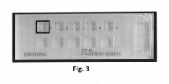
- BIOCHIP TM -Mosaics Frozen sections from a biopsy of one PV and two BP patients, respectively, were combined to BIOCHIP TM -Mosaics on one slide with HEK293 cells transfected with Desmoglein 1 (EUROIMMUN, substrate FD 1495-50; DSG1), Desmoglein 3 (EUROIMMUN substrate FD 1496-50; DSG3), BP230 (EUROIMMUN substrate FD 1502-56; BP230gC (SEQ ID NO 18)) or anti-BP180-NC16A-4X (EUROIMMUN substrate FD 1502-52; BP180-NC16A-4X) ( Figs. 1 and 2 ).
- tissue slices, acetone or formaldehyde fixed recombinant cell preparations or antigen preparations are used as antigenic substrates.
- the technology described in EP Patent 0 117 262 was used. Briefly, very thin glass plates chemically coated with spontaneously reacting aldehyde groups were covered with the antigens. Cell cultures directly grew on the glass plates. Free amino groups of the tissue slices, particularly the hydroxyl lysine of collagen, covalently adhered to the carrier material so that the tissue could not be removed by the following incubations with liquid reagents.
- the glass plates were industrially cut in very small glass chips with an edge length of 1-2 mm. Glass chips coated with different antigens can be combined in one reaction field of a slide to obtain a so called BIOCHIP-Mosaic TM .
- the individual mosaics were incubated with 30 ⁇ l PBS-Tween20 (2%) for 15 hours at 4°C. Bound antibodies were visualised by a FITC-labeled anti-human IgG antibody (EUROIMMUN, product no. AF 102, FITC-labelled anti-human-IgG (goat)). The incubation is carried out for 30 min. at room temperature (20-25° C). The slides were washed with PBS-Tween20 (0.2%) for 5 min. after both incubation steps. Then they were covered and evaluated under a fluorescence microscope (Axio Scope A1, Zeiss, Jena, Germany, article no. 490035-9100-000).
- Co-incubation of the PV biopsy showed parallel reactivity to desmosomes and Dsg3 (see Fig. 6 for an exemplary image), while co-incubation of the two BP biopsies resulted in staining of the epidermal basement membrane and BP180 and BP230 antigens (see Fig. 5 for an exemplary image).
- antibodies are detected which are present in residues of body fluids within the biopsy using the co-incubation technology.
Landscapes
- Health & Medical Sciences (AREA)
- Life Sciences & Earth Sciences (AREA)
- Immunology (AREA)
- Engineering & Computer Science (AREA)
- Chemical & Material Sciences (AREA)
- Hematology (AREA)
- Biomedical Technology (AREA)
- Molecular Biology (AREA)
- Urology & Nephrology (AREA)
- General Health & Medical Sciences (AREA)
- Analytical Chemistry (AREA)
- Physics & Mathematics (AREA)
- Pathology (AREA)
- General Physics & Mathematics (AREA)
- Biochemistry (AREA)
- Cell Biology (AREA)
- Microbiology (AREA)
- Biotechnology (AREA)
- Food Science & Technology (AREA)
- Medicinal Chemistry (AREA)
- Chemical Kinetics & Catalysis (AREA)
- Clinical Laboratory Science (AREA)
- Tropical Medicine & Parasitology (AREA)
- Rehabilitation Therapy (AREA)
- Rheumatology (AREA)
- Zoology (AREA)
- Virology (AREA)
- Physiology (AREA)
- Peptides Or Proteins (AREA)
- Medicines Containing Antibodies Or Antigens For Use As Internal Diagnostic Agents (AREA)
- Investigating Or Analysing Biological Materials (AREA)
Claims (9)
- Ein in-vitro-Verfahren zur Diagnose einer Autoimmunerkrankung, umfassendInkontaktbringen eines Spendergewebeschnitts mit einer Flüssigkeit, die in der Lage ist, einen Autoantikörper aus dem Spendergewebeschnitt zu extrahieren, und Inkontaktbringen der Flüssigkeit mit einem Akzeptormaterial, das ein Antigen umfasst, wobei der Spendergewebeschnitt von einem zu diagnostizierenden Patienten erhalten wurde,gefolgt von dem Nachweis eines Komplexes, umfassend den Autoantikörper und das Antigen, wobei der Nachweis des Komplexes indiziert, dass der Patient an einer Autoimmunerkrankung leidet,wobei der Spendergewebeschnitt und das Akzeptormaterial, das ein Antigen umfasst, gemeinsam in der Flüssigkeit inkubiert werden,wobei sowohl der Spendergewebeschnitt als auch das Akzeptormaterial zur gleichen Zeit mit der Flüssigkeit in Kontakt gebracht werden, zu der der Autoantikörper aus dem Spendergewebeschnitt extrahiert und zu dem Akzeptormaterial transportiert wird,wobei die Flüssigkeit eine jegliche Flüssigkeit ist, die bei Kontakt mit dem Spendergewebeschnitt die in dem Spendergewebeschnitt vorliegenden Autoantikörper aufnimmt, ihre chemische Zusammensetzung, Struktur oder Bindungseigenschaften, wie die Fähigkeit, spezifisch an ein Antigen zu binden, bewahrt und ihren Übergang zu dem Akzeptormaterial, das ein Antigen umfasst, ermöglicht.
- Das Verfahren nach Anspruch 1, wobei das Akzeptormaterial, das ein Antigen umfasst,eine Gewebeprobe, die das Antigen, vorzugsweise in der Form eines nativen Polypeptids, umfasst,eine Zelle, die das Antigen produziert, oderein isoliertes Polypeptid ist.
- Das Verfahren nach Anspruch 1 oder 2, wobei der Spendergewebeschnitt ein gefrorener Gewebeschnitt ist.
- Das Verfahren nach einem der Ansprüche 1 bis 3, wobeidie Krankheit eine gastrointestinale Autoimmunkrankheit, bevorzugt Zöliakie, ist,der Spendergewebeschnitt ein gastrointestinales, bevorzugt duodenales Gewebe, ist unddas Antigen Gewebetransglutaminase oder deamidiertes Gliadin oder eine Variante davon, bevorzugt ein Oligomer von deamidiertem Gliadin oder einer Variante davon, ist,wobei die Variante mindestens 90 % Sequenzidentität zu SEQ ID NO:1, SEQ ID NO:2, SEQ ID NO:3, SEQ ID NO:4 oder SEQ ID NO:5 oder einer Sequenz gemäß dem UniProt-Datenbankcode: P21980 aufweist.
- Das Verfahren nach einem der Ansprüche 1 bis 4, wobei dieKrankheit Pemphigus und/oder Pemphigoid, bevorzugt bullöses Pemphigoid, ist,der Spendergewebeschnitt erkranktes Hautgewebe ist unddas Akzeptormaterial ein oder mehrere Antigene aus der Gruppe umfasst, die Desmoglein-1 (Dsg1), Desmoglein-3 (Dsg3), bullöses Pemphigoid-Antigen 2 (BP180), 230 kDa bullöses Pemphigoid-Antigen (BP 230), Laminin-Untereinheit alpha-3 (LAMA3), Laminin332, beta4-Integrin und Kollagen Typ VII, nicht-kollagene Domäne A von BP180 (NC16A) und eine Variante davon umfasst,wobei die Variante mindestens 90 % Sequenzidentität zu SEQ ID NO:6, SEQ ID NO:7, SEQ ID NO:8, SEQ ID NO:14 oder SEQ ID NO:17 oder einer Sequenz gemäß dem UniProt-Datenbankcode: Q02413, P32926 oder Q02388 aufweist.
- Das Verfahren nach einem der Ansprüche 1 bis 5, wobei dieKrankheit Goodpasture-Syndrom oder systemischer Lupus erythematodes (SLE) ist,der Spendergewebeschnitt erkranktes Nierengewebe ist unddas Akzeptormaterial, das das Antigen umfasst, ein Material ist oder von einem Material abgeleitet ist, das aus der Gruppe, umfassend antiglomeruläre Basalmembran, doppelsträngige DNA (dsDNA), humane Epithelzellen (HEp-2), sekretorischer Phospholipase-A2-Rezeptor (pLA2R) und Thrombospondin-Typ-1-Domäne-enthaltendes Protein 7A (THSD7A) oder eine Variante von SEQ ID NO:9 oder SEQ ID NO:10, ausgewählt ist,wobei die Variante mindestens 90 % Sequenzidentität zu SEQ ID NO:9 oder SEQ ID NO:10 aufweist.
- Das Verfahren nach einem der Ansprüche 1 bis 6, wobeidie Krankheit Morbus Crohn ist,der Spendergewebeschnitt erkranktes Darmgewebe ist unddas Akzeptormaterial, das das Antigen umfasst, aus der Gruppe ausgewählt ist, die gesundes Pankreasgewebe, CUB und Zona pellucida-artige Domäne-enthaltendes Protein 1 (CUZD1) und pankreatisches Zymogengranula-Membranprotein GP-2 (GP2) und eine Variante von SEQ ID NO:11 oder SEQ ID NO:12 umfasst, wobei die beiden letzteren bevorzugt in der Form einer Zelle vorliegen, die CUZD1 und/oder GP2 oder eine Variante von SEQ ID NO:11 oder SEQ ID NO:12 exprimiert,wobei die Variante mindestens 90 % Sequenzidentität zu SEQ ID NO:11 oder SEQ ID NO: 12 aufweist.
- Das Verfahren nach einem der Ansprüche 1 bis 7, wobeidie Krankheit Morbus Basedow und/oder die Hashimoto-Krankheit ist,der Spendergewebeschnitt von der Schilddrüse abgeleitet ist unddas Akzeptormaterial, das das Antigen umfasst, gesundes Schilddrüsengewebe ist oder davon abgeleitet ist.
- Das Verfahren nach Anspruch 1, wobei die Flüssigkeit eine wässrige Flüssigkeit ist, die einen physiologischen Puffer mit einem pH-Wert von 5 bis 9 umfasst.
Applications Claiming Priority (1)
| Application Number | Priority Date | Filing Date | Title |
|---|---|---|---|
| EP16001395 | 2016-06-21 |
Publications (2)
| Publication Number | Publication Date |
|---|---|
| EP3260864A1 EP3260864A1 (de) | 2017-12-27 |
| EP3260864B1 true EP3260864B1 (de) | 2024-08-07 |
Family
ID=56203077
Family Applications (1)
| Application Number | Title | Priority Date | Filing Date |
|---|---|---|---|
| EP17001041.7A Active EP3260864B1 (de) | 2016-06-21 | 2017-06-21 | Diagnostischer co-inkubations-assay |
Country Status (3)
| Country | Link |
|---|---|
| US (1) | US10393734B2 (de) |
| EP (1) | EP3260864B1 (de) |
| CN (1) | CN107525919B (de) |
Families Citing this family (5)
| Publication number | Priority date | Publication date | Assignee | Title |
|---|---|---|---|---|
| SG10201908826UA (en) | 2018-10-22 | 2020-05-28 | Euroimmun Medizinische Labordiagnostika Ag | Diagnosis of blistering autoimmune diseases |
| CN112111015B (zh) * | 2020-09-24 | 2021-12-07 | 苏州携创生物技术有限公司 | PLA2R、C1q、THSD7A融合蛋白及其构建方法、应用 |
| CN112557670A (zh) * | 2020-12-07 | 2021-03-26 | 东南大学 | 一种基于尿液外泌体pla2r检测imn的方法、试剂盒及其应用 |
| CN114656572B (zh) * | 2022-02-25 | 2024-07-19 | 北京大学第一医院 | 一种bp180抗体快速检测试剂盒及其制备方法 |
| CN116482375A (zh) * | 2023-02-15 | 2023-07-25 | 杭州赛基生物科技有限公司 | 人膜性肾病相关指标检测试剂盒及其制备方法和使用方法 |
Family Cites Families (6)
| Publication number | Priority date | Publication date | Assignee | Title |
|---|---|---|---|---|
| ATE33717T1 (de) | 1983-02-25 | 1988-05-15 | Winfried Dr Med Stoecker | Verfahren und vorrichtungen fuer untersuchungen an unbeweglich gemachtem biologischem material. |
| EP1978364B1 (de) * | 2007-04-06 | 2011-06-01 | Zedira GmbH | Transglutaminase 6 als diagnostischer Indikator für Autoimmunerkrankungen |
| US7972804B2 (en) * | 2007-07-03 | 2011-07-05 | One Lambda | Methods of detecting antibodies specific for denatured HLA antigens |
| EP2191893B1 (de) * | 2008-11-19 | 2019-03-06 | Euroimmun Medizinische Labordiagnostika AG | Analyseverfahren und Vorrichtungen für biologische Reaktionen zwischen einer flüssigen und einer festen Phase |
| RU2491552C1 (ru) * | 2012-04-23 | 2013-08-27 | Государственное бюджетное учреждение здравоохранения города Москвы Научно-исследовательский институт скорой помощи имени Н.В. Склифосовского Департамента здравоохранения г. Москвы | Диагностикум для определения донор-специфических антител к главному комплексу гистосовместимости и способ его получения |
| CA2934070A1 (en) * | 2013-12-19 | 2015-06-25 | Opexa Therapeutics, Inc. | Methods of t cell epitope profiling, making t cell compositions, and treating diseases |
-
2017
- 2017-06-21 EP EP17001041.7A patent/EP3260864B1/de active Active
- 2017-06-21 CN CN201710475732.XA patent/CN107525919B/zh active Active
- 2017-06-21 US US15/629,528 patent/US10393734B2/en active Active
Also Published As
| Publication number | Publication date |
|---|---|
| CN107525919A (zh) | 2017-12-29 |
| EP3260864A1 (de) | 2017-12-27 |
| US20170370911A1 (en) | 2017-12-28 |
| US10393734B2 (en) | 2019-08-27 |
| CN107525919B (zh) | 2023-03-28 |
Similar Documents
| Publication | Publication Date | Title |
|---|---|---|
| EP3260864B1 (de) | Diagnostischer co-inkubations-assay | |
| EP2606356B1 (de) | Verfahren und mittel zur diagnose von spondyloarthritis mittels auto-antikörper-marker | |
| US8148084B2 (en) | Diagnosis of autoimmune disease | |
| US20120046181A1 (en) | Biomarkers for the Diagnosis of Renal Allograft and Kidney Status | |
| EP3364193B1 (de) | Verbesserter assay zur diagnose von erdnussallergie | |
| US11598772B2 (en) | Method and products for the diagnosis of a seafood allergy | |
| CN109738653B (zh) | 用于阿尔茨海默症的检测、诊断或风险预测的抗原蛋白组合以及包含其的试剂盒 | |
| US20100035360A1 (en) | Reagent for detection of autoantibody and kit for diagnosis of autoimmune disease | |
| US20150133322A1 (en) | Method for the diagnosis of early rheumatoid arthritis | |
| JP5924502B2 (ja) | リンパ球性漏斗下垂体後葉炎のバイオマーカー及びその用途 | |
| KR102441107B1 (ko) | 데옥시하이푸신·신타제 유전자를 지표로서 사용하는 동맥 경화 및 암의 검출 방법 | |
| EP2535714A1 (de) | Biomarker für Autoimmun-Lebererkrankungen und Verwendungen davon | |
| EP3477303A1 (de) | Methode und mittel zur diagnose von autoimmuner hepatitis mittels autoantikörper-marker | |
| WO2006054806A1 (ja) | クローン病関連自己抗原 | |
| JP2016223774A (ja) | リンパ球性漏斗下垂体後葉炎検査試薬及びその用途 | |
| KR102803104B1 (ko) | 새우에 대한 아나필락시스 진단용 바이오마커 조성물 및 이의 용도 | |
| EP2444805A2 (de) | Erkennung von Mesothelin-/Megakaryozyten zur Potenzierung von faktorbezogenen Peptiden in peritonealer Flüssigkeit zur Bewertung des Peritoneum und Peritonealhöhle | |
| EP4123306B1 (de) | Mmunologisches verfahren zum nachweis von mit zöliakie in zusammenhang stehenden antikörpern | |
| EP2816354B1 (de) | Biomarker für die Krankheit "complex regional pain syndrome" (CRPS) | |
| US5312628A (en) | Isolation of a soluble 42 KD pancreatic islet cell autoantigen | |
| CN121324669A (zh) | 检测抗kctd3自身抗体的试剂在诊断神经系统自身免疫性疾病中的应用 | |
| JP2023004699A (ja) | Abo血液型不適合腎移植を行う被験動物における抗体関連型拒絶反応の可能性を試験する方法 | |
| CN112285363A (zh) | 自身免疫类神经疾病的诊断 | |
| KR20200077779A (ko) | 퇴행성 뇌질환 발병 위험성 예측용 조성물 및 이를 이용한 퇴행성 뇌질환의 발병 위험성 예측 방법 | |
| WO2016005916A1 (en) | A method and kit for the detection of a biomarker of thyroid cancer in biological samples |
Legal Events
| Date | Code | Title | Description |
|---|---|---|---|
| PUAI | Public reference made under article 153(3) epc to a published international application that has entered the european phase |
Free format text: ORIGINAL CODE: 0009012 |
|
| STAA | Information on the status of an ep patent application or granted ep patent |
Free format text: STATUS: THE APPLICATION HAS BEEN PUBLISHED |
|
| AK | Designated contracting states |
Kind code of ref document: A1 Designated state(s): AL AT BE BG CH CY CZ DE DK EE ES FI FR GB GR HR HU IE IS IT LI LT LU LV MC MK MT NL NO PL PT RO RS SE SI SK SM TR |
|
| AX | Request for extension of the european patent |
Extension state: BA ME |
|
| STAA | Information on the status of an ep patent application or granted ep patent |
Free format text: STATUS: REQUEST FOR EXAMINATION WAS MADE |
|
| 17P | Request for examination filed |
Effective date: 20180626 |
|
| RBV | Designated contracting states (corrected) |
Designated state(s): AL AT BE BG CH CY CZ DE DK EE ES FI FR GB GR HR HU IE IS IT LI LT LU LV MC MK MT NL NO PL PT RO RS SE SI SK SM TR |
|
| STAA | Information on the status of an ep patent application or granted ep patent |
Free format text: STATUS: EXAMINATION IS IN PROGRESS |
|
| 17Q | First examination report despatched |
Effective date: 20181102 |
|
| GRAP | Despatch of communication of intention to grant a patent |
Free format text: ORIGINAL CODE: EPIDOSNIGR1 |
|
| STAA | Information on the status of an ep patent application or granted ep patent |
Free format text: STATUS: GRANT OF PATENT IS INTENDED |
|
| GRAJ | Information related to disapproval of communication of intention to grant by the applicant or resumption of examination proceedings by the epo deleted |
Free format text: ORIGINAL CODE: EPIDOSDIGR1 |
|
| STAA | Information on the status of an ep patent application or granted ep patent |
Free format text: STATUS: EXAMINATION IS IN PROGRESS |
|
| INTG | Intention to grant announced |
Effective date: 20201117 |
|
| INTC | Intention to grant announced (deleted) | ||
| GRAJ | Information related to disapproval of communication of intention to grant by the applicant or resumption of examination proceedings by the epo deleted |
Free format text: ORIGINAL CODE: EPIDOSDIGR1 |
|
| GRAP | Despatch of communication of intention to grant a patent |
Free format text: ORIGINAL CODE: EPIDOSNIGR1 |
|
| GRAP | Despatch of communication of intention to grant a patent |
Free format text: ORIGINAL CODE: EPIDOSNIGR1 |
|
| STAA | Information on the status of an ep patent application or granted ep patent |
Free format text: STATUS: GRANT OF PATENT IS INTENDED |
|
| INTG | Intention to grant announced |
Effective date: 20221205 |
|
| GRAJ | Information related to disapproval of communication of intention to grant by the applicant or resumption of examination proceedings by the epo deleted |
Free format text: ORIGINAL CODE: EPIDOSDIGR1 |
|
| STAA | Information on the status of an ep patent application or granted ep patent |
Free format text: STATUS: EXAMINATION IS IN PROGRESS |
|
| INTC | Intention to grant announced (deleted) | ||
| RIN1 | Information on inventor provided before grant (corrected) |
Inventor name: JAHNKE, ANNIKA Inventor name: TEEGEN, BIANCA Inventor name: STOECKER, WINFRIED |
|
| GRAP | Despatch of communication of intention to grant a patent |
Free format text: ORIGINAL CODE: EPIDOSNIGR1 |
|
| STAA | Information on the status of an ep patent application or granted ep patent |
Free format text: STATUS: GRANT OF PATENT IS INTENDED |
|
| INTG | Intention to grant announced |
Effective date: 20240318 |
|
| GRAS | Grant fee paid |
Free format text: ORIGINAL CODE: EPIDOSNIGR3 |
|
| GRAA | (expected) grant |
Free format text: ORIGINAL CODE: 0009210 |
|
| STAA | Information on the status of an ep patent application or granted ep patent |
Free format text: STATUS: THE PATENT HAS BEEN GRANTED |
|
| AK | Designated contracting states |
Kind code of ref document: B1 Designated state(s): AL AT BE BG CH CY CZ DE DK EE ES FI FR GB GR HR HU IE IS IT LI LT LU LV MC MK MT NL NO PL PT RO RS SE SI SK SM TR |
|
| P01 | Opt-out of the competence of the unified patent court (upc) registered |
Free format text: CASE NUMBER: APP_39741/2024 Effective date: 20240703 |
|
| REG | Reference to a national code |
Ref country code: GB Ref legal event code: FG4D |
|
| REG | Reference to a national code |
Ref country code: CH Ref legal event code: EP |
|
| REG | Reference to a national code |
Ref country code: IE Ref legal event code: FG4D |
|
| REG | Reference to a national code |
Ref country code: DE Ref legal event code: R096 Ref document number: 602017083827 Country of ref document: DE |
|
| REG | Reference to a national code |
Ref country code: LT Ref legal event code: MG9D |
|
| REG | Reference to a national code |
Ref country code: NL Ref legal event code: MP Effective date: 20240807 |
|
| PG25 | Lapsed in a contracting state [announced via postgrant information from national office to epo] |
Ref country code: NO Free format text: LAPSE BECAUSE OF FAILURE TO SUBMIT A TRANSLATION OF THE DESCRIPTION OR TO PAY THE FEE WITHIN THE PRESCRIBED TIME-LIMIT Effective date: 20241107 |
|
| REG | Reference to a national code |
Ref country code: AT Ref legal event code: MK05 Ref document number: 1711497 Country of ref document: AT Kind code of ref document: T Effective date: 20240807 |
|
| PG25 | Lapsed in a contracting state [announced via postgrant information from national office to epo] |
Ref country code: PT Free format text: LAPSE BECAUSE OF FAILURE TO SUBMIT A TRANSLATION OF THE DESCRIPTION OR TO PAY THE FEE WITHIN THE PRESCRIBED TIME-LIMIT Effective date: 20241209 Ref country code: PL Free format text: LAPSE BECAUSE OF FAILURE TO SUBMIT A TRANSLATION OF THE DESCRIPTION OR TO PAY THE FEE WITHIN THE PRESCRIBED TIME-LIMIT Effective date: 20240807 Ref country code: FI Free format text: LAPSE BECAUSE OF FAILURE TO SUBMIT A TRANSLATION OF THE DESCRIPTION OR TO PAY THE FEE WITHIN THE PRESCRIBED TIME-LIMIT Effective date: 20240807 Ref country code: NL Free format text: LAPSE BECAUSE OF FAILURE TO SUBMIT A TRANSLATION OF THE DESCRIPTION OR TO PAY THE FEE WITHIN THE PRESCRIBED TIME-LIMIT Effective date: 20240807 Ref country code: GR Free format text: LAPSE BECAUSE OF FAILURE TO SUBMIT A TRANSLATION OF THE DESCRIPTION OR TO PAY THE FEE WITHIN THE PRESCRIBED TIME-LIMIT Effective date: 20241108 |
|
| PG25 | Lapsed in a contracting state [announced via postgrant information from national office to epo] |
Ref country code: BG Free format text: LAPSE BECAUSE OF FAILURE TO SUBMIT A TRANSLATION OF THE DESCRIPTION OR TO PAY THE FEE WITHIN THE PRESCRIBED TIME-LIMIT Effective date: 20240807 |
|
| PG25 | Lapsed in a contracting state [announced via postgrant information from national office to epo] |
Ref country code: LV Free format text: LAPSE BECAUSE OF FAILURE TO SUBMIT A TRANSLATION OF THE DESCRIPTION OR TO PAY THE FEE WITHIN THE PRESCRIBED TIME-LIMIT Effective date: 20240807 |
|
| PG25 | Lapsed in a contracting state [announced via postgrant information from national office to epo] |
Ref country code: AT Free format text: LAPSE BECAUSE OF FAILURE TO SUBMIT A TRANSLATION OF THE DESCRIPTION OR TO PAY THE FEE WITHIN THE PRESCRIBED TIME-LIMIT Effective date: 20240807 Ref country code: IS Free format text: LAPSE BECAUSE OF FAILURE TO SUBMIT A TRANSLATION OF THE DESCRIPTION OR TO PAY THE FEE WITHIN THE PRESCRIBED TIME-LIMIT Effective date: 20241207 |
|
| PG25 | Lapsed in a contracting state [announced via postgrant information from national office to epo] |
Ref country code: HR Free format text: LAPSE BECAUSE OF FAILURE TO SUBMIT A TRANSLATION OF THE DESCRIPTION OR TO PAY THE FEE WITHIN THE PRESCRIBED TIME-LIMIT Effective date: 20240807 |
|
| PG25 | Lapsed in a contracting state [announced via postgrant information from national office to epo] |
Ref country code: ES Free format text: LAPSE BECAUSE OF FAILURE TO SUBMIT A TRANSLATION OF THE DESCRIPTION OR TO PAY THE FEE WITHIN THE PRESCRIBED TIME-LIMIT Effective date: 20240807 Ref country code: RS Free format text: LAPSE BECAUSE OF FAILURE TO SUBMIT A TRANSLATION OF THE DESCRIPTION OR TO PAY THE FEE WITHIN THE PRESCRIBED TIME-LIMIT Effective date: 20241107 |
|
| PG25 | Lapsed in a contracting state [announced via postgrant information from national office to epo] |
Ref country code: RS Free format text: LAPSE BECAUSE OF FAILURE TO SUBMIT A TRANSLATION OF THE DESCRIPTION OR TO PAY THE FEE WITHIN THE PRESCRIBED TIME-LIMIT Effective date: 20241107 Ref country code: PT Free format text: LAPSE BECAUSE OF FAILURE TO SUBMIT A TRANSLATION OF THE DESCRIPTION OR TO PAY THE FEE WITHIN THE PRESCRIBED TIME-LIMIT Effective date: 20241209 Ref country code: PL Free format text: LAPSE BECAUSE OF FAILURE TO SUBMIT A TRANSLATION OF THE DESCRIPTION OR TO PAY THE FEE WITHIN THE PRESCRIBED TIME-LIMIT Effective date: 20240807 Ref country code: NO Free format text: LAPSE BECAUSE OF FAILURE TO SUBMIT A TRANSLATION OF THE DESCRIPTION OR TO PAY THE FEE WITHIN THE PRESCRIBED TIME-LIMIT Effective date: 20241107 Ref country code: NL Free format text: LAPSE BECAUSE OF FAILURE TO SUBMIT A TRANSLATION OF THE DESCRIPTION OR TO PAY THE FEE WITHIN THE PRESCRIBED TIME-LIMIT Effective date: 20240807 Ref country code: LV Free format text: LAPSE BECAUSE OF FAILURE TO SUBMIT A TRANSLATION OF THE DESCRIPTION OR TO PAY THE FEE WITHIN THE PRESCRIBED TIME-LIMIT Effective date: 20240807 Ref country code: IS Free format text: LAPSE BECAUSE OF FAILURE TO SUBMIT A TRANSLATION OF THE DESCRIPTION OR TO PAY THE FEE WITHIN THE PRESCRIBED TIME-LIMIT Effective date: 20241207 Ref country code: HR Free format text: LAPSE BECAUSE OF FAILURE TO SUBMIT A TRANSLATION OF THE DESCRIPTION OR TO PAY THE FEE WITHIN THE PRESCRIBED TIME-LIMIT Effective date: 20240807 Ref country code: GR Free format text: LAPSE BECAUSE OF FAILURE TO SUBMIT A TRANSLATION OF THE DESCRIPTION OR TO PAY THE FEE WITHIN THE PRESCRIBED TIME-LIMIT Effective date: 20241108 Ref country code: FI Free format text: LAPSE BECAUSE OF FAILURE TO SUBMIT A TRANSLATION OF THE DESCRIPTION OR TO PAY THE FEE WITHIN THE PRESCRIBED TIME-LIMIT Effective date: 20240807 Ref country code: ES Free format text: LAPSE BECAUSE OF FAILURE TO SUBMIT A TRANSLATION OF THE DESCRIPTION OR TO PAY THE FEE WITHIN THE PRESCRIBED TIME-LIMIT Effective date: 20240807 Ref country code: BG Free format text: LAPSE BECAUSE OF FAILURE TO SUBMIT A TRANSLATION OF THE DESCRIPTION OR TO PAY THE FEE WITHIN THE PRESCRIBED TIME-LIMIT Effective date: 20240807 Ref country code: AT Free format text: LAPSE BECAUSE OF FAILURE TO SUBMIT A TRANSLATION OF THE DESCRIPTION OR TO PAY THE FEE WITHIN THE PRESCRIBED TIME-LIMIT Effective date: 20240807 |
|
| PG25 | Lapsed in a contracting state [announced via postgrant information from national office to epo] |
Ref country code: RO Free format text: LAPSE BECAUSE OF FAILURE TO SUBMIT A TRANSLATION OF THE DESCRIPTION OR TO PAY THE FEE WITHIN THE PRESCRIBED TIME-LIMIT Effective date: 20240807 Ref country code: DK Free format text: LAPSE BECAUSE OF FAILURE TO SUBMIT A TRANSLATION OF THE DESCRIPTION OR TO PAY THE FEE WITHIN THE PRESCRIBED TIME-LIMIT Effective date: 20240807 Ref country code: SM Free format text: LAPSE BECAUSE OF FAILURE TO SUBMIT A TRANSLATION OF THE DESCRIPTION OR TO PAY THE FEE WITHIN THE PRESCRIBED TIME-LIMIT Effective date: 20240807 |
|
| PG25 | Lapsed in a contracting state [announced via postgrant information from national office to epo] |
Ref country code: EE Free format text: LAPSE BECAUSE OF FAILURE TO SUBMIT A TRANSLATION OF THE DESCRIPTION OR TO PAY THE FEE WITHIN THE PRESCRIBED TIME-LIMIT Effective date: 20240807 |
|
| PG25 | Lapsed in a contracting state [announced via postgrant information from national office to epo] |
Ref country code: CZ Free format text: LAPSE BECAUSE OF FAILURE TO SUBMIT A TRANSLATION OF THE DESCRIPTION OR TO PAY THE FEE WITHIN THE PRESCRIBED TIME-LIMIT Effective date: 20240807 |
|
| PG25 | Lapsed in a contracting state [announced via postgrant information from national office to epo] |
Ref country code: SK Free format text: LAPSE BECAUSE OF FAILURE TO SUBMIT A TRANSLATION OF THE DESCRIPTION OR TO PAY THE FEE WITHIN THE PRESCRIBED TIME-LIMIT Effective date: 20240807 |
|
| REG | Reference to a national code |
Ref country code: DE Ref legal event code: R084 Ref document number: 602017083827 Country of ref document: DE |
|
| REG | Reference to a national code |
Ref country code: DE Ref legal event code: R097 Ref document number: 602017083827 Country of ref document: DE |
|
| PLBE | No opposition filed within time limit |
Free format text: ORIGINAL CODE: 0009261 |
|
| STAA | Information on the status of an ep patent application or granted ep patent |
Free format text: STATUS: NO OPPOSITION FILED WITHIN TIME LIMIT |
|
| PGFP | Annual fee paid to national office [announced via postgrant information from national office to epo] |
Ref country code: DE Payment date: 20250618 Year of fee payment: 9 |
|
| REG | Reference to a national code |
Ref country code: GB Ref legal event code: 746 Effective date: 20250617 |
|
| PGFP | Annual fee paid to national office [announced via postgrant information from national office to epo] |
Ref country code: GB Payment date: 20250620 Year of fee payment: 9 |
|
| 26N | No opposition filed |
Effective date: 20250508 |
|
| PGFP | Annual fee paid to national office [announced via postgrant information from national office to epo] |
Ref country code: FR Payment date: 20250626 Year of fee payment: 9 |
|
| PG25 | Lapsed in a contracting state [announced via postgrant information from national office to epo] |
Ref country code: SE Free format text: LAPSE BECAUSE OF FAILURE TO SUBMIT A TRANSLATION OF THE DESCRIPTION OR TO PAY THE FEE WITHIN THE PRESCRIBED TIME-LIMIT Effective date: 20240807 |
|
| REG | Reference to a national code |
Ref country code: CH Ref legal event code: H13 Free format text: ST27 STATUS EVENT CODE: U-0-0-H10-H13 (AS PROVIDED BY THE NATIONAL OFFICE) Effective date: 20260127 |
|
| PG25 | Lapsed in a contracting state [announced via postgrant information from national office to epo] |
Ref country code: IT Free format text: LAPSE BECAUSE OF FAILURE TO SUBMIT A TRANSLATION OF THE DESCRIPTION OR TO PAY THE FEE WITHIN THE PRESCRIBED TIME-LIMIT Effective date: 20240807 |
|
| PG25 | Lapsed in a contracting state [announced via postgrant information from national office to epo] |
Ref country code: MC Free format text: LAPSE BECAUSE OF FAILURE TO SUBMIT A TRANSLATION OF THE DESCRIPTION OR TO PAY THE FEE WITHIN THE PRESCRIBED TIME-LIMIT Effective date: 20240807 |
|
| PG25 | Lapsed in a contracting state [announced via postgrant information from national office to epo] |
Ref country code: LU Free format text: LAPSE BECAUSE OF NON-PAYMENT OF DUE FEES Effective date: 20250621 |
|
| REG | Reference to a national code |
Ref country code: BE Ref legal event code: MM Effective date: 20250630 |
|
| PG25 | Lapsed in a contracting state [announced via postgrant information from national office to epo] |
Ref country code: IE Free format text: LAPSE BECAUSE OF NON-PAYMENT OF DUE FEES Effective date: 20250621 |
|
| PG25 | Lapsed in a contracting state [announced via postgrant information from national office to epo] |
Ref country code: BE Free format text: LAPSE BECAUSE OF NON-PAYMENT OF DUE FEES Effective date: 20250630 |
|
| PG25 | Lapsed in a contracting state [announced via postgrant information from national office to epo] |
Ref country code: CH Free format text: LAPSE BECAUSE OF NON-PAYMENT OF DUE FEES Effective date: 20250630 |