EP3280344B1 - Système d'utilisation de la réalité augmentée pour améliorer une opération chirurgicale - Google Patents
Système d'utilisation de la réalité augmentée pour améliorer une opération chirurgicale Download PDFInfo
- Publication number
- EP3280344B1 EP3280344B1 EP16718475.3A EP16718475A EP3280344B1 EP 3280344 B1 EP3280344 B1 EP 3280344B1 EP 16718475 A EP16718475 A EP 16718475A EP 3280344 B1 EP3280344 B1 EP 3280344B1
- Authority
- EP
- European Patent Office
- Prior art keywords
- target area
- projection
- dimensional
- user
- image stack
- Prior art date
- Legal status (The legal status is an assumption and is not a legal conclusion. Google has not performed a legal analysis and makes no representation as to the accuracy of the status listed.)
- Active
Links
Images




Classifications
-
- A—HUMAN NECESSITIES
- A61—MEDICAL OR VETERINARY SCIENCE; HYGIENE
- A61B—DIAGNOSIS; SURGERY; IDENTIFICATION
- A61B34/00—Computer-aided surgery; Manipulators or robots specially adapted for use in surgery
- A61B34/25—User interfaces for surgical systems
-
- A—HUMAN NECESSITIES
- A61—MEDICAL OR VETERINARY SCIENCE; HYGIENE
- A61B—DIAGNOSIS; SURGERY; IDENTIFICATION
- A61B34/00—Computer-aided surgery; Manipulators or robots specially adapted for use in surgery
- A61B34/10—Computer-aided planning, simulation or modelling of surgical operations
-
- A—HUMAN NECESSITIES
- A61—MEDICAL OR VETERINARY SCIENCE; HYGIENE
- A61B—DIAGNOSIS; SURGERY; IDENTIFICATION
- A61B34/00—Computer-aided surgery; Manipulators or robots specially adapted for use in surgery
- A61B34/20—Surgical navigation systems; Devices for tracking or guiding surgical instruments, e.g. for frameless stereotaxis
-
- A—HUMAN NECESSITIES
- A61—MEDICAL OR VETERINARY SCIENCE; HYGIENE
- A61B—DIAGNOSIS; SURGERY; IDENTIFICATION
- A61B90/00—Instruments, implements or accessories specially adapted for surgery or diagnosis and not covered by any of the groups A61B1/00 - A61B50/00, e.g. for luxation treatment or for protecting wound edges
- A61B90/36—Image-producing devices or illumination devices not otherwise provided for
- A61B90/37—Surgical systems with images on a monitor during operation
-
- G—PHYSICS
- G06—COMPUTING OR CALCULATING; COUNTING
- G06T—IMAGE DATA PROCESSING OR GENERATION, IN GENERAL
- G06T7/00—Image analysis
- G06T7/70—Determining position or orientation of objects or cameras
- G06T7/73—Determining position or orientation of objects or cameras using feature-based methods
-
- A—HUMAN NECESSITIES
- A61—MEDICAL OR VETERINARY SCIENCE; HYGIENE
- A61B—DIAGNOSIS; SURGERY; IDENTIFICATION
- A61B17/00—Surgical instruments, devices or methods
- A61B2017/00681—Aspects not otherwise provided for
- A61B2017/00694—Aspects not otherwise provided for with means correcting for movement of or for synchronisation with the body
-
- A—HUMAN NECESSITIES
- A61—MEDICAL OR VETERINARY SCIENCE; HYGIENE
- A61B—DIAGNOSIS; SURGERY; IDENTIFICATION
- A61B34/00—Computer-aided surgery; Manipulators or robots specially adapted for use in surgery
- A61B34/10—Computer-aided planning, simulation or modelling of surgical operations
- A61B2034/101—Computer-aided simulation of surgical operations
- A61B2034/105—Modelling of the patient, e.g. for ligaments or bones
-
- A—HUMAN NECESSITIES
- A61—MEDICAL OR VETERINARY SCIENCE; HYGIENE
- A61B—DIAGNOSIS; SURGERY; IDENTIFICATION
- A61B34/00—Computer-aided surgery; Manipulators or robots specially adapted for use in surgery
- A61B34/10—Computer-aided planning, simulation or modelling of surgical operations
- A61B2034/107—Visualisation of planned trajectories or target regions
-
- A—HUMAN NECESSITIES
- A61—MEDICAL OR VETERINARY SCIENCE; HYGIENE
- A61B—DIAGNOSIS; SURGERY; IDENTIFICATION
- A61B34/00—Computer-aided surgery; Manipulators or robots specially adapted for use in surgery
- A61B34/20—Surgical navigation systems; Devices for tracking or guiding surgical instruments, e.g. for frameless stereotaxis
- A61B2034/2046—Tracking techniques
- A61B2034/2048—Tracking techniques using an accelerometer or inertia sensor
-
- A—HUMAN NECESSITIES
- A61—MEDICAL OR VETERINARY SCIENCE; HYGIENE
- A61B—DIAGNOSIS; SURGERY; IDENTIFICATION
- A61B34/00—Computer-aided surgery; Manipulators or robots specially adapted for use in surgery
- A61B34/20—Surgical navigation systems; Devices for tracking or guiding surgical instruments, e.g. for frameless stereotaxis
- A61B2034/2046—Tracking techniques
- A61B2034/2055—Optical tracking systems
-
- A—HUMAN NECESSITIES
- A61—MEDICAL OR VETERINARY SCIENCE; HYGIENE
- A61B—DIAGNOSIS; SURGERY; IDENTIFICATION
- A61B90/00—Instruments, implements or accessories specially adapted for surgery or diagnosis and not covered by any of the groups A61B1/00 - A61B50/00, e.g. for luxation treatment or for protecting wound edges
- A61B90/36—Image-producing devices or illumination devices not otherwise provided for
- A61B2090/363—Use of fiducial points
-
- A—HUMAN NECESSITIES
- A61—MEDICAL OR VETERINARY SCIENCE; HYGIENE
- A61B—DIAGNOSIS; SURGERY; IDENTIFICATION
- A61B90/00—Instruments, implements or accessories specially adapted for surgery or diagnosis and not covered by any of the groups A61B1/00 - A61B50/00, e.g. for luxation treatment or for protecting wound edges
- A61B90/36—Image-producing devices or illumination devices not otherwise provided for
- A61B2090/364—Correlation of different images or relation of image positions in respect to the body
- A61B2090/365—Correlation of different images or relation of image positions in respect to the body augmented reality, i.e. correlating a live optical image with another image
-
- A—HUMAN NECESSITIES
- A61—MEDICAL OR VETERINARY SCIENCE; HYGIENE
- A61B—DIAGNOSIS; SURGERY; IDENTIFICATION
- A61B90/00—Instruments, implements or accessories specially adapted for surgery or diagnosis and not covered by any of the groups A61B1/00 - A61B50/00, e.g. for luxation treatment or for protecting wound edges
- A61B90/36—Image-producing devices or illumination devices not otherwise provided for
- A61B2090/364—Correlation of different images or relation of image positions in respect to the body
- A61B2090/367—Correlation of different images or relation of image positions in respect to the body creating a 3D dataset from 2D images using position information
-
- A—HUMAN NECESSITIES
- A61—MEDICAL OR VETERINARY SCIENCE; HYGIENE
- A61B—DIAGNOSIS; SURGERY; IDENTIFICATION
- A61B90/00—Instruments, implements or accessories specially adapted for surgery or diagnosis and not covered by any of the groups A61B1/00 - A61B50/00, e.g. for luxation treatment or for protecting wound edges
- A61B90/36—Image-producing devices or illumination devices not otherwise provided for
- A61B2090/364—Correlation of different images or relation of image positions in respect to the body
- A61B2090/368—Correlation of different images or relation of image positions in respect to the body changing the image on a display according to the operator's position
-
- A—HUMAN NECESSITIES
- A61—MEDICAL OR VETERINARY SCIENCE; HYGIENE
- A61B—DIAGNOSIS; SURGERY; IDENTIFICATION
- A61B90/00—Instruments, implements or accessories specially adapted for surgery or diagnosis and not covered by any of the groups A61B1/00 - A61B50/00, e.g. for luxation treatment or for protecting wound edges
- A61B90/39—Markers, e.g. radio-opaque or breast lesions markers
- A61B2090/3904—Markers, e.g. radio-opaque or breast lesions markers specially adapted for marking specified tissue
- A61B2090/3908—Soft tissue, e.g. breast tissue
-
- A—HUMAN NECESSITIES
- A61—MEDICAL OR VETERINARY SCIENCE; HYGIENE
- A61B—DIAGNOSIS; SURGERY; IDENTIFICATION
- A61B90/00—Instruments, implements or accessories specially adapted for surgery or diagnosis and not covered by any of the groups A61B1/00 - A61B50/00, e.g. for luxation treatment or for protecting wound edges
- A61B90/39—Markers, e.g. radio-opaque or breast lesions markers
- A61B2090/3937—Visible markers
-
- A—HUMAN NECESSITIES
- A61—MEDICAL OR VETERINARY SCIENCE; HYGIENE
- A61B—DIAGNOSIS; SURGERY; IDENTIFICATION
- A61B90/00—Instruments, implements or accessories specially adapted for surgery or diagnosis and not covered by any of the groups A61B1/00 - A61B50/00, e.g. for luxation treatment or for protecting wound edges
- A61B90/50—Supports for surgical instruments, e.g. articulated arms
- A61B2090/502—Headgear, e.g. helmet, spectacles
-
- G—PHYSICS
- G02—OPTICS
- G02B—OPTICAL ELEMENTS, SYSTEMS OR APPARATUS
- G02B27/00—Optical systems or apparatus not provided for by any of the groups G02B1/00 - G02B26/00, G02B30/00
- G02B27/01—Head-up displays
- G02B27/0101—Head-up displays characterised by optical features
- G02B2027/0132—Head-up displays characterised by optical features comprising binocular systems
- G02B2027/0134—Head-up displays characterised by optical features comprising binocular systems of stereoscopic type
-
- G—PHYSICS
- G02—OPTICS
- G02B—OPTICAL ELEMENTS, SYSTEMS OR APPARATUS
- G02B27/00—Optical systems or apparatus not provided for by any of the groups G02B1/00 - G02B26/00, G02B30/00
- G02B27/01—Head-up displays
- G02B27/017—Head mounted
- G02B2027/0178—Eyeglass type
-
- G—PHYSICS
- G02—OPTICS
- G02B—OPTICAL ELEMENTS, SYSTEMS OR APPARATUS
- G02B27/00—Optical systems or apparatus not provided for by any of the groups G02B1/00 - G02B26/00, G02B30/00
- G02B27/01—Head-up displays
- G02B27/017—Head mounted
-
- G—PHYSICS
- G06—COMPUTING OR CALCULATING; COUNTING
- G06T—IMAGE DATA PROCESSING OR GENERATION, IN GENERAL
- G06T19/00—Manipulating three-dimensional [3D] models or images for computer graphics
- G06T19/006—Mixed reality
-
- G—PHYSICS
- G06—COMPUTING OR CALCULATING; COUNTING
- G06T—IMAGE DATA PROCESSING OR GENERATION, IN GENERAL
- G06T2207/00—Indexing scheme for image analysis or image enhancement
- G06T2207/10—Image acquisition modality
- G06T2207/10024—Color image
-
- G—PHYSICS
- G06—COMPUTING OR CALCULATING; COUNTING
- G06T—IMAGE DATA PROCESSING OR GENERATION, IN GENERAL
- G06T2207/00—Indexing scheme for image analysis or image enhancement
- G06T2207/10—Image acquisition modality
- G06T2207/10072—Tomographic images
-
- G—PHYSICS
- G06—COMPUTING OR CALCULATING; COUNTING
- G06T—IMAGE DATA PROCESSING OR GENERATION, IN GENERAL
- G06T2207/00—Indexing scheme for image analysis or image enhancement
- G06T2207/20—Special algorithmic details
- G06T2207/20212—Image combination
- G06T2207/20221—Image fusion; Image merging
-
- G—PHYSICS
- G06—COMPUTING OR CALCULATING; COUNTING
- G06T—IMAGE DATA PROCESSING OR GENERATION, IN GENERAL
- G06T2207/00—Indexing scheme for image analysis or image enhancement
- G06T2207/30—Subject of image; Context of image processing
- G06T2207/30004—Biomedical image processing
-
- G—PHYSICS
- G06—COMPUTING OR CALCULATING; COUNTING
- G06T—IMAGE DATA PROCESSING OR GENERATION, IN GENERAL
- G06T2207/00—Indexing scheme for image analysis or image enhancement
- G06T2207/30—Subject of image; Context of image processing
- G06T2207/30204—Marker
Definitions
- embodiments described herein illustrate methods, apparatus, and systems for utilizing augmented reality, based on the use of three dimensional (3D) medical imaging to enhance the use of surgical tools such as a neuronavigator (a machine able to track the position of surgical probes inside the brain or other operating areas of a patient, and visualize it on MRI or CT scans of the patient itself).
- 3D three dimensional
- a surgeon may utilize a head-mounted display to perceive the 3D model of a target area during a surgery.
- This 3D model can be extracted from a scan, and visualized directly as a superimposition onto the actual target area of the patient undergoing surgery using augmented reality glasses.
- Neuronavigation systems are unique tools used to access lesions or tumors or other target areas located far too deep inside the brain for traditional surgical methods and which commonly result in surgical sites that do not have clear visibility or access.
- the set-up of assistive machinery in the operating room e.g., a neuronavigator or the like
- the precision by which surgical tools associated with the neuronavigator can be tracked is limited by the fact that the system cannot take into account the natural movements and deformations (swelling) occurring in the target area (e.g., the brain) during surgery.
- the use of the assistive machine itself is not very comfortable because the surgeon needs to focus attention on a screen that illustrates the actual position of the probe, while the surgeon's hands are operating on an operatory field that is oriented differently from this field of view. It is not uncommon, for instance, for the orientation of the images on the screen to be the reverse of the physical orientation in the actual working area, a situation that requires an extra level of concentration and may limit a surgeon's operative capabilities. These issues limit the distribution and use of assistive devices such as neuronavigators.
- the inventors have identified a need for simplifying the preparation of the operating room, improving the comfort of the surgeon by enabling the direct visualization of the position and the path of the surgical probes superimposed on the real field of view of the surgeon, and improving the tracking of both the surgical instruments and the target areas upon which surgery is performed, in order to proficiently operate on small targets.
- example embodiments described herein utilize improved digital imaging tools that provide a three dimensional visual guide during surgery.
- a head-mounted display provides augmented reality visualizations that improve a surgeon's understanding of the relative locations of a target area within the context of the surgeon's field of view, and, in some embodiments, also illustrates a relative location of the surgeon's surgical tools.
- augmented reality 3D glasses example embodiments deploy the 3D tracking technology and the 3D visualization of the surgical field in a manner that allows the surgeon to focus his attention (sight and hands) on the operatory field and on the patient rather than on separate screen. Similarly, using example embodiments described below, the surgeon is not required to interpolate movements that appear on an external display in one orientation on the fly to properly respond with movements in the actual working area.
- Figure 1 shows an example system that may perform the operations described herein for utilizing three dimensional augmented reality visualizations to assist surgery.
- computing device 100 may be configured to perform various operations in accordance with example embodiments of the present invention, such as in conjunction with the operations described in conjunction with Figures 4 through 8 below.
- the components, devices, and elements described herein may not be mandatory in every embodiment of the computing device 100, and some may be omitted in certain embodiments. Additionally, some embodiments may include further or different components, devices or elements beyond those shown and described herein.
- the computing device 100 may include or otherwise be in communication with a processing system including, for example, processing circuitry 102 that is configurable to perform actions in accordance with example embodiments described herein.
- the processing circuitry 102 may be configured to perform data processing, application execution and/or other processing and management services according to an example embodiment of the present invention.
- the computing device 100 or the processing circuitry 102 may be embodied as a chip or chip set.
- the computing device 100 or the processing circuitry 102 may comprise one or more physical packages (e.g., chips) including materials, components and/or wires on a structural assembly (e.g., a baseboard).
- the computing device 100 or the processing circuitry 102 may, in some cases, be configured to implement an embodiment of the present invention on a single chip or as a single "system on a chip.” As such, in some cases, a chip or chipset may constitute means for performing one or more operations for providing the functionalities described herein.
- the processing circuitry 102 may include a processor 104 and memory 106 that may be in communication with or otherwise control a head-mounted display 108 and, in some embodiments, a separate user interface 110.
- the processing circuitry 102 may further be in communication or otherwise control a scanner 114 (which may be external to the computing device 100 or, in some embodiments, may be included as a component of the computing device 100).
- the processing circuitry 102 may be embodied as a circuit chip (e.g., an integrated circuit chip) configured (e.g., with hardware or a combination of hardware and software) to perform or direct performance of operations described herein.
- the processor 104 may be embodied in a number of different ways.
- the processor 104 may be embodied as various processing means such as one or more of a microprocessor or other processing element, a coprocessor, a controller or various other computing or processing devices including integrated circuits such as, for example, an ASIC (application specific integrated circuit), an FPGA (field programmable gate array), or the like.
- the processor 104 may be configured to execute program code instructions stored in the memory 106 or otherwise accessible to the processor 104.
- the processor 104 may represent an entity (e.g., physically embodied in circuitry) capable of performing operations according to embodiments of the present invention while configured accordingly.
- the memory 106 may include one or more non-transitory memory devices such as, for example, volatile and/or non-volatile memory that may be either fixed or removable.
- the memory 106 may be configured to store information, data, applications, instructions or the like for enabling the computing device 100 to carry out various functions in accordance with example embodiments of the present invention.
- the memory 106 could be configured to buffer input data for processing by the processor 104.
- the memory 106 could be configured to store instructions for execution by the processor 104.
- the memory 106 may include one of a plurality of databases that may store a variety of files, contents or data sets.
- applications may be stored for execution by the processor 104 in order to carry out the functionality associated with each respective application.
- the memory 106 may be in communication with the processor 104 via a bus for passing information among components of the computing device 100.
- the head-mounted display 108 includes one or more interface mechanisms for augmenting a user's view of the real-world environment.
- the head-mounted display 108 may, in this regard, comprise augmented reality glasses that augment a user's perceived view of the real-world environment.
- augmented reality glasses may include a display (e.g., an LED or other similar display) in one lens (or both lenses) that produces a video feed that interleaves computer-generated sensory input with the view captured by a camera on the front of the lens (or lenses) of the augmented reality glasses.
- the user cannot see directly through the lens (or lenses) providing this interleaved video feed, and instead only sees the interleaved video feed itself.
- the user can see through the lenses of the augmented reality glasses, and a display is provided in a portion of one lens (or both) that illustrates computer-generated sensory input relating to the user's view.
- the computer-generated sensory input may be projected onto a lens (or both lenses) by a projector located in the augmented reality glasses.
- the lens may comprise a partially reflective material that beams the projected video into the eye(s) of the user, thus adding the computer-generated sensory input directly to the user's natural view through the lenses of the augmented reality glasses without disrupting the natural view.
- the head-mounted display 108 may further include spatial awareness sensors (e.g., gyroscope, accelerometer, compass, or the like) that enable the computing system 100 to compute the real-time position and orientation of the head-mounted display 108.
- spatial awareness sensors e.g., gyroscope, accelerometer, compass, or the like
- the head-mounted display 108 may itself incorporate the rest of the computing device 100, and thus the various elements of the computing device 100 may communicate via one or more buses. In other embodiments, the head-mounted display 108 may not be co-located with the rest of the computing device 100, in which case the head-mounted display may communicate with the computing device 100, by a wired connection, or by a wireless connection, which may utilize short-range communication mechanisms (e.g., infrared, Bluetooth TM , or the like), a local area network, or a wide area network (e.g., the Internet). In embodiments utilizing wireless communication, the head-mounted display 108 may include, for example, an antenna (or multiple antennas) and may support hardware and/or software for enabling this communication.
- an antenna or multiple antennas
- a separate user interface 110 may be in communication with the processing circuitry 102 and may receive an indication of user input and/or provide an audible, visual, mechanical or other output to the user.
- the user interface 110 may include, for example, a keyboard, a mouse, a joystick, a display, a touch screen, a microphone, a speaker, or any other input/output mechanisms.
- the computing device 100 need not always include a separate user interface 110.
- the head-mounted display 108 may be configured both to provide output to the user and to receive user input (such as, for instance, by examination of eye motion or facial movements made by the user).
- a separate communication interface 110 may not be necessary.
- the communication interface 110 is shown in dashed lines in Figure 1 .
- Communication interface 112 includes means such as a device or circuitry embodied in either hardware, or a combination of hardware and software that is configured to receive and/or transmit data between any device or module of the computing device 100 and any external device, such as by a wired connection, a local area network, or a wide area network (e.g., the Internet).
- the head-mounted display 108 may include, for example, an antenna (or multiple antennas) and may support hardware and/or software for enabling communications with a wireless communication network and/or a communication modem or other hardware/software for supporting this communication.
- the scanner 114 may be a tomograph or any other device that can use penetrating waves to capture data regarding an object.
- scanner 114 may comprise a computed tomography (CT) scanner, a positronic emission tomography (PET) scanner, a single-photon emission computed tomography (SPECT) scanner, or a magnetic resonance imaging (MRI) scanner.
- CT computed tomography
- PET positronic emission tomography
- SPECT single-photon emission computed tomography
- MRI magnetic resonance imaging
- the scanner 114 is configured to produce a tomogram, which constitutes a two dimensional image illustrating a slice, or section, of the imaged object.
- the scanner 114 may produce a series of tomograms (referred to herein as an image stack) that, when analyzed in combination, can be used to generate a three dimensional mesh representative of the scanned object.
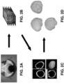
- Figures 2A through 2D a series of images are shown illustrating an order of operations used to generate a 3D reconstruction of a target area (in these illustrations, the example used is a human brain).
- a patient must undergo a scan (using scanner 114 described above) to generate an image stack of one part of the patient's body.
- Figure 2A illustrates a patient in a MRI scanner, although any other tomographic scanner (or alternative type of scanner) is contemplated, so long as it can produce an image stack (i.e., a series or sequence of images showing an object of interest) with mm resolution that can subsequently be converted into a 3D mesh.
- Figure 2B depicts a series of images forming an image stack that can subsequently be used to generate the 3D mesh.
- Figure 2C illustrates the importation of the image stack into application software that can be used to generate a 3D reconstruction of the target area via image segmentation and mesh generation.
- a 3D reconstruction of a target area can be performed by loading the image stack into iLastik (a software application for biological image segmentation).
- the image stack can thus be segmented using the carving module of iLastik, which allow fast, accurate, semi-automated segmentation of the z stack, and to extract a mesh of the 3D object.
- Figure 2D illustrates a visual representation of a 3D mesh generated in accordance with example embodiments described in greater detail below.
- this mesh Once this mesh is extracted, its size (in meters or centimeters) may need to be adjusted. Also, eventual compression on the z axis might need to be taken into account, if the voxel size of the 3D mesh is not isotropic (due, for instance to possible difference between the thickness of the optical slides and the xy resolution).
- This can be achieved using a Blender add-on (e.g., Neuromorph, a free collection of add-ons for Blender 3D modeling software, which allows importing 3D meshes and resizing them if the voxel size is known).
- a Blender add-on e.g., Neuromorph, a free collection of add-ons for Blender 3D modeling software, which allows importing 3D meshes and resizing them if the voxel size is known.
- the 3D object of the target area can thus comprise a simple file in standard .obj format, which can then be loaded into augmented reality (AR) glasses (e.g., via a USB cable, SD card, or the like). The surgeon can then load the 3D model into augmented reality software within augmented reality glasses.
- AR augmented reality
- Figure 3A a surgeon is illustrated utilizing traditional assistive machinery for surgery.
- Figure 3A demonstrates the use of a 2D representation of a target area (in this case, a brain) that requires the surgeon to alternately view the surgical site and then an external display illustrating the target area.
- the surgeon can utilize augmented reality glasses, such as those shown in Figure 3B , to address this deficiency of traditional assistive machinery.
- the augmented reality glasses of example embodiments of the present invention project the three dimensional model of the target area onto the field of view of the surgeon.
- Figure 3C illustrates an example augmented reality display enabled using augmented reality glasses.
- FIG. 4 a flow diagram illustrates example operations for utilizing three dimensional augmented reality visualizations to assist surgery.
- the computing device 100 may include means, such as scanner 114 or the like, for scanning a target area to generate an image stack representing the target area.
- the scanner 114 may be a tomograph or any other device that can use penetrating waves to capture image data regarding an object.
- scanner 114 may comprise a computed tomography (CT) scanner, a positronic emission tomography (PET) scanner, a single-photon emission computed tomography (SPECT) scanner, or a magnetic resonance imaging (MRI) scanner.
- CT computed tomography
- PET positronic emission tomography
- SPECT single-photon emission computed tomography
- MRI magnetic resonance imaging
- the scanner 114 is configured to produce a tomogram, which constitutes a two dimensional image illustrating a slice, or section, of the imaged object.
- the scanner 114 may produces a series of tomograms (referred to herein as an image stack) that, when analyzed in combination, can be used to generate a three
- a head-mounted display 108 (or other similar wearable device), superimposes a 3D projection of the 3D reconstruction onto the field of view of the user (e.g., surgeon).
- the head-mounted display 108 may in some embodiments be an element of the computing device 100, although in other embodiments, the head-mounted display 108 may be a separate apparatus connected to the computing device 100 via wireless communication media, as described previously. Specific aspects of superimposing the 3D projection are described in greater detail below in connection with Figure 6 .
- the computing device 100 includes means, such as processing circuitry 102, head-mounted display 108, user interface 110, or the like, for maintaining alignment between the projection and the user's actual view of the target area using a plurality of fiducial markers associated with the target area.
- these fiducial markers are deployed into the environment in advance of surgery and provide reference points that enable accurate relative positioning of the surgeon, surgical tools, and provide a better understanding of the location, orientation, and swelling of target areas.
- a subset of the fiducial markers may be pre-existing within the operating room environment while another subset of the fiducial markers may be inserted or otherwise disposed on different portions of the patient.
- a subset of the plurality of fiducial markers may be located directly on the target area that is the subject of surgery (e.g., the brain), or on related tissue, such as on secondary target areas whose movement and swelling are intended to be tracked.
- the location of these markers must also be digitally placed into the 3D model during the preprocessing step so that the physical locations exactly match the corresponding positions in the virtual world.
- the camera in the augmented reality glasses can then recognize the fiducial markers in the real world, and match them with their digital representation. This will allow superimposing the real and the virtual world correctly.
- At least one of these fiducial markers should be placed directly on the organ of interest (i.e. brain). Because the organ can move and swell, this marker will be moving and follow the deformation of the organ, allowing the system to compute a deformation that can be applied to the 3D mesh loaded into the system. In this latter regard, specific operations for maintaining alignment of the 3D projection and the user's actual view of the target area are described in greater detail below in connection with Figures 7 and 8 .
- the computing device 100 includes means, such as processing circuitry 102, communication interface 110, or the like, for receiving relative position and orientation information regarding a surgical tool.
- assistive machinery e.g., a neuronavigator or the like
- Such assistive machinery generally utilizes its own tracking mechanisms to identify relative locations and orientations of the surgical tools and which can accordingly provide the spatial information of the position of the surgical tools inside that are not visible during a surgical procedure.
- the computing device 100 includes means, such as processing circuitry 102, head-mounted display 108, or the like, for superimposing a projection of the surgical tool onto the field of view of the user. Having received the location and orientation of surgical tools from the assistive machinery, the surgical tools can be projected onto the field of view of the surgeon in the same manner as described above in connection with operation 406 and below in connection with Figure 6 .
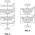
- FIG. 5 example operations are illustrated for generating a three dimensional reconstruction of a target area based on an image stack received from a scan of the target area, in accordance with some example embodiments of the present invention.
- the procedure may turn to operation 502, in which the computing device 100 includes means, such as processing circuitry 102 or the like, for pre-processing the image stack.
- pre-processing the image stack may in some embodiments include aligning images of the image stack.
- pre-processing the image stack may in some embodiments include filtering images of the image stack to increase the SNR (signal-to-noise ratio) of the images.
- the computing device 100 includes means, such as processing circuitry 102 or the like, for performing image segmentation on all images of the image stack to identify structures within the image stack.
- image segmentation may utilize a seeded watershed algorithm.
- the algorithm can detect the edges of each image to be segmented based on that image's gradient, then afterwards each edge can be followed on the 3 rd dimension (z) by following the spatial direction of the edge on the sequence of images.
- the procedure may, in some embodiments, be iterated for the both a target area (e.g., an organ) and a lesion (previously recognized by the physician) that appears within the reconstructed organ, removal of which may be the primary purpose of the surgical procedure.
- the computing device 100 includes means, such as processing circuitry 102 or the like, for generating a 3D mesh defining boundaries of the identified structures.
- This 3D mesh may comprise a collection of points and surfaces in a 3D space that approximate the shape of the segmented structure.
- the 3D reconstruction discussed above in connection with Figure 4 may comprise this 3D mesh.
- Scanner 114 typically provides information about pixel size and z thickness of an image stack.
- the relative thickness of the images in the image stack and the XY resolution of the images of the image stack can be evaluated to determine if the generated 3D mesh includes isotropic voxels, in which case the 3D mesh can be utilized to present a 3D projection to the user.
- the voxel size of the 3D mesh is not isotropic, a common result is that the Z dimension of the 3D mesh may be compressed or elongated.
- optional operation 508 may be performed, in which the computing device 100 includes means, such as processing circuitry 102 or the like, for rescaling the three dimensional mesh to render its voxels isotropic (correcting the compression or elongation caused by the asymmetry between the Z axis thickness and the XY-axis resolution of the images in the image stack.
- the 3D augmented reality projection may change over time with movement of the head-mounted display.
- the augmented reality glasses utilize a front camera configured to recognize fiducial markers placed in the operating room that the computing device 100 can recognize as fixed points in the environment whose position relative to the patient is known. This enables correct alignment of the 3D model, as well as the tracking of the surgeon's head position.
- tracking of the augmented reality glasses can be improved using sensors disposed on the augmented reality glasses (e.g., gyroscope(s), accelerometer(s), or the like) embedded in the augmented reality glasses.
- the computing device 100 includes means, such as processing circuitry 102 or the like, for calculating a relative location of the target area from the perspective of the head-mounted display.
- the computing device 100 includes means, such as head-mounted display 108, user interface 110, or the like, for presenting an interface requesting user adjustment of the alignment between the projection and the user's actual view of the target area.
- the computing device 100 includes means, such as head-mounted display 108, user interface 110, or the like, for receiving one or more responsive adjustments.
- the use of the fiducial markers should be as accurate as possible, in order to grant a perfect tracing of the position of the 3D model relative to the real world. Nevertheless, because the model will be superimposed on the eyes of the surgeon, the surgeon may be provided with the ability to finely adjust the position of the model by using a graphical interface, to correct millimetric adjusting errors (on the x, y, or z axes).
- the computing device 100 includes means, such as processing circuitry 102, head-mounted display 108, or the like, for updating the projection of the three dimensional reconstruction in response to receiving the one or more responsive adjustments. These adjustments may be made u
- the computing device 100 includes means, such as processing circuitry 102 or the like, for detecting a change in location or orientation of at least two fiducial markers disposed on the target area, wherein movement of the at least two fiducial markers disposed on the target area indicates a change in location or shape of the target area.
- the computing device 100 includes means, such as processing circuitry 102 or the like, for computing a deformation of the three dimensional reconstruction based on the detected change. Subsequently, in operation 806, the computing device 100 includes means, such as processing circuitry 102 or the like, for applying the deformation to the three dimensional reconstruction. Finally, in operation 808, the computing device 100 includes means, such as processing circuitry 102 or the like, for updating the projection of the three dimensional reconstruction in response to applying the deformation to the three dimensional reconstruction.
- example embodiments deploy 3D tracking and 3D visualization of the surgical field in a manner that allows the surgeon to focus attention (sight and hands) on the operatory field and on the patient rather than on separate screen.
- the surgeon is not required to interpolate movements that appear on an external display in one orientation on the fly to properly respond with movements in the actual working area. Accordingly, example embodiments described herein provide significant benefits for surgical applications having little or no direct visibility and surgical applications utilizing assistive machinery.
- Blocks of the flowchart support combinations of means for performing the specified functions and combinations of operations for performing the specified functions. It will be understood that one or more blocks of the flowchart, and combinations of blocks in the flowchart, can be implemented by special purpose hardware-based computer systems which perform the specified functions, or combinations of special purpose hardware and computer instructions.
Landscapes
- Health & Medical Sciences (AREA)
- Engineering & Computer Science (AREA)
- Surgery (AREA)
- Life Sciences & Earth Sciences (AREA)
- Nuclear Medicine, Radiotherapy & Molecular Imaging (AREA)
- General Health & Medical Sciences (AREA)
- Veterinary Medicine (AREA)
- Biomedical Technology (AREA)
- Heart & Thoracic Surgery (AREA)
- Medical Informatics (AREA)
- Molecular Biology (AREA)
- Animal Behavior & Ethology (AREA)
- Public Health (AREA)
- Robotics (AREA)
- Human Computer Interaction (AREA)
- Computer Vision & Pattern Recognition (AREA)
- Physics & Mathematics (AREA)
- General Physics & Mathematics (AREA)
- Theoretical Computer Science (AREA)
- Gynecology & Obstetrics (AREA)
- Radiology & Medical Imaging (AREA)
- Oral & Maxillofacial Surgery (AREA)
- Pathology (AREA)
- Apparatus For Radiation Diagnosis (AREA)
- Processing Or Creating Images (AREA)
Claims (8)
- Système permettant d'utiliser la visualisation par réalité augmentée en chirurgie, le système comprenant :
une pluralité de marqueurs de repère comprenant :un premier sous-ensemble de la pluralité de marqueurs de repère configurés pour être placés dans une salle d'opération en tant que points fixes dont la position par rapport à un patient est connue, etun deuxième sous-ensemble de la pluralité de marqueurs de repère configuré pour être placé sur une zone cible d'un patient ;un appareil (100) configuré pour générer (404) une reconstruction tridimensionnelle d'une pile d'images représentant la zone cible du patient parréalisation (504) d'une segmentation d'image sur toutes les images de la pile d'images pour identifier les structures au sein de la pile d'images, etgénération (506) d'un maillage tridimensionnel définissant les limites des structures identifiées, la reconstruction tridimensionnelle comprenant le maillage tridimensionnel,dans lequel, lorsque les marqueurs de repère ont été placés, l'appareil est en outre configuré pour placer de façon numérique les emplacements correspondants d'une représentation numérique de la pluralité de marqueurs de repère dans la reconstruction tridimensionnelle de la pile d'images ;un visiocasque (108) configuré pour superposer (406) une projection tridimensionnelle de la reconstruction tridimensionnelle sur un champ de vision d'un utilisateur ; etune caméra dans le visiocasque configurée pour identifier (702) un emplacement de chacun de la pluralité de marqueurs de repère, et pour faire correspondre la pluralité de marqueurs de repère avec leur représentation numérique, de sorte que l'appareil est configuré pour maintenir (408) l'alignement entre la projection et la vue réelle de l'utilisateur de la zone cible à l'aide de la pluralité de marqueurs de repère,dans lequel la caméra est configurée pour détecter (802) un changement de l'emplacement du deuxième sous-ensemble de la pluralité de marqueurs de repère indiquant un changement d'emplacement ou de forme de la zone cible, de sorte que l'appareil est configuré pour calculer (804) une déformation de la reconstruction tridimensionnelle, et le visiocasque est configuré pour mettre à jour (808) la projection de la reconstruction tridimensionnelle. - Système selon la revendication 1, dans lequel l'appareil est configuré pour maintenir l'alignement entre la projection et la vue réelle de l'utilisateur de la zone cible parcalcul d'un emplacement relatif de la zone cible du point de vue du visiocasque, etgénération de la projection de la reconstruction tridimensionnelle en fonction de l'emplacement relatif calculé de la zone cible.
- Système selon la revendication 1, comprenant en outre :
un scanner qui balaye la partie du corps pour générer la pile d'images, le scanner étant l'un parmi :un scanner de tomodensitométrie (TDM) qui balaye la zone cible ;un scanner d'imagerie par résonance magnétique (IRM) qui scanne la zone cible ;un scanner de tomographie par émission de positons (TEP) qui balaye la zone cible ; ouun scanner de tomographie par émission monophotonique (SPECT) qui balaye la zone cible. - Système selon la revendication 1, dans lequel la génération par l'appareil de la reconstruction tridimensionnelle de la pile d'images représentant la zone cible comprend :
le prétraitement de la pile d'images. - Système selon la revendication 4, dans lequel le prétraitement de la pile d'images par l'appareil comprend au moins l'un parmi :l'alignement des images de la pile d'images ; oule filtrage des images de la pile d'images.
- Système selon la revendication 1, dans lequel, dans un cas dans lequel une taille de voxel du maillage tridimensionnel n'est pas isotrope, l'appareil redimensionne le maillage tridimensionnel.
- Système selon la revendication 1, dans lequel le visiocasque superpose la projection tridimensionnelle de la reconstruction tridimensionnelle sur le champ de vision de l'utilisateur par :affichage d'une première projection de la reconstruction tridimensionnelle à un oeil de l'utilisateur ; etaffichage d'une deuxième projection de la reconstruction tridimensionnelle à l'autre oeil de l'utilisateur,dans lequel les différences entre la première projection et la deuxième projection sont conçues pour générer la projection tridimensionnelle de la reconstruction tridimensionnelle.
- Système selon la revendication 1, dans lequel l'appareil maintient en outre l'alignement entre la projection et la vue réelle de l'utilisateur de la zone cible par :
présentation d'une interface utilisateur demandant à l'utilisateur d'ajuster l'alignement entre la projection et la vue réelle de l'utilisateur de la zone cible.
Applications Claiming Priority (2)
| Application Number | Priority Date | Filing Date | Title |
|---|---|---|---|
| US201562143923P | 2015-04-07 | 2015-04-07 | |
| PCT/IB2016/051924 WO2016162789A2 (fr) | 2015-04-07 | 2016-04-05 | Procédé, appareil et système d'utilisation de la réalité augmentée pour améliorer une opération chirurgicale |
Publications (2)
| Publication Number | Publication Date |
|---|---|
| EP3280344A2 EP3280344A2 (fr) | 2018-02-14 |
| EP3280344B1 true EP3280344B1 (fr) | 2025-01-29 |
Family
ID=55808801
Family Applications (1)
| Application Number | Title | Priority Date | Filing Date |
|---|---|---|---|
| EP16718475.3A Active EP3280344B1 (fr) | 2015-04-07 | 2016-04-05 | Système d'utilisation de la réalité augmentée pour améliorer une opération chirurgicale |
Country Status (3)
| Country | Link |
|---|---|
| US (1) | US20180140362A1 (fr) |
| EP (1) | EP3280344B1 (fr) |
| WO (1) | WO2016162789A2 (fr) |
Families Citing this family (89)
| Publication number | Priority date | Publication date | Assignee | Title |
|---|---|---|---|---|
| US11547499B2 (en) | 2014-04-04 | 2023-01-10 | Surgical Theater, Inc. | Dynamic and interactive navigation in a surgical environment |
| US10154239B2 (en) | 2014-12-30 | 2018-12-11 | Onpoint Medical, Inc. | Image-guided surgery with surface reconstruction and augmented reality visualization |
| US10013808B2 (en) | 2015-02-03 | 2018-07-03 | Globus Medical, Inc. | Surgeon head-mounted display apparatuses |
| CN118717223A (zh) | 2015-02-13 | 2024-10-01 | 瑟西纳斯医疗技术有限责任公司 | 用于在骨骼中放置医疗设备的系统和方法 |
| CA2997965C (fr) | 2015-10-14 | 2021-04-27 | Surgical Theater LLC | Navigation chirurgicale a realite augmentee |
| CN111329552B (zh) | 2016-03-12 | 2021-06-22 | P·K·朗 | 包括机器人的用于引导骨切除的增强现实可视化 |
| US11839433B2 (en) * | 2016-09-22 | 2023-12-12 | Medtronic Navigation, Inc. | System for guided procedures |
| GB2570263B (en) * | 2016-10-21 | 2021-03-17 | Synaptive Medical Inc | Mixed reality training system |
| US10499997B2 (en) | 2017-01-03 | 2019-12-10 | Mako Surgical Corp. | Systems and methods for surgical navigation |
| EP3568070B1 (fr) | 2017-01-16 | 2024-01-03 | Philipp K. Lang | Guidage optique pour procédures chirurgicales, médicales et dentaires |
| US10010379B1 (en) | 2017-02-21 | 2018-07-03 | Novarad Corporation | Augmented reality viewing and tagging for medical procedures |
| EP3585254B1 (fr) | 2017-02-24 | 2024-03-20 | Masimo Corporation | Câble de dispositif médical et procédé de partage de données entre des dispositifs médicaux connectés |
| WO2018156809A1 (fr) * | 2017-02-24 | 2018-08-30 | Masimo Corporation | Système de réalité augmentée permettant d'afficher des données de patient |
| WO2018162079A1 (fr) * | 2017-03-10 | 2018-09-13 | Brainlab Ag | Pré-enregistrement de réalité augmentée |
| US9892564B1 (en) * | 2017-03-30 | 2018-02-13 | Novarad Corporation | Augmenting real-time views of a patient with three-dimensional data |
| EP3612127A1 (fr) * | 2017-04-20 | 2020-02-26 | The Cleveland Clinic Foundation | Système et méthode pour procédures endovasculaires percutanées guidées par images holographiques |
| JP7159208B2 (ja) | 2017-05-08 | 2022-10-24 | マシモ・コーポレイション | ドングルを使用することによって医療システムをネットワークコントローラとペアリングするためのシステム |
| US10470645B2 (en) | 2017-05-22 | 2019-11-12 | Gustav Lo | Imaging system and method |
| EP3668426A4 (fr) | 2017-08-14 | 2021-10-20 | Circinus Medical Technology, LLC | Système et procédé utilisant la réalité augmentée avec un alignement de formes pour la pose d'un dispositif médical dans un os |
| EP3470006B1 (fr) | 2017-10-10 | 2020-06-10 | Holo Surgical Inc. | Segmentation automatisée d'images de structure osseuse tridimensionnelles |
| EP3445048B8 (fr) | 2017-08-15 | 2026-01-14 | Augmedics, Inc. | Interface utilisateur graphique pour un système de navigation chirurgical pour fournir une image de réalité augmentée pendant le fonctionnement |
| US10861236B2 (en) | 2017-09-08 | 2020-12-08 | Surgical Theater, Inc. | Dual mode augmented reality surgical system and method |
| US11801114B2 (en) | 2017-09-11 | 2023-10-31 | Philipp K. Lang | Augmented reality display for vascular and other interventions, compensation for cardiac and respiratory motion |
| WO2019059044A1 (fr) * | 2017-09-20 | 2019-03-28 | 日本電気株式会社 | Dispositif de traitement d'informations, procédé de commande, et programme |
| US20190175059A1 (en) | 2017-12-07 | 2019-06-13 | Medtronic Xomed, Inc. | System and Method for Assisting Visualization During a Procedure |
| US11348257B2 (en) | 2018-01-29 | 2022-05-31 | Philipp K. Lang | Augmented reality guidance for orthopedic and other surgical procedures |
| US20190254753A1 (en) | 2018-02-19 | 2019-08-22 | Globus Medical, Inc. | Augmented reality navigation systems for use with robotic surgical systems and methods of their use |
| EP3764932B1 (fr) | 2018-03-13 | 2025-07-16 | Intuitive Surgical Operations, Inc. | Procédés et systemes de guidage de mouvement manuel de systèmes médicaux |
| TWI678181B (zh) * | 2018-04-30 | 2019-12-01 | 長庚大學 | 手術導引系統 |
| US10869727B2 (en) * | 2018-05-07 | 2020-12-22 | The Cleveland Clinic Foundation | Live 3D holographic guidance and navigation for performing interventional procedures |
| WO2019229275A1 (fr) * | 2018-05-30 | 2019-12-05 | Zubizarreta Macho Alvaro | Procédé pour transférer une planification thérapeutique à un dispositif de réalité mixte |
| WO2019245862A1 (fr) | 2018-06-19 | 2019-12-26 | Tornier, Inc. | Visualisation de plans chirurgicaux à modification peropératoire |
| JP2020000649A (ja) * | 2018-06-29 | 2020-01-09 | 富士通株式会社 | 可視化装置、可視化方法、および可視化プログラム |
| CN110794950A (zh) * | 2018-08-01 | 2020-02-14 | 深圳先进技术研究院 | 一种数据监测实验方法、装置、终端设备及存储介质 |
| EP4710888A3 (fr) | 2018-08-10 | 2026-03-25 | Augmedics Inc. | Identification assistée par ordinateur d'une structure anatomique appropriée pour le placement d'un dispositif médical pendant une intervention chirurgicale |
| US12544180B2 (en) | 2018-10-26 | 2026-02-10 | Intuitive Surgical Operations, Inc. | Mixed reality systems and methods for indicating an extent of a field of view of an imaging device |
| US12008721B2 (en) | 2018-10-26 | 2024-06-11 | Intuitive Surgical Operations, Inc. | Mixed reality systems and methods for indicating an extent of a field of view of an imaging device |
| US11287874B2 (en) | 2018-11-17 | 2022-03-29 | Novarad Corporation | Using optical codes with augmented reality displays |
| EP3689218B1 (fr) * | 2019-01-30 | 2023-10-18 | DENTSPLY SIRONA Inc. | Procédé et système de guidage d'un balayage intra-oral |
| WO2020160129A1 (fr) | 2019-01-31 | 2020-08-06 | Intuitive Surgical Operations, Inc. | Systèmes et procédés pour permettre l'insertion d'un instrument chirurgical dans un espace chirurgical |
| US11553969B1 (en) | 2019-02-14 | 2023-01-17 | Onpoint Medical, Inc. | System for computation of object coordinates accounting for movement of a surgical site for spinal and other procedures |
| US11857378B1 (en) | 2019-02-14 | 2024-01-02 | Onpoint Medical, Inc. | Systems for adjusting and tracking head mounted displays during surgery including with surgical helmets |
| CN109925053B (zh) * | 2019-03-04 | 2021-06-22 | 杭州三坛医疗科技有限公司 | 手术路径的确定方法、装置和系统、可读存储介质 |
| EP3955841A4 (fr) | 2019-04-15 | 2023-04-12 | Circinus Medical Technologies LLC | Appareil d'attache pour fixer un dispositif d'alignement médical pour aligner un outil |
| AU2020257211A1 (en) | 2019-04-15 | 2021-11-11 | Circinus Medical Technology Llc | Orientation calibration system for image capture |
| JP7322183B2 (ja) | 2019-05-14 | 2023-08-07 | ホウメディカ・オステオニクス・コーポレイション | 整形外科インプラント埋植のための骨壁追跡及びガイダンス |
| CN112168348A (zh) * | 2019-07-03 | 2021-01-05 | 钜旺生技股份有限公司 | 用于手术的定位与导航系统及其运作方法 |
| US10832486B1 (en) | 2019-07-17 | 2020-11-10 | Gustav Lo | Systems and methods for displaying augmented anatomical features |
| US11288802B2 (en) | 2019-07-17 | 2022-03-29 | Gustav Lo | Systems and methods for displaying augmented anatomical features |
| US12211151B1 (en) | 2019-07-30 | 2025-01-28 | Onpoint Medical, Inc. | Systems for optimizing augmented reality displays for surgical procedures |
| US11672607B2 (en) * | 2019-08-14 | 2023-06-13 | Warsaw Orthopedic, Inc. | Systems, devices, and methods for surgical navigation with anatomical tracking |
| WO2021044523A1 (fr) * | 2019-09-03 | 2021-03-11 | 株式会社インキュビット | Dispositif d'assistance chirurgicale |
| CN110728756B (zh) * | 2019-09-30 | 2024-02-09 | 亮风台(上海)信息科技有限公司 | 一种基于增强现实的远程指导的方法与设备 |
| US11210865B2 (en) | 2019-10-03 | 2021-12-28 | International Business Machines Corporation | Visually interacting with three dimensional data in augmented or virtual reality |
| WO2021108268A1 (fr) | 2019-11-26 | 2021-06-03 | Tornier, Inc. | Guidage virtuel pour corriger une installation de broche chirurgicale |
| US12220176B2 (en) | 2019-12-10 | 2025-02-11 | Globus Medical, Inc. | Extended reality instrument interaction zone for navigated robotic |
| US12133772B2 (en) | 2019-12-10 | 2024-11-05 | Globus Medical, Inc. | Augmented reality headset for navigated robotic surgery |
| US11992373B2 (en) | 2019-12-10 | 2024-05-28 | Globus Medical, Inc | Augmented reality headset with varied opacity for navigated robotic surgery |
| AU2020404991B2 (en) | 2019-12-18 | 2023-10-19 | Howmedica Osteonics Corp. | Surgical guidance for surgical tools |
| US11237627B2 (en) | 2020-01-16 | 2022-02-01 | Novarad Corporation | Alignment of medical images in augmented reality displays |
| CA3168826A1 (fr) | 2020-01-22 | 2021-07-29 | Photonic Medical Inc. | Loupe numerique etalonnee, multimodale, a vision ouverte avec detection de profondeur |
| US11464581B2 (en) | 2020-01-28 | 2022-10-11 | Globus Medical, Inc. | Pose measurement chaining for extended reality surgical navigation in visible and near infrared spectrums |
| US11382699B2 (en) | 2020-02-10 | 2022-07-12 | Globus Medical Inc. | Extended reality visualization of optical tool tracking volume for computer assisted navigation in surgery |
| US11207150B2 (en) | 2020-02-19 | 2021-12-28 | Globus Medical, Inc. | Displaying a virtual model of a planned instrument attachment to ensure correct selection of physical instrument attachment |
| WO2021221749A1 (fr) | 2020-04-29 | 2021-11-04 | Howmedica Osteonics Corp. | Dimensionnement et placement de tête humérale à réalité mixte |
| US11607277B2 (en) | 2020-04-29 | 2023-03-21 | Globus Medical, Inc. | Registration of surgical tool with reference array tracked by cameras of an extended reality headset for assisted navigation during surgery |
| US11153555B1 (en) | 2020-05-08 | 2021-10-19 | Globus Medical Inc. | Extended reality headset camera system for computer assisted navigation in surgery |
| US11382700B2 (en) | 2020-05-08 | 2022-07-12 | Globus Medical Inc. | Extended reality headset tool tracking and control |
| US11510750B2 (en) | 2020-05-08 | 2022-11-29 | Globus Medical, Inc. | Leveraging two-dimensional digital imaging and communication in medicine imagery in three-dimensional extended reality applications |
| US12211154B2 (en) | 2020-05-11 | 2025-01-28 | Intuitive Surgical Operations, Inc. | Systems and methods for region-based presentation of augmented content |
| US11737831B2 (en) | 2020-09-02 | 2023-08-29 | Globus Medical Inc. | Surgical object tracking template generation for computer assisted navigation during surgical procedure |
| EP3995098A1 (fr) | 2020-11-09 | 2022-05-11 | Koninklijke Philips N.V. | Planification de trajet cérébral profond utilisant la réalité augmentée |
| EP4247254A4 (fr) | 2020-11-19 | 2024-05-22 | Circinus Medical Technology LLC | Systèmes et procédés pour une analyse d'image basée sur l'intelligence artificielle pour le positionnement d'un appareil chirurgical |
| US12053247B1 (en) | 2020-12-04 | 2024-08-06 | Onpoint Medical, Inc. | System for multi-directional tracking of head mounted displays for real-time augmented reality guidance of surgical procedures |
| US12016633B2 (en) | 2020-12-30 | 2024-06-25 | Novarad Corporation | Alignment of medical images in augmented reality displays |
| US11289196B1 (en) | 2021-01-12 | 2022-03-29 | Emed Labs, Llc | Health testing and diagnostics platform |
| US12064186B2 (en) | 2021-02-02 | 2024-08-20 | Circinus Medical Technology Llc | Systems and methods for simulating three-dimensional orientations of surgical hardware devices about an insertion point of an anatomy |
| WO2022192585A1 (fr) | 2021-03-10 | 2022-09-15 | Onpoint Medical, Inc. | Guidage de réalité augmentée pour systèmes d'imagerie et chirurgie robotique |
| US11615888B2 (en) | 2021-03-23 | 2023-03-28 | Emed Labs, Llc | Remote diagnostic testing and treatment |
| US11929168B2 (en) | 2021-05-24 | 2024-03-12 | Emed Labs, Llc | Systems, devices, and methods for diagnostic aid kit apparatus |
| JP2024516547A (ja) | 2021-04-14 | 2024-04-16 | サーキヌス メディカル テクノロジー エルエルシー | Lidarベースの解剖学的マッピングに係るシステム及び方法 |
| US11369454B1 (en) | 2021-05-24 | 2022-06-28 | Emed Labs, Llc | Systems, devices, and methods for diagnostic aid kit apparatus |
| US11610682B2 (en) | 2021-06-22 | 2023-03-21 | Emed Labs, Llc | Systems, methods, and devices for non-human readable diagnostic tests |
| US12014829B2 (en) | 2021-09-01 | 2024-06-18 | Emed Labs, Llc | Image processing and presentation techniques for enhanced proctoring sessions |
| US12433761B1 (en) | 2022-01-20 | 2025-10-07 | Onpoint Medical, Inc. | Systems and methods for determining the shape of spinal rods and spinal interbody devices for use with augmented reality displays, navigation systems and robots in minimally invasive spine procedures |
| US12175612B2 (en) | 2022-02-16 | 2024-12-24 | Microsoft Technology Licensing, Llc. | Mixed reality environment display using surface reconstruction mesh and live video overlay |
| CN114627271A (zh) * | 2022-03-15 | 2022-06-14 | 上海诠视传感技术有限公司 | 一种查看3d可视化数据的方法、ar眼镜、系统及存储介质 |
| US12458467B2 (en) * | 2023-12-31 | 2025-11-04 | Xironetic Llc | Systems and methods for augmented reality-aided implant placement |
| WO2025149847A1 (fr) * | 2024-01-10 | 2025-07-17 | Intradys Sas | Système et procédé de positionnement d'une partie du corps d'un patient pendant une intervention fonctionnelle |
Citations (6)
| Publication number | Priority date | Publication date | Assignee | Title |
|---|---|---|---|---|
| US20050203380A1 (en) * | 2004-02-17 | 2005-09-15 | Frank Sauer | System and method for augmented reality navigation in a medical intervention procedure |
| US20060176242A1 (en) * | 2005-02-08 | 2006-08-10 | Blue Belt Technologies, Inc. | Augmented reality device and method |
| US20130267838A1 (en) * | 2012-04-09 | 2013-10-10 | Board Of Regents, The University Of Texas System | Augmented Reality System for Use in Medical Procedures |
| US20140275760A1 (en) * | 2013-03-13 | 2014-09-18 | Samsung Electronics Co., Ltd. | Augmented reality image display system and surgical robot system comprising the same |
| WO2014200016A1 (fr) * | 2013-06-11 | 2014-12-18 | Tanji Atsushi | Système, procédé, dispositif et programme d'aide à la chirurgie, et dispositif de traitement d'informations |
| US20150049081A1 (en) * | 2013-08-13 | 2015-02-19 | Dane Coffey | Computer Visualization of Anatomical Items |
Family Cites Families (7)
| Publication number | Priority date | Publication date | Assignee | Title |
|---|---|---|---|---|
| JP3619063B2 (ja) * | 1999-07-08 | 2005-02-09 | キヤノン株式会社 | 立体画像処理装置、その方法、立体視パラメータ設定装置、その方法そしてコンピュータプログラム記憶媒体 |
| US20030053669A1 (en) * | 2001-07-18 | 2003-03-20 | Marconi Medical Systems, Inc. | Magnetic resonance angiography method and apparatus |
| CA2797302C (fr) * | 2010-04-28 | 2019-01-15 | Ryerson University | Systeme et procedes de retroaction de guidage peroperatoire |
| US9142062B2 (en) * | 2011-03-29 | 2015-09-22 | Qualcomm Incorporated | Selective hand occlusion over virtual projections onto physical surfaces using skeletal tracking |
| EP2956814A1 (fr) * | 2013-02-14 | 2015-12-23 | Seiko Epson Corporation | Visiocasque et son procédé de commande |
| US8922589B2 (en) * | 2013-04-07 | 2014-12-30 | Laor Consulting Llc | Augmented reality apparatus |
| US10070120B2 (en) * | 2014-09-17 | 2018-09-04 | Qualcomm Incorporated | Optical see-through display calibration |
-
2016
- 2016-04-05 EP EP16718475.3A patent/EP3280344B1/fr active Active
- 2016-04-05 US US15/564,347 patent/US20180140362A1/en not_active Abandoned
- 2016-04-05 WO PCT/IB2016/051924 patent/WO2016162789A2/fr not_active Ceased
Patent Citations (6)
| Publication number | Priority date | Publication date | Assignee | Title |
|---|---|---|---|---|
| US20050203380A1 (en) * | 2004-02-17 | 2005-09-15 | Frank Sauer | System and method for augmented reality navigation in a medical intervention procedure |
| US20060176242A1 (en) * | 2005-02-08 | 2006-08-10 | Blue Belt Technologies, Inc. | Augmented reality device and method |
| US20130267838A1 (en) * | 2012-04-09 | 2013-10-10 | Board Of Regents, The University Of Texas System | Augmented Reality System for Use in Medical Procedures |
| US20140275760A1 (en) * | 2013-03-13 | 2014-09-18 | Samsung Electronics Co., Ltd. | Augmented reality image display system and surgical robot system comprising the same |
| WO2014200016A1 (fr) * | 2013-06-11 | 2014-12-18 | Tanji Atsushi | Système, procédé, dispositif et programme d'aide à la chirurgie, et dispositif de traitement d'informations |
| US20150049081A1 (en) * | 2013-08-13 | 2015-02-19 | Dane Coffey | Computer Visualization of Anatomical Items |
Non-Patent Citations (4)
| Title |
|---|
| MATHIAS MARKERT ET AL: "Tracking of the liver for navigation in open surgery", INTERNATIONAL JOURNAL OF COMPUTER ASSISTED RADIOLOGY AND SURGERY, vol. 5, no. 3, 7 August 2009 (2009-08-07), DE, pages 229 - 235, XP055593706, ISSN: 1861-6410, DOI: 10.1007/s11548-009-0395-x * |
| OLIVEIRA-SANTOS THIAGO ET AL: "Passive Single Marker Tracking for Organ Motion and Deformation Detection in Open Liver Surgery", 22 June 2011, INTERNATIONAL CONFERENCE ON COMPUTER ANALYSIS OF IMAGES AND PATTERNS. CAIP 2017: COMPUTER ANALYSIS OF IMAGES AND PATTERNS; [LECTURE NOTES IN COMPUTER SCIENCE; LECT.NOTES COMPUTER], SPRINGER, BERLIN, HEIDELBERG, PAGE(S) 156 - 167, ISBN: 978-3-642-17318-9, XP047369683 * |
| SKRINJAR O M ET AL: "REAL TIME 3D BRAIN SHIFT COMPENSATION", INFORMATION PROCESSING IN MEDICAL IMAGING. INTERNATIONALCONFERENCE. PROCEED, XX, XX, vol. 1613, 25 June 1999 (1999-06-25), pages 42 - 55, XP008010724 * |
| SUN KAY ET AL: "Near Real-Time Computer Assisted Surgery for Brain Shift Correction Using Biomechanical Models", IEEE JOURNAL OF TRANSLATIONAL ENGINEERING IN HEALTH AND MEDICINE, vol. 2, 30 May 2014 (2014-05-30), pages 1 - 13, XP011552671, DOI: 10.1109/JTEHM.2014.2327628 * |
Also Published As
| Publication number | Publication date |
|---|---|
| US20180140362A1 (en) | 2018-05-24 |
| WO2016162789A3 (fr) | 2016-11-17 |
| WO2016162789A2 (fr) | 2016-10-13 |
| EP3280344A2 (fr) | 2018-02-14 |
Similar Documents
| Publication | Publication Date | Title |
|---|---|---|
| EP3280344B1 (fr) | Système d'utilisation de la réalité augmentée pour améliorer une opération chirurgicale | |
| US11759261B2 (en) | Augmented reality pre-registration | |
| EP2452649A1 (fr) | Visualisation de données anatomiques à réalité améliorée | |
| EP3328305B1 (fr) | Suivi de microscope sur la base d'une analyse vidéo | |
| US20260120301A1 (en) | Combining angiographic information with fluoroscopic images | |
| US20220022964A1 (en) | System for displaying an augmented reality and method for generating an augmented reality | |
| US12112437B2 (en) | Positioning medical views in augmented reality | |
| CA3085814C (fr) | Reconstruction d'hypersurface de vue microscopique | |
| CN111028326A (zh) | 用于使3d对象的空间表面曲率可视化的方法、系统和介质 | |
| WO2025073380A1 (fr) | Panorama sans recomposition dans l'acquisition d'images médicales | |
| US12190410B2 (en) | Augmenting a medical image with an intelligent ruler | |
| JP2006227774A (ja) | 画像表示方法 | |
| EP4409594B1 (fr) | Détermination de la hauteur de la colonne vertébrale à l'aide de la réalité augmentée | |
| US20240331329A1 (en) | Method and system for superimposing two-dimensional (2d) images over deformed surfaces | |
| KR20260062993A (ko) | 광학 코드를 이용한 증강 현실 디스플레이 |
Legal Events
| Date | Code | Title | Description |
|---|---|---|---|
| STAA | Information on the status of an ep patent application or granted ep patent |
Free format text: STATUS: THE INTERNATIONAL PUBLICATION HAS BEEN MADE |
|
| PUAI | Public reference made under article 153(3) epc to a published international application that has entered the european phase |
Free format text: ORIGINAL CODE: 0009012 |
|
| STAA | Information on the status of an ep patent application or granted ep patent |
Free format text: STATUS: REQUEST FOR EXAMINATION WAS MADE |
|
| 17P | Request for examination filed |
Effective date: 20171103 |
|
| AK | Designated contracting states |
Kind code of ref document: A2 Designated state(s): AL AT BE BG CH CY CZ DE DK EE ES FI FR GB GR HR HU IE IS IT LI LT LU LV MC MK MT NL NO PL PT RO RS SE SI SK SM TR |
|
| AX | Request for extension of the european patent |
Extension state: BA ME |
|
| DAV | Request for validation of the european patent (deleted) | ||
| DAX | Request for extension of the european patent (deleted) | ||
| PUAG | Search results despatched under rule 164(2) epc together with communication from examining division |
Free format text: ORIGINAL CODE: 0009017 |
|
| STAA | Information on the status of an ep patent application or granted ep patent |
Free format text: STATUS: EXAMINATION IS IN PROGRESS |
|
| 17Q | First examination report despatched |
Effective date: 20190612 |
|
| B565 | Issuance of search results under rule 164(2) epc |
Effective date: 20190612 |
|
| RIC1 | Information provided on ipc code assigned before grant |
Ipc: A61B 34/20 20160101AFI20190606BHEP Ipc: A61B 17/00 20060101ALI20190606BHEP Ipc: G02B 27/01 20060101ALI20190606BHEP Ipc: A61B 34/10 20160101ALI20190606BHEP Ipc: A61B 90/00 20160101ALI20190606BHEP Ipc: A61B 34/00 20160101ALI20190606BHEP Ipc: G06T 7/00 20170101ALI20190606BHEP Ipc: G06T 19/00 20110101ALN20190606BHEP Ipc: G06T 7/73 20170101ALI20190606BHEP Ipc: A61B 90/50 20160101ALI20190606BHEP |
|
| P01 | Opt-out of the competence of the unified patent court (upc) registered |
Effective date: 20230621 |
|
| GRAP | Despatch of communication of intention to grant a patent |
Free format text: ORIGINAL CODE: EPIDOSNIGR1 |
|
| STAA | Information on the status of an ep patent application or granted ep patent |
Free format text: STATUS: GRANT OF PATENT IS INTENDED |
|
| RIC1 | Information provided on ipc code assigned before grant |
Ipc: G06T 7/73 20170101ALI20240725BHEP Ipc: A61B 34/00 20160101ALI20240725BHEP Ipc: G06T 7/00 20170101ALI20240725BHEP Ipc: G02B 27/01 20060101ALI20240725BHEP Ipc: A61B 34/10 20160101ALI20240725BHEP Ipc: A61B 17/00 20060101ALI20240725BHEP Ipc: A61B 90/50 20160101ALI20240725BHEP Ipc: A61B 90/00 20160101ALI20240725BHEP Ipc: A61B 34/20 20160101AFI20240725BHEP Ipc: G06T 19/00 20110101ALN20240725BHEP |
|
| INTG | Intention to grant announced |
Effective date: 20240821 |
|
| GRAS | Grant fee paid |
Free format text: ORIGINAL CODE: EPIDOSNIGR3 |
|
| GRAA | (expected) grant |
Free format text: ORIGINAL CODE: 0009210 |
|
| STAA | Information on the status of an ep patent application or granted ep patent |
Free format text: STATUS: THE PATENT HAS BEEN GRANTED |
|
| AK | Designated contracting states |
Kind code of ref document: B1 Designated state(s): AL AT BE BG CH CY CZ DE DK EE ES FI FR GB GR HR HU IE IS IT LI LT LU LV MC MK MT NL NO PL PT RO RS SE SI SK SM TR |
|
| REG | Reference to a national code |
Ref country code: GB Ref legal event code: FG4D |
|
| REG | Reference to a national code |
Ref country code: CH Ref legal event code: EP |
|
| REG | Reference to a national code |
Ref country code: DE Ref legal event code: R096 Ref document number: 602016091077 Country of ref document: DE |
|
| REG | Reference to a national code |
Ref country code: IE Ref legal event code: FG4D |
|
| REG | Reference to a national code |
Ref country code: NL Ref legal event code: MP Effective date: 20250129 |
|
| PG25 | Lapsed in a contracting state [announced via postgrant information from national office to epo] |
Ref country code: NL Free format text: LAPSE BECAUSE OF FAILURE TO SUBMIT A TRANSLATION OF THE DESCRIPTION OR TO PAY THE FEE WITHIN THE PRESCRIBED TIME-LIMIT Effective date: 20250129 |
|
| PG25 | Lapsed in a contracting state [announced via postgrant information from national office to epo] |
Ref country code: RS Free format text: LAPSE BECAUSE OF FAILURE TO SUBMIT A TRANSLATION OF THE DESCRIPTION OR TO PAY THE FEE WITHIN THE PRESCRIBED TIME-LIMIT Effective date: 20250429 |
|
| PG25 | Lapsed in a contracting state [announced via postgrant information from national office to epo] |
Ref country code: FI Free format text: LAPSE BECAUSE OF FAILURE TO SUBMIT A TRANSLATION OF THE DESCRIPTION OR TO PAY THE FEE WITHIN THE PRESCRIBED TIME-LIMIT Effective date: 20250129 |
|
| PG25 | Lapsed in a contracting state [announced via postgrant information from national office to epo] |
Ref country code: PL Free format text: LAPSE BECAUSE OF FAILURE TO SUBMIT A TRANSLATION OF THE DESCRIPTION OR TO PAY THE FEE WITHIN THE PRESCRIBED TIME-LIMIT Effective date: 20250129 |
|
| PGFP | Annual fee paid to national office [announced via postgrant information from national office to epo] |
Ref country code: DE Payment date: 20250422 Year of fee payment: 10 |
|
| PG25 | Lapsed in a contracting state [announced via postgrant information from national office to epo] |
Ref country code: ES Free format text: LAPSE BECAUSE OF FAILURE TO SUBMIT A TRANSLATION OF THE DESCRIPTION OR TO PAY THE FEE WITHIN THE PRESCRIBED TIME-LIMIT Effective date: 20250129 |
|
| REG | Reference to a national code |
Ref country code: LT Ref legal event code: MG9D |
|
| PG25 | Lapsed in a contracting state [announced via postgrant information from national office to epo] |
Ref country code: IS Free format text: LAPSE BECAUSE OF FAILURE TO SUBMIT A TRANSLATION OF THE DESCRIPTION OR TO PAY THE FEE WITHIN THE PRESCRIBED TIME-LIMIT Effective date: 20250529 Ref country code: NO Free format text: LAPSE BECAUSE OF FAILURE TO SUBMIT A TRANSLATION OF THE DESCRIPTION OR TO PAY THE FEE WITHIN THE PRESCRIBED TIME-LIMIT Effective date: 20250429 |
|
| PGFP | Annual fee paid to national office [announced via postgrant information from national office to epo] |
Ref country code: IT Payment date: 20250429 Year of fee payment: 10 |
|
| REG | Reference to a national code |
Ref country code: AT Ref legal event code: MK05 Ref document number: 1762706 Country of ref document: AT Kind code of ref document: T Effective date: 20250129 |
|
| PG25 | Lapsed in a contracting state [announced via postgrant information from national office to epo] |
Ref country code: HR Free format text: LAPSE BECAUSE OF FAILURE TO SUBMIT A TRANSLATION OF THE DESCRIPTION OR TO PAY THE FEE WITHIN THE PRESCRIBED TIME-LIMIT Effective date: 20250129 |
|
| PG25 | Lapsed in a contracting state [announced via postgrant information from national office to epo] |
Ref country code: LV Free format text: LAPSE BECAUSE OF FAILURE TO SUBMIT A TRANSLATION OF THE DESCRIPTION OR TO PAY THE FEE WITHIN THE PRESCRIBED TIME-LIMIT Effective date: 20250129 Ref country code: PT Free format text: LAPSE BECAUSE OF FAILURE TO SUBMIT A TRANSLATION OF THE DESCRIPTION OR TO PAY THE FEE WITHIN THE PRESCRIBED TIME-LIMIT Effective date: 20250529 |
|
| PGFP | Annual fee paid to national office [announced via postgrant information from national office to epo] |
Ref country code: FR Payment date: 20250425 Year of fee payment: 10 |
|
| PG25 | Lapsed in a contracting state [announced via postgrant information from national office to epo] |
Ref country code: GR Free format text: LAPSE BECAUSE OF FAILURE TO SUBMIT A TRANSLATION OF THE DESCRIPTION OR TO PAY THE FEE WITHIN THE PRESCRIBED TIME-LIMIT Effective date: 20250430 Ref country code: BG Free format text: LAPSE BECAUSE OF FAILURE TO SUBMIT A TRANSLATION OF THE DESCRIPTION OR TO PAY THE FEE WITHIN THE PRESCRIBED TIME-LIMIT Effective date: 20250129 |
|
| PG25 | Lapsed in a contracting state [announced via postgrant information from national office to epo] |
Ref country code: AT Free format text: LAPSE BECAUSE OF FAILURE TO SUBMIT A TRANSLATION OF THE DESCRIPTION OR TO PAY THE FEE WITHIN THE PRESCRIBED TIME-LIMIT Effective date: 20250129 |
|
| PG25 | Lapsed in a contracting state [announced via postgrant information from national office to epo] |
Ref country code: SE Free format text: LAPSE BECAUSE OF FAILURE TO SUBMIT A TRANSLATION OF THE DESCRIPTION OR TO PAY THE FEE WITHIN THE PRESCRIBED TIME-LIMIT Effective date: 20250129 |
|
| PG25 | Lapsed in a contracting state [announced via postgrant information from national office to epo] |
Ref country code: SM Free format text: LAPSE BECAUSE OF FAILURE TO SUBMIT A TRANSLATION OF THE DESCRIPTION OR TO PAY THE FEE WITHIN THE PRESCRIBED TIME-LIMIT Effective date: 20250129 |
|
| PG25 | Lapsed in a contracting state [announced via postgrant information from national office to epo] |
Ref country code: DK Free format text: LAPSE BECAUSE OF FAILURE TO SUBMIT A TRANSLATION OF THE DESCRIPTION OR TO PAY THE FEE WITHIN THE PRESCRIBED TIME-LIMIT Effective date: 20250129 |
|
| PG25 | Lapsed in a contracting state [announced via postgrant information from national office to epo] |
Ref country code: CZ Free format text: LAPSE BECAUSE OF FAILURE TO SUBMIT A TRANSLATION OF THE DESCRIPTION OR TO PAY THE FEE WITHIN THE PRESCRIBED TIME-LIMIT Effective date: 20250129 Ref country code: EE Free format text: LAPSE BECAUSE OF FAILURE TO SUBMIT A TRANSLATION OF THE DESCRIPTION OR TO PAY THE FEE WITHIN THE PRESCRIBED TIME-LIMIT Effective date: 20250129 |
|
| PG25 | Lapsed in a contracting state [announced via postgrant information from national office to epo] |
Ref country code: RO Free format text: LAPSE BECAUSE OF FAILURE TO SUBMIT A TRANSLATION OF THE DESCRIPTION OR TO PAY THE FEE WITHIN THE PRESCRIBED TIME-LIMIT Effective date: 20250129 |
|
| PG25 | Lapsed in a contracting state [announced via postgrant information from national office to epo] |
Ref country code: SK Free format text: LAPSE BECAUSE OF FAILURE TO SUBMIT A TRANSLATION OF THE DESCRIPTION OR TO PAY THE FEE WITHIN THE PRESCRIBED TIME-LIMIT Effective date: 20250129 |
|
| REG | Reference to a national code |
Ref country code: DE Ref legal event code: R097 Ref document number: 602016091077 Country of ref document: DE |
|
| REG | Reference to a national code |
Ref country code: CH Ref legal event code: H13 Free format text: ST27 STATUS EVENT CODE: U-0-0-H10-H13 (AS PROVIDED BY THE NATIONAL OFFICE) Effective date: 20251125 |
|
| PLBE | No opposition filed within time limit |
Free format text: ORIGINAL CODE: 0009261 |
|
| STAA | Information on the status of an ep patent application or granted ep patent |
Free format text: STATUS: NO OPPOSITION FILED WITHIN TIME LIMIT |
|
| PG25 | Lapsed in a contracting state [announced via postgrant information from national office to epo] |
Ref country code: LU Free format text: LAPSE BECAUSE OF NON-PAYMENT OF DUE FEES Effective date: 20250405 |
|
| PG25 | Lapsed in a contracting state [announced via postgrant information from national office to epo] |
Ref country code: MC Free format text: LAPSE BECAUSE OF FAILURE TO SUBMIT A TRANSLATION OF THE DESCRIPTION OR TO PAY THE FEE WITHIN THE PRESCRIBED TIME-LIMIT Effective date: 20250129 |
|
| GBPC | Gb: european patent ceased through non-payment of renewal fee |
Effective date: 20250429 |
|
| REG | Reference to a national code |
Ref country code: BE Ref legal event code: MM Effective date: 20250430 |
|
| 26N | No opposition filed |
Effective date: 20251030 |
|
| PG25 | Lapsed in a contracting state [announced via postgrant information from national office to epo] |
Ref country code: GB Free format text: LAPSE BECAUSE OF NON-PAYMENT OF DUE FEES Effective date: 20250429 |
|
| PG25 | Lapsed in a contracting state [announced via postgrant information from national office to epo] |
Ref country code: BE Free format text: LAPSE BECAUSE OF NON-PAYMENT OF DUE FEES Effective date: 20250430 |
|
| PG25 | Lapsed in a contracting state [announced via postgrant information from national office to epo] |
Ref country code: CH Free format text: LAPSE BECAUSE OF NON-PAYMENT OF DUE FEES Effective date: 20250430 |
|
| PG25 | Lapsed in a contracting state [announced via postgrant information from national office to epo] |
Ref country code: IE Free format text: LAPSE BECAUSE OF NON-PAYMENT OF DUE FEES Effective date: 20250405 |