EP3285675B1 - Verfahren zum registrieren von präoperativen bildern eines subjekts in einem ultraschallbehandlungsraum - Google Patents
Verfahren zum registrieren von präoperativen bildern eines subjekts in einem ultraschallbehandlungsraum Download PDFInfo
- Publication number
- EP3285675B1 EP3285675B1 EP16782704.7A EP16782704A EP3285675B1 EP 3285675 B1 EP3285675 B1 EP 3285675B1 EP 16782704 A EP16782704 A EP 16782704A EP 3285675 B1 EP3285675 B1 EP 3285675B1
- Authority
- EP
- European Patent Office
- Prior art keywords
- skull
- ultrasound
- medical image
- transducer element
- transducer
- Prior art date
- Legal status (The legal status is an assumption and is not a legal conclusion. Google has not performed a legal analysis and makes no representation as to the accuracy of the status listed.)
- Active
Links
Images




Classifications
-
- A—HUMAN NECESSITIES
- A61—MEDICAL OR VETERINARY SCIENCE; HYGIENE
- A61B—DIAGNOSIS; SURGERY; IDENTIFICATION
- A61B5/00—Measuring for diagnostic purposes; Identification of persons
- A61B5/0033—Features or image-related aspects of imaging apparatus, e.g. for MRI, optical tomography or impedance tomography apparatus; Arrangements of imaging apparatus in a room
- A61B5/004—Features or image-related aspects of imaging apparatus, e.g. for MRI, optical tomography or impedance tomography apparatus; Arrangements of imaging apparatus in a room adapted for image acquisition of a particular organ or body part
- A61B5/0042—Features or image-related aspects of imaging apparatus, e.g. for MRI, optical tomography or impedance tomography apparatus; Arrangements of imaging apparatus in a room adapted for image acquisition of a particular organ or body part for the brain
-
- A—HUMAN NECESSITIES
- A61—MEDICAL OR VETERINARY SCIENCE; HYGIENE
- A61B—DIAGNOSIS; SURGERY; IDENTIFICATION
- A61B5/00—Measuring for diagnostic purposes; Identification of persons
- A61B5/0033—Features or image-related aspects of imaging apparatus, e.g. for MRI, optical tomography or impedance tomography apparatus; Arrangements of imaging apparatus in a room
- A61B5/0035—Features or image-related aspects of imaging apparatus, e.g. for MRI, optical tomography or impedance tomography apparatus; Arrangements of imaging apparatus in a room adapted for acquisition of images from more than one imaging mode, e.g. combining MRI and optical tomography
-
- A—HUMAN NECESSITIES
- A61—MEDICAL OR VETERINARY SCIENCE; HYGIENE
- A61B—DIAGNOSIS; SURGERY; IDENTIFICATION
- A61B5/00—Measuring for diagnostic purposes; Identification of persons
- A61B5/0033—Features or image-related aspects of imaging apparatus, e.g. for MRI, optical tomography or impedance tomography apparatus; Arrangements of imaging apparatus in a room
- A61B5/0037—Performing a preliminary scan, e.g. a prescan for identifying a region of interest
-
- A—HUMAN NECESSITIES
- A61—MEDICAL OR VETERINARY SCIENCE; HYGIENE
- A61B—DIAGNOSIS; SURGERY; IDENTIFICATION
- A61B6/00—Apparatus or devices for radiation diagnosis; Apparatus or devices for radiation diagnosis combined with radiation therapy equipment
- A61B6/02—Arrangements for diagnosis sequentially in different planes; Stereoscopic radiation diagnosis
- A61B6/03—Computed tomography [CT]
-
- A—HUMAN NECESSITIES
- A61—MEDICAL OR VETERINARY SCIENCE; HYGIENE
- A61B—DIAGNOSIS; SURGERY; IDENTIFICATION
- A61B6/00—Apparatus or devices for radiation diagnosis; Apparatus or devices for radiation diagnosis combined with radiation therapy equipment
- A61B6/02—Arrangements for diagnosis sequentially in different planes; Stereoscopic radiation diagnosis
- A61B6/03—Computed tomography [CT]
- A61B6/032—Transmission computed tomography [CT]
-
- A—HUMAN NECESSITIES
- A61—MEDICAL OR VETERINARY SCIENCE; HYGIENE
- A61B—DIAGNOSIS; SURGERY; IDENTIFICATION
- A61B6/00—Apparatus or devices for radiation diagnosis; Apparatus or devices for radiation diagnosis combined with radiation therapy equipment
- A61B6/50—Apparatus or devices for radiation diagnosis; Apparatus or devices for radiation diagnosis combined with radiation therapy equipment specially adapted for specific body parts; specially adapted for specific clinical applications
- A61B6/501—Apparatus or devices for radiation diagnosis; Apparatus or devices for radiation diagnosis combined with radiation therapy equipment specially adapted for specific body parts; specially adapted for specific clinical applications for diagnosis of the head, e.g. neuroimaging or craniography
-
- A—HUMAN NECESSITIES
- A61—MEDICAL OR VETERINARY SCIENCE; HYGIENE
- A61B—DIAGNOSIS; SURGERY; IDENTIFICATION
- A61B6/00—Apparatus or devices for radiation diagnosis; Apparatus or devices for radiation diagnosis combined with radiation therapy equipment
- A61B6/52—Devices using data or image processing specially adapted for radiation diagnosis
- A61B6/5211—Devices using data or image processing specially adapted for radiation diagnosis involving processing of medical diagnostic data
- A61B6/5229—Devices using data or image processing specially adapted for radiation diagnosis involving processing of medical diagnostic data combining image data of a patient, e.g. combining a functional image with an anatomical image
- A61B6/5247—Devices using data or image processing specially adapted for radiation diagnosis involving processing of medical diagnostic data combining image data of a patient, e.g. combining a functional image with an anatomical image combining images from an ionising-radiation diagnostic technique and a non-ionising radiation diagnostic technique, e.g. X-ray and ultrasound
-
- A—HUMAN NECESSITIES
- A61—MEDICAL OR VETERINARY SCIENCE; HYGIENE
- A61B—DIAGNOSIS; SURGERY; IDENTIFICATION
- A61B8/00—Diagnosis using ultrasonic, sonic or infrasonic waves
- A61B8/08—Clinical applications
- A61B8/0808—Clinical applications for diagnosis of the brain
-
- A—HUMAN NECESSITIES
- A61—MEDICAL OR VETERINARY SCIENCE; HYGIENE
- A61B—DIAGNOSIS; SURGERY; IDENTIFICATION
- A61B8/00—Diagnosis using ultrasonic, sonic or infrasonic waves
- A61B8/13—Tomography
-
- A—HUMAN NECESSITIES
- A61—MEDICAL OR VETERINARY SCIENCE; HYGIENE
- A61B—DIAGNOSIS; SURGERY; IDENTIFICATION
- A61B8/00—Diagnosis using ultrasonic, sonic or infrasonic waves
- A61B8/52—Devices using data or image processing specially adapted for diagnosis using ultrasonic, sonic or infrasonic waves
- A61B8/5215—Devices using data or image processing specially adapted for diagnosis using ultrasonic, sonic or infrasonic waves involving processing of medical diagnostic data
- A61B8/5238—Devices using data or image processing specially adapted for diagnosis using ultrasonic, sonic or infrasonic waves involving processing of medical diagnostic data for combining image data of patient, e.g. merging several images from different acquisition modes into one image
- A61B8/5261—Devices using data or image processing specially adapted for diagnosis using ultrasonic, sonic or infrasonic waves involving processing of medical diagnostic data for combining image data of patient, e.g. merging several images from different acquisition modes into one image combining images from different diagnostic modalities, e.g. ultrasound and X-ray
-
- A—HUMAN NECESSITIES
- A61—MEDICAL OR VETERINARY SCIENCE; HYGIENE
- A61B—DIAGNOSIS; SURGERY; IDENTIFICATION
- A61B90/00—Instruments, implements or accessories specially adapted for surgery or diagnosis and not covered by any of the groups A61B1/00 - A61B50/00, e.g. for luxation treatment or for protecting wound edges
- A61B90/36—Image-producing devices or illumination devices not otherwise provided for
-
- A—HUMAN NECESSITIES
- A61—MEDICAL OR VETERINARY SCIENCE; HYGIENE
- A61N—ELECTROTHERAPY; MAGNETOTHERAPY; RADIATION THERAPY; ULTRASOUND THERAPY
- A61N7/00—Ultrasound therapy
-
- A—HUMAN NECESSITIES
- A61—MEDICAL OR VETERINARY SCIENCE; HYGIENE
- A61N—ELECTROTHERAPY; MAGNETOTHERAPY; RADIATION THERAPY; ULTRASOUND THERAPY
- A61N7/00—Ultrasound therapy
- A61N7/02—Localised ultrasound hyperthermia
-
- G—PHYSICS
- G06—COMPUTING OR CALCULATING; COUNTING
- G06T—IMAGE DATA PROCESSING OR GENERATION, IN GENERAL
- G06T7/00—Image analysis
- G06T7/10—Segmentation; Edge detection
-
- G—PHYSICS
- G06—COMPUTING OR CALCULATING; COUNTING
- G06T—IMAGE DATA PROCESSING OR GENERATION, IN GENERAL
- G06T7/00—Image analysis
- G06T7/30—Determination of transform parameters for the alignment of images, i.e. image registration
-
- G—PHYSICS
- G06—COMPUTING OR CALCULATING; COUNTING
- G06T—IMAGE DATA PROCESSING OR GENERATION, IN GENERAL
- G06T7/00—Image analysis
- G06T7/30—Determination of transform parameters for the alignment of images, i.e. image registration
- G06T7/33—Determination of transform parameters for the alignment of images, i.e. image registration using feature-based methods
-
- G—PHYSICS
- G06—COMPUTING OR CALCULATING; COUNTING
- G06T—IMAGE DATA PROCESSING OR GENERATION, IN GENERAL
- G06T7/00—Image analysis
- G06T7/70—Determining position or orientation of objects or cameras
- G06T7/73—Determining position or orientation of objects or cameras using feature-based methods
-
- A—HUMAN NECESSITIES
- A61—MEDICAL OR VETERINARY SCIENCE; HYGIENE
- A61B—DIAGNOSIS; SURGERY; IDENTIFICATION
- A61B90/00—Instruments, implements or accessories specially adapted for surgery or diagnosis and not covered by any of the groups A61B1/00 - A61B50/00, e.g. for luxation treatment or for protecting wound edges
- A61B90/36—Image-producing devices or illumination devices not otherwise provided for
- A61B2090/364—Correlation of different images or relation of image positions in respect to the body
-
- A—HUMAN NECESSITIES
- A61—MEDICAL OR VETERINARY SCIENCE; HYGIENE
- A61B—DIAGNOSIS; SURGERY; IDENTIFICATION
- A61B8/00—Diagnosis using ultrasonic, sonic or infrasonic waves
- A61B8/44—Constructional features of the ultrasonic, sonic or infrasonic diagnostic device
- A61B8/4483—Constructional features of the ultrasonic, sonic or infrasonic diagnostic device characterised by features of the ultrasound transducer
- A61B8/4488—Constructional features of the ultrasonic, sonic or infrasonic diagnostic device characterised by features of the ultrasound transducer the transducer being a phased array
-
- G—PHYSICS
- G06—COMPUTING OR CALCULATING; COUNTING
- G06T—IMAGE DATA PROCESSING OR GENERATION, IN GENERAL
- G06T2207/00—Indexing scheme for image analysis or image enhancement
- G06T2207/10—Image acquisition modality
- G06T2207/10028—Range image; Depth image; 3D point clouds
-
- G—PHYSICS
- G06—COMPUTING OR CALCULATING; COUNTING
- G06T—IMAGE DATA PROCESSING OR GENERATION, IN GENERAL
- G06T2207/00—Indexing scheme for image analysis or image enhancement
- G06T2207/10—Image acquisition modality
- G06T2207/10072—Tomographic images
- G06T2207/10081—Computed x-ray tomography [CT]
-
- G—PHYSICS
- G06—COMPUTING OR CALCULATING; COUNTING
- G06T—IMAGE DATA PROCESSING OR GENERATION, IN GENERAL
- G06T2207/00—Indexing scheme for image analysis or image enhancement
- G06T2207/10—Image acquisition modality
- G06T2207/10072—Tomographic images
- G06T2207/10088—Magnetic resonance imaging [MRI]
-
- G—PHYSICS
- G06—COMPUTING OR CALCULATING; COUNTING
- G06T—IMAGE DATA PROCESSING OR GENERATION, IN GENERAL
- G06T2207/00—Indexing scheme for image analysis or image enhancement
- G06T2207/10—Image acquisition modality
- G06T2207/10132—Ultrasound image
-
- G—PHYSICS
- G06—COMPUTING OR CALCULATING; COUNTING
- G06T—IMAGE DATA PROCESSING OR GENERATION, IN GENERAL
- G06T2207/00—Indexing scheme for image analysis or image enhancement
- G06T2207/30—Subject of image; Context of image processing
- G06T2207/30004—Biomedical image processing
- G06T2207/30008—Bone
-
- G—PHYSICS
- G06—COMPUTING OR CALCULATING; COUNTING
- G06T—IMAGE DATA PROCESSING OR GENERATION, IN GENERAL
- G06T2207/00—Indexing scheme for image analysis or image enhancement
- G06T2207/30—Subject of image; Context of image processing
- G06T2207/30004—Biomedical image processing
- G06T2207/30016—Brain
-
- G—PHYSICS
- G06—COMPUTING OR CALCULATING; COUNTING
- G06T—IMAGE DATA PROCESSING OR GENERATION, IN GENERAL
- G06T2207/00—Indexing scheme for image analysis or image enhancement
- G06T2207/30—Subject of image; Context of image processing
- G06T2207/30204—Marker
Definitions
- the field of the invention is systems and methods for medical imaging and medical image-guided treatment. More particularly, the invention relates to systems and methods for registering pre-operative medical images of a subject to an ultrasound treatment space.
- FUS Focused ultrasound
- MRI magnetic resonance imaging
- CT computed tomography
- the pre-operative CT images are registered with the MR-images during the treatment planning stage to bring the CT data into the ultrasound treatment space.
- a stereotactic frame is used to ensure that the patient's head does not move during the treatment.
- the present invention is defined in the appended set of claims and overcomes the aforementioned drawbacks by providing a computer-implemented method for registering a medical image with a coordinate space of an ultrasound treatment system.
- CT computed tomography
- the method begins by providing pre-operative medical image data of a subject, such as pre-operative CT data, as indicated at step 102.
- the pre-operative images are then processed to identify landmarks in the images, as indicated at step 104.
- the landmarks can be anatomical landmarks, including bony surfaces or tissue interfaces, or in some embodiments can include fiducial or other markers that are depicted in the pre-operative images.
- the images are CT images that depict the skull of a subject and these CT images are processed to segment the outer surface of the skull.
- the segmented skull can then be defined in three dimensions by a series of vertices and faces. In some embodiments, these vertices can be downsampled to reduce the computational time.
- ultrasound data are acquired from the subject, as indicated at step 106.
- ultrasound data are acquired using an ultrasound system, such as the example ultrasound system illustrated in FIG. 3 , which is described below in more detail.
- an electric voltage spike can be transmitted to each ultrasound transducer element in sequence using a pulser/receiver. Once the ultrasound burst hits the skull surface, an echo is generated and received by the transmitting transducer element and captured using an oscilloscope. This process can be repeated for all of the elements in the array.
- the time of flight ⁇ t i is determined, as indicated at step 108.
- the time of flight can be determined by identifying the rising edge of the echo wavefront.
- the data can be digitally filtered with a 4th order Butterworth bandpass filter (0.1-20 MHz) and a Hilbert transform taken to extract the signal envelope.
- the rising edge of the signal envelope can then be located and followed backwards to the closest inflection point.
- the time of flight, ⁇ t i can be determined as the time associated with the location of this inflection point.
- the pre-operative images are registered to the ultrasound treatment space, as indicated at step 110.
- the registration can proceed via one of two methods: a point-based method as according to the claimed invention, or a plane-based method which does not form part of the claimed invention and is mentioned here for illustrative purposes only.
- the optimization for either method can be solved using a constrained or unconstrained solver for multiple different starting vectors.
- the optimization can also be performed without proprietary software by implementing established optimization algorithms (e.g., quasi-newton methods, gradient descent) in C++ or another appropriate programming language.
- the optimization problem can be solved in two steps. First, an unconstrained solver can be used to solve for the displacements to center the CT-derived data within the ultrasound space. Then, a constrained solver can be used to solve the full transformation matrix using the solution to the first stage as the initial starting vector, and restricting rotations about and translations along each axis by selected amounts. For example, rotations about each axis can be restricted to ⁇ 3 degrees and translations along each axis can be restricted to ⁇ 3 mm, respectively.
- the cost function can be evaluated at each iteration for the transducer elements providing the best fit to the skull surface. This can be done to avoid errors in the calculation of ⁇ t i for some elements biasing the optimization results. For example, for elements near the top of a hemispherical dome array, scattering may result in an artificially shortened ⁇ t i .
- the distance to the point on the skull where the first reflection of the sound occurred is determined from the previously calculated time of flight measurements.
- a point on the skull surface could then be assumed to fall on a spherical surface centered about the i th transducer element and having radius, ( ⁇ t i c )/2, where c is the speed of sound.
- the skull position within the dome is the position where the skull sits on the multiple spherical bounding surfaces, as shown in FIG. 2B .
- the transformation matrix T(x), where ⁇ x ⁇ is a vector containing three Euler angles and displacements along the three Cartesian directions, that transforms the CT data into the ultrasound coordinate space can be determined.
- the vector r m can be defined based on the landmarks identified in the pre-operative images. As described above, these landmarks can be defined by segmenting the skull surface from the images, or based on fiducial or other markers that are positioned on the subject and depicted in the pre-operative images.
- the penalty function, P i ( x ) is double valued, having a value of zero when the skull sits above the spherical surface, and having the value of the distance the skull has penetrated the surface, times a weighting factor a , if any points on the skull surface lie closer to the i th transducer element than the radius of the bounding surface determined from the ultrasound measurements.
- the second method for determining the registration between the medical image and the ultrasound treatment space is a plane-based method which does not form part of the claimed invention.
- the cost function in this plane-based method is the same as in the point-based method, but R i ( x ) and p i ( x ) are modified such that they describe the distance between a face on the skull and the spherical control surface for the i th transducer element.
- the optimization for either the point-based or the plane-based method can be solved using a constrained or unconstrained solver for multiple different starting vectors.
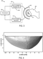
- the method of the present invention can be carried out using an ultrasound system such as the one illustrated in FIG. 3 .
- the ultrasound system 300 generally includes a transducer array 302 that is capable of delivering ultrasound to a subject 304 and receiving responsive signals therefrom.
- the transducer array 302 is preferably configured to surround an extent of the subject's head.
- the transducer array 302 may be an approximately hemispherical array of transducer elements.
- the ultrasound system 300 also generally includes a processor 306 that is in communication with a pulser/receiver 307, which may include a multi-channel transmitter 308 and a multi-channel receiver 310.
- the multi-channel transmitter 308 receives driving signals from the processor 306 and, in turn, directs the transducer elements of the transducer array 302 to generate ultrasound energy.
- the multi-channel receiver 310 receives acoustic signals during and/or after sonications and relays these signals to the processor 306 for processing in accordance with embodiments of the present invention.
- the processor 306 may also be configured to adjust the driving signals in response to the acoustic signals received by the multi-channel receiver 310.
- the phase and/or amplitude of the driving signals may be adjusted so that ultrasound energy is more efficiently transmitted through the skull of the subject 304 and into the target volume-of-interest 312.
- the acoustic signals may also be analyzed to determine whether and how the extent of the focal region should be adjusted.
- the transducer array 302 may be an approximately hemispherical phased array with multiple transmit-receive ultrasound elements sparsely distributed in such a manner that the variation in the distance between elements is maximized.
- the diameter of the array 302 may be, for example, 30 centimeters.
- the array 302 may contain, for example, 128, 256, or more elements that are mounted on a hemispherical surface.
- the transducer array 302 can be a sparse hemispherical ultrasound transducer array, such as an array of 128 lead zirconate titanate ("PZT") transducer elements fabricated and installed on the inner surface of a 30 cm diameter hemisphere.
- the elements in such an array can be constructed as squares with dimensions of 2 x 2 mm 2 and center frequencies of approximately 11 MHz. Electrical connections in the transducer array 302 can be made by soldering to the element electrodes.
- the transducer elements in the transducer array 302 can be backed using a 3:1 (by weight) mixture of PZT powder (Del Piezo Specialties, LLC., West Palm Beach, FL, USA) and epoxy (301 epoxy, Epoxy Technology Inc, Billerica, MA, USA) in order to improve the transducer bandwidth.
- this backing layer can be approximately 1 cm thick, with a bottom surface that is angled to prevent reflection of sound back towards the transducer element.
- the transducer array 302 can be configured such that the receiver elements are sparsely distributed in a pseudo-random configuration over a whole hemisphere to optimize the imaging resolution; although, it is contemplated that the placement (or the number of elements) may not be critical as long as adequate sampling of the skull surface can be obtained.
- the transmit elements can be selected as a subset of all of the elements in the array 302.
- the array may contain 1372 transducer elements, of which only 128 are transmit elements.
- the center frequency of the transmit array can be selected to be sufficiently low so as to undergo minimal distortion and attenuation through the skull bone.
- an ultrasound-based registration method is provided to register medical images, such as CT skull data, to an ultrasound treatment coordinate space.
- the method provides significant benefits for the development of a low-cost, transcranial ultrasound treatment platform.
- a previously developed transcranial ultrasound propagation model based on ray-acoustics was employed to assess the transmit focusing error resulting from imperfections in the US-based registration.
- the location of the hippocampus within the brain region of five triangulated skull meshes was targeted in silico using a clinical transcranial phased array (ExAblate 4000, InSightec, Haifa, Israel) applicator (1024 elements, 1 cm x 1 cm squares, 30 cm diameter array aperture).
- the phases used to target the hippocampus were determined from the US-registered data, and were applied to landmark-registered (gold-standard) skull configurations to determine the impact of the misalignment on trans-skull focusing.
- FIGS. 5 and 6 show the absolute displacement errors and absolute rotational errors associated with the experimental fixtures (frame error), across multiple CT datasets from the same skull and across multiple different skulls (inter-skull mean).
- the frame error was calculated using the landmarks on the array frame to register the repeated CT data sets from Skull #5 to the same coordinate space.
- the skull plate was then registered across the data sets using holes drilled into the plate as references.
- the frame errors (0.50 ⁇ 0.12 mm; 0.47 ⁇ 0.17 degrees) thus represent the possible differences in the true orientation of the skull between the US and CT measurements.
- the displacement and rotation errors associated with repeat measurements of one skull were determined by registering the same US data to the three different CT stacks obtained for Skull #5. With average errors of 1.18 ⁇ 0.15 mm and 1.26 ⁇ 0.08 degrees, Skull #5 had the highest registration error of all the skulls. The low intra-skull standard deviations show that the registration errors did not vary greatly across different CT datasets of the same skull. Across all skulls (using the mean values for Skull #5), on average sub-millimeter (0.95 ⁇ 0.20 mm) and sub-degree errors (0.79 ⁇ 0.36 degrees) were obtained, but larger standard deviations were observed, particularly for the rotational error.
- FIGS. 7A-7C show the dependency of this registration scheme on different algorithm parameters averaged across the five skulls.
- FIG. 7A the impact of the skull surface discretization from the CT data is shown.
- the results are expressed in terms of a percentage of the initial number of vertices, with 100 percent corresponding to no refinement of the surface (10.2 ⁇ 0.6 x 10⁴ vertices, 0.27 ⁇ 0.06 mm 2 face area).
- the results show a small increase in the mean errors with decreasing discretization.
- FIG. 7C illustrates the errors as a function of array elements.
- the full array (128 elements)
- only 75 percent (96) of the elements produced usable signals and the optimization cost function was summed across 50 percent (64).
- Table 1 shows focal pressure and positional shift due to skull-array misregistration relative to the ideal case of perfect registration. Results are shown for 230 and 650 kHz.
- the targeting error is the shift of the anatomical target due to the registration error.
- Table 1 Simulation Results Skull Pressure Ratio (%) (230 kHz/ 650 kHz) Focal shift (mm) (230 kHz/ 650 kHz) Targeting Error (mm) 1 100/99 0.6/0.7 0.7 2 99/99 0.8/0.9 0.8 3 99/99 0.9/1.0 1.0 4 99/99 1.2/1.0 1.0 5(1) 99/99 1.0/1.0 1.0 5(2) 99/99 1.1/1.2 1.2 5(3) 99/98 1.2/1.3 1.3
- FIG. 8 shows example echoes from a single channel on one skull, with and without the scalp. Although attenuated, the echo from the skull bone can be clearly seen. Also shown are the magnitude of the Hilbert transform for each case, showing a clear rising edge at the tissue-skull interface. From the echo data the scalp thickness at this location was estimated to be about 3.7 mm. This is within the reported range of scalp thickness for adults (3-5 mm). At the cut edge, the scalp used in this study was measured with Vernier calipers to be 7.5 mm thick at its thickest point.
- the methods described here are two possible implementations to register CT images of the skull to discrete ultrasound measurements of the skull surface. This method could also be readily adapted to register other 3D data sets of the skull, such as MRI, to ultrasound data.
- the point-based method described above is computationally faster than the plane-based method, but is more likely to converge to a local minimum rather than a global minimum, particularly if the number of vertices describing the skull surface is downsampled.
- the cost function was described in terms of a root-mean-squared value, but could also be expressed in terms of a linear sum or with individual element weightings.
- the distance term, R i ( x ), and the face, m i were expressed in terms of the minimum of an absolute value, or the distance between the skull vertex or face and the closest point. Alternatively, these terms could be expressed as the absolute value of the minimum of the signed distance. This alternative could be used in place of, or in conjunction with, the penalty term to address penetration of the skull surface through the spherical control surfaces.
- a relatively simple method was implemented to make the registration more robust by including only the best 50 percent of the elements. More sophisticated methods, such as M-estimators (e.g., the bi-weight, Talwar) could also be used.
- 128 transducer elements is sufficient for implementing the registration methods described here, even considering that some transducer elements may not provide useable signals.
- algorithms that use triangulation of signals received by other elements may be used to recover information from some of the transducer elements where the transmitted sound is not reflected back to the element, but scattered elsewhere in the dome. These algorithms could potentially improve the accuracy of the measurements.
- the registration methods described here can be advantageously used with cavitation-mediated therapies.
- the methods could also have potential application in thermal therapies if ultrasound-based treatment monitoring techniques, such as local harmonic imaging or ultrasound thermometry, can be robustly implemented transcranially.
- ultrasound imaging is fast and the data from all transducer elements can be acquired on the order of milliseconds, even if the elements are excited one at a time, the methods described here have the potential to track head motion during treatment, thereby removing the need for a stereotactic frame.
- Ultrasound data acquisitions can also be accelerated by transmitting from multiple elements at the same time. It may also be possible to use different frequency transmissions from each of the elements to allow more overlapping transmissions.
- a temporary frame may be used during the CT imaging and treatment to provide a rough initial alignment of the two data sets prior to optimizing the registration.
- the echo signals from the skull are expected to be significantly larger than that from the scalp. Additionally, it is noted that scalp thickness ranges from approximately 3-5 mm in adults, and at 11.25 MHz the ultrasound wavelength in soft tissue is approximately 0.13 mm, suggesting that the water-scalp and scalp-skull interfaces should be resolvable in the pulse-echo data. Naturally, other frequencies that provide the needed precision in the localization can be used.
Landscapes
- Health & Medical Sciences (AREA)
- Life Sciences & Earth Sciences (AREA)
- Engineering & Computer Science (AREA)
- Nuclear Medicine, Radiotherapy & Molecular Imaging (AREA)
- Medical Informatics (AREA)
- Radiology & Medical Imaging (AREA)
- Physics & Mathematics (AREA)
- General Health & Medical Sciences (AREA)
- Public Health (AREA)
- Animal Behavior & Ethology (AREA)
- Veterinary Medicine (AREA)
- Biomedical Technology (AREA)
- Surgery (AREA)
- Molecular Biology (AREA)
- Pathology (AREA)
- Heart & Thoracic Surgery (AREA)
- Biophysics (AREA)
- Computer Vision & Pattern Recognition (AREA)
- High Energy & Nuclear Physics (AREA)
- Optics & Photonics (AREA)
- Theoretical Computer Science (AREA)
- Neurology (AREA)
- General Physics & Mathematics (AREA)
- Oral & Maxillofacial Surgery (AREA)
- Neurosurgery (AREA)
- Dentistry (AREA)
- Pulmonology (AREA)
- Ultra Sonic Daignosis Equipment (AREA)
- Apparatus For Radiation Diagnosis (AREA)
Claims (6)
- Computerimplementiertes Verfahren zur Registrierung eines medizinischen Bildes mit einem Koordinatenraum eines Ultraschallbehandlungssystems, wobei die Schritte des Verfahrens umfassen:(a) Bereitstellen eines medizinischen Bildes eines Subjekts, das ein anatomisches Merkmal abbildet, wobei das anatomische Merkmal ein Schädel eines Subjekts ist;(b) Erfassen von Ultraschalldaten von dem Subjekt unter Verwendung einer Anordnung von Ultraschallwandlerelementen, die einen Teil eines Ultraschallbehandlungssystems bilden, wobei die Ultraschallwandlerelemente bekannte räumliche Orte haben;(c) Berechnen einer Flugzeitmessung für jedes Wandlerelement aus den Ultraschalldaten;(d) Bestimmen von Orten, die mit dem anatomischen Merkmal aus dem bereitgestellten medizinischen Bild des Subjekts verbunden sind;(e) Bestimmen von Registrierungsparametern aus den berechneten Flugzeitmessungen und den in Schritt (d) bestimmten Orten; und(f) Registrieren des bereitgestellten medizinischen Bildes mit dem Koordinatenraum des Ultraschallsystems unter Verwendung der in Schritt (e) bestimmten Registrierungsparameter;wobei Schritt (e) die Minimierung einer Kostenfunktion beinhaltet, die zumindest teilweise auf Entfernungen basiert, welche jeweils mit einem anderen Wandlerelement verbunden sind, wobei jede Entfernung die minimale Entfernung zwischen einer Kugeloberfläche, die um ein Wandlerelement zentriert ist, und den bestimmten Orten, die mit dem anatomischen Merkmal verbunden sind, ist;wobei die Kostenfunktion zusätzlich für jedes Wandlerelement eine Straffunktion enthält, die weitere Lösungen der Kostenfunktion benachteiligtwobei die Straffunktion einen Nullwert hat, wenn der Schädel über einer um das Wandlerelement zentrierten Kugeloberfläche sitzt, und den Wert der Entfernung hat, die der Schädel in die um das Wandlerelement zentrierte Kugeloberfläche eingedrungen ist, multipliziert mit einem Gewichtungsfaktor α gleich 0.25, 0.5, 0.75 oder 1, wenn irgendwelche Punkte auf der Schädeloberfläche näher am Wandlerelement liegen als der Radius der um das Wandlerelement zentrierten Kugeloberfläche, undwobei eine Kugeloberfläche, die um einen Wandler zentriert ist, einen Radius aufweist, der durch die berechnete Flugzeitmessung für das zugehörige Wandlerelement definiert ist.
- Verfahren nach Anspruch 1, wobei der Radius der Kugeloberfläche definiert ist als die berechnete Flugzeitmessung geteilt durch das Doppelte der Schallgeschwindigkeit.
- Verfahren nach Anspruch 1, wobei Schritt (d) das Segmentieren des bereitgestellten medizinischen Bildes und das Bestimmen von Orten, die mit dem anatomischen Merkmal verbunden sind, auf der Grundlage des segmentierten medizinischen Bildes umfasst.
- Verfahren nach Anspruch 1, wobei Schritt (d) das Identifizieren mindestens einer Markierung in dem bereitgestellten medizinischen Bild und das Bestimmen mindestens eines der mit dem anatomischen Merkmal verbundenen Orts als Ort der mindestens einen Markierung umfasst.
- Verfahren nach Anspruch 1, wobei das bereitgestellte medizinische Bild ein Computertomographie-Bild ist.
- Verfahren nach Anspruch 1, wobei das bereitgestellte medizinische Bild ein Magnetresonanzbild ist.
Applications Claiming Priority (2)
| Application Number | Priority Date | Filing Date | Title |
|---|---|---|---|
| US201562152565P | 2015-04-24 | 2015-04-24 | |
| PCT/IB2016/000627 WO2016170427A1 (en) | 2015-04-24 | 2016-04-25 | Method for registering pre-operative images of a subject to an ultrasound treatment space |
Publications (3)
| Publication Number | Publication Date |
|---|---|
| EP3285675A1 EP3285675A1 (de) | 2018-02-28 |
| EP3285675A4 EP3285675A4 (de) | 2018-11-07 |
| EP3285675B1 true EP3285675B1 (de) | 2024-07-24 |
Family
ID=57142905
Family Applications (1)
| Application Number | Title | Priority Date | Filing Date |
|---|---|---|---|
| EP16782704.7A Active EP3285675B1 (de) | 2015-04-24 | 2016-04-25 | Verfahren zum registrieren von präoperativen bildern eines subjekts in einem ultraschallbehandlungsraum |
Country Status (4)
| Country | Link |
|---|---|
| US (1) | US10285593B2 (de) |
| EP (1) | EP3285675B1 (de) |
| CA (1) | CA2980976C (de) |
| WO (1) | WO2016170427A1 (de) |
Families Citing this family (21)
| Publication number | Priority date | Publication date | Assignee | Title |
|---|---|---|---|---|
| EP4230262A3 (de) | 2015-06-24 | 2023-11-22 | The Regents Of The University Of Michigan | System für histotripsietherapie zur behandlung des hirngewebes |
| EP3487410B1 (de) * | 2016-08-01 | 2025-02-12 | Cordance Medical Inc. | Ultraschallgeführte öffnung der blut-hirn-schranke |
| US10799208B2 (en) * | 2016-11-04 | 2020-10-13 | Siemens Medical Solutions Usa, Inc. | Compressional sound speed imaging using ultrasound |
| GB2557915B (en) * | 2016-12-16 | 2020-06-10 | Calderon Agudo Oscar | Method of and apparatus for non invasive medical imaging using waveform inversion |
| EP3844717A4 (de) * | 2018-08-29 | 2022-04-06 | Agency for Science, Technology and Research | Läsionslokalisierung in einem organ |
| CN113286552B (zh) | 2018-11-28 | 2025-05-02 | 希斯托索尼克斯公司 | 组织摧毁术系统及方法 |
| CA3138023A1 (en) * | 2019-05-31 | 2020-12-03 | Sunnybrook Research Institute | Systems and methods for reducing thermal skull-induced aberrations during transcranial ultrasound therapeutic procedures |
| CN110464459B (zh) * | 2019-07-10 | 2022-04-15 | 丽水市中心医院 | 基于ct-mri融合的介入计划导航系统及其导航方法 |
| CN110433406B (zh) * | 2019-09-04 | 2021-05-28 | 珠海医凯电子科技有限公司 | 超声聚焦定位图像优化方法 |
| GB202000247D0 (en) * | 2020-01-08 | 2020-02-19 | Forkbeard Tech As | Positioning using cost terms |
| WO2021155026A1 (en) | 2020-01-28 | 2021-08-05 | The Regents Of The University Of Michigan | Systems and methods for histotripsy immunosensitization |
| GB202009079D0 (en) | 2020-06-15 | 2020-07-29 | Oxsonics Ltd | Mapping of cavitation activity |
| JP2023530477A (ja) | 2020-06-18 | 2023-07-18 | ヒストソニックス,インコーポレーテッド | 組織破砕音響/患者結合システムおよび方法 |
| EP4204084A4 (de) | 2020-08-27 | 2024-10-09 | The Regents Of The University Of Michigan | Ultraschallwandler mit sende-empfangs-fähigkeit für histotripsie |
| EP4351718A4 (de) | 2021-06-07 | 2025-03-26 | The Regents of The University of Michigan | Minimal invasive histotripsiesysteme und verfahren |
| IL308943A (en) | 2021-06-07 | 2024-01-01 | Univ Michigan Regents | All-inclusive ultrasound systems and methods that include histotripsy |
| JP2025502042A (ja) | 2022-01-04 | 2025-01-24 | ユニバーシティ オブ ユタ リサーチ ファウンデーション | 脳深部回路のモデュレーションのためのシステムおよび方法 |
| WO2024009143A1 (en) * | 2022-07-07 | 2024-01-11 | Insightec Ltd. | Systems and methods for registering images obtained using different imaging modalities |
| AU2023366591A1 (en) | 2022-10-28 | 2025-04-24 | Histosonics, Inc. | Histotripsy systems and methods |
| WO2024221001A2 (en) | 2023-04-20 | 2024-10-24 | Histosonics, Inc. | Histotripsy systems and associated methods including user interfaces and workflows for treatment planning and therapy |
| WO2026062551A1 (en) * | 2024-09-20 | 2026-03-26 | Medtronic Navigation, Inc. | System and method for navigation |
Citations (1)
| Publication number | Priority date | Publication date | Assignee | Title |
|---|---|---|---|---|
| US7175599B2 (en) * | 2003-04-17 | 2007-02-13 | Brigham And Women's Hospital, Inc. | Shear mode diagnostic ultrasound |
Family Cites Families (8)
| Publication number | Priority date | Publication date | Assignee | Title |
|---|---|---|---|---|
| US6106464A (en) * | 1999-02-22 | 2000-08-22 | Vanderbilt University | Apparatus and method for bone surface-based registration of physical space with tomographic images and for guiding an instrument relative to anatomical sites in the image |
| US6896657B2 (en) | 2003-05-23 | 2005-05-24 | Scimed Life Systems, Inc. | Method and system for registering ultrasound image in three-dimensional coordinate system |
| US20080249419A1 (en) * | 2006-12-12 | 2008-10-09 | Sekins K Michael | Time-of-flight triangulation based methods of device spatial registration for multiple-transducer therapeutic ultrasound systems |
| US8182975B2 (en) | 2007-03-28 | 2012-05-22 | Fujifilm Corporation | Positive resist composition and pattern forming method using the same |
| EP2131212A3 (de) | 2008-06-05 | 2011-10-05 | Medison Co., Ltd. | Nicht starre Registrierung zwischen CT-Bildern und Ultraschallbildern |
| WO2014134188A1 (en) * | 2013-02-28 | 2014-09-04 | Rivanna Medical, LLC | Systems and methods for ultrasound imaging |
| US9230331B2 (en) | 2013-10-21 | 2016-01-05 | Samsung Electronics Co., Ltd. | Systems and methods for registration of ultrasound and CT images |
| US9934570B2 (en) * | 2015-10-09 | 2018-04-03 | Insightec, Ltd. | Systems and methods for registering images obtained using various imaging modalities and verifying image registration |
-
2016
- 2016-04-25 EP EP16782704.7A patent/EP3285675B1/de active Active
- 2016-04-25 US US15/564,308 patent/US10285593B2/en active Active
- 2016-04-25 CA CA2980976A patent/CA2980976C/en active Active
- 2016-04-25 WO PCT/IB2016/000627 patent/WO2016170427A1/en not_active Ceased
Patent Citations (1)
| Publication number | Priority date | Publication date | Assignee | Title |
|---|---|---|---|---|
| US7175599B2 (en) * | 2003-04-17 | 2007-02-13 | Brigham And Women's Hospital, Inc. | Shear mode diagnostic ultrasound |
Non-Patent Citations (1)
| Title |
|---|
| G T CLEMENT ET AL: "A non-invasive method for focusing ultrasound through the human skull", PHYS. MED. BIOL, vol. 47, 5 April 2002 (2002-04-05), pages 1219 - 1236, XP055763472 * |
Also Published As
| Publication number | Publication date |
|---|---|
| WO2016170427A1 (en) | 2016-10-27 |
| US20180132723A1 (en) | 2018-05-17 |
| CA2980976A1 (en) | 2016-10-27 |
| EP3285675A4 (de) | 2018-11-07 |
| US10285593B2 (en) | 2019-05-14 |
| EP3285675A1 (de) | 2018-02-28 |
| CA2980976C (en) | 2023-03-21 |
| HK1251432A1 (en) | 2019-02-01 |
Similar Documents
| Publication | Publication Date | Title |
|---|---|---|
| EP3285675B1 (de) | Verfahren zum registrieren von präoperativen bildern eines subjekts in einem ultraschallbehandlungsraum | |
| US12290403B2 (en) | Systems and methods for performing transcranial ultrasound therapeutic and imaging procedures | |
| US20210097702A1 (en) | Systems and methods for registering images obtained using various imaging modalities and verifying image registration | |
| EP2964328B1 (de) | Frequenzoptimierung bei der ultraschallbehandlung | |
| EP2964096B1 (de) | System und verfahren zur messung und korrektur von ultraschall-phasenverzerrungen durch ablenkende medien | |
| JP4558504B2 (ja) | 超音波治療における組織異常の修正 | |
| WO2018138576A1 (en) | Cavitation localization | |
| CN107205720B (zh) | 一种超声适应性波束形成方法及其对经颅成像的应用 | |
| US10485992B2 (en) | Ultrasound guided radiotherapy system | |
| Zubair et al. | 3D synthetic aperture imaging with a therapeutic spherical random phased array for transcostal applications | |
| O'Reilly et al. | Registration of human skull computed tomography data to an ultrasound treatment space using a sparse high frequency ultrasound hemispherical array | |
| China et al. | FLEX: FLexible transducer with EXternal tracking for ultrasound imaging with patient-specific geometry estimation | |
| HK1251432B (en) | Method for registering pre-operative images of a subject to an ultrasound treatment space | |
| Hajian et al. | Accurate 3-D profile extraction of skull bone using an ultrasound matrix array | |
| JP4394440B2 (ja) | 超音波治療 | |
| Mehdizadeh et al. | Joint Beamforming and Feature Detection for Enhanced Visualization of Spinal Bone Surfaces in Ultrasound Images |
Legal Events
| Date | Code | Title | Description |
|---|---|---|---|
| STAA | Information on the status of an ep patent application or granted ep patent |
Free format text: STATUS: THE INTERNATIONAL PUBLICATION HAS BEEN MADE |
|
| PUAI | Public reference made under article 153(3) epc to a published international application that has entered the european phase |
Free format text: ORIGINAL CODE: 0009012 |
|
| STAA | Information on the status of an ep patent application or granted ep patent |
Free format text: STATUS: REQUEST FOR EXAMINATION WAS MADE |
|
| 17P | Request for examination filed |
Effective date: 20171011 |
|
| AK | Designated contracting states |
Kind code of ref document: A1 Designated state(s): AL AT BE BG CH CY CZ DE DK EE ES FI FR GB GR HR HU IE IS IT LI LT LU LV MC MK MT NL NO PL PT RO RS SE SI SK SM TR |
|
| AX | Request for extension of the european patent |
Extension state: BA ME |
|
| DAV | Request for validation of the european patent (deleted) | ||
| DAX | Request for extension of the european patent (deleted) | ||
| A4 | Supplementary search report drawn up and despatched |
Effective date: 20181010 |
|
| RIC1 | Information provided on ipc code assigned before grant |
Ipc: G06T 7/30 20170101ALI20181004BHEP Ipc: A61B 8/13 20060101ALI20181004BHEP Ipc: A61N 7/00 20060101ALI20181004BHEP Ipc: A61B 34/10 20160101AFI20181004BHEP Ipc: A61B 6/03 20060101ALI20181004BHEP Ipc: A61B 34/00 20160101ALI20181004BHEP Ipc: A61B 5/055 20060101ALI20181004BHEP |
|
| REG | Reference to a national code |
Ref country code: HK Ref legal event code: DE Ref document number: 1251432 Country of ref document: HK |
|
| STAA | Information on the status of an ep patent application or granted ep patent |
Free format text: STATUS: EXAMINATION IS IN PROGRESS |
|
| 17Q | First examination report despatched |
Effective date: 20210115 |
|
| GRAP | Despatch of communication of intention to grant a patent |
Free format text: ORIGINAL CODE: EPIDOSNIGR1 |
|
| STAA | Information on the status of an ep patent application or granted ep patent |
Free format text: STATUS: GRANT OF PATENT IS INTENDED |
|
| INTG | Intention to grant announced |
Effective date: 20240215 |
|
| GRAS | Grant fee paid |
Free format text: ORIGINAL CODE: EPIDOSNIGR3 |
|
| GRAA | (expected) grant |
Free format text: ORIGINAL CODE: 0009210 |
|
| STAA | Information on the status of an ep patent application or granted ep patent |
Free format text: STATUS: THE PATENT HAS BEEN GRANTED |
|
| AK | Designated contracting states |
Kind code of ref document: B1 Designated state(s): AL AT BE BG CH CY CZ DE DK EE ES FI FR GB GR HR HU IE IS IT LI LT LU LV MC MK MT NL NO PL PT RO RS SE SI SK SM TR |
|
| REG | Reference to a national code |
Ref country code: GB Ref legal event code: FG4D |
|
| REG | Reference to a national code |
Ref country code: CH Ref legal event code: EP |
|
| REG | Reference to a national code |
Ref country code: DE Ref legal event code: R096 Ref document number: 602016088553 Country of ref document: DE |
|
| REG | Reference to a national code |
Ref country code: IE Ref legal event code: FG4D |
|
| REG | Reference to a national code |
Ref country code: LT Ref legal event code: MG9D |
|
| REG | Reference to a national code |
Ref country code: NL Ref legal event code: MP Effective date: 20240724 |
|
| PG25 | Lapsed in a contracting state [announced via postgrant information from national office to epo] |
Ref country code: PT Free format text: LAPSE BECAUSE OF FAILURE TO SUBMIT A TRANSLATION OF THE DESCRIPTION OR TO PAY THE FEE WITHIN THE PRESCRIBED TIME-LIMIT Effective date: 20241125 |
|
| REG | Reference to a national code |
Ref country code: AT Ref legal event code: MK05 Ref document number: 1705572 Country of ref document: AT Kind code of ref document: T Effective date: 20240724 |
|
| PG25 | Lapsed in a contracting state [announced via postgrant information from national office to epo] |
Ref country code: NL Free format text: LAPSE BECAUSE OF FAILURE TO SUBMIT A TRANSLATION OF THE DESCRIPTION OR TO PAY THE FEE WITHIN THE PRESCRIBED TIME-LIMIT Effective date: 20240724 |
|
| PG25 | Lapsed in a contracting state [announced via postgrant information from national office to epo] |
Ref country code: PT Free format text: LAPSE BECAUSE OF FAILURE TO SUBMIT A TRANSLATION OF THE DESCRIPTION OR TO PAY THE FEE WITHIN THE PRESCRIBED TIME-LIMIT Effective date: 20241125 Ref country code: NL Free format text: LAPSE BECAUSE OF FAILURE TO SUBMIT A TRANSLATION OF THE DESCRIPTION OR TO PAY THE FEE WITHIN THE PRESCRIBED TIME-LIMIT Effective date: 20240724 |
|
| PG25 | Lapsed in a contracting state [announced via postgrant information from national office to epo] |
Ref country code: NO Free format text: LAPSE BECAUSE OF FAILURE TO SUBMIT A TRANSLATION OF THE DESCRIPTION OR TO PAY THE FEE WITHIN THE PRESCRIBED TIME-LIMIT Effective date: 20241024 |
|
| PG25 | Lapsed in a contracting state [announced via postgrant information from national office to epo] |
Ref country code: FI Free format text: LAPSE BECAUSE OF FAILURE TO SUBMIT A TRANSLATION OF THE DESCRIPTION OR TO PAY THE FEE WITHIN THE PRESCRIBED TIME-LIMIT Effective date: 20240724 Ref country code: GR Free format text: LAPSE BECAUSE OF FAILURE TO SUBMIT A TRANSLATION OF THE DESCRIPTION OR TO PAY THE FEE WITHIN THE PRESCRIBED TIME-LIMIT Effective date: 20241025 Ref country code: PL Free format text: LAPSE BECAUSE OF FAILURE TO SUBMIT A TRANSLATION OF THE DESCRIPTION OR TO PAY THE FEE WITHIN THE PRESCRIBED TIME-LIMIT Effective date: 20240724 |
|
| PG25 | Lapsed in a contracting state [announced via postgrant information from national office to epo] |
Ref country code: BG Free format text: LAPSE BECAUSE OF FAILURE TO SUBMIT A TRANSLATION OF THE DESCRIPTION OR TO PAY THE FEE WITHIN THE PRESCRIBED TIME-LIMIT Effective date: 20240724 |
|
| PG25 | Lapsed in a contracting state [announced via postgrant information from national office to epo] |
Ref country code: LV Free format text: LAPSE BECAUSE OF FAILURE TO SUBMIT A TRANSLATION OF THE DESCRIPTION OR TO PAY THE FEE WITHIN THE PRESCRIBED TIME-LIMIT Effective date: 20240724 |
|
| PG25 | Lapsed in a contracting state [announced via postgrant information from national office to epo] |
Ref country code: AT Free format text: LAPSE BECAUSE OF FAILURE TO SUBMIT A TRANSLATION OF THE DESCRIPTION OR TO PAY THE FEE WITHIN THE PRESCRIBED TIME-LIMIT Effective date: 20240724 Ref country code: IS Free format text: LAPSE BECAUSE OF FAILURE TO SUBMIT A TRANSLATION OF THE DESCRIPTION OR TO PAY THE FEE WITHIN THE PRESCRIBED TIME-LIMIT Effective date: 20241124 |
|
| PG25 | Lapsed in a contracting state [announced via postgrant information from national office to epo] |
Ref country code: HR Free format text: LAPSE BECAUSE OF FAILURE TO SUBMIT A TRANSLATION OF THE DESCRIPTION OR TO PAY THE FEE WITHIN THE PRESCRIBED TIME-LIMIT Effective date: 20240724 |
|
| PG25 | Lapsed in a contracting state [announced via postgrant information from national office to epo] |
Ref country code: ES Free format text: LAPSE BECAUSE OF FAILURE TO SUBMIT A TRANSLATION OF THE DESCRIPTION OR TO PAY THE FEE WITHIN THE PRESCRIBED TIME-LIMIT Effective date: 20240724 Ref country code: RS Free format text: LAPSE BECAUSE OF FAILURE TO SUBMIT A TRANSLATION OF THE DESCRIPTION OR TO PAY THE FEE WITHIN THE PRESCRIBED TIME-LIMIT Effective date: 20241024 |
|
| PG25 | Lapsed in a contracting state [announced via postgrant information from national office to epo] |
Ref country code: RS Free format text: LAPSE BECAUSE OF FAILURE TO SUBMIT A TRANSLATION OF THE DESCRIPTION OR TO PAY THE FEE WITHIN THE PRESCRIBED TIME-LIMIT Effective date: 20241024 Ref country code: PL Free format text: LAPSE BECAUSE OF FAILURE TO SUBMIT A TRANSLATION OF THE DESCRIPTION OR TO PAY THE FEE WITHIN THE PRESCRIBED TIME-LIMIT Effective date: 20240724 Ref country code: NO Free format text: LAPSE BECAUSE OF FAILURE TO SUBMIT A TRANSLATION OF THE DESCRIPTION OR TO PAY THE FEE WITHIN THE PRESCRIBED TIME-LIMIT Effective date: 20241024 Ref country code: LV Free format text: LAPSE BECAUSE OF FAILURE TO SUBMIT A TRANSLATION OF THE DESCRIPTION OR TO PAY THE FEE WITHIN THE PRESCRIBED TIME-LIMIT Effective date: 20240724 Ref country code: IS Free format text: LAPSE BECAUSE OF FAILURE TO SUBMIT A TRANSLATION OF THE DESCRIPTION OR TO PAY THE FEE WITHIN THE PRESCRIBED TIME-LIMIT Effective date: 20241124 Ref country code: HR Free format text: LAPSE BECAUSE OF FAILURE TO SUBMIT A TRANSLATION OF THE DESCRIPTION OR TO PAY THE FEE WITHIN THE PRESCRIBED TIME-LIMIT Effective date: 20240724 Ref country code: GR Free format text: LAPSE BECAUSE OF FAILURE TO SUBMIT A TRANSLATION OF THE DESCRIPTION OR TO PAY THE FEE WITHIN THE PRESCRIBED TIME-LIMIT Effective date: 20241025 Ref country code: FI Free format text: LAPSE BECAUSE OF FAILURE TO SUBMIT A TRANSLATION OF THE DESCRIPTION OR TO PAY THE FEE WITHIN THE PRESCRIBED TIME-LIMIT Effective date: 20240724 Ref country code: ES Free format text: LAPSE BECAUSE OF FAILURE TO SUBMIT A TRANSLATION OF THE DESCRIPTION OR TO PAY THE FEE WITHIN THE PRESCRIBED TIME-LIMIT Effective date: 20240724 Ref country code: BG Free format text: LAPSE BECAUSE OF FAILURE TO SUBMIT A TRANSLATION OF THE DESCRIPTION OR TO PAY THE FEE WITHIN THE PRESCRIBED TIME-LIMIT Effective date: 20240724 Ref country code: AT Free format text: LAPSE BECAUSE OF FAILURE TO SUBMIT A TRANSLATION OF THE DESCRIPTION OR TO PAY THE FEE WITHIN THE PRESCRIBED TIME-LIMIT Effective date: 20240724 |
|
| PG25 | Lapsed in a contracting state [announced via postgrant information from national office to epo] |
Ref country code: SM Free format text: LAPSE BECAUSE OF FAILURE TO SUBMIT A TRANSLATION OF THE DESCRIPTION OR TO PAY THE FEE WITHIN THE PRESCRIBED TIME-LIMIT Effective date: 20240724 Ref country code: RO Free format text: LAPSE BECAUSE OF FAILURE TO SUBMIT A TRANSLATION OF THE DESCRIPTION OR TO PAY THE FEE WITHIN THE PRESCRIBED TIME-LIMIT Effective date: 20240724 Ref country code: DK Free format text: LAPSE BECAUSE OF FAILURE TO SUBMIT A TRANSLATION OF THE DESCRIPTION OR TO PAY THE FEE WITHIN THE PRESCRIBED TIME-LIMIT Effective date: 20240724 |
|
| PG25 | Lapsed in a contracting state [announced via postgrant information from national office to epo] |
Ref country code: EE Free format text: LAPSE BECAUSE OF FAILURE TO SUBMIT A TRANSLATION OF THE DESCRIPTION OR TO PAY THE FEE WITHIN THE PRESCRIBED TIME-LIMIT Effective date: 20240724 |
|
| PG25 | Lapsed in a contracting state [announced via postgrant information from national office to epo] |
Ref country code: CZ Free format text: LAPSE BECAUSE OF FAILURE TO SUBMIT A TRANSLATION OF THE DESCRIPTION OR TO PAY THE FEE WITHIN THE PRESCRIBED TIME-LIMIT Effective date: 20240724 |
|
| REG | Reference to a national code |
Ref country code: DE Ref legal event code: R097 Ref document number: 602016088553 Country of ref document: DE |
|
| PG25 | Lapsed in a contracting state [announced via postgrant information from national office to epo] |
Ref country code: SK Free format text: LAPSE BECAUSE OF FAILURE TO SUBMIT A TRANSLATION OF THE DESCRIPTION OR TO PAY THE FEE WITHIN THE PRESCRIBED TIME-LIMIT Effective date: 20240724 Ref country code: IT Free format text: LAPSE BECAUSE OF FAILURE TO SUBMIT A TRANSLATION OF THE DESCRIPTION OR TO PAY THE FEE WITHIN THE PRESCRIBED TIME-LIMIT Effective date: 20240724 |
|
| PLBE | No opposition filed within time limit |
Free format text: ORIGINAL CODE: 0009261 |
|
| STAA | Information on the status of an ep patent application or granted ep patent |
Free format text: STATUS: NO OPPOSITION FILED WITHIN TIME LIMIT |
|
| 26N | No opposition filed |
Effective date: 20250425 |
|
| PGFP | Annual fee paid to national office [announced via postgrant information from national office to epo] |
Ref country code: DE Payment date: 20250429 Year of fee payment: 10 |
|
| PGFP | Annual fee paid to national office [announced via postgrant information from national office to epo] |
Ref country code: GB Payment date: 20250429 Year of fee payment: 10 |
|
| PGFP | Annual fee paid to national office [announced via postgrant information from national office to epo] |
Ref country code: FR Payment date: 20250429 Year of fee payment: 10 |
|
| PGFP | Annual fee paid to national office [announced via postgrant information from national office to epo] |
Ref country code: CH Payment date: 20250508 Year of fee payment: 10 |
|
| PG25 | Lapsed in a contracting state [announced via postgrant information from national office to epo] |
Ref country code: SE Free format text: LAPSE BECAUSE OF FAILURE TO SUBMIT A TRANSLATION OF THE DESCRIPTION OR TO PAY THE FEE WITHIN THE PRESCRIBED TIME-LIMIT Effective date: 20240724 |
|
| PG25 | Lapsed in a contracting state [announced via postgrant information from national office to epo] |
Ref country code: LU Free format text: LAPSE BECAUSE OF NON-PAYMENT OF DUE FEES Effective date: 20250425 |
|
| PG25 | Lapsed in a contracting state [announced via postgrant information from national office to epo] |
Ref country code: MC Free format text: LAPSE BECAUSE OF FAILURE TO SUBMIT A TRANSLATION OF THE DESCRIPTION OR TO PAY THE FEE WITHIN THE PRESCRIBED TIME-LIMIT Effective date: 20240724 |
|
| REG | Reference to a national code |
Ref country code: BE Ref legal event code: MM Effective date: 20250430 |
|
| PG25 | Lapsed in a contracting state [announced via postgrant information from national office to epo] |
Ref country code: BE Free format text: LAPSE BECAUSE OF NON-PAYMENT OF DUE FEES Effective date: 20250430 |
|
| PG25 | Lapsed in a contracting state [announced via postgrant information from national office to epo] |
Ref country code: IE Free format text: LAPSE BECAUSE OF NON-PAYMENT OF DUE FEES Effective date: 20250425 |