EP3287143A1 - Agent antitumoral ciblant moléculairement ckap4 - Google Patents
Agent antitumoral ciblant moléculairement ckap4 Download PDFInfo
- Publication number
- EP3287143A1 EP3287143A1 EP16755122.5A EP16755122A EP3287143A1 EP 3287143 A1 EP3287143 A1 EP 3287143A1 EP 16755122 A EP16755122 A EP 16755122A EP 3287143 A1 EP3287143 A1 EP 3287143A1
- Authority
- EP
- European Patent Office
- Prior art keywords
- ckap4
- cells
- cancer
- antibody
- dkk1
- Prior art date
- Legal status (The legal status is an assumption and is not a legal conclusion. Google has not performed a legal analysis and makes no representation as to the accuracy of the status listed.)
- Granted
Links
Images


Classifications
-
- A—HUMAN NECESSITIES
- A61—MEDICAL OR VETERINARY SCIENCE; HYGIENE
- A61K—PREPARATIONS FOR MEDICAL, DENTAL OR TOILETRY PURPOSES
- A61K31/00—Medicinal preparations containing organic active ingredients
- A61K31/70—Carbohydrates; Sugars; Derivatives thereof
- A61K31/7088—Compounds having three or more nucleosides or nucleotides
- A61K31/7105—Natural ribonucleic acids, i.e. containing only riboses attached to adenine, guanine, cytosine or uracil and having 3'-5' phosphodiester links
-
- C—CHEMISTRY; METALLURGY
- C07—ORGANIC CHEMISTRY
- C07K—PEPTIDES
- C07K16/00—Immunoglobulins [IG], e.g. monoclonal or polyclonal antibodies
- C07K16/18—Immunoglobulins [IG], e.g. monoclonal or polyclonal antibodies against material from animals or humans
- C07K16/28—Immunoglobulins [IG], e.g. monoclonal or polyclonal antibodies against material from animals or humans against receptors, cell surface antigens or cell surface determinants
-
- A—HUMAN NECESSITIES
- A61—MEDICAL OR VETERINARY SCIENCE; HYGIENE
- A61K—PREPARATIONS FOR MEDICAL, DENTAL OR TOILETRY PURPOSES
- A61K31/00—Medicinal preparations containing organic active ingredients
- A61K31/70—Carbohydrates; Sugars; Derivatives thereof
- A61K31/7088—Compounds having three or more nucleosides or nucleotides
- A61K31/713—Double-stranded nucleic acids or oligonucleotides
-
- A—HUMAN NECESSITIES
- A61—MEDICAL OR VETERINARY SCIENCE; HYGIENE
- A61K—PREPARATIONS FOR MEDICAL, DENTAL OR TOILETRY PURPOSES
- A61K39/00—Medicinal preparations containing antigens or antibodies
- A61K39/395—Antibodies; Immunoglobulins; Immune serum, e.g. antilymphocytic serum
-
- A—HUMAN NECESSITIES
- A61—MEDICAL OR VETERINARY SCIENCE; HYGIENE
- A61K—PREPARATIONS FOR MEDICAL, DENTAL OR TOILETRY PURPOSES
- A61K48/00—Medicinal preparations containing genetic material which is inserted into cells of the living body to treat genetic diseases; Gene therapy
-
- A—HUMAN NECESSITIES
- A61—MEDICAL OR VETERINARY SCIENCE; HYGIENE
- A61K—PREPARATIONS FOR MEDICAL, DENTAL OR TOILETRY PURPOSES
- A61K49/00—Preparations for testing in vivo
-
- A—HUMAN NECESSITIES
- A61—MEDICAL OR VETERINARY SCIENCE; HYGIENE
- A61P—SPECIFIC THERAPEUTIC ACTIVITY OF CHEMICAL COMPOUNDS OR MEDICINAL PREPARATIONS
- A61P1/00—Drugs for disorders of the alimentary tract or the digestive system
- A61P1/18—Drugs for disorders of the alimentary tract or the digestive system for pancreatic disorders, e.g. pancreatic enzymes
-
- A—HUMAN NECESSITIES
- A61—MEDICAL OR VETERINARY SCIENCE; HYGIENE
- A61P—SPECIFIC THERAPEUTIC ACTIVITY OF CHEMICAL COMPOUNDS OR MEDICINAL PREPARATIONS
- A61P11/00—Drugs for disorders of the respiratory system
-
- A—HUMAN NECESSITIES
- A61—MEDICAL OR VETERINARY SCIENCE; HYGIENE
- A61P—SPECIFIC THERAPEUTIC ACTIVITY OF CHEMICAL COMPOUNDS OR MEDICINAL PREPARATIONS
- A61P35/00—Antineoplastic agents
-
- A—HUMAN NECESSITIES
- A61—MEDICAL OR VETERINARY SCIENCE; HYGIENE
- A61P—SPECIFIC THERAPEUTIC ACTIVITY OF CHEMICAL COMPOUNDS OR MEDICINAL PREPARATIONS
- A61P35/00—Antineoplastic agents
- A61P35/02—Antineoplastic agents specific for leukemia
-
- A—HUMAN NECESSITIES
- A61—MEDICAL OR VETERINARY SCIENCE; HYGIENE
- A61P—SPECIFIC THERAPEUTIC ACTIVITY OF CHEMICAL COMPOUNDS OR MEDICINAL PREPARATIONS
- A61P43/00—Drugs for specific purposes, not provided for in groups A61P1/00-A61P41/00
-
- C—CHEMISTRY; METALLURGY
- C07—ORGANIC CHEMISTRY
- C07K—PEPTIDES
- C07K16/00—Immunoglobulins [IG], e.g. monoclonal or polyclonal antibodies
- C07K16/18—Immunoglobulins [IG], e.g. monoclonal or polyclonal antibodies against material from animals or humans
- C07K16/28—Immunoglobulins [IG], e.g. monoclonal or polyclonal antibodies against material from animals or humans against receptors, cell surface antigens or cell surface determinants
- C07K16/30—Immunoglobulins [IG], e.g. monoclonal or polyclonal antibodies against material from animals or humans against receptors, cell surface antigens or cell surface determinants from tumour cells
- C07K16/3023—Lung
-
- C—CHEMISTRY; METALLURGY
- C07—ORGANIC CHEMISTRY
- C07K—PEPTIDES
- C07K16/00—Immunoglobulins [IG], e.g. monoclonal or polyclonal antibodies
- C07K16/18—Immunoglobulins [IG], e.g. monoclonal or polyclonal antibodies against material from animals or humans
- C07K16/28—Immunoglobulins [IG], e.g. monoclonal or polyclonal antibodies against material from animals or humans against receptors, cell surface antigens or cell surface determinants
- C07K16/30—Immunoglobulins [IG], e.g. monoclonal or polyclonal antibodies against material from animals or humans against receptors, cell surface antigens or cell surface determinants from tumour cells
- C07K16/303—Liver or Pancreas
-
- C—CHEMISTRY; METALLURGY
- C12—BIOCHEMISTRY; BEER; SPIRITS; WINE; VINEGAR; MICROBIOLOGY; ENZYMOLOGY; MUTATION OR GENETIC ENGINEERING
- C12N—MICROORGANISMS OR ENZYMES; COMPOSITIONS THEREOF; PROPAGATING, PRESERVING, OR MAINTAINING MICROORGANISMS; MUTATION OR GENETIC ENGINEERING; CULTURE MEDIA
- C12N15/00—Mutation or genetic engineering; DNA or RNA concerning genetic engineering, vectors, e.g. plasmids, or their isolation, preparation or purification; Use of hosts therefor
- C12N15/09—Recombinant DNA-technology
- C12N15/11—DNA or RNA fragments; Modified forms thereof; Non-coding nucleic acids having a biological activity
- C12N15/113—Non-coding nucleic acids modulating the expression of genes, e.g. antisense oligonucleotides; Antisense DNA or RNA; Triplex- forming oligonucleotides; Catalytic nucleic acids, e.g. ribozymes; Nucleic acids used in co-suppression or gene silencing
- C12N15/1138—Non-coding nucleic acids modulating the expression of genes, e.g. antisense oligonucleotides; Antisense DNA or RNA; Triplex- forming oligonucleotides; Catalytic nucleic acids, e.g. ribozymes; Nucleic acids used in co-suppression or gene silencing against receptors or cell surface proteins
-
- C—CHEMISTRY; METALLURGY
- C12—BIOCHEMISTRY; BEER; SPIRITS; WINE; VINEGAR; MICROBIOLOGY; ENZYMOLOGY; MUTATION OR GENETIC ENGINEERING
- C12Q—MEASURING OR TESTING PROCESSES INVOLVING ENZYMES, NUCLEIC ACIDS OR MICROORGANISMS; COMPOSITIONS OR TEST PAPERS THEREFOR; PROCESSES OF PREPARING SUCH COMPOSITIONS; CONDITION-RESPONSIVE CONTROL IN MICROBIOLOGICAL OR ENZYMOLOGICAL PROCESSES
- C12Q1/00—Measuring or testing processes involving enzymes, nucleic acids or microorganisms; Compositions therefor; Processes of preparing such compositions
- C12Q1/68—Measuring or testing processes involving enzymes, nucleic acids or microorganisms; Compositions therefor; Processes of preparing such compositions involving nucleic acids
- C12Q1/6876—Nucleic acid products used in the analysis of nucleic acids, e.g. primers or probes
- C12Q1/6883—Nucleic acid products used in the analysis of nucleic acids, e.g. primers or probes for diseases caused by alterations of genetic material
- C12Q1/6886—Nucleic acid products used in the analysis of nucleic acids, e.g. primers or probes for diseases caused by alterations of genetic material for cancer
-
- G—PHYSICS
- G01—MEASURING; TESTING
- G01N—INVESTIGATING OR ANALYSING MATERIALS BY DETERMINING THEIR CHEMICAL OR PHYSICAL PROPERTIES
- G01N33/00—Investigating or analysing materials by specific methods not covered by groups G01N1/00 - G01N31/00
- G01N33/48—Biological material, e.g. blood, urine; Haemocytometers
- G01N33/50—Chemical analysis of biological material, e.g. blood, urine; Testing involving biospecific ligand binding methods; Immunological testing
- G01N33/5005—Chemical analysis of biological material, e.g. blood, urine; Testing involving biospecific ligand binding methods; Immunological testing involving human or animal cells
- G01N33/5008—Chemical analysis of biological material, e.g. blood, urine; Testing involving biospecific ligand binding methods; Immunological testing involving human or animal cells for testing or evaluating the effect of chemical or biological compounds, e.g. drugs, cosmetics
- G01N33/5011—Chemical analysis of biological material, e.g. blood, urine; Testing involving biospecific ligand binding methods; Immunological testing involving human or animal cells for testing or evaluating the effect of chemical or biological compounds, e.g. drugs, cosmetics for testing antineoplastic activity
-
- G—PHYSICS
- G01—MEASURING; TESTING
- G01N—INVESTIGATING OR ANALYSING MATERIALS BY DETERMINING THEIR CHEMICAL OR PHYSICAL PROPERTIES
- G01N33/00—Investigating or analysing materials by specific methods not covered by groups G01N1/00 - G01N31/00
- G01N33/48—Biological material, e.g. blood, urine; Haemocytometers
- G01N33/50—Chemical analysis of biological material, e.g. blood, urine; Testing involving biospecific ligand binding methods; Immunological testing
- G01N33/5005—Chemical analysis of biological material, e.g. blood, urine; Testing involving biospecific ligand binding methods; Immunological testing involving human or animal cells
- G01N33/5008—Chemical analysis of biological material, e.g. blood, urine; Testing involving biospecific ligand binding methods; Immunological testing involving human or animal cells for testing or evaluating the effect of chemical or biological compounds, e.g. drugs, cosmetics
- G01N33/502—Chemical analysis of biological material, e.g. blood, urine; Testing involving biospecific ligand binding methods; Immunological testing involving human or animal cells for testing or evaluating the effect of chemical or biological compounds, e.g. drugs, cosmetics for testing non-proliferative effects
- G01N33/5023—Chemical analysis of biological material, e.g. blood, urine; Testing involving biospecific ligand binding methods; Immunological testing involving human or animal cells for testing or evaluating the effect of chemical or biological compounds, e.g. drugs, cosmetics for testing non-proliferative effects on expression patterns
-
- G—PHYSICS
- G01—MEASURING; TESTING
- G01N—INVESTIGATING OR ANALYSING MATERIALS BY DETERMINING THEIR CHEMICAL OR PHYSICAL PROPERTIES
- G01N33/00—Investigating or analysing materials by specific methods not covered by groups G01N1/00 - G01N31/00
- G01N33/48—Biological material, e.g. blood, urine; Haemocytometers
- G01N33/50—Chemical analysis of biological material, e.g. blood, urine; Testing involving biospecific ligand binding methods; Immunological testing
- G01N33/53—Immunoassay; Biospecific binding assay; Materials therefor
- G01N33/575—Immunoassay; Biospecific binding assay; Materials therefor for cancer
- G01N33/5752—Immunoassay; Biospecific binding assay; Materials therefor for cancer of the lungs
-
- G—PHYSICS
- G01—MEASURING; TESTING
- G01N—INVESTIGATING OR ANALYSING MATERIALS BY DETERMINING THEIR CHEMICAL OR PHYSICAL PROPERTIES
- G01N33/00—Investigating or analysing materials by specific methods not covered by groups G01N1/00 - G01N31/00
- G01N33/48—Biological material, e.g. blood, urine; Haemocytometers
- G01N33/50—Chemical analysis of biological material, e.g. blood, urine; Testing involving biospecific ligand binding methods; Immunological testing
- G01N33/53—Immunoassay; Biospecific binding assay; Materials therefor
- G01N33/575—Immunoassay; Biospecific binding assay; Materials therefor for cancer
- G01N33/57525—Immunoassay; Biospecific binding assay; Materials therefor for cancer of the liver or pancreas
-
- G—PHYSICS
- G01—MEASURING; TESTING
- G01N—INVESTIGATING OR ANALYSING MATERIALS BY DETERMINING THEIR CHEMICAL OR PHYSICAL PROPERTIES
- G01N33/00—Investigating or analysing materials by specific methods not covered by groups G01N1/00 - G01N31/00
- G01N33/48—Biological material, e.g. blood, urine; Haemocytometers
- G01N33/50—Chemical analysis of biological material, e.g. blood, urine; Testing involving biospecific ligand binding methods; Immunological testing
- G01N33/53—Immunoassay; Biospecific binding assay; Materials therefor
- G01N33/575—Immunoassay; Biospecific binding assay; Materials therefor for cancer
- G01N33/5758—Immunoassay; Biospecific binding assay; Materials therefor for cancer involving compounds serving as markers for tumours, cancers or neoplasias, e.g. cellular determinants, receptors, heat shock/stress proteins, A-protein, oligosaccharides or metabolites
- G01N33/57585—Immunoassay; Biospecific binding assay; Materials therefor for cancer involving compounds serving as markers for tumours, cancers or neoplasias, e.g. cellular determinants, receptors, heat shock/stress proteins, A-protein, oligosaccharides or metabolites involving compounds identifiable in body fluids
-
- G—PHYSICS
- G01—MEASURING; TESTING
- G01N—INVESTIGATING OR ANALYSING MATERIALS BY DETERMINING THEIR CHEMICAL OR PHYSICAL PROPERTIES
- G01N33/00—Investigating or analysing materials by specific methods not covered by groups G01N1/00 - G01N31/00
- G01N33/48—Biological material, e.g. blood, urine; Haemocytometers
- G01N33/50—Chemical analysis of biological material, e.g. blood, urine; Testing involving biospecific ligand binding methods; Immunological testing
- G01N33/53—Immunoassay; Biospecific binding assay; Materials therefor
- G01N33/575—Immunoassay; Biospecific binding assay; Materials therefor for cancer
- G01N33/5758—Immunoassay; Biospecific binding assay; Materials therefor for cancer involving compounds serving as markers for tumours, cancers or neoplasias, e.g. cellular determinants, receptors, heat shock/stress proteins, A-protein, oligosaccharides or metabolites
- G01N33/5759—Immunoassay; Biospecific binding assay; Materials therefor for cancer involving compounds serving as markers for tumours, cancers or neoplasias, e.g. cellular determinants, receptors, heat shock/stress proteins, A-protein, oligosaccharides or metabolites involving compounds localised on the membrane of tumour or cancer cells
-
- C—CHEMISTRY; METALLURGY
- C07—ORGANIC CHEMISTRY
- C07K—PEPTIDES
- C07K2317/00—Immunoglobulins specific features
- C07K2317/70—Immunoglobulins specific features characterized by effect upon binding to a cell or to an antigen
- C07K2317/76—Antagonist effect on antigen, e.g. neutralization or inhibition of binding
-
- C—CHEMISTRY; METALLURGY
- C12—BIOCHEMISTRY; BEER; SPIRITS; WINE; VINEGAR; MICROBIOLOGY; ENZYMOLOGY; MUTATION OR GENETIC ENGINEERING
- C12N—MICROORGANISMS OR ENZYMES; COMPOSITIONS THEREOF; PROPAGATING, PRESERVING, OR MAINTAINING MICROORGANISMS; MUTATION OR GENETIC ENGINEERING; CULTURE MEDIA
- C12N2310/00—Structure or type of the nucleic acid
- C12N2310/10—Type of nucleic acid
- C12N2310/14—Type of nucleic acid interfering nucleic acids [NA]
-
- C—CHEMISTRY; METALLURGY
- C12—BIOCHEMISTRY; BEER; SPIRITS; WINE; VINEGAR; MICROBIOLOGY; ENZYMOLOGY; MUTATION OR GENETIC ENGINEERING
- C12N—MICROORGANISMS OR ENZYMES; COMPOSITIONS THEREOF; PROPAGATING, PRESERVING, OR MAINTAINING MICROORGANISMS; MUTATION OR GENETIC ENGINEERING; CULTURE MEDIA
- C12N2310/00—Structure or type of the nucleic acid
- C12N2310/50—Physical structure
- C12N2310/53—Physical structure partially self-complementary or closed
- C12N2310/531—Stem-loop; Hairpin
-
- C—CHEMISTRY; METALLURGY
- C12—BIOCHEMISTRY; BEER; SPIRITS; WINE; VINEGAR; MICROBIOLOGY; ENZYMOLOGY; MUTATION OR GENETIC ENGINEERING
- C12Q—MEASURING OR TESTING PROCESSES INVOLVING ENZYMES, NUCLEIC ACIDS OR MICROORGANISMS; COMPOSITIONS OR TEST PAPERS THEREFOR; PROCESSES OF PREPARING SUCH COMPOSITIONS; CONDITION-RESPONSIVE CONTROL IN MICROBIOLOGICAL OR ENZYMOLOGICAL PROCESSES
- C12Q2600/00—Oligonucleotides characterized by their use
- C12Q2600/118—Prognosis of disease development
-
- C—CHEMISTRY; METALLURGY
- C12—BIOCHEMISTRY; BEER; SPIRITS; WINE; VINEGAR; MICROBIOLOGY; ENZYMOLOGY; MUTATION OR GENETIC ENGINEERING
- C12Q—MEASURING OR TESTING PROCESSES INVOLVING ENZYMES, NUCLEIC ACIDS OR MICROORGANISMS; COMPOSITIONS OR TEST PAPERS THEREFOR; PROCESSES OF PREPARING SUCH COMPOSITIONS; CONDITION-RESPONSIVE CONTROL IN MICROBIOLOGICAL OR ENZYMOLOGICAL PROCESSES
- C12Q2600/00—Oligonucleotides characterized by their use
- C12Q2600/136—Screening for pharmacological compounds
-
- C—CHEMISTRY; METALLURGY
- C12—BIOCHEMISTRY; BEER; SPIRITS; WINE; VINEGAR; MICROBIOLOGY; ENZYMOLOGY; MUTATION OR GENETIC ENGINEERING
- C12Q—MEASURING OR TESTING PROCESSES INVOLVING ENZYMES, NUCLEIC ACIDS OR MICROORGANISMS; COMPOSITIONS OR TEST PAPERS THEREFOR; PROCESSES OF PREPARING SUCH COMPOSITIONS; CONDITION-RESPONSIVE CONTROL IN MICROBIOLOGICAL OR ENZYMOLOGICAL PROCESSES
- C12Q2600/00—Oligonucleotides characterized by their use
- C12Q2600/158—Expression markers
-
- C—CHEMISTRY; METALLURGY
- C12—BIOCHEMISTRY; BEER; SPIRITS; WINE; VINEGAR; MICROBIOLOGY; ENZYMOLOGY; MUTATION OR GENETIC ENGINEERING
- C12Q—MEASURING OR TESTING PROCESSES INVOLVING ENZYMES, NUCLEIC ACIDS OR MICROORGANISMS; COMPOSITIONS OR TEST PAPERS THEREFOR; PROCESSES OF PREPARING SUCH COMPOSITIONS; CONDITION-RESPONSIVE CONTROL IN MICROBIOLOGICAL OR ENZYMOLOGICAL PROCESSES
- C12Q2600/00—Oligonucleotides characterized by their use
- C12Q2600/178—Oligonucleotides characterized by their use miRNA, siRNA or ncRNA
-
- G—PHYSICS
- G01—MEASURING; TESTING
- G01N—INVESTIGATING OR ANALYSING MATERIALS BY DETERMINING THEIR CHEMICAL OR PHYSICAL PROPERTIES
- G01N2333/00—Assays involving biological materials from specific organisms or of a specific nature
- G01N2333/435—Assays involving biological materials from specific organisms or of a specific nature from animals; from humans
- G01N2333/46—Assays involving biological materials from specific organisms or of a specific nature from animals; from humans from vertebrates
- G01N2333/47—Assays involving proteins of known structure or function as defined in the subgroups
- G01N2333/4701—Details
- G01N2333/4703—Regulators; Modulating activity
- G01N2333/4704—Inhibitors; Supressors
-
- G—PHYSICS
- G01—MEASURING; TESTING
- G01N—INVESTIGATING OR ANALYSING MATERIALS BY DETERMINING THEIR CHEMICAL OR PHYSICAL PROPERTIES
- G01N2333/00—Assays involving biological materials from specific organisms or of a specific nature
- G01N2333/435—Assays involving biological materials from specific organisms or of a specific nature from animals; from humans
- G01N2333/705—Assays involving receptors, cell surface antigens or cell surface determinants
-
- G—PHYSICS
- G01—MEASURING; TESTING
- G01N—INVESTIGATING OR ANALYSING MATERIALS BY DETERMINING THEIR CHEMICAL OR PHYSICAL PROPERTIES
- G01N2500/00—Screening for compounds of potential therapeutic value
- G01N2500/02—Screening involving studying the effect of compounds C on the interaction between interacting molecules A and B (e.g. A = enzyme and B = substrate for A, or A = receptor and B = ligand for the receptor)
Definitions
- the present invention relates to an antitumor agent. More specifically, the present invention relates to an antitumor agent which can target cytoskeleton-associated protein 4 (CKAP4, 63-KDa, cytoskeleton-linking membrane protein (CLIMP-63), P63, ERGIC-63)) as a target molecule and can suppress the proliferation of tumor cells through the suppression of the expression or function of CKAP4.
- CKAP4 cytoskeleton-associated protein 4
- CLIMP-63 cytoskeleton-linking membrane protein
- ERGIC-63 cytoskeleton-linking membrane protein
- LDL receptor-related protein 5 or 6 LDL receptor-related protein 5 or 6
- Wnt which is an extracellular secreted glycoprotein
- a Wnt signal is involved in various types of cell function regulations. It is known that abnormalities in the Wnt signaling pathway can induce cancer, bone diseases, inflammations, infections and the like.
- Dkkl (Dickkopfl) is known to be a factor which can be expressed by a Wnt signal and can inhibit the binding between a Wnt ligand and LDL receptor-related protein 6 (LRP6) to regulate the Wnt signaling pathway, and Dkk1 forms a negative feedback mechanism.
- LRP6 LDL receptor-related protein 6
- a Wnt signal can positively regulate the proliferation of cells. Therefore, it has been believed previously that Dkk1 can negatively regulate the proliferation of cells.
- Non-Patent Document 1 reports that Dkkl is overexpressed in multiple myeloma, hepatoblastoma, Wilms tumor, prostate cancer, renal cancer, breast cancer, esophageal cancer, lung cancer and the like and the suppression of the expression of Dkkl and an anti-Dkk1 antibody are effective for the suppression of the proliferation of tumor cells.
- Dkk1 can negatively regulate the proliferation of cells in the Wnt signaling pathway
- Dkk1 also has a function to enhance the proliferation of cells. Therefore, it is assumed that Dkk1 can enhance the proliferation of cells through a signaling pathway that is independent from the Wnt signaling pathway. However, a signaling pathway through which the proliferation of cells can be enhanced by Dkk1 is not clarified yet.
- CKAP4 is a transmembrane receptor, and an anti-proliferative factor (APF), surfactant protein A (SPA), a tissue plasminogen activator (TPA) and the like are known as the ligands for CKAP4.
- APF anti-proliferative factor
- SPA surfactant protein A
- TPA tissue plasminogen activator
- the present inventors have established a Dkkl-expressing strain (an MDCK/Dkk1-FLAG cells) using MDCKs cell originated from a canine renal tubule, and it is confirmed that Dkk1 can enhance the proliferation ability of the MDCK cell, as demonstrated in the section "Examples" below. From these facts, it is assumed that Dkkl can enhance the proliferation of cells through a signaling pathway that is independent from the Wnt signaling pathway.
- one object of the present invention is to identify a target molecule that is involved in Dkkl-induced hyperproliferation of tumor cells and to provide a novel antitumor agent.
- Another object of the present invention is to provide a method for screening for an antitumor agent using the target molecule.
- Still another object of the present invention is to provide: a test method for predicting the postoperative prognosis in a cancer patient; and a test method for predicting the presence or absence of the development of cancer.
- the present inventors have made intensive and extensive studies for the purpose of clarifying a signaling pathway through which the proliferation of tumor cells can be enhanced by Dkk1, and it is found that CKAP4 acts as a receptor for Dkk1 in a signaling pathway that is independent from the Wnt signaling pathway.
- Dkk1 can enhance the proliferation of tumor cells through the association between CKAP4 and PI3K, and it is found that CKAP4 can regulate the cell hyperproliferation effect by Dkk1 specifically and is partially involved in a cell proliferation regulation mechanism that is not known in the past.
- the present inventors also found that the proliferation of tumor cells can be suppressed by suppressing the expression or function of CKAP4 in the tumor cells.
- the present invention has been accomplished by making further studies on the basis of these findings.
- the present invention provides the following aspects of inventions.
- the antitumor agent according to the present invention is developed on the basis of a novel finding that CKAP4 acts as a receptor for Dkk1 and Dkk1 transmits a signal capable of enhancing the proliferation of tumor cells through CKAP4. According to the present invention, it becomes possible to suppress the proliferation of tumor cells effectively by suppressing the expression or function of CKAP4 that serves as a target molecule. Actually, an in vivo tumor suppression effect of an anti-CKAP4 antibody on various types of cancer cells is confirmed.
- the antitumor agent according to the present invention has a high specificity to tumors and is therefore superior with respect to safety.
- the development of an excellent anti-malignant tumor agent can be accelerated by employing CKAP4 as a target molecule and screening for a substance capable of suppressing the expression or function of CKAP4, and therefore cancer patients can get benefits.
- the postoperative prognosis can be predicted with high accuracy, and therefore the method is helpful for the determination of the best therapy strategy for the prognosis in a cancer patient.
- the presence or absence of the development of cancer can be predicted using exosomes collected from a body fluid from a subject, and therefore it becomes possible to perform the diagnosis of cancer in a low-invasive or non-invasive manner.
- the antitumor agent according to the present invention is characterized by containing a substance capable of suppressing the expression or function of CKAP4 as an active ingredient.
- a substance capable of suppressing the expression or function of CKAP4 as an active ingredient.
- CKAP4 is involved in the mechanism for the regulation of the proliferation of tumor cells as a receptor for Dkk1, and has an activity to enhance the proliferation of tumor cells. In the antitumor agent according to the present invention, it becomes possible to effectively suppress the proliferation of tumor cells by suppressing the expression or function of CKAP4.
- CKAP4 is known as a transmembrane protein, and the amino acid sequence and the nucleotide sequence for CKAP4 are also known.
- amino acid sequence for human CKAP4 is known to be the amino acid sequence represented by SEQ ID NO: 1
- nucleotide sequence for a gene encoding human CKAP4 is known to be the nucleotide sequence represented by SEQ ID NO: 2.
- the "substance capable of suppressing the expression of CKAP4" is not particularly limited, as long as the substance is pharmaceutically acceptable and can suppress the expression of CKAP4 from DNA encoding CKAP4 (i.e., CKAP4 gene).
- the substance capable of suppressing the expression of CKAP4 may exhibit the effect to suppress the expression of CKAP4 in any stage, including a CKAP4 gene transcription stage, a CKAP4 gene post-transcriptional regulation, a CKAP4 translation stage, a CKAP4 post-translational modification stage and the like.
- nucleic acid medicines such as: a nucleic acid molecule capable of suppressing the transcription of CKAP4 gene (e.g., a decoy nucleic acid); an RNA molecule having an RNA interference activity on mRNA for CKAP4 (e.g., siRNA, shRNA, dsRNA) or a precursor thereof; and an nucleic acid molecule capable of suppressing the transcription of mRNA for CKAP4 (e.g., miRNA, an antisense nucleic acid (antisense DNA, antisense RNA), a ribozyme).
- nucleic acid medicines may be used singly, or a combination of two or more of them may be used.
- the nucleotide sequence for each of the nucleic acid medicines may be designed appropriately by a person skilled in the art by any know method on the basis of the information on the nucleotide sequence for CKAP4 gene.
- these nucleic acid medicines siRNA, shRNA, dsRNA, miRNA and an antisense nucleic acid are preferred, and siRNA and shRNA are more preferred, from the viewpoint of the easiness of the application to clinical situations.
- the RNA molecule may be designed in such a manner that the RNA molecule can be produced in vivo. More specifically, the RNA molecule may be one in which DNA encoding the RNA molecule is inserted into an expression vector to be used in a mammalian cell.
- the expression vector include a virus vector such as a retrovirus vector, a lentivirus vector, an adenovirus vector, an adeno-associated virus vector, a herpesvirus vector and a sendai virus vector, and an animal cell expression plasmid.
- the "substance capable of suppressing the function of CKAP4" is not particularly limited, as long as the substance is pharmaceutically acceptable and can suppress the function of CKAP4 located on cell membranes of tumor cells.
- Specific examples of the substance capable of suppressing the function of CKAP4 include an antibody capable of binding specifically to CKAP4 or a fragment of the antibody, an aptamer capable of binding specifically to CKAP4, and a low-molecular-weight compound capable of suppressing the function of CKAP4. These substances may be used singly, or two or more of them may be used. Among these substances, an antibody capable of binding specifically to CKAP4 or a fragment of the antibody is preferred.
- the antibody capable of binding specifically to CKAP4 may be either one of a monoclonal antibody and a polyclonal antibody, and is preferably a monoclonal antibody.
- the isotype of the antibody is not particularly limited and may be any one of IgG, IgM, IgA and the like, and is preferably IgG.
- the antibody is preferably an antibody of which the antigenicity in a human body is decreased. More specifically, the antibody is preferably a complete human antibody, a humanized antibody, a mouse-human chimeric antibody, a chicken-human chimeric antibody or the like, and is more preferably a complete human antibody or a humanized antibody among them.
- the antibody-binding site in CKAP4 is desirably a site in CKAP4 which is exposed on the outside of a cell (e.g., a site lying between position-128 to position-602 in SEQ ID NO: 1, preferably a site lying between position-128 to position-503 in SEQ ID NO: 1 for human CKAP4).
- a leucine zipper domain of CKAP4 position-468 to position-503 in SEQ ID NO: 1 is needed. Therefore, the CKAP4-binding site in the antibody may be adjusted in a manner such that the binding between CKAP4 and Dkk1 in the leucine zipper domain can be suppressed.
- the fragment of the antibody may be any one, as long as the fragment has at least a complementarity determination region (CDR) for specifically recognizing a target antigen and for specifically binding to the target antigen.
- CDR complementarity determination region
- Specific examples of the fragment include Fab, Fab', F(ab') 2 , scFv and scFv-Fc.
- the antibody and a fragment thereof can be produced in a genetically engineering manner by a conventional method.
- CKAP4 is expressed in a tumor cell to enhance the proliferation of the tumor cells.
- the proliferation of tumor cells can be suppressed by suppressing the expression or function of CKAP4. Therefore, the antitumor agent according to the present invention can be used for the treatment of cancer.
- the type of cancer for which the antitumor agent according to the present invention can be used is not particularly limited.
- cancer such as lung cancer, pancreatic cancer, colorectal cancer, colon cancer, gastric cancer, rectal cancer, liver cancer, breast cancer, bladder cancer, prostate cancer, cervical cancer, head and neck cancer, bile duct cancer, gallbladder cancer, oral cancer, tongue cancer, pharyngeal cancer, laryngeal cancer, brain tumor, glioma, glioblastoma and multiple neural glioblastoma; and hematological cancer, such as leukemia and malignant lymphoma.
- solid cancer such as lung cancer, pancreatic cancer, colorectal cancer, colon cancer, gastric cancer, rectal cancer, liver cancer, breast cancer, bladder cancer, prostate cancer, cervical cancer, head and neck cancer, bile duct cancer, gallbladder cancer, oral cancer, tongue cancer, pharyngeal cancer, laryngeal cancer, brain tumor, glioma, glioblastoma and multiple neural glioblastoma
- hematological cancer such as leukemia and mal
- the antitumor agent according to the present invention can exert the tumor cell proliferation suppressing effect thereof effectively against a tumor cell in which CKAP4 is expressed, particularly a tumor cell in which both of CKAP4 and Dkkl are expressed. Therefore, cancer in which CKAP4 is expressed, particularly cancer in which both of CKAP4 and Dkk1 are expressed, is a preferred disease to which the antitumor agent can be applied.
- cancer in which CKAP4 is expressed particularly cancer in which both of CKAP4 and Dkk1 are expressed, is a preferred disease to which the antitumor agent can be applied.
- lung cancer and pancreatic cancer are particularly preferred diseases to which the anti-tumor agent according to the present invention is to be applied, because CKAP4 and Dkk1 are expressed at high frequencies in lung cancer and pancreatic cancer.
- the occurrence of expression of CKAP4 in cancer can be confirmed by carrying out the tissue immunization of a collected cancer tissue. More specifically, a collected cancer tissue is immunostained with an anti-CKAP4 antibody, and it is determined that CKAP4 is expressed when a region in which the expression of CKAP4 is observed makes up 5% or more of a tumor region.
- a preferred example of the types of cancer to which the antitumor agent according to the present invention can apply is a type of cancer in which the CKAP4-expressed region makes up 5% or more, more preferably 20% or more, particularly preferably 50% or more, of a tumor region.
- the occurrence of expression of CKAP4 in cancer can also be determined by collecting RNA from a collected cancer tissue and then measuring the RNA by a quantitative PCR.
- the determination of the presence or absence of the expression of CKAP4 may be performed employing a non-cancer tissue in the same case as a measure. More specifically, it is determined that CKAP4 is expressed in cancer when the amount of CKAP4 in a cell lysate of a cancer tissue is larger than that in a cell lysate of a non-cancer tissue in the same case.
- the occurrence of expression of Dkkl in cancer can also be confirmed by a method in which a collected cancer tissue is subjected to tissue immunization, a method in which RNA is collected from a collected cancer tissue and then the RNA is measured by a quantitative PCR and the like, as in the case of CKAP4.
- the collected cancer tissue is immunostained with an anti-Dkk1 antibody
- Dkk1 is expressed when a region in which the expression of Dkk1 is observed makes up 5% or more of a tumor region.
- a preferred example of the type of cancer to which the antitumor agent according to the present invention can apply is a type of cancer in which the Dkk1-expressed region makes up 5% or more, more preferably 20% or more, particularly preferably 50% or more, of a tumor region.
- RNA is collected from a collected cancer tissue to measure Dkk1
- Dkk1 is expressed in cancer when the amount of Dkk1 in a cell lysate of a cancer tissue is larger than that in a cell lysate of a non-cancer tissue in the same case.
- the antitumor agent according to the present invention can be used for a mammal such as bovine, pig, canine, feline, goat, rat, mouse and rabbit as well as human, and can be used suitably as a medicine for human.
- the dosage form of the antitumor agent according to the present invention may be of any one of oral administration and parenteral (non-oral) administration as long as an antitumor effect can be produced, and may be selected appropriately depending on the types of the active ingredient to be used.
- Specific examples of the dosage form of the antitumor agent according to the present invention include oral administration; injection administration (e.g., intravenous injection, subcutaneous injection, muscle injection, intraperitoneal injection, topical injection to an affected part); and parenteral administration such as suppository administration.
- the dose amount of the antitumor agent according to the present invention may be selected appropriately depending on the types of the active ingredient to be used, the dosage form, the types of cancer to which the antitumor agent is to be applied, the levels of clinical conditions of patients and the like.
- the nucleic acid molecule may be usually administered at a single dose of about 0.01 ⁇ g to 1000 mg/kg body weight, preferably about 0.1 to 100 ⁇ g/kg body weight.
- the substance may be usually administered at a single dose of about 0.1 mg to 20 mg/kg body weight at a frequency of about once per 1 to 3 weeks.
- the antitumor agent according to the present invention may be used singly, or may be used in combination with at least one other drug having an anti-tumor effect and/or at least one radiation therapy.
- the antitumor agent according to the present invention can be prepared in a dosage form that is suitable for the types of the active ingredient and the forms of administration.
- Examples of the dosage form of the antitumor agent according to the present invention include: a solid preparation including tablets, capsules, pills, a powder, granules and a suppository; and a liquid preparation including a solution, a suspension, an emulsion and an injection.
- the antitumor agent according to the present invention can be formulated by adding a pharmaceutically acceptable carrier and an additive, depending on the dosage form thereof.
- a pharmaceutically acceptable carrier for example, in the case of a solid preparation, the solid preparation can be formulated by adding an excipient, a binder, a disintegrating agent, a lubricant and the like.
- the liquid preparation can be formulated by adding physiological saline, a buffer and the like.
- the antitumor agent in the case where a nucleic acid molecule is used as the active ingredient, it is desirable that the antitumor agent is formulated together with a nucleic acid introduction aid so that the nucleic acid molecule can migrate into a tumor cell easily.
- a nucleic acid introduction aid include lipofectamine, oligofectamine, RNAi fect, a liposome, a polyamine, DEAE dextran, calcium phosphate and a dendrimer.
- the screening method according to the present invention is a method for screening for an active ingredient of the antitumor agent, and is characterized by comprising: step 1 of detecting the ability of each of substances of interest (wherein the substances of interest also referred to as "test substances", hereinafter) to suppress the expression of CKAP4 or the ability of each of the test substances to bind to CKAP4 located on cell membranes; and step 2 of selecting, among from the test substances, a test substance that is confirmed to have the ability to suppress the expression of CKAP4 or the ability to bind to CKAP4 located on cell membranes as a candidate for the active ingredient of the antitumor agent.
- test substances also referred to as "test substances”, hereinafter
- the test substance may be any one, as long as the test substance can be employed as a candidate substance for the active ingredient of the antitumor agent.
- the type of the test substance is not particularly limited, and may be, for example, a naturally occurring substance such as a biological substance or a synthetic substance that is synthesized chemically.
- Specific examples of the test substance include: a low-molecular-weight compound; a protein such as an antibody; and a nucleic acid molecule such as siRNA, shRNA, dsRNA, miRNA, antisense DNA, antisense RNA and an aptamer.
- step 1 the ability to suppress the expression of CKAP4 or the ability to bind to CKAP4 located on cell membranes is detected first.
- step 1-a and step 1-b as mentioned below are carried out:
- the cell expressing CKAP4, which is to be used in step 1-a, is not particularly limited, as long as CKAP4 can be expressed in the cell.
- a cell which can express CKAP4 endogenously e.g., a tumor cell
- a cell which is produced by transfecting CKAP4 gene into a normal cell to acquire a CKAP4-expressing ability, and the like can be used.
- the transfection of CKAP4 gene into a normal cell can be achieved by a known genetic engineering technique.
- step 1-a in order to bring the test substance into contact with cells expressing CKAP4, the following process can be carried out: the cells and the test substance are added to a culture medium in which the cells can grow and then the cells are cultured at a temperature at which the cells can grow.
- a nucleic acid molecule such as siRNA, shRNA, dsRNA, miRNA, antisense DNA and antisense RNA
- the type of the nucleic acid introduction aid is as mentioned in the section "1. Antitumor agent".
- step 1-b the measurement of the amount of CKAP4 expressed in the cells can be achieved by measuring the amount of mRNA for CKAP4 and/or the amount of CKAP4 (the amount of a protein) by a known technique.
- the concrete method for detecting the ability of the test substance to bind to CKAP4 located on cell membranes is not particularly limited, and an example of the method is a method in which step 1-i and step 1-ii as mentioned below are carried out:
- Step 1-i can be carried out by the same method as that employed in step 1-a mentioned above.
- step 1-b the concrete technique for confirming whether or not the test substance is bound to CKAP4 located on cell membranes is not particularly limited, and the confirmation can be determined by measuring the amount of the test substance by a known immunostaining method.
- the confirmation can also be carried out by collecting CKAP4 from the cells subsequent to step 1-i and then measuring the amount of the test substance bound to CKAP4.
- step 2 a test substance which is confirmed to have the ability to suppress the expression of CKAP4 or the ability to bind to CKAP4 located on cell membranes in step 1 as a candidate for the active ingredient of the antitumor agent.
- such a test substance that the expression amount of CKAP4 when step 1 is carried out with the addition of the test substance is smaller compared with the expression amount of CKAP4 when step 1 is carried out without the addition of the test substance is determined to have the ability to suppress the expression of CKAP4.
- step 2 a test substance which is confirmed to have the ability to suppress the expression of CKAP4 or the ability to suppress the binding between CKAP4 and Dkkl is determined to have the effect to suppress the proliferation of tumor cells, and the test substance is selected as a candidate for the active ingredient of the antitumor agent.
- the test substance thus selected may be actually tested with respect to the effect to suppress the proliferation of tumor cells to determine whether or not the test substance can be used clinically as an antitumor agent.
- the method for testing to examine the postoperative prognosis in a cancer patient according to the present invention is characterized by comprising a step of measuring the expression of CKAP4 and Dkkl in a cancer tissue collected from the cancer patient. According to the test method of the present invention, it becomes possible to predict the postoperative prognosis in a cancer patient by employing the expression of both of CKAP4 and Dkkl in a cancer tissue collected from the cancer patient as a measure.
- the type of cancer in a patient which is to be examined in the prognosis test method is not particularly limited.
- Specific examples of the type of cancer include: solid cancer such as lung cancer, pancreatic cancer, colorectal cancer, colon cancer, gastric cancer, rectal cancer, liver cancer, breast cancer, bladder cancer, prostate cancer, cervical cancer, head and neck cancer, bile duct cancer, gallbladder cancer, oral cancer, tongue cancer, pharyngeal cancer, laryngeal cancer, brain tumor, glioma, glioblastoma and multiple neural glioblastoma; and hematological cancer such as leukemia and malignant lymphoma.
- preferred examples of the type of cancer to be examined in the test method according to the present invention include lung cancer and pancreatic cancer.
- the measurement of the expression of CKAP4 in a cancer tissue collected from a cancer patient can be carried out using a reagent capable of detecting CKAP4.
- An example of the method for measuring the expression of CKAP4 is a method in which a collected cancer tissue is immunostained using an anti-CKAP4 antibody. More specifically, a collected cancer tissue is immunostained using an anti-CKAP4 antibody, and it is determined that CKAP4 is expressed when a region in which the expression of CKAP4 is observed makes up 5% or more of a tumor region.
- the measurement of the expression of CKAP4 can also be carried out by, for example, collecting RNA from a collected cancer tissue and then carrying out a quantitative PCR.
- the determination on the presence or absence of the expression of CKAP4 may be performed by employing a value in a non-cancer tissue in the same case as a reference. More specifically, it is determined that CKAP4 is expressed in cancer when the amount of CKAP4 in a cell lysate of a cancer tissue is larger than that in a cell lysate of a non-cancer tissue in the same case.
- the test method according to the present invention from the viewpoint of the further improvement in the accuracy of prediction of the postoperative prognosis in a patient, it is desirable to also measure the expression of Dkk1 in a cancer tissue collected from the cancer patient.
- the measurement of the expression of Dkk1 in a cancer tissue collected from the cancer patient can be carried out using a reagent capable of detecting Dkk1.
- the expression can be confirmed by a method in which a collected cancer tissue is tissue-immunized, a method in which RNA is collected from a collected cancer tissue and then a quantitative PCR is carried out, and the like, as is in the case of the measurement of the expression of CKAP4.
- a collected cancer tissue is immunostained using an anti-Dkk1 antibody
- Dkk1 is expressed in the cancer when a region in which the expression of Dkk1 is observed makes up 5% or more of a tumor region.
- RNA is collected from a collected cancer tissue to measure Dkk1
- Dkk1 is expressed in the cancer when the amount of Dkk1 in a cell lysate of a cancer tissue is larger than that in a cell lysate of a non-cancer tissue in the same case.
- a cancer patient in whom the expression of both of CKAP4 and Dkk1 is observed is predicted as a patient whose postoperative prognosis is poor, namely whose overall survival period is short or relapse-free survival period is short or the like.
- the present invention further provides a reagent for detecting CKAP4 in a cancer tissue, as a test kit for the above-mentioned test method.
- the reagent for detecting CKAP4 is not particularly limited, and examples of the reagent include an anti-CKAP4 antibody and a fragment thereof.
- a reagent for detecting Dkk1 in a cancer tissue may also be included. If necessary, the antibody may be labeled with biotin, a fluorescent label, a magnetic bead or the like.
- the method for testing to check on cancer according to the present invention is characterized by comprising a step of measuring CKAP4 in exosomes collected from a subject. According to the present invention, it becomes possible to diagnose whether or not a subject is suffering from cancer by employing the presence or absence of CKAP4 in exosomes collected from the subject as a measure.
- the type of cancer to be tested is not particularly limited, and specific examples of the type of cancer include: solid cancer such as lung cancer, pancreatic cancer, colorectal cancer, colon cancer, gastric cancer, rectal cancer, liver cancer, breast cancer, bladder cancer, prostate cancer, cervical cancer, head and neck cancer, bile duct cancer, gallbladder cancer, oral cancer, tongue cancer, pharyngeal cancer, laryngeal cancer, brain tumor, glioma, glioblastoma and multiple neural glioblastoma; and hematological cancer such as leukemia and malignant lymphoma.
- preferred examples of the type of cancer to be examined by the test method according to the present invention include lung cancer and pancreatic cancer.
- the origin of the exosomes is not particularly limited, and examples of the origin include body fluids including serum and urine. Among these body fluids, serum is preferred.
- the method for collecting exosomes from a subject is not particularly limited, as long as exosomes that can be used as a sample for the detection of CKAP4 can be obtained.
- the method may be selected appropriately among from the conventional known methods.
- the separation of exosomes can be carried out by an ultracentrifugation treatment.
- the separation of exosomes can be carried out more simply by using a commercially available kit. Specific examples of the commercially available exosome separation kit include an ExoQuickTM Exosome precipitation solution and Exoquick-TC (both manufactured by System Biosciences).
- CKAP4 in exosomes can be detected by eluting a protein from the exosomes and then detecting CKAP4 using a reagent for detecting CKAP4 such as an anti-CKAP4 antibody.
- test method it is predicted that a subject has a high possibility of being suffering from cancer when the presence of CKAP4 is observed in the exosomes.
- the present invention further provides a reagent for detecting CKAP4 in exosomes, as a test kit for carrying out the above-mentioned test method.
- the reagent for detecting CKAP4 is not particularly limited, and examples of the reagent include an anti-CKAP4 antibody and a fragment thereof. If necessary, the anti-CKAP4 antibody may be labeled with biotin, a fluorescent label, a magnetic bead or the like.
- a reagent for separating exosomes from a body fluid or the like may be further included.
- a transfected cell is sometimes referred to as a "X/Y cell” (e.g., a MDCK/Dkkl-FLAG-GPI cell).
- This cell is a cell produced by transfecting an X cell with DNA encoding a protein Y.
- a protein is sometimes referred to as "A-B” (e.g., Dkkl-FLAG).
- This protein is a protein produced by fusing a peptide A to a peptide B (e.g., a tag).
- Transfected cells other than those produce by the production method mentioned below were produced by a method similar to the below-mentioned production method or a known genetic engineering technique.
- Dkk1-FLAG-GPI A lentivirus vector harboring Dkk1 gene (Dkk1-FLAG-GPI), into which a FLAG epitope and a glycosylphosphatidylinositol (GPI) anchor signal sequence were inserted tandemly into the C-terminal side, was constructed.
- MDCK cells which are normal cells originated from a canine renal tubule, were transfected with this vector, and MDCK cells each of which was transfected with human Dkk1 gene (Dkk1-FLAG-GPI; the nucleotide sequence for human Dkk1 is represented by SEQ ID NO: 3 and the nucleotide sequence for Dkk1-FLAG-GPI is represented by SEQ ID NO: 4) (i.e., MDCK/Dkk1-FLAG-GPI cells) were selected in Dulbecco's modified Eagle's medium(DMEM) supplemented with 10 ⁇ g/ml of blasticidin S.
- DMEM Dulbecco's modified Eagle's medium
- Each of three 10-cm culture dishes containing confluent MDCK/Dkk1-FLAG-GPI cells was added with 0.5 mg/ml of sulfo-NHS-LC-biotin, and the culture dishes were incubated for 30 minutes at 4°C and then washed with 5 ml of 1 ⁇ PBS containing 50 mM of NH 4 Cl three times. Subsequently, cells were collected and then lysed using 1 ml of NP40 buffer (20 mM Tris-HCl, pH 8.0, 10 % Glycerol, 137 mM NaCl, and 1 % NP40) containing a protease inhibitor (10 g/ml Leupeptin or Aprotinin, or 1 mM PMSF).
- NP40 buffer 20 mM Tris-HCl, pH 8.0, 10 % Glycerol, 137 mM NaCl, and 1 % NP40
- the beads were washed with 1 ml of NP40 buffer three times and then further washed with TBS (25 mM Tris-HCl, pH 7.4, 150mM NaCl) three times.
- TBS 25 mM Tris-HCl, pH 7.4, 150mM NaCl
- the collected beads were eluted by carrying out 30-minute incubation three times at 4°C with TBS (50 ⁇ l) containing 0.2 mg/ml of Flag peptide.
- An eluted sample (up to 150 ⁇ l) was added with 40 ⁇ l of a 50% slurry of NeutrAvidin beads, and the resultant solution was incubated for 2 hours at 4°C while shaking.
- the beads were washed twice with 1ml of NP40 buffer and further washed once with 1 ml of 10 mM Tris-HCl (pH 7.5), and a Dkkl-bound complex was eluted with 20 ⁇ l of laemli sample buffer. Subsequently, a protein that was bound to Dkkl was detected by protein silver staining. Seventeen bands (arrowheads in Fig. 2 (b) ) were cut from the gel and then analyzed by mass spectrometry.
- MDCK cells (2 ⁇ 10 5 cells) expressing Dkk1 were seeded on a Transwell polycarbonate filter (Corning Costar Quality Biological, Gaithersburg, MD, USA). After 48 hours, the culture medium was replaced and then further cultured for 24 hour. In order to detect Dkk1 secreted in the culture medium, Blue Sepharose was added to each of culture media respectively collected from the apical side and the basolateral side, and the resultant solutions were incubated for 2 hours at 4°C and then subjected to centrifugation. Precipitates were collected, and Dkk1 was detected with an anti-Dkk1 antibody.
- MDCK type I (MDCK I) renal tubule cells were kindly provided by Dr. S. Tsukita (Osaka University, Japan)
- A549 and NCI-H1579 human lung adenocarcinoma cells were kindly provided by Dr. Y. Shintani (Osaka University, Japan)
- HeLaS3 human cervical cancer cells were kindly provided by Dr. K. Matsumoto (Nagoya University, Japan)
- AGS human gastric cancer cells were kindly provided by Dr. M. Hatakeyama (Tokyo University, Japan)
- KKLS was kindly provided by Dr. W. Yasui (Hiroshima University, Japan)
- HepG2 human hepatoblastoma cells were kindly provided by Dr. Y.
- S2-CP8 and SUIT-2 pancreatic cancer cells were purchased from Cell Resource Center for Biomedical Research, Institute of Development, Aging and Cancer Tohoku University.
- X293T cells were purchased from Takara Bio Inc.
- DMEM Dulbecco's modified Eagle's medium
- FBS fetal bovine serum
- RPMI-1640 fetal bovine serum
- MDCK I cells were seeded at a density of 1 ⁇ 10 5 cells/ml in a 35-mm dish.
- MDCK I cells were cultured in DMEM supplemented with 10% of FBS, and the other cells were cultured in DMEM supplemented with 1% of FBS. The number of cells was counted after a lapse of a predetermined number of days.
- the test by three-dimensional culture was carried out as previously reported ( Matsumoto et al., EMBO J, 2014 ).
- 40 ⁇ l of a matrigel was mounted on a round glass slide and then incubated for 30 minutes at 37°C to solidify the gel.
- MDCK I cells (3 ⁇ 10 4 cells) suspended in DMEM containing 10% of FBS and 2% of the matrigel and S2-CP8 cells (2 ⁇ 10 4 cells) or A549 cells (2 ⁇ 10 4 cells) suspended in DMEM containing 0.1% of bovine serum albumin and 2% of the matrigel were added on the solidified matrigel and then cultured.
- the tumor xenograft analysis was carried out by a previously reported technique ( Fujii et al., Oncogene, 2014 ) with modification.
- Six-week-old male BALB/cAnNCrj-nu nude mice (Charles River Laboratory Japan Inc, Osaka, Japan) were anesthetized with a combination of medetomidine (0.3 mg/kg body weight) and midazolam (4 mg/kg body weight), and then S2-CP8 cells (5 ⁇ 10 6 cells) or A549 cells (5 ⁇ 10 6 cells) suspended in 100 ⁇ l of phosphate buffered saline (PBS) were injected subcutaneously to the mice. Subsequently, the nude mice were subjected to analysis 21 days after the transplantation (for S2-CP8 cells) or 28 days after the transplantation (for A549 cells).
- PBS phosphate buffered saline
- the nude mice were divided into two groups at the time point at which the average tumor size reached to 50 mm 3 .
- An anti-pCKAP4 antibody (150 ⁇ g/body) or an anti-glutathione-S-transferase (GST) antibody (150 ⁇ g/body) was injected into the intraperitoneal cavity for 2 weeks (on days 0, 4, 8, 12) (for S2-CP8 cells) or 3.5 weeks (on days 0, 4, 9, 12, 16, 18, 22) (for A549 cells) at a frequency of twice per week.
- the nude mice were subjected to analysis on day 14 (for S2-CP8 cells) or day 25 (for A549 cells) after the administration of each of the antibodies.
- the volume and weight of a tumor xenograft formed at the transplanted site were measured, and the tumor xenograft was further subjected to an immunohistochemical analyses.
- the tumor volume was calculated in accordance with the formula: (major axis) ⁇ (minor axis) ⁇ (minor axis) ⁇ 0.5. All of the animal experiments carried in this study were approved by the Animal Research Committee of Osaka University, Japan (No. 21-048-1 ).
- FLAG-p110 ⁇ and FLAG-p110 ⁇ in pcDNA3mycTEVflag and p85 ⁇ in pCAGGS were kindly provided by Dr. T. Asano (Hiroshima University, Japan).
- CKAP4 cDNA was amplified from an A549 cDNA library, and all mutants were produced by a standard technique.
- a lentivirus vector was constructed by inserting cDNA into CSII-CMV-MCS-IRES2-Bsd that was kindly provided by Dr. H. Miyoshi (RIKEN BioResource Center, Ibaraki, Japan).
- a lentivirus vector harboring shRNA was constructed by cloning an oligo DNA fragment containing a H1 promoter and shRNA into CS-RfA-EVBsd using Gateway technology (Invitrogen).
- the target proteins for shRNA and the target sequences in the target proteins used in this test are shown in Table 3.
- ShRNA Target sequences luciferase GTGCGTTGCTAGTACCAAC (SEQ ID NO: 7) human Dkkl GGGTTTCTTGGAATGACGA (SEQ ID NO: 8) human CKAP4 GCAGATTAACCTCAGAAAT (SEQ ID NO: 9)
- X293T cells were transfected with each of packaging vectors (pCAG-HIV-gp and pCMV-VSV-G-RSV-Rev) and the lentivirus vector using a FuGENE HD transfection reagent (Roche Applied Science, Basel, Switzerland) to amplifying the lentivirus.
- the lentivirus was introduced into parental cells (5 ⁇ 10 4 cells/well in a 12-well plate) in the presence of a conditioned medium and 10 ⁇ g/mL of polybrene.
- the cells were centrifugated at 1200 ⁇ g for 1 hour, and then further incubated for another 24 hours to produce MDCK I cells, S2-CP8 cells and A549 cells each of which expressed Dkk1-Flag, CKAP4-HA, Dkk1 shRNA or CKAP4 shRNA stably.
- the cells were seeded on a glass slide and then fixed with 4% of paraformaldehyde for 20 minutes at room temperature. Subsequently, the cells were washed three times with PBS, and then blocked for 20 minutes with PBS containing 1% of BSA and 0.05% of Tween-20. Subsequently, a primary antibody was added to the solution, and the resultant solution was incubated overnight. After the cells were washed three times with PBS, a fluorescently labeling secondary antibody was added to the solution, and the resultant solution was incubated for 1 hour at room temperature. Subsequently, the glass slide was washed extensively with PBS and then sealed with PBS containing 50% of glycerol. The operations and measurements in this test were carried out using LSM510 system (Carl Zeiss Microscopy Co., Ltd, Jena, Germany).
- pancreatic cancer patients 59
- lung adenocarcinoma patients 67
- lung squamous cell carcinoma patients 61
- lung atypical adenomatous hyperplasia patients 11
- the ages of the pancreatic cancer patients ranged from 47 to 83 years (median, 70 years).
- the ages of the lung adenocarcinoma patients ranged from 39 to 85 years (median, 68 years).
- the ages of the lung squamous cell carcinoma patients ranged from 38 to 82 years (median, 71 years).
- the ages of the lung atypical adenomatous hyperplasia patients ranged from 58 to 78 years (median, 67 years).
- each of the resected specimens was observed with a microscope to measure the location and size of a tumor, each of the specimens was fixed in 10 vol% of formalin to prepare a paraffin-embedded block, and then the paraffin-embedded block was analyzed histologically.
- Each of the specimens used in the experiment was sectioned at a thickness of 4 ⁇ m and then stained with hematoxylin and eosin (H&E) or immunoperoxidase for the individual analyses.
- H&E hematoxylin and eosin
- the protocol for this study was approved by the ethical review board of the graduate School of Medicine, Osaka University, Japan (No. 13455 ).
- the immunohistochemical analysis was carried out by a previously reported technique ( Fujii et al., Oncogene, 2014 ) with modification.
- the concrete procedure was briefly as follows. First, all tissue sections were stained using DakoRealTM EnVisionTM Detection System (Dako, Carpentaria, CA, USA) in accordance with the manufacturer's recommendations. More specifically, antigens were activated using Pascal pressure chamber (Dako), and then Dako REAL Peroxidase-Blocking Solution was reacted for 5 minutes to block a non-specific binding site.
- tissue sections were treated with an anti-Dkkl antibody (1:100), an anti-CKAP4 antibody (1:100) or a pAKT antibody (1:50) for 16 hours at 4°C.
- an anti-Dkkl antibody (1:100) an anti-CKAP4 antibody (1:100) or a pAKT antibody (1:50) for 16 hours at 4°C.
- goat anti-mouse IgG labeled with horseradish peroxidase (HRP) was added to each of the tissue sections, then the tissue sections were incubated for 1 hour, and then diaminobenzidine (DAB) (Dako) was added to the resultant solutions.
- DAB diaminobenzidine
- the tissue sections were counterstained with 0.1% (w/v) of hematoxylin. A tumor in which more than 5% of the whole area was stained was classified as being "Dkkl -positive" or "CKAP4-positive".
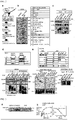
- Fig. 1 (a) the results from which it was confirmed that Dkkl, which was tagged with FLAG at the C-terminal thereof, was expressed in MDCK cells that expressed Dkk1 gene (Dkk1-FLAG) are shown.
- the "control" shown in Fig. 1 (a) was the results obtained using MDCK cells that were parental cells. HSP90 was used as a loading control.
- the lower panel in Fig. 1 (a) the result of the analysis of the expression of Dkk1 which were obtained by culturing MDCK/Dkk1-FLAG cells cultured on a Transwell polycarbonate filter to establish apico-basal polarity and then dividing the cells into apical side cells and basal side cells is shown.
- Fig. 1 (b) the results of the staining of the MDCK/Dkk1-FLAG cells, which were fixed on a Transwell polycarbonate filter, with each of an anti-Dkk1 antibody (green), an anti-E-cadherin antibody (red) and DRAQ5 DNA Dye (blue) are shown. E-cadherin was used as a maker for a basolateral membrane.
- Fig. 1 (b) "Ap" represents an apical side, "Bl” represents a basal side, and the scale bar indicates 20 ⁇ m. From these results, it was confirmed that Dkk1-FLAG was localized in an apical membrane in the MDCK/Dkk1-FLAG cells.
- Fig. 1 (c) the results of the immunostaining of a tissue section of the kidney of a mouse at embryonic day 13 (day E13), which was embedded in paraffin, with an anti-Dkkl antibody.
- the upper right panel in Fig. 1 (c) is an enlarged image of a region boxed in the upper left panel.
- the scale bar shown in the upper right panel in Fig. 1 (c) indicates 100 ⁇ m.
- the results of the immunostaining of the tissue section with an anti-Dkk1 antibody green
- an anti-aPKC antibody red.
- aPKC is a marker for an apical membrane.
- the median is represented with a bold line, the box represents the 25 th -75 th percentile range, the error bars represent the 5 th -95 th percentile ranges, * represents P ⁇ 0.05; ** represents P ⁇ 0.01, the scale bar in (d) indicates 100 ⁇ m, and the scale bar in (e) indicates 20 ⁇ m. From these results, it was confirmed that the MDCK/Dkk1-FLAG cells had an improved proliferation ability compared with MDCK cells and Dkkl enhanced the proliferation ability of cells.
- MDCK cells were cultured two-dimensionally on Transwell polycarbonate filter for 5 days, then 250 ng/ml of Dkkl was added to an apical side or a basal side, and then the cells were cultured for 24 hours. Subsequently, the cells were fixed and then stained with an anti-Ki67 antibody (red) and DRAQ5 DNA Dye (blue). The results are shown in Fig. 1 (f) .
- the percentage of Ki67-positive cases was calculated as the ratio of the number of anti-Ki67 antibody-stained cells to the number of nuclear-stained cells (in 5 fields). Each of the results is expressed in an average value ⁇ s. d. of the results of the three independent tests.
- Fig. 1 (f) The percentage of Ki67-positive cases was calculated as the ratio of the number of anti-Ki67 antibody-stained cells to the number of nuclear-stained cells (in 5 fields). Each of the results is expressed in an average value ⁇ s. d. of the results of the three independent
- NT represents a case where Dkk1 was not added
- AP represent a case where Dkkl was added to the apical side
- Bl represents a case where Dkk1 was added to the basal side
- ** represents P ⁇ 0.01
- the scale bar indicates 20 ⁇ m.
- the MDCK/Dkk1-FLAG cells were cultured two-dimensionally for 3 days, and then the number of the cells was counted. The results are shown in Fig. 1 (g) . As a control, MDCK cells were used. From the results, it was also confirmed that the MDCK/Dkk1-FLAG cells showed a higher proliferation ability compared with the control and Dkkl enhanced the proliferation of cells.
- FIG. 2 (a) The schematic illustration of the experimental procedure for screening for Dkkl -binding proteins located on an apical side surface is shown in Fig. 2 (a) .
- proteins shown in the table in the right-side table in Fig. 2 (b) were detected.
- CKAP4 was detected as band 10.
- a lysate (Input) of the control (wild-type MDCK cells), MDCK/Dkk1-FLAG cells or MDCK/Dkk1-FLAG-GPI cells was immunoprecipitated with an anti-FLAG antibody.
- the lysate (Input) and the immunoprecipitates (IP) were subjected to the detection of CKAP4, LRP6 or Dkk1 using an anti-CKAP4 antibody, an anti-LRP6 antibody or an anti-Dkk1 antibody, respectively.
- the results are shown in Fig. 2 (c) .
- LRP6 is a known Dkk1-binding protein and is used as a positive control. From the results, it was found that, like LRP6, CKAP4 can also form a complex with Dkkl, and an unprecedented novel finding that CKAP4 was a Dkk1-binding protein was obtained.
- a lysate (Input) of each of the control (MDCK cells) and MDCK cells that expressed wild-type Dkk1-FLAG (WT) or a deletion mutant Dkk1-FLAG ( ⁇ CRD-1, ⁇ CRD-2) was immunoprecipitated with an anti-FLAG antibody.
- the cell lysate (Input) and the immunoprecipitates (IP) were subjected to the detection of CKAP4, LRP6 or Dkkl using an anti-CKAP4 antibody, an anti-LRP6 antibody or an anti-Dkk1 antibody, respectively.
- the results are shown in the lower panel in Fig. 2 (d) . As illustrated in the lower panel in Fig.
- each of WT-FLAG and ⁇ CRD-2-FLAG formed a complex with CKAP4 but ⁇ CRD-1-FLAG did not form a complex with CKAP4.
- each of WT-FLAG and ⁇ CRD-2-FLAG formed a complex with LRP6 but ⁇ CRD-2-FLAG did not form a complex with LRP6. Consequently, it was found that Dkkl formed a complex with each of CKAP4 and LRP6 through a different domain.
- ICD represents an intracellular domain
- ECD represents an extracellular domain
- E represents an endoplasmic reticulum anchor domain
- MB represents a microtubule-binding domain
- M represents a transmembrane domain
- T represents a tyrosine-sulfated domain
- LZ represents a leucine zipper domain
- ⁇ represents an ⁇ -helix domain.
- a lysate (Input) of X293T/Dkk1-FLAG cells in which wild-type CKAP4-HA(WT) or a deletion mutant CKAP4-HA (ECD, ⁇ C1, AC2, ⁇ C3) was transiently expressed was immunoprecipitated with an anti-FLAG antibody. Subsequently, each of the cell lysate (Input) and the immunoprecipitates (IP) was probed with an anti-HA antibody or an anti-Dkkl antibody. The results are shown in the lower left panels in Fig. 2 (e) .
- Dkk1 formed a complex with ⁇ C1 but did not form a complex with ⁇ C2 or ⁇ C3 in an extracellular domain of CKAP4. Namely, a possibility that the N-terminal side of Dkkl was bound to a leucine zipper domain of CKAP4 was suggested.
- a lysate (Input) of X293T/Dkk1-FLAG cells in which wild-type CKAP4-HA (WT) or a deletion mutant CKAP4-HA ( ⁇ LZ) was transiently expressed was immunoprecipitated with an anti-FLAG antibody, and then the cell lysate (Input) and the immunoprecipitates (IP) were subjected to the detection of HA and Dkk1 using an anti-HA antibody and an anti-Dkk1 antibody, respectively.
- the results are shown in the lower right panel in Fig. 2 (e) . From the results, it was found that Dkk1 did not form a complex with ⁇ LZ. Namely, it was confirmed that a CRD-1 domain located on the N-terminal side of Dkk1 and a leucine zipper domain of CKAP4 were necessary for the binding between Dkkl and CKAP4.
- GST-CKAP4-ECD A polypeptide (GST-CKAP4-ECD) (a mutant in which an intracellular domain was deleted) was provided, in which glutathione-S-transferase (GST) was linked to the N-terminal of an extracellular domain (ECD) of CKAP4 (which is located between position-128 to position-602 in the amino acid sequence represented by SEQ ID NO: 1).
- GST glutathione-S-transferase
- ECD extracellular domain
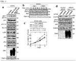
- a lysate of MDCK cells (WT), MDCK/Dkk1-FLAG cells (Dkk1-FLAG) or MDCK/Dkk1-GPI1-FLAG cells (Dkkl-FLAG-GPI) was reacted with an antibody against each of various proteins shown in Fig. 4 (a) .
- the phosphorylation of AKT was enhanced but the enhancement of the phosphorylation of ErK1/2, JNK and Src was not observed in MDCK cells when Dkk1 was overexpressed. Namely, it was suggested that Dkk1 activated AKT.
- MDCK cells was treated with 1 nM of nocodazole for 24 hours, and then the MDCK cells were stimulated with a preparation solution containing 250 ng/ml of Dkk1 or a Dkkl-free preparation solution for 60 minutes. The cells were collected 0, 10, 30 and 60 minutes after the initiation of the stimulation to prepare cell lysates. Subsequently, the cell lysates were subjected to the detection of pAKT, AKT and clathrin using an anti-pAKT antibody, an anti-AKT antibody and an anti-clathrin antibody, respectively. Clathrin was used as a loading control. The results are shown in Fig. 4 (b) . From the results, it was demonstrated that the phosphorylation of AKT in MDCK cells was enhanced in the presence of Dkkl, and it was suggested that Dkkl activated AKT in a time-dependent manner.
- MDCK cells control or MDCK/Dkk1-FLAG cells (Dkkl-FLAG) were transfected with control siRNA or CKAP4 siRNA.
- the resultant cells were subjected to a cell proliferation assay.
- the results are shown in Fig. 4 (c) . From the results, it was confirmed that the decrease in cell proliferation ability was observed when the expression of CKAP4 was suppressed in the Dkk1-expressing cells and therefore Dkk1 enhanced the proliferation of cells through CKAP4.
- MDCK cells control or MDCK/Dkk1-FLAG cells were transfected with control siRNA or CKAP4 siRNA.
- a lysate of the resultant cells was prepared, and then the lysate was reacted with an antibody against each of various proteins shown in Fig. 4 (d) , thereby detecting the various proteins.
- the results are shown in Fig. 4 (d) .
- Dkkl was expressed, the phosphorylation of AKT was significantly decreased by suppressing the expression of CKAP4. Therefore, it was suggested that Dkk1 and CKAP4 were needed for the activation of AKT in MDCK cells.
- a lysate (Input) of each of a control (MDCK cells) and MDCK cells (MDCK/Dkk1-FLAG) overexpressing wild-type Dkkl-FLAG(WT) was immunoprecipitated with an anti-CKAP4 antibody.
- the cell lysate (Input) and the immunoprecipitates (IP) were subjected to the detection of p85 ⁇ , p110 ⁇ , CKAP4 and Dkkl using an anti-p85 ⁇ antibody, an anti-p110 ⁇ antibody, an anti-CKAP4 antibody and an anti-Dkk1 antibody, respectively.
- the results are shown in Fig. 5 (a) and (b) .
- CKAP4 was bound to p85 ⁇ and p110 ⁇ , which constitute PI3K, in cells in which Dkkl was overexpressed. Namely, from the results, it was suggested that CKAP4 was bound to PI3K to activate AKT in cells in which Dkk1 was overexpressed.
- CKAP4-HA or CKAP4-ECD-HA was caused to be expressed together with p85 ⁇ .
- a lysate (Input) of the resultant cells was prepared. The lysate was immunoprecipitated with an anti-HA antibody. Subsequently, the cell lysate (Input) and the immunoprecipitates (IP) were subjected to the detection of p85 ⁇ and CKAP4 using an anti-p85 ⁇ antibody and an anti-HA antibody, respectively. The results are shown in Fig. 5 (c) . From the results, it was found that an intracellular domain of CKAP4 was needed for the Dkkl-dependent binding between CKAP4 and p85 ⁇ .
- CKAP4-HA or CKAP4-HA ECD was caused to be expressed together with FLAG-p110 ⁇ .
- a lysate (Input) of the resultant cells was prepared.
- the lysate was immunoprecipitated with an anti-HA antibody.
- the cell lysate (Input) and the immunoprecipitates (IP) were subjected to the detection of p110 ⁇ and CKAP4 using an anti-p110 ⁇ antibody or an anti-HA antibody, respectively.
- the results are shown in Fig. 5 (d) . From the results, it was found that an intracellular domain of CKAP4 was needed for the Dkkl-dependent binding between CKAP4 and p110 ⁇ .
- a lysate (Input) of S2-CP8 cells in which control shRNA or Dkkl shRNA (Dkkl) was stably expressed was prepared.
- the lysate was immunoprecipitated with an anti-CKAP4 antibody.
- the cell lysate (Input) and the immunoprecipitates (IP) were subjected to the detection of p85 ⁇ , p110 ⁇ , CKAP4 and Dkkl using an anti-p85 ⁇ antibody, an anti-p110 ⁇ antibody, an anti-CKAP4 antibody and an anti-Dkkl antibody, respectively.
- the results are shown in Fig. 5 (e) and (f) . From the results, it was found that CKAP4 also formed a complex with each of p85 ⁇ and p110 ⁇ in a Dkkl-dependent manner in S2-CP8 cells.
- a gene encoding a CKAP4 mutant (PA mutant-CKAP4-HA), in which some of proline residues were respectively substituted by alanine residues in the proline-rich domain of CKAP4, was produced.
- a lysate of X293T/Dkkl-FLAG cell transfected with a gene encoding CKAP4-HA or PA mutant-CKAP4-HA was immunoprecipitated with an anti-HA antibody or a non-immune antibody.
- the cell lysate (Input) and the immunoprecipitates (IP) were subjected to the detection of p85 ⁇ and HA using an anti-p85 ⁇ antibody and an anti-HA antibody, respectively.
- the results are shown in the lower panel in Fig. 5 (g) . From the results, it was confirmed that, when a proline residue in the proline-rich domain of CKAP4 was substituted by an alanine residue, the binding between CKAP4 and PI3K was inhibited.
- FIG. 5 (h) the schematic illustration of the domain structure of p85 ⁇ is shown.
- genes respectively encoding wild-type p85 ⁇ (WT) (GFP-WT), ⁇ SH3 in which SH3 domain was deleted (GFP- ⁇ SH3) and ⁇ SH2 in which SH2 and iSH2 domains were deleted (GFP- ⁇ SH2) were produced.
- a lysate of X293T/Dkk1-FLAG cells transfected with a gene encoding GFP-WT, GFP- ⁇ SH3 or GFP- ⁇ SH2 was immunoprecipitated with an anti-CKAP4 antibody or a non-immune antibody.
- the cell lysate (Input) and the immunoprecipitates (IP) were subjected to the detection of GFP(p85 ⁇ ) and CKAP4 using an anti-GFP(p85 ⁇ ) antibody and an anti-CKAP4 antibody, respectively.
- the results are shown in the lower panel in Fig. 5 (h) . From the results, it was found that a SH3 domain of p85 ⁇ (PI3K) involved in the binding to CKAP4.
- a lysate of each of lung cancer cells (A549, Calu-6 and NCl-H1579), pancreatic cancer cells (SUIT-2 and S2-CP8), cervical canal cancer cells (HeLaS3), gastric cancer cells (AGS and KKLS), esophageal squamous cell carcinoma cells (KYSE-70 and TE-11), hepatoblastoma cells (HepG2) and fetal kidney cells (X239T) was reacted with each of an anti-Dkkl antibody, an anti-CKAP4 antibody and an anti-HSP90 antibody, and the amounts of Dkkl, CKAP4 and HSP90 (control) were measured. The results are shown in Fig. 6 . From the results, it was confirmed that Dkkl was not expressed in normal cells and was expressed in some of cancer cells while CKAP4 was expressed in all of the cells.
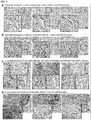
- the boxed areas are enlarged images of tumor regions.
- the boxed areas are enlarged images of non-tumor tumor regions.
- the scale bar indicates 50 ⁇ m.
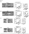
- Fig. 7 (b) and (c) the results obtained by classifying each type of samples on the basis of the presence or absence of the expression of Dkkl and CKAP4 and analyzing the relationship between days after operation and an overall survival rate and a relapse-free survival rate are shown. From these results, it was found that, in a pancreatic cancer patient in whom both of Dkk1 and CKAP4 were expressed, the shortening of a post-operative overall survival period and a relapse-free survival period was observed.
- each of boxed areas is an enlarged image of a tumor region or a non-tumor region.
- the scale bar indicates 100 ⁇ m.
- Fig. 9 Cancer tissues of each of human pancreatic cancer, human lung adenocarcinoma and human lung squamous cell carcinoma were immunostained with an anti-Dkkl antibody, an anti-CKAP4 antibody or an anti-pAKT antibody and haematoxylin. The results are shown in Fig. 9 .
- each of black boxes is an enlarged image, and the bar scale indicates 200 ⁇ m. From the results, it was confirmed that, in human pancreatic cancer, human lung adenocarcinoma and human lung squamous cell carcinoma examined in the test, AKT was activated in a tumor region in which Dkkl and CKAP4 were expressed.
- S2-CP8 cells were transfected with control shRNA, Dkkl shRNA, Dkkl shRNA and Dkkl -FLAG, CKAP4 shRNA, or CKAP4shRNA and CKAP4-HA.
- a lysate of the resultant cells was prepared, and the lysate was subjected to the detection of various proteins shown in the upper panels in Fig. 10 (a) using antibodies against the proteins, respectively.
- the cells were also subjected to a cell proliferation assay. The results are shown in the lower panel in Fig. 10 (a) .
- A549 cells were transfected with control shRNA, Dkkl shRNA, Dkkl shRNA and Dkkl -FLAG, CKAP4 shRNA, or CKAP4 shRNA and CKAP4-HA.
- a lysate of the resultant cells was prepared, and the lysate was subjected to the detection of various proteins shown in the upper panels in Fig. 10 (b) using antibodies against the proteins, respectively.
- the cells were also subjected to a cell proliferation assay. The results are shown in the lower panel in Fig. 10 (b) .
- S2-CP8 cells (2 ⁇ 10 4 cells) or A549 cells (2 ⁇ 10 4 cells) expressing control shRNA, Dkkl shRNA, Dkkl shRNA and Dkkl-FLAG, or CKAP4 shRNA or CKAP4 shRNA and CKAP4-HA were cultured three-dimensionally on a matrigel for 5 days. The number of spheroids per 1 mm 2 was counted (in five fields). The results are shown in Fig. 10 (c) and (d) .
- S2-CP8 cells (5 ⁇ 10 6 cells) expressing control shRNA, CKAP4 shRNA, or CKAP4 shRNA and CKAP4-HA were injected subcutaneously to the back of each of nude mice. After 21 days, a tumor xenograft in a transplanted site in each of the nude mice was analyzed. The results are shown in Fig. 11 (a) . In the left panel in Fig. 11 (a) , the results of the observation of the nude mice from above after 21 days and the results of the observation of the extirpated tumor xenografts are shown. In Fig.
- the broken lines represent the outline forms of the tumor xenografts, and each of the scale bars indicates 10 mm.
- the results of the measurement of the sizes of the tumor tissue sections are shown.
- the results of the measurement of the weights of the tumor xenografts are shown.
- the median is represented with a bold line, the box represents the 25 th -75 th percentile range, the error bars represent the 5 th -95 th percentile ranges, * represents P ⁇ 0.05; and ** represents P ⁇ 0.01. From the results, it was confirmed that the tumor formation ability of S2-CP8 cells decreased by suppressing the expression of CKAP4 and recovered by causing the overexpression of CKAP4.
- A549 cells (5 ⁇ 10 6 cells) expressing control shRNA, CKAP4 shRNA, or CKAP4 shRNA and CKAP4-HA were injected subcutaneously to the back of each of nude mice. After 28 days, a tumor xenograft in a transplanted site in each of the nude mice was analyzed. The results are shown in Fig. 11 (b) . In the left panel in Fig. 11 (b) , the results of the observation of the nude mice from above after 28 days and the results of the observation of the extirpated tumor xenografts are shown. In Fig. 11 (b) , the broken lines represent the outline forms of the tumor xenografts, and each of the scale bars indicates 10 mm.
- the results of the measurement of the sizes of the tumor tissue sections are shown.
- the results of the measurement of the weights of the tumor xenografts are shown.
- the median is represented with a bold line, the box represents the 25 th -75 th percentile range, the error bars represent the 5 th -95 th percentile ranges, * represents P ⁇ 0.05; and ** represents P ⁇ 0.01. From the results, it was confirmed that the tumor formation ability of A549 cells decreased by suppressing the expression of CKAP4 and recovered by causing the overexpression of CKAP4.
- S2-CP8 cells (5 ⁇ 10 6 cells) expressing control shRNA or Dkkl shRNA were injected subcutaneously to the back of each of nude mice. After 21 days, a tumor xenograft in a transplanted site in each of the nude mice was analyzed. The results are shown in Fig. 11 (c) .
- the left panel in Fig. 11 (c) the results of the observation of the nude mice from above after 21 days and the results of the observation of the extirpated tumor xenografts are shown.
- the broken lines represent the outline forms of the tumor xenografts, and each of the scale bars indicates 10 mm.
- A549 cells (5 ⁇ 10 6 cells) expressing control shRNA or Dkkl shRNA were injected subcutaneously to the back of each of nude mice. After 28 days, a tumor xenograft in a transplanted site in each of the nude mice was analyzed. The results are shown in Fig. 11 (d) .
- the left panel in Fig. 11 (d) the results of the observation of the nude mice from above after 28 days and the results of the observation of the extirpated tumor xenografts are shown.
- the broken lines represent the outline forms of the tumor xenografts, and each of the scale bars indicates 10 mm.
- CKAP4 pAb polyclonal anti-CKAP4 antibody
- a lysate of S2-CP8 (control) or S2-CP8 cells expressing CKAP4 cDNA or CKAP4 shRNA was prepared, and the lysate was subjected to the detection of CKAP4 using an anti-CKAP4 antibody.
- the results are shown in the right panel in Fig. 12 (a) . From the results, it was confirmed that the expression amount of CKAP4 in S2-CP8 cells expressing CKAP4 cDNA increased compared with that in the control and the expression amount of CKAP4 in S2-CP8 cells expressing CKAP4 shRNA was suppressed compared with the control. Consequently, it was demonstrated that the produced CKAP4 antibody acted property.
- S2-CP8 cells proliferated on a collagen-coated glass slide was stained in an unfixed state with an anti-CKAP4 antibody (green).
- the actin cytoskeleton and nucleus of each of the cells were stained with phalloidin (red) and DRAQ5 DNA Dye (blue).
- the results are shown in Fig. 12 (b) . From the results, it was confirmed that CKAP4 was expressed intensively on the cell membranes of S2-CP8 cells.
- MDCK cells were treated with 1 nM of nocodazole for 24 hours, and then the cells were incubated together with 25 ⁇ g/ml of an anti-CKAP4 antibody or an anti-GST antibody for 4 hours. Subsequently, the cells were stimulated with 250 ng/ml of Dkk1 or 1 nM of insulin for 30 minutes in the presence of an anti-CKAP4 antibody or an anti-GST antibody. Subsequently, the cells were collected, then a lysate of the cells was prepared, and then the lysate was subjected to the detection of pAKT, AKT and clathrin using an anti-pAKT antibody, an anti-AKT antibody and an anti-clathrin antibody, respectively.
- the anti-GST antibody was used as a control, and clathrin was used as a loading control.
- the results are shown in Fig. 12 (d) . From the results, it was confirmed that the anti-CKAP4 antibody suppressed the Dkkl-dependent activation of AKT but did not affect the insulin-dependent activity of AKT.
- S2-CP8 cells or A549 cells were incubated together with 5 ⁇ g/ml of an anti-CKAP4 antibody or an anti-GST antibody for 4 hours. Subsequently, the cells were collected, then a lysate of the cells was prepared, and then the lysate was subjected to the detection of pAKT, AKT and clathrin using an anti-pAKT antibody, an anti-AKT antibody and an anti-clathrin antibody, respectively.
- the anti-GST antibody was used as a control, and clathrin was used as a loading control. The results are shown in Fig. 12 (e) . From the results, it was confirmed that the anti-CKAP4 antibody suppressed the activation of AKT in cancer cells.
- S2-CP8 cells (2 ⁇ 10 4 cells) or A549 cells (2 ⁇ 10 4 cells) were cultured three-dimensionally on a matrigel for 5 days in the presence of 5 ⁇ g/ml of an anti-GST antibody or 5 ⁇ g/ml of an anti-CKAP4 antibody, and the number of spheroids per one field was counted (in five fields).
- the anti-GST antibody was used as a control.
- the results are shown in Fig. 12 (f) . ** represents P ⁇ 0.01.
- S2-CP8 cells (2 ⁇ 10 4 cells) were cultured on a matrigel three-dimensionally for 7 days in the presence of 25 ⁇ g/ml of an anti-GST antibody, 12.5 ⁇ g/ml of an anti-CKAP4 antibody or 25 ⁇ g/ml of an anti-CKAP4 antibody.
- the number of spheroids per 1 mm 2 was counted.
- the results are shown in Fig. 12 (g) .
- the scale bar indicates 200 ⁇ m.
- Surgical specimens or pleural effusions were provided by two cancer patients from whom agreement was obtained in Center for Adult Diseases, Osaka.
- Each of the two samples (LC189 and LB95) was dispersed mechanically and enzymatically to collect 40 to 250 ⁇ m of a tissue section, and then the tissue section was subjected to floating culture using StemPro hESC (Invitrogen, Carlsbad, CA) to produce cancer tissue-originated spheroids (CTOSs).
- the CTOSs were transplanted into a NOD/scid mouse and the mouse was raised to form a xenograft tumor at the transplanted site in the mouse.
- CTOSs which were isolated and cryopreserved again from the xenograft tumor obtained above, were thawed and then subjected to the following proliferation assay. More specifically, in the proliferation assay, the CTOSs were embedded in Matrigel Growth Factor Reduced (GFR) (BD Biosciences, Bedford, MA) and then cultured in a StemPro hESC medium supplemented with an anti-CKAP4 antibody or an anti-GST antibody at each of various concentrations for 7 days at 37°C. During the culture period, the area of the CTOSs was measured using Image J.
- GFR Matrigel Growth Factor Reduced
- the proliferation suppression effect was not observed even in the presence of the anti-CKAP4 antibody in the CTOSs of the sample LB95 in which the expression amounts of Dkk1 and CKAP4 were small while the proliferation suppression effect was observed significantly in the presence of the anti-CKAP4 antibody in the CTOSs of the sample LC189 in which the expression amounts of Dkkl and CKAP4 were large.
- Human pancreatic cancer cells (S2-CP8) (5 ⁇ 10 6 cells) or human lung cancer cells (A549) (5 ⁇ 10 6 cells) were injected subcutaneously to the back of each of nude mice. At the time point at which the average tumor size reached 50 mm 3 , the nude mice were randomly divided into two groups.
- Tumor xenografts at the transplanted sites in the nude mice were analyzed after 14 days for S2-CP8 cells and after 25 days for A549 cells.
- the results for the human pancreatic cancer cells (S2-CP8) are shown in Fig. 13 (a)
- the results for the human lung cancer cell (A549) (5 ⁇ 10 6 cells) are shown in Fig. 13 (b) .
- the results of the observation of the nude mice from above after 14 days and 25 days are shown.
- the results of the observation of the extirpated tumor xenografts are shown.
- the broken lines represent the outline forms of the tumor xenografts.
- each of the scale bar indicates 10 mm.
- the results of the measurement of the volumes of the tumor xenografts are shown.
- the results of the measurement of the weights of the tumor xenografts are shown.
- each of the results is expressed in an average value ⁇ s. d. * represents P ⁇ 0.05, and ** represents P ⁇ 0.01. From the results, it was confirmed that the tumor formation ability of pancreatic cancer cells (S2-CP8) and lung cancer cells (A549) decreased by the administration of the anti-CKAP4 antibody.
- pancreatic cancer cells SUIT-2 and S2-CP8
- lung cancer cells A549 and Calu-6
- fetal normal kidney cells X293T
- canine renal tubule-originated normal cells MDCK
- the culture supernatant was centrifuged under the conditions of 2,000 ⁇ g and 10 minutes and then further centrifuged under the conditions of 10,000 ⁇ g and 10 minutes to separate a cell debris.
- a collected supernatant was ultracentrifuged under the conditions of 4°C, 100,000 ⁇ g and 3 hours to produce exosome pellets.
- the exosome pellets were washed with 1.2 ml of PBS, and the washed exosome pellets were subjected to the additional separation by ultracentrifugation under the conditions of 4°C, 100,000 ⁇ g and 2 hours. Subsequently, a supernatant was discarded to collect exosomes, and the exosomes were lysed in Laemmli sample buffer.
- the lysate of the exosomes and a lysate of each of the cells were subjected to the detection of CKAP4, TSG101 and clathrin using an anti-CKAP4 antibody, an anti-TSG101 antibody and an anti-clathrin antibody, respectively.
- CKAP4 was detected in the exosomes contained in the culture supernatants of the pancreatic cancer cells and lung cancer cells while CKAP4 was not detected in the exosomes contained in the culture supernatants of the normal cells. Namely, it was confirmed that it became possible to diagnose with respect to the presence or absence of the development of cancer by employing CKAP4 contained in exosomes as a measure.
Landscapes
- Health & Medical Sciences (AREA)
- Life Sciences & Earth Sciences (AREA)
- Chemical & Material Sciences (AREA)
- Engineering & Computer Science (AREA)
- Immunology (AREA)
- Molecular Biology (AREA)
- General Health & Medical Sciences (AREA)
- Biomedical Technology (AREA)
- Medicinal Chemistry (AREA)
- Organic Chemistry (AREA)
- Biochemistry (AREA)
- Hematology (AREA)
- Urology & Nephrology (AREA)
- Biotechnology (AREA)
- Genetics & Genomics (AREA)
- Microbiology (AREA)
- Bioinformatics & Cheminformatics (AREA)
- Public Health (AREA)
- Veterinary Medicine (AREA)
- Animal Behavior & Ethology (AREA)
- Physics & Mathematics (AREA)
- Analytical Chemistry (AREA)
- Pathology (AREA)
- Pharmacology & Pharmacy (AREA)
- Cell Biology (AREA)
- Epidemiology (AREA)
- Food Science & Technology (AREA)
- General Physics & Mathematics (AREA)
- Proteomics, Peptides & Aminoacids (AREA)
- Zoology (AREA)
- Wood Science & Technology (AREA)
- Biophysics (AREA)
- General Engineering & Computer Science (AREA)
- Chemical Kinetics & Catalysis (AREA)
- General Chemical & Material Sciences (AREA)
- Nuclear Medicine, Radiotherapy & Molecular Imaging (AREA)
- Tropical Medicine & Parasitology (AREA)
- Toxicology (AREA)
- Oncology (AREA)
- Hospice & Palliative Care (AREA)
Applications Claiming Priority (2)
| Application Number | Priority Date | Filing Date | Title |
|---|---|---|---|
| JP2015038343 | 2015-02-27 | ||
| PCT/JP2016/052485 WO2016136372A1 (fr) | 2015-02-27 | 2016-01-28 | Agent antitumoral ciblant moléculairement ckap4 |
Publications (3)
| Publication Number | Publication Date |
|---|---|
| EP3287143A1 true EP3287143A1 (fr) | 2018-02-28 |
| EP3287143A4 EP3287143A4 (fr) | 2018-09-05 |
| EP3287143B1 EP3287143B1 (fr) | 2026-03-04 |
Family
ID=56788351
Family Applications (1)
| Application Number | Title | Priority Date | Filing Date |
|---|---|---|---|
| EP16755122.5A Active EP3287143B1 (fr) | 2015-02-27 | 2016-01-28 | Agent antitumoral ciblant ckap4 |
Country Status (6)
| Country | Link |
|---|---|
| US (1) | US10618954B2 (fr) |
| EP (1) | EP3287143B1 (fr) |
| JP (2) | JP6316498B2 (fr) |
| CN (1) | CN107249636B (fr) |
| CA (1) | CA2977917C (fr) |
| WO (1) | WO2016136372A1 (fr) |
Families Citing this family (9)
| Publication number | Priority date | Publication date | Assignee | Title |
|---|---|---|---|---|
| GB2574785A (en) | 2017-09-15 | 2019-12-25 | Jan Loetvall | Method and system for identifying membrane proteins on extracellular vesicles |
| US11807679B2 (en) * | 2017-09-26 | 2023-11-07 | Osaka University | Anti-CKAP4 monoclonal antibody |
| US12397047B2 (en) | 2019-01-09 | 2025-08-26 | Exocure Sweden Ab | Bacteria-derived vesicles and uses thereof |
| CN110007071A (zh) * | 2019-04-01 | 2019-07-12 | 河南省生物工程技术研究中心有限公司 | 一种ckap4标记抗体-荧光微球复合物的制备方法及其应用 |
| WO2021050975A1 (fr) | 2019-09-13 | 2021-03-18 | Exocure Biosciences | Utilisation de nanovésicules fantômes en tant qu'agents thérapeutiques |
| JP7623124B2 (ja) * | 2020-10-20 | 2025-01-28 | 大塚製薬株式会社 | 細胞外小胞の内包物の測定方法および測定キット |
| CN114563571B (zh) * | 2022-01-20 | 2025-01-28 | 湖南大学 | 检测标本中ckap4试剂的应用及膀胱癌检测试剂盒 |
| CN114720700A (zh) * | 2022-05-07 | 2022-07-08 | 浙江大学 | 检测抗细胞骨架相关蛋白4-IgG自身抗体的试剂在制备检测血管内皮损伤试剂盒的应用 |
| WO2025088835A1 (fr) * | 2023-10-25 | 2025-05-01 | 国立大学法人大阪大学 | Anticorps anti-ckap4 humanisé ou fragment de liaison à l'antigène de celui-ci |
Family Cites Families (4)
| Publication number | Priority date | Publication date | Assignee | Title |
|---|---|---|---|---|
| WO2008014484A1 (fr) | 2006-07-27 | 2008-01-31 | University Of Maryland, Baltimore | Récepteur cellulaire du facteur antiprolifération |
| WO2008144485A2 (fr) | 2007-05-18 | 2008-11-27 | University Of Maryland, Baltimore | Inhibition de l'expression et/ou de l'activité de palmitoyl acyle transférase pour la régulation d'activité du facteur antiprolifératif |
| EP2519634B1 (fr) * | 2009-12-29 | 2016-06-01 | CuRNA, Inc. | Traitement de maladies liées à la protéine tumorale 63 (p63) par l'inhibition du produit de transcription antisens naturel de p63 |
| EP2670435B1 (fr) * | 2011-02-01 | 2017-08-23 | The University of Hong Kong | Utilisation d'un anticorps monoclonal anti-dkk1 pour le traitement du cancer du foie |
-
2016
- 2016-01-28 CN CN201680012273.5A patent/CN107249636B/zh active Active
- 2016-01-28 EP EP16755122.5A patent/EP3287143B1/fr active Active
- 2016-01-28 WO PCT/JP2016/052485 patent/WO2016136372A1/fr not_active Ceased
- 2016-01-28 JP JP2017501998A patent/JP6316498B2/ja active Active
- 2016-01-28 CA CA2977917A patent/CA2977917C/fr active Active
- 2016-01-28 US US15/553,941 patent/US10618954B2/en active Active
-
2018
- 2018-03-27 JP JP2018059655A patent/JP2018158919A/ja active Pending
Also Published As
| Publication number | Publication date |
|---|---|
| CA2977917A1 (fr) | 2016-09-01 |
| CA2977917C (fr) | 2021-10-12 |
| US10618954B2 (en) | 2020-04-14 |
| JP2018158919A (ja) | 2018-10-11 |
| EP3287143A4 (fr) | 2018-09-05 |
| CN107249636A (zh) | 2017-10-13 |
| CN107249636B (zh) | 2023-07-28 |
| WO2016136372A1 (fr) | 2016-09-01 |
| JPWO2016136372A1 (ja) | 2018-01-25 |
| JP6316498B2 (ja) | 2018-04-25 |
| EP3287143B1 (fr) | 2026-03-04 |
| US20180037649A1 (en) | 2018-02-08 |
Similar Documents
| Publication | Publication Date | Title |
|---|---|---|
| EP3287143B1 (fr) | Agent antitumoral ciblant ckap4 | |
| Li et al. | NLRP7 deubiquitination by USP10 promotes tumor progression and tumor-associated macrophage polarization in colorectal cancer | |
| Wang-Johanning et al. | Immunotherapeutic potential of anti-human endogenous retrovirus-K envelope protein antibodies in targeting breast tumors | |
| US8486903B2 (en) | TAZ/WWTR1 for diagnosis and treatment of cancer | |
| BRPI0715844A2 (pt) | Composição farmacêutica para tratamento do colangiocarcinoma, método de inibição do crescimento ou invasão do colangiocarcinoma e método de tratamento do colangiocarcinoma | |
| US12140594B2 (en) | Early and non invasive method for assessing a subject's risk of having pancreatic ductal adenocarcinoma and methods of treatment of such disease | |
| Pasqualon et al. | A cytoplasmic C-terminal fragment of Syndecan-1 is generated by sequential proteolysis and antagonizes Syndecan-1 dependent lung tumor cell migration | |
| US20180080939A1 (en) | Cancer marker and the use thereof | |
| WO2022049867A1 (fr) | Régulateur de microenvironnement immunitaire tumoral et son utilisation préventive, thérapeutique ou de diagnostic | |
| US20160103121A1 (en) | Cancer examination method and examination kit | |
| ES2839212T3 (es) | Métodos para determinar el estado metabólico de linfomas B | |
| Kong et al. | CPT1A promotes the proliferation and immune evasion of lung cancer by inducing CTLA-4 K154 succinylation | |
| JP7638527B2 (ja) | 性ホルモン非感受性greb1陽性腫瘍の治療剤 | |
| WO2018025869A1 (fr) | Biomarqueur pour prédiction de l'effet thérapeutique d'un inhibiteur de fstl1 chez un patient cancéreux | |
| WO2026013153A1 (fr) | Méthodes de pronostic et de traitement de patients souffrant de tumeur à haut niveau de myc | |
| CN120026107A (zh) | Rpl6基因及其编码蛋白作为肝癌的标志物和治疗靶点的应用 | |
| Wang | Mechanisms That Promote Adhesion Defects and Genomic Instability to Produce Invasive Prostate Cancer | |
| EP3574917A1 (fr) | Agent anticancéreux, procédé de criblage pour agent anticancéreux, kit de détermination de l'efficacité sur le cancer et procédé de détermination de l'efficacité sur le cancer | |
| WO2012133994A1 (fr) | Procédé de dépistage destiné à un agent de traitement du cancer utilisant une interaction entre le pauf et l'un de ses partenaires de liaison | |
| BR122024021115A2 (pt) | Uso de um agente de ligação ao amhrii humano e método para determinar se um indivíduo é responsivo a um tratamento de câncer com um agente de ligação ao amhrii |
Legal Events
| Date | Code | Title | Description |
|---|---|---|---|
| STAA | Information on the status of an ep patent application or granted ep patent |
Free format text: STATUS: THE INTERNATIONAL PUBLICATION HAS BEEN MADE |
|
| PUAI | Public reference made under article 153(3) epc to a published international application that has entered the european phase |
Free format text: ORIGINAL CODE: 0009012 |
|
| STAA | Information on the status of an ep patent application or granted ep patent |
Free format text: STATUS: REQUEST FOR EXAMINATION WAS MADE |
|
| 17P | Request for examination filed |
Effective date: 20170920 |
|
| AK | Designated contracting states |
Kind code of ref document: A1 Designated state(s): AL AT BE BG CH CY CZ DE DK EE ES FI FR GB GR HR HU IE IS IT LI LT LU LV MC MK MT NL NO PL PT RO RS SE SI SK SM TR |
|
| AX | Request for extension of the european patent |
Extension state: BA ME |
|
| DAV | Request for validation of the european patent (deleted) | ||
| DAX | Request for extension of the european patent (deleted) | ||
| A4 | Supplementary search report drawn up and despatched |
Effective date: 20180808 |
|
| RIC1 | Information provided on ipc code assigned before grant |
Ipc: A61P 35/00 20060101ALI20180802BHEP Ipc: G01N 33/50 20060101ALI20180802BHEP Ipc: A61K 49/00 20060101ALI20180802BHEP Ipc: G01N 33/15 20060101ALI20180802BHEP Ipc: A61K 45/00 20060101AFI20180802BHEP Ipc: A61K 31/7105 20060101ALI20180802BHEP Ipc: A61P 35/02 20060101ALI20180802BHEP Ipc: A61K 31/713 20060101ALI20180802BHEP Ipc: A61K 39/395 20060101ALI20180802BHEP Ipc: A61K 48/00 20060101ALI20180802BHEP |
|
| STAA | Information on the status of an ep patent application or granted ep patent |
Free format text: STATUS: EXAMINATION IS IN PROGRESS |
|
| 17Q | First examination report despatched |
Effective date: 20200204 |
|
| REG | Reference to a national code |
Ref country code: DE Ref legal event code: R079 Free format text: PREVIOUS MAIN CLASS: A61K0045000000 Ipc: A61K0039395000 Ref document number: 602016094852 Country of ref document: DE |
|
| GRAP | Despatch of communication of intention to grant a patent |
Free format text: ORIGINAL CODE: EPIDOSNIGR1 |
|
| STAA | Information on the status of an ep patent application or granted ep patent |
Free format text: STATUS: GRANT OF PATENT IS INTENDED |
|
| RIC1 | Information provided on ipc code assigned before grant |
Ipc: A61K 39/395 20060101AFI20251002BHEP Ipc: A61K 31/7105 20060101ALI20251002BHEP Ipc: A61K 31/713 20060101ALI20251002BHEP Ipc: A61K 45/00 20060101ALI20251002BHEP Ipc: A61K 48/00 20060101ALI20251002BHEP Ipc: A61K 49/00 20060101ALI20251002BHEP Ipc: A61P 35/00 20060101ALI20251002BHEP Ipc: A61P 35/02 20060101ALI20251002BHEP Ipc: G01N 33/15 20060101ALI20251002BHEP Ipc: G01N 33/50 20060101ALI20251002BHEP |
|
| INTG | Intention to grant announced |
Effective date: 20251010 |
|
| GRAS | Grant fee paid |
Free format text: ORIGINAL CODE: EPIDOSNIGR3 |
|
| GRAA | (expected) grant |
Free format text: ORIGINAL CODE: 0009210 |
|
| STAA | Information on the status of an ep patent application or granted ep patent |
Free format text: STATUS: THE PATENT HAS BEEN GRANTED |
|
| AK | Designated contracting states |
Kind code of ref document: B1 Designated state(s): AL AT BE BG CH CY CZ DE DK EE ES FI FR GB GR HR HU IE IS IT LI LT LU LV MC MK MT NL NO PL PT RO RS SE SI SK SM TR |
|
| REG | Reference to a national code |
Ref country code: CH Ref legal event code: F10 Free format text: ST27 STATUS EVENT CODE: U-0-0-F10-F00 (AS PROVIDED BY THE NATIONAL OFFICE) Effective date: 20260304 Ref country code: GB Ref legal event code: FG4D |
|
| REG | Reference to a national code |
Ref country code: IE Ref legal event code: FG4D |
|
| REG | Reference to a national code |
Ref country code: DE Ref legal event code: R096 Ref document number: 602016094852 Country of ref document: DE |