EP3291733B1 - Schätzung der verteilung der knochenmineraldichte in mindestens einem teil des skeletts einer person - Google Patents
Schätzung der verteilung der knochenmineraldichte in mindestens einem teil des skeletts einer person Download PDFInfo
- Publication number
- EP3291733B1 EP3291733B1 EP16720837.0A EP16720837A EP3291733B1 EP 3291733 B1 EP3291733 B1 EP 3291733B1 EP 16720837 A EP16720837 A EP 16720837A EP 3291733 B1 EP3291733 B1 EP 3291733B1
- Authority
- EP
- European Patent Office
- Prior art keywords
- ray
- given
- value
- attenuation
- person
- Prior art date
- Legal status (The legal status is an assumption and is not a legal conclusion. Google has not performed a legal analysis and makes no representation as to the accuracy of the status listed.)
- Not-in-force
Links
Images
Classifications
-
- A—HUMAN NECESSITIES
- A61—MEDICAL OR VETERINARY SCIENCE; HYGIENE
- A61B—DIAGNOSIS; SURGERY; IDENTIFICATION
- A61B6/00—Apparatus or devices for radiation diagnosis; Apparatus or devices for radiation diagnosis combined with radiation therapy equipment
- A61B6/50—Apparatus or devices for radiation diagnosis; Apparatus or devices for radiation diagnosis combined with radiation therapy equipment specially adapted for specific body parts; specially adapted for specific clinical applications
- A61B6/505—Apparatus or devices for radiation diagnosis; Apparatus or devices for radiation diagnosis combined with radiation therapy equipment specially adapted for specific body parts; specially adapted for specific clinical applications for diagnosis of bone
-
- A—HUMAN NECESSITIES
- A61—MEDICAL OR VETERINARY SCIENCE; HYGIENE
- A61B—DIAGNOSIS; SURGERY; IDENTIFICATION
- A61B6/00—Apparatus or devices for radiation diagnosis; Apparatus or devices for radiation diagnosis combined with radiation therapy equipment
- A61B6/02—Arrangements for diagnosis sequentially in different planes; Stereoscopic radiation diagnosis
-
- A—HUMAN NECESSITIES
- A61—MEDICAL OR VETERINARY SCIENCE; HYGIENE
- A61B—DIAGNOSIS; SURGERY; IDENTIFICATION
- A61B6/00—Apparatus or devices for radiation diagnosis; Apparatus or devices for radiation diagnosis combined with radiation therapy equipment
- A61B6/40—Arrangements for generating radiation specially adapted for radiation diagnosis
- A61B6/4007—Arrangements for generating radiation specially adapted for radiation diagnosis characterised by using a plurality of source units
-
- A—HUMAN NECESSITIES
- A61—MEDICAL OR VETERINARY SCIENCE; HYGIENE
- A61B—DIAGNOSIS; SURGERY; IDENTIFICATION
- A61B6/00—Apparatus or devices for radiation diagnosis; Apparatus or devices for radiation diagnosis combined with radiation therapy equipment
- A61B6/40—Arrangements for generating radiation specially adapted for radiation diagnosis
- A61B6/4007—Arrangements for generating radiation specially adapted for radiation diagnosis characterised by using a plurality of source units
- A61B6/4014—Arrangements for generating radiation specially adapted for radiation diagnosis characterised by using a plurality of source units arranged in multiple source-detector units
-
- A—HUMAN NECESSITIES
- A61—MEDICAL OR VETERINARY SCIENCE; HYGIENE
- A61B—DIAGNOSIS; SURGERY; IDENTIFICATION
- A61B6/00—Apparatus or devices for radiation diagnosis; Apparatus or devices for radiation diagnosis combined with radiation therapy equipment
- A61B6/42—Arrangements for detecting radiation specially adapted for radiation diagnosis
- A61B6/4266—Arrangements for detecting radiation specially adapted for radiation diagnosis characterised by using a plurality of detector units
-
- A—HUMAN NECESSITIES
- A61—MEDICAL OR VETERINARY SCIENCE; HYGIENE
- A61B—DIAGNOSIS; SURGERY; IDENTIFICATION
- A61B6/00—Apparatus or devices for radiation diagnosis; Apparatus or devices for radiation diagnosis combined with radiation therapy equipment
- A61B6/52—Devices using data or image processing specially adapted for radiation diagnosis
- A61B6/5205—Devices using data or image processing specially adapted for radiation diagnosis involving processing of raw data to produce diagnostic data
-
- A—HUMAN NECESSITIES
- A61—MEDICAL OR VETERINARY SCIENCE; HYGIENE
- A61B—DIAGNOSIS; SURGERY; IDENTIFICATION
- A61B6/00—Apparatus or devices for radiation diagnosis; Apparatus or devices for radiation diagnosis combined with radiation therapy equipment
- A61B6/52—Devices using data or image processing specially adapted for radiation diagnosis
- A61B6/5211—Devices using data or image processing specially adapted for radiation diagnosis involving processing of medical diagnostic data
- A61B6/5217—Devices using data or image processing specially adapted for radiation diagnosis involving processing of medical diagnostic data extracting a diagnostic or physiological parameter from medical diagnostic data
-
- G—PHYSICS
- G16—INFORMATION AND COMMUNICATION TECHNOLOGY [ICT] SPECIALLY ADAPTED FOR SPECIFIC APPLICATION FIELDS
- G16H—HEALTHCARE INFORMATICS, i.e. INFORMATION AND COMMUNICATION TECHNOLOGY [ICT] SPECIALLY ADAPTED FOR THE HANDLING OR PROCESSING OF MEDICAL OR HEALTHCARE DATA
- G16H50/00—ICT specially adapted for medical diagnosis, medical simulation or medical data mining; ICT specially adapted for detecting, monitoring or modelling epidemics or pandemics
- G16H50/30—ICT specially adapted for medical diagnosis, medical simulation or medical data mining; ICT specially adapted for detecting, monitoring or modelling epidemics or pandemics for calculating health indices; for individual health risk assessment
Definitions
- the invention relates to the field of methods for estimating the distribution of bone mineral density in at least a portion of an individual skeleton.
- radiological images of the skeleton of the individual are made by projecting from X-ray sources, such as X-ray tubes, X-rays at different levels of energy onto the individual and detecting, for each of these energy levels, the attenuation of these X-rays by the tissues of the individual.
- X-ray sources such as X-ray tubes
- the analysis of the differential attenuation between the tissues allows to visualize on a plane of projection of the skeleton of the individual, a surface distribution of the bone mineral density.
- a disadvantage of this process known as “Dual X-Ray Absortiometry" for Bi-photon X-ray Absorptiometry is that when the X-ray apparatus requires a time between the generation of the two energy levels, the individual must remain immobile, without which one can not realize differential analysis between the two images obtained at the two energy levels.
- Another problem with this method is that it requires a high level of training for radiology operators as well as specific facilities for the generation of bi-energy X-rays.
- An object of the present invention is to provide an alternative method of estimating the distribution of bone mineral density in at least one skeletal portion of an individual.
- this skeleton being composed of bone tissues, these bone tissues. being surrounded by soft tissues themselves surrounded by an outer envelope of the individual.
- X-ray corresponds to at least one X-ray or more X-ray parallel to each other and forming a beam.
- the term attenuation coefficient denotes a coefficient to quantify the attenuation of the intensity of a given X-ray by a given material as a function of the thickness of this material traversed by this X-ray.
- Each X-ray projected from a projection source propagates in space and up to the detection surface that corresponds to it by following a rectilinear trajectory.
- each projected ray passes through the soft tissues and bone tissues of the skeleton of the individual along a straight line. Knowing the source of the ray and its point of projection, that is to say its point of impact on the detection surface, it is possible to determine the position in the space of each X-ray with respect to the detection surfaces and by compared to the sources, knowing that these detection surfaces and sources are fixed relative to each other at least for the time necessary for the projection and that the relative positions between these detection surfaces and the sources are known, in particular by calibration of the radiographic environment.
- each X-ray traversing a material is attenuated by this material according to an attenuation coefficient specific to this material, it is found that each X-ray passing through the envelope of the individual, the soft tissues and / or bone tissues of the individual is attenuated by all the tissues he has passed through.
- each point of the radiological image has a level of gray or color or contrast that is representative / a function of the intensity of the X-ray arriving at a corresponding point of the detection surface, it can be determined by observation of each point of the radiological image, a level of intensity of the X-ray (s) that have been projected onto the point corresponding of the corresponding detection surface.
- the method according to the invention provides a solution to this problem since it makes it possible to estimate the attenuation share of a given X-ray which is related to the only soft tissues traversed by this given X-ray and the attenuation part of this given X-ray which is linked to the bone tissue traversed by this X-ray.
- the software searches for a particular X-ray that traverses only the soft tissues of the individual without crossing bone tissue and from the position of this particular X-ray and the three-dimensional geometric representation of the portion of the outer envelope, the software estimates the soft tissue thickness value traversed by that particular X-ray, and by observing a point of projection of this particular x-ray, the software determines an attenuation value experienced by this particular X-ray when it passes through only the soft tissues of the individual, then knowing said thickness value of soft tissue crossed by this particular X-ray and said attenuation value experienced by that particular X-ray, the software calculates the attenuation coefficient associated with only the soft tissues of the individual.
- said value representative of the bone mineral density of the bone tissue traversed by this given X-ray is a coefficient of attenuation of the bone tissue traversed by this given x-ray.
- This method consists in placing one or more test pieces, each of which has at least one known attenuation coefficient, next to the individual when the rays are projected from the sources to the detection surfaces.
- mu * thickness mu being the attenuation coefficient specific to the material traversed by an X-ray and the thickness being the thickness of this material. crossed by the ray.
- said three-dimensional geometrical representation of said at least one portion of the outer envelope containing said part of the skeleton of the individual is deduced by means of at least one observation of the envelope made using optical capture means.
- the three-dimensional geometrical representation of the envelope of the individual as well as the first and second radiological images are respectively positioned in the same spatial reference and preferably correspond to the same instant of observation of the individual, which allows to facilitate the calculations necessary for measuring the distribution of bone mineral density in the observed part of the skeleton.
- the software identifies, on the first and second radiological images, characteristic parameters of the individual such as relative spatial positions between characteristic points of the skeleton and / or the outer envelope of the individual visible on these first and second images, or dimensions or distances between characteristic points of the skeleton and / or the outer envelope visible on these first and second images.
- the software generates a geometric representation in three dimensions of the envelope of the individual, using the characteristic parameters of the individual thus identified and using a previously developed statistical database with a population of individuals, this database illustrating relationships between characteristic parameters of individuals and expected three-dimensional representations of the individual's envelope.
- the invention relates to a method for estimating the distribution of bone mineral density in at least a portion V of an individual skeleton.
- the invention also relates to a device for implementing this method.
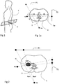
- this device may comprise first and second detection surfaces P1, P2 which are respectively flat and are oriented together at an angle of 90 ° with an angle uncertainty of more or less than 60 ° and preferably of plus or minus 5 ° of angle and preferably less than 1 ° of angle, as on the Figures 1a and 2 .
- the device also comprises a calculator and a software making it possible to carry out calculations or estimates or deductions necessary for the implementation of the method according to the invention, this device thus being automated or at least semi automated to allow an estimation of the surface distribution. or volume of bone mineral density, hereinafter DMO, in at least a portion of the skeleton of an individual.
- DMO volume of bone mineral density
- the device may consist of a mobile source and calibration means to know in each of the views the global radiological environment (position of the sources relative to the films and relative position of the sources), and if necessary a tracking on the subject to position a subject in a global environment.
- the device may also consist of a single source and a rotation and calibration device for rotating the subject and defining the pair of images taking into account the rotation of the subject to have the two different incidences.
- the device according to the invention may also comprise optical capture means for observing the outer envelope E of the individual.
- optical capture means which are connected to the computer make it possible to generate a three-dimensional geometrical representation of at least a portion of the outer envelope E containing said part of the skeleton of the individual.
- optical capture means may be a body scanner or a camera or camera, stereoscopic or not.
- optical capture means may optionally be associated with means for projecting moire or raster fringes possibly generated via light projections on the envelope of the individual.
- These projection means may be for example adapted to project the fringes or frames in the form of laser light.
- the positions of the first and second detection surfaces and the positions of the first and second X-ray sources as well as possibly those of the optical capture means are located in a same space using a spatial landmark.
- the objective of the method according to the invention is to allow the provision of an estimate of bone mineral BMD in a skeletal portion of an individual.
- the particularly targeted part of the skeleton for estimating the distribution of BMD is essentially a vertebral body or a vertebra, or a femoral neck or more generally the femur. These parts are targeted because they are particularly suited to the characterization of the risk of osteoporosis or a change in mineral density.
- these first and second images respectively correspond to the same instant of detection of X-rays projected on the first and second detection surfaces.
- there is a time shift between the detections required for the generation of the first and second images which is less than 1 second and preferably less than 0.5 seconds.
- the first and second images are made to illustrate projections of the individual while standing .
- the standing position of the individual is the most representative of the risks associated with osteoporosis.
- the optical capture means to ensure the accuracy of representation of the outer envelope and the use of the first and second images to ensure that the representation of the envelope is well positioned in the same spatial reference as the three-dimensional representation of the part of the skeleton that we want to visualize.
- the optical capture means would provide only one view of the outer envelope and considering a 3D vertebra pattern from the estimated single radiographic image, then it is possible to estimate the soft tissue thicknesses. crossed and thus evaluate the attenuation of X-rays by these soft tissues.
- the surface BMD of the vertebra can thus be estimated but with reduced accuracy.
- the software estimates a value representative of the bone mineral density of the bone tissue V traversed by this given X-ray.
- the soft tissues that is, the organs other than the hard skeletal tissues, have an X-ray attenuation coefficient which is substantially homogeneous.
- I 0 be the intensity of the incident beam emitted by the source
- I the intensity of the beam / radius as detected on a pixel of the image
- ⁇ the attenuation coefficient of the material traversed by the beam
- x l crossed thickness of this material.
- I I 0 e - ⁇ m The 1 - ⁇ m The 2 - ⁇ ⁇ vi x vi
- Calibration can be done with a calibration phantom (specimen), usually in lucite (whose attenuation coefficient is close to the soft tissues) and aluminum (whose attenuation coefficient is close to the bone), then the signal is redistributed between this range of values to enhance the bone signal, which allows the final to have surface BMD, and this with a single energy image.
- the value of I corr makes it possible to obtain a so-called surface BMD value, which is generally obtained from the reference imaging modalities (DXA corresponding to the two-photon X-ray absorptiometry imaging method), this surface BMD value does not take into account the heterogeneity of the vertebral tissue traversed.
- Bone mineral density values may be associated with at least some of the crossing points.
Landscapes
- Health & Medical Sciences (AREA)
- Life Sciences & Earth Sciences (AREA)
- Engineering & Computer Science (AREA)
- Medical Informatics (AREA)
- Public Health (AREA)
- Pathology (AREA)
- General Health & Medical Sciences (AREA)
- Biomedical Technology (AREA)
- Surgery (AREA)
- Optics & Photonics (AREA)
- Radiology & Medical Imaging (AREA)
- Nuclear Medicine, Radiotherapy & Molecular Imaging (AREA)
- Heart & Thoracic Surgery (AREA)
- Molecular Biology (AREA)
- Physics & Mathematics (AREA)
- Animal Behavior & Ethology (AREA)
- High Energy & Nuclear Physics (AREA)
- Biophysics (AREA)
- Veterinary Medicine (AREA)
- Computer Vision & Pattern Recognition (AREA)
- Physiology (AREA)
- Orthopedic Medicine & Surgery (AREA)
- Dentistry (AREA)
- Oral & Maxillofacial Surgery (AREA)
- Databases & Information Systems (AREA)
- Epidemiology (AREA)
- Primary Health Care (AREA)
- Data Mining & Analysis (AREA)
- Apparatus For Radiation Diagnosis (AREA)
Claims (13)
- Schätzverfahren zum Schätzen der Verteilung der Knochenmineraldichte in mindestens einem Teil eines Skeletts einer Einzelperson, wobei dieses Skelett aus Knochengewebe besteht, wobei diese Knochengewebe von Weichgeweben umgeben sind, die selbst von einer äußeren Hülle der Einzelperson umgeben sind, wobei das Verfahren dadurch gekennzeichnet ist, dass es umfasst das Erzeugen:- einer dreidimensionalen geometrischen Darstellung des genannten Teils des Skeletts der Einzelperson; und- einer dreidimensionalen geometrischen Darstellung mindestens eines Abschnittes der äußeren Hülle (E), die den genannten Teil des Skeletts der Einzelperson enthält;- mindestens eines ersten Röntgenbildes des genannten mindestens einen Teil des Skeletts, wobei dieses erste Bild durch Beobachtung einer Projektion von Röntgenstrahlen, die aus einer ersten Quelle stammen, auf eine erste Erfassungsfläche erzeugt wird, wobei dieses erste Bild durch Projektion von Röntgenstrahlen erzeugt wird und die beobachtete Einzelperson gemäß einem ersten Beobachtungseinfallswinkel darstellt;- mindestens eines zweiten Röntgenbildes des genannten mindestens einen Teils des Skeletts, wobei dieses zweite Bild durch Projektion von Röntgenstrahlen erzeugt wird und die beobachtete Einzelperson gemäß einem zweiten Beobachtungseinfallswinkel darstellt, der sich von dem ersten Beobachtungseinfallswinkel unterscheidet; und dann mit Hilfe einer vorgegebenen Software und für mehrere gegebene Röntgenstrahlen unter den genannten projizierten Röntgenstrahlen für jeden gegebenen Röntgenstrahl geschätzt wird:- ein Wert für die Dicke der Weichgewebe, die von diesem gegebenen Röntgenstrahl durchlaufen werden;- ein Wert für die Dicke der Knochengewebe, die von diesem gegebenen Röntgenstrahl durchlaufen werden; und- ein Wert für die Gesamtdämpfung dieses gegebenen Röntgenstrahls zwischen seiner Quelle und seiner Projektion auf die Erfassungsfläche, auf die er projiziert wird; undmit Hilfe des Wertes für die Dicke der Weichgewebe, die von diesem gegebenen Röntgenstrahl durchlaufen werden, und eines Schätzwertes eines Dämpfungskoeffizienten der Weichgewebe, die von diesem gegebenen Röntgenstrahl durchlaufen werden, einerseits ein Wert für die Dämpfung dieses gegebenen Röntgenstrahls durch die Dicke der Weichgewebe, die er durchlaufen hat, und andererseits ein Wert für die Dämpfung dieses gegebenen Röntgenstrahls durch die Dicke der Knochengewebe, die er durchlaufen hat, hergeleitet wird; und dann mit Hilfe des Wertes für die Dicke der Knochengewebe, die von diesem gegebenen Röntgenstrahl durchlaufen werden, des Wertes für die Dämpfung dieses gegebenen Röntgenstrahls durch die Dicke der Knochengewebe, die er durchlaufen hat, und des Gesamtdämpfungswertes für diesen gegebenen Röntgenstrahl die Software einen Wert schätzt, der repräsentativ für die Knochenmineraldichte der von diesem gegebenen Röntgenstrahl durchlaufenen Knochengewebe ist.
- Schätzverfahren nach Anspruch 1, bei dem das genannte zweite Röntgenbild des genannten mindestens einen Teils des Skeletts durch Beobachtung einer Projektion von Röntgenstrahlen, die aus einer zweiten Quelle stammen, die verschieden und entfernt zur ersten Quelle ist, auf eine zweite Erfassungsfläche erzeugt wird.
- Schätzverfahren nach Anspruch 2, bei dem die erste und die zweite Erfassungsfläche jeweils eben und zueinander um einen Winkel von 90° plus oder minus einem Winkel von 60° und vorzugsweise plus oder minus einem Winkel von 5° ausgerichtet sind.
- Schätzverfahren nach einem der Ansprüche 1 bis 3, bei dem die Software zum Herleiten des genannten Dämpfungswertes des genannten gegebenen Röntgenstrahls durch die Dicke der Weichgewebe, die er durchlaufen hat, einen Dämpfungskoeffizienten für die Weichgewebe der Einzelperson schätzt.
- Schätzverfahren nach Anspruch 4, bei dem die Software zum Schätzen des Dämpfungskoeffizienten für die Weichgewebe der Einzelperson einen besonderen Röntgenstrahl sucht, der einzig die Weichgewebe der Einzelperson durchläuft, ohne die Knochengewebe zu durchlaufen, und anhand der Position dieses besonderen Röntgenstrahls und der dreidimensionalen geometrischen Darstellung des Abschnittes der äußeren Hülle (E) die Software den Wert für die Dicke der Weichgewebe schätzt, die von diesem besonderen Röntgenstrahl durchlaufen werden, und durch Beobachtung eines Projektionspunktes dieses besonderen Röntgenstrahls die Software einen Wert für die Dämpfung bestimmt, die von diesem besonderen Röntgenstrahl während seines Durchlaufens einzig der Weichgewebe der Einzelperson erfahren wird, und dann in Kenntnis dieses genannten Wertes für die Dicke der Weichgewebe, die von diesem besonderen Röntgenstrahl durchlaufen werden, und des genannten Wertes für die Dämpfung, die von diesem besonderen Röntgenstrahl erfahren wird, die Software den Dämpfungskoeffizienten berechnet, der einzig mit den Weichgeweben der Einzelperson in Verbindung steht.
- Schätzverfahren nach Anspruch 5, bei dem in Kenntnis:- des Dämpfungskoeffizienten für die Weichgewebe der Einzelperson;- der Dicke der von diesem gegebenen Röntgenstrahl durchlaufenen Weichgewebe;- einer Intensität (I0) dieses gegebenen Strahls an seiner Quelle; und- einer Intensität (I) dieses gegebenen Strahls, wie er auf der Erfassungsfläche (P1, P2), auf die er projiziert wird, erfasst wird, die Software berechnet:- einerseits einen Wert für die Dämpfung dieses gegebenen Röntgenstrahls durch die Dicke der Weichgewebe, die er durchlaufen hat; und- andererseits einen Gesamtdämpfungswert für diesen gegebenen Röntgenstrahl zwischen seiner Quelle und der Erfassungsfläche, auf die er projiziert wird; und dann- durch Korrigieren dieses Gesamtdämpfungswerts für diesen gegebenen Röntgenstrahl mit Hilfe des genannten Wertes für die Dämpfung dieses gegebenen Röntgenstrahls durch die Dicke der Weichgewebe, die er durchlaufen hat, die Software den Wert für die Dämpfung dieses gegebenen Röntgenstrahls durch die Dicke der Knochengewebe berechnet, die er durchlaufen hat.
- Verfahren nach einem der Ansprüche 1 bis 6, bei dem der genannte Wert, der repräsentativ für die Knochenmineraldichte der Knochengewebe ist, die von diesem Röntgenstrahl durchlaufen werden, ein Dämpfungskoeffizient des Knochengewebes ist, das von diesem gegebenen Röntgenstrahl durchlaufen wird.
- Verfahren nach einem der Ansprüche 1 bis 7, bei dem das genannte erste und das genannte zweite Bild einem selben Zeitpunkt des Erfassens der Röntgenstrahlen entsprechen, die auf die erste bzw. die zweite Erfassungsfläche projiziert werden.
- Verfahren nach einem der Ansprüche 1 bis 7, bei dem das genannte erste und das genannte zweite Bild zwei unterschiedlichen Zeitpunkten des Erfassens der Röntgenstrahlen entsprechen, die auf die erste bzw. die zweite Erfassungsfläche projiziert werden.
- Verfahren nach einem der Ansprüche 1 bis 9, bei dem das erste und das zweite Bild Projektionen der Einzelperson in der aufrechten Position zeigen.
- Verfahren nach einem der Ansprüche 1 bis 10, bei dem die genannte dreidimensionale geometrische Darstellung des genannten mindestens einen Abschnitts der äußeren Hülle, die den genannten Teil des Skeletts der Einzelperson enthält, anhand des genannten ersten und zweiten Röntgenbildes hergeleitet wird.
- Verfahren nach einem der Ansprüche 1 bis 11, bei dem die genannte dreidimensionale geometrische Darstellung des genannten mindestens einen Abschnitts der äußeren Hülle, die den genannten Teil des Skeletts der Einzelperson enthält, mit Hilfe einer Beobachtung der Hülle hergeleitet wird, die mit Hilfe optischer Erfassungsmittel erzeugt wird.
- Vorrichtung, umfassend:- einen Rechner;- optische Erfassungsmittel, die mit dem Rechner verbunden sind, um eine äußere Hülle (E) einer Einzelperson zu beobachten;- Erzeugungsmittel zur Erzeugung mindestens eines ersten Röntgenbildes mindestens eines Teils des Skeletts der Einzelperson durch Beobachtung gemäß einem ersten Beobachtungseinfallswinkel einer Projektion von Röntgenstrahlen, die aus einer ersten Quelle stammen, auf eine erste Erfassungsfläche;- Erzeugungsmittel zur Erzeugung mindestens eines zweiten Röntgenbildes des genannten mindestens einen Teils des Skeletts durch Projektion von Röntgenstrahlen gemäß einem zweiten Beobachtungseinfallswinkels, der sich von dem ersten Beobachtungseinfallswinkel unterscheidet,dadurch gekennzeichnet, dass
die Vorrichtung eine Software umfasst, die geeignet ist, um Berechnungen oder Schätzungen oder Herleitungen durchzuführen, die zum Durchführen des Schätzverfahrens zum Schätzen der Verteilung der Knochenmineraldichte nach einem der Ansprüche 1 bis 12 erforderlich sind.
Applications Claiming Priority (2)
| Application Number | Priority Date | Filing Date | Title |
|---|---|---|---|
| FR1554150A FR3035785B1 (fr) | 2015-05-07 | 2015-05-07 | Procede d'estimation de la repartition de la densite minerale osseuse dans au moins une partie de squelette d'un individu |
| PCT/EP2016/060035 WO2016177798A1 (fr) | 2015-05-07 | 2016-05-04 | Procede d'estimation de la repartition de la densite minerale osseuse dans au moins une partie de squelette d'un individu |
Publications (2)
| Publication Number | Publication Date |
|---|---|
| EP3291733A1 EP3291733A1 (de) | 2018-03-14 |
| EP3291733B1 true EP3291733B1 (de) | 2019-04-10 |
Family
ID=54014956
Family Applications (1)
| Application Number | Title | Priority Date | Filing Date |
|---|---|---|---|
| EP16720837.0A Not-in-force EP3291733B1 (de) | 2015-05-07 | 2016-05-04 | Schätzung der verteilung der knochenmineraldichte in mindestens einem teil des skeletts einer person |
Country Status (6)
| Country | Link |
|---|---|
| US (1) | US10478141B2 (de) |
| EP (1) | EP3291733B1 (de) |
| JP (1) | JP2018514363A (de) |
| CN (1) | CN107690311B (de) |
| FR (1) | FR3035785B1 (de) |
| WO (1) | WO2016177798A1 (de) |
Families Citing this family (12)
| Publication number | Priority date | Publication date | Assignee | Title |
|---|---|---|---|---|
| US9865050B2 (en) | 2010-03-23 | 2018-01-09 | Hologic, Inc. | Measuring intramuscular fat |
| US9642585B2 (en) | 2013-11-25 | 2017-05-09 | Hologic, Inc. | Bone densitometer |
| AU2016222642B2 (en) | 2015-02-26 | 2021-07-15 | Hologic, Inc. | Methods for physiological state determination in body scans |
| AU2018243634B2 (en) | 2017-03-31 | 2024-03-21 | Hologic, Inc. | Multiple modality body composition analysis |
| US12033318B2 (en) * | 2018-09-10 | 2024-07-09 | Kyocera Corporation | Estimation apparatus, estimation system, and computer-readable non-transitory medium storing estimation program |
| KR20200095859A (ko) | 2019-02-01 | 2020-08-11 | 삼성전자주식회사 | 엑스선 영상 처리 방법 및 그에 따른 엑스선 영상 처리 장치 |
| KR102167859B1 (ko) * | 2019-02-20 | 2020-10-21 | 하이윈 테크놀로지스 코포레이션 | 컴퓨터 단층 촬영장치의 스캔 정보를 골격 파라미터로 전환하는 방법 |
| EP3975848A4 (de) | 2019-05-28 | 2023-06-14 | Hologic, Inc. | System und verfahren zur kontinuierlichen kalibrierung von röntgenaufnahmen |
| WO2021059668A1 (ja) * | 2019-09-27 | 2021-04-01 | 富士フイルム株式会社 | 情報処理装置、情報処理方法およびプログラム |
| CN113662567B (zh) * | 2020-05-14 | 2024-08-27 | 镇江慧影科技发展有限公司 | 一种结合表面几何采集的x射线骨密度测量系统及方法 |
| JP7301792B2 (ja) * | 2020-06-23 | 2023-07-03 | 富士フイルム株式会社 | 放射線画像処理装置、方法およびプログラム |
| TWI891074B (zh) * | 2023-09-28 | 2025-07-21 | 宏碁智醫股份有限公司 | 骨質密度預測方法與裝置 |
Family Cites Families (16)
| Publication number | Priority date | Publication date | Assignee | Title |
|---|---|---|---|---|
| JPH01118792A (ja) * | 1987-10-31 | 1989-05-11 | Shimadzu Corp | Dpa装置 |
| JPH0722580B2 (ja) * | 1992-05-20 | 1995-03-15 | アロカ株式会社 | 骨評価装置 |
| CN1068200A (zh) * | 1992-06-15 | 1993-01-20 | 南京市中医院 | 骨痂生长及骨矿物密度的定量分析方法及x线机图像系统 |
| US5850836A (en) * | 1995-09-22 | 1998-12-22 | Hologic, Inc. | Morphometric x-ray absorptiometry (MXA) |
| US5917877A (en) * | 1997-09-05 | 1999-06-29 | Cyberlogic, Inc. | Plain x-ray bone densitometry apparatus and method |
| US6173038B1 (en) * | 1999-12-01 | 2001-01-09 | Cyberlogic, Inc. | Plain x-ray bone densitometry apparatus and method |
| US6501819B2 (en) * | 2000-12-18 | 2002-12-31 | Ge Medical Systems Global Technology Company, Llc | Medical diagnostic method and apparatus to control dual energy exposure techniques based on image information |
| FR2849241B1 (fr) * | 2002-12-20 | 2005-06-24 | Biospace Instr | Procede et dispositif d'imagerie radiographique |
| FR2856170B1 (fr) | 2003-06-10 | 2005-08-26 | Biospace Instr | Procede d'imagerie radiographique pour la reconstruction tridimensionnelle, dispositif et programme d'ordinateur pour mettre en oeuvre ce procede |
| JP5085031B2 (ja) * | 2005-11-10 | 2012-11-28 | 株式会社東芝 | X線アンギオ撮影装置 |
| US8660329B2 (en) | 2007-05-25 | 2014-02-25 | Ecole Nationale Superieure D'arts Et Metiers (Ensam) | Method for reconstruction of a three-dimensional model of a body structure |
| CN101158653B (zh) * | 2007-11-16 | 2010-12-15 | 西北工业大学 | 一种锥束ct系统的散射测定和校正方法 |
| CN101226642A (zh) * | 2008-01-25 | 2008-07-23 | 西安交通大学 | 基于ct数据一致性的投影射束硬化校正方法 |
| JP2010051689A (ja) * | 2008-08-29 | 2010-03-11 | Fujifilm Corp | 放射線画像検出器、及び放射線画像撮影装置 |
| US8295570B2 (en) * | 2009-09-10 | 2012-10-23 | General Electric Company | Methods and apparatus for measuring body circumference |
| EP2858571B1 (de) * | 2012-06-07 | 2019-01-23 | The Johns Hopkins University | Integration von quantitativen kalibriersystemen in ct-scanner |
-
2015
- 2015-05-07 FR FR1554150A patent/FR3035785B1/fr not_active Expired - Fee Related
-
2016
- 2016-05-04 US US15/572,374 patent/US10478141B2/en active Active
- 2016-05-04 EP EP16720837.0A patent/EP3291733B1/de not_active Not-in-force
- 2016-05-04 JP JP2018509983A patent/JP2018514363A/ja active Pending
- 2016-05-04 CN CN201680026326.9A patent/CN107690311B/zh not_active Expired - Fee Related
- 2016-05-04 WO PCT/EP2016/060035 patent/WO2016177798A1/fr not_active Ceased
Non-Patent Citations (1)
| Title |
|---|
| None * |
Also Published As
| Publication number | Publication date |
|---|---|
| EP3291733A1 (de) | 2018-03-14 |
| FR3035785A1 (fr) | 2016-11-11 |
| US20180146942A1 (en) | 2018-05-31 |
| WO2016177798A1 (fr) | 2016-11-10 |
| US10478141B2 (en) | 2019-11-19 |
| JP2018514363A (ja) | 2018-06-07 |
| CN107690311B (zh) | 2020-11-24 |
| CN107690311A (zh) | 2018-02-13 |
| FR3035785B1 (fr) | 2017-06-02 |
Similar Documents
| Publication | Publication Date | Title |
|---|---|---|
| EP3291733B1 (de) | Schätzung der verteilung der knochenmineraldichte in mindestens einem teil des skeletts einer person | |
| EP1788525B1 (de) | Bildgebendes Verfahren für die dreidimensionale Rekonstruktion, Vorrichtung und Computerprogramm zur Durchführung besagten Verfahrens | |
| EP2309462B1 (de) | Verfahren und Vorrichtung zur dreidimensionalen Rekonstruktion von Röntgenbilder mit niedriger Strahlungsdosis | |
| EP3612095B1 (de) | Strahlaufhärtungskorrektur in der röntgendunkelfeldabbildung | |
| EP2059904B1 (de) | Computergestütztes bildaufnahmeverfahren zur dreidimensionalen rekonstruktion aus zweidimensionalen radiologischen bildern und vorrichtung zu dessen ausführung | |
| CN101396272B (zh) | 用于产生物质选择的立体图像的方法 | |
| JP2009285356A (ja) | 医療用撮影システム、画像処理装置、画像処理方法、およびプログラム | |
| CN101960298A (zh) | X射线成像装置、x射线成像方法和x射线成像装置的控制方法 | |
| Duarte et al. | Computed stereo lensless X-ray imaging | |
| FR2849241A1 (fr) | Procede et dispositif d'imagerie radiographique | |
| FR2801776A1 (fr) | Procede d'utilisation d'un systeme d'osteodensitometrie, par rayonnement x bi-energie, a faisceau conique | |
| Mechlem et al. | Spectral differential phase contrast x-ray radiography | |
| Williams et al. | Fresnel coherent diffractive imaging: treatment and analysis of data | |
| JP2014083234A (ja) | 散乱線補正方法、画像処理装置および断層撮影装置 | |
| EP3830628B1 (de) | Vorrichtung und verfahren zur erfassung mikroskopischer plenoptischer bilder mit turbulenzdämpfung | |
| FR3071715A1 (fr) | Procede d'imagerie radiographique, dispositif de traitement d'image radiographique et dispositif d'imagerie radiographique. | |
| US20080002807A1 (en) | Method and system for generating a scatter corrected X-ray image | |
| Ritter et al. | Simultaneous maximum-likelihood reconstruction for x-ray grating based phase-contrast tomography avoiding intermediate phase retrieval | |
| Abdel Gawad et al. | Classification of human teeth caries using custom non-invasive optical imaging system | |
| JP4137580B2 (ja) | 位相情報復元方法及び位相情報復元装置、並びに、位相情報復元プログラム | |
| Diemoz et al. | Comparison of analyzer-based imaging computed tomography extraction algorithms and application to bone-cartilage imaging | |
| US20030202688A1 (en) | Method, apparatus, and program for restoring phase information | |
| Kachatkou et al. | In situ X-ray beam imaging using an off-axis magnifying coded aperture camera system | |
| Tromans et al. | Investigating the replacement of the physical anti-scatter grid with digital image processing | |
| Reinhold | Property Estimation of Thin Cortical Bone from Clinical QCT: An Analysis by Synthesis Approach: Going beyond Nyquist |
Legal Events
| Date | Code | Title | Description |
|---|---|---|---|
| STAA | Information on the status of an ep patent application or granted ep patent |
Free format text: STATUS: THE INTERNATIONAL PUBLICATION HAS BEEN MADE |
|
| PUAI | Public reference made under article 153(3) epc to a published international application that has entered the european phase |
Free format text: ORIGINAL CODE: 0009012 |
|
| STAA | Information on the status of an ep patent application or granted ep patent |
Free format text: STATUS: REQUEST FOR EXAMINATION WAS MADE |
|
| 17P | Request for examination filed |
Effective date: 20171107 |
|
| AK | Designated contracting states |
Kind code of ref document: A1 Designated state(s): AL AT BE BG CH CY CZ DE DK EE ES FI FR GB GR HR HU IE IS IT LI LT LU LV MC MK MT NL NO PL PT RO RS SE SI SK SM TR |
|
| AX | Request for extension of the european patent |
Extension state: BA ME |
|
| DAV | Request for validation of the european patent (deleted) | ||
| DAX | Request for extension of the european patent (deleted) | ||
| GRAP | Despatch of communication of intention to grant a patent |
Free format text: ORIGINAL CODE: EPIDOSNIGR1 |
|
| STAA | Information on the status of an ep patent application or granted ep patent |
Free format text: STATUS: GRANT OF PATENT IS INTENDED |
|
| INTG | Intention to grant announced |
Effective date: 20181029 |
|
| GRAS | Grant fee paid |
Free format text: ORIGINAL CODE: EPIDOSNIGR3 |
|
| GRAA | (expected) grant |
Free format text: ORIGINAL CODE: 0009210 |
|
| STAA | Information on the status of an ep patent application or granted ep patent |
Free format text: STATUS: THE PATENT HAS BEEN GRANTED |
|
| AK | Designated contracting states |
Kind code of ref document: B1 Designated state(s): AL AT BE BG CH CY CZ DE DK EE ES FI FR GB GR HR HU IE IS IT LI LT LU LV MC MK MT NL NO PL PT RO RS SE SI SK SM TR |
|
| REG | Reference to a national code |
Ref country code: GB Ref legal event code: FG4D Free format text: NOT ENGLISH |
|
| REG | Reference to a national code |
Ref country code: CH Ref legal event code: EP Ref country code: AT Ref legal event code: REF Ref document number: 1117592 Country of ref document: AT Kind code of ref document: T Effective date: 20190415 |
|
| REG | Reference to a national code |
Ref country code: DE Ref legal event code: R096 Ref document number: 602016012282 Country of ref document: DE |
|
| REG | Reference to a national code |
Ref country code: IE Ref legal event code: FG4D Free format text: LANGUAGE OF EP DOCUMENT: FRENCH |
|
| REG | Reference to a national code |
Ref country code: NL Ref legal event code: MP Effective date: 20190410 |
|
| REG | Reference to a national code |
Ref country code: LT Ref legal event code: MG4D |
|
| REG | Reference to a national code |
Ref country code: AT Ref legal event code: MK05 Ref document number: 1117592 Country of ref document: AT Kind code of ref document: T Effective date: 20190410 |
|
| PG25 | Lapsed in a contracting state [announced via postgrant information from national office to epo] |
Ref country code: NL Free format text: LAPSE BECAUSE OF FAILURE TO SUBMIT A TRANSLATION OF THE DESCRIPTION OR TO PAY THE FEE WITHIN THE PRESCRIBED TIME-LIMIT Effective date: 20190410 |
|
| PG25 | Lapsed in a contracting state [announced via postgrant information from national office to epo] |
Ref country code: HR Free format text: LAPSE BECAUSE OF FAILURE TO SUBMIT A TRANSLATION OF THE DESCRIPTION OR TO PAY THE FEE WITHIN THE PRESCRIBED TIME-LIMIT Effective date: 20190410 Ref country code: SE Free format text: LAPSE BECAUSE OF FAILURE TO SUBMIT A TRANSLATION OF THE DESCRIPTION OR TO PAY THE FEE WITHIN THE PRESCRIBED TIME-LIMIT Effective date: 20190410 Ref country code: PT Free format text: LAPSE BECAUSE OF FAILURE TO SUBMIT A TRANSLATION OF THE DESCRIPTION OR TO PAY THE FEE WITHIN THE PRESCRIBED TIME-LIMIT Effective date: 20190910 Ref country code: LT Free format text: LAPSE BECAUSE OF FAILURE TO SUBMIT A TRANSLATION OF THE DESCRIPTION OR TO PAY THE FEE WITHIN THE PRESCRIBED TIME-LIMIT Effective date: 20190410 Ref country code: ES Free format text: LAPSE BECAUSE OF FAILURE TO SUBMIT A TRANSLATION OF THE DESCRIPTION OR TO PAY THE FEE WITHIN THE PRESCRIBED TIME-LIMIT Effective date: 20190410 Ref country code: NO Free format text: LAPSE BECAUSE OF FAILURE TO SUBMIT A TRANSLATION OF THE DESCRIPTION OR TO PAY THE FEE WITHIN THE PRESCRIBED TIME-LIMIT Effective date: 20190710 Ref country code: AL Free format text: LAPSE BECAUSE OF FAILURE TO SUBMIT A TRANSLATION OF THE DESCRIPTION OR TO PAY THE FEE WITHIN THE PRESCRIBED TIME-LIMIT Effective date: 20190410 Ref country code: FI Free format text: LAPSE BECAUSE OF FAILURE TO SUBMIT A TRANSLATION OF THE DESCRIPTION OR TO PAY THE FEE WITHIN THE PRESCRIBED TIME-LIMIT Effective date: 20190410 |
|
| PG25 | Lapsed in a contracting state [announced via postgrant information from national office to epo] |
Ref country code: GR Free format text: LAPSE BECAUSE OF FAILURE TO SUBMIT A TRANSLATION OF THE DESCRIPTION OR TO PAY THE FEE WITHIN THE PRESCRIBED TIME-LIMIT Effective date: 20190711 Ref country code: BG Free format text: LAPSE BECAUSE OF FAILURE TO SUBMIT A TRANSLATION OF THE DESCRIPTION OR TO PAY THE FEE WITHIN THE PRESCRIBED TIME-LIMIT Effective date: 20190710 Ref country code: RS Free format text: LAPSE BECAUSE OF FAILURE TO SUBMIT A TRANSLATION OF THE DESCRIPTION OR TO PAY THE FEE WITHIN THE PRESCRIBED TIME-LIMIT Effective date: 20190410 Ref country code: PL Free format text: LAPSE BECAUSE OF FAILURE TO SUBMIT A TRANSLATION OF THE DESCRIPTION OR TO PAY THE FEE WITHIN THE PRESCRIBED TIME-LIMIT Effective date: 20190410 Ref country code: LV Free format text: LAPSE BECAUSE OF FAILURE TO SUBMIT A TRANSLATION OF THE DESCRIPTION OR TO PAY THE FEE WITHIN THE PRESCRIBED TIME-LIMIT Effective date: 20190410 |
|
| REG | Reference to a national code |
Ref country code: CH Ref legal event code: PL |
|
| PG25 | Lapsed in a contracting state [announced via postgrant information from national office to epo] |
Ref country code: IS Free format text: LAPSE BECAUSE OF FAILURE TO SUBMIT A TRANSLATION OF THE DESCRIPTION OR TO PAY THE FEE WITHIN THE PRESCRIBED TIME-LIMIT Effective date: 20190810 Ref country code: AT Free format text: LAPSE BECAUSE OF FAILURE TO SUBMIT A TRANSLATION OF THE DESCRIPTION OR TO PAY THE FEE WITHIN THE PRESCRIBED TIME-LIMIT Effective date: 20190410 |
|
| REG | Reference to a national code |
Ref country code: DE Ref legal event code: R097 Ref document number: 602016012282 Country of ref document: DE |
|
| PG25 | Lapsed in a contracting state [announced via postgrant information from national office to epo] |
Ref country code: DK Free format text: LAPSE BECAUSE OF FAILURE TO SUBMIT A TRANSLATION OF THE DESCRIPTION OR TO PAY THE FEE WITHIN THE PRESCRIBED TIME-LIMIT Effective date: 20190410 Ref country code: MC Free format text: LAPSE BECAUSE OF FAILURE TO SUBMIT A TRANSLATION OF THE DESCRIPTION OR TO PAY THE FEE WITHIN THE PRESCRIBED TIME-LIMIT Effective date: 20190410 Ref country code: LI Free format text: LAPSE BECAUSE OF NON-PAYMENT OF DUE FEES Effective date: 20190531 Ref country code: SK Free format text: LAPSE BECAUSE OF FAILURE TO SUBMIT A TRANSLATION OF THE DESCRIPTION OR TO PAY THE FEE WITHIN THE PRESCRIBED TIME-LIMIT Effective date: 20190410 Ref country code: CH Free format text: LAPSE BECAUSE OF NON-PAYMENT OF DUE FEES Effective date: 20190531 Ref country code: CZ Free format text: LAPSE BECAUSE OF FAILURE TO SUBMIT A TRANSLATION OF THE DESCRIPTION OR TO PAY THE FEE WITHIN THE PRESCRIBED TIME-LIMIT Effective date: 20190410 Ref country code: RO Free format text: LAPSE BECAUSE OF FAILURE TO SUBMIT A TRANSLATION OF THE DESCRIPTION OR TO PAY THE FEE WITHIN THE PRESCRIBED TIME-LIMIT Effective date: 20190410 Ref country code: EE Free format text: LAPSE BECAUSE OF FAILURE TO SUBMIT A TRANSLATION OF THE DESCRIPTION OR TO PAY THE FEE WITHIN THE PRESCRIBED TIME-LIMIT Effective date: 20190410 |
|
| REG | Reference to a national code |
Ref country code: BE Ref legal event code: MM Effective date: 20190531 |
|
| PLBE | No opposition filed within time limit |
Free format text: ORIGINAL CODE: 0009261 |
|
| STAA | Information on the status of an ep patent application or granted ep patent |
Free format text: STATUS: NO OPPOSITION FILED WITHIN TIME LIMIT |
|
| PG25 | Lapsed in a contracting state [announced via postgrant information from national office to epo] |
Ref country code: LU Free format text: LAPSE BECAUSE OF NON-PAYMENT OF DUE FEES Effective date: 20190504 Ref country code: SM Free format text: LAPSE BECAUSE OF FAILURE TO SUBMIT A TRANSLATION OF THE DESCRIPTION OR TO PAY THE FEE WITHIN THE PRESCRIBED TIME-LIMIT Effective date: 20190410 |
|
| 26N | No opposition filed |
Effective date: 20200113 |
|
| PG25 | Lapsed in a contracting state [announced via postgrant information from national office to epo] |
Ref country code: TR Free format text: LAPSE BECAUSE OF FAILURE TO SUBMIT A TRANSLATION OF THE DESCRIPTION OR TO PAY THE FEE WITHIN THE PRESCRIBED TIME-LIMIT Effective date: 20190410 |
|
| PG25 | Lapsed in a contracting state [announced via postgrant information from national office to epo] |
Ref country code: IE Free format text: LAPSE BECAUSE OF NON-PAYMENT OF DUE FEES Effective date: 20190504 |
|
| PG25 | Lapsed in a contracting state [announced via postgrant information from national office to epo] |
Ref country code: BE Free format text: LAPSE BECAUSE OF NON-PAYMENT OF DUE FEES Effective date: 20190531 Ref country code: SI Free format text: LAPSE BECAUSE OF FAILURE TO SUBMIT A TRANSLATION OF THE DESCRIPTION OR TO PAY THE FEE WITHIN THE PRESCRIBED TIME-LIMIT Effective date: 20190410 |
|
| PG25 | Lapsed in a contracting state [announced via postgrant information from national office to epo] |
Ref country code: CY Free format text: LAPSE BECAUSE OF FAILURE TO SUBMIT A TRANSLATION OF THE DESCRIPTION OR TO PAY THE FEE WITHIN THE PRESCRIBED TIME-LIMIT Effective date: 20190410 |
|
| PG25 | Lapsed in a contracting state [announced via postgrant information from national office to epo] |
Ref country code: HU Free format text: LAPSE BECAUSE OF FAILURE TO SUBMIT A TRANSLATION OF THE DESCRIPTION OR TO PAY THE FEE WITHIN THE PRESCRIBED TIME-LIMIT; INVALID AB INITIO Effective date: 20160504 Ref country code: MT Free format text: LAPSE BECAUSE OF FAILURE TO SUBMIT A TRANSLATION OF THE DESCRIPTION OR TO PAY THE FEE WITHIN THE PRESCRIBED TIME-LIMIT Effective date: 20190410 |
|
| PG25 | Lapsed in a contracting state [announced via postgrant information from national office to epo] |
Ref country code: MK Free format text: LAPSE BECAUSE OF FAILURE TO SUBMIT A TRANSLATION OF THE DESCRIPTION OR TO PAY THE FEE WITHIN THE PRESCRIBED TIME-LIMIT Effective date: 20190410 |
|
| PGFP | Annual fee paid to national office [announced via postgrant information from national office to epo] |
Ref country code: GB Payment date: 20240521 Year of fee payment: 9 |
|
| PGFP | Annual fee paid to national office [announced via postgrant information from national office to epo] |
Ref country code: DE Payment date: 20240521 Year of fee payment: 9 |
|
| PGFP | Annual fee paid to national office [announced via postgrant information from national office to epo] |
Ref country code: FR Payment date: 20240528 Year of fee payment: 9 |
|
| PGFP | Annual fee paid to national office [announced via postgrant information from national office to epo] |
Ref country code: IT Payment date: 20240524 Year of fee payment: 9 |
|
| REG | Reference to a national code |
Ref country code: DE Ref legal event code: R119 Ref document number: 602016012282 Country of ref document: DE |
|
| GBPC | Gb: european patent ceased through non-payment of renewal fee |
Effective date: 20250504 |
|
| PG25 | Lapsed in a contracting state [announced via postgrant information from national office to epo] |
Ref country code: GB Free format text: LAPSE BECAUSE OF NON-PAYMENT OF DUE FEES Effective date: 20250504 |
|
| PG25 | Lapsed in a contracting state [announced via postgrant information from national office to epo] |
Ref country code: DE Free format text: LAPSE BECAUSE OF NON-PAYMENT OF DUE FEES Effective date: 20251202 |
|
| PG25 | Lapsed in a contracting state [announced via postgrant information from national office to epo] |
Ref country code: IT Free format text: LAPSE BECAUSE OF NON-PAYMENT OF DUE FEES Effective date: 20250504 |
|
| PG25 | Lapsed in a contracting state [announced via postgrant information from national office to epo] |
Ref country code: FR Free format text: LAPSE BECAUSE OF NON-PAYMENT OF DUE FEES Effective date: 20250531 |