EP3291735B1 - Système et procédé de compensation de mouvement dans des procédures médicales - Google Patents
Système et procédé de compensation de mouvement dans des procédures médicales Download PDFInfo
- Publication number
- EP3291735B1 EP3291735B1 EP16724717.0A EP16724717A EP3291735B1 EP 3291735 B1 EP3291735 B1 EP 3291735B1 EP 16724717 A EP16724717 A EP 16724717A EP 3291735 B1 EP3291735 B1 EP 3291735B1
- Authority
- EP
- European Patent Office
- Prior art keywords
- image
- pose
- baseline
- view
- live
- Prior art date
- Legal status (The legal status is an assumption and is not a legal conclusion. Google has not performed a legal analysis and makes no representation as to the accuracy of the status listed.)
- Active
Links
Images

Classifications
-
- A—HUMAN NECESSITIES
- A61—MEDICAL OR VETERINARY SCIENCE; HYGIENE
- A61B—DIAGNOSIS; SURGERY; IDENTIFICATION
- A61B8/00—Diagnosis using ultrasonic, sonic or infrasonic waves
- A61B8/42—Details of probe positioning or probe attachment to the patient
- A61B8/4245—Details of probe positioning or probe attachment to the patient involving determining the position of the probe, e.g. with respect to an external reference frame or to the patient
-
- A—HUMAN NECESSITIES
- A61—MEDICAL OR VETERINARY SCIENCE; HYGIENE
- A61B—DIAGNOSIS; SURGERY; IDENTIFICATION
- A61B8/00—Diagnosis using ultrasonic, sonic or infrasonic waves
- A61B8/48—Diagnostic techniques
- A61B8/483—Diagnostic techniques involving the acquisition of a 3D volume of data
-
- A—HUMAN NECESSITIES
- A61—MEDICAL OR VETERINARY SCIENCE; HYGIENE
- A61B—DIAGNOSIS; SURGERY; IDENTIFICATION
- A61B8/00—Diagnosis using ultrasonic, sonic or infrasonic waves
- A61B8/52—Devices using data or image processing specially adapted for diagnosis using ultrasonic, sonic or infrasonic waves
- A61B8/5215—Devices using data or image processing specially adapted for diagnosis using ultrasonic, sonic or infrasonic waves involving processing of medical diagnostic data
- A61B8/5238—Devices using data or image processing specially adapted for diagnosis using ultrasonic, sonic or infrasonic waves involving processing of medical diagnostic data for combining image data of patient, e.g. merging several images from different acquisition modes into one image
- A61B8/5246—Devices using data or image processing specially adapted for diagnosis using ultrasonic, sonic or infrasonic waves involving processing of medical diagnostic data for combining image data of patient, e.g. merging several images from different acquisition modes into one image combining images from the same or different imaging techniques, e.g. color Doppler and B-mode
-
- A—HUMAN NECESSITIES
- A61—MEDICAL OR VETERINARY SCIENCE; HYGIENE
- A61B—DIAGNOSIS; SURGERY; IDENTIFICATION
- A61B8/00—Diagnosis using ultrasonic, sonic or infrasonic waves
- A61B8/52—Devices using data or image processing specially adapted for diagnosis using ultrasonic, sonic or infrasonic waves
- A61B8/5269—Devices using data or image processing specially adapted for diagnosis using ultrasonic, sonic or infrasonic waves involving detection or reduction of artifacts
- A61B8/5276—Devices using data or image processing specially adapted for diagnosis using ultrasonic, sonic or infrasonic waves involving detection or reduction of artifacts due to motion
-
- G—PHYSICS
- G06—COMPUTING OR CALCULATING; COUNTING
- G06T—IMAGE DATA PROCESSING OR GENERATION, IN GENERAL
- G06T7/00—Image analysis
- G06T7/30—Determination of transform parameters for the alignment of images, i.e. image registration
-
- A—HUMAN NECESSITIES
- A61—MEDICAL OR VETERINARY SCIENCE; HYGIENE
- A61B—DIAGNOSIS; SURGERY; IDENTIFICATION
- A61B8/00—Diagnosis using ultrasonic, sonic or infrasonic waves
- A61B8/52—Devices using data or image processing specially adapted for diagnosis using ultrasonic, sonic or infrasonic waves
- A61B8/5215—Devices using data or image processing specially adapted for diagnosis using ultrasonic, sonic or infrasonic waves involving processing of medical diagnostic data
- A61B8/5238—Devices using data or image processing specially adapted for diagnosis using ultrasonic, sonic or infrasonic waves involving processing of medical diagnostic data for combining image data of patient, e.g. merging several images from different acquisition modes into one image
- A61B8/5261—Devices using data or image processing specially adapted for diagnosis using ultrasonic, sonic or infrasonic waves involving processing of medical diagnostic data for combining image data of patient, e.g. merging several images from different acquisition modes into one image combining images from different diagnostic modalities, e.g. ultrasound and X-ray
-
- G—PHYSICS
- G06—COMPUTING OR CALCULATING; COUNTING
- G06T—IMAGE DATA PROCESSING OR GENERATION, IN GENERAL
- G06T2207/00—Indexing scheme for image analysis or image enhancement
- G06T2207/10—Image acquisition modality
- G06T2207/10132—Ultrasound image
-
- G—PHYSICS
- G06—COMPUTING OR CALCULATING; COUNTING
- G06T—IMAGE DATA PROCESSING OR GENERATION, IN GENERAL
- G06T2207/00—Indexing scheme for image analysis or image enhancement
- G06T2207/20—Special algorithmic details
- G06T2207/20092—Interactive image processing based on input by user
Definitions
- This disclosure relates to motion compensation and more particularly to systems and methods for accounting for patient motion in medical images.
- Multi-modality "fusion" imaging of ultrasound with a prior image can be enabled using electromagnetic (EM) tracking of an ultrasound probe and registration of the EM coordinate system with the coordinate system of the prior image.
- EM electromagnetic
- Automatic methods to establish the registration may be based on acquisition of an EM-tracked three-dimensional (3D) ultrasound (US) volume (called a baseline 3DUS), followed by manual or automatic image-based registration of the baseline 3DUS to the prior static image (e.g., a computed tomography (CT) image).
- 3D three-dimensional
- CT computed tomography
- the registration between the live ultrasound imaging and the prior image will no longer be accurate.
- the operator is planning an intervention such as a needle insertion into a tumor, the operator will typically request a breath hold to interrupt the tumor motion.
- the position of the tumor during this breath hold typically differs from the position during baseline 3DUS acquisition.
- the fusion image with the prior static image may suffer from inaccuracies.
- Image-based registration methods to re-register a current or "live” ultrasound image back to the baseline 3DUS have been attempted to compensate for organ motion.
- registration methods are not robust or accurate if the overlap or similarity between the images to be registered is insufficient.
- US Patent Publication No. 2010/256495 shows an image-based registration method to register a current ultrasound image back to a baseline image that uses a tracking system that is registered to the current and baseline images. A quantitative analysis is performed on the current ultrasound image to determine whether to acquire additional ultrasound images.
- US Patent Publication No. 2010/268085 shows an ultrasound imaging system that provides a concurrent display of a reference contrast enhanced image and a current ultrasound image in either of a side-by-side presentation or with the ultrasound images overlaid over each other.
- US Patent Publication No. 2014/213906 shows a system for correcting misalignments or offsets between a series or sequence of medical images that determines an image offset between the images to enable placing the sequence of images into a common frame of reference.
- a search is performed for a transformation that derives a maximum of similarity for the pixels between the images.
- US Patent Publication No. 2013/266178 shows a calibration checking system that utilizes validation testing including a testing of an absolute differential between an image based volume motion (VMIB) and a tracking based volume motion (VMTB) relative to a calibration threshold (CT) to determine whether there are problems with the calibration.
- VMIB image based volume motion
- VMTB tracking based volume motion
- CT calibration threshold
- systems and methods provide feedback to an operator about success parameters of a current view, and may provide guidance for a user to achieve a view with optimal success parameters.
- Success parameters are parameters related to overlap or similarity between images (fields of view (FOV)) to be registered.
- FOV field of view
- fusion imaging becomes less accurate and less useful when organ motion occurs.
- Manual motion compensation is cumbersome, inaccurate, and user-dependent.
- Automatic image-based motion compensation is fast and accurate only if a live image to be used for registration has sufficient overlap and similarity (“success parameters") with the baseline image. Since it is not trivial for the operator to assess the success parameters of an ultrasound view, the operator may acquire 3DUS (three-dimensional ultrasound) images and attempt motion compensation by "trial and error", resulting in failed registration attempts, wasted time, and operator dissatisfaction.
- the present principles provide feedback about the success parameters of the current view, before the image is acquired and motion compensation is attempted, and provide guidance to a view that has high or optimal success parameters.
- the present principles result in efficient motion compensation in multi-modality "fusion" imaging procedures.
- Motion is compensated using image-based registration of a "live” or "current” ultrasound volume relative to a prior “static” ultrasound volume.
- the static volume may be pre-registered to another modality such as a computed tomography (CT) image.
- CT computed tomography
- the registration-based motion compensation uses the live and static images to find sufficient similarity and overlap (success parameters).
- fields of view are compared to identify similar poses for imaging equipment to enable sufficient overlap (success parameters) between baseline and live images.
- An interface provides live feedback on such success parameters to guide the user to an acquisition of the live image that permits successful and accurate registration with the static image.
- the operator acquires a live, electromagnetic (EM)-tracked 3D ultrasound (3DUS) image that can be registered with a prior static 3DUS. Based on the EM tracking and the known field-of-view (FOV) of the 3DUS, the relative pose and overlap between the current view and the static image can be computed. This information is provided to the operator to identify a view that is suited for motion compensation while also imaging a desired target area (e.g., a tumor).
- the operator inputs the desired target area, and the system computes one or several suitable views to image the target area with sufficient overlap and pose similarity to the static image to permit successful motion compensation. The system may then provide guidance to the operator to place an ultrasound probe in or near a pre-computed pose for 3DUS acquisition.
- image similarity between baseline and current views may be employed to find an optimal field of view match.
- the present invention will be described in terms of medical imaging instruments; however, the teachings of the present invention are much broader and are applicable to any imaging instruments where motion compensation is useful.
- the present principles are employed in tracking or analyzing complex biological or mechanical systems.
- the present principles are applicable to internal tracking procedures of biological systems and in procedures in all areas of the body, such as the lungs, gastro-intestinal tract, excretory organs, blood vessels, etc.
- the elements depicted in the FIGS. may be implemented in various combinations of hardware and software and provide functions which may be combined in a single element or multiple elements.
- processor or “controller” should not be construed to refer exclusively to hardware capable of executing software, and can implicitly include, without limitation, digital signal processor ("DSP") hardware, read-only memory (“ROM”) for storing software, random access memory (“RAM”), non-volatile storage, etc.
- DSP digital signal processor
- ROM read-only memory
- RAM random access memory
- non-volatile storage etc.
- embodiments of the present invention can take the form of a computer program product accessible from a computer-usable or computer-readable storage medium providing program code for use by or in connection with a computer or any instruction execution system.
- a computer-usable or computer readable storage medium can be any apparatus that may include, store, communicate, propagate, or transport the program for use by or in connection with the instruction execution system, apparatus, or device.
- the medium can be an electronic, magnetic, optical, electromagnetic, infrared, or semiconductor system (or apparatus or device) or a propagation medium.
- Examples of a computer-readable medium include a semiconductor or solid state memory, magnetic tape, a removable computer diskette, a random access memory (RAM), a read-only memory (ROM), a rigid magnetic disk and an optical disk.
- Current examples of optical disks include compact disk - read only memory (CD-ROM), compact disk - read/write (CD-R/W), Blu-Ray TM and DVD.
- Memory 116 includes a pose analyzer unit 132, which receives spatial pose tracking information from a baseline 3DUS stored in tracked baseline memory 124 and from live/current image 111, which may be stored in tracked current memory 126 when acquired at a current/live position of an imaging probe 102, to compute success parameters.
- the imaging probe 102 may include an ultrasound probe for live or real-time imaging of a subject 130.
- the imaging probe 102 is tracked using electromagnetic (EM) tracking 115, although other tracking technologies may be employed.
- Pose analyzer unit 132 passes the information (success parameters) to a display 118, and optionally to a pose guidance unit 138.
- the pose guidance unit 138 may receive user input (target area) via a graphical user interface on display 118 and/or interface 120.
- the pose analyzer unit 132 computes overlap and similarity (success parameters) for measuring field of view similarity
- the pose guidance unit 138 provides direct information on how to reposition the probe 102 to obtain optimal fields of view for images.
- the pose analyzer unit 132 may provide an overlap percentage (e.g., 55% overlap) as feedback, while the pose guidance unit 138 could provide instructions, e.g., "move probe to the left", to achieve a higher overlap percentage.
- the user is provided with real time feedback and/or guidance to achieve a best pose for performing a task, replicating the baseline image, increasing the probability of a good registration with prior static images, etc.
- Display 118 may also permit a user to interact with the workstation 112 and its components and functions, or any other element within the system 100. This is further facilitated by the interface 120 which may include a keyboard, mouse, a joystick, a haptic device, microphone, speakers, lights or any other peripheral or control to permit user feedback from and interaction with the workstation 112.
- the interface 120 may provide feedback and guidance that is displayed to the operator and is thus visible on the system's display 118, audible if audio feedback is employed, vibrational if haptic feedback is employed, etc.
- System 100 provides an ultrasound fusion imaging system based on spatial tracking (e.g., EM tracking using and EM tracking device 140 and EM probe tracking 115) of the ultrasound probe 102. It should be understood that other tracking technologies may also be employed, e.g., optical shape sensing, etc.
- a registration unit 134 e.g., a multi-modality registration module, provides registration for different modalities with or to a prior static image stored in memory 122.
- the prior static image 122 may be captured by any imaging modality 108, such as, e.g., CT, magnetic resonance, X-ray, US, etc.
- the imaging modality 108 may be present during a procedure or the static images 122 may be supplied from a previous procedure or captured image.
- Registration unit 134 registers the prior static image from memory 122 with an acquired spatially tracked baseline 3DUS volume stored in memory 124.
- the information from live probe tracking 115 and a known field-of-view of the ultrasound images 111 from a live imaging modality 110 are employed to continuously compute and display overlap and pose similarity (e.g., relative rotations, etc. that provide success parameters) of a current view with the baseline 3DUS stored in memory 124 before a new image 3DUS 126 is acquired, stored and/or used for registration.
- overlap and pose similarity e.g., relative rotations, etc. that provide success parameters
- the success parameters may also measure image similarity (e.g., image contrast, brightness, information content or other features) extracted from live images. Note that the image-based success parameters (from live-images) need the acquisition and processing of 2D or preliminary images (111) (e.g., live and/or baseline images), whereas the other success parameters only need the pose tracking information (and knowledge of the ultrasound field of view - but no images needed).
- image similarity e.g., image contrast, brightness, information content or other features
- a motion compensation unit 128 accounts for motion between the new image 3DUS 126 and the baseline 3DUS image 124 and provides the motion compensation information to a registration unit 136, which is employed to register an EM coordinate system of EM tracker 140 with the static image 122 and provide registration for the baseline image 124 registered to the static images 122 from the registration unit 134.
- the motion compensation registration unit 128 computes the differences in images between the baseline images 124 and the tracked current image 126. This information is employed by the pose analyzer unit 132 to compute the success parameters for a current pose before acquiring a new image. It should be understood that while registration units and modules are described individually, registration of the various coordinate systems, computation of transforms and other registration functions may be performed by a single or multiple registration units, engines or programs.
- the system 100 also computes and provides guidance to acquire an optimal view using the post guidance unit 138.
- a computation of the optimal view may use operator input of a desired target area for imaging, such as a tumor, to find a view that images the tumor while maintaining sufficient success parameters for motion compensation.
- the system 100 is operable on a tracking-based ultrasound fusion imaging system, such as the Philips ® PercuNav ® product, although the present principles may be applied to other devices and may include other imaging modalities.
- the present principles are operable on a system that acquires 3DUS baseline images 124, which, in turn, are registered by the registration unit 134 to the prior static image (e.g., CT) 122 that the live ultrasound image 126 is to be fused with.
- CT prior static image
- One goal is to provide an efficient workflow and method for compensation of organ motion that may have occurred since the time the 3DUS baseline image 124 was captured.
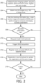
- the ultrasound system is typically in "live 2D" mode, and the live 2D images 111 from the ultrasound scanner are fused (via the current registration units 134, 136) with the prior static image (CT) 122.
- the operator is interested in fusion-imaging of a particular target area, such as a tumor, and will move the ultrasound probe 102 to explore different ways of visualizing the tumor. Different views of the tumor are possible, but only some may have sufficient overlap and pose similarity (success parameters) with the baseline 3DUS 124 for motion compensation.
- the system 100 continuously computes and displays the success parameters, enabling the operator to identify a view of the tumor with sufficient success parameters.
- the operator optionally proceeds to obtain a breath hold from the patient, acquires a live 2D/3DUS image 111 of the tumor, which is stored in the current view memory 126 once acquired.
- the motion compensation unit 128 is triggered with the acquisition of the new 3DUS image (stored in memory 126).
- the system 100 carries out the motion compensation by registering the live 3DUS image 126 with the baseline 3DUS 124, and using the registration result from registration unit 134 to update the displayed fusion image.
- the updated fusion image can now be used by the operator to visualize the tumor or to carry out an intervention (such as a needle insertion).
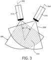
- FIG. 3 a diagram shows ultrasound imaging overlap to describe the computation of success parameters in the pose analyzer unit 132 of FIG. 1 .
- Image overlap and relative pose can be computed as success parameters (shown in FIG. 3 in 2D for simplicity).
- Ultrasonic probes 312, 314 are each positioned to provide different views 302 and 304, respectively.
- An image angle difference, ⁇ is computed directly from the relative pose transform T current2base .
- Success parameters may also include parameters, such as, e.g., brightness, contrast or other image features, extracted from the 2D US images acquired in the different views.
- the system 100 may provide feedback and guidance for optimizing the success parameters of the ultrasound view.
- the pose analyzer unit 132 will be connected to the pose guidance unit 138 to compute the ultrasound probe motion needed to increase or optimize the parameters.
- This information is passed onto the display 118 to show the instructions to the operator (e.g., "move left", “rotate clockwise”).
- the pose guidance unit 138 may use the prior static image 122 (e.g., CT) and information derived from it (e.g., skin surface) to determine ultrasound poses that are acceptable (e.g., ultrasound probe touching skin).
- the system 100 may provide feedback and guidance to optimize the success parameters of an ultrasound view that images the user-provided target area.
- the interface 120 will permit the operator to enter a target area in the 3DUS baseline 124 or prior static image 122.
- the information is passed on to the pose guidance unit 138 to compute guidance toward a view that images that target area while maximizing the success parameters.
- the pose optimization problem can be computationally solved by the pose analyzer unit 132 by considering the probe positions and rotations as input parameters (possibly constrained to positions on the patient's skin, as derived from the prior static image 122) that are to be optimized, and defining a cost function, f, that is to be minimized, which is inversely related to the likelihood of motion compensation succeeding for a 3DUS acquired at the current pose.
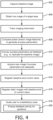
- a method for fusing images to account for motion compensation is illustratively shown in accordance with the present principles.
- a baseline image is captured.
- live images of a target area are obtained.
- the live images may be 2D images or provide a preliminary image.
- an imaging instrument is tracked to obtain a pose for capturing the baseline image and to obtain a pose for a current view of the live images such that a tracking system has its coordinate system registered with the baseline image and the live images.
- a pose for a current view is analyzed to compare field of view differences between the pose for the baseline image and the pose for the current view using the tracking system to generate success parameters.
- parameters/image features from the live image and baseline image may be computed and compared as well.
- the success parameters are conveyed to provide feedback on optimal image acquisition for motion compensation between the baseline image and the current view.
- the success parameters measure field of view overlap and pose similarity between the baseline images and the current view image.
- the success parameters may include different parameters for achieving overlap, pose similarity and image similarity, e.g., angles, positions, areas, percentages, image contrast, brightness or other quantities or features.
- a new image at the pose of the current view is acquired. This may be a full blown 3D image as opposed to a 2D image or preliminary image.
- the adequacy of the success parameters may be determined by a user or may be set automatically or as a default. It is determined whether a pose provides a field of view for a current pose comparable to that of the pose of the baseline image to permit a user to replicate the baseline image field of view. For example, a threshold, say, .e.g., 90% overlap, between a baseline field of view and a current field of view may be set to determine adequacy. Other criteria are also contemplated.
- the baseline images and the current view image may be registered.
- static images e.g., CT, MRI, etc.
- This registration may occur at any time and preferably occurs during an early stage (e.g., planning) with regards to the baseline image being registered to the static image.
- the pose guidance unit may include and compute a cost function to evaluate overlap between different image fields of view.
- the cost function may also consider image parameters between, e.g., a live image (e.g., a preliminary 2D image) and the baseline image.
- the image parameters may include contrast, brightness, content information, etc.
- the feedback activity may include at least one of a visual signal (e.g., flash, images), text commands, audio signals (e.g., beeping, voice commands), haptic signals (e.g., vibration intensity, vibration changes), etc.
- a visual signal e.g., flash, images
- audio signals e.g., beeping, voice commands
- haptic signals e.g., vibration intensity, vibration changes
Landscapes
- Health & Medical Sciences (AREA)
- Life Sciences & Earth Sciences (AREA)
- Engineering & Computer Science (AREA)
- Physics & Mathematics (AREA)
- Radiology & Medical Imaging (AREA)
- Medical Informatics (AREA)
- Veterinary Medicine (AREA)
- Biophysics (AREA)
- Nuclear Medicine, Radiotherapy & Molecular Imaging (AREA)
- Pathology (AREA)
- Public Health (AREA)
- Biomedical Technology (AREA)
- Heart & Thoracic Surgery (AREA)
- General Health & Medical Sciences (AREA)
- Molecular Biology (AREA)
- Surgery (AREA)
- Animal Behavior & Ethology (AREA)
- Computer Vision & Pattern Recognition (AREA)
- Theoretical Computer Science (AREA)
- General Physics & Mathematics (AREA)
- Apparatus For Radiation Diagnosis (AREA)
- Ultra Sonic Daignosis Equipment (AREA)
Claims (13)
- Système d'imagerie par fusion ultrasonore (100) pour fusionner des images pour tenir compte de la compensation de mouvement, comprenant :une sonde d'imagerie ultrasonore (102) configurée pour capturer une image de base et des images en direct d'une zone cible ;un système de suivi en direct (115) configuré pour suivre la sonde d'imagerie ultrasonore (102) pendant la capture de l'image de base et des images en direct, le système de suivi en direct présentant un système de coordonnées enregistré avec l'image de base et les images en direct ; etune unité d'analyse de position (132) configurée pour comparer les différences de champ de vision entre la position de la sonde (102) pour l'image de base et la position de la sonde (102) pour une vue actuelle d'une image en direct déterminée à l'aide du système de suivi en direct pour générer des paramètres de réussite représentant une mesure de chevauchement entre l'image de base et l'image de vue actuelle et une similarité entre les positions, et pour transmettre les paramètres de réussite à un opérateur pour fournir un retour d'informations sur l'acquisition d'image pour une compensation de mouvement entre l'image de base et l'image de vue actuelle,le système comprenant en outre une unité d'enregistrement de compensation de mouvement (128) pour enregistrer l'image de base avec l'image de vue actuelle.
- Système selon la revendication 1, dans lequel les paramètres de réussite représentent en outre une mesure de similarité d'image entre l'image de base et l'image en direct prise en une position actuelle.
- Système selon la revendication 1, dans lequel l'analyseur de position (132) est en outre configuré pour déterminer si une position fournit un champ de vision pour une position actuelle qui est similaire à celui d'une position de l'image de base pour permettre à un utilisateur de reproduire le champ de vision de l'image de base.
- Système selon la revendication 1, comprenant en outre une unité de guidage de position (138) configurée pour fournir des informations de direction à un utilisateur indiquant une direction pour déplacer la sonde d'imagerie ultrasonore (102) pour obtenir une position souhaitée pour l'image de vue actuelle.
- Système selon la revendication 4, dans lequel l'unité de guidage de position (138) inclut une fonction coût configurée pour évaluer le chevauchement et/ou la rotation entre différents champs de vision de l'image et/ou les paramètres d'image entre l'image de base et l'image de vue actuelle.
- Système selon la revendication 1, comprenant en outre une interface (120) configurée pour guider les actions de l'utilisateur par l'intermédiaire d'une activité de retour d'informations afin d'obtenir une position souhaitée pour l'image de vue actuelle.
- Système selon la revendication 5, dans lequel l'activité de retour d'informations inclut au moins un élément parmi un signal visuel, une commande textuelle, un signal audio et un signal haptique pour guider les actions d'utilisateur vers une position souhaitée.
- Procédé der fusion des images pour tenir compte de la compensation de mouvement, comprenant :la capture (402) d'une image de base avec une sonde d'imagerie ultrasonore (102) d'un système d'imagerie par fusion ultrasonore (100) ;l'obtention (404) d'images en direct d'une zone cible avec la sonde ultrasonore (102) ;le suivi (406) de la sonde ultrasonore (102) pour obtenir une position de la sonde (102) pour capturer l'image de base et pour obtenir une position de la sonde (102) pour une vue actuelle d'une image en direct de telle sorte qu'un système de suivi (115) présente son système de coordonnées enregistré avec l'image de base et les images en direct ;l'analyse (408), avec une unité d'analyse de position (132), d'une position pour une image de vue actuelle afin de comparer les différences de champ de vision entre la position pour l'image de base et la position pour l'image de vue actuelle en utilisant le système de suivi pour générer des paramètres de réussite, les paramètres de réussite représentant une mesure de chevauchement entre l'image de base et l'image de vue actuelle ;la transmission (410) des paramètres de réussite à un opérateur pour fournir un retour d'informations sur l'acquisition d'image pour une compensation de mouvement entre l'image de base et l'image de vue actuelle ;si les paramètres de réussite sont adéquats, l'acquisition (412) d'une nouvelle image à la position de la vue actuelle, etl'enregistrement (414) de l'image de base avec l'image de vue actuelle à l'aide d'une unité d'enregistrement de compensation de mouvement (128).
- Procédé selon la revendication 8, dans lequel les paramètres de réussite représentent en outre une mesure de similarité d'image entre l'image de base et l'image en direct prise à la position de la vue actuelle.
- Procédé selon la revendication 8, comprenant en outre la détermination (410) pour établir si une position fournit un champ de vision pour une position actuelle qui est similaire à celui de la position de l'image de base pour permettre à un utilisateur de reproduire le champ de vision de l'image de base.
- Procédé selon la revendication 8, comprenant en outre la fourniture (418) d'informations de direction à un utilisateur indiquant une direction pour déplacer la sonde ultrasonore (102) pour obtenir une position pour la vue actuelle en utilisant une unité de guidage de position (138).
- Procédé selon la revendication 11, dans lequel l'unité de guidage de position inclut une fonction coût évaluant le chevauchement entre différents champs de vision d'image et/ou paramètres d'image entre l'image de base et l'image de vue actuelle.
- Procédé selon la revendication 8, comprenant en outre le guidage (418) des actions d'utilisateur par l'intermédiaire d'une activité de retour d'informations pour obtenir une position pour l'image de vue actuelle, dans lequel l'activité de retour d'informations inclut au moins un élément parmi des signaux visuels, des commandes textuelles, des signaux audio et des signaux haptiques guidant les actions d'utilisateur vers une position souhaitée.
Applications Claiming Priority (2)
| Application Number | Priority Date | Filing Date | Title |
|---|---|---|---|
| US201562158011P | 2015-05-07 | 2015-05-07 | |
| PCT/IB2016/052623 WO2016178198A1 (fr) | 2015-05-07 | 2016-05-09 | Système et procédé de compensation de mouvement dans des procédures médicales |
Publications (2)
| Publication Number | Publication Date |
|---|---|
| EP3291735A1 EP3291735A1 (fr) | 2018-03-14 |
| EP3291735B1 true EP3291735B1 (fr) | 2024-10-09 |
Family
ID=56072372
Family Applications (1)
| Application Number | Title | Priority Date | Filing Date |
|---|---|---|---|
| EP16724717.0A Active EP3291735B1 (fr) | 2015-05-07 | 2016-05-09 | Système et procédé de compensation de mouvement dans des procédures médicales |
Country Status (5)
| Country | Link |
|---|---|
| US (1) | US10893851B2 (fr) |
| EP (1) | EP3291735B1 (fr) |
| JP (1) | JP6714019B2 (fr) |
| CN (1) | CN107980148B (fr) |
| WO (1) | WO2016178198A1 (fr) |
Families Citing this family (14)
| Publication number | Priority date | Publication date | Assignee | Title |
|---|---|---|---|---|
| WO2016175758A2 (fr) * | 2015-04-28 | 2016-11-03 | Analogic Corporation | Pilotage guidé par image d'un réseau de transducteurs et/ou d'un instrument |
| EP3291735B1 (fr) * | 2015-05-07 | 2024-10-09 | Koninklijke Philips N.V. | Système et procédé de compensation de mouvement dans des procédures médicales |
| RU2769065C2 (ru) * | 2017-05-11 | 2022-03-28 | Конинклейке Филипс Н.В. | Технологический процесс, система и способ компенсации движения при ультразвуковых процедурах |
| US20200275915A1 (en) * | 2017-09-08 | 2020-09-03 | Koninklijke Philips N.V. | Ultrasound probe localization with drift correction |
| EP3482690A1 (fr) * | 2017-11-14 | 2019-05-15 | Koninklijke Philips N.V. | Suivi et visualisation ultrasonore |
| US11276167B2 (en) * | 2017-12-04 | 2022-03-15 | Koninklijke Philips N.V. | Image data processing method, device and system |
| FI3793447T3 (fi) * | 2018-05-15 | 2023-03-18 | Univ New York | Järjestelmä ja menetelmä ultraäänikuvien ottamisen suuntaamiseksi |
| CN111292277B (zh) * | 2018-12-10 | 2021-02-09 | 深圳迈瑞生物医疗电子股份有限公司 | 超声融合成像方法及超声融合成像导航系统 |
| KR102761431B1 (ko) * | 2019-03-13 | 2025-02-05 | 삼성전자주식회사 | 전자 장치 및 그 제어 방법 |
| NL2023588B1 (en) * | 2019-07-29 | 2021-02-18 | Elitac | Vibrotactile feedback arrangement |
| US11607200B2 (en) * | 2019-08-13 | 2023-03-21 | GE Precision Healthcare LLC | Methods and system for camera-aided ultrasound scan setup and control |
| US20210045716A1 (en) * | 2019-08-13 | 2021-02-18 | GE Precision Healthcare LLC | Method and system for providing interaction with a visual artificial intelligence ultrasound image segmentation module |
| CN112130134B (zh) * | 2020-08-17 | 2023-12-05 | 河北汉光重工有限责任公司 | 一种基于时间补偿的实时基线修正方法 |
| US12167937B2 (en) * | 2021-12-03 | 2024-12-17 | GE Precision Healthcare LLC | Methods and systems for live image acquisition |
Family Cites Families (42)
| Publication number | Priority date | Publication date | Assignee | Title |
|---|---|---|---|---|
| US7127090B2 (en) * | 2001-07-30 | 2006-10-24 | Accuimage Diagnostics Corp | Methods and systems for combining a plurality of radiographic images |
| DE10149795B4 (de) * | 2001-10-09 | 2006-04-06 | Siemens Ag | Semiautomatische Registrierung zur Überlagerung zweier medizinischer Bilddatensätze |
| JP4088104B2 (ja) * | 2002-06-12 | 2008-05-21 | 株式会社東芝 | 超音波診断装置 |
| JP4705104B2 (ja) * | 2004-08-09 | 2011-06-22 | ブラッコ・シュイス・ソシエテ・アノニム | 複数のマスクに基づく医療画像処理のためのイメージ登録方法および装置 |
| US20060093192A1 (en) * | 2004-11-03 | 2006-05-04 | Bechtel J S | Finger guide device |
| JP4470187B2 (ja) * | 2004-12-03 | 2010-06-02 | 株式会社日立メディコ | 超音波装置、超音波撮像プログラム及び超音波撮像方法 |
| US7840042B2 (en) * | 2006-01-20 | 2010-11-23 | 3M Innovative Properties Company | Superposition for visualization of three-dimensional data acquisition |
| US20080186378A1 (en) * | 2007-02-06 | 2008-08-07 | Feimo Shen | Method and apparatus for guiding towards targets during motion |
| US9597041B2 (en) * | 2007-03-30 | 2017-03-21 | General Electric Company | Sequential image acquisition with updating method and system |
| CN101053531A (zh) * | 2007-05-17 | 2007-10-17 | 上海交通大学 | 基于多模式增敏成像融合的早期肿瘤定位跟踪方法 |
| EP2208182B1 (fr) * | 2007-11-14 | 2011-06-08 | Koninklijke Philips Electronics N.V. | Système et procédé pour une analyse ceus en 3d quantitative |
| US9651662B2 (en) * | 2007-11-16 | 2017-05-16 | Koninklijke Philips N.V. | Interventional navigation using 3D contrast-enhanced ultrasound |
| WO2009085204A2 (fr) * | 2007-12-23 | 2009-07-09 | Oraya Therapeutics, Inc. | Procédés et dispositifs permettant de détecter, contrôler et prévoir l'administration d'un rayonnement |
| US7801271B2 (en) * | 2007-12-23 | 2010-09-21 | Oraya Therapeutics, Inc. | Methods and devices for orthovoltage ocular radiotherapy and treatment planning |
| JP2009200713A (ja) * | 2008-02-20 | 2009-09-03 | Sony Corp | 画像処理装置、画像処理方法、プログラム |
| US8348846B2 (en) | 2008-09-30 | 2013-01-08 | Mediri Gmbh | 3D motion detection and correction by object tracking in ultrasound images |
| EP2210844B1 (fr) * | 2009-01-26 | 2012-04-04 | Neopost Technologies | Procédé et appareil d'introduction et de pliage de feuilles |
| US9412044B2 (en) * | 2009-06-09 | 2016-08-09 | Siemens Aktiengesellschaft | Method of compensation of respiratory motion in cardiac imaging |
| DE102010009295B4 (de) * | 2010-02-25 | 2019-02-21 | Siemens Healthcare Gmbh | Verfahren zur Darstellung eines zu untersuchenden und/oder behandelnden Bereichs |
| CA2797302C (fr) * | 2010-04-28 | 2019-01-15 | Ryerson University | Systeme et procedes de retroaction de guidage peroperatoire |
| US20140194793A1 (en) * | 2010-05-14 | 2014-07-10 | Kai Medical, Inc. | Systems and methods for non-contact multiparameter vital signs monitoring, apnea therapy, apnea diagnosis, and snore therapy |
| WO2011143631A2 (fr) * | 2010-05-14 | 2011-11-17 | Kai Medical, Inc. | Systèmes et procédés pour le monitorage des signes vitaux multi-paramètres sans contact, la thérapie de l'apnée, l'annulation des vibrations, l'identification de patient, et capteurs de monitorage de sujet |
| JP5848762B2 (ja) * | 2010-06-28 | 2016-01-27 | コーニンクレッカ フィリップス エヌ ヴェKoninklijke Philips N.V. | Emキャリブレーションのリアルタイム品質管理 |
| EP2680778B1 (fr) * | 2011-03-03 | 2019-07-31 | Koninklijke Philips N.V. | Système et procédé d'initialisation et d'enregistrement automatiques d'un système de navigation |
| US8810640B2 (en) * | 2011-05-16 | 2014-08-19 | Ut-Battelle, Llc | Intrinsic feature-based pose measurement for imaging motion compensation |
| JP2011205681A (ja) * | 2011-05-27 | 2011-10-13 | Hitachi Ltd | 記録方法 |
| US8588501B2 (en) * | 2011-07-29 | 2013-11-19 | Siemens Aktiengesellschaft | Automatic pose initialization for accurate 2-D/3-D registration applied to abdominal aortic aneurysm endovascular repair |
| KR20140049137A (ko) * | 2012-10-12 | 2014-04-25 | 삼성전자주식회사 | 의료영상 분석장치 및 방법 |
| US9001226B1 (en) * | 2012-12-04 | 2015-04-07 | Lytro, Inc. | Capturing and relighting images using multiple devices |
| US10456062B2 (en) * | 2013-01-11 | 2019-10-29 | The Cleveland Clinic Foundation | Alignment of manipulable sensor assembly |
| US9131922B2 (en) * | 2013-01-29 | 2015-09-15 | Eigen, Inc. | Calibration for 3D reconstruction of medical images from a sequence of 2D images |
| EP2973417B1 (fr) * | 2013-03-11 | 2020-07-15 | Carestream Dental Technology Topco Limited | Procédé et système d'imagerie tridimensionnelle |
| US10278584B2 (en) * | 2013-03-11 | 2019-05-07 | Carestream Dental Technology Topco Limited | Method and system for three-dimensional imaging |
| US10357092B2 (en) * | 2013-11-06 | 2019-07-23 | Koninklijke Phiilps N.V. | System and a method for guiding a user during a shaving procedure |
| US10068373B2 (en) * | 2014-07-01 | 2018-09-04 | Samsung Electronics Co., Ltd. | Electronic device for providing map information |
| US20160106381A1 (en) * | 2014-10-20 | 2016-04-21 | General Electric Company | Ultrasound probe with tactile indicator |
| US9626589B1 (en) * | 2015-01-19 | 2017-04-18 | Ricoh Co., Ltd. | Preview image acquisition user interface for linear panoramic image stitching |
| JP6436442B2 (ja) * | 2015-04-10 | 2018-12-12 | キヤノン株式会社 | 光音響装置および画像処理方法 |
| EP3291735B1 (fr) * | 2015-05-07 | 2024-10-09 | Koninklijke Philips N.V. | Système et procédé de compensation de mouvement dans des procédures médicales |
| US10646199B2 (en) * | 2015-10-19 | 2020-05-12 | Clarius Mobile Health Corp. | Systems and methods for remote graphical feedback of ultrasound scanning technique |
| US10964424B2 (en) * | 2016-03-09 | 2021-03-30 | EchoNous, Inc. | Ultrasound image recognition systems and methods utilizing an artificial intelligence network |
| EP3574504A1 (fr) * | 2017-01-24 | 2019-12-04 | Tietronix Software, Inc. | Système et procédé de guidage de réalité augmentée tridimensionnelle pour l'utilisation d'un équipement médical |
-
2016
- 2016-05-09 EP EP16724717.0A patent/EP3291735B1/fr active Active
- 2016-05-09 JP JP2017557317A patent/JP6714019B2/ja active Active
- 2016-05-09 WO PCT/IB2016/052623 patent/WO2016178198A1/fr not_active Ceased
- 2016-05-09 CN CN201680026452.4A patent/CN107980148B/zh active Active
- 2016-05-09 US US15/567,648 patent/US10893851B2/en active Active
Also Published As
| Publication number | Publication date |
|---|---|
| JP6714019B2 (ja) | 2020-06-24 |
| US20180146955A1 (en) | 2018-05-31 |
| US10893851B2 (en) | 2021-01-19 |
| EP3291735A1 (fr) | 2018-03-14 |
| JP2018518226A (ja) | 2018-07-12 |
| CN107980148A (zh) | 2018-05-01 |
| WO2016178198A1 (fr) | 2016-11-10 |
| CN107980148B (zh) | 2022-06-28 |
Similar Documents
| Publication | Publication Date | Title |
|---|---|---|
| EP3291735B1 (fr) | Système et procédé de compensation de mouvement dans des procédures médicales | |
| US20170084036A1 (en) | Registration of video camera with medical imaging | |
| US10424067B2 (en) | Image processing apparatus, image processing method and storage medium | |
| CN107111875B (zh) | 用于多模态自动配准的反馈 | |
| US20150235369A1 (en) | Image processing apparatus and image processing method | |
| US20140147027A1 (en) | Intra-operative image correction for image-guided interventions | |
| JP2013517909A (ja) | 気管支鏡検査法ガイダンスに適用される画像ベースのグローバル登録 | |
| CN103985147A (zh) | 终端用户特定诊断用医学图像读取的标志检测模型的现场学习的方法和系统 | |
| JP2016147026A (ja) | 画像処理装置、画像処理方法およびプログラム | |
| WO2012020547A1 (fr) | Dispositif, procédé et programme d'assistance à l'imagerie diagnostique | |
| JP2022031179A (ja) | デバイス対画像レジストレーションの方法、装置及び記憶媒体 | |
| US12507864B2 (en) | Interactive augmented reality system for laparoscopic and video assisted surgeries | |
| US20210166805A1 (en) | Method and system for synchronizing medical image analysis and reporting | |
| CN106687048A (zh) | 医学成像装置 | |
| US20240206907A1 (en) | System and Method for Device Tracking in Magnetic Resonance Imaging Guided Inerventions | |
| EP2601637A1 (fr) | Système et procédé de segmentation multi-modalité d'un tissu interne avec rétroaction en direct | |
| EP4128145B1 (fr) | Combinaison d'informations angiographiques avec des images fluoroscopiques | |
| US20250252584A1 (en) | Method for tracker-less image registration and system for carrying out said method | |
| CN107106106A (zh) | 针对具有减小的角范围的旋转c型臂计算机断层摄影的自适应分割 | |
| WO2025073380A1 (fr) | Panorama sans recomposition dans l'acquisition d'images médicales | |
| US10299864B1 (en) | Co-localization of multiple internal organs based on images obtained during surgery | |
| EP4687642A1 (fr) | Procédé et système pour effectuer des balayages par des systèmes d'imagerie multiples | |
| JP2017225670A (ja) | 画像処理装置およびその制御方法 | |
| CN121817944A (zh) | 一种超声图像处理方法、装置和超声设备 | |
| CN116763401A (zh) | 一种穿刺路径规划系统、方法及手术机器人 |
Legal Events
| Date | Code | Title | Description |
|---|---|---|---|
| STAA | Information on the status of an ep patent application or granted ep patent |
Free format text: STATUS: THE INTERNATIONAL PUBLICATION HAS BEEN MADE |
|
| PUAI | Public reference made under article 153(3) epc to a published international application that has entered the european phase |
Free format text: ORIGINAL CODE: 0009012 |
|
| STAA | Information on the status of an ep patent application or granted ep patent |
Free format text: STATUS: REQUEST FOR EXAMINATION WAS MADE |
|
| 17P | Request for examination filed |
Effective date: 20171207 |
|
| AK | Designated contracting states |
Kind code of ref document: A1 Designated state(s): AL AT BE BG CH CY CZ DE DK EE ES FI FR GB GR HR HU IE IS IT LI LT LU LV MC MK MT NL NO PL PT RO RS SE SI SK SM TR |
|
| AX | Request for extension of the european patent |
Extension state: BA ME |
|
| DAV | Request for validation of the european patent (deleted) | ||
| DAX | Request for extension of the european patent (deleted) | ||
| RAP1 | Party data changed (applicant data changed or rights of an application transferred) |
Owner name: KONINKLIJKE PHILIPS N.V. |
|
| STAA | Information on the status of an ep patent application or granted ep patent |
Free format text: STATUS: EXAMINATION IS IN PROGRESS |
|
| 17Q | First examination report despatched |
Effective date: 20210412 |
|
| GRAP | Despatch of communication of intention to grant a patent |
Free format text: ORIGINAL CODE: EPIDOSNIGR1 |
|
| STAA | Information on the status of an ep patent application or granted ep patent |
Free format text: STATUS: GRANT OF PATENT IS INTENDED |
|
| RIC1 | Information provided on ipc code assigned before grant |
Ipc: G06T 7/30 20170101ALI20240415BHEP Ipc: A61B 8/08 20060101ALI20240415BHEP Ipc: G06T 7/00 20060101ALI20240415BHEP Ipc: A61B 8/00 20060101AFI20240415BHEP |
|
| INTG | Intention to grant announced |
Effective date: 20240510 |
|
| GRAS | Grant fee paid |
Free format text: ORIGINAL CODE: EPIDOSNIGR3 |
|
| GRAA | (expected) grant |
Free format text: ORIGINAL CODE: 0009210 |
|
| STAA | Information on the status of an ep patent application or granted ep patent |
Free format text: STATUS: THE PATENT HAS BEEN GRANTED |
|
| AK | Designated contracting states |
Kind code of ref document: B1 Designated state(s): AL AT BE BG CH CY CZ DE DK EE ES FI FR GB GR HR HU IE IS IT LI LT LU LV MC MK MT NL NO PL PT RO RS SE SI SK SM TR |
|
| REG | Reference to a national code |
Ref country code: CH Ref legal event code: EP |
|
| REG | Reference to a national code |
Ref country code: DE Ref legal event code: R096 Ref document number: 602016089741 Country of ref document: DE |
|
| REG | Reference to a national code |
Ref country code: IE Ref legal event code: FG4D |
|
| REG | Reference to a national code |
Ref country code: LT Ref legal event code: MG9D |
|
| REG | Reference to a national code |
Ref country code: NL Ref legal event code: MP Effective date: 20241009 |
|
| REG | Reference to a national code |
Ref country code: AT Ref legal event code: MK05 Ref document number: 1729674 Country of ref document: AT Kind code of ref document: T Effective date: 20241009 |
|
| PG25 | Lapsed in a contracting state [announced via postgrant information from national office to epo] |
Ref country code: NL Free format text: LAPSE BECAUSE OF FAILURE TO SUBMIT A TRANSLATION OF THE DESCRIPTION OR TO PAY THE FEE WITHIN THE PRESCRIBED TIME-LIMIT Effective date: 20241009 |
|
| PG25 | Lapsed in a contracting state [announced via postgrant information from national office to epo] |
Ref country code: NL Free format text: LAPSE BECAUSE OF FAILURE TO SUBMIT A TRANSLATION OF THE DESCRIPTION OR TO PAY THE FEE WITHIN THE PRESCRIBED TIME-LIMIT Effective date: 20241009 |
|
| PG25 | Lapsed in a contracting state [announced via postgrant information from national office to epo] |
Ref country code: PT Free format text: LAPSE BECAUSE OF FAILURE TO SUBMIT A TRANSLATION OF THE DESCRIPTION OR TO PAY THE FEE WITHIN THE PRESCRIBED TIME-LIMIT Effective date: 20250210 Ref country code: IS Free format text: LAPSE BECAUSE OF FAILURE TO SUBMIT A TRANSLATION OF THE DESCRIPTION OR TO PAY THE FEE WITHIN THE PRESCRIBED TIME-LIMIT Effective date: 20250209 Ref country code: HR Free format text: LAPSE BECAUSE OF FAILURE TO SUBMIT A TRANSLATION OF THE DESCRIPTION OR TO PAY THE FEE WITHIN THE PRESCRIBED TIME-LIMIT Effective date: 20241009 |
|
| PG25 | Lapsed in a contracting state [announced via postgrant information from national office to epo] |
Ref country code: FI Free format text: LAPSE BECAUSE OF FAILURE TO SUBMIT A TRANSLATION OF THE DESCRIPTION OR TO PAY THE FEE WITHIN THE PRESCRIBED TIME-LIMIT Effective date: 20241009 |
|
| PG25 | Lapsed in a contracting state [announced via postgrant information from national office to epo] |
Ref country code: BG Free format text: LAPSE BECAUSE OF FAILURE TO SUBMIT A TRANSLATION OF THE DESCRIPTION OR TO PAY THE FEE WITHIN THE PRESCRIBED TIME-LIMIT Effective date: 20241009 |
|
| PG25 | Lapsed in a contracting state [announced via postgrant information from national office to epo] |
Ref country code: ES Free format text: LAPSE BECAUSE OF FAILURE TO SUBMIT A TRANSLATION OF THE DESCRIPTION OR TO PAY THE FEE WITHIN THE PRESCRIBED TIME-LIMIT Effective date: 20241009 |
|
| PG25 | Lapsed in a contracting state [announced via postgrant information from national office to epo] |
Ref country code: NO Free format text: LAPSE BECAUSE OF FAILURE TO SUBMIT A TRANSLATION OF THE DESCRIPTION OR TO PAY THE FEE WITHIN THE PRESCRIBED TIME-LIMIT Effective date: 20250109 |
|
| PG25 | Lapsed in a contracting state [announced via postgrant information from national office to epo] |
Ref country code: LV Free format text: LAPSE BECAUSE OF FAILURE TO SUBMIT A TRANSLATION OF THE DESCRIPTION OR TO PAY THE FEE WITHIN THE PRESCRIBED TIME-LIMIT Effective date: 20241009 Ref country code: AT Free format text: LAPSE BECAUSE OF FAILURE TO SUBMIT A TRANSLATION OF THE DESCRIPTION OR TO PAY THE FEE WITHIN THE PRESCRIBED TIME-LIMIT Effective date: 20241009 Ref country code: GR Free format text: LAPSE BECAUSE OF FAILURE TO SUBMIT A TRANSLATION OF THE DESCRIPTION OR TO PAY THE FEE WITHIN THE PRESCRIBED TIME-LIMIT Effective date: 20250110 |
|
| PG25 | Lapsed in a contracting state [announced via postgrant information from national office to epo] |
Ref country code: PL Free format text: LAPSE BECAUSE OF FAILURE TO SUBMIT A TRANSLATION OF THE DESCRIPTION OR TO PAY THE FEE WITHIN THE PRESCRIBED TIME-LIMIT Effective date: 20241009 |
|
| PG25 | Lapsed in a contracting state [announced via postgrant information from national office to epo] |
Ref country code: RS Free format text: LAPSE BECAUSE OF FAILURE TO SUBMIT A TRANSLATION OF THE DESCRIPTION OR TO PAY THE FEE WITHIN THE PRESCRIBED TIME-LIMIT Effective date: 20250109 |
|
| PG25 | Lapsed in a contracting state [announced via postgrant information from national office to epo] |
Ref country code: SM Free format text: LAPSE BECAUSE OF FAILURE TO SUBMIT A TRANSLATION OF THE DESCRIPTION OR TO PAY THE FEE WITHIN THE PRESCRIBED TIME-LIMIT Effective date: 20241009 |
|
| PGFP | Annual fee paid to national office [announced via postgrant information from national office to epo] |
Ref country code: DE Payment date: 20250528 Year of fee payment: 10 |
|
| PG25 | Lapsed in a contracting state [announced via postgrant information from national office to epo] |
Ref country code: DK Free format text: LAPSE BECAUSE OF FAILURE TO SUBMIT A TRANSLATION OF THE DESCRIPTION OR TO PAY THE FEE WITHIN THE PRESCRIBED TIME-LIMIT Effective date: 20241009 |
|
| REG | Reference to a national code |
Ref country code: DE Ref legal event code: R097 Ref document number: 602016089741 Country of ref document: DE |
|
| PG25 | Lapsed in a contracting state [announced via postgrant information from national office to epo] |
Ref country code: EE Free format text: LAPSE BECAUSE OF FAILURE TO SUBMIT A TRANSLATION OF THE DESCRIPTION OR TO PAY THE FEE WITHIN THE PRESCRIBED TIME-LIMIT Effective date: 20241009 |
|
| PG25 | Lapsed in a contracting state [announced via postgrant information from national office to epo] |
Ref country code: RO Free format text: LAPSE BECAUSE OF FAILURE TO SUBMIT A TRANSLATION OF THE DESCRIPTION OR TO PAY THE FEE WITHIN THE PRESCRIBED TIME-LIMIT Effective date: 20241009 |
|
| PG25 | Lapsed in a contracting state [announced via postgrant information from national office to epo] |
Ref country code: SK Free format text: LAPSE BECAUSE OF FAILURE TO SUBMIT A TRANSLATION OF THE DESCRIPTION OR TO PAY THE FEE WITHIN THE PRESCRIBED TIME-LIMIT Effective date: 20241009 |
|
| PG25 | Lapsed in a contracting state [announced via postgrant information from national office to epo] |
Ref country code: CZ Free format text: LAPSE BECAUSE OF FAILURE TO SUBMIT A TRANSLATION OF THE DESCRIPTION OR TO PAY THE FEE WITHIN THE PRESCRIBED TIME-LIMIT Effective date: 20241009 |
|
| PG25 | Lapsed in a contracting state [announced via postgrant information from national office to epo] |
Ref country code: IT Free format text: LAPSE BECAUSE OF FAILURE TO SUBMIT A TRANSLATION OF THE DESCRIPTION OR TO PAY THE FEE WITHIN THE PRESCRIBED TIME-LIMIT Effective date: 20241009 |
|
| PLBE | No opposition filed within time limit |
Free format text: ORIGINAL CODE: 0009261 |
|
| STAA | Information on the status of an ep patent application or granted ep patent |
Free format text: STATUS: NO OPPOSITION FILED WITHIN TIME LIMIT |
|
| PG25 | Lapsed in a contracting state [announced via postgrant information from national office to epo] |
Ref country code: SE Free format text: LAPSE BECAUSE OF FAILURE TO SUBMIT A TRANSLATION OF THE DESCRIPTION OR TO PAY THE FEE WITHIN THE PRESCRIBED TIME-LIMIT Effective date: 20241009 |
|
| 26N | No opposition filed |
Effective date: 20250710 |
|
| REG | Reference to a national code |
Ref country code: CH Ref legal event code: H13 Free format text: ST27 STATUS EVENT CODE: U-0-0-H10-H13 (AS PROVIDED BY THE NATIONAL OFFICE) Effective date: 20251223 |
|
| PG25 | Lapsed in a contracting state [announced via postgrant information from national office to epo] |
Ref country code: LU Free format text: LAPSE BECAUSE OF NON-PAYMENT OF DUE FEES Effective date: 20250509 |
|
| PG25 | Lapsed in a contracting state [announced via postgrant information from national office to epo] |
Ref country code: CH Free format text: LAPSE BECAUSE OF NON-PAYMENT OF DUE FEES Effective date: 20250531 |
|
| REG | Reference to a national code |
Ref country code: BE Ref legal event code: MM Effective date: 20250531 |
|
| PG25 | Lapsed in a contracting state [announced via postgrant information from national office to epo] |
Ref country code: MC Free format text: LAPSE BECAUSE OF FAILURE TO SUBMIT A TRANSLATION OF THE DESCRIPTION OR TO PAY THE FEE WITHIN THE PRESCRIBED TIME-LIMIT Effective date: 20241009 |
|
| PGFP | Annual fee paid to national office [announced via postgrant information from national office to epo] |
Ref country code: GB Payment date: 20260304 Year of fee payment: 11 |
|
| PG25 | Lapsed in a contracting state [announced via postgrant information from national office to epo] |
Ref country code: IE Free format text: LAPSE BECAUSE OF NON-PAYMENT OF DUE FEES Effective date: 20250509 |
|
| PG25 | Lapsed in a contracting state [announced via postgrant information from national office to epo] |
Ref country code: BE Free format text: LAPSE BECAUSE OF NON-PAYMENT OF DUE FEES Effective date: 20250531 |
|
| PG25 | Lapsed in a contracting state [announced via postgrant information from national office to epo] |
Ref country code: FR Free format text: LAPSE BECAUSE OF NON-PAYMENT OF DUE FEES Effective date: 20250531 |