EP3296904A1 - Appareil médical et procédé d'affichage d'image l'utilisant - Google Patents
Appareil médical et procédé d'affichage d'image l'utilisant Download PDFInfo
- Publication number
- EP3296904A1 EP3296904A1 EP17194186.7A EP17194186A EP3296904A1 EP 3296904 A1 EP3296904 A1 EP 3296904A1 EP 17194186 A EP17194186 A EP 17194186A EP 3296904 A1 EP3296904 A1 EP 3296904A1
- Authority
- EP
- European Patent Office
- Prior art keywords
- image
- processing
- types
- processed
- post
- Prior art date
- Legal status (The legal status is an assumption and is not a legal conclusion. Google has not performed a legal analysis and makes no representation as to the accuracy of the status listed.)
- Granted
Links
Images


Classifications
-
- G—PHYSICS
- G16—INFORMATION AND COMMUNICATION TECHNOLOGY [ICT] SPECIALLY ADAPTED FOR SPECIFIC APPLICATION FIELDS
- G16H—HEALTHCARE INFORMATICS, i.e. INFORMATION AND COMMUNICATION TECHNOLOGY [ICT] SPECIALLY ADAPTED FOR THE HANDLING OR PROCESSING OF MEDICAL OR HEALTHCARE DATA
- G16H30/00—ICT specially adapted for the handling or processing of medical images
- G16H30/40—ICT specially adapted for the handling or processing of medical images for processing medical images, e.g. editing
-
- A—HUMAN NECESSITIES
- A61—MEDICAL OR VETERINARY SCIENCE; HYGIENE
- A61B—DIAGNOSIS; SURGERY; IDENTIFICATION
- A61B6/00—Apparatus or devices for radiation diagnosis; Apparatus or devices for radiation diagnosis combined with radiation therapy equipment
- A61B6/02—Arrangements for diagnosis sequentially in different planes; Stereoscopic radiation diagnosis
- A61B6/03—Computed tomography [CT]
-
- A—HUMAN NECESSITIES
- A61—MEDICAL OR VETERINARY SCIENCE; HYGIENE
- A61B—DIAGNOSIS; SURGERY; IDENTIFICATION
- A61B5/00—Measuring for diagnostic purposes; Identification of persons
- A61B5/05—Detecting, measuring or recording for diagnosis by means of electric currents or magnetic fields; Measuring using microwaves or radio waves
- A61B5/055—Detecting, measuring or recording for diagnosis by means of electric currents or magnetic fields; Measuring using microwaves or radio waves involving electronic [EMR] or nuclear [NMR] magnetic resonance, e.g. magnetic resonance imaging
-
- A—HUMAN NECESSITIES
- A61—MEDICAL OR VETERINARY SCIENCE; HYGIENE
- A61B—DIAGNOSIS; SURGERY; IDENTIFICATION
- A61B8/00—Diagnosis using ultrasonic, sonic or infrasonic waves
-
- G—PHYSICS
- G06—COMPUTING OR CALCULATING; COUNTING
- G06T—IMAGE DATA PROCESSING OR GENERATION, IN GENERAL
- G06T7/00—Image analysis
-
- A—HUMAN NECESSITIES
- A61—MEDICAL OR VETERINARY SCIENCE; HYGIENE
- A61B—DIAGNOSIS; SURGERY; IDENTIFICATION
- A61B8/00—Diagnosis using ultrasonic, sonic or infrasonic waves
- A61B8/56—Details of data transmission or power supply
- A61B8/565—Details of data transmission or power supply involving data transmission via a network
Definitions
- Exemplary embodiments relate to a medical apparatus which displays images processed through image processing, and an image display method using the same.
- medical image equipment such as a computer tomography (CT) apparatus, a magnetic resonance imager (MRI), an X-ray apparatus and an ultrasonic apparatus, have become essential equipment in modern medical science.
- CT computer tomography
- MRI magnetic resonance imager
- X-ray apparatus X-ray apparatus
- ultrasonic apparatus ultrasonic apparatus
- a medical service allows an abnormal region of a patient to be photographed using medical image equipment and then printed on a film.
- the film must be transmitted to a doctor in charge of the patient.
- the medical service has an ineffective structure in that it takes considerable time and a lot of manpower to finally make a clinical diagnosis and causes difficulty in performing rapid and precise treatment of the patient.
- a system of providing a medical service using the computer and data communication technology has been researched and developed in the medical community.
- a medical image system in which a computer communication network is installed throughout the entirety of a hospital, all X-ray films are converted into digital data, a database of the digital data is stored in a large storage medium connected to a server, and an X-ray image of a desired patient is searched through a computer monitor in each medical office as needed, has been introduced.
- an aspect of an exemplary embodiment provides a medical image processing apparatus which displays additional images to which various types of image processing is applied, and an image displaying method using the same.
- a medical apparatus includes features of the appended independent medical image processing apparatus claim.
- the controller may magnify and display the processed image represented by the object on the display unit.
- the medical apparatus may further include an input unit (e.g., an input, an input device, etc.) including a remote controller, a mouse, a keyboard, a speech recognition device or a motion recognition device, and the instructions to the object may be input through the input unit.
- an input unit e.g., an input, an input device, etc.
- a remote controller e.g., a mouse, a keyboard, a speech recognition device or a motion recognition device, and the instructions to the object may be input through the input unit.
- the display unit may comprise a touchscreen, and the instructions to the object may be input by touching the object displayed on the display unit.
- the objects may include thumbnail pictures of the images processed through the various or multiple predetermined types of image processing or text representing features of the various predetermined types of image processing.
- the various or multiple predetermined types of image processing may include post-processing of an image having undergone pre-processing.
- the display unit may display the overall image of the subject, information regarding the image, a list of viewable images, tools to process the image and the objects of the images processed by the various or multiple predetermined types of image processing, and display, if instructions to an object are input, the processed image represented by the object.
- a control method of a medical image processing apparatus includes features of the appended independent method claim.
- the processed image represented by the object may be magnified and displayed on the display unit.
- the medical apparatus may include an input unit including a remote controller, a mouse, a keyboard, a speech recognition device or a motion recognition device, and the instructions to the object may be input through the input unit.
- an input unit including a remote controller, a mouse, a keyboard, a speech recognition device or a motion recognition device, and the instructions to the object may be input through the input unit.
- the display unit may use a touchscreen, and the instructions to the object may be input by touching the object displayed on the display unit.
- the objects may include thumbnail pictures of the images processed through the various or multiple predetermined types of image processing or text representing features of the various or multiple predetermined types of image processing.
- the overall image of the subject may be displayed.
- the objects together with the overall image of the subject may be displayed on the display unit of the medical apparatus.
- the processing of the image through various or multiple predetermined types of image processing, when the image of the subject is received, may include pre-processing the image, when the image of the subject is received, and post-processing the image through the various or multiple predetermined types of image processing, separately from the processing of the image.
- a non-transitory computer readable recording medium in which a program implementing the control method is recorded.
- a control method of a medical apparatus includes processing an image of a subject, when the image of the subject is received, and displaying the processed image on a display unit of the medical apparatus, respectively processing the image through various or multiple predetermined types of image processing and displaying objects corresponding to the processed image on the display unit, during the processing of the image separately from the processing of the image, and when instructions to an object are input, magnifying and displaying the processed image represented by the object on the display unit.
- the display unit may use a touchscreen, and the instructions to the object may be input by touching the object displayed on the display unit.
- the medical apparatus may include an input unit including a remote controller, a mouse, a keyboard, a speech recognition device or a motion recognition device, and the instructions to the object may be input through the input unit.
- an input unit including a remote controller, a mouse, a keyboard, a speech recognition device or a motion recognition device, and the instructions to the object may be input through the input unit.
- the objects may include thumbnail pictures of the images processed through the various or multiple predetermined types of image processing or texts representing features of the various predetermined types of image processing.
- the various or multiple predetermined types of image processing may include post-processing of an image having undergone pre-processing.
- the post-processing may adjust at least one of parameters of the image having undergone pre-processing, including brightness, contrast, sharpness, saturation, film-effect and window/level.
- a non-transitory computer readable recording medium in which a program implementing the control method is recorded.
- a control method of a medical apparatus includes: processing an image of a subject through multiple predetermined types of image processing and displaying the processed image on a display of the medical apparatus; generating and displaying an object corresponding to the processed image on the display; and in response to inputting instructions into the object, magnifying and displaying the processed image represented by the object on the display.
- the image of the subject may include at least one from among an MRI image, a CT image, an X-ray image, and an ultrasonic image.
- the processing of the image of the subject may adjust at least one of parameters of the image having undergone pre-processing, including brightness, contrast, sharpness, saturation, film-effect and window/level.
- Full and parallel processing of the second types of image processing together with first type of image processing is executed with respect to the image, optionally having undergone pre-processing.
- various types of (post-)processing are respectively fully executed and various objects, such as thumb nails, of the image processed through the respective second types of (post-)processing are generated, and thus, although second types of image processing may take a longer time to be generated, full magnified display in the second region is quickly available upon receiving the user's selection of one of the objects representing one of the second types of image processing, wherein the images resulting from the second types of image processing may be generated beforehand, e.g. while the user may be involved in study of the first type processed image displayed initially in the second region.
- FIGS. 1 and 2 are views illustrating the configuration of a medical image system according to an aspect of an exemplary embodiment
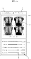
- FIGS. 3 and 4 are views illustrating the configuration of a user interface according to an aspect of an exemplary embodiment.
- the medical image system includes an image acquisition apparatus 1 acquiring an image of a subject, for example, a patient, and a medical apparatus 3 processing the image processed by the image acquisition apparatus 1 and displaying the processed image.
- the medical apparatus 3 is a workstation controlling the overall operation of the medical image system, processes the image processed by the image acquisition apparatus 1, and displays the processed image.
- the medical apparatus 3 may be a picture archiving communication system (PACS) viewer which receives the image processed by the image acquisition apparatus 1 from a server 2 or receives the image directly from the image acquisition apparatus 1, processes the received image, and displays the processed image.
- PPS picture archiving communication system
- the medical apparatus 3 includes a communication unit 5 (e.g., a communicator, transceiver, receiver, receiving unit, etc.) receiving image information of the subject from the image acquisition apparatus 1 through communication with the image acquisition apparatus 1, an input unit 6 providing an interface for operation of the medical apparatus 3, a controller 7 controlling the overall operation of the medical image system including the medical apparatus 3 and the image acquisition apparatus 1 according to instructions input through the input unit 6 and processing the image of the subject received from the communication unit 5, a storage unit 21 storing the processed image and various pieces of information, and a display unit 8 (e.g., a display, etc.) displaying the state of the medical apparatus 3 or the image acquisition apparatus 1 and the various pieces of information and displaying the image processed by the controller 7.
- a communication unit 5 e.g., a communicator, transceiver, receiver, receiving unit, etc.
- Such a medical apparatus 3 may include a PC, a portable smart-phone or a tablet PC, and a PACS viewer supporting a digital imaging and communication in medicine (DICOM) protocol.
- DICOM digital imaging and communication in medicine
- the image acquisition apparatus 1 acquires the image of a subject, for example, a patient, and is an MRI, a CT apparatus, an X-ray apparatus or an ultrasonic apparatus.
- the image of the subject may be at least one from among an MRI image, a CT image, an X-ray image, and an ultrasonic image.
- the image acquisition apparatus 1 acquires the image of the subject, and particularly, acquires the image of the inside of the subject, according to the function thereof.
- Image data of the subject may be based on the digital imaging and communication in medicine (DICOM) protocol which is a standard protocol.
- DICOM digital imaging and communication in medicine
- the communication unit 5 may receive various medical images from a server 2 in figure 1 in which images processed by various image acquisition apparatuses are sorted and stored, or may receive medical images via the Internet 4 or from other computers or other medical apparatuses 3-1.
- the communication unit 5 may transmit control instructions to control the operation of the image acquisition apparatus 1 from the controller 7 to the image acquisition apparatus 1, or may transmit various pieces of information modified or processed by the controller 7 to the server 2, the Internet 4, or other medical apparatuses 3.
- the server 2 may be a PACS server supporting the DICOM protocol.
- the communication unit 5 may employ various known wired and wireless communication methods in communication with external devices.
- the input unit 6 provides the interface so as to allow a user of the medical apparatus 3, for example, medical staff, to operate the overall function of the medical image system including the medical apparatus 3 and the image acquisition apparatus 1, and may include a remote controller, a mouse, a keyboard, a speech recognition device or a motion recognition device.
- the medical staff may properly operate the overall function of the medical image system, as needed, through peculiar functions of the above-described various input devices.
- the display unit 8 uses a touchscreen, a user may operate the function of the medical apparatus 3 by directly touching various icons displayed on the display unit 8. That is, the display unit 8 may perform the function of the input unit 6 in addition to the function of displaying various pieces of information.
- the display unit 8 provides a user interface, and the user interface may include a plurality of regions in which windows of various sizes and shapes having peculiar functions are displayed.
- the user interface provides information regarding processing of the image of a subject, i.e., image processing, and provides environments allowing a user to perform operations related to image processing.
- the user interface provided by the display unit 8 includes a first region 13 displaying objects 17 of respective images processed through various predetermined kinds or types of image processing, a second region 12 displaying an image processed through image processing, a third region 10 displaying information regarding the image of a subject, a fourth region 11 displaying a list of selectable image files, and a fifth region 14 providing image processing tools to process the image displayed in the second region 12.
- the third region 10 may display the information regarding the subject. On the assumption that the subject is a patient, the third region 10 displays information, such as personal information and disease history of the patient, so that a user may refer to such information.
- the fourth region 11 may display a list of image files viewable by the user.
- the fourth region 11 may display images transmitted from other medical apparatuses 3, the Internet 4 and the server 2, as described above, as well as the image transmitted from the image acquisition apparatus 1.
- the images are classified based on the devices or media transmitting the images, and may thus be sorted and displayed according to a device or medium selected by the user.
- the user touches or clicks an image file desired to view from among the image files displayed on the list displayed in the fourth region 11, the corresponding image file is displayed in the fourth region 11, and thus the user may confirm the image of the selected file.
- the fifth region 14 displays the image processing tools so as to allow the user to process the image displayed in the second region 12 as desired.
- the fifth region 14 may display a tool for adjusting the size of an image displayed on the display unit 8, a tool for rotating the image, a tool for cutting out a desired portion of the image, a tool for magnifying or reducing a desired portion of the image, etc.
- These tools may be expressed by symbolized icons intuitively representing the functions of the tools. If the display unit 8 uses a touchscreen, the user may touch the icon expressing a designated tool or click the icon through the input unit 6, thus being capable of processing the image according to the function of the corresponding tool. When the image is processed by the tool selected by the user, the processed image is displayed in the second region 12.
- the first region 13 may be designed to possess the same area on the user interface in common with the fifth region 14, and the user may select the first region 13 or the fifth region 14 providing desired contents through tabs 15 and 16 to select the first region 13 and the fifth region 14, displayed on the upper portion of the corresponding area.
- the first region 13 and the fifth region 14 may be displayed in separate areas, since the first region 13 and the fifth region 14 provide contents executing similar functions for the purpose of image processing, the first region 13 and the fifth region 14 may be displayed in the same area so as to allow the user to more easily understand the configuration of the interface.
- the tabs 15 and 16 are provided so as to allow the user to select the contents displayed in the corresponding area, and thus the user interface may be designed to have a simpler structure.
- the second region 12 displaying the image processed through image processing and the first region 13 displaying objects 17 of respective images processed through various predetermined kinds of image processing will be described later with reference to image processing.
- the controller 7 controls the overall operation of the medical image system including the medical apparatus 3 and the image acquisition apparatus 1 according to information received through the communication unit 5 or external instructions input through the input unit 6 or the display unit 8.
- the controller 7 may control the display unit 8 so as to display a medical image or a diagnosis record, and, if the medical image or the diagnosis record is amended or updated, may store the amended or updated medical image or diagnosis record in the storage unit 21, and may transmit the amended or updated medical image or diagnosis record to the server 2 through the communication unit 5 so as to store the amended or updated medical image or diagnosis record in the server 2.
- the controller 7 basically performs image processing to provide an image having a more improved quality, when the controller 7 receives the image of the subject transmitted from the image acquisition apparatus 1 through the communication unit 5.
- Image processing may be divided into pre-processing and post-processing.
- Pre-processing is a process of removing artifacts or errors generated due to the physical structure of the image acquisition apparatus 1
- post-processing is a process of improving the quality of an image having undergone pre-processing and during post-processing, contrast, sharpness and brightness are determined according to set parameters.
- the controller 7 displays the image processed through image processing on the display unit 8.
- the image having undergone the basic image processing is displayed in the second region 12 of the user interface provided by the display unit 8.
- the controller 7 executes various predetermined kinds of image processing with respect to the received image. More specifically, the controller 7 executes various kinds of post-processing of the image having undergone pre-processing.
- the controller 7 executes various kinds of post-processing exhibiting various effects, such as brightness, contrast, sharpness, saturation, film-effect, window/level, etc., with respect to the image from which the artifacts or errors are removed through pre-processing.
- first image processing the above-described basic image processing
- second image processing the various kinds of predetermined image processing executed separately from the first image processing
- the controller 7 amends artifacts or errors of the image through pre-processing, executes first image processing by executing first post-processing with respect to the image having undergone pre-processing, and executes second image processing by respectively executing the above-described various kinds of second post-processing with respect to the image having undergone pre-processing.
- the various kinds of second post-processing may include all kinds of post-processing which may be executed with respect to the image.
- the controller 7 may select kinds of post-processing which a user often uses by calculating the use frequencies of the kinds of post-processing selected by the user, and may form second post-processing consisting of the selected kinds of post-processing.
- the controller 7 generates images variously processed by executing second image processing, and displays objects 17 of the images in the first region 13 of the display unit 8.
- the objects 17 include thumbnail pictures of the images processed by executing various kinds of post-processing and texts expressing features of the respective kinds of post-processing.
- the text representing the features of a kind of post-processing means, for example, if the kind of post-processing applied to the image is change of contrast, a text expressing the feature of the corresponding kind of post-processing, such as contrast, of the image having undergone the corresponding kind of post-processing.
- the objects 17 may be expressed by thumbnail pictures or expressed by both thumbnail pictures and texts.
- Second image processing together with first image processing is executed with respect to the image having undergone pre-processing. Differently from first image processing, in second image processing, various kinds of post-processing are respectively executed and various images processed by executing the respective kinds of post-processing are generated, as described above, and thus second image processing may take a longer time than first image processing.
- first image processing When first image processing is completed, an image processed by executing first image processing is displayed in the second region 12 of the user interface, and if second image processing is not completed, the thumbnail picture of the image is not displayed. Therefore, until second image processing is completed, the objects 17 are expressed by texts in the first region 13, and when second image processing is completed, the texts may be changed to thumbnail pictures, or both the texts and the thumbnail pictures may be displayed. Of course, when second image processing is completed simultaneously with first image processing or is completed prior to first image processing, the objects 17 may be expressed by thumbnail pictures.
- the respective objects 17 represent images to which various kinds of post-processing are respectively applied, when a user inputs instructions to an object 17, a processed image represented by the object 17 to which the instructions are input is magnified and displayed on the display unit 8.
- the image displayed in the second region 12 when the user touches or clicks the thumbnail picture of the image in which contrast is changed from among several thumbnail pictures 17, the image represented by the corresponding thumbnail picture is magnified and displayed in the second region 12.
- the user may only touch or click a thumbnail picture while confirming an image processed by executing a kind of post-processing regarding a corresponding parameter and expressed by the thumbnail picture in advance without a troublesome operation, such as a manual adjustment of the parameter, and thus may more easily and simply process the image as the user desires.
- the second region 12 displays an image to which first image processing is applied
- the first region 13 displays thumbnail pictures of respective images to which second image processing is applied.
- the tab 16 to select the first region 13 from among the first region 13 and the fifth region 14 providing processing tools to process an image displayed in the same area is touched or clicked, contents provided by the first region 13 is displayed in the corresponding area. That is, the first region 13 displays thumbnail pictures of respective images to which second image processing is applied.
- a thumbnail picture to which a desired kind of post-processing is applied is touched or clicked from among these several thumbnail pictures, a processed image represented by the thumbnail picture is magnified and displayed in the second region 12.
- a window 22 displaying an image before it is changed into a processed image may be provided in the second region 12, and when the window 22 is touched or clicked, the image before the change into the processed image may be displayed again. That is, the image before the change and the image after the change may be compared and be converted directly therebetween.
- the window 22 displaying the image before the change may be provided to occupy a small-sized area at the corner of the second region 12 if display of the image after the change is emphasized, and may be provided to occupy the same-sized area or a similar-sized area as or to the image after the change if a comparison between the image before the change and the image after the change is emphasized. This may be adjusted according to a change of setting by a user.
- a parameter adjustment window 18 may be provided in the first region 13 so as to be manipulated when a user desires more detailed post-processing.
- the user may input specific parameter values into value input windows 20 displayed in the parameter adjustment window 18, or may drag icons 19 to adjust parameters.
- FIG. 5 is a view illustrating the user interface according to an aspect of an exemplary embodiment.
- FIG. 5 specifically illustrates the above-described first region 13.
- contents provided by the first region 13 may be displayed, as shown in FIG. 5 .
- Thumbnail pictures 17 and texts of images processed by executing various predetermined kinds of image processing, i.e., second image processing, are displayed at the upper portion of the first region 13.
- the user may touch or click the thumbnail picture 17 to which a desired kind of image processing is applied from among the displayed thumbnail pictures 17, thus confirming the processed image represented by the corresponding thumbnail picture 17 in the second region 12.
- the parameter adjustment window 18 may be displayed at the lower portion of the first region 13 so as to be manipulated when the user desires more detailed post-processing.
- the user may input specific parameter values into the value input windows 20 displayed in the parameter adjustment window 18, or may drag the icons 19 to adjust parameters.
- FIG. 5 is an example of FIG. 4 , the exemplary embodiments not limited thereto and the user interface of FIG. 5 may be modified in various ways.
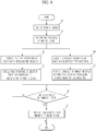
- FIG. 6 is a flowchart illustrating an image displaying method according to an aspect of an exemplary embodiment.
- an image of a subject is received (Operation 30).
- the communication unit 5 receives the image of the subject from the image acquisition apparatus 1, another medical apparatus 3, the Internet 4 or the server 2.
- Pre-processing is a process of removing artifacts or errors generated due to the physical structure of the image acquisition apparatus 1, and is basically executed with respect to the received image.
- the controller 7 executes first post-processing of the image, pre-processing of which is completed (Operation 32).
- Pre-processing and first post-processing are image processing basically executed to improve the quality of the received image, and correspond to the above-described first image processing.
- the controller 7 displays the image processed by executing the first post-processing in the second region 12 of the user interface (Operation 33).
- controller 7 executes second post-processing of the image, pre-processing of which is completed (Operation 34).
- the controller 7 processes the image, pre-processing of which is completed, by executing various kinds of post-processing and adjusting various effects, such as brightness, contrast, sharpness, saturation, film-effect, window/level, etc. Therefore, images exhibiting different effects are generated according to types of applied post-processing.
- the controller 7 When second post-processing is completed, the controller 7 generates thumbnail pictures of the respective images and displays the generated thumbnail pictures in the first region 13 of the user interface (Operation 35). Operations 34 and 35 may be executed simultaneously with Operations 32 and 33. Only the thumbnail pictures may be displayed, or the thumbnail pictures together with texts representing features of the kinds of post-processing may be displayed.
- the image to which first post-processing is applied is displayed in the second region 12, and the thumbnail pictures of the images to which kinds of second post-processing are applied are displayed in the first region 13.
- the controller 7 magnifies a processed image represented by the thumbnail picture and displays the processed image in the second region 12 (Operation 37).
- the instructions to the thumbnail picture may be input by touching the thumbnail picture if the display unit 8 employs a touchscreen, or be input by clicking the thumbnail picture through the input unit 6 including a remote controller, a mouse, a keyboard, a speech recognition device or a motion recognition device.
- magnification does not mean exaggeration of a specific part but means fitting of the image represented by the thumbnail to the size of the second region 12.
- the medical apparatus provides the user interface displaying thumbnail pictures of images to which various types of post-processing are applied in advance.
- the user may process the image by touching or clicking a thumbnail picture exhibiting the desired effect from among various thumbnail pictures in which various types of post-processing are executed, without direct change of parameters or direct input of values. Since an image exhibiting the desired effect may be intuitively recognized and selected through the thumbnail picture, the user may more easily and simply process the image.
Landscapes
- Health & Medical Sciences (AREA)
- Life Sciences & Earth Sciences (AREA)
- Engineering & Computer Science (AREA)
- Medical Informatics (AREA)
- Nuclear Medicine, Radiotherapy & Molecular Imaging (AREA)
- Physics & Mathematics (AREA)
- Public Health (AREA)
- General Health & Medical Sciences (AREA)
- Radiology & Medical Imaging (AREA)
- Pathology (AREA)
- Animal Behavior & Ethology (AREA)
- Veterinary Medicine (AREA)
- Biophysics (AREA)
- Surgery (AREA)
- Biomedical Technology (AREA)
- Heart & Thoracic Surgery (AREA)
- Molecular Biology (AREA)
- High Energy & Nuclear Physics (AREA)
- Epidemiology (AREA)
- Primary Health Care (AREA)
- Theoretical Computer Science (AREA)
- General Physics & Mathematics (AREA)
- Computer Vision & Pattern Recognition (AREA)
- Optics & Photonics (AREA)
- Measuring And Recording Apparatus For Diagnosis (AREA)
- Apparatus For Radiation Diagnosis (AREA)
Applications Claiming Priority (2)
| Application Number | Priority Date | Filing Date | Title |
|---|---|---|---|
| KR1020110139025A KR101474768B1 (ko) | 2011-12-21 | 2011-12-21 | 의료기기 및 이를 이용한 영상표시방법 |
| EP12198718.4A EP2608094B1 (fr) | 2011-12-21 | 2012-12-20 | Appareil médical et procédé d'affichage d'image l'utilisant |
Related Parent Applications (1)
| Application Number | Title | Priority Date | Filing Date |
|---|---|---|---|
| EP12198718.4A Division EP2608094B1 (fr) | 2011-12-21 | 2012-12-20 | Appareil médical et procédé d'affichage d'image l'utilisant |
Publications (2)
| Publication Number | Publication Date |
|---|---|
| EP3296904A1 true EP3296904A1 (fr) | 2018-03-21 |
| EP3296904B1 EP3296904B1 (fr) | 2020-07-29 |
Family
ID=47747283
Family Applications (2)
| Application Number | Title | Priority Date | Filing Date |
|---|---|---|---|
| EP12198718.4A Active EP2608094B1 (fr) | 2011-12-21 | 2012-12-20 | Appareil médical et procédé d'affichage d'image l'utilisant |
| EP17194186.7A Active EP3296904B1 (fr) | 2011-12-21 | 2012-12-20 | Appareil médical et procédé d'affichage d'image l'utilisant |
Family Applications Before (1)
| Application Number | Title | Priority Date | Filing Date |
|---|---|---|---|
| EP12198718.4A Active EP2608094B1 (fr) | 2011-12-21 | 2012-12-20 | Appareil médical et procédé d'affichage d'image l'utilisant |
Country Status (4)
| Country | Link |
|---|---|
| US (1) | US20130162687A1 (fr) |
| EP (2) | EP2608094B1 (fr) |
| KR (1) | KR101474768B1 (fr) |
| CN (1) | CN103177183A (fr) |
Families Citing this family (14)
| Publication number | Priority date | Publication date | Assignee | Title |
|---|---|---|---|---|
| KR102244258B1 (ko) * | 2013-10-04 | 2021-04-27 | 삼성전자주식회사 | 디스플레이 장치 및 이를 이용한 영상표시방법 |
| JP6241197B2 (ja) * | 2013-10-25 | 2017-12-06 | コニカミノルタ株式会社 | 放射線画像撮影システムおよびコンソール |
| EP3637632B1 (fr) | 2015-01-14 | 2021-09-08 | Telefonaktiebolaget LM Ericsson (publ) | Signalisation de limitation d'un sous-ensemble de livre de codes |
| US10991137B2 (en) * | 2015-06-11 | 2021-04-27 | Canon Kabushiki Kaisha | Information processing apparatus, information processing method, and information processing system for display of medical images |
| JP6838554B2 (ja) | 2015-10-02 | 2021-03-03 | ソニー株式会社 | 医療用制御装置、制御方法、プログラム、および医療用制御システム |
| KR102517104B1 (ko) * | 2016-02-17 | 2023-04-04 | 삼성전자주식회사 | 가상 현실 시스템에서 이미지 처리 방법 및 장치 |
| KR102227873B1 (ko) * | 2016-09-28 | 2021-03-15 | 지멘스 메디컬 솔루션즈 유에스에이, 인크. | 초음파 영상의 화질을 조절하는 초음파 시스템 및 방법 |
| KR101923183B1 (ko) * | 2016-12-14 | 2018-11-28 | 삼성전자주식회사 | 의료 영상 표시 방법 및 의료 영상 표시 장치 |
| JP7177611B2 (ja) * | 2017-07-10 | 2022-11-24 | キヤノン株式会社 | 画像表示装置、画像表示方法及びプログラム |
| CN108184097B (zh) * | 2018-01-15 | 2020-02-28 | 浙江大学 | 一种远程医疗中图像的实时查看方法 |
| KR102422712B1 (ko) * | 2019-10-23 | 2022-07-19 | 경북대학교 산학협력단 | 의료영상 기반 스마트 ui 제공방법 및 장치 |
| CN113497898B (zh) * | 2020-04-02 | 2023-04-07 | 抖音视界有限公司 | 视频特效配置文件生成方法、视频渲染方法及装置 |
| CN111477305B (zh) * | 2020-05-11 | 2023-10-31 | 青岛海信医疗设备股份有限公司 | 一种胶片数字化处理方法、终端和拍摄终端 |
| CN112235547A (zh) * | 2020-10-22 | 2021-01-15 | 北京德为智慧科技有限公司 | 智能观片装置及其控制方法 |
Citations (4)
| Publication number | Priority date | Publication date | Assignee | Title |
|---|---|---|---|---|
| US20020094119A1 (en) | 2001-01-18 | 2002-07-18 | Velayudhan Sahadevan | High resolution digitized image analysis of chest x-rays for diagnosis of difficult to visualize evolving very ealrly stage lung cancer, pnumoconiosis and pulmonary diseases |
| US20070140536A1 (en) * | 2005-12-19 | 2007-06-21 | Eastman Kodak Company | Medical image processing method and apparatus |
| WO2008064120A2 (fr) * | 2006-11-17 | 2008-05-29 | Amigent, Inc. | Procédé pour afficher les mesures et les changements dans le temps des images de surface de la peau |
| WO2011044295A2 (fr) | 2009-10-07 | 2011-04-14 | Hologic, Inc. | Traitement et affichage d'informations de détection assistée par ordinateur, associées à des images radiologiques du sein |
Family Cites Families (14)
| Publication number | Priority date | Publication date | Assignee | Title |
|---|---|---|---|---|
| US6175614B1 (en) * | 1999-05-07 | 2001-01-16 | Oec Medical Systems, Inc. | Method and apparatus for automatic sizing and positioning of ABS sampling window in an x-ray imaging system |
| US7054473B1 (en) * | 2001-11-21 | 2006-05-30 | R2 Technology, Inc. | Method and apparatus for an improved computer aided diagnosis system |
| US6574307B1 (en) * | 2001-11-21 | 2003-06-03 | Ge Medical Systems Global Technology Company Llc | Method and apparatus for enhancing the contrast of a medical diagnostic image that includes foreign objects |
| GB2382509B (en) * | 2001-11-23 | 2003-10-08 | Voxar Ltd | Handling of image data created by manipulation of image data sets |
| JP2003316524A (ja) * | 2002-04-24 | 2003-11-07 | Konica Minolta Holdings Inc | 記録媒体及びプログラム |
| US7236264B2 (en) * | 2002-10-28 | 2007-06-26 | Kabushiki Kaisha Toshiba | System and method for adjusting color reproduction |
| JP2004283325A (ja) * | 2003-03-20 | 2004-10-14 | Konica Minolta Holdings Inc | 医用画像処理装置、医用ネットワークシステム及び医用画像処理装置のためのプログラム |
| JP4468672B2 (ja) * | 2003-10-06 | 2010-05-26 | オリンパス株式会社 | 手術支援システム |
| US7502499B2 (en) * | 2004-11-02 | 2009-03-10 | Siemens Medical Solutions Usa, Inc. | System and method for filtering noise from a medical image |
| JP4720387B2 (ja) * | 2005-09-07 | 2011-07-13 | ソニー株式会社 | 撮像装置、画像処理装置、および方法、並びにコンピュータ・プログラム |
| WO2008015751A1 (fr) * | 2006-08-03 | 2008-02-07 | Pioneer Corporation | Dispositif d'affichage d'images amenant des moyens d'affichage à afficher une image sur la base de donnés d'image traitées obtenues par soumission de données d'image prédéterminées à un procédé prédéterminé, dispositif de traitement d'images, proc& |
| JP5247224B2 (ja) * | 2008-05-02 | 2013-07-24 | キヤノン株式会社 | 画像処理装置、画像処理の調整値の変更方法及びプログラム |
| US20090319897A1 (en) * | 2008-06-20 | 2009-12-24 | Microsoft Corporation | Enhanced user interface for editing images |
| KR100969510B1 (ko) * | 2010-05-31 | 2010-07-09 | 주식회사 인트로메딕 | 클라이언트 단말의 의료 데이터 프로세싱 장치 및 그 방법 |
-
2011
- 2011-12-21 KR KR1020110139025A patent/KR101474768B1/ko active Active
-
2012
- 2012-12-20 EP EP12198718.4A patent/EP2608094B1/fr active Active
- 2012-12-20 EP EP17194186.7A patent/EP3296904B1/fr active Active
- 2012-12-21 CN CN2012105635914A patent/CN103177183A/zh active Pending
- 2012-12-21 US US13/724,132 patent/US20130162687A1/en not_active Abandoned
Patent Citations (4)
| Publication number | Priority date | Publication date | Assignee | Title |
|---|---|---|---|---|
| US20020094119A1 (en) | 2001-01-18 | 2002-07-18 | Velayudhan Sahadevan | High resolution digitized image analysis of chest x-rays for diagnosis of difficult to visualize evolving very ealrly stage lung cancer, pnumoconiosis and pulmonary diseases |
| US20070140536A1 (en) * | 2005-12-19 | 2007-06-21 | Eastman Kodak Company | Medical image processing method and apparatus |
| WO2008064120A2 (fr) * | 2006-11-17 | 2008-05-29 | Amigent, Inc. | Procédé pour afficher les mesures et les changements dans le temps des images de surface de la peau |
| WO2011044295A2 (fr) | 2009-10-07 | 2011-04-14 | Hologic, Inc. | Traitement et affichage d'informations de détection assistée par ordinateur, associées à des images radiologiques du sein |
Non-Patent Citations (1)
| Title |
|---|
| "PhotoImpact 10 User Guide", 1 August 2004, ULEAD SYSTEMS INC., article "PhotoImpact 10 User Guide", pages: 1 - 366, XP055057026 * |
Also Published As
| Publication number | Publication date |
|---|---|
| EP3296904B1 (fr) | 2020-07-29 |
| EP2608094A1 (fr) | 2013-06-26 |
| KR101474768B1 (ko) | 2014-12-19 |
| KR20130071676A (ko) | 2013-07-01 |
| EP2608094B1 (fr) | 2017-10-11 |
| CN103177183A (zh) | 2013-06-26 |
| US20130162687A1 (en) | 2013-06-27 |
Similar Documents
| Publication | Publication Date | Title |
|---|---|---|
| EP3296904B1 (fr) | Appareil médical et procédé d'affichage d'image l'utilisant | |
| US11631495B2 (en) | Systems and methods for contextual imaging workflow | |
| US7834891B2 (en) | System and method for perspective-based procedure analysis | |
| CN106445435B (zh) | 医用图像显示装置、系统和方法 | |
| US20190302997A1 (en) | Medical image display apparatus and recording medium | |
| JP2017191461A (ja) | 医用レポート作成装置及びその制御方法、医用画像閲覧装置及びその制御方法、並びに、プログラム | |
| KR20100117530A (ko) | 촬영 정보처리장치 및 촬영 정보처리방법 | |
| JP2015016067A (ja) | 画像表示方法および装置並びにプログラム | |
| JP6158690B2 (ja) | 画像表示装置 | |
| KR101806816B1 (ko) | 의료기기 및 이를 이용한 영상표시방법 | |
| JP2010187758A (ja) | 医用画像表示装置、医用画像表示方法、及びプログラム | |
| JP6146249B2 (ja) | 画像表示装置及び画像表示方法 | |
| JP5870764B2 (ja) | 電子カルテ装置 | |
| US20150243065A1 (en) | Medical information display apparatus | |
| KR101855734B1 (ko) | 의료기기 및 이를 이용한 영상표시방법 | |
| US20180046760A1 (en) | Data management device and image display system | |
| US12548112B2 (en) | Recording medium, display device, display system and display method that displays lesion candidate areas or display information thereof | |
| JP5825063B2 (ja) | 医用情報処理装置 | |
| US12082949B2 (en) | Selection support system and storage medium | |
| JP5707685B2 (ja) | 医用画像表示装置及びプログラム | |
| JP5825064B2 (ja) | 医用情報処理装置 | |
| JP2020098438A (ja) | 医用画像表示装置及び医用画像表示システム | |
| JP2008295947A (ja) | 医用画像処理装置及びプログラム |
Legal Events
| Date | Code | Title | Description |
|---|---|---|---|
| PUAI | Public reference made under article 153(3) epc to a published international application that has entered the european phase |
Free format text: ORIGINAL CODE: 0009012 |
|
| STAA | Information on the status of an ep patent application or granted ep patent |
Free format text: STATUS: REQUEST FOR EXAMINATION WAS MADE |
|
| 17P | Request for examination filed |
Effective date: 20170929 |
|
| AC | Divisional application: reference to earlier application |
Ref document number: 2608094 Country of ref document: EP Kind code of ref document: P |
|
| AK | Designated contracting states |
Kind code of ref document: A1 Designated state(s): AL AT BE BG CH CY CZ DE DK EE ES FI FR GB GR HR HU IE IS IT LI LT LU LV MC MK MT NL NO PL PT RO RS SE SI SK SM TR |
|
| RBV | Designated contracting states (corrected) |
Designated state(s): AL AT BE BG CH CY CZ DE DK EE ES FI FR GB GR HR HU IE IS IT LI LT LU LV MC MK MT NL NO PL PT RO RS SE SI SK SM TR |
|
| STAA | Information on the status of an ep patent application or granted ep patent |
Free format text: STATUS: EXAMINATION IS IN PROGRESS |
|
| 17Q | First examination report despatched |
Effective date: 20190311 |
|
| REG | Reference to a national code |
Ref country code: DE Ref legal event code: R079 Ref document number: 602012071563 Country of ref document: DE Free format text: PREVIOUS MAIN CLASS: G06F0019000000 Ipc: G16H0030400000 |
|
| GRAP | Despatch of communication of intention to grant a patent |
Free format text: ORIGINAL CODE: EPIDOSNIGR1 |
|
| STAA | Information on the status of an ep patent application or granted ep patent |
Free format text: STATUS: GRANT OF PATENT IS INTENDED |
|
| RIC1 | Information provided on ipc code assigned before grant |
Ipc: G16H 30/40 20180101AFI20200128BHEP |
|
| INTG | Intention to grant announced |
Effective date: 20200219 |
|
| GRAS | Grant fee paid |
Free format text: ORIGINAL CODE: EPIDOSNIGR3 |
|
| GRAA | (expected) grant |
Free format text: ORIGINAL CODE: 0009210 |
|
| STAA | Information on the status of an ep patent application or granted ep patent |
Free format text: STATUS: THE PATENT HAS BEEN GRANTED |
|
| AC | Divisional application: reference to earlier application |
Ref document number: 2608094 Country of ref document: EP Kind code of ref document: P |
|
| AK | Designated contracting states |
Kind code of ref document: B1 Designated state(s): AL AT BE BG CH CY CZ DE DK EE ES FI FR GB GR HR HU IE IS IT LI LT LU LV MC MK MT NL NO PL PT RO RS SE SI SK SM TR |
|
| REG | Reference to a national code |
Ref country code: CH Ref legal event code: EP |
|
| REG | Reference to a national code |
Ref country code: AT Ref legal event code: REF Ref document number: 1296754 Country of ref document: AT Kind code of ref document: T Effective date: 20200815 |
|
| REG | Reference to a national code |
Ref country code: IE Ref legal event code: FG4D |
|
| REG | Reference to a national code |
Ref country code: DE Ref legal event code: R096 Ref document number: 602012071563 Country of ref document: DE |
|
| REG | Reference to a national code |
Ref country code: LT Ref legal event code: MG4D |
|
| REG | Reference to a national code |
Ref country code: NL Ref legal event code: MP Effective date: 20200729 |
|
| REG | Reference to a national code |
Ref country code: AT Ref legal event code: MK05 Ref document number: 1296754 Country of ref document: AT Kind code of ref document: T Effective date: 20200729 |
|
| PG25 | Lapsed in a contracting state [announced via postgrant information from national office to epo] |
Ref country code: SE Free format text: LAPSE BECAUSE OF FAILURE TO SUBMIT A TRANSLATION OF THE DESCRIPTION OR TO PAY THE FEE WITHIN THE PRESCRIBED TIME-LIMIT Effective date: 20200729 Ref country code: PT Free format text: LAPSE BECAUSE OF FAILURE TO SUBMIT A TRANSLATION OF THE DESCRIPTION OR TO PAY THE FEE WITHIN THE PRESCRIBED TIME-LIMIT Effective date: 20201130 Ref country code: HR Free format text: LAPSE BECAUSE OF FAILURE TO SUBMIT A TRANSLATION OF THE DESCRIPTION OR TO PAY THE FEE WITHIN THE PRESCRIBED TIME-LIMIT Effective date: 20200729 Ref country code: LT Free format text: LAPSE BECAUSE OF FAILURE TO SUBMIT A TRANSLATION OF THE DESCRIPTION OR TO PAY THE FEE WITHIN THE PRESCRIBED TIME-LIMIT Effective date: 20200729 Ref country code: GR Free format text: LAPSE BECAUSE OF FAILURE TO SUBMIT A TRANSLATION OF THE DESCRIPTION OR TO PAY THE FEE WITHIN THE PRESCRIBED TIME-LIMIT Effective date: 20201030 Ref country code: FI Free format text: LAPSE BECAUSE OF FAILURE TO SUBMIT A TRANSLATION OF THE DESCRIPTION OR TO PAY THE FEE WITHIN THE PRESCRIBED TIME-LIMIT Effective date: 20200729 Ref country code: BG Free format text: LAPSE BECAUSE OF FAILURE TO SUBMIT A TRANSLATION OF THE DESCRIPTION OR TO PAY THE FEE WITHIN THE PRESCRIBED TIME-LIMIT Effective date: 20201029 Ref country code: AT Free format text: LAPSE BECAUSE OF FAILURE TO SUBMIT A TRANSLATION OF THE DESCRIPTION OR TO PAY THE FEE WITHIN THE PRESCRIBED TIME-LIMIT Effective date: 20200729 Ref country code: NO Free format text: LAPSE BECAUSE OF FAILURE TO SUBMIT A TRANSLATION OF THE DESCRIPTION OR TO PAY THE FEE WITHIN THE PRESCRIBED TIME-LIMIT Effective date: 20201029 Ref country code: ES Free format text: LAPSE BECAUSE OF FAILURE TO SUBMIT A TRANSLATION OF THE DESCRIPTION OR TO PAY THE FEE WITHIN THE PRESCRIBED TIME-LIMIT Effective date: 20200729 |
|
| PG25 | Lapsed in a contracting state [announced via postgrant information from national office to epo] |
Ref country code: IS Free format text: LAPSE BECAUSE OF FAILURE TO SUBMIT A TRANSLATION OF THE DESCRIPTION OR TO PAY THE FEE WITHIN THE PRESCRIBED TIME-LIMIT Effective date: 20201129 Ref country code: PL Free format text: LAPSE BECAUSE OF FAILURE TO SUBMIT A TRANSLATION OF THE DESCRIPTION OR TO PAY THE FEE WITHIN THE PRESCRIBED TIME-LIMIT Effective date: 20200729 Ref country code: RS Free format text: LAPSE BECAUSE OF FAILURE TO SUBMIT A TRANSLATION OF THE DESCRIPTION OR TO PAY THE FEE WITHIN THE PRESCRIBED TIME-LIMIT Effective date: 20200729 Ref country code: LV Free format text: LAPSE BECAUSE OF FAILURE TO SUBMIT A TRANSLATION OF THE DESCRIPTION OR TO PAY THE FEE WITHIN THE PRESCRIBED TIME-LIMIT Effective date: 20200729 |
|
| PG25 | Lapsed in a contracting state [announced via postgrant information from national office to epo] |
Ref country code: NL Free format text: LAPSE BECAUSE OF FAILURE TO SUBMIT A TRANSLATION OF THE DESCRIPTION OR TO PAY THE FEE WITHIN THE PRESCRIBED TIME-LIMIT Effective date: 20200729 |
|
| PG25 | Lapsed in a contracting state [announced via postgrant information from national office to epo] |
Ref country code: RO Free format text: LAPSE BECAUSE OF FAILURE TO SUBMIT A TRANSLATION OF THE DESCRIPTION OR TO PAY THE FEE WITHIN THE PRESCRIBED TIME-LIMIT Effective date: 20200729 Ref country code: DK Free format text: LAPSE BECAUSE OF FAILURE TO SUBMIT A TRANSLATION OF THE DESCRIPTION OR TO PAY THE FEE WITHIN THE PRESCRIBED TIME-LIMIT Effective date: 20200729 Ref country code: CZ Free format text: LAPSE BECAUSE OF FAILURE TO SUBMIT A TRANSLATION OF THE DESCRIPTION OR TO PAY THE FEE WITHIN THE PRESCRIBED TIME-LIMIT Effective date: 20200729 Ref country code: SM Free format text: LAPSE BECAUSE OF FAILURE TO SUBMIT A TRANSLATION OF THE DESCRIPTION OR TO PAY THE FEE WITHIN THE PRESCRIBED TIME-LIMIT Effective date: 20200729 Ref country code: IT Free format text: LAPSE BECAUSE OF FAILURE TO SUBMIT A TRANSLATION OF THE DESCRIPTION OR TO PAY THE FEE WITHIN THE PRESCRIBED TIME-LIMIT Effective date: 20200729 Ref country code: EE Free format text: LAPSE BECAUSE OF FAILURE TO SUBMIT A TRANSLATION OF THE DESCRIPTION OR TO PAY THE FEE WITHIN THE PRESCRIBED TIME-LIMIT Effective date: 20200729 |
|
| REG | Reference to a national code |
Ref country code: DE Ref legal event code: R097 Ref document number: 602012071563 Country of ref document: DE |
|
| PG25 | Lapsed in a contracting state [announced via postgrant information from national office to epo] |
Ref country code: AL Free format text: LAPSE BECAUSE OF FAILURE TO SUBMIT A TRANSLATION OF THE DESCRIPTION OR TO PAY THE FEE WITHIN THE PRESCRIBED TIME-LIMIT Effective date: 20200729 |
|
| PLBE | No opposition filed within time limit |
Free format text: ORIGINAL CODE: 0009261 |
|
| STAA | Information on the status of an ep patent application or granted ep patent |
Free format text: STATUS: NO OPPOSITION FILED WITHIN TIME LIMIT |
|
| PG25 | Lapsed in a contracting state [announced via postgrant information from national office to epo] |
Ref country code: SK Free format text: LAPSE BECAUSE OF FAILURE TO SUBMIT A TRANSLATION OF THE DESCRIPTION OR TO PAY THE FEE WITHIN THE PRESCRIBED TIME-LIMIT Effective date: 20200729 |
|
| 26N | No opposition filed |
Effective date: 20210430 |
|
| REG | Reference to a national code |
Ref country code: CH Ref legal event code: PL |
|
| GBPC | Gb: european patent ceased through non-payment of renewal fee |
Effective date: 20201220 |
|
| PG25 | Lapsed in a contracting state [announced via postgrant information from national office to epo] |
Ref country code: MC Free format text: LAPSE BECAUSE OF FAILURE TO SUBMIT A TRANSLATION OF THE DESCRIPTION OR TO PAY THE FEE WITHIN THE PRESCRIBED TIME-LIMIT Effective date: 20200729 Ref country code: SI Free format text: LAPSE BECAUSE OF FAILURE TO SUBMIT A TRANSLATION OF THE DESCRIPTION OR TO PAY THE FEE WITHIN THE PRESCRIBED TIME-LIMIT Effective date: 20200729 |
|
| REG | Reference to a national code |
Ref country code: BE Ref legal event code: MM Effective date: 20201231 |
|
| PG25 | Lapsed in a contracting state [announced via postgrant information from national office to epo] |
Ref country code: IE Free format text: LAPSE BECAUSE OF NON-PAYMENT OF DUE FEES Effective date: 20201220 Ref country code: FR Free format text: LAPSE BECAUSE OF NON-PAYMENT OF DUE FEES Effective date: 20201231 Ref country code: LU Free format text: LAPSE BECAUSE OF NON-PAYMENT OF DUE FEES Effective date: 20201220 |
|
| PG25 | Lapsed in a contracting state [announced via postgrant information from national office to epo] |
Ref country code: CH Free format text: LAPSE BECAUSE OF NON-PAYMENT OF DUE FEES Effective date: 20201231 Ref country code: GB Free format text: LAPSE BECAUSE OF NON-PAYMENT OF DUE FEES Effective date: 20201220 Ref country code: LI Free format text: LAPSE BECAUSE OF NON-PAYMENT OF DUE FEES Effective date: 20201231 |
|
| PG25 | Lapsed in a contracting state [announced via postgrant information from national office to epo] |
Ref country code: IS Free format text: LAPSE BECAUSE OF FAILURE TO SUBMIT A TRANSLATION OF THE DESCRIPTION OR TO PAY THE FEE WITHIN THE PRESCRIBED TIME-LIMIT Effective date: 20201129 Ref country code: TR Free format text: LAPSE BECAUSE OF FAILURE TO SUBMIT A TRANSLATION OF THE DESCRIPTION OR TO PAY THE FEE WITHIN THE PRESCRIBED TIME-LIMIT Effective date: 20200729 Ref country code: MT Free format text: LAPSE BECAUSE OF FAILURE TO SUBMIT A TRANSLATION OF THE DESCRIPTION OR TO PAY THE FEE WITHIN THE PRESCRIBED TIME-LIMIT Effective date: 20200729 Ref country code: CY Free format text: LAPSE BECAUSE OF FAILURE TO SUBMIT A TRANSLATION OF THE DESCRIPTION OR TO PAY THE FEE WITHIN THE PRESCRIBED TIME-LIMIT Effective date: 20200729 |
|
| PG25 | Lapsed in a contracting state [announced via postgrant information from national office to epo] |
Ref country code: MK Free format text: LAPSE BECAUSE OF FAILURE TO SUBMIT A TRANSLATION OF THE DESCRIPTION OR TO PAY THE FEE WITHIN THE PRESCRIBED TIME-LIMIT Effective date: 20200729 |
|
| PG25 | Lapsed in a contracting state [announced via postgrant information from national office to epo] |
Ref country code: BE Free format text: LAPSE BECAUSE OF NON-PAYMENT OF DUE FEES Effective date: 20201231 |
|
| PGFP | Annual fee paid to national office [announced via postgrant information from national office to epo] |
Ref country code: DE Payment date: 20251120 Year of fee payment: 14 |