EP3300672A1 - Medizinische arterienvorrichtung und verfahren zur verwendung davon - Google Patents
Medizinische arterienvorrichtung und verfahren zur verwendung davon Download PDFInfo
- Publication number
- EP3300672A1 EP3300672A1 EP17193799.8A EP17193799A EP3300672A1 EP 3300672 A1 EP3300672 A1 EP 3300672A1 EP 17193799 A EP17193799 A EP 17193799A EP 3300672 A1 EP3300672 A1 EP 3300672A1
- Authority
- EP
- European Patent Office
- Prior art keywords
- medical apparatus
- tubular wall
- section
- circumference
- blood vessel
- Prior art date
- Legal status (The legal status is an assumption and is not a legal conclusion. Google has not performed a legal analysis and makes no representation as to the accuracy of the status listed.)
- Granted
Links
Images


Classifications
-
- A—HUMAN NECESSITIES
- A61—MEDICAL OR VETERINARY SCIENCE; HYGIENE
- A61B—DIAGNOSIS; SURGERY; IDENTIFICATION
- A61B17/00—Surgical instruments, devices or methods
- A61B17/12—Surgical instruments, devices or methods for ligaturing or otherwise compressing tubular parts of the body, e.g. blood vessels or umbilical cord
-
- A—HUMAN NECESSITIES
- A61—MEDICAL OR VETERINARY SCIENCE; HYGIENE
- A61B—DIAGNOSIS; SURGERY; IDENTIFICATION
- A61B17/00—Surgical instruments, devices or methods
- A61B17/12—Surgical instruments, devices or methods for ligaturing or otherwise compressing tubular parts of the body, e.g. blood vessels or umbilical cord
- A61B17/12022—Occluding by internal devices, e.g. balloons or releasable wires
- A61B17/12027—Type of occlusion
- A61B17/12036—Type of occlusion partial occlusion
-
- A—HUMAN NECESSITIES
- A61—MEDICAL OR VETERINARY SCIENCE; HYGIENE
- A61B—DIAGNOSIS; SURGERY; IDENTIFICATION
- A61B17/00—Surgical instruments, devices or methods
- A61B17/12—Surgical instruments, devices or methods for ligaturing or otherwise compressing tubular parts of the body, e.g. blood vessels or umbilical cord
- A61B17/12022—Occluding by internal devices, e.g. balloons or releasable wires
- A61B17/12099—Occluding by internal devices, e.g. balloons or releasable wires characterised by the location of the occluder
- A61B17/12109—Occluding by internal devices, e.g. balloons or releasable wires characterised by the location of the occluder in a blood vessel
-
- A—HUMAN NECESSITIES
- A61—MEDICAL OR VETERINARY SCIENCE; HYGIENE
- A61B—DIAGNOSIS; SURGERY; IDENTIFICATION
- A61B17/00—Surgical instruments, devices or methods
- A61B17/12—Surgical instruments, devices or methods for ligaturing or otherwise compressing tubular parts of the body, e.g. blood vessels or umbilical cord
- A61B17/12022—Occluding by internal devices, e.g. balloons or releasable wires
- A61B17/12131—Occluding by internal devices, e.g. balloons or releasable wires characterised by the type of occluding device
- A61B17/12168—Occluding by internal devices, e.g. balloons or releasable wires characterised by the type of occluding device having a mesh structure
-
- A—HUMAN NECESSITIES
- A61—MEDICAL OR VETERINARY SCIENCE; HYGIENE
- A61B—DIAGNOSIS; SURGERY; IDENTIFICATION
- A61B17/00—Surgical instruments, devices or methods
- A61B17/12—Surgical instruments, devices or methods for ligaturing or otherwise compressing tubular parts of the body, e.g. blood vessels or umbilical cord
- A61B17/12022—Occluding by internal devices, e.g. balloons or releasable wires
- A61B17/12131—Occluding by internal devices, e.g. balloons or releasable wires characterised by the type of occluding device
- A61B17/12168—Occluding by internal devices, e.g. balloons or releasable wires characterised by the type of occluding device having a mesh structure
- A61B17/12172—Occluding by internal devices, e.g. balloons or releasable wires characterised by the type of occluding device having a mesh structure having a pre-set deployed three-dimensional shape
-
- A—HUMAN NECESSITIES
- A61—MEDICAL OR VETERINARY SCIENCE; HYGIENE
- A61F—FILTERS IMPLANTABLE INTO BLOOD VESSELS; PROSTHESES; DEVICES PROVIDING PATENCY TO, OR PREVENTING COLLAPSING OF, TUBULAR STRUCTURES OF THE BODY, e.g. STENTS; ORTHOPAEDIC, NURSING OR CONTRACEPTIVE DEVICES; FOMENTATION; TREATMENT OR PROTECTION OF EYES OR EARS; BANDAGES, DRESSINGS OR ABSORBENT PADS; FIRST-AID KITS
- A61F2/00—Filters implantable into blood vessels; Prostheses, i.e. artificial substitutes or replacements for parts of the body; Appliances for connecting them with the body; Devices providing patency to, or preventing collapsing of, tubular structures of the body, e.g. stents
- A61F2/82—Devices providing patency to, or preventing collapsing of, tubular structures of the body, e.g. stents
- A61F2/86—Stents in a form characterised by the wire-like elements; Stents in the form characterised by a net-like or mesh-like structure
- A61F2/90—Stents in a form characterised by the wire-like elements; Stents in the form characterised by a net-like or mesh-like structure characterised by a net-like or mesh-like structure
-
- A—HUMAN NECESSITIES
- A61—MEDICAL OR VETERINARY SCIENCE; HYGIENE
- A61B—DIAGNOSIS; SURGERY; IDENTIFICATION
- A61B17/00—Surgical instruments, devices or methods
- A61B17/12—Surgical instruments, devices or methods for ligaturing or otherwise compressing tubular parts of the body, e.g. blood vessels or umbilical cord
- A61B17/12022—Occluding by internal devices, e.g. balloons or releasable wires
- A61B17/12131—Occluding by internal devices, e.g. balloons or releasable wires characterised by the type of occluding device
- A61B17/1214—Coils or wires
- A61B17/1215—Coils or wires comprising additional materials, e.g. thrombogenic, having filaments, having fibers, being coated
-
- A—HUMAN NECESSITIES
- A61—MEDICAL OR VETERINARY SCIENCE; HYGIENE
- A61B—DIAGNOSIS; SURGERY; IDENTIFICATION
- A61B17/00—Surgical instruments, devices or methods
- A61B2017/00526—Methods of manufacturing
-
- A—HUMAN NECESSITIES
- A61—MEDICAL OR VETERINARY SCIENCE; HYGIENE
- A61B—DIAGNOSIS; SURGERY; IDENTIFICATION
- A61B17/00—Surgical instruments, devices or methods
- A61B17/12—Surgical instruments, devices or methods for ligaturing or otherwise compressing tubular parts of the body, e.g. blood vessels or umbilical cord
- A61B2017/12004—Surgical instruments, devices or methods for ligaturing or otherwise compressing tubular parts of the body, e.g. blood vessels or umbilical cord for haemostasis, for prevention of bleeding
-
- A—HUMAN NECESSITIES
- A61—MEDICAL OR VETERINARY SCIENCE; HYGIENE
- A61B—DIAGNOSIS; SURGERY; IDENTIFICATION
- A61B5/00—Measuring for diagnostic purposes; Identification of persons
- A61B5/48—Other medical applications
- A61B5/4848—Monitoring or testing the effects of treatment, e.g. of medication
-
- A—HUMAN NECESSITIES
- A61—MEDICAL OR VETERINARY SCIENCE; HYGIENE
- A61F—FILTERS IMPLANTABLE INTO BLOOD VESSELS; PROSTHESES; DEVICES PROVIDING PATENCY TO, OR PREVENTING COLLAPSING OF, TUBULAR STRUCTURES OF THE BODY, e.g. STENTS; ORTHOPAEDIC, NURSING OR CONTRACEPTIVE DEVICES; FOMENTATION; TREATMENT OR PROTECTION OF EYES OR EARS; BANDAGES, DRESSINGS OR ABSORBENT PADS; FIRST-AID KITS
- A61F2/00—Filters implantable into blood vessels; Prostheses, i.e. artificial substitutes or replacements for parts of the body; Appliances for connecting them with the body; Devices providing patency to, or preventing collapsing of, tubular structures of the body, e.g. stents
- A61F2/02—Prostheses implantable into the body
- A61F2/04—Hollow or tubular parts of organs, e.g. bladders, tracheae, bronchi or bile ducts
- A61F2/06—Blood vessels
-
- A—HUMAN NECESSITIES
- A61—MEDICAL OR VETERINARY SCIENCE; HYGIENE
- A61F—FILTERS IMPLANTABLE INTO BLOOD VESSELS; PROSTHESES; DEVICES PROVIDING PATENCY TO, OR PREVENTING COLLAPSING OF, TUBULAR STRUCTURES OF THE BODY, e.g. STENTS; ORTHOPAEDIC, NURSING OR CONTRACEPTIVE DEVICES; FOMENTATION; TREATMENT OR PROTECTION OF EYES OR EARS; BANDAGES, DRESSINGS OR ABSORBENT PADS; FIRST-AID KITS
- A61F2/00—Filters implantable into blood vessels; Prostheses, i.e. artificial substitutes or replacements for parts of the body; Appliances for connecting them with the body; Devices providing patency to, or preventing collapsing of, tubular structures of the body, e.g. stents
- A61F2/02—Prostheses implantable into the body
- A61F2/04—Hollow or tubular parts of organs, e.g. bladders, tracheae, bronchi or bile ducts
- A61F2/06—Blood vessels
- A61F2002/068—Modifying the blood flow model, e.g. by diffuser or deflector
Definitions
- the current invention relates to a medical device to be positioned within the main pulmonary artery and/or the pulmonary artery branches, and to methods of use thereof for treating, reducing the severity of, or reducing symptoms associated with, or any combination thereof, congestive heart failure, including left ventricular failure, wherein use may in certain embodiments, affect the position and function of the interventricular septum during systole.
- CHF Congestive heart failure
- systolic failure a reduced ability of the heart muscle to contract
- diastolic failure a mechanical problem that limits the ability of the heart's chambers to fill with blood
- LV left ventricle
- CHF is a progressive disease. Failure of the left side of the heart (left-heart failure/left-sided failure/left-ventricle failure) is the most common form of the disease.
- CHF affects people of all ages including children, but it occurs most frequently in those over age 60, and is the leading cause of hospitalization and death in that age group.
- Current treatments of CHF include lifestyle changes, medications, and surgery to bypass blocked blood vessels, replace regurgitant or stenotic valves, install stents to open narrowed coronary vessels, install pump assist devices or transplantation of the heart.
- Surgical pulmonary artery banding is a technique that was described more than 60 years ago and is still in use today for children and infants with congenital heart defects, such as overflow of blood to the lungs and volume overload of the RV.
- PAB is typically performed through a thoracotomy and involves wrapping a band around the exterior of the main pulmonary artery (MPA) and fixing the band in place, often with the use of sutures. Once applied, the band is tightened, narrowing the diameter of the MPA, increasing resistance to flow, reducing blood flow to the lungs, and reducing downstream pulmonary artery (PA) pressure.
- MPA main pulmonary artery
- Surgical PAB procedures involve the risks present with all surgical procedures.
- use of PAB has a number of particular disadvantages and drawbacks.
- Primary among these drawbacks is the inability of the surgeon performing the procedure to accurately assess, from the hemodynamic standpoint, the optimal final diameter to which the PA should be adjusted.
- the surgeon must rely upon his or her experience in adjusting the band to achieve acceptable forward flow while decreasing the blood flow sufficiently to protect the pulmonary vasculature.
- the band It is also not uncommon for the band to migrate towards one of the main pulmonary branches (usually the left), resulting in stenosis of the other main pulmonary branch (usually the right). There have also been reports of hardening of the vessels around the band due to buildup of calcium deposits and scarring of the PA wall beneath the band, which can also inhibit blood flow. Flow resistance due to PAB may change over time, and additional surgeries to adjust band tightness occur in up to one third of patients. The band is typically removed in a subsequent operation, for example, when a congenital malformation is corrected in the child or infant.
- PAB left ventricle dilated cardiomyopathy
- This method includes increasing the pressure load on the right ventricle by placing a band around the pulmonary artery. The increased pressure in the right ventricle caused a leftward shift of the interventricular septum and improvement of left ventricle function. It was found that the optimal degree of constriction was achieved when the RV pressure was approximately 60% to 70% of the systemic level and so that the interventricular septum slightly moved to a midline position.
- the MPA is not a favorable location for positioning an implant due to its relatively large diameter ( ⁇ 30mm) and short length ( ⁇ 50mm).
- the full length of the MPA is not usable for an implant due to the proximity to the pulmonary valve on one end, and the bifurcation to the pulmonary branches on the other. It is estimated that the usable length of the MPA for the implant is approximately 30mm. Implantation of a short, wide device into the MPA is very difficult, and there is significant danger that the device will rotate or otherwise not be placed concentric with the MPA, in which case near complete blockage of the MPA could occur. In addition, the device may erroneously be placed either too close to the pulmonary valve or to the bifurcation.
- the methods and apparatuses of this invention describe a medical apparatus configured to reduce a diameter of a blood vessel for treating or at least reducing the severity of a congestive failure of the heart, such as but not limited to: systolic heart failure, diastolic heart failure, left ventricle (LV) heart failure, right ventricle (RV) heart failure, congenital defects of the heart for which surgical pulmonary artery banding (PAB) is used, and any other condition which requires pulmonary artery banding (PAB).
- systolic heart failure astolic heart failure
- diastolic heart failure diastolic heart failure
- left ventricle (LV) heart failure left ventricle (LV) heart failure
- RV right ventricle
- PAB pulmonary artery banding
- PAB pulmonary artery banding
- the first tubular wall is connected at least at one end to the second tubular wall.
- At least a portion of the second tubular wall is coated with a coating material.
- first and second tubular walls are concentric.
- material of the first and second tubular walls comprises at least one of the group consisting of: Nitinol, stainless steel, Titanium, Cobalt-Chromium alloy, Tantalum alloy, polymer, Platinum alloy and any combination thereof
- the first tubular wall is made of a first material and the second tubular wall is made of a second material.
- the first and second tubular walls are manufactured by a process selected from: laser cutting, braiding, any combination thereof
- the ratio between the medical apparatus's longitudinal length (L) and diameter of the first tubular wall (Dout) is smaller than 2.
- the constricting element comprises a loop section.
- the second tubular wall comprises at least one fixation element, configured to anchor the loop section and prevent it from longitudinal movement.
- the constriction element further comprises a tail section configured to be pulled and/or pushed to adjust the circumference of the loop section.
- the circumference of the loop section is locked at the adjusted circumference
- the tail is configured to be detached after adjustment of the loop section circumference.
- the tail section is configured to extend out of the blood vessel and into a subcutaneous space.
- the constricting element is configured for circumference adjustment of the second tubular wall, while the medical apparatus is within the anatomic vessel.
- the constriction provides the second tubular wall with a radial neck section, configured for reduction of the effective diameter of the anatomical blood vessel.
- the medical apparatus is collapsible and configured to be delivered into the anatomic vessel via a catheter.
- the step of constricting is configured for forming a diametrical reducer for the anatomical blood vessel.
- the step of providing further comprises providing the medical apparatus with a constricting element configured for the constricting of the portion of the second tubular wall.
- the constricting element comprises a loop section and a tail section.
- the step of constricting further comprises adjusting the constriction, before, during, and/or after the step of the deploying.
- the step of the deploying comprises delivering the medical apparatus into the anatomical blood vessel via a catheter.
- the method further comprises a step of expanding the constricted portion of the second tubular wall, while within the anatomical blood vessel.
- the method further comprises a step of temporarily or permanently locking the constricted section of the second tabular wall to a specific circumference.
- the method further comprises a step of detaching the tail section from the constricting element.
- the method further comprises monitoring at least one physiological parameter.
- the term “about”, refers to a deviance of between 0.0001-5% from the indicated number or range of numbers. In one embodiment, the term “about”, refers to a deviance of between 1 - 10% from the indicated number or range of numbers. In one embodiment, the term “about”, refers to a deviance of up to 25% from the indicated number or range of numbers.
- the term “a” or “one” or “an” refers to at least one.
- the present invention provides a stent-like medical apparatus, as demonstrated at least in Fig. 1 , which is configured to reduce the effective diameter of an anatomical blood vessel (150).
- the medical apparatus (100) comprising:
- the combination of the outer tubular wall (110), the inner tubular wall (120) and the constricting element (130) forms a diametrical reducer, which is suitable to be implanted within an anatomical blood vessel (150).
- the constriction (121) provides the inner tubular wall (120) with a radial neck section (125).
- the neck section (125) is configured for reduction of the effective diameter of an anatomical blood vessel (150).
- a gradual adjustment of the constricting element (130) can cause a gradual reduction of the effective diameter of the anatomical blood vessel.
- the inner and outer tubular walls (110,120) are connected at their proximal ends (as demonstrated at least in Figs. 3 , 10 and 11 ), at their distal ends (as demonstrated in Figs. 5 and 12 ) or at both ends (not shown). According to a preferred embodiment, the inner and outer tubular walls (110,120) are connected at their proximal ends, configured for more stable blood flow within the medical apparatus (100).
- the constriction or an adjustment of the constriction can cause a change in the overall longitudinal length (L in ) of the inner tubular wall (120).
- the medical apparatus further comprises an arched section (140), for a non-limiting example a torus-like portion (as demonstrated at least in Fig. 2 ), configured to connect between the proximal ends and/or the distal ends of the inner and outer tubular walls (110,120).
- an arched section for a non-limiting example a torus-like portion (as demonstrated at least in Fig. 2 ), configured to connect between the proximal ends and/or the distal ends of the inner and outer tubular walls (110,120).
- the outer and inner tubular walls (110,120) are concentric. According to some embodiments, the medical apparatus is axisymmetric.
- the material of the outer and inner tubular walls comprises at least one from the group consisting of: Nitinol, stainless steel, Titanium, Cobalt-Chromium alloy, Tantalum alloy, polymer, Platinum alloy and any combination thereof
- the inner tubular wall is coated (128), as demonstrated in Fig. 5 in order to cause the blood flow in the blood vessel (150) to primarily flow through the inner tubular wall (120).
- the coating material can be selected, for a non-limiting example, from: silicone elastomers, urethane containing polymers (such as polyurethane, silicone-polyurethane, polycarbonate urethanes, and silicone polycarbonate urethanes), PTFE, PLA (including PLGA, PLLA), xenograft or allograft tissue (such as pericardial tissue).
- At least one of the outer and inner tubular walls (110,120) is made of a collapsible memory shape material, therefore self-expanding material.
- at least one of the outer and inner tubular walls (110,120) is manufactured by laser cut process.
- at least one of the outer and inner tubular walls (110,120) is manufactured by braiding.
- the outer tubular wall (110) is manufactured by a laser cut process and the inner tubular wall (120) is manufactured by braiding (as shown in Fig. 5 ).
- a predetermined numeral L/ D out
- the constricting element (130) comprises a loop section (131), which can comprise one of: a ring, a band, a hoop, a noose, a hitch and any combination thereof.
- the circumference of the loop section (131) is predetermined and fixed before delivery into the anatomical blood vessel.
- the circumference of the loop section (131) can be adjusted.
- the circumference adjustment can be conducted prior to the insertion of the medical apparatus (100) into the anatomical blood vessel (150).
- a physician's (or a care giver) prior to insertion adjustment is according to the patient's clinical condition.
- the circumference adjustment can be conducted while the medical apparatus is within the blood vessel (150), e.g. during the implantation procedure and/or any time after implantation.
- the adjustment can be gradual, i.e., over hours, days or weeks after implantation.
- the adjustable loop section (131) can be only tightened, and according to some embodiments it can be expandable as well, for a non-limiting example the adjustable loop section (131) can be expanded by an inflatable balloon (330), as demonstrated in Fig. 8A , and after expansion in Fig. 8B .
- the loop section (131) can be positioned at any location along the longitudinal axis X of the inner tubular wall (120). According to some embodiments, the loop section (131) is deployed at a predetermined location, prior to insertion of the medical apparatus to the blood vessel (150), as demonstrated in Figs. 6A and 11. Fig. 11 demonstrates fixation of the loop section (131) at the distal end of the inner tubular wall (120), where the medical apparatus (100) is shown after its implantation within the anatomical blood vessel.
- the loop section (131) is fixated at a particular location along the longitudinal axis X by at least partially weaving the loop section (131) through the struts of the outer tubular wall (110), as demonstrated in Figs. 9C and 12 , thereby securing the loop section (131) in place.
- the medical apparatus (100) is collapsible and self-expanding and is configured to be delivered into an anatomical blood vessel (150) via a delivering system, e.g. a catheter (310), as demonstrated in Fig. 6B .
- a catheter e.g. a catheter (310)
- the catheter's tip is marked by a large arrow (311) pointing to the direction of its' insertion; the withdrawal direction of the catheter's outer sheath (312), which releases the medical apparatus (100) into the anatomical blood vessel (150), is marked by small arrows (313).
- the medical apparatus (100) can be provided to the physician (or a care giver) together with the delivering catheter (310), where the medical apparatus (100) is already crimped there within, as shown in Fig. 6B .
- the constricting element (130) further comprises a tail section (132) configured to be pulled (or pushed, if the tail section is non-flexible), in some embodiments, by a mechanism in the handle of the delivery system and thereby adjust the circumference of the loop section (131) and accordingly the neck section (125) of the inner tubular wall.
- the constricting element comprises an adjusting mechanism (320).
- the adjusting mechanism (320) comprises a folded wire, where the folded section is passed through a tube element (321) and forms the loop section (136), which extends out the distal end of the tube element (321), and where two tail sections (132) extend out of the proximal end of the tube element (321).
- the adjusting mechanism (320) comprises a looped wire with a sliding knot (hence the loop section), with the single wire passed through a tube element (321) and extending out of its distal end, and where the wire's single tail (hence tail section), extends out of the tube element's (321) proximal end.
- the wire's loop section (136) is tightened and thereby its circumference is reduced.
- the proximal end of the adjusting mechanism (320) can be extended out of the blood vessel and into a subcutaneous space, which can provide access for post procedure adjustment (e.g. post implantation adjustment). More specifically, the wire's tail section/s (132) together with the proximal end of the tube element (321) can be extended out of the blood vessel and into a subcutaneous space.
- the tail section (132) extends out of the blood vessel, for a non-limiting example, through the right side of the heart and to a subcutaneous space.
- an actuator (350) can further pull and/or push the proximal end of the tail section (132) for adjustment of the circumference of the loop section (131) of the constricting element (130), as demonstrated in Fig. 7 .
- the loop section (131), after its circumference adjustment, can be temporarily or permanently fixed or locked to the selected circumference, and according to some embodiments, the tail section (132) can be detached (133) from the loop section (131), for a non-limiting example, at a point near the medical apparatus, as demonstrated in Fig. 6C .
- the medical apparatus (100) is delivered as a single tubular wall (115) where the inner tubular (120) wall is formed by radially folding at least one end (distal and/or proximal end) of the single tubular wall (115) there-into and into the loop section (130), and thereby to form the inner tubular wall (120) with a diameter D in ⁇ D out .
- the folding and forming of the inner tubular wall (120) can be conducted at a prior stage, e.g. at the manufacturing stage, as demonstrated in Figs. 4A and 4B , or while within the anatomical blood vessel (150), as demonstrated in Figs. 9A-9C .
- the present invention provides a method (200) for reducing the effective diameter of an anatomic vessel (150).
- the method (200) comprising steps of:
- the step of constricting (230) is configured for forming a diametrical reducer for the anatomical blood vessel (150).
- the constricting is gradual, by means of step wise constriction over a predetermined period of time, for a non-limiting example, every couple of days or weeks; which can lead to a gradual and controlled reduction of the effective diameter of the anatomical blood vessel.
- the step of providing (210) further comprises providing (211) the medical apparatus with a constricting element (130) configured for the constricting of the portion (121) of the inner tubular wall (120).
- the constricting element (130) comprises a loop section (131) and can further comprise a tail section (132), as detailed above.
- the step of constricting (230) further comprises adjusting (231) the constriction. Adjusting the constriction can be at any time of the treatment procedure, i.e. before deployment of the medical apparatus (as in Fig. 6A ), during the deployment (while at least part of the delivery system is in the anatomical vessel), and after the step of the deploying (220); according to the latter (after the deployment), the step of adjusting (231) is conducted while the medical apparatus is within the anatomical vessel (150), as demonstrated in Figs. 6C , 7 and 10 .
- the adjusting can be gradual, for gradual and controlled adjustment of the effective diameter of the anatomical blood vessel.
- the step of deploying (220) comprises delivering (221) the medical apparatus by a trans-catheter procedure into the anatomical blood vessel (150) via a catheter (310), as demonstrated in Figs. 6B and 9A .
- the method (200) further comprises a step of expanding (240) the constricted portion of the inner tubular wall, while within the anatomical blood vessel (150), for a non-limiting example: by an inflatable balloon (330), delivered and controlled by a catheter (331), as demonstrated in Fig. 8A , or according to another non-limiting example by the adjusting mechanism (320), as described in Figs. 6C and 12 , in the case where the wire is non-flexible.
- the expanding takes place either at the end of the treatment, in which the constriction is expanded to terminate the therapy, or during the deployment process, where the expanding may be required in the case where the medical apparatus has been constricted too tightly, and needs to be expanded and retightened again.
- the expanding can be gradual.
- the method (200) further comprises a step of temporarily or permanently locking (250) the constricted section of the inner tabular wall (120) to a specific circumference.
- the locking can be performed by using a locking mechanism (372) for the constricted area of the inner tubular wall (120), for a non-limiting example as demonstrated in Fig. 14 .
- the method (200) further comprises a step of detaching (260) the tail section (132) of the constricting element (130), according to some embodiments after the step of locking (250), as demonstrated in Fig. 6C (133).
- the tail section (132) can then be retracted out of the anatomical blood vessel and patient's body together with the delivery system.
- the providing (210) comprises forming (212) the inner tubular wall (120) by pushing in at least one end-portion (111) of the single tubular wall (115) and radially folding thereof into.
- the step of forming (212) can be performed before the step of deploying (220) the medical apparatus; according to other embodiments the step of forming (212) can be performed after the step of deploying (220), while the medical apparatus (100) is within the anatomical blood vessel (150), by using a catheter (360).
- the forming (212), before or while the medical apparatus is within the blood vessel can be achieved when at least part of the single tubular wall (115) is manufactured by a braiding process.
- the method (200) further comprises monitoring (270) at least one physiological parameter, in order examine the effects of treatment by use of the medical apparatus.
- the monitoring (270) is performed during at least one of the steps of: deploying (220), delivering (221), forming (212), constricting (230), adjusting (231), expanding (240), locking (250) and detaching (260).
- the monitoring (270) is configured for the adjusting (231) of the constriction of the inner tubular wall (120).
- the at least one monitored physiological parameter can be compared to a baseline reading of the same (e.g. prior to treatment), in order examine the effects of treatment.
- monitoring (270) comprises at least one of:
- a therapeutic result for the use of the medical apparatus (100) can be at least one of the group consisting of:
- Figs. 1 and 2 side views
- the constricting element (130) is configured to constrict a circumference (121) ofa portion of the inner tubular wall (120) and provide the inner tubular wall with a radial neck section (125), as shown in Figs. 1 and 2 , before and after constriction (respectively).
- Fig. 3 side cross-sectional view
- Fig. 3 shows: the medical apparatus's longitudinal axis X, radial axis r and angular axis ⁇ , according to a cylindrical coordinate system; the outer diameter of the medical apparatus (100), which is actually the diameter of the outer tubular wall D out , the diameter at the narrowest section of the inner tubular wall D neck ; the longitudinal length of the inner tubular wall L in ; and the total length of the medical apparatus L, which in this case is the length of the outer tubular wall.
- the medical apparatus (100) is manufactured as a single tubular wall (115).
- the inner tubular (120) wall is formed by radially folding at least one end (distal and/or proximal) of the single tubular wall (115) there-into and according to some embodiments also into the loop section, and thereby forming the inner tubular wall (120) with a diameter D in ⁇ D out .
- Figs. 4A and 4B demonstrate the folding and forming of the inner tubular wall (120) conducted at a prior stage, e.g. at the manufacturing stage.
- Fig. 4A demonstrates a longitudinal view at the beginning of the folding process, while Fig. 4B depicts a frontal view, demonstrating the folded arched section (140).
- Figs. 9A-9C demonstrate the folding and forming of the inner tubular wall (120) conducted while the medical apparatus is within the anatomical blood vessel (150).
- the forming, before or while the medical apparatus is within the blood vessel can be achieved when at least part of the single tubular wall (115) is manufactured by a braiding process.
- Fig. 5 side view
- Fig. 5 conceptually depicts the medical apparatus (100), according to some embodiments of the invention, where the outer tubular wall (110) is manufactured by laser cut process (119), and the inner tubular wall (120) is manufactured by braiding process (129).
- Fig. 5 further demonstrates the inner tubular wall (120) coated with a coating material (128) configured to cause the blood flowing through the blood vessel to primarily flow through the constricted section of the inner tubular wall.
- Fig. 5 further demonstrates that the inner and outer tubular walls (110,120) are connected at their distal end (127) by suture connection or by any other means known in the art.
- FIG. 6A illustrates a step of providing of the medical apparatus, where the circumference of the constricting element (130) is set or adjusted at a prior stage, e.g. at the time of manufacture and/or before the deployment of the medical apparatus (100) within the blood vessel.
- Fig. 6B illustrates a step of delivering into the blood vessel, where the medical apparatus is in a collapsed configuration, being delivered be a catheter (310).
- the catheter's tip is marked by a large arrow (311) which points to the direction of its' insertion, and the small arrows point to the withdrawal direction of the catheter's sheath (312), which releases the medical apparatus (100) into the anatomical blood vessel (150).
- Fig. 6C illustrates a step of adjusting of the constricting element (130).
- the at least a portion (121) of the inner tubular wall (120) is tightened and adjusted by an adjusting mechanism (320).
- the adjusting mechanism (320) comprises a folded wire which is at least partially passed through a tube element (321). The wire is therefore configured to have the loop section (136) which extends out the distal end of tube element (321) and two tail wires, as the tail section (132), which extend out of the proximal end of the tube element (321).
- the adjusting mechanism (320) comprises a looped wire with a sliding knot with the single wire passed through a tube element (321). The wire is therefore configured to have the loop section (136) which extends out the distal end of tube element (321) and the tail section (132) which extends out of the proximal end of the tube element (321).
- a counter-force holding the tube element (321) stationary, while the wire tail/s (132) is/are pulled or pushed, can tighten or expand the circumference of the wire loop section (136), respectively.
- the tube element and the wire tail section extend out of the blood vessels and to a subcutaneous space for access for later adjustment.
- the tube and the wire tail are connected to, and operated by, the delivery system.
- Fig. 6C further demonstrates the optional step of detaching (260) at least a part of the tail section from the constricting element (133); the tail section (132) can then be retracted out of the anatomical blood vessel and out of the patient's body.
- Fig. 7 (side cross section view), which conceptually illustrates methods of use of the medical apparatus, according to some embodiments of the invention.
- Fig. 7 demonstrates the step of monitoring (260), as mentioned above, optionally using a monitoring device (340) for displaying the monitoring features.
- Fig. 7 further demonstrates an actuator or a pulling mechanism (350) connected to the tail section (132) of the constricting element (130), configured for the step of adjusting (231), where the tail section (132) is pulled by a mechanism in the handle (not shown) of the delivery system, or where the tail section extends out of the blood vessel and to a position in the subcutaneous space, where it can be later used to adjust the level of constriction.
- an actuator or a pulling mechanism (350) connected to the tail section (132) of the constricting element (130), configured for the step of adjusting (231), where the tail section (132) is pulled by a mechanism in the handle (not shown) of the delivery system, or where the tail section extends out of the blood vessel and
- Figs. 8A and 8B side cross section views, which conceptually illustrate methods use of the medical apparatus, according to some embodiments of the invention.
- Fig. 8A demonstrates the step of expanding (240) of the constricted section together with the loop section (131), using an inflatable balloon (330) being delivered and controlled be a catheter (331).
- Fig. 8B demonstrates the constricted section after the expansion.
- the steps of adjusting, expanding and detaching can be monitored and displayed by the monitoring device (340).
- FIG. 9A demonstrates the step of delivering the medical apparatus into a blood vessel (150), as a collapsed long single tubular wall (115), delivered by catheter (310).
- Fig. 9B and 9C demonstrate the step of forming (212) the inner tubular wall (120), by pushing the proximal end (111) of the long tubular wall (115) and radially folding it there-into.
- the loop section (131) can be pre-positioned within the single tubular wall (115) close to the inner circumference of what will later be the outer tubular wall (but not attached to it), and after the folding step, the loop section (131) is positioned between the outer (110) and the inner (120) tubular walls.
- the loop section (131) can be initially attached (for example braided) to the single tubular wall (115) along the outer circumference of what will later be the inner tubular wall, and then be radially folded together with the proximal end (111); therefore after the folding step, the loop section (131) remains along the outer circumference of the newly formed inner tubular wall (120).
- the forming is conducted by the delivery system (310) having a tubular pushing tube (360).
- Fig. 10 side cross section view
- Fig. 10 conceptually illustrates the medical apparatus, according to some embodiments of the invention, where both the outer (110) and the inner (120) tubular walls are manufactured by laser cut process, and therefore both are configured with a firm construction.
- FIG. 11 side cross section view
- Fig. 11 conceptually illustrates the medical apparatus, according to some embodiments of the invention, where the constricting element (130) is positioned at the distal end of the inner tubular wall, and thereby providing the medical apparatus with a nozzle configuration.
- Fig. 12 side cross section view
- the medical apparatus (100) comprises non-concentric tubular walls (110,122).
- Fig. 12 describes a non-limiting example where the loop section (131) is connected or anchored to the inner surface of the outer tubular wall (110) at least at one circumferential location (117) which anchors the loop section (131) and prevents it from moving in a longitudinal direction, or dislodging from the medical apparatus.
- the anchored loop section (131) is further prevented from radial migration of the connection location (117), and therefore the constricted inner tubular wall (120) is pulled towards the anchoring location (117).
- Fig.12 further demonstrates the configuration of the constricting element configured as the adjusting mechanism (320) having a wire and a tube element.
- Fig. 13 side cross section view
- the constricting element comprises a wide band (371), configured for a longer section of constricted diameter, which in some embodiments provides a more stable flow pattern and/or a more pronounce pressure differential across the medical apparatus during blood flow.
- Fig. 14 side cross section view
- the locking mechanism (372) comprises a band with ratchet pins (373) and a screw connection (374), such that the band can be can be tightened by tightening the screw connection, and remain fixed at the set tightened position.
- the screw is operated by a mechanism in the delivery system.
- Fig. 15A and 15B side cross section view and front cross section view, respectively), which conceptually illustrate the medical apparatus, according to some embodiments of the invention, where the constriction element (130) further comprises at least one fixation element (135), which in this example is located on the exterior circumference of the inner tubular wall.
- the at least one fixation element (135) is configured to specifically position the loop section (131) of the constriction element and prevent it from longitudinal movement, while allowing its degree of constriction to be adjusted.
- the fixation element/s (135) comprise at least one of: a ring, a band, a suture, a hook, a hoop, a noose, a hitch and any combination thereof.
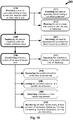
- FIG. 16 conceptually illustrates optional method of use steps for the medical apparatus, according to some embodiments of the invention.
- Fig. 16 shows the method steps in a map-like configuration, to conceptually demonstrate the optional method paths.
Landscapes
- Health & Medical Sciences (AREA)
- Life Sciences & Earth Sciences (AREA)
- Surgery (AREA)
- Engineering & Computer Science (AREA)
- Biomedical Technology (AREA)
- Animal Behavior & Ethology (AREA)
- Veterinary Medicine (AREA)
- Vascular Medicine (AREA)
- Public Health (AREA)
- Heart & Thoracic Surgery (AREA)
- General Health & Medical Sciences (AREA)
- Molecular Biology (AREA)
- Medical Informatics (AREA)
- Nuclear Medicine, Radiotherapy & Molecular Imaging (AREA)
- Reproductive Health (AREA)
- Cardiology (AREA)
- Oral & Maxillofacial Surgery (AREA)
- Transplantation (AREA)
- Media Introduction/Drainage Providing Device (AREA)
- Prostheses (AREA)
- Surgical Instruments (AREA)
Applications Claiming Priority (1)
| Application Number | Priority Date | Filing Date | Title |
|---|---|---|---|
| US201662400695P | 2016-09-28 | 2016-09-28 |
Publications (3)
| Publication Number | Publication Date |
|---|---|
| EP3300672A1 true EP3300672A1 (de) | 2018-04-04 |
| EP3300672C0 EP3300672C0 (de) | 2024-09-18 |
| EP3300672B1 EP3300672B1 (de) | 2024-09-18 |
Family
ID=59974355
Family Applications (1)
| Application Number | Title | Priority Date | Filing Date |
|---|---|---|---|
| EP17193799.8A Active EP3300672B1 (de) | 2016-09-28 | 2017-09-28 | Medizinische arterienvorrichtung |
Country Status (4)
| Country | Link |
|---|---|
| US (2) | US11771434B2 (de) |
| EP (1) | EP3300672B1 (de) |
| CN (1) | CN107865677B (de) |
| IL (1) | IL254791B (de) |
Cited By (12)
| Publication number | Priority date | Publication date | Assignee | Title |
|---|---|---|---|---|
| WO2018225059A1 (en) | 2017-06-05 | 2018-12-13 | Restore Medical Ltd | Double walled fixed length stent like apparatus and methods of use thereof |
| WO2021226014A3 (en) * | 2020-05-04 | 2021-12-16 | Vahaticor Llc | Vascular flow and pressure modulator |
| US11717425B2 (en) | 2014-07-20 | 2023-08-08 | Restore Medical Ltd. | Pulmonary artery implant apparatus and methods of use thereof |
| US11771434B2 (en) | 2016-09-28 | 2023-10-03 | Restore Medical Ltd. | Artery medical apparatus and methods of use thereof |
| US11883030B2 (en) | 2022-04-29 | 2024-01-30 | inQB8 Medical Technologies, LLC | Systems, devices, and methods for controllably and selectively occluding, restricting, and diverting flow within a patient's vasculature |
| EP4135627A4 (de) * | 2020-04-16 | 2024-04-24 | Shifamed Holdings, LLC | Einstellbare interatriale vorrichtungen sowie zugehörige systeme und verfahren |
| US11974751B2 (en) | 2022-04-29 | 2024-05-07 | inQB8 Medical Technologies, LLC | Systems, devices, and methods for controllably and selectively occluding, restricting, and diverting flow within a patient's vasculature |
| US12090290B2 (en) | 2021-03-09 | 2024-09-17 | Shifamed Holdings, Llc | Shape memory actuators for adjustable shunting systems, and associated systems and methods |
| US12151071B2 (en) | 2019-09-09 | 2024-11-26 | Shifamed Holdings, Llc | Adjustable shunts and associated systems and methods |
| WO2025109602A1 (en) * | 2023-11-24 | 2025-05-30 | Restore Medical Ltd. | Adjustable constrictor for reducing the diameter of implantable medical device |
| US12491347B2 (en) | 2020-08-25 | 2025-12-09 | Shifamed Holdings, Llc | Adjustable interatrial shunts and associated systems and methods |
| US12588910B1 (en) | 2021-11-08 | 2026-03-31 | VahatiCor, Inc. | Vascular flow restriction devices and related systems and methods |
Families Citing this family (25)
| Publication number | Priority date | Publication date | Assignee | Title |
|---|---|---|---|---|
| WO2005074367A2 (en) | 2004-02-03 | 2005-08-18 | Atria Medical Inc. | Device and method for controlling in-vivo pressure |
| US12453626B2 (en) | 2009-05-04 | 2025-10-28 | V-Wave Ltd. | Shunt for redistributing atrial blood volume |
| US9034034B2 (en) | 2010-12-22 | 2015-05-19 | V-Wave Ltd. | Devices for reducing left atrial pressure, and methods of making and using same |
| US11135054B2 (en) | 2011-07-28 | 2021-10-05 | V-Wave Ltd. | Interatrial shunts having biodegradable material, and methods of making and using same |
| US11291807B2 (en) | 2017-03-03 | 2022-04-05 | V-Wave Ltd. | Asymmetric shunt for redistributing atrial blood volume |
| US10898698B1 (en) * | 2020-05-04 | 2021-01-26 | V-Wave Ltd. | Devices with dimensions that can be reduced and increased in vivo, and methods of making and using the same |
| EP3740163A1 (de) | 2018-01-20 | 2020-11-25 | V-Wave Ltd. | Vorrichtungen und verfahren zur bereitstellung eines durchgangs zwischen herzkammern |
| WO2019221971A1 (en) | 2018-05-12 | 2019-11-21 | Venacore Inc. | Controlling rate of blood flow to right atrium |
| CN113924138B (zh) * | 2019-04-17 | 2024-04-16 | W.L.戈尔及同仁股份有限公司 | 用于急性治疗在患有心力衰竭的患者体内的流体超负荷的方法和装置 |
| CN113840572B (zh) * | 2019-04-18 | 2025-03-04 | 爱德华兹生命科学公司 | 经腔静脉文丘里管 |
| EP3958750A1 (de) * | 2019-04-22 | 2022-03-02 | Eolo Medical Inc. | Vorrichtungen zur behandlung von lungenerkrankungen mit implantierbaren ventilen |
| US12533500B2 (en) | 2019-06-18 | 2026-01-27 | Shifamed Holdings, Llc | Adjustable interatrial shunts and associated systems and methods |
| WO2021150761A1 (en) * | 2020-01-21 | 2021-07-29 | Shifamed Holdings, Llc | Interatrial shunts with anchoring mechanisms and associated systems and methods |
| US12599756B2 (en) | 2020-05-04 | 2026-04-14 | V-Wave Ltd. | Devices with dimensions that can be reduced and increased In Vivo |
| GB202007488D0 (en) * | 2020-05-20 | 2020-07-01 | Oxford Endovascular Ltd | An expandable tube for deployment within a blood vessel |
| US11234702B1 (en) | 2020-11-13 | 2022-02-01 | V-Wave Ltd. | Interatrial shunt having physiologic sensor |
| US20220257395A1 (en) * | 2021-02-15 | 2022-08-18 | John I. Shipp | Stent |
| EP4304524A4 (de) * | 2021-03-12 | 2025-01-22 | Cardio-Renal Solutions, Inc. | Vorrichtung und verfahren für okklusion mit variablem blutfluss |
| US20220387009A1 (en) * | 2021-06-04 | 2022-12-08 | V-Wave Ltd. | Systems and methods for temporary shunting between heart chambers |
| US20250041042A1 (en) | 2021-07-07 | 2025-02-06 | Restore Medical Ltd. | Implantable device to form a constriction within a blood vessel lumen |
| AU2023401922A1 (en) * | 2022-11-28 | 2025-06-19 | Fluidda NV | Apparatus for treating hyperinflation and associated complications in lung regions |
| WO2024196885A2 (en) * | 2023-03-17 | 2024-09-26 | Cardio-Renal Solutions, Inc. | Device and method for variable blood flow occlusion |
| AU2024363044A1 (en) | 2023-10-18 | 2026-04-16 | V-Wave Ltd. | Hybrid devices with dimensions that can be adjusted in vivo and methods of manufacturing thereof |
| CN117224277B (zh) * | 2023-11-10 | 2024-02-23 | 广东脉搏医疗科技有限公司 | 冠状窦缩窄装置及冠状窦缩窄系统 |
| USD1087344S1 (en) * | 2025-01-02 | 2025-08-05 | Pulmair Medical, Inc. | Implantable artificial bronchus |
Citations (6)
| Publication number | Priority date | Publication date | Assignee | Title |
|---|---|---|---|---|
| WO2001035861A1 (en) * | 1999-10-29 | 2001-05-25 | C.R. Bard, Inc. | Method of, and device for, installing a stent in a sleeve |
| WO2003028522A2 (en) * | 2001-03-27 | 2003-04-10 | Neovasc Medical Ltd. | Flow reducing implant |
| EP1870057A1 (de) * | 2006-06-21 | 2007-12-26 | Morel d'Arleux, Eric | Endoprothese des Stent-Typs |
| US20130073026A1 (en) * | 2011-09-20 | 2013-03-21 | Aga Medical Corporation | Device and method for treating vascular abnormalities |
| US20130261531A1 (en) * | 2012-03-30 | 2013-10-03 | Medtronic Vascular, Inc. | Arteriovenous Shunt Having a Flow Control Mechanism |
| WO2016096529A1 (en) * | 2014-12-18 | 2016-06-23 | Intellistent Ag | Stent and kit of stents for adjustable interventional reduction of blood flow |
Family Cites Families (179)
| Publication number | Priority date | Publication date | Assignee | Title |
|---|---|---|---|---|
| US3730186A (en) | 1971-03-05 | 1973-05-01 | Univ California | Adjustable implantable artery-constricting device |
| US4183102A (en) | 1977-09-08 | 1980-01-15 | Jacques Guiset | Inflatable prosthetic device for lining a body duct |
| US4425908A (en) | 1981-10-22 | 1984-01-17 | Beth Israel Hospital | Blood clot filter |
| US4546499A (en) | 1982-12-13 | 1985-10-15 | Possis Medical, Inc. | Method of supplying blood to blood receiving vessels |
| US4950276A (en) | 1988-02-25 | 1990-08-21 | Vince Dennis J | Prosthesis for banding of an artery capable of dilation by a balloon dilator |
| GB8927282D0 (en) | 1989-12-01 | 1990-01-31 | Univ Strathclyde | Vascular surgical devices |
| US5156620A (en) | 1991-02-04 | 1992-10-20 | Pigott John P | Intraluminal graft/stent and balloon catheter for insertion thereof |
| US5167628A (en) | 1991-05-02 | 1992-12-01 | Boyles Paul W | Aortic balloon catheter assembly for indirect infusion of the coronary arteries |
| FR2683449A1 (fr) | 1991-11-08 | 1993-05-14 | Cardon Alain | Endoprothese pour implantation transluminale. |
| US5332403A (en) | 1992-08-17 | 1994-07-26 | Jack Kolff | LVAD with t-shape and unidirectional valve |
| DE4334140C2 (de) | 1993-10-07 | 1996-04-18 | Angiomed Ag | Stent und Vorrichtung mit Stent |
| US5423851A (en) | 1994-03-06 | 1995-06-13 | Samuels; Shaun L. W. | Method and apparatus for affixing an endoluminal device to the walls of tubular structures within the body |
| US5683411A (en) | 1994-04-06 | 1997-11-04 | William Cook Europe A/S | Medical article for implantation into the vascular system of a patient |
| US5554185A (en) | 1994-07-18 | 1996-09-10 | Block; Peter C. | Inflatable prosthetic cardiovascular valve for percutaneous transluminal implantation of same |
| US5662711A (en) | 1995-06-07 | 1997-09-02 | Douglas; William | Flow adjustable artery shunt |
| US5554180A (en) | 1995-07-07 | 1996-09-10 | Aeroquip Corporation | Intraluminal stenting graft |
| JP2750569B2 (ja) | 1995-12-07 | 1998-05-13 | 幸夫 堀口 | 血管内装着型血流調節器及びバイパス用人工血管 |
| US6936057B1 (en) | 1997-05-19 | 2005-08-30 | Cardio Medical Solutions, Inc. (Cms) | Device and method for partially occluding blood vessels using flow-through balloon |
| US6120534A (en) * | 1997-10-29 | 2000-09-19 | Ruiz; Carlos E. | Endoluminal prosthesis having adjustable constriction |
| US7169160B1 (en) * | 1998-07-28 | 2007-01-30 | Medtronic, Inc. | Device for anchoring tubular element |
| US6406488B1 (en) | 1998-08-27 | 2002-06-18 | Heartstent Corporation | Healing transmyocardial implant |
| US6083239A (en) * | 1998-11-24 | 2000-07-04 | Embol-X, Inc. | Compliant framework and methods of use |
| EP1072282A1 (de) | 1999-07-19 | 2001-01-31 | EndoArt S.A. | Durchflusskontrollvorrichtuing |
| US6471635B1 (en) | 2000-02-10 | 2002-10-29 | Obtech Medical Ag | Anal incontinence disease treatment with controlled wireless energy supply |
| IL131863A0 (en) | 1999-09-10 | 2001-03-19 | Bruckheimer Elchanan | Intravascular device and method using it |
| US6312462B1 (en) | 1999-09-22 | 2001-11-06 | Impra, Inc. | Prosthesis for abdominal aortic aneurysm repair |
| US6585758B1 (en) | 1999-11-16 | 2003-07-01 | Scimed Life Systems, Inc. | Multi-section filamentary endoluminal stent |
| US20070043435A1 (en) | 1999-11-17 | 2007-02-22 | Jacques Seguin | Non-cylindrical prosthetic valve system for transluminal delivery |
| WO2001045486A2 (en) | 2000-02-11 | 2001-06-28 | Potencia Medical Ag | Urinary incontinence treatment apparatus |
| IL153753A0 (en) | 2002-12-30 | 2003-07-06 | Neovasc Medical Ltd | Varying-diameter vascular implant and balloon |
| US6953476B1 (en) | 2000-03-27 | 2005-10-11 | Neovasc Medical Ltd. | Device and method for treating ischemic heart disease |
| US6589030B2 (en) | 2000-06-20 | 2003-07-08 | Ntn Corporation | Magnetically levitated pump apparatus |
| US7158832B2 (en) | 2000-09-27 | 2007-01-02 | Cvrx, Inc. | Electrode designs and methods of use for cardiovascular reflex control devices |
| US8086314B1 (en) | 2000-09-27 | 2011-12-27 | Cvrx, Inc. | Devices and methods for cardiovascular reflex control |
| US6616624B1 (en) | 2000-10-30 | 2003-09-09 | Cvrx, Inc. | Systems and method for controlling renovascular perfusion |
| DE10102045B4 (de) | 2001-01-17 | 2004-02-26 | Bionethos Holding Gmbh | Medizinische Vorrichtung zur Behandlung von Gefäßen oder sonstigen Körperröhren |
| US20050182483A1 (en) | 2004-02-11 | 2005-08-18 | Cook Incorporated | Percutaneously placed prosthesis with thromboresistant valve portion |
| US20020107536A1 (en) | 2001-02-07 | 2002-08-08 | Hussein Hany M. | Device and method for preventing kidney failure |
| US6595980B1 (en) | 2001-02-23 | 2003-07-22 | Coaxia, Inc. | Devices and methods for preventing distal embolization using flow reversal by occlusion of the brachiocephalic artery |
| US6562066B1 (en) | 2001-03-02 | 2003-05-13 | Eric C. Martin | Stent for arterialization of the coronary sinus and retrograde perfusion of the myocardium |
| US8091556B2 (en) | 2001-04-20 | 2012-01-10 | V-Wave Ltd. | Methods and apparatus for reducing localized circulatory system pressure |
| WO2005074367A2 (en) | 2004-02-03 | 2005-08-18 | Atria Medical Inc. | Device and method for controlling in-vivo pressure |
| DE10121210B4 (de) | 2001-04-30 | 2005-11-17 | Universitätsklinikum Freiburg | Verankerungselement zur intraluminalen Verankerung eines Herzklappenersatzes und Verfahren zu seiner Herstellung |
| US20030032976A1 (en) | 2001-05-21 | 2003-02-13 | Boucek Mark M. | Catheter deployed partial occlusion devices and methods |
| US6533800B1 (en) | 2001-07-25 | 2003-03-18 | Coaxia, Inc. | Devices and methods for preventing distal embolization using flow reversal in arteries having collateral blood flow |
| US6638257B2 (en) | 2002-03-01 | 2003-10-28 | Aga Medical Corporation | Intravascular flow restrictor |
| AU2003234505A1 (en) | 2002-05-03 | 2003-11-17 | The General Hospital Corporation | Involuted endovascular valve and method of construction |
| EP1507492A1 (de) | 2002-05-10 | 2005-02-23 | Cordis Corporation | Verfahren zur herstellung einer medizinischen vorrichtung mit einer dünnwandigen rohrförmigen membran über einem tragrahmen |
| AU2003219503A1 (en) | 2002-08-08 | 2004-02-25 | Neovasc Medical Ltd. | Geometric flow regulator |
| WO2004014474A1 (en) | 2002-08-08 | 2004-02-19 | Neovasc Medical Ltd. | Flow reducing implant |
| FR2847155B1 (fr) | 2002-11-20 | 2005-08-05 | Younes Boudjemline | Procede de fabrication d'un implant medical a structure ajouree et implant obtenu par ce procede |
| US20040111006A1 (en) | 2002-12-17 | 2004-06-10 | Scout Medical Technologies, Llc | System and method for regulating blood pressure |
| US8372112B2 (en) | 2003-04-11 | 2013-02-12 | St. Jude Medical, Cardiology Division, Inc. | Closure devices, related delivery methods, and related methods of use |
| US7226473B2 (en) | 2003-05-23 | 2007-06-05 | Brar Balbir S | Treatment of stenotic regions |
| US7201772B2 (en) | 2003-07-08 | 2007-04-10 | Ventor Technologies, Ltd. | Fluid flow prosthetic device |
| US20050075725A1 (en) | 2003-10-02 | 2005-04-07 | Rowe Stanton J. | Implantable prosthetic valve with non-laminar flow |
| US7070616B2 (en) | 2003-10-31 | 2006-07-04 | Cordis Corporation | Implantable valvular prosthesis |
| IL158960A0 (en) | 2003-11-19 | 2004-05-12 | Neovasc Medical Ltd | Vascular implant |
| US20050165344A1 (en) | 2003-11-26 | 2005-07-28 | Dobak John D.Iii | Method and apparatus for treating heart failure |
| DE10394350B4 (de) | 2003-12-23 | 2018-05-17 | Cormove | In ein Lumen zu implantierender Teilesatz und Prothese denselben enthaltend |
| US7998220B2 (en) | 2004-02-04 | 2011-08-16 | Murphy Timothy P | Methods for treating obesity |
| EP1720584B1 (de) | 2004-03-02 | 2020-11-18 | Peter William Walsh | Gefäss- oder sackwand-behandlung und herzunterstützungsvorrichtung |
| US8777974B2 (en) | 2004-03-19 | 2014-07-15 | Aga Medical Corporation | Multi-layer braided structures for occluding vascular defects |
| US20050228434A1 (en) | 2004-03-19 | 2005-10-13 | Aga Medical Corporation | Multi-layer braided structures for occluding vascular defects |
| CA2558922A1 (en) | 2004-03-23 | 2005-10-13 | Boston Scientific Limited | Agent eluting stent and catheter |
| WO2005099374A2 (en) | 2004-04-05 | 2005-10-27 | Genesee Biomedical, Inc. | Method and apparatus for the surgical treatment of congestive heart failure |
| US7641686B2 (en) | 2004-04-23 | 2010-01-05 | Direct Flow Medical, Inc. | Percutaneous heart valve with stentless support |
| CA2828619C (en) | 2004-05-05 | 2018-09-25 | Direct Flow Medical, Inc. | Prosthetic valve with an elastic stent and a sealing structure |
| US20060122692A1 (en) | 2004-05-10 | 2006-06-08 | Ran Gilad | Stent valve and method of using same |
| US8623067B2 (en) | 2004-05-25 | 2014-01-07 | Covidien Lp | Methods and apparatus for luminal stenting |
| ITTO20050074A1 (it) | 2005-02-10 | 2006-08-11 | Sorin Biomedica Cardio Srl | Protesi valvola cardiaca |
| SE531468C2 (sv) | 2005-04-21 | 2009-04-14 | Edwards Lifesciences Ag | En anordning för styrning av blodflöde |
| WO2006131930A2 (en) | 2005-06-10 | 2006-12-14 | Sagax Inc. | Implant device particularly useful for implantation in the intravascular system for diverting emboli |
| US9642726B2 (en) | 2005-07-25 | 2017-05-09 | Vascular Dynamics, Inc. | Devices and methods for control of blood pressure |
| US7983765B1 (en) | 2005-08-19 | 2011-07-19 | Pacesetter, Inc. | Left chamber pressure sensor lead delivery system |
| BRPI0505102A (pt) | 2005-11-22 | 2007-08-07 | Renato Samy Assad | aperfeiçoamentos introduzidos em dispositivo de bandagem do tronco pulmonar |
| US20070213813A1 (en) | 2005-12-22 | 2007-09-13 | Symetis Sa | Stent-valves for valve replacement and associated methods and systems for surgery |
| US20070179590A1 (en) | 2005-12-29 | 2007-08-02 | Wenfeng Lu | Hybrid intraluminal device with varying expansion force |
| US9681948B2 (en) | 2006-01-23 | 2017-06-20 | V-Wave Ltd. | Heart anchor device |
| EP1849440A1 (de) | 2006-04-28 | 2007-10-31 | Younes Boudjemline | Vaskuläre Stents mit verschiedenen Durchmessern |
| AU2007266459B2 (en) | 2006-05-31 | 2013-01-17 | Star Bp, Inc. | Heart assist device |
| US7935144B2 (en) | 2006-10-19 | 2011-05-03 | Direct Flow Medical, Inc. | Profile reduction of valve implant |
| US20080208329A1 (en) | 2006-10-20 | 2008-08-28 | Gordon Bishop | Handle mechanism to adjust a medical device |
| US7862502B2 (en) | 2006-10-20 | 2011-01-04 | Ellipse Technologies, Inc. | Method and apparatus for adjusting a gastrointestinal restriction device |
| CA2664557C (en) | 2006-11-07 | 2015-05-26 | Corvia Medical, Inc. | Devices and methods for the treatment of heart failure |
| US8740962B2 (en) | 2006-11-07 | 2014-06-03 | Dc Devices, Inc. | Prosthesis for retrieval and deployment |
| US8105392B2 (en) | 2006-11-08 | 2012-01-31 | Boston Scientific Scimed, Inc. | Pyloric obesity valve |
| US20080140110A1 (en) | 2006-12-12 | 2008-06-12 | Spence Paul A | Implant, systems and methods for physically diverting material in blood flow away from the head |
| US20090112312A1 (en) | 2007-02-26 | 2009-04-30 | Larose Jeffrey A | Intravascular ventricular assist device |
| US8425593B2 (en) | 2007-09-26 | 2013-04-23 | St. Jude Medical, Inc. | Collapsible prosthetic heart valves |
| US9532868B2 (en) | 2007-09-28 | 2017-01-03 | St. Jude Medical, Inc. | Collapsible-expandable prosthetic heart valves with structures for clamping native tissue |
| US9848981B2 (en) | 2007-10-12 | 2017-12-26 | Mayo Foundation For Medical Education And Research | Expandable valve prosthesis with sealing mechanism |
| WO2009061389A2 (en) | 2007-11-05 | 2009-05-14 | St. Jude Medical, Inc. | Collapsible/expandable prosthetic heart valves with non-expanding stent posts and retrieval features |
| US8764772B2 (en) | 2008-02-21 | 2014-07-01 | Cook Medical Technologies Llc | Occlusion device |
| US9687242B2 (en) | 2008-04-03 | 2017-06-27 | Cook Medical Technologies Llc | Occlusion device |
| US8114147B2 (en) | 2008-06-16 | 2012-02-14 | Boston Scientific Scimed, Inc. | Continuous double layered stent for migration resistance |
| AU2009271574B2 (en) | 2008-07-15 | 2015-05-21 | St. Jude Medical, Inc. | Axially anchoring collapsible and re-expandable prosthetic heart valves for various disease states |
| DE202009018984U1 (de) | 2008-07-15 | 2015-01-29 | St. Jude Medical, Inc. | Tasche zur Verwendung in einer Herzklappenprothese |
| US9232992B2 (en) * | 2008-07-24 | 2016-01-12 | Aga Medical Corporation | Multi-layered medical device for treating a target site and associated method |
| EP2346405B1 (de) | 2008-09-26 | 2019-03-13 | Vascular Dynamics Inc. | Vorrichtungen und verfahren zur kontrolle des blutdrucks |
| US20100125288A1 (en) | 2008-11-17 | 2010-05-20 | G&L Consulting, Llc | Method and apparatus for reducing renal blood pressure |
| US20100222633A1 (en) | 2009-02-27 | 2010-09-02 | Victor Poirier | Blood pump system with controlled weaning |
| CN104739461A (zh) * | 2009-04-09 | 2015-07-01 | 心血管科技股份有限公司 | 组织封闭装置、输送装置和系统、用于其的套件和方法 |
| EP2427143B1 (de) | 2009-05-04 | 2017-08-02 | V-Wave Ltd. | Vorrichtung zur druckregelung in einer herzkammer |
| US9034034B2 (en) | 2010-12-22 | 2015-05-19 | V-Wave Ltd. | Devices for reducing left atrial pressure, and methods of making and using same |
| US8052737B2 (en) | 2009-05-05 | 2011-11-08 | Medtronic Vascular, Inc. | Implantable temporary flow restrictor device |
| JP5654013B2 (ja) | 2009-07-22 | 2015-01-14 | ザ テキサス エー アンド エム ユニヴァーシティー システムThe Texas A&M University System | 心臓病態を治療するための拡張期リコイル方法及びデバイス |
| US9439652B2 (en) | 2009-08-24 | 2016-09-13 | Qualimed Innovative Medizinprodukte Gmbh | Implantation device with handle and method of use thereof |
| WO2011025887A1 (en) | 2009-08-27 | 2011-03-03 | Boston Scientific Scimed, Inc. | Stent with variable cross section braiding filament and method for making same |
| US9603708B2 (en) | 2010-05-19 | 2017-03-28 | Dfm, Llc | Low crossing profile delivery catheter for cardiovascular prosthetic implant |
| US9987153B2 (en) | 2010-06-08 | 2018-06-05 | The Regents Of The University Of Minnesota | Vascular elastance |
| US9592008B2 (en) | 2010-07-01 | 2017-03-14 | Pulmonx Corporation | Devices and systems for lung treatment |
| US9901433B2 (en) | 2010-07-02 | 2018-02-27 | Myopowers Medical Technologies France | Medical device comprising an artificial contractile structure |
| US20130274648A1 (en) | 2010-07-26 | 2013-10-17 | The Trustees Of Columbia University In The City Of New York | Blood flow controllers and methods |
| EP2629699B1 (de) | 2010-10-21 | 2017-01-04 | Medtronic, Inc. | Mitralbioprothese mit niedrigem ventrikulären profil |
| US8632455B2 (en) | 2010-11-12 | 2014-01-21 | Ethicon Endo-Surgery, Inc. | Gastric band with asymmetrical member |
| WO2012148367A2 (en) | 2011-03-29 | 2012-11-01 | Oran Bulent | Transarterial heart assist device |
| EP2522307B1 (de) * | 2011-05-08 | 2020-09-30 | ITSO Medical AB | Vorrichtung zur Lieferung von medizinischen Geräten an eine Herzklappe |
| EP2529705B1 (de) * | 2011-06-03 | 2019-02-06 | Cook Medical Technologies LLC | Einführer zum Einsetzen eines Implantats |
| US11135054B2 (en) | 2011-07-28 | 2021-10-05 | V-Wave Ltd. | Interatrial shunts having biodegradable material, and methods of making and using same |
| US9629715B2 (en) | 2011-07-28 | 2017-04-25 | V-Wave Ltd. | Devices for reducing left atrial pressure having biodegradable constriction, and methods of making and using same |
| EP2567663A1 (de) | 2011-09-09 | 2013-03-13 | Occlutech Holding AG | Faltbare medizinische Verschlussvorrichtung, Verfahren und medizinisches System zur Abgabe eines Objekts |
| CA2852266C (en) | 2011-10-18 | 2019-11-26 | Anaxiom Corporation | Method and apparatus for treating a patient by intentionally occluding a blood vessel, including method and apparatus for inducing weight loss in a patient by intentionally occluding the celiac artery |
| US20130103162A1 (en) | 2011-10-25 | 2013-04-25 | Kieran Costello | Coated stent |
| CN102462565B (zh) | 2011-10-25 | 2014-03-26 | 张石江 | 可回收可调节血管内缩窄介入支架 |
| EP3656354B1 (de) | 2011-12-06 | 2021-02-03 | Aortic Innovations LLC | Vorrichtung für endovaskuläre aortenoperation |
| WO2013096548A1 (en) | 2011-12-23 | 2013-06-27 | Volcano Corporation | Methods and apparatus for regulating blood pressure |
| EP2809262A4 (de) * | 2012-02-02 | 2015-10-07 | Inceptus Medical LLC | Implantatvorrichtungen und verfahren für aneurysma |
| EP2822506A4 (de) | 2012-03-05 | 2015-10-28 | Univ Pennsylvania | Superabsorbierende beschichtete stents für vaskuläre reduktion und zur verankerung von ersatzklappen |
| US9649480B2 (en) | 2012-07-06 | 2017-05-16 | Corvia Medical, Inc. | Devices and methods of treating or ameliorating diastolic heart failure through pulmonary valve intervention |
| CN102764170B (zh) | 2012-07-18 | 2015-09-16 | 吕文峰 | 一种复合功能的血管内支架 |
| US9561035B2 (en) | 2012-08-03 | 2017-02-07 | Harold Carrison | Vessel flow control devices and methods |
| US9943427B2 (en) | 2012-11-06 | 2018-04-17 | Covidien Lp | Shaped occluding devices and methods of using the same |
| CN102961200B (zh) | 2012-11-30 | 2015-08-12 | 宁波健世生物科技有限公司 | 带锚定机构的肺动脉瓣膜支架 |
| US9775636B2 (en) | 2013-03-12 | 2017-10-03 | Corvia Medical, Inc. | Devices, systems, and methods for treating heart failure |
| US9339274B2 (en) | 2013-03-12 | 2016-05-17 | St. Jude Medical, Cardiology Division, Inc. | Paravalvular leak occlusion device for self-expanding heart valves |
| US20140350668A1 (en) | 2013-03-13 | 2014-11-27 | Symetis Sa | Prosthesis Seals and Methods for Sealing an Expandable Prosthesis |
| US9849006B2 (en) | 2013-03-15 | 2017-12-26 | Children's Medical Center Corporation | Methods and apparatuses for treating vessels |
| CN103202735B (zh) | 2013-04-01 | 2016-02-10 | 杭州启明医疗器械有限公司 | 肺动脉瓣膜置换装置及其支架 |
| WO2014175996A2 (en) | 2013-04-26 | 2014-10-30 | Boston Scientific Scimed, Inc. | Devices for obstructing passage of air or other contaminants into a portion of a lung and methods of use |
| CN105208943A (zh) * | 2013-05-13 | 2015-12-30 | 爱德华兹生命科学公司 | 主动脉阻断装置 |
| US9713696B2 (en) | 2013-05-21 | 2017-07-25 | V-Wave Ltd. | Apparatus and methods for delivering devices for reducing left atrial pressure |
| US9375301B2 (en) | 2013-06-06 | 2016-06-28 | Coloplast A/S | Artificial urinary sphincter having a multi-compartment cuff |
| US11291452B2 (en) | 2013-06-26 | 2022-04-05 | W. L. Gore & Associates, Inc. | Medical device deployment system |
| US10010328B2 (en) | 2013-07-31 | 2018-07-03 | NeuVT Limited | Endovascular occlusion device with hemodynamically enhanced sealing and anchoring |
| US9622863B2 (en) | 2013-11-22 | 2017-04-18 | Edwards Lifesciences Corporation | Aortic insufficiency repair device and method |
| US9750603B2 (en) | 2014-01-27 | 2017-09-05 | Medtronic Vascular Galway | Stented prosthetic heart valve with variable stiffness and methods of use |
| US10675450B2 (en) | 2014-03-12 | 2020-06-09 | Corvia Medical, Inc. | Devices and methods for treating heart failure |
| US9668861B2 (en) | 2014-03-15 | 2017-06-06 | Rex Medical, L.P. | Vascular device for treating venous valve insufficiency |
| US10667931B2 (en) | 2014-07-20 | 2020-06-02 | Restore Medical Ltd. | Pulmonary artery implant apparatus and methods of use thereof |
| CN204106100U (zh) | 2014-09-15 | 2015-01-21 | 北京泰杰伟业科技有限公司 | 可用于血管瘤体栓塞治疗的弹簧圈系统 |
| US9693860B2 (en) | 2014-12-01 | 2017-07-04 | Medtronic, Inc. | Segmented transcatheter valve prosthesis having an unsupported valve segment |
| GB2534194B (en) | 2015-01-16 | 2017-02-08 | Cook Medical Technologies Llc | Cone Expanding Collapsible Medical Device |
| US10568731B2 (en) | 2015-02-12 | 2020-02-25 | Hemodynamx-Technologies Ltd. | Aortic implant method |
| US20160296317A1 (en) | 2015-04-09 | 2016-10-13 | Oregon Health & Science University | Reducing stent for a transjugular intrahepatic portosystemic shunt |
| US20170042551A1 (en) | 2015-08-13 | 2017-02-16 | The Brain Protection Company PTY LTD | Implantable damping devices for treating dementia and associated systems and methods of use |
| US20190307459A1 (en) | 2015-08-13 | 2019-10-10 | The Brain Protection Company PTY LTD | Implantable damping devices for treating dementia and associated systems and methods of use |
| US9393384B1 (en) | 2015-08-17 | 2016-07-19 | Tufts Medical Center, Inc. | Systems and methods for treating acute and chronic heart failure |
| US10568634B2 (en) | 2016-02-01 | 2020-02-25 | Vivonics, Inc. | Transcatheter device and minimally invasive method for constricting and adjusting blood flow through a blood vessel |
| EP3243451B1 (de) | 2016-05-12 | 2020-08-05 | IntelliStent AG | Vorrichtung zur einstellbaren einschränkung von intravaskulärem strom |
| US10835394B2 (en) | 2016-05-31 | 2020-11-17 | V-Wave, Ltd. | Systems and methods for making encapsulated hourglass shaped stents |
| US20170340460A1 (en) | 2016-05-31 | 2017-11-30 | V-Wave Ltd. | Systems and methods for making encapsulated hourglass shaped stents |
| US11224503B2 (en) | 2016-08-12 | 2022-01-18 | Hemodynamx-Techologies Ltd. | Aortic implant |
| US11771434B2 (en) | 2016-09-28 | 2023-10-03 | Restore Medical Ltd. | Artery medical apparatus and methods of use thereof |
| US11135050B2 (en) | 2016-10-04 | 2021-10-05 | Boston Scientific Scimed, Inc. | Stent including anchoring members |
| US11529224B2 (en) | 2016-10-05 | 2022-12-20 | Pulmonx Corporation | High resistance implanted bronchial isolation devices and methods |
| IL271184B2 (en) | 2017-06-05 | 2024-04-01 | Restore Medical Ltd | Fixed-length, double-walled stent-like device and methods of using the same |
| GB2598251B (en) | 2017-06-28 | 2022-05-25 | Cook Medical Technologies Llc | Implantable medical device including valve member |
| CN111587097B (zh) | 2017-11-15 | 2023-12-08 | 赫默丹奈科斯科技有限公司 | 主动脉压力损失降低设备和方法 |
| US20210161643A1 (en) | 2018-07-23 | 2021-06-03 | Eolo Medical Inc. | Methods and devices for the treatment of pulmonary disorders with implantable valves |
| EP3826585B1 (de) | 2018-07-24 | 2025-10-01 | W. L. Gore & Associates, Inc. | Flussbegrenzendes stenttransplantat |
| EP3883482B1 (de) | 2018-11-19 | 2023-07-19 | Pulmair Medical, Inc. | Implantierbare künstliche bronchien |
| EP3886760A1 (de) | 2018-11-26 | 2021-10-06 | Nephronyx Ltd. | Strömungsmodifikationsvorrichtungen in körperlumen |
| CN121714327A (zh) | 2018-12-04 | 2026-03-24 | 脑部保护私人有限公司 | 包括用于治疗病症的可植入阻尼装置和治疗剂的组合疗法及相关系统和使用方法 |
| CN113891686B (zh) | 2019-01-23 | 2024-12-27 | 冲击波医疗公司 | 具有覆盖物的流改变装置 |
| WO2020234787A1 (en) | 2019-05-21 | 2020-11-26 | Hemodynamx-Technologies Ltd | Aortic pressure loss reduction apparatus and methods |
| WO2021113670A1 (en) | 2019-12-05 | 2021-06-10 | Shifamed Holdings, Llc | Implantable shunt systems and methods |
| US12533249B2 (en) | 2020-05-28 | 2026-01-27 | Nephronyx Ltd. | Acute and chronic devices for modifying flow in body lumens and methods of use thereof |
| US20250041042A1 (en) | 2021-07-07 | 2025-02-06 | Restore Medical Ltd. | Implantable device to form a constriction within a blood vessel lumen |
-
2017
- 2017-09-27 US US15/716,667 patent/US11771434B2/en active Active
- 2017-09-28 CN CN201710896715.3A patent/CN107865677B/zh active Active
- 2017-09-28 IL IL254791A patent/IL254791B/en unknown
- 2017-09-28 EP EP17193799.8A patent/EP3300672B1/de active Active
-
2020
- 2020-08-04 US US16/984,166 patent/US11890017B2/en active Active
Patent Citations (6)
| Publication number | Priority date | Publication date | Assignee | Title |
|---|---|---|---|---|
| WO2001035861A1 (en) * | 1999-10-29 | 2001-05-25 | C.R. Bard, Inc. | Method of, and device for, installing a stent in a sleeve |
| WO2003028522A2 (en) * | 2001-03-27 | 2003-04-10 | Neovasc Medical Ltd. | Flow reducing implant |
| EP1870057A1 (de) * | 2006-06-21 | 2007-12-26 | Morel d'Arleux, Eric | Endoprothese des Stent-Typs |
| US20130073026A1 (en) * | 2011-09-20 | 2013-03-21 | Aga Medical Corporation | Device and method for treating vascular abnormalities |
| US20130261531A1 (en) * | 2012-03-30 | 2013-10-03 | Medtronic Vascular, Inc. | Arteriovenous Shunt Having a Flow Control Mechanism |
| WO2016096529A1 (en) * | 2014-12-18 | 2016-06-23 | Intellistent Ag | Stent and kit of stents for adjustable interventional reduction of blood flow |
Cited By (18)
| Publication number | Priority date | Publication date | Assignee | Title |
|---|---|---|---|---|
| US11717425B2 (en) | 2014-07-20 | 2023-08-08 | Restore Medical Ltd. | Pulmonary artery implant apparatus and methods of use thereof |
| US12138185B2 (en) | 2014-07-20 | 2024-11-12 | Restore Medical Ltd. | Pulmonary artery implant apparatus and methods of use thereof |
| US11890017B2 (en) | 2016-09-28 | 2024-02-06 | Restore Medical Ltd. | Artery medical apparatus and methods of use thereof |
| US11771434B2 (en) | 2016-09-28 | 2023-10-03 | Restore Medical Ltd. | Artery medical apparatus and methods of use thereof |
| EP3624704A4 (de) * | 2017-06-05 | 2021-03-10 | Restore Medical Ltd | Doppelwandige stentartige vorrichtung mit fester länge und verfahren zur verwendung davon |
| WO2018225059A1 (en) | 2017-06-05 | 2018-12-13 | Restore Medical Ltd | Double walled fixed length stent like apparatus and methods of use thereof |
| US11364132B2 (en) | 2017-06-05 | 2022-06-21 | Restore Medical Ltd. | Double walled fixed length stent like apparatus and methods of use thereof |
| US12151071B2 (en) | 2019-09-09 | 2024-11-26 | Shifamed Holdings, Llc | Adjustable shunts and associated systems and methods |
| US12343487B2 (en) | 2019-09-09 | 2025-07-01 | Shifamed Holdings, Llc | Adjustable shunts and associated systems and methods |
| EP4135627A4 (de) * | 2020-04-16 | 2024-04-24 | Shifamed Holdings, LLC | Einstellbare interatriale vorrichtungen sowie zugehörige systeme und verfahren |
| WO2021226014A3 (en) * | 2020-05-04 | 2021-12-16 | Vahaticor Llc | Vascular flow and pressure modulator |
| US12491347B2 (en) | 2020-08-25 | 2025-12-09 | Shifamed Holdings, Llc | Adjustable interatrial shunts and associated systems and methods |
| US12090290B2 (en) | 2021-03-09 | 2024-09-17 | Shifamed Holdings, Llc | Shape memory actuators for adjustable shunting systems, and associated systems and methods |
| US12588910B1 (en) | 2021-11-08 | 2026-03-31 | VahatiCor, Inc. | Vascular flow restriction devices and related systems and methods |
| US11974751B2 (en) | 2022-04-29 | 2024-05-07 | inQB8 Medical Technologies, LLC | Systems, devices, and methods for controllably and selectively occluding, restricting, and diverting flow within a patient's vasculature |
| US11883030B2 (en) | 2022-04-29 | 2024-01-30 | inQB8 Medical Technologies, LLC | Systems, devices, and methods for controllably and selectively occluding, restricting, and diverting flow within a patient's vasculature |
| US12569251B2 (en) | 2022-04-29 | 2026-03-10 | Relief Cardiovascular, Inc. | Systems, devices, and methods for controllably and selectively occluding, restricting, and diverting flow within a patient's vasculature |
| WO2025109602A1 (en) * | 2023-11-24 | 2025-05-30 | Restore Medical Ltd. | Adjustable constrictor for reducing the diameter of implantable medical device |
Also Published As
| Publication number | Publication date |
|---|---|
| IL254791A0 (en) | 2017-12-31 |
| US20200360024A1 (en) | 2020-11-19 |
| CN107865677A (zh) | 2018-04-03 |
| US20180085128A1 (en) | 2018-03-29 |
| US11771434B2 (en) | 2023-10-03 |
| CN107865677B (zh) | 2023-02-03 |
| US11890017B2 (en) | 2024-02-06 |
| EP3300672C0 (de) | 2024-09-18 |
| EP3300672B1 (de) | 2024-09-18 |
| IL254791B (en) | 2022-01-01 |
Similar Documents
| Publication | Publication Date | Title |
|---|---|---|
| US11890017B2 (en) | Artery medical apparatus and methods of use thereof | |
| US20230128309A1 (en) | Vascular implant | |
| US12171651B2 (en) | System and method to radially constrict a stent graft | |
| US11364132B2 (en) | Double walled fixed length stent like apparatus and methods of use thereof | |
| EP3932373B1 (de) | Ausgabesystem zur radialen einschränkung eines stentimplantats | |
| US5713907A (en) | Apparatus and method for dilating a lumen and for inserting an intraluminal graft | |
| US5749918A (en) | Intraluminal graft and method for inserting the same | |
| US20190269498A1 (en) | Constrainable Stent Graft, Delivery System and Methods of Use | |
| CN115023200A (zh) | 用于将植入式医疗器械固定在输送系统上的方法和组件 | |
| JP7609796B2 (ja) | 大動脈解離インプラント | |
| JP2007535335A (ja) | 弁輪の縮小システム |
Legal Events
| Date | Code | Title | Description |
|---|---|---|---|
| PUAI | Public reference made under article 153(3) epc to a published international application that has entered the european phase |
Free format text: ORIGINAL CODE: 0009012 |
|
| STAA | Information on the status of an ep patent application or granted ep patent |
Free format text: STATUS: THE APPLICATION HAS BEEN PUBLISHED |
|
| AK | Designated contracting states |
Kind code of ref document: A1 Designated state(s): AL AT BE BG CH CY CZ DE DK EE ES FI FR GB GR HR HU IE IS IT LI LT LU LV MC MK MT NL NO PL PT RO RS SE SI SK SM TR |
|
| AX | Request for extension of the european patent |
Extension state: BA ME |
|
| STAA | Information on the status of an ep patent application or granted ep patent |
Free format text: STATUS: REQUEST FOR EXAMINATION WAS MADE |
|
| 17P | Request for examination filed |
Effective date: 20180928 |
|
| RBV | Designated contracting states (corrected) |
Designated state(s): AL AT BE BG CH CY CZ DE DK EE ES FI FR GB GR HR HU IE IS IT LI LT LU LV MC MK MT NL NO PL PT RO RS SE SI SK SM TR |
|
| STAA | Information on the status of an ep patent application or granted ep patent |
Free format text: STATUS: EXAMINATION IS IN PROGRESS |
|
| 17Q | First examination report despatched |
Effective date: 20200103 |
|
| RAP3 | Party data changed (applicant data changed or rights of an application transferred) |
Owner name: RESTORE MEDICAL LTD |
|
| GRAP | Despatch of communication of intention to grant a patent |
Free format text: ORIGINAL CODE: EPIDOSNIGR1 |
|
| STAA | Information on the status of an ep patent application or granted ep patent |
Free format text: STATUS: GRANT OF PATENT IS INTENDED |
|
| RIC1 | Information provided on ipc code assigned before grant |
Ipc: A61F 2/06 20130101ALN20240306BHEP Ipc: A61F 2/90 20130101ALI20240306BHEP Ipc: A61F 2/07 20130101ALI20240306BHEP Ipc: A61B 17/12 20060101AFI20240306BHEP |
|
| RIC1 | Information provided on ipc code assigned before grant |
Ipc: A61F 2/06 20130101ALN20240313BHEP Ipc: A61F 2/90 20130101ALI20240313BHEP Ipc: A61F 2/07 20130101ALI20240313BHEP Ipc: A61B 17/12 20060101AFI20240313BHEP |
|
| INTG | Intention to grant announced |
Effective date: 20240327 |
|
| GRAJ | Information related to disapproval of communication of intention to grant by the applicant or resumption of examination proceedings by the epo deleted |
Free format text: ORIGINAL CODE: EPIDOSDIGR1 |
|
| STAA | Information on the status of an ep patent application or granted ep patent |
Free format text: STATUS: EXAMINATION IS IN PROGRESS |
|
| GRAP | Despatch of communication of intention to grant a patent |
Free format text: ORIGINAL CODE: EPIDOSNIGR1 |
|
| STAA | Information on the status of an ep patent application or granted ep patent |
Free format text: STATUS: GRANT OF PATENT IS INTENDED |
|
| INTC | Intention to grant announced (deleted) | ||
| GRAS | Grant fee paid |
Free format text: ORIGINAL CODE: EPIDOSNIGR3 |
|
| INTG | Intention to grant announced |
Effective date: 20240716 |
|
| RIC1 | Information provided on ipc code assigned before grant |
Ipc: A61F 2/06 20130101ALN20240709BHEP Ipc: A61F 2/90 20130101ALI20240709BHEP Ipc: A61F 2/07 20130101ALI20240709BHEP Ipc: A61B 17/12 20060101AFI20240709BHEP |
|
| GRAA | (expected) grant |
Free format text: ORIGINAL CODE: 0009210 |
|
| STAA | Information on the status of an ep patent application or granted ep patent |
Free format text: STATUS: THE PATENT HAS BEEN GRANTED |
|
| RIN1 | Information on inventor provided before grant (corrected) |
Inventor name: FRIESEM, BEN ADAM Inventor name: BELLOMO, STEPHEN F. |
|
| AK | Designated contracting states |
Kind code of ref document: B1 Designated state(s): AL AT BE BG CH CY CZ DE DK EE ES FI FR GB GR HR HU IE IS IT LI LT LU LV MC MK MT NL NO PL PT RO RS SE SI SK SM TR |
|
| REG | Reference to a national code |
Ref country code: GB Ref legal event code: FG4D |
|
| REG | Reference to a national code |
Ref country code: CH Ref legal event code: EP |
|
| REG | Reference to a national code |
Ref country code: IE Ref legal event code: FG4D |
|
| REG | Reference to a national code |
Ref country code: DE Ref legal event code: R096 Ref document number: 602017084890 Country of ref document: DE |
|
| U01 | Request for unitary effect filed |
Effective date: 20241010 |
|
| U07 | Unitary effect registered |
Designated state(s): AT BE BG DE DK EE FI FR IT LT LU LV MT NL PT RO SE SI Effective date: 20241029 |
|
| U20 | Renewal fee for the european patent with unitary effect paid |
Year of fee payment: 8 Effective date: 20241126 |
|
| PG25 | Lapsed in a contracting state [announced via postgrant information from national office to epo] |
Ref country code: NO Free format text: LAPSE BECAUSE OF FAILURE TO SUBMIT A TRANSLATION OF THE DESCRIPTION OR TO PAY THE FEE WITHIN THE PRESCRIBED TIME-LIMIT Effective date: 20241218 |
|
| PG25 | Lapsed in a contracting state [announced via postgrant information from national office to epo] |
Ref country code: GR Free format text: LAPSE BECAUSE OF FAILURE TO SUBMIT A TRANSLATION OF THE DESCRIPTION OR TO PAY THE FEE WITHIN THE PRESCRIBED TIME-LIMIT Effective date: 20241219 |
|
| PG25 | Lapsed in a contracting state [announced via postgrant information from national office to epo] |
Ref country code: HR Free format text: LAPSE BECAUSE OF FAILURE TO SUBMIT A TRANSLATION OF THE DESCRIPTION OR TO PAY THE FEE WITHIN THE PRESCRIBED TIME-LIMIT Effective date: 20240918 |
|
| PG25 | Lapsed in a contracting state [announced via postgrant information from national office to epo] |
Ref country code: RS Free format text: LAPSE BECAUSE OF FAILURE TO SUBMIT A TRANSLATION OF THE DESCRIPTION OR TO PAY THE FEE WITHIN THE PRESCRIBED TIME-LIMIT Effective date: 20241218 |
|
| PG25 | Lapsed in a contracting state [announced via postgrant information from national office to epo] |
Ref country code: RS Free format text: LAPSE BECAUSE OF FAILURE TO SUBMIT A TRANSLATION OF THE DESCRIPTION OR TO PAY THE FEE WITHIN THE PRESCRIBED TIME-LIMIT Effective date: 20241218 Ref country code: NO Free format text: LAPSE BECAUSE OF FAILURE TO SUBMIT A TRANSLATION OF THE DESCRIPTION OR TO PAY THE FEE WITHIN THE PRESCRIBED TIME-LIMIT Effective date: 20241218 Ref country code: HR Free format text: LAPSE BECAUSE OF FAILURE TO SUBMIT A TRANSLATION OF THE DESCRIPTION OR TO PAY THE FEE WITHIN THE PRESCRIBED TIME-LIMIT Effective date: 20240918 Ref country code: GR Free format text: LAPSE BECAUSE OF FAILURE TO SUBMIT A TRANSLATION OF THE DESCRIPTION OR TO PAY THE FEE WITHIN THE PRESCRIBED TIME-LIMIT Effective date: 20241219 |
|
| PG25 | Lapsed in a contracting state [announced via postgrant information from national office to epo] |
Ref country code: IS Free format text: LAPSE BECAUSE OF FAILURE TO SUBMIT A TRANSLATION OF THE DESCRIPTION OR TO PAY THE FEE WITHIN THE PRESCRIBED TIME-LIMIT Effective date: 20250118 |
|
| PG25 | Lapsed in a contracting state [announced via postgrant information from national office to epo] |
Ref country code: SM Free format text: LAPSE BECAUSE OF FAILURE TO SUBMIT A TRANSLATION OF THE DESCRIPTION OR TO PAY THE FEE WITHIN THE PRESCRIBED TIME-LIMIT Effective date: 20240918 |
|
| PG25 | Lapsed in a contracting state [announced via postgrant information from national office to epo] |
Ref country code: ES Free format text: LAPSE BECAUSE OF FAILURE TO SUBMIT A TRANSLATION OF THE DESCRIPTION OR TO PAY THE FEE WITHIN THE PRESCRIBED TIME-LIMIT Effective date: 20240918 |
|
| PG25 | Lapsed in a contracting state [announced via postgrant information from national office to epo] |
Ref country code: PL Free format text: LAPSE BECAUSE OF FAILURE TO SUBMIT A TRANSLATION OF THE DESCRIPTION OR TO PAY THE FEE WITHIN THE PRESCRIBED TIME-LIMIT Effective date: 20240918 Ref country code: CZ Free format text: LAPSE BECAUSE OF FAILURE TO SUBMIT A TRANSLATION OF THE DESCRIPTION OR TO PAY THE FEE WITHIN THE PRESCRIBED TIME-LIMIT Effective date: 20240918 |
|
| PG25 | Lapsed in a contracting state [announced via postgrant information from national office to epo] |
Ref country code: SK Free format text: LAPSE BECAUSE OF FAILURE TO SUBMIT A TRANSLATION OF THE DESCRIPTION OR TO PAY THE FEE WITHIN THE PRESCRIBED TIME-LIMIT Effective date: 20240918 |
|
| REG | Reference to a national code |
Ref country code: CH Ref legal event code: PL |
|
| PG25 | Lapsed in a contracting state [announced via postgrant information from national office to epo] |
Ref country code: MC Free format text: LAPSE BECAUSE OF FAILURE TO SUBMIT A TRANSLATION OF THE DESCRIPTION OR TO PAY THE FEE WITHIN THE PRESCRIBED TIME-LIMIT Effective date: 20240918 |
|
| PG25 | Lapsed in a contracting state [announced via postgrant information from national office to epo] |
Ref country code: CH Free format text: LAPSE BECAUSE OF NON-PAYMENT OF DUE FEES Effective date: 20240930 |
|
| PLBE | No opposition filed within time limit |
Free format text: ORIGINAL CODE: 0009261 |
|
| STAA | Information on the status of an ep patent application or granted ep patent |
Free format text: STATUS: NO OPPOSITION FILED WITHIN TIME LIMIT |
|
| PG25 | Lapsed in a contracting state [announced via postgrant information from national office to epo] |
Ref country code: IE Free format text: LAPSE BECAUSE OF NON-PAYMENT OF DUE FEES Effective date: 20240928 |
|
| 26N | No opposition filed |
Effective date: 20250619 |
|
| PGFP | Annual fee paid to national office [announced via postgrant information from national office to epo] |
Ref country code: GB Payment date: 20250919 Year of fee payment: 9 |
|
| U20 | Renewal fee for the european patent with unitary effect paid |
Year of fee payment: 9 Effective date: 20250924 |
|
| PG25 | Lapsed in a contracting state [announced via postgrant information from national office to epo] |
Ref country code: CY Free format text: LAPSE BECAUSE OF FAILURE TO SUBMIT A TRANSLATION OF THE DESCRIPTION OR TO PAY THE FEE WITHIN THE PRESCRIBED TIME-LIMIT; INVALID AB INITIO Effective date: 20170928 |
|
| PG25 | Lapsed in a contracting state [announced via postgrant information from national office to epo] |
Ref country code: HU Free format text: LAPSE BECAUSE OF FAILURE TO SUBMIT A TRANSLATION OF THE DESCRIPTION OR TO PAY THE FEE WITHIN THE PRESCRIBED TIME-LIMIT; INVALID AB INITIO Effective date: 20170928 |