EP3307169B1 - Echtzeit-kollimation und roi-filterpositionierung in der röntgenbildgebung durch die automatische erkennung der interessenspunkte - Google Patents
Echtzeit-kollimation und roi-filterpositionierung in der röntgenbildgebung durch die automatische erkennung der interessenspunkte Download PDFInfo
- Publication number
- EP3307169B1 EP3307169B1 EP15730033.6A EP15730033A EP3307169B1 EP 3307169 B1 EP3307169 B1 EP 3307169B1 EP 15730033 A EP15730033 A EP 15730033A EP 3307169 B1 EP3307169 B1 EP 3307169B1
- Authority
- EP
- European Patent Office
- Prior art keywords
- landmark
- image
- procedure
- landmarks
- roi
- Prior art date
- Legal status (The legal status is an assumption and is not a legal conclusion. Google has not performed a legal analysis and makes no representation as to the accuracy of the status listed.)
- Active
Links
Images



Classifications
-
- A—HUMAN NECESSITIES
- A61—MEDICAL OR VETERINARY SCIENCE; HYGIENE
- A61B—DIAGNOSIS; SURGERY; IDENTIFICATION
- A61B6/00—Apparatus or devices for radiation diagnosis; Apparatus or devices for radiation diagnosis combined with radiation therapy equipment
- A61B6/48—Diagnostic techniques
- A61B6/486—Diagnostic techniques involving generating temporal series of image data
- A61B6/487—Diagnostic techniques involving generating temporal series of image data involving fluoroscopy
-
- A—HUMAN NECESSITIES
- A61—MEDICAL OR VETERINARY SCIENCE; HYGIENE
- A61B—DIAGNOSIS; SURGERY; IDENTIFICATION
- A61B6/00—Apparatus or devices for radiation diagnosis; Apparatus or devices for radiation diagnosis combined with radiation therapy equipment
- A61B6/06—Diaphragms
-
- A—HUMAN NECESSITIES
- A61—MEDICAL OR VETERINARY SCIENCE; HYGIENE
- A61B—DIAGNOSIS; SURGERY; IDENTIFICATION
- A61B6/00—Apparatus or devices for radiation diagnosis; Apparatus or devices for radiation diagnosis combined with radiation therapy equipment
- A61B6/12—Arrangements for detecting or locating foreign bodies
-
- A—HUMAN NECESSITIES
- A61—MEDICAL OR VETERINARY SCIENCE; HYGIENE
- A61B—DIAGNOSIS; SURGERY; IDENTIFICATION
- A61B6/00—Apparatus or devices for radiation diagnosis; Apparatus or devices for radiation diagnosis combined with radiation therapy equipment
- A61B6/46—Arrangements for interfacing with the operator or the patient
- A61B6/467—Arrangements for interfacing with the operator or the patient characterised by special input means
- A61B6/469—Arrangements for interfacing with the operator or the patient characterised by special input means for selecting a region of interest [ROI]
-
- A—HUMAN NECESSITIES
- A61—MEDICAL OR VETERINARY SCIENCE; HYGIENE
- A61B—DIAGNOSIS; SURGERY; IDENTIFICATION
- A61B6/00—Apparatus or devices for radiation diagnosis; Apparatus or devices for radiation diagnosis combined with radiation therapy equipment
- A61B6/50—Apparatus or devices for radiation diagnosis; Apparatus or devices for radiation diagnosis combined with radiation therapy equipment specially adapted for specific body parts; specially adapted for specific clinical applications
- A61B6/504—Apparatus or devices for radiation diagnosis; Apparatus or devices for radiation diagnosis combined with radiation therapy equipment specially adapted for specific body parts; specially adapted for specific clinical applications for diagnosis of blood vessels, e.g. by angiography
-
- A—HUMAN NECESSITIES
- A61—MEDICAL OR VETERINARY SCIENCE; HYGIENE
- A61B—DIAGNOSIS; SURGERY; IDENTIFICATION
- A61B6/00—Apparatus or devices for radiation diagnosis; Apparatus or devices for radiation diagnosis combined with radiation therapy equipment
- A61B6/52—Devices using data or image processing specially adapted for radiation diagnosis
- A61B6/5211—Devices using data or image processing specially adapted for radiation diagnosis involving processing of medical diagnostic data
-
- A—HUMAN NECESSITIES
- A61—MEDICAL OR VETERINARY SCIENCE; HYGIENE
- A61B—DIAGNOSIS; SURGERY; IDENTIFICATION
- A61B6/00—Apparatus or devices for radiation diagnosis; Apparatus or devices for radiation diagnosis combined with radiation therapy equipment
- A61B6/54—Control of apparatus or devices for radiation diagnosis
- A61B6/545—Control of apparatus or devices for radiation diagnosis involving automatic set-up of acquisition parameters
-
- G—PHYSICS
- G06—COMPUTING OR CALCULATING; COUNTING
- G06V—IMAGE OR VIDEO RECOGNITION OR UNDERSTANDING
- G06V10/00—Arrangements for image or video recognition or understanding
- G06V10/20—Image preprocessing
- G06V10/25—Determination of region of interest [ROI] or a volume of interest [VOI]
-
- G—PHYSICS
- G16—INFORMATION AND COMMUNICATION TECHNOLOGY [ICT] SPECIALLY ADAPTED FOR SPECIFIC APPLICATION FIELDS
- G16H—HEALTHCARE INFORMATICS, i.e. INFORMATION AND COMMUNICATION TECHNOLOGY [ICT] SPECIALLY ADAPTED FOR THE HANDLING OR PROCESSING OF MEDICAL OR HEALTHCARE DATA
- G16H50/00—ICT specially adapted for medical diagnosis, medical simulation or medical data mining; ICT specially adapted for detecting, monitoring or modelling epidemics or pandemics
- G16H50/20—ICT specially adapted for medical diagnosis, medical simulation or medical data mining; ICT specially adapted for detecting, monitoring or modelling epidemics or pandemics for computer-aided diagnosis, e.g. based on medical expert systems
-
- G—PHYSICS
- G06—COMPUTING OR CALCULATING; COUNTING
- G06V—IMAGE OR VIDEO RECOGNITION OR UNDERSTANDING
- G06V10/00—Arrangements for image or video recognition or understanding
- G06V10/20—Image preprocessing
- G06V10/24—Aligning, centring, orientation detection or correction of the image
- G06V10/245—Aligning, centring, orientation detection or correction of the image by locating a pattern; Special marks for positioning
-
- G—PHYSICS
- G06—COMPUTING OR CALCULATING; COUNTING
- G06V—IMAGE OR VIDEO RECOGNITION OR UNDERSTANDING
- G06V2201/00—Indexing scheme relating to image or video recognition or understanding
- G06V2201/03—Recognition of patterns in medical or anatomical images
- G06V2201/034—Recognition of patterns in medical or anatomical images of medical instruments
Definitions
- This disclosure is directed to non-transitory program storage devices for performing methods for real-time collimation and region-of-interest (ROI)-filter positioning in X-ray imaging in interventional procedures via automatic detection and tracking of landmarks of interest.
- ROI region-of-interest
- X-ray fluoroscopy For imaging and guidance. These interventional procedures are normally performed with computerized X-ray imaging systems.
- the system is called a monoplane system if it uses only one X-ray imaging arm and a biplane system if it uses two arms. Biplane systems provide two X-ray images which enable better 3-D imaging and real-time navigation.
- Most of X-ray imaging systems include an X-ray source, an image intensifier and a recording medium for each X-ray imaging arm.
- One concern today is to prevent scattered X-rays from reaching the image intensifier during interventional procedures because the scattered X-rays may present a health concern for medical personnel who are in the operation room performing the procedures.
- the concern can be addressed by using a collimator to collimate the X-ray beam to a region of interest for imaging the target of interest.
- the collimator is adjusted to optimally cover parts of the scene where there is no body part. However, in some interventional procedures the collimator is adjusted to cover some body parts which are not of interest to the physicians.
- the ROI-filter may be positioned so that regions surrounding the ROI are imaged with attenuated radiation, resulting in a less intense and noisier fluoroscopic image.
- contrast media is injected into a patient and is captured with the X-ray imaging device.
- Document US 2013/343631 A1 discloses a non-transitory program storage device readable by a computer tangibly embodying a program of instructions executed by the computer to perform methods steps for real time collimation and ROI filter positioning in X-ray imaging.
- Exemplary embodiments of the disclosure as described herein generally include systems for real-time collimation and ROI-filter positioning in X-ray imaging in interventional procedures.
- a non-transitory program storage device readable by a computer, tangibly embodying a program of instructions executed by the computer to perform the method steps for real-time collimation and ROI-filter positioning in X-ray imaging in interventional procedures.
- a method for real-time collimation and ROI-filter positioning in X-ray imaging in interventional procedures including acquiring an image of a region-of-interest (ROI) at a beginning of a medical intervention procedure on a subject, classifying the image based on low-level features in the image to determine a type of procedure being performed, determining a list of landmarks in the image from the type of procedure being performed, and loading a pre-trained landmark model for each landmark in the list of landmarks, where landmarks include anatomical structures of the subject and medical devices being used in the medical intervention procedure, and computing collimator settings of an X-ray imaging device from ROI filter margins and bounding boxes of the landmarks calculated using the landmark models.
- ROI region-of-interest
- the method includes presenting to an operator the computed collimator settings of the X-ray imaging device, and receiving a confirmation of the operator the computed collimator settings from the operator.
- the method includes, during the medical intervention procedure, detecting that a landmark has moved into the ROI, and recomputing the ROI filter margins and the collimator settings of the X-ray imaging device.
- the landmark models are used to detect landmarks in the image.
- the landmarks include balloon markers, guiding catheters, guidewires, intravascular ultrasound transducers, vessels, ablation catheter electrodes, circumferential mapping catheter electrodes, coronary sinus catheter electrodes, calcium, and bone structures.
- the method further comprises initializing the landmark, receiving positive samples of the landmark from a user, receiving negative samples of the landmark from other regions of the image, determining an appearance of the image from an image patch that represents an appearance of the initialized landmark, and training a landmark model using the positive samples, the negative samples, and the image appearance.
- the landmark is initialized with one of a line segment, a curve, a bounding box, a circle or other similar types of structures.
- the low-level features include edges, Haar-like features, scale-invariant features, histogram of ordered gradients, and local ternary patterns (LTP).
- the image is classified by a classifier based on the type of procedure being performed, where the procedure types include an electrophysiology procedure, an intravascular ultrasound procedure, a chronic total occlusion (CTO) procedure, and a balloon angioplasty procedure.
- procedure types include an electrophysiology procedure, an intravascular ultrasound procedure, a chronic total occlusion (CTO) procedure, and a balloon angioplasty procedure.
- CTO chronic total occlusion
- a method for real-time collimation and ROI-filter positioning in X-ray imaging in interventional procedures including acquiring an image of a region-of-interest (ROI) at a beginning of a medical intervention procedure on a subject, classifying the image based on a type of procedure being performed, detecting landmarks in the image, where the landmarks being detected are determined from the type of procedure being performed, where landmarks include anatomical structures of the subject and medical devices being used in the medical intervention procedure, and computing collimator settings of an X-ray imaging device from ROI filter margins and bounding boxes of the landmarks calculated using the detected landmarks.
- ROI region-of-interest
- the method includes determining a list of landmarks from the type of procedure being performed and loading a pre-trained landmark model for each landmark in the list of landmarks, where the landmark models are used to detect landmarks in the image.
- the method further comprises initializing the landmark, receiving positive samples of the landmark from a user, receiving negative samples of the landmark from other regions of the image, determining an appearance of the image from an image patch that represents an appearance of the initialized landmark, and training a landmark model using the positive samples, the negative samples, and the image appearance.
- the method includes presenting to an operator the computed collimator settings of the X-ray imaging device, and receiving a confirmation of the operator the computed collimator settings from the operator.
- the method includes, during the medical intervention procedure, detecting that a landmark has moved into the ROI, and recomputing the ROI filter margins and the collimator settings of the X-ray imaging device.
- the image is classified using low-level features, where the low-level features include edges, Haar-like features, scale-invariant features, histogram of ordered gradients, and local ternary patterns (LTPs).
- the low-level features include edges, Haar-like features, scale-invariant features, histogram of ordered gradients, and local ternary patterns (LTPs).
- Exemplary embodiments of the disclosure as described herein generally include systems for real-time collimation and ROI-filter positioning in X-ray imaging in interventional procedures. Accordingly, while the disclosure is susceptible to various modifications and alternative forms, specific embodiments thereof are shown by way of example in the drawings and will herein be described in detail.
- the term "image” refers to multi-dimensional data composed of discrete image elements (e.g., pixels for 2-dimensional images and voxels for 3- dimensional images).
- the image may be, for example, a medical image of a subject collected by computer tomography, magnetic resonance imaging, ultrasound, or any other medical imaging system known to one of skill in the art.
- the image may also be provided from non-medical contexts, such as, for example, remote sensing systems, electron microscopy, etc.
- an image can be thought of as a function from R 3 to R or R 7 , the methods of the disclosure are not limited to such images, and can be applied to images of any dimension, e.g., a 2- dimensional picture or a 3- dimensional volume.
- the domain of the image is typically a 2- or 3-dimensional rectangular array, wherein each pixel or voxel can be addressed with reference to a set of 2 or 3 mutually orthogonal axes.
- digital and digitized as used herein will refer to images or volumes, as appropriate, in a digital or digitized format acquired via a digital acquisition system or via conversion from an analog image.
- the present disclosure can provide methods for automatically setting a collimator and an ROI-filter in an optimal way and in real time to minimize scattered X-rays by taking into account the aforementioned characteristics.
- Methods according to the disclosure can classify a scene into different categories based on the presence of medical devices or structures in the fluoroscopic images. According to the disclosure, structures of the subject and the medical devices used in the procedure are referred to as "landmarks". Based on the detected scene category, models of the corresponding landmarks can be loaded to detect the locations of the landmarks in the image and to generate the collimator and ROI-filter settings. For example, if an atrial fibrillation procedure is detected, models of the ablation catheter, the coronary sinus catheter and the circumferential mapping catheter are automatically loaded.
- the landmark is considered to be new, and a "tag and learn" method is used to allow a technician to initialize the landmark, and then to detect similar landmarks in the X-ray image.
- the detected new landmarks are added to a landmark database.
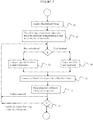
- FIG. 2 is a flowchart of an exemplary, non-limiting method for real-time collimation and ROI-filter positioning in X-ray imaging in interventional procedures via automatic detection and tracking the landmarks of interest.
- a method starts at step 21 by acquiring a fluoroscopic image.
- the image is a monoplane image if the fluoroscopic imaging system is a monoplane system or a biplane frame otherwise.
- the image is acquired at the beginning of a procedure and includes the medical devices at the time, and after the X-ray imaging device has been centered at the region of interest of the patient or subject.
- the image or scene can be classified at step 22 into different categories based on the procedure being performed, since different medical procedures employ different medical devices and the fluoroscopic images have different regions of interest in the patient body.
- the scene categorization uses low level image features, such as edges, Haar-like features, scale-invariant features, histograms of ordered gradients, local ternary patterns (LTP), etc., and employs a classifier to classify the scene.
- An exemplary, non-limiting list of interventional procedures for classifying an image includes an electrophysiology procedure, an intravascular ultrasound procedure, a chronic total occlusion (CTO) procedure, a balloon angioplasty procedure, etc.
- CTO chronic total occlusion
- pre-trained classifiers can be loaded at step 23 to detect those landmarks.
- the determination of which landmarks are present in the image can be performed manually by an operator, or automatically. In manual mode, an operator examines the image and selects a list of landmarks from a list of candidate landmarks. Alternatively, in an automatic mode, all pre-trained landmark models are loaded to detect the presence of the landmarks in the image, from which a list of landmark models is selected.
- the pre-trained landmark models are landmark specific and are learned from a large number of annotated X-ray images.
- An exemplary, non-limiting list of pre-trained models for the following landmarks is as follows:
- models of the following landmarks are selected: (1) the guiding catheter; (2) the balloon markers; and (3) the guidewire.
- the list of landmark models can be stored in a database.
- Exemplary, non-limiting algorithms according to the disclosure for detecting these three landmarks are as follows.
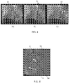
- FIGS. 4(a)-(c) illustrate the detection of electrodes on a coronary sinus catheter.
- arrows 41 indicate the electrodes;
- points 42 are on the candidate electrode locations; and
- FIG. 4(c) shows the detected candidate locations 43 after non max suppression.
- the catheter tip and electrodes can be detected as oriented points (x, y, ⁇ ), and can be parameterized by their position (x, y) and orientation ( ⁇ ).
- Marginal Space Learning can be used to first detect just the tip and electrode positions (x, y) and then at promising positions search for all orientations ( ⁇ ).
- the tip and electrode positions (x, y) can be detected using trained binary classifiers.
- Exemplary, non-limiting classifiers use about 100,000 Haar features in a centered window of size (k ⁇ k).
- PBT Probabilistic Boosting Tree
- the catheter tip is different from other electrodes in term of context and appearance and it can be detected more reliably than other electrodes.
- the detected electrode and candidate positions are then augmented with a set of discrete orientations and provided to a trained oriented point detector, and the same applies for the detected tip positions.
- Exemplary oriented point detectors use a richer feature pool, including steerable feature responses and image intensity differences relative to the query position and orientation.
- the set of detected electrodes and tips at each frame is provided to a non-maximal suppression (NMS) stage that selects the clustered detections. In each frame, at most K electrodes and F tips are selected.
- NMS non-
- a balloon marker detection includes two steps. In a first step, a PBT classifier detects the candidate marker locations, while in a second step, a pair of two candidate markers is evaluated by another PBT classifier.
- FIG. 5 is an X-ray image of balloon markers 51, a guidewire 52, a stent 53 and a guiding catheter 54.
- An exemplary, non-limiting detection algorithm according the disclosure of the guidewire includes a semantic guidewire model that contains three parts, including a catheter tip, a guidewire tip and a guidewire body. Detection of these different parts are integrated into a Bayesian formula for detection of the whole guidewire. A hierarchical and multi-resolution scheme is applied to efficiently detect the guidewires.
- Landmark detection according to the disclosure can be performed in parallel when no other computation workloads, such as visualization and GUI interaction, are required.
- a tag-and-learn method can be used at step 24 to learn the model online from user initialization.
- a flow chart of a tag-and-learn method according to the disclosure is shown in FIG. 6 .
- a new landmark can be initialized manually or semiautomatically by the operator at step 61.
- Exemplary, non-liming initializations include a line segment, a curve, a bounding box, a circle or other similar types of structures.
- examples of positive samples are provided by user initialization, negative samples are acquired from the remaining regions of the image, and an image appearance is acquired by learning a template of the initialized landmark, where a template is an image patch which represents the appearance of the landmark.
- a template is an image patch which represents the appearance of the landmark.
- the learning of the new landmark model is performed at step 63 by utilizing the positive samples, the negative samples, and the image appearance.
- the collimation is computed and the collimator settings are generated at step 25.
- Bounding boxes (B_left, B right, B_top, B_bottom) of all landmarks are computed from the landmark models, along with margins (M_left, M_right, M_top, M_bottom) of the ROI-filter. From these, the collimator settings, (C_left, C_right, C_top, C_bottom) can be generated.
- the bounding boxes and ROI-filter margins can be determined automatically based on what type of landmark is important in a given scene. For example, for a stent implantation scene, then the location of the two balloon markers are important and the images should define an ROI around this pair of balloon markers.
- Exemplary, non-limiting collimator settings are
- a system can track landmarks of interest when one or more landmarks move into the collimation area.
- the collimator and ROI filter settings are re-computed and shown to the operator for confirmation using the above steps 22-26.
- a method according to the disclosure can work automatically in real time with little or no human input except during system initialization and confirmation of the updated collimator and ROI-filter settings, however, an operator can manually set these parameters at any time.
- an embodiment of the present disclosure can be implemented in various forms of hardware, software, firmware, special purpose processes, or a combination thereof.
- an embodiment of the present disclosure can be implemented in software as an application program tangible embodied on a computer readable program storage device.
- the application program can be uploaded to, and executed by, a machine comprising any suitable architecture.
- FIG. 7 is a block diagram of an exemplary computer system for implementing a method for real-time collimation and ROI-filter positioning in X-ray imaging in interventional procedures according to an embodiment of the disclosure.
- a computer system 71 for implementing an embodiment of the present disclosure can comprise, inter alia, a central processing unit (CPU) 72, a memory 73 and an input/output (I/O) interface 74.
- the computer system 71 is generally coupled through the I/O interface 74 to a display 75 and various input devices 76 such as a mouse and a keyboard.
- the support circuits can include circuits such as cache, power supplies, clock circuits, and a communication bus.
- the memory 73 can include random access memory (RAM), read only memory (ROM), disk drive, tape drive, etc., or a combinations thereof.
- RAM random access memory
- ROM read only memory
- Embodiments of the present disclosure can be implemented as a routine 77 that is stored in memory 73 and executed by the CPU 72 to process the signal from the signal source 78.
- the computer system 71 is a general purpose computer system that becomes a specific purpose computer system when executing the routine 77 of the present disclosure.
- the computer system 71 also includes an operating system and micro instruction code.
- the various processes and functions described herein can either be part of the micro instruction code or part of the application program (or combination thereof) which is executed via the operating system.
- various other peripheral devices can be connected to the computer platform such as an additional data storage device and a printing device.
Landscapes
- Health & Medical Sciences (AREA)
- Life Sciences & Earth Sciences (AREA)
- Engineering & Computer Science (AREA)
- Medical Informatics (AREA)
- Physics & Mathematics (AREA)
- Public Health (AREA)
- Biomedical Technology (AREA)
- General Health & Medical Sciences (AREA)
- Pathology (AREA)
- Molecular Biology (AREA)
- Veterinary Medicine (AREA)
- Optics & Photonics (AREA)
- High Energy & Nuclear Physics (AREA)
- Radiology & Medical Imaging (AREA)
- Biophysics (AREA)
- Heart & Thoracic Surgery (AREA)
- Nuclear Medicine, Radiotherapy & Molecular Imaging (AREA)
- Surgery (AREA)
- Animal Behavior & Ethology (AREA)
- Oral & Maxillofacial Surgery (AREA)
- Vascular Medicine (AREA)
- Dentistry (AREA)
- Computer Vision & Pattern Recognition (AREA)
- General Physics & Mathematics (AREA)
- Multimedia (AREA)
- Theoretical Computer Science (AREA)
- Human Computer Interaction (AREA)
- Data Mining & Analysis (AREA)
- Databases & Information Systems (AREA)
- Epidemiology (AREA)
- Primary Health Care (AREA)
- Apparatus For Radiation Diagnosis (AREA)
Claims (9)
- Nicht-transitorische Programmspeichervorrichtung, die von einem Computer lesbar ist und ein Programm von Anweisungen greifbar verkörpert, die von dem Computer ausgeführt werden, wodurch der Computer veranlasst wird, ein Verfahren zur Echtzeit-Kollimation und ROI-Filter-Positionierung bei der Röntgenbildgebung in Interventionsverfahren durchzuführen, wobei das Verfahren die folgenden Schritte umfasst:Erfassen eines Bildes einer Region von Interesse (ROI) zu Beginn eines medizinischen Interventionsverfahrens an einem Subjekt;Klassifizieren des Bildes basierend auf Merkmalen niedriger Ebene im Bild, um eine Art des Verfahrens zu bestimmen, das durchgeführt wird;Bestimmen einer Liste von Referenzpunkten im Bild aus der Art des durchgeführten Verfahrens und Laden eines vortrainierten Referenzpunktmodells für jeden Referenzpunkt in der Liste der Referenzpunkte, wobei die Referenzpunkte anatomische Strukturen des Subjekts und medizinische Vorrichtungen umfassen, die bei dem medizinischen Interventionsverfahren verwendet werden; undBerechnen von Kollimatoreinstellungen einer Röntgenbildgebungsvorrichtung aus ROI-Filterrändern und Begrenzungsboxen der Referenzpunkte, die mit den Referenzpunktmodellen berechnet wurden.
- Computerlesbare Programmspeichervorrichtung nach Anspruch 1, wobei das Verfahren ferner ein Darstellen der berechneten Kollimatoreinstellungen der Röntgenbildgebungsvorrichtung für einen Bediener und ein Empfangen einer Bestätigung des Bedieners über die berechneten Kollimatoreinstellungen vom Bediener (25, 26) umfasst.
- Computerlesbare Programmspeichervorrichtung nach Anspruch 1, wobei das Verfahren ferner ein Detektieren während des medizinischen Interventionsverfahrens, ob sich ein Referenzpunkt in die ROI bewegt hat, und ein erneutes Berechnen der ROI-Filterränder und der Kollimatoreinstellungen der Röntgenbildgebungsvorrichtung umfasst.
- Computerlesbare Programmspeichervorrichtung nach Anspruch 1, wobei die Referenzpunktmodelle zum Detektieren von Referenzpunkten im Bild verwendet werden.
- Computerlesbare Programmspeichervorrichtung nach Anspruch 4, wobei die Referenzpunkte Ballonmarkierungen (51), Führungskatheter (54), Führungsdrähte (52), intravaskuläre Ultraschallwandler, Gefäße, Ablationskatheterelektroden, Katheterelektroden zur Umfangskartierung, Koronarsinuskatheterelektroden, Kalzium und Knochenstrukturen umfassen.
- Computerlesbare Programmspeichervorrichtung nach Anspruch 4, wobei, wenn kein vortrainiertes Referenzpunktmodell für einen Referenzpunkt existiert, das Verfahren ferner ein Initialisieren des Referenzpunktes (61), ein Empfangen positiver Proben des Referenzpunktes von einem Benutzer, ein Empfangen negativer Proben des Referenzpunktes von anderen Regionen des Bildes, ein Bestimmen eines Erscheinungsbildes des Bildes aus einem Bildfeld, das ein Erscheinungsbild des initialisierten Referenzpunktes (61) darstellt, und ein Trainieren eines Referenzpunktmodells unter Verwendung der positiven Proben, der negativen Proben und des Erscheinungsbildes des Bildes (62, 63) umfasst.
- Computerlesbare Programmspeichervorrichtung nach Anspruch 6, wobei der Referenzpunkt mit einem Liniensegment, einer Kurve, einer Begrenzungsbox, einem Kreis oder anderen ähnlichen Arten von Strukturen initialisiert wird (61).
- Computerlesbare Programmspeichervorrichtung nach Anspruch 1, wobei die Merkmale auf niedriger Ebene Kanten, Haar-ähnliche Merkmale, skaleninvariante Merkmale, ein Histogramm geordneter Gradienten und lokale ternäre Muster (LTP) umfassen.
- Computerlesbare Programmspeichervorrichtung nach Anspruch 1, wobei das Bild durch einen Klassifizierer basierend auf einer Art des durchgeführten Verfahrens klassifiziert wird, wobei die Arten des Verfahrens ein elektrophysiologisches Verfahren, ein intravaskuläres Ultraschallverfahren, ein chronisches Totalokklusionsverfahren (CTO) und ein Ballon-Angioplastie-Verfahren umfassen.
Priority Applications (1)
| Application Number | Priority Date | Filing Date | Title |
|---|---|---|---|
| EP19204288.5A EP3656309A1 (de) | 2015-06-09 | 2015-06-09 | Echtzeitkollimation und roi-filter-positionierung in der röntgenbildgebung über den automatischen nachweis von leitstrukturen von interesse |
Applications Claiming Priority (1)
| Application Number | Priority Date | Filing Date | Title |
|---|---|---|---|
| PCT/US2015/034792 WO2016200370A1 (en) | 2015-06-09 | 2015-06-09 | Real-time collimation and roi-filter positioning in x-ray imaging via automatic detection of the landmarks of interest |
Related Child Applications (2)
| Application Number | Title | Priority Date | Filing Date |
|---|---|---|---|
| EP19204288.5A Division-Into EP3656309A1 (de) | 2015-06-09 | 2015-06-09 | Echtzeitkollimation und roi-filter-positionierung in der röntgenbildgebung über den automatischen nachweis von leitstrukturen von interesse |
| EP19204288.5A Division EP3656309A1 (de) | 2015-06-09 | 2015-06-09 | Echtzeitkollimation und roi-filter-positionierung in der röntgenbildgebung über den automatischen nachweis von leitstrukturen von interesse |
Publications (2)
| Publication Number | Publication Date |
|---|---|
| EP3307169A1 EP3307169A1 (de) | 2018-04-18 |
| EP3307169B1 true EP3307169B1 (de) | 2020-07-29 |
Family
ID=53433322
Family Applications (2)
| Application Number | Title | Priority Date | Filing Date |
|---|---|---|---|
| EP15730033.6A Active EP3307169B1 (de) | 2015-06-09 | 2015-06-09 | Echtzeit-kollimation und roi-filterpositionierung in der röntgenbildgebung durch die automatische erkennung der interessenspunkte |
| EP19204288.5A Withdrawn EP3656309A1 (de) | 2015-06-09 | 2015-06-09 | Echtzeitkollimation und roi-filter-positionierung in der röntgenbildgebung über den automatischen nachweis von leitstrukturen von interesse |
Family Applications After (1)
| Application Number | Title | Priority Date | Filing Date |
|---|---|---|---|
| EP19204288.5A Withdrawn EP3656309A1 (de) | 2015-06-09 | 2015-06-09 | Echtzeitkollimation und roi-filter-positionierung in der röntgenbildgebung über den automatischen nachweis von leitstrukturen von interesse |
Country Status (4)
| Country | Link |
|---|---|
| US (1) | US10268915B2 (de) |
| EP (2) | EP3307169B1 (de) |
| CN (1) | CN107809955B (de) |
| WO (1) | WO2016200370A1 (de) |
Families Citing this family (15)
| Publication number | Priority date | Publication date | Assignee | Title |
|---|---|---|---|---|
| US10417763B2 (en) * | 2014-07-25 | 2019-09-17 | Samsung Electronics Co., Ltd. | Image processing apparatus, image processing method, x-ray imaging apparatus and control method thereof |
| US10342505B2 (en) | 2016-03-31 | 2019-07-09 | General Electric Company | System and method for adjusting a radiation dose during imaging of an object within a subject |
| CN108937975A (zh) | 2017-05-19 | 2018-12-07 | 上海西门子医疗器械有限公司 | X-射线曝光区域调节方法、存储介质和x-射线系统 |
| CN107871319B (zh) * | 2017-11-21 | 2021-09-17 | 上海联影医疗科技股份有限公司 | 限束器区域的检测方法、装置、x射线系统和存储介质 |
| GB2575795A (en) * | 2018-07-23 | 2020-01-29 | Medsolve Ltd | Imaging system for use in a fluoroscopy procedure |
| ES2951825T3 (es) * | 2019-02-14 | 2023-10-25 | Brainlab Ag | Ajuste automático de parámetros de formación de imágenes |
| WO2020193640A1 (en) * | 2019-03-26 | 2020-10-01 | Koninklijke Philips N.V. | Persistent guide wire identification |
| US11707255B2 (en) * | 2019-04-02 | 2023-07-25 | Siemens Medical Solutions Usa, Inc. | Image-based probe positioning |
| JP2020185145A (ja) * | 2019-05-14 | 2020-11-19 | 株式会社島津製作所 | X線撮影装置 |
| DE102020204454A1 (de) | 2019-05-14 | 2020-11-19 | Siemens Healthcare Gmbh | Überwachung einer Behandlung eines Objekts |
| EP4030996A4 (de) * | 2019-09-20 | 2023-10-25 | Canon U.S.A. Inc. | Co-registrierung und markerdetektion mit künstlicher intelligenz, einschliesslich maschinenlernen und verwendung der ergebnisse davon |
| CN113679400B (zh) * | 2020-05-18 | 2024-09-24 | 西门子(深圳)磁共振有限公司 | 介入治疗中的图像呈现方法、系统、成像系统和存储介质 |
| DE102020213035A1 (de) * | 2020-10-15 | 2022-04-21 | Siemens Healthcare Gmbh | Verfahren zur Ansteuerung eines Röntgengerätes und medizinisches System |
| DE102021214738A1 (de) | 2021-12-20 | 2023-06-22 | Siemens Healthcare Gmbh | Ansteuerverfahren für Röntgengerät und Röntgengerät |
| CN115841601A (zh) * | 2022-09-28 | 2023-03-24 | 上海奥朋医疗科技有限公司 | 介入式导管头端形态的识别判断方法、系统、设备及介质 |
Family Cites Families (23)
| Publication number | Priority date | Publication date | Assignee | Title |
|---|---|---|---|---|
| US5278887A (en) | 1992-06-29 | 1994-01-11 | Siemens Corporate Research, Inc. | Apparatus and method for reducing X-ray dosage during a fluoroscopic procedure |
| DE69529857T2 (de) * | 1994-03-25 | 2004-01-08 | Kabushiki Kaisha Toshiba, Kawasaki | Strahlentherapie-System |
| US6055295A (en) | 1998-01-29 | 2000-04-25 | Siemens Corporate Research, Inc. | Method and apparatus for automatic collimation in x-ray peripheral imaging |
| JP3513700B2 (ja) | 2000-03-29 | 2004-03-31 | 稔 植松 | 移動式放射線遮蔽装置 |
| ATE485770T1 (de) | 2003-07-30 | 2010-11-15 | Koninkl Philips Electronics Nv | Röntgeneinrichtung mit automatisch einstellbarem kollimator |
| US8423121B2 (en) | 2008-08-11 | 2013-04-16 | Siemens Aktiengesellschaft | Method and system for guidewire tracking in fluoroscopic image sequences |
| CN102202576B (zh) * | 2008-10-10 | 2015-04-08 | 皇家飞利浦电子股份有限公司 | 用于为了在微创的x射线引导的介入中减少x射线剂量而产生覆盖经分割的目标结构或病变的减小视场的、具有自动快门适应的血管造影图像采集系统和方法 |
| US8311308B2 (en) | 2009-12-10 | 2012-11-13 | Siemens Corporation | Stent viewing using a learning based classifier in medical imaging |
| US8548213B2 (en) | 2010-03-16 | 2013-10-01 | Siemens Corporation | Method and system for guiding catheter detection in fluoroscopic images |
| US8565859B2 (en) | 2010-06-29 | 2013-10-22 | Siemens Aktiengesellschaft | Method and system for image based device tracking for co-registration of angiography and intravascular ultrasound images |
| US8892186B2 (en) | 2010-09-20 | 2014-11-18 | Siemens Aktiengesellschaft | Method and system for detection and tracking of coronary sinus catheter electrodes in fluoroscopic images |
| WO2012123850A1 (en) * | 2011-03-15 | 2012-09-20 | Koninklijke Philips Electronics N.V. | Medical imaging device for providing an image representation supporting in positioning an intervention device |
| US9002436B2 (en) | 2011-09-19 | 2015-04-07 | Siemens Aktiengesellschaft | Method and system for ablation catheter and circumferential mapping catheter tracking in fluoroscopic images |
| CN103999088B (zh) * | 2011-12-14 | 2018-07-24 | 皇家飞利浦有限公司 | 用于防止高剂量c型拱几何位置的实时反馈 |
| US9700276B2 (en) | 2012-02-28 | 2017-07-11 | Siemens Healthcare Gmbh | Robust multi-object tracking using sparse appearance representation and online sparse appearance dictionary update |
| US20130272504A1 (en) * | 2012-04-16 | 2013-10-17 | Meir Deutsch | X-Ray Dose Reduction by Controlled Shutter Speed |
| JP6258938B2 (ja) * | 2012-08-27 | 2018-01-10 | コーニンクレッカ フィリップス エヌ ヴェKoninklijke Philips N.V. | 光学3dシーン検出及び解釈に基づく患者個別型及び自動x線システム調節 |
| BR112015003888A2 (pt) | 2012-08-27 | 2017-07-04 | Koninklijke Philips Nv | controlador de colimador, método de controle de um colimador em um formador de imagens de raio x para obter uma imagem de um objeto, sistema formador de imagens de raio x, elemento de programa de computador para controlar um aparelho e mídia legível por computador |
| JP2014054425A (ja) * | 2012-09-13 | 2014-03-27 | Toshiba Corp | X線診断装置 |
| US20140187923A1 (en) * | 2012-12-28 | 2014-07-03 | General Electric Company | Collimator for imaging system and method for using the same |
| US10736588B2 (en) * | 2013-04-03 | 2020-08-11 | Koninklijke Philips N.V. | Interventional x-ray |
| US9420976B2 (en) * | 2014-03-19 | 2016-08-23 | General Electric Company | Systems and methods for optimized source collimation |
| KR20160007121A (ko) * | 2014-07-11 | 2016-01-20 | 삼성전자주식회사 | 엑스선 장치 |
-
2015
- 2015-06-09 EP EP15730033.6A patent/EP3307169B1/de active Active
- 2015-06-09 CN CN201580080774.2A patent/CN107809955B/zh active Active
- 2015-06-09 US US15/574,203 patent/US10268915B2/en active Active
- 2015-06-09 EP EP19204288.5A patent/EP3656309A1/de not_active Withdrawn
- 2015-06-09 WO PCT/US2015/034792 patent/WO2016200370A1/en not_active Ceased
Non-Patent Citations (1)
| Title |
|---|
| None * |
Also Published As
| Publication number | Publication date |
|---|---|
| WO2016200370A1 (en) | 2016-12-15 |
| CN107809955B (zh) | 2021-01-19 |
| US20180129896A1 (en) | 2018-05-10 |
| CN107809955A (zh) | 2018-03-16 |
| US10268915B2 (en) | 2019-04-23 |
| EP3307169A1 (de) | 2018-04-18 |
| EP3656309A1 (de) | 2020-05-27 |
Similar Documents
| Publication | Publication Date | Title |
|---|---|---|
| EP3307169B1 (de) | Echtzeit-kollimation und roi-filterpositionierung in der röntgenbildgebung durch die automatische erkennung der interessenspunkte | |
| US9155470B2 (en) | Method and system for model based fusion on pre-operative computed tomography and intra-operative fluoroscopy using transesophageal echocardiography | |
| US9687204B2 (en) | Method and system for registration of ultrasound and physiological models to X-ray fluoroscopic images | |
| US8423121B2 (en) | Method and system for guidewire tracking in fluoroscopic image sequences | |
| US9247880B2 (en) | Image fusion for interventional guidance | |
| US10111726B2 (en) | Risk indication for surgical procedures | |
| US20150223773A1 (en) | Method and Apparatus for Image Fusion Based Planning of C-Arm Angulation for Structural Heart Disease | |
| EP3224804B1 (de) | Vorrichtung zur bestimmung von objektpositionen in einem projektionsbild | |
| JP2017185007A (ja) | 放射線撮影装置、放射線画像の対象物検出プログラムおよび放射線画像における対象物検出方法 | |
| US9730609B2 (en) | Method and system for aortic valve calcification evaluation | |
| US20170116751A1 (en) | System and Method For Dynamic Device Tracking Using Medical Imaging Systems | |
| US9186087B2 (en) | Combined cardiac and respiratory motion compensation for atrial fibrillation ablation procedures | |
| US20210174523A1 (en) | Method for registration of image data and for provision of corresponding trained facilities, apparatus for doing so and corresponding computer program product | |
| US20240315779A1 (en) | Systems and apparatuses for for navigation and procedural guidance of laser leaflet resection under intracardiac echocardiography | |
| US9681856B2 (en) | Image fusion for interventional guidance | |
| Ma et al. | Real-time registration of 3D echo to x-ray fluoroscopy based on cascading classifiers and image registration | |
| KR20240128966A (ko) | 타겟 영역의 위치를 확정하는 방법, 전자장비 및 매체 | |
| EP2956065B1 (de) | Vorrichtung zur auf einer bildfusion beruhenden planung einer c-arm-abwinkelung für strukturelle herzkrankheit | |
| US20240245466A1 (en) | Identification of guidewire position during a procedure | |
| Wu et al. | Real-Time catheter extraction from 2d x-ray fluoroscopic and 3d echocardiographic images for cardiac interventions | |
| Chen et al. | Deep learning based non-rigid device tracking in ultrasound image | |
| JP7303144B2 (ja) | 放射線撮影装置、放射線画像の対象物検出プログラムおよび放射線画像における対象物検出方法 | |
| Sun et al. | Towards automated ultrasound transesophageal echocardiography and x-ray fluoroscopy fusion using an image-based co-registration method | |
| Ma et al. | Attention on the Wires (AttWire): A Foundation Model for Detecting Devices and Catheters in X-ray Fluoroscopic Images | |
| Zheng et al. | Part-based object detection and segmentation |
Legal Events
| Date | Code | Title | Description |
|---|---|---|---|
| STAA | Information on the status of an ep patent application or granted ep patent |
Free format text: STATUS: THE INTERNATIONAL PUBLICATION HAS BEEN MADE |
|
| PUAI | Public reference made under article 153(3) epc to a published international application that has entered the european phase |
Free format text: ORIGINAL CODE: 0009012 |
|
| STAA | Information on the status of an ep patent application or granted ep patent |
Free format text: STATUS: REQUEST FOR EXAMINATION WAS MADE |
|
| 17P | Request for examination filed |
Effective date: 20171117 |
|
| AK | Designated contracting states |
Kind code of ref document: A1 Designated state(s): AL AT BE BG CH CY CZ DE DK EE ES FI FR GB GR HR HU IE IS IT LI LT LU LV MC MK MT NL NO PL PT RO RS SE SI SK SM TR |
|
| AX | Request for extension of the european patent |
Extension state: BA ME |
|
| DAV | Request for validation of the european patent (deleted) | ||
| DAX | Request for extension of the european patent (deleted) | ||
| RIN1 | Information on inventor provided before grant (corrected) |
Inventor name: OSTERMEIER, MARTIN Inventor name: CHEN, TERRENCE Inventor name: COMANICIU, DORIN Inventor name: WU, WEN Inventor name: NEKOVAR, ANTON |
|
| STAA | Information on the status of an ep patent application or granted ep patent |
Free format text: STATUS: EXAMINATION IS IN PROGRESS |
|
| 17Q | First examination report despatched |
Effective date: 20190626 |
|
| GRAP | Despatch of communication of intention to grant a patent |
Free format text: ORIGINAL CODE: EPIDOSNIGR1 |
|
| STAA | Information on the status of an ep patent application or granted ep patent |
Free format text: STATUS: GRANT OF PATENT IS INTENDED |
|
| INTG | Intention to grant announced |
Effective date: 20200102 |
|
| GRAS | Grant fee paid |
Free format text: ORIGINAL CODE: EPIDOSNIGR3 |
|
| GRAA | (expected) grant |
Free format text: ORIGINAL CODE: 0009210 |
|
| STAA | Information on the status of an ep patent application or granted ep patent |
Free format text: STATUS: THE PATENT HAS BEEN GRANTED |
|
| AK | Designated contracting states |
Kind code of ref document: B1 Designated state(s): AL AT BE BG CH CY CZ DE DK EE ES FI FR GB GR HR HU IE IS IT LI LT LU LV MC MK MT NL NO PL PT RO RS SE SI SK SM TR |
|
| REG | Reference to a national code |
Ref country code: CH Ref legal event code: EP |
|
| REG | Reference to a national code |
Ref country code: AT Ref legal event code: REF Ref document number: 1294873 Country of ref document: AT Kind code of ref document: T Effective date: 20200815 |
|
| REG | Reference to a national code |
Ref country code: IE Ref legal event code: FG4D |
|
| REG | Reference to a national code |
Ref country code: DE Ref legal event code: R096 Ref document number: 602015056494 Country of ref document: DE |
|
| REG | Reference to a national code |
Ref country code: LT Ref legal event code: MG4D |
|
| REG | Reference to a national code |
Ref country code: NL Ref legal event code: MP Effective date: 20200729 |
|
| REG | Reference to a national code |
Ref country code: AT Ref legal event code: MK05 Ref document number: 1294873 Country of ref document: AT Kind code of ref document: T Effective date: 20200729 |
|
| PG25 | Lapsed in a contracting state [announced via postgrant information from national office to epo] |
Ref country code: NO Free format text: LAPSE BECAUSE OF FAILURE TO SUBMIT A TRANSLATION OF THE DESCRIPTION OR TO PAY THE FEE WITHIN THE PRESCRIBED TIME-LIMIT Effective date: 20201029 Ref country code: ES Free format text: LAPSE BECAUSE OF FAILURE TO SUBMIT A TRANSLATION OF THE DESCRIPTION OR TO PAY THE FEE WITHIN THE PRESCRIBED TIME-LIMIT Effective date: 20200729 Ref country code: AT Free format text: LAPSE BECAUSE OF FAILURE TO SUBMIT A TRANSLATION OF THE DESCRIPTION OR TO PAY THE FEE WITHIN THE PRESCRIBED TIME-LIMIT Effective date: 20200729 Ref country code: BG Free format text: LAPSE BECAUSE OF FAILURE TO SUBMIT A TRANSLATION OF THE DESCRIPTION OR TO PAY THE FEE WITHIN THE PRESCRIBED TIME-LIMIT Effective date: 20201029 Ref country code: GR Free format text: LAPSE BECAUSE OF FAILURE TO SUBMIT A TRANSLATION OF THE DESCRIPTION OR TO PAY THE FEE WITHIN THE PRESCRIBED TIME-LIMIT Effective date: 20201030 Ref country code: FI Free format text: LAPSE BECAUSE OF FAILURE TO SUBMIT A TRANSLATION OF THE DESCRIPTION OR TO PAY THE FEE WITHIN THE PRESCRIBED TIME-LIMIT Effective date: 20200729 Ref country code: LT Free format text: LAPSE BECAUSE OF FAILURE TO SUBMIT A TRANSLATION OF THE DESCRIPTION OR TO PAY THE FEE WITHIN THE PRESCRIBED TIME-LIMIT Effective date: 20200729 Ref country code: HR Free format text: LAPSE BECAUSE OF FAILURE TO SUBMIT A TRANSLATION OF THE DESCRIPTION OR TO PAY THE FEE WITHIN THE PRESCRIBED TIME-LIMIT Effective date: 20200729 Ref country code: PT Free format text: LAPSE BECAUSE OF FAILURE TO SUBMIT A TRANSLATION OF THE DESCRIPTION OR TO PAY THE FEE WITHIN THE PRESCRIBED TIME-LIMIT Effective date: 20201130 Ref country code: SE Free format text: LAPSE BECAUSE OF FAILURE TO SUBMIT A TRANSLATION OF THE DESCRIPTION OR TO PAY THE FEE WITHIN THE PRESCRIBED TIME-LIMIT Effective date: 20200729 |
|
| PG25 | Lapsed in a contracting state [announced via postgrant information from national office to epo] |
Ref country code: LV Free format text: LAPSE BECAUSE OF FAILURE TO SUBMIT A TRANSLATION OF THE DESCRIPTION OR TO PAY THE FEE WITHIN THE PRESCRIBED TIME-LIMIT Effective date: 20200729 Ref country code: RS Free format text: LAPSE BECAUSE OF FAILURE TO SUBMIT A TRANSLATION OF THE DESCRIPTION OR TO PAY THE FEE WITHIN THE PRESCRIBED TIME-LIMIT Effective date: 20200729 Ref country code: PL Free format text: LAPSE BECAUSE OF FAILURE TO SUBMIT A TRANSLATION OF THE DESCRIPTION OR TO PAY THE FEE WITHIN THE PRESCRIBED TIME-LIMIT Effective date: 20200729 Ref country code: IS Free format text: LAPSE BECAUSE OF FAILURE TO SUBMIT A TRANSLATION OF THE DESCRIPTION OR TO PAY THE FEE WITHIN THE PRESCRIBED TIME-LIMIT Effective date: 20201129 |
|
| PG25 | Lapsed in a contracting state [announced via postgrant information from national office to epo] |
Ref country code: NL Free format text: LAPSE BECAUSE OF FAILURE TO SUBMIT A TRANSLATION OF THE DESCRIPTION OR TO PAY THE FEE WITHIN THE PRESCRIBED TIME-LIMIT Effective date: 20200729 |
|
| PG25 | Lapsed in a contracting state [announced via postgrant information from national office to epo] |
Ref country code: DK Free format text: LAPSE BECAUSE OF FAILURE TO SUBMIT A TRANSLATION OF THE DESCRIPTION OR TO PAY THE FEE WITHIN THE PRESCRIBED TIME-LIMIT Effective date: 20200729 Ref country code: CZ Free format text: LAPSE BECAUSE OF FAILURE TO SUBMIT A TRANSLATION OF THE DESCRIPTION OR TO PAY THE FEE WITHIN THE PRESCRIBED TIME-LIMIT Effective date: 20200729 Ref country code: IT Free format text: LAPSE BECAUSE OF FAILURE TO SUBMIT A TRANSLATION OF THE DESCRIPTION OR TO PAY THE FEE WITHIN THE PRESCRIBED TIME-LIMIT Effective date: 20200729 Ref country code: EE Free format text: LAPSE BECAUSE OF FAILURE TO SUBMIT A TRANSLATION OF THE DESCRIPTION OR TO PAY THE FEE WITHIN THE PRESCRIBED TIME-LIMIT Effective date: 20200729 Ref country code: RO Free format text: LAPSE BECAUSE OF FAILURE TO SUBMIT A TRANSLATION OF THE DESCRIPTION OR TO PAY THE FEE WITHIN THE PRESCRIBED TIME-LIMIT Effective date: 20200729 Ref country code: SM Free format text: LAPSE BECAUSE OF FAILURE TO SUBMIT A TRANSLATION OF THE DESCRIPTION OR TO PAY THE FEE WITHIN THE PRESCRIBED TIME-LIMIT Effective date: 20200729 |
|
| REG | Reference to a national code |
Ref country code: DE Ref legal event code: R097 Ref document number: 602015056494 Country of ref document: DE |
|
| PG25 | Lapsed in a contracting state [announced via postgrant information from national office to epo] |
Ref country code: AL Free format text: LAPSE BECAUSE OF FAILURE TO SUBMIT A TRANSLATION OF THE DESCRIPTION OR TO PAY THE FEE WITHIN THE PRESCRIBED TIME-LIMIT Effective date: 20200729 |
|
| PLBE | No opposition filed within time limit |
Free format text: ORIGINAL CODE: 0009261 |
|
| STAA | Information on the status of an ep patent application or granted ep patent |
Free format text: STATUS: NO OPPOSITION FILED WITHIN TIME LIMIT |
|
| PG25 | Lapsed in a contracting state [announced via postgrant information from national office to epo] |
Ref country code: SK Free format text: LAPSE BECAUSE OF FAILURE TO SUBMIT A TRANSLATION OF THE DESCRIPTION OR TO PAY THE FEE WITHIN THE PRESCRIBED TIME-LIMIT Effective date: 20200729 |
|
| 26N | No opposition filed |
Effective date: 20210430 |
|
| PG25 | Lapsed in a contracting state [announced via postgrant information from national office to epo] |
Ref country code: SI Free format text: LAPSE BECAUSE OF FAILURE TO SUBMIT A TRANSLATION OF THE DESCRIPTION OR TO PAY THE FEE WITHIN THE PRESCRIBED TIME-LIMIT Effective date: 20200729 |
|
| PG25 | Lapsed in a contracting state [announced via postgrant information from national office to epo] |
Ref country code: MC Free format text: LAPSE BECAUSE OF FAILURE TO SUBMIT A TRANSLATION OF THE DESCRIPTION OR TO PAY THE FEE WITHIN THE PRESCRIBED TIME-LIMIT Effective date: 20200729 |
|
| REG | Reference to a national code |
Ref country code: CH Ref legal event code: PL |
|
| REG | Reference to a national code |
Ref country code: BE Ref legal event code: MM Effective date: 20210630 |
|
| PG25 | Lapsed in a contracting state [announced via postgrant information from national office to epo] |
Ref country code: LU Free format text: LAPSE BECAUSE OF NON-PAYMENT OF DUE FEES Effective date: 20210609 |
|
| PG25 | Lapsed in a contracting state [announced via postgrant information from national office to epo] |
Ref country code: LI Free format text: LAPSE BECAUSE OF NON-PAYMENT OF DUE FEES Effective date: 20210630 Ref country code: IE Free format text: LAPSE BECAUSE OF NON-PAYMENT OF DUE FEES Effective date: 20210609 Ref country code: CH Free format text: LAPSE BECAUSE OF NON-PAYMENT OF DUE FEES Effective date: 20210630 |
|
| PG25 | Lapsed in a contracting state [announced via postgrant information from national office to epo] |
Ref country code: BE Free format text: LAPSE BECAUSE OF NON-PAYMENT OF DUE FEES Effective date: 20210630 |
|
| PG25 | Lapsed in a contracting state [announced via postgrant information from national office to epo] |
Ref country code: CY Free format text: LAPSE BECAUSE OF FAILURE TO SUBMIT A TRANSLATION OF THE DESCRIPTION OR TO PAY THE FEE WITHIN THE PRESCRIBED TIME-LIMIT Effective date: 20200729 |
|
| PG25 | Lapsed in a contracting state [announced via postgrant information from national office to epo] |
Ref country code: HU Free format text: LAPSE BECAUSE OF FAILURE TO SUBMIT A TRANSLATION OF THE DESCRIPTION OR TO PAY THE FEE WITHIN THE PRESCRIBED TIME-LIMIT; INVALID AB INITIO Effective date: 20150609 |
|
| REG | Reference to a national code |
Ref country code: DE Ref legal event code: R081 Ref document number: 602015056494 Country of ref document: DE Owner name: SIEMENS HEALTHINEERS AG, DE Free format text: FORMER OWNER: SIEMENS HEALTHCARE GMBH, MUENCHEN, DE |
|
| PG25 | Lapsed in a contracting state [announced via postgrant information from national office to epo] |
Ref country code: MK Free format text: LAPSE BECAUSE OF FAILURE TO SUBMIT A TRANSLATION OF THE DESCRIPTION OR TO PAY THE FEE WITHIN THE PRESCRIBED TIME-LIMIT Effective date: 20200729 |
|
| PG25 | Lapsed in a contracting state [announced via postgrant information from national office to epo] |
Ref country code: TR Free format text: LAPSE BECAUSE OF FAILURE TO SUBMIT A TRANSLATION OF THE DESCRIPTION OR TO PAY THE FEE WITHIN THE PRESCRIBED TIME-LIMIT Effective date: 20200729 |
|
| PG25 | Lapsed in a contracting state [announced via postgrant information from national office to epo] |
Ref country code: MT Free format text: LAPSE BECAUSE OF FAILURE TO SUBMIT A TRANSLATION OF THE DESCRIPTION OR TO PAY THE FEE WITHIN THE PRESCRIBED TIME-LIMIT Effective date: 20200729 |
|
| PGFP | Annual fee paid to national office [announced via postgrant information from national office to epo] |
Ref country code: FR Payment date: 20250613 Year of fee payment: 11 |
|
| PGFP | Annual fee paid to national office [announced via postgrant information from national office to epo] |
Ref country code: DE Payment date: 20250820 Year of fee payment: 11 |
|
| PGFP | Annual fee paid to national office [announced via postgrant information from national office to epo] |
Ref country code: GB Payment date: 20250710 Year of fee payment: 11 |