EP3312608B1 - Verfahren zur identifizierung einer nicht heilenden hautwunde und zur überwachung der heilung von hautwunden - Google Patents
Verfahren zur identifizierung einer nicht heilenden hautwunde und zur überwachung der heilung von hautwunden Download PDFInfo
- Publication number
- EP3312608B1 EP3312608B1 EP16002266.1A EP16002266A EP3312608B1 EP 3312608 B1 EP3312608 B1 EP 3312608B1 EP 16002266 A EP16002266 A EP 16002266A EP 3312608 B1 EP3312608 B1 EP 3312608B1
- Authority
- EP
- European Patent Office
- Prior art keywords
- wound
- time point
- marker
- skin wound
- cells
- Prior art date
- Legal status (The legal status is an assumption and is not a legal conclusion. Google has not performed a legal analysis and makes no representation as to the accuracy of the status listed.)
- Active
Links
Images














Classifications
-
- C—CHEMISTRY; METALLURGY
- C12—BIOCHEMISTRY; BEER; SPIRITS; WINE; VINEGAR; MICROBIOLOGY; ENZYMOLOGY; MUTATION OR GENETIC ENGINEERING
- C12Q—MEASURING OR TESTING PROCESSES INVOLVING ENZYMES, NUCLEIC ACIDS OR MICROORGANISMS; COMPOSITIONS OR TEST PAPERS THEREFOR; PROCESSES OF PREPARING SUCH COMPOSITIONS; CONDITION-RESPONSIVE CONTROL IN MICROBIOLOGICAL OR ENZYMOLOGICAL PROCESSES
- C12Q1/00—Measuring or testing processes involving enzymes, nucleic acids or microorganisms; Compositions therefor; Processes of preparing such compositions
- C12Q1/68—Measuring or testing processes involving enzymes, nucleic acids or microorganisms; Compositions therefor; Processes of preparing such compositions involving nucleic acids
- C12Q1/6876—Nucleic acid products used in the analysis of nucleic acids, e.g. primers or probes
- C12Q1/6883—Nucleic acid products used in the analysis of nucleic acids, e.g. primers or probes for diseases caused by alterations of genetic material
-
- G—PHYSICS
- G01—MEASURING; TESTING
- G01N—INVESTIGATING OR ANALYSING MATERIALS BY DETERMINING THEIR CHEMICAL OR PHYSICAL PROPERTIES
- G01N33/00—Investigating or analysing materials by specific methods not covered by groups G01N1/00 - G01N31/00
- G01N33/48—Biological material, e.g. blood, urine; Haemocytometers
- G01N33/50—Chemical analysis of biological material, e.g. blood, urine; Testing involving biospecific ligand binding methods; Immunological testing
- G01N33/5005—Chemical analysis of biological material, e.g. blood, urine; Testing involving biospecific ligand binding methods; Immunological testing involving human or animal cells
- G01N33/5091—Chemical analysis of biological material, e.g. blood, urine; Testing involving biospecific ligand binding methods; Immunological testing involving human or animal cells for testing the pathological state of an organism
-
- G—PHYSICS
- G01—MEASURING; TESTING
- G01N—INVESTIGATING OR ANALYSING MATERIALS BY DETERMINING THEIR CHEMICAL OR PHYSICAL PROPERTIES
- G01N33/00—Investigating or analysing materials by specific methods not covered by groups G01N1/00 - G01N31/00
- G01N33/48—Biological material, e.g. blood, urine; Haemocytometers
- G01N33/50—Chemical analysis of biological material, e.g. blood, urine; Testing involving biospecific ligand binding methods; Immunological testing
- G01N33/5005—Chemical analysis of biological material, e.g. blood, urine; Testing involving biospecific ligand binding methods; Immunological testing involving human or animal cells
- G01N33/5008—Chemical analysis of biological material, e.g. blood, urine; Testing involving biospecific ligand binding methods; Immunological testing involving human or animal cells for testing or evaluating the effect of chemical or biological compounds, e.g. drugs, cosmetics
-
- G—PHYSICS
- G01—MEASURING; TESTING
- G01N—INVESTIGATING OR ANALYSING MATERIALS BY DETERMINING THEIR CHEMICAL OR PHYSICAL PROPERTIES
- G01N33/00—Investigating or analysing materials by specific methods not covered by groups G01N1/00 - G01N31/00
- G01N33/48—Biological material, e.g. blood, urine; Haemocytometers
- G01N33/50—Chemical analysis of biological material, e.g. blood, urine; Testing involving biospecific ligand binding methods; Immunological testing
- G01N33/5005—Chemical analysis of biological material, e.g. blood, urine; Testing involving biospecific ligand binding methods; Immunological testing involving human or animal cells
- G01N33/5008—Chemical analysis of biological material, e.g. blood, urine; Testing involving biospecific ligand binding methods; Immunological testing involving human or animal cells for testing or evaluating the effect of chemical or biological compounds, e.g. drugs, cosmetics
- G01N33/502—Chemical analysis of biological material, e.g. blood, urine; Testing involving biospecific ligand binding methods; Immunological testing involving human or animal cells for testing or evaluating the effect of chemical or biological compounds, e.g. drugs, cosmetics for testing non-proliferative effects
- G01N33/5023—Chemical analysis of biological material, e.g. blood, urine; Testing involving biospecific ligand binding methods; Immunological testing involving human or animal cells for testing or evaluating the effect of chemical or biological compounds, e.g. drugs, cosmetics for testing non-proliferative effects on expression patterns
-
- G—PHYSICS
- G01—MEASURING; TESTING
- G01N—INVESTIGATING OR ANALYSING MATERIALS BY DETERMINING THEIR CHEMICAL OR PHYSICAL PROPERTIES
- G01N33/00—Investigating or analysing materials by specific methods not covered by groups G01N1/00 - G01N31/00
- G01N33/48—Biological material, e.g. blood, urine; Haemocytometers
- G01N33/50—Chemical analysis of biological material, e.g. blood, urine; Testing involving biospecific ligand binding methods; Immunological testing
- G01N33/5005—Chemical analysis of biological material, e.g. blood, urine; Testing involving biospecific ligand binding methods; Immunological testing involving human or animal cells
- G01N33/5008—Chemical analysis of biological material, e.g. blood, urine; Testing involving biospecific ligand binding methods; Immunological testing involving human or animal cells for testing or evaluating the effect of chemical or biological compounds, e.g. drugs, cosmetics
- G01N33/5044—Chemical analysis of biological material, e.g. blood, urine; Testing involving biospecific ligand binding methods; Immunological testing involving human or animal cells for testing or evaluating the effect of chemical or biological compounds, e.g. drugs, cosmetics involving specific cell types
-
- G—PHYSICS
- G01—MEASURING; TESTING
- G01N—INVESTIGATING OR ANALYSING MATERIALS BY DETERMINING THEIR CHEMICAL OR PHYSICAL PROPERTIES
- G01N33/00—Investigating or analysing materials by specific methods not covered by groups G01N1/00 - G01N31/00
- G01N33/48—Biological material, e.g. blood, urine; Haemocytometers
- G01N33/50—Chemical analysis of biological material, e.g. blood, urine; Testing involving biospecific ligand binding methods; Immunological testing
- G01N33/5005—Chemical analysis of biological material, e.g. blood, urine; Testing involving biospecific ligand binding methods; Immunological testing involving human or animal cells
- G01N33/5008—Chemical analysis of biological material, e.g. blood, urine; Testing involving biospecific ligand binding methods; Immunological testing involving human or animal cells for testing or evaluating the effect of chemical or biological compounds, e.g. drugs, cosmetics
- G01N33/5044—Chemical analysis of biological material, e.g. blood, urine; Testing involving biospecific ligand binding methods; Immunological testing involving human or animal cells for testing or evaluating the effect of chemical or biological compounds, e.g. drugs, cosmetics involving specific cell types
- G01N33/5047—Cells of the immune system
- G01N33/5055—Cells of the immune system involving macrophages
-
- G—PHYSICS
- G16—INFORMATION AND COMMUNICATION TECHNOLOGY [ICT] SPECIALLY ADAPTED FOR SPECIFIC APPLICATION FIELDS
- G16H—HEALTHCARE INFORMATICS, i.e. INFORMATION AND COMMUNICATION TECHNOLOGY [ICT] SPECIALLY ADAPTED FOR THE HANDLING OR PROCESSING OF MEDICAL OR HEALTHCARE DATA
- G16H50/00—ICT specially adapted for medical diagnosis, medical simulation or medical data mining; ICT specially adapted for detecting, monitoring or modelling epidemics or pandemics
- G16H50/30—ICT specially adapted for medical diagnosis, medical simulation or medical data mining; ICT specially adapted for detecting, monitoring or modelling epidemics or pandemics for calculating health indices; for individual health risk assessment
-
- G—PHYSICS
- G16—INFORMATION AND COMMUNICATION TECHNOLOGY [ICT] SPECIALLY ADAPTED FOR SPECIFIC APPLICATION FIELDS
- G16H—HEALTHCARE INFORMATICS, i.e. INFORMATION AND COMMUNICATION TECHNOLOGY [ICT] SPECIALLY ADAPTED FOR THE HANDLING OR PROCESSING OF MEDICAL OR HEALTHCARE DATA
- G16H50/00—ICT specially adapted for medical diagnosis, medical simulation or medical data mining; ICT specially adapted for detecting, monitoring or modelling epidemics or pandemics
- G16H50/50—ICT specially adapted for medical diagnosis, medical simulation or medical data mining; ICT specially adapted for detecting, monitoring or modelling epidemics or pandemics for simulation or modelling of medical disorders
-
- C—CHEMISTRY; METALLURGY
- C12—BIOCHEMISTRY; BEER; SPIRITS; WINE; VINEGAR; MICROBIOLOGY; ENZYMOLOGY; MUTATION OR GENETIC ENGINEERING
- C12Q—MEASURING OR TESTING PROCESSES INVOLVING ENZYMES, NUCLEIC ACIDS OR MICROORGANISMS; COMPOSITIONS OR TEST PAPERS THEREFOR; PROCESSES OF PREPARING SUCH COMPOSITIONS; CONDITION-RESPONSIVE CONTROL IN MICROBIOLOGICAL OR ENZYMOLOGICAL PROCESSES
- C12Q1/00—Measuring or testing processes involving enzymes, nucleic acids or microorganisms; Compositions therefor; Processes of preparing such compositions
- C12Q1/68—Measuring or testing processes involving enzymes, nucleic acids or microorganisms; Compositions therefor; Processes of preparing such compositions involving nucleic acids
- C12Q1/6876—Nucleic acid products used in the analysis of nucleic acids, e.g. primers or probes
- C12Q1/6883—Nucleic acid products used in the analysis of nucleic acids, e.g. primers or probes for diseases caused by alterations of genetic material
- C12Q1/6886—Nucleic acid products used in the analysis of nucleic acids, e.g. primers or probes for diseases caused by alterations of genetic material for cancer
-
- C—CHEMISTRY; METALLURGY
- C12—BIOCHEMISTRY; BEER; SPIRITS; WINE; VINEGAR; MICROBIOLOGY; ENZYMOLOGY; MUTATION OR GENETIC ENGINEERING
- C12Q—MEASURING OR TESTING PROCESSES INVOLVING ENZYMES, NUCLEIC ACIDS OR MICROORGANISMS; COMPOSITIONS OR TEST PAPERS THEREFOR; PROCESSES OF PREPARING SUCH COMPOSITIONS; CONDITION-RESPONSIVE CONTROL IN MICROBIOLOGICAL OR ENZYMOLOGICAL PROCESSES
- C12Q2600/00—Oligonucleotides characterized by their use
- C12Q2600/118—Prognosis of disease development
-
- C—CHEMISTRY; METALLURGY
- C12—BIOCHEMISTRY; BEER; SPIRITS; WINE; VINEGAR; MICROBIOLOGY; ENZYMOLOGY; MUTATION OR GENETIC ENGINEERING
- C12Q—MEASURING OR TESTING PROCESSES INVOLVING ENZYMES, NUCLEIC ACIDS OR MICROORGANISMS; COMPOSITIONS OR TEST PAPERS THEREFOR; PROCESSES OF PREPARING SUCH COMPOSITIONS; CONDITION-RESPONSIVE CONTROL IN MICROBIOLOGICAL OR ENZYMOLOGICAL PROCESSES
- C12Q2600/00—Oligonucleotides characterized by their use
- C12Q2600/136—Screening for pharmacological compounds
-
- C—CHEMISTRY; METALLURGY
- C12—BIOCHEMISTRY; BEER; SPIRITS; WINE; VINEGAR; MICROBIOLOGY; ENZYMOLOGY; MUTATION OR GENETIC ENGINEERING
- C12Q—MEASURING OR TESTING PROCESSES INVOLVING ENZYMES, NUCLEIC ACIDS OR MICROORGANISMS; COMPOSITIONS OR TEST PAPERS THEREFOR; PROCESSES OF PREPARING SUCH COMPOSITIONS; CONDITION-RESPONSIVE CONTROL IN MICROBIOLOGICAL OR ENZYMOLOGICAL PROCESSES
- C12Q2600/00—Oligonucleotides characterized by their use
- C12Q2600/158—Expression markers
-
- G—PHYSICS
- G01—MEASURING; TESTING
- G01N—INVESTIGATING OR ANALYSING MATERIALS BY DETERMINING THEIR CHEMICAL OR PHYSICAL PROPERTIES
- G01N2800/00—Detection or diagnosis of diseases
- G01N2800/20—Dermatological disorders
-
- G—PHYSICS
- G01—MEASURING; TESTING
- G01N—INVESTIGATING OR ANALYSING MATERIALS BY DETERMINING THEIR CHEMICAL OR PHYSICAL PROPERTIES
- G01N2800/00—Detection or diagnosis of diseases
- G01N2800/52—Predicting or monitoring the response to treatment, e.g. for selection of therapy based on assay results in personalised medicine; Prognosis
-
- G—PHYSICS
- G01—MEASURING; TESTING
- G01N—INVESTIGATING OR ANALYSING MATERIALS BY DETERMINING THEIR CHEMICAL OR PHYSICAL PROPERTIES
- G01N2800/00—Detection or diagnosis of diseases
- G01N2800/60—Complex ways of combining multiple protein biomarkers for diagnosis
Definitions
- the present invention relates to an in vitro method for identifying a skin wound in an individual as being a non-healing skin wound or healing skin wound, in vitro methods for monitoring the healing of a skin wound in an individual, methods for screening for compounds suitable for modulating skin wound healing, as well as kits related thereto.
- Chronic wounds have a multifactorial etiology and are dependent on different variables: a) underlying disease, e.g. diabetes, arterial or venous insufficiency, b) pressure, c) age and nutritional status and d) microbial environment.
- Chronic wounds are generally understood as those wounds that have not healed within 2 months. They are a major health issue worldwide. In developed countries, including the US and the EU, it has been estimated that 1 to 2% of the total population will experience a chronic wound during their lifetime [ Gottrup F (2004) Am J Surg 187:38S-43S ]. In the US alone, approximately 5.7 million patients are affected. This number is expected to increase due to the aging population and growing incidence of metabolic diseases.
- Venous ulcers are defects in pathologically altered tissue on the lower leg based on chronic venous insufficiency, often accompanied by deep venous thrombosis.
- Pressure ulcers are the results of severe tissue hypoxemia in immobilized patients. Diabetic foot ulceration can affect up to 25% of patients with diabetes throughout their lifetime and often results in lower limb amputation.
- the standard of care for all of these wounds includes wound dressings, surgical and biological (maggot) debridement, infection control and negative pressure therapy.
- Regranex® (PDGF: platelet-derived growth factor) is the only registered pharmacological treatment, but its therapeutic efficacy is minor, as is the success of cell-based therapies. Recurrence is a problem in one third of all chronic wounds, regardless of their treatment.
- Non-steroidal anti-inflammatory drugs e.g. ibuprofen, are only effective in ameliorating wound pain [Dissemond j et al (2014)].
- Mendez et al., J Vascular Surgery (1999), 30(4):734-743 discloses that venous ulcer wound fluids inhibit the proliferation of fibroblasts.
- markers are known in the context of wound healing. Furthermore, it has been described in the past for certain markers, that some markers are elevated or decreased in groups of non-healing wounds in general (either pooled or separately, determining average values) as compared to healing wounds in general (again either pooled or separately, determining average values).
- the absolute values of such markers in single individuals often vary tremendously and do not allow for monitoring or diagnosing a single skin wound in a single individual reliably.
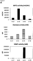
- Myeloperoxidase, Matrix-Metalloproteinase and Elastase enzyme activities are found to be generally lower in healing skin wound exudates as compared to the non-healing wounds.
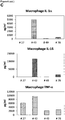
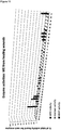
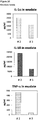
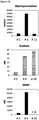
- IL-1 ⁇ , IL-1 ⁇ and TNF- ⁇ cytokine levels are elevated in general in non-healing skin as compared to healing skin wounds ( Figure 11 ).
- the individual values again vary strongly.
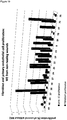
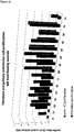
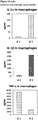
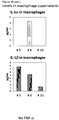
- cytokine levels are in the same range as in non-healing wound exudates for some of the healing wound exudates ( Figure 13 ), and only about 50% of the wound exudate samples from non-healing wounds induce cytokine secretion by macrophages ( Figure 15 ).
- Methods for reliably identifying non-healing skin wounds and/or for identifying that the healing of a skin wound worsens would provide an opportunity to further treat such skin wounds in time for the individual affected, thereby avoiding or attenuating recurrence and/or worsening of the healing status.
- pathogenic phagocytes release a variety of factors, including cytokines, proteases and toxic oxygen radicals into the wound tissue to destroy tissue cells, extracellular matrix and growth factors [ Clark RAF et al (2007) J Invest Dermatol 127:1018-1029 ]. These pathogenic factors are contained in the wound itself, e.g. in the fibrin clot or in the wound exudate (wound fluid).
- the wound fluid called wound exudate (WE) is the extracellular fluid containing a molecular fingerprint of wound cells and can be referred to as a "liquid biopsy”.
- the methods developed and established are particularly suitable for the identifying a skin wound in an individual as healing or non-healing skin wound, and/or for monitoring the healing of a skin wound in an individual and/or for evaluating the efficacy of known and unknown compounds, and/or for compound screening, in the context of skin would healing.
- the present invention relates to an in vitro method for identifying a skin wound in an individual as being a non-healing skin wound or healing skin wound, the method comprising:
- one skin wound may be a healing skin wound
- another skin wound of the same individual at the same or a different time point may be a non-healing skin wound, e.g. due to infections or an underlying disease affecting a specific skin wound.
- the present invention relates to an in vitro method which allows for identifying a skin wound in an individual as being a non-healing skin wound or healing skin wound. Therefore, the present invention allows, for example, for assessing specifically different skin wounds of the same individual.
- Identifying a skin wound of an individual as a non-healing skin wound allows for patient surveillance, monitoring of the wound healing and specific therapeutic interventions to stabilize, ameliorate and/or improve the healing of the skin wound.
- the skin wound identified as non-healing skin wound using a method of the invention may be treated with one or more of the following: compression, wound dressings, surgical debridement, biological debridement, infection control, antibiotic therapy, negative pressure therapy, proteins, in particular growth factors, antibodies, peptides, sugars, cells or cell constituents, artificial skin, human blood-derived products, gene therapy or genetically engineered wound bed modifications, drugs, herbal medicines, plant extracts.
- a wound is understood as damage to a tissue of a living individual, such as cuts, tears, burns, or breaks, preferably a wound is understood as open injury of a tissue of a living individual.
- a skin wound is understood as a damage to a skin of a living individual, such as cuts, tears, burns, or breaks.
- a skin wound is understood as open injury of the skin of a living individual.
- the skin may be located at any area of an individual, such as for example the head, the arms, the legs, the chest, or the back. Further, the individual may have one, two, three, four or more skin wounds. Further, the area of a skin wound may differ. In a preferred embodiment, the skin wound forms wound exudate.
- the skin wound may for example be selected from a wound of a diabetic patient, a wound which is infected by at least one microorganism, an ischemic wound, a wound in a patient suffering from deficient blood supply or venous stasis, an ulcer, such a diabetic ulcer, venous ulcer, arterial ulcer (e.g. ulcus cruris arteriosum), mixed ulcer, or pressure ulcer, a neuropathic wound, ulcus cruris, surgical wound, burn, dehiscence, neoplastic ulcer and rare ulcer.
- an individual's skin wound is not affected by a further disease mechanically preventing wound closure, such as calcinosis, where calcium crystals in the wound mechanically prevent wound closure, or exudative dermatitis.
- Ulcers can result in complete loss of the epidermis and often portions of the dermis and even subcutaneous fat.
- a non-healing skin wound refers to a skin wound which does not heal at an expected rate, in particular, as a skin wound which does not close within 2 months under standard therapy, preferably within 3 or more months under standard therapy.
- a non-healing skin wound is characterized by a lack of wound closure, an increase of the area and/or depth of the wound, necrosis and/or infections of the skin wound, and/or lack of granulation.
- a "healing skin wound” is understood as a skin wound which heals at an expected rate, in particular, as a skin wound which closes within 2 months under standard therapy.
- a healing skin wound is characterized by ongoing wound closure, granulation, absence of necrosis and/or absence of infections.
- Standard therapy is understood as a treatment recommended in general by physicians for skin wounds, in particular one or more selected from wound dressings, surgical and biological (maggot) debridement, infection control, negative pressure therapy, and therapy with a biological or cell treatment.
- the skin wound is untreated or treated with standard therapy or with one or more of the following: compression, wound dressings, surgical debridement, biological debridement, infection control, antibiotic therapy, negative pressure therapy, proteins, in particular growth factors, antibodies, peptides, sugars, cells or cell constituents, artificial skin, human blood-derived products, gene therapy or genetically engineered wound bed modifications, drugs, herbal medicines, plant extracts.
- the individual is an animal, preferably the individual is a vertebrate, in particular a mammal, more preferably a human.
- the individual may be an otherwise healthy individual or may exhibit further diseases and/or co-morbidities, and/or is treated with medication(s) for further diseases and/or co-morbidities.
- the skin wound of the individual may be untreated or treated with standard therapy or with one or more of the following: compression, wound dressings, surgical debridement, biological debridement, infection control, antibiotic therapy, negative pressure therapy, proteins, in particular growth factors, antibodies, peptides, sugars, cells or cell constituents, artificial skin, human blood-derived products, gene therapy or genetically engineered wound bed modifications, drugs, herbal medicines, plant extracts.
- step a) the method of the invention includes measuring
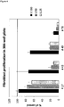
- Measuring the proliferation of primary fibroblast cells in the presence of a wound exudate sample obtained from said skin wound may be performed as shown in the examples, in particular in Example 3.1.1.
- primary fibroblast cells are used, which may be primary mammal dermal fibroblasts, preferably primary human dermal fibroblasts.
- Methods for obtaining cultured primary human dermal fibroblast cells are known in the art and are for example described in the examples.
- the cells may be cultured using DMEM medium containing FCS.
- the cells are incubated on a solid support, thereby allowing the cells to adhere to the support, as for example described in the Examples, where multiwell plates were used.
- the cells are contacted with the wound exudate sample, which is optionally diluted, e.g. diluted with medium or a saline aqueous liquid.
- the contacting may be performed before or after adherence of the cells occurs.
- the contacting may be achieved by adding the optionally diluted, liquid wound exudate sample, to the cells either prior to adherence, for example at the seeding of the cells, or after adherence.
- the contacting may be achieved e.g. by pipetting, and optionally gentle mixing.
- the cells are incubated for an appropriate time, such as for 6 hours to 300 hours, more preferably 12 hours to 200 hours, even more preferably 24 hours to 120 hours. In the examples, 72 hours were successfully used.
- a corresponding liquid without wound exudate such as medium or a saline aqueous liquid may be added or no liquid is added.
- the amount, preferably the cell number, including the formation of extracellular matrix, of the primary fibroblast cells is determined, such as by fixing cells and determining total protein content.
- the cells may for example be fixed using paraformaldehyde.
- a suitable dye, such as sulforhodamine B may be used for determining the amount, preferably the cell number, including the formation of extracellular matrix, of the primary fibroblast cells.
- the stained cells including the extracellular matrix formed may then be quantified e.g. by determining absorbance or fluorescence at a suitable wavelength, depending on the dye.
- the method is performed in 2D cell culture, which allows for culturing the cells adherently on a solid support.
- the method step includes the following steps:
- the culturing of cells in methods of the present invention is preferably performed at about 20°C to 40°C, more preferably 25°C to 38°C, even more preferably at about 37°C.
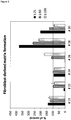
- Measuring the fibroblast-derived matrix formation by primary fibroblast cells in the presence of a wound exudate sample obtained from a skin wound may be performed as shown in the examples, in particular in Example 3.1.2.
- primary fibroblast cells are used, which may be primary mammal dermal fibroblasts, preferably primary human dermal fibroblasts.
- primary human dermal fibroblast cells are seeded on a support, which is preferably pre-coated with an adhesion enhancing agent, such as gelatin.
- the coating may be achieved by incubating the support with a solution or suspension containing the adhesion enhancing agent, such as gelatin. In the examples, a 0,2% gelatin solution was successfully used.
- the cells are cultured until confluence is reached. Subsequently, the cells are contacted with (i) a matrix promoting supplement, and (ii) the wound exudate sample, which is optionally diluted, wherein (i) and (ii) may be contacted simultaneously or sequentially.
- the matrix promoting supplement which is preferably selected from a solution comprising Vitamin C or a physiologically acceptable salt thereof, such the sodium salt, or 2-phospho-L-ascorbic acid or a physiologically acceptable salt thereof, and a combination of EGF and insulin, is added to the cells, e.g. by pipetting, and optionally gentle mixing.
- the wound exudate sample, which is optionally diluted may be contacted simultaneously or sequentially.
- the optionally diluted wound exudate sample may be mixed with the matrix promoting supplement, and the mixture may be added to the cells.
- the optionally diluted wound exudate sample may be added separately, but simultaneously, or separately, but subsequent to or prior to the matrix promoting supplement.
- the components (i) and (ii) are preferably contacted within 1 hour.
- the cells are subsequently incubated, preferably for 12 hours to 20 days, wherein the medium is optionally replaced at least one time with fresh medium supplemented with optionally diluted wound exudate and matrix promoting supplement. In the example, the medium was replaced once after 4 days of incubation, and the total incubation was 8 days.
- the solid support preferably contains at least one cavity which allows for filling of the space and therefore allows for a 3D cell culture.
- the amount of the fibroblast-derived matrix is determined, such as by fixing cells and determining total protein content.
- the cells may for example be fixed using paraformaldehyde.
- a suitable dye such as sulforhodamine B may be used for determining the amount, preferably the cell number, including the formation of extracellular matrix, of the primary fibroblast cells.
- the stained cells including the formation of extracellular matrix may then be quantified e.g. by determining absorbance or fluorescence at a suitable wavelength, depending on the dye.
- the method step preferably includes the following steps:
- fibroblast-derived matrix or “FDM” is understood as the extracellular matrix (ECM) formed by living fibroblast cells in an environment conducive for matrix formation, e.g. in the presence of a matrix promoting supplement.
- FDM is obtainable as described in the examples.
- FDM is obtainable by (i) seeding primary human dermal fibroblast cells on a support, which is pre-coated with an adhesion enhancing agent, such as gelatin, (ii) culturing the cells on the support, preferably until confluence is reached and (iii) contacting the cells with a matrix promoting supplement, such as Vitamin C or a physiologically acceptable salt thereof, or 2-phospho-L-ascorbic acid or a physiologically acceptable salt thereof, or a combination of EGF and insulin.
- an adhesion enhancing agent such as gelatin
- a "matrix promoting supplement” is understood as a compound or composition which promotes the formation of fibroblast-derived matrix by living fibroblast cells in an in vitro cell culture.
- Suitable matrix promoting supplements are Vitamin C or a physiologically acceptable salt thereof, such the sodium salt, or 2-phospho-L-ascorbic acid or a physiologically acceptable salt thereof, and a combination of EGF and insulin, as well as compositions comprising the compounds, such as solutions or suspensions.
- a combination of EGF and insulin may be provided to the cell culture separately, e.g. as separate solutions comprising EGF or insulin respectively, or together, e.g. as solution comprising EGF and insulin.
- an "adhesion enhancing agent” is an agent which enhances adhesion of cells to a solid support, such as a plastic support, but which does not substantially interfere with the viability of the cells.
- the adhesion enhancing agent is gelatin or fibronectin, more preferably gelatin.
- 2D cell culture is understood as a cell culture wherein the cells are cultured in a planar or substantially planar surface. In a preferred embodiment, the 2D cell culture is culturing of adherent cells.
- 3D cell culture is understood as a cell culture wherein the cells are cultured on a non-planar or substantially non-planar surface.
- the 3D cell culture is culturing of adherent cells and/or culturing of cells within a matrix, such as ECM, in particular FDM.
- a “support” or “solid support” is preferably selected from a chip, array, such as a microarray or nanoarray, a plate, such as a multiwell plate, or a dish.
- the solid support is preferably suitable for culturing cells, for example the support may be a plastic support.
- “Wound exudate” is understood as the extracellular fluid located within and above a skin wound. The wound exudate is also referred to a "liquid biopsy".
- wound exudate sample or "WE” is understood as a sample of wound exudate obtained from a skin wound of an individual.
- Methods for obtaining a wound exudate sample are known in the art.
- a wound exudate sample may be obtained by a physical or chemical method, in particular by applying negative pressure to the skin wound, such as by using a negative pressure drainage device, a method using capillary forces, collecting wound exudate in a film dressing or membrane, collecting wound exudate in a syringe, applying an absorptive material, such as absorptive beads, or a filter, or by using a swab, such as a cotton swab.
- the volume of wound exudate sample may vary and may be in the range of 1 nl to 1 l, 10 nl to 10 1 l, or 100 nl to 1 l, such as 1 ⁇ l to 1 l, 1 ml to 1 l or 10 ml to 1 l.
- wound exudate samples investigated in the examples had a volume of up to 400 ml and typically had a volume of 10 to 100 ml, in particular 10 to 50 ml.
- the wound exudate sample may be used the methods of the invention directly after obtaining the sample or may be stored, in particular stored at ⁇ 4°C, ⁇ 0°C or ⁇ 10°C before usage in the methods of the invention.
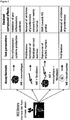
- the above assays relating to measuring the proliferation of primary fibroblast cells and the fibroblast-derived matrix formation by primary fibroblast cells can reliably identify skin wounds as healing skin wounds, or non-healing skin wounds, respectively. In particular, it is possible to reliably identify skin wounds as non-healing skin wounds. Therefore, one or both of these assays may be used for the method of the invention.
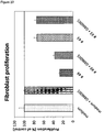
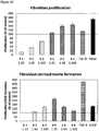
- the wound exudate of a healing wound has a dose-dependent positive effect on the proliferation of primary fibroblast cells and the fibroblast-derived matrix formation by primary fibroblast cells, as compared to a control in absence of wound exudate.
- value(s) obtained in the assay(s) which is/are equal to or above a control value established in the absence of wound exudate are indicative of a healing wound. Accordingly, value(s) obtained in the assay(s) which is/are below a control value established in the absence of wound exudate are indicative of a non-healing wound.
- control value(s) may be determined in parallel or may be established independently, preferably in parallel.
- the skin wound is identified as being a non-healing skin wound in case the value(s) obtained in i) and/or ii) is/are below a control value established in the absence of wound exudate, preferably at least 10% below the respective control value(s), more preferably at least 15%, even more preferably are at least 20% below the respective control value(s), or the skin wound is identified as being a healing skin wound in case the value(s) obtained in i) and/or ii) is/are equal to or above a control value established in the absence of wound exudate, at least 10% above the respective control value(s), more preferably at least 15%, even more preferably are at least 20% above the respective control value(s), more preferably wherein the value obtained in i) is at least 10%, more preferably at least 15%, above the respective control value and/or the value obtained in ii) is at least 50%, more preferably at least 100%, above the respective control value.
- the measurements at least in triplicate and/or to establish a statistical significance to improve the accuracy of the diagnosis.
- Statistical methods are known in the art. For example, the standard deviation may be determined. Further, the statistical significance may be p ⁇ 0,05, p ⁇ 0,001 or p ⁇ 0,001 for identifying a skin wound as a healing or non-healing skin wound.
- the value(s) in step a) is/are measured at least in triplicate and/or a statistical significance is established.
- a skin wound can be identified reliably as a non-healing skin wound in case the values obtained in both assays are below a control value established in the absence of wound exudate. Further, a skin wound can be identified reliably as a healing skin wound in case the values obtained in both assays are equal to or above a control value established in the absence of wound exudate.
- values in both assays which are each at least 10% above the respective control values, more preferably wherein the values obtained in the assay are each at least 15%, even more preferably are at least 20%, 30%, 40% or 50% above the respective control values are identifying the skin wound as healing skin wound.
- values in both assays which are each at least 10% below the respective control values, more preferably wherein the values obtained in the assay are each at least 15%, even more preferably are at least 20%, 30%, 40% or 50% below the respective control values are identifying the skin wound as non-healing skin wound.
- the method of the invention comprises
- the accuracy and reliability can be further increased by including a further assay in the method, which measures the proliferation of keratinocyte cells, such as primary keratinocyte cells or HaCaT cells. Further, it was found in the Examples that the use of HaCaT cells is to be preferred as compared to primary keratinocytes and allows for reliable prediction, in combination with the fibroblast-based assays described above.
- a skin wound can be identified reliably as a healing skin wound in case at least two, preferably all three of the values obtained with the assays measuring the proliferation of primary fibroblast cells, measuring the fibroblast-derived matrix formation by primary fibroblast cells and measuring the proliferation of keratinocyte cells are equal to or above the respective control values established in the absence of wound exudate, more preferably wherein the values obtained in the assays measuring the proliferation of primary fibroblast cells and/or the fibroblast-derived matrix formation by primary fibroblast cells, and the assay measuring the proliferation of keratinocyte cells are at least 10%, more preferably at least 15%, 20%, 30%, 40% or 50% above the respective control value.
- a skin wound can be identified reliably as a non-healing skin wound in case at least two, preferably all three of the values obtained with the assays measuring the proliferation of primary fibroblast cells, measuring the fibroblast-derived matrix formation by primary fibroblast cells and measuring the proliferation of keratinocyte cells are below the respective control values established in the absence of wound exudate, more preferably wherein the values obtained in the assays measuring the proliferation of primary fibroblast cells and/or the fibroblast-derived matrix formation by primary fibroblast cells, and the assay measuring the proliferation of keratinocyte cells are at least 10%, more preferably at least 15%, 20%, 30%, 40% or 50% below the respective control value.
- step a) further comprises the following step: iiia) measuring the proliferation of keratinocyte cells in the presence of a wound exudate sample obtained from said skin wound, and wherein step b) comprises: b) identifying the skin wound as being a non-healing skin wound in case at least two, preferably three of the values obtained in i) to iiia) are below the respective control values established in the absence of wound exudate, more preferably wherein the values obtained in i) and/or ii) and iiia) are at least 10%, more preferably at least 15%, below the respective control value, or identifying the skin wound as being a healing skin wound in case at least two, preferably three of the values obtained in i) to iii) are equal to or above the respective control values established in the absence of wound exudate, more preferably wherein the values obtained in i) and/or ii) and iiia) are at least 10%,
- the proliferation of keratinocyte cells is measured in the presence of a wound exudate sample obtained from said skin wound.
- the keratinocyte proliferation assay preferably includes culturing human primary keratinocyte cells, or HaCaT cells, which is a standard keratinocyte cell line, under standard conditions, such as by using DMEM containing FCS as medium, as for example described in the Examples.
- the cells are subsequently incubated on a solid support, thereby allowing the cells to adhere to the support. Further, the cells are contacted with the wound exudate sample, which is optionally diluted, wherein the contacting may be performed before or after adherence of the cells occurs.
- the optionally diluted wound exudate sample may be added to the adherent cells, for example by pipetting or otherwise adding the liquid, or the optionally diluted wound exudate sample may be added to the non-adherent cells, for example by pipetting or otherwise adding the liquid to the cells, followed by allowing the keratinocyte cells to adhere.
- the cells are subsequently incubated, preferably for 6 hours to 200 hours, preferably 24 hours to 100 hours. In the examples, the cells are incubated for 72 hours.
- the amount, preferably the cell number, of the keratinocyte cells is determined, such as by fixing cells and determining total protein content.
- the cells may for example be fixed using paraformaldehyde.
- a suitable dye such as sulforhodamine B may be used for determining the amount, preferably the cell number, of the keratinocyte cells.
- the stained cells may then be quantified e.g. by determining absorbance or fluorescence at a suitable wavelength, depending on the dye.
- the method is performed in 2D cell culture, which allows for culturing the cells adherently on a solid support.
- a keratinocyte cell may be a primary keratinocyte cell or a keratinocyte cell line, in particular a human primary keratinocyte cell or a human keratinocyte cell line.
- the keratinocyte cells used in the present invention are selected from HaCaT cells and primary keratinocyte cells.
- HaCaT cells represent an established and widely used human keratinocyte cell line.
- the keratinocyte cells used in the present invention are HaCaT cells.
- measuring the proliferation of keratinocyte cells in the presence of a wound exudate sample obtained from a skin wound includes the following steps:
- the accuracy and reliability can be further increased by including one or more further assays in the method which determine macrophage M1 and M2 markers in the context of wound exudate.
- markers may be cell surface protein markers, protein markers in the supernatant of macrophages or marker mRNAs in macrophages.
- Macrophages are tissue-resident professional phagocytes and antigen-presenting cells (APC), which differentiate from circulating peripheral blood monocytes. Activated macrophages of different phenotypes are classified by skilled persons into M1-macrophages and M2 macrophages. M1-macrophages are activated macrophages which comprise immune effector cells with an acute inflammatory phenotype. These are highly aggressive against bacteria and produce large amounts of lymphokines. The M2-macrophages are alternatively activated and anti-inflammatory.
- APC antigen-presenting cells
- M2 marker is understood as a protein marker which is specific for M2 macrophages. Preferably, the marker is secreted by the macrophages.
- the M2 markers are known in the art and are selected from CCL22 and CCL18. The markers may determined by methods known in the art, e.g. by using an immunological assay, even more preferably by using an ELISA assay.
- M1 marker is understood as a protein marker which is specific for M1 macrophages. Preferably, the marker is secreted by the macrophages.
- the M1 markers are known in the art and are selected from CXCL10 and IL-23p19. The markers may determined by methods known in the art, e.g. by using an immunological assay, even more preferably by using an ELISA assay.
- M1 cell surface marker is understood as a protein marker which is expressed at the surface of macrophages, and which is specific for M1 macrophages.
- the M1 cell surface markers are known in the art and are selected from CD38, CD64 and CD197.
- the amount(s) and/or frequency distribution(s) of the cell surface markers may be determined by an immunological assay and/or a fluorescence assay, in particular by FACS analysis, whereby typically a frequency distribution is determined.
- M2 cell surface marker is understood as a protein marker which is expressed at the surface of macrophages, and which is specific for M2 macrophages.
- the M2 cell surface markers are known in the art and are selected from CD200 receptor (CD200R), CD206 and CD209.
- CD200R CD200 receptor
- CD206 CD206
- CD209 CD200 receptor
- the amount(s) and/or frequency distribution(s) of the cell surface markers may be determined by an immunological assay and/or a fluorescence assay, in particular by FACS analysis, whereby typically a frequency distribution is determined.
- M2 marker mRNA is understood as an mRNA which is expressed by macrophages, and which is specific for M2 macrophages.
- the M2 marker mRNAs are known in the art and are selected CD200 receptor (CD200R), CD206, CD209, CCL22 and CCL18.
- the marker mRNAs may be determined by methods known in the art. Preferably, the amount may be determined by contacting a probe which specifically binds to a marker mRNA, wherein the probe is optionally labelled, with the macrophage RNA under conditions which are conducive to hybridization, and detecting the hybridized probe. For example, the mRNA may be reversely transcribed into cDNA prior to detection.
- M1 marker mRNA is understood as an mRNA which is expressed by macrophages, and which is specific for M1 macrophages.
- the M1 marker mRNAs are known in the art and are selected from CD38, CD64, CD197, CXCL10 and IL-23p19.
- the amount may be determined by contacting a probe which specifically binds to a marker mRNA, wherein the probe is optionally labelled, with the macrophage RNA under conditions which are conducive to hybridization, and detecting the hybridized probe.
- the mRNA may be reversely transcribed into cDNA prior to detection.
- the ratio of M1/M2 markers is indicative of a healing or non-healing skin wound, in combination with one or more cellular assays described above relating to measuring the proliferation of primary fibroblast cells, measuring the fibroblast-derived matrix (FDM) formation by primary fibroblast cells and measuring the proliferation of keratinocyte cells.
- FDM fibroblast-derived matrix
- an elevated ratio of M1/M2 markers, M1/M2 cell surface markers or M1/M2 marker mRNAs is indicative of a non-healing skin wound
- a low ratio of M1/M2 markers, M1/M2 cell surface markers or M1/M2 marker mRNAs is indicative of a healing skin wound.
- step a) of the method of the invention further comprises one, two or three of the following steps iiib) to iiid):
- the frequency distribution may be determined by determining the %age of cells which are positive for a given marker within a population, which is the most commonly used readout in FACS analysis.
- the amount may be determined by determining the quantity of cell surface expression, as a surrogate for the number of labelled molecules on the cell surface per individual cell when using labelled binding agents for the markers, as for example measured by the mean fluorescence intensity.
- measuring the amount(s) of one or more M1 marker(s) and one or more M2 marker(s) in the supernatant of macrophages incubated with a wound exudate sample obtained from a skin wound includes the following steps:
- primary human monocyte cells may be co-cultured with human dermal fibroblast cells in 2D cell culture, or with fibroblast-derived matrices. Methods for generating fibroblast-derived matrices are described above, as well as in the examples. Subsequently, the cells are incubated until macrophage differentiation is reached. For example, CD163 can be used as a cell surface marker of macrophage differentiation. Further, the cells are contacted with a wound exudate sample, which is optionally diluted, for example by pipetting the sample to the cells, and optionally gentle mixing. Further, the cells are incubated, preferably for 1 hour 100 hours, e.g. 4 hours to 100 hours.
- measuring the amount(s) and/or frequency distribution(s) of one or more M1 cell surface marker(s) and one or more M2 cell surface marker(s) on macrophages incubated with a wound exudate sample obtained from a skin wound includes the following steps:
- primary human monocyte cells may be co-cultured with human dermal fibroblast cells in 2D cell culture, or with fibroblast-derived matrices. Methods for generating fibroblast-derived matrices are described above, as well as in the examples. Subsequently, the cells are incubated until macrophage differentiation is reached. For example, CD163 can be used as a cell surface marker of macrophage differentiation. Further, the cells are contacted with a wound exudate sample, which is optionally diluted, for example by pipetting the sample to the cells, and optionally gentle mixing. Further, the cells are incubated, preferably for 1 hour 100 hours, e.g. 4 hours to 100 hours.
- the amount(s) and/or frequency distribution(s) of one or more M1 marker(s) and one or more M2 marker(s) on the cell surface of macrophages is determined.
- the cells may be harvested and subjected to FACS analysis, gating on the monocyte/macrophage population. Geometric means of mean fluorescence intensities can be used to quantify surface marker expression.
- the one or more M1 cell surface markers are selected from CD38, CD64 and CD197, and/or the one or more M2 cell surface markers are selected from CD200 receptor (CD200R), CD206 and CD209, more preferably wherein the amount(s) and/or frequency distribution(s) of the cell surface markers are determined by an immunological assay and/or a fluorescence assay, in particular by FACS analysis.
- step (iv) comprises contacting the macrophages with binding agents, preferably antibodies, which specifically recognize one or more M1 surface marker(s) and one or more M2 surface marker(s), wherein the binding agents are optionally labelled, in particular labelled with a fluorescent label, and determining the amount of binding molecules bound to the macrophages, in particular by determining mean fluorescence intensity, thereby determining the amount(s) of the cell surface markers.
- binding agents preferably antibodies, which specifically recognize one or more M1 surface marker(s) and one or more M2 surface marker(s)
- the binding agents are optionally labelled, in particular labelled with a fluorescent label
- determining the amount of binding molecules bound to the macrophages in particular by determining mean fluorescence intensity, thereby determining the amount(s) of the cell surface markers.
- antibodies specifically recognizing the surface markers and which contain a fluorescent label may be used.
- step (iv) comprises contacting the macrophages with binding agents, preferably antibodies, which specifically recognize one or more M1 surface marker(s) and one or more M2 surface marker(s), wherein the binding agents are optionally labelled, in particular labelled with a fluorescent label, and determining the percentages of cells which are positive for the one or more M1 surface marker(s) and the one or more M2 surface marker(s), respectively, within a cell population, in particular wherein FACS analysis is performed, thereby determining the frequency distribution(s) of the cell surface markers.
- binding agents preferably antibodies, which specifically recognize one or more M1 surface marker(s) and one or more M2 surface marker(s), wherein the binding agents are optionally labelled, in particular labelled with a fluorescent label, and determining the percentages of cells which are positive for the one or more M1 surface marker(s) and the one or more M2 surface marker(s), respectively, within a cell population, in particular wherein FACS analysis is performed, thereby determining the frequency distribution(s) of the cell
- Determination of proteins as binding agents of a marker protein can be performed using any of a number of known methods for identifying and obtaining proteins that specifically interact with proteins or polypeptides, for example, a yeast two-hybrid screening system such as that described in U.S. Pat. No. 5,283,173 and U.S. Pat. No. 5,468,614 , or the equivalent.
- a binding agent which specifically recognizes a marker has preferably at least an affinity of 10 7 l/mol for its corresponding target molecule.
- the binding agent which specifically recognizes a marker preferably has an affinity of 10 8 l/mol or even more preferred of 10 9 l/mol for its target marker molecule.
- the term specific is used to indicate that other biomolecules present in the sample do not significantly bind to the binding agent which specifically recognizes the marker.
- the level of binding to a biomolecule other than the target marker molecule results in a binding affinity which is only 10% or less, more preferably only 5% or less of the affinity to the target marker molecule, respectively.
- a preferred specific binding agent will fulfill both the above minimum criteria for affinity as well as for specificity.
- a binding agent which specifically recognizes a marker preferably is an antibody reactive with the marker.
- the term antibody refers to a polyclonal antibody, a monoclonal antibody, antigen binding fragments of such antibodies, single chain antibodies as well as to genetic constructs comprising the binding domain of an antibody.
- antibody includes polyclonal antibodies, monoclonal antibodies, fragments thereof such as F(ab')2, and Fab fragments, as well as any naturally occurring or recombinantly produced binding partners, which are molecules that specifically bind to a marker protein. Any antibody fragment retaining the above criteria of a specific binding agent can be used. Antibodies are generated by state of the art procedures, e.g., as described in Tijssen ( Tijssen, P., Practice and theory of enzyme immunoassays, Elsevier Science Publishers B.V., Amsterdam (1990 ), the whole book, especially pages 43-78). In addition, the skilled person is well aware of methods based on immunosorbents that can be used for the specific isolation of antibodies. By these means the quality of polyclonal antibodies and hence their performance in immunoassays can be enhanced (Tijssen, P., supra , pages 108-115).
- polyclonal antibodies raised in e.g. goats may be used.
- polyclonal antibodies from different species e.g., rats, rabbits or guinea pigs, as well as monoclonal antibodies can be used. Since monoclonal antibodies can be produced in any amount required with constant properties, they represent ideal tools in development of an assay for clinical routine.
- the sample obtained from an individual is incubated with the binding agent that specifically recognizes the marker in question under conditions appropriate for formation of a binding agent marker-complex. Such conditions need not be specified, since the skilled artisan without any inventive effort can easily identify such appropriate incubation conditions.
- the amount of binding agent marker-complex is measured and used in the methods and uses of the invention. As the skilled artisan will appreciate there are numerous methods to measure the amount of the specific binding agent marker-complex all described in detail in relevant textbooks (cf., e.g., Tijssen P., supra, or Diamandis, E.P. and Christopoulos, T.K. (eds.), Immunoassay, Academic Press, Boston (1996 )).
- monoclonal antibodies to the marker(s) are used in a quantitative (amount or concentration of the marker(s) is determined) immunoassay.
- the marker may be detected in a sandwich type assay format.
- a first specific binding agent is used to capture the marker in question on the one side and a second specific binding agent (e.g. a second antibody), which is labeled to be directly or indirectly detectable, is used on the other side.
- the second specific binding agent may contain a detectable reporter moiety or label such as an enzyme, dye, radionuclide, luminescent group, fluorescent group or biotin, or the like. Any reporter moiety or label could be used with the methods disclosed herein so long as the signal of such is directly related or proportional to the quantity of binding agent remaining on the support after wash.
- the amount of the second binding agent that remains bound to the solid support is then determined using a method appropriate for the specific detectable reporter moiety or label.
- a method appropriate for the specific detectable reporter moiety or label For radioactive groups, scintillation counting or autoradiographic methods are generally appropriate.
- Antibody-enzyme conjugates can be prepared using a variety of coupling techniques (for review see, e.g., Scouten, W. H., Methods in Enzymology 135:30-65, 1987 ). Spectroscopic methods can be used to detect dyes (including, for example, colorimetric products of enzyme reactions), luminescent groups and fluorescent groups. Biotin can be detected using avidin or streptavidin, coupled to a different reporter group (commonly a radioactive or fluorescent group or an enzyme).
- Enzyme reporter groups can generally be detected by the addition of substrate (generally for a specific period of time), followed by spectroscopic, spectrophotometric or other analysis of the reaction products. Standards and standard additions can be used to determine the level of antigen in a sample, using well known techniques.
- Immunoassays for measuring marker proteins of the invention include for example ELISA, enzyme immunoassay (EIA) and electro-chemiluminescence immunoassay (ECLIA) for the quantitative determination of a marker protein described herein.
- EIA enzyme immunoassay
- ELIA electro-chemiluminescence immunoassay
- measuring the expression level(s) of one or more M1 marker mRNA(s) and one or more M2 marker mRNA(s) in macrophages incubated with a wound exudate sample obtained from a skin wound includes the following steps:
- the one or more M1 marker mRNA(s) are selected from CD38, CD64, CD197, CXCL10 and IL-23p19, and/or the one or more M2 marker mRNA(s) are selected from CD200 receptor (CD200R), CD206, CD209, CCL22 and CCL18, more preferably wherein the method comprises contacting a probe which specifically binds to a marker mRNA, wherein the probe is optionally labelled, with the macrophage RNA under conditions which are conducive to hybridization, and detecting the hybridized probe.
- primary human monocyte cells may be co-cultured with human dermal fibroblast cells in 2D cell culture, or with fibroblast-derived matrices. Methods for generating fibroblast-derived matrices are described above, as well as in the examples. Subsequently, the cells are incubated until macrophage differentiation is reached. For example, CD163 can be used as a cell surface marker of macrophage differentiation. Further, the cells are contacted with a wound exudate sample, which is optionally diluted, for example by pipetting the sample to the cells, and optionally gentle mixing. Further, the cells are incubated, preferably for 1 hour 100 hours, e.g. 4 hours to 100 hours.
- the expression level(s) of one or more M1 marker mRNA(s) and one or more M2 marker mRNA(s) in the macrophages is determined.
- the cells may be harvested and mRNA expression level(s) may be determined using suitable probes.
- the expression level of a housekeeping gene such as actin or GAPDH may be determined and the expression level(s) of M1 or M2 marker RNA(s) may be determined as expression level relative to a housekeeping gene.
- Sensitivity and specificity are statistical measures of the performance of a binary classification test, also known in statistics as classification function: Sensitivity (also called the true positive rate) measures the proportion of positives that are correctly identified as such (e.g., the percentage of healing skin wounds identified as healing skin wound, respectively).
- Specificity measures the proportion of negatives that are correctly identified as such (e.g., the percentage of healing skin wounds identified as being a non-healing skin wound).
- scanners may be set to trigger on low-risk items like belt buckles and keys (low specificity), in order to reduce the risk of missing objects that do pose a threat to the aircraft and those aboard (high sensitivity).
- This trade-off can be represented graphically as a receiver operating characteristic curve.
- a perfect predictor would be described as 100% sensitive (e.g., all non-healing skin wounds are identified as non-healing skin wounds) and 100% specific (e.g., all healing skin wounds are not identified as non-healing skin wounds); however, theoretically any predictor will possess a minimum error bound known as the Bayes error rate.
- the cut-off can be set in order to either increase sensitivity or specificity.
- a combined value may be calculated using the two or more values obtained in the assays.
- the combined value is compared to the combined value of the respective control, which has been obtained using the same mathematical procedure.
- the combined value is obtained by weighted calculation of the values. This means that one of the assays is given a higher weighting that the other.
- the weighting factors have been obtained by analyzing a reference population of skin wounds.
- identifying a skin wound as non-healing skin wound preferably requires that at least the value(s) obtained in the assays measuring the proliferation of primary fibroblast cells and/or measuring the fibroblast-derived matrix (FDM) formation by primary fibroblast cells is/are below the respective control value(s) established in the absence of wound exudate.
- identifying a skin wound as healing skin wound preferably requires that at least the value(s) obtained in the assays measuring the proliferation of primary fibroblast cells and/or measuring the fibroblast-derived matrix (FDM) formation by primary fibroblast cells is/are equal or above the respective control value(s) established in the absence of wound exudate.
- FDM fibroblast-derived matrix
- step b) comprises:
- the skin wound as identified as being a healing skin wound in case at least two, preferably three, four, five or six of (1) to (6) above for a healing skin wound are fulfilled, with the proviso that at least the value(s) obtained in i) and/or ii) is/are at least 10% above the respective control values, more preferably wherein the values obtained in i) and/or ii) is/are at least 15%, even more preferably at least 20%, 30%, 40% or 50% above the respective control values, even more preferably wherein the value obtained in i) is at least 15%, even more preferably at least 20%, 30%, 40% or 50% above the respective control value and the value obtained in ii) is at least 50%, more preferably at least 100%, even more preferably 110%, 120%, 130% or 140% above the respective control value.
- the skin wound as identified as being a non-healing skin wound in case at least two, preferably three, four, five or six of (1) to (6) above for a non-healing skin wound are fulfilled, with the proviso that at least the value(s) obtained in i) and/or ii) is/are at least 10% below the respective control values, more preferably wherein the values obtained in i) and/or ii) is/are at least 15%, even more preferably at least 20%, 30%, 40% or 50% below the respective control values.
- the cellular assays are performed as described above for the methods of identifying.
- the method of the invention allows for reliably monitoring and predicting the wound healing of skin wounds in a patient.
- the method of the invention correctly predicted the subsequent worsening of skin wound healing in a patient, at a time point where visual inspection concluded an improvement in wound healing.
- the present invention relates to an in vitro method for monitoring the healing of a skin wound in an individual, the method comprising:
- the second time point is understood to be later than the first time point.
- the first time point and the second time point are separated by at least 6 hours, in particular by between 6 hours and 12 months.
- the first time point and the second time point are separated by 12 hours to 3, 6 or 12 months, or 1 day, 1 week or 1 month to 12 months.
- the monitoring is surprisingly reliable for determining a skin wound at a second time point to exhibit improved healing as compared to the first time point when an increase in the value(s) for one or both of the fibroblast-based assays is determined at the later time point as compared to the earlier time point, in cases where the value obtained in one or both of the assays measuring the proliferation of primary fibroblast cells in the presence of wound exudate samples obtained from a skin wound, and measuring the fibroblast-derived matrix formation by primary fibroblast cells in the presence of a wound exudate sample is/are equal to or below a control value established in the absence of wound exudate at the first, earlier time point. Accordingly, the monitoring of an improved healing is particularly reliable in case the wound is a non-healing wound at the first, earlier time point, as determined by a method of identifying the present invention.
- the method for monitoring the healing of a skin wound in an individual of the invention includes identifying the skin wound as being a non-healing skin wound or healing skin wound, respectively, at the respective time point, by performing the methods of the invention.
- the monitoring is surprisingly reliable for determining a skin wound at a second time point to exhibit worsened healing when a decrease in the value(s) for the one or both of the fibroblast-based assays is determined at the later time point as compared to the earlier time point, in case the value obtained in one or both of the assays measuring the proliferation of primary fibroblast cells in the presence of wound exudate samples obtained from a skin wound, and measuring the fibroblast-derived matrix formation by primary fibroblast cells in the presence of a wound exudate sample at the second later time point is/are equal to or below 100% of a control value established in the absence of wound exudate at the first, earlier time point. Accordingly, the monitoring of a worsened healing is particularly reliable in case the wound is a non-healing wound at the second, later time point, as determined by a method for identifying of the present invention.
- the present invention relates to an in vitro method for monitoring the healing of a skin wound in an individual, the method comprising:
- the method of the invention comprises:
- steps a) to e) may be repeated at two, three, four, five, six, seven, eight, nine, 10 or more later time points.
- a skin wound at a second time point is identified to exhibit improved healing in case the value obtained in c)i) at said second time point is at least at least 10%, more preferably 15%, even more preferably 20%, 25%, 30%, 40% or 50% higher than the value obtained in a)i) at said first time point, and the value obtained in c)ii) at said second time point is at least 10%, more preferably 15%, even more preferably 20%, 25%, 30%, 40% or 50% higher than the value obtained in a)ii) at said first time point, with the proviso that the values obtained in a)i) at said first time point and a)ii) at said first time point are equal to or below a respective control value established in the absence of wound exudate, preferably wherein the value(s) at said first time point is/are at least 10% below the respective control value, more preferably at least 15%, even more preferably at least 20%, 30%, 40% or 50% below the respective control value established in the absence of wound exudate at said first time point.
- a skin wound at a second time point is identified to exhibit worsened healing in case the value obtained in c)i) at said second time point is at least at least 10%, more preferably 15%, even more preferably 20%, 25%, 30%, 40% or 50% lower than the value obtained in a)i) at said first time point, and the value obtained in c)ii) at said second time point is at least 10%, more preferably 15%, even more preferably 20%, 25%, 30%, 40% or 50% lower than the value obtained in a)ii) at said first time point, with the proviso that the values obtained in c)i) and c)ii) at said second time point are equal to or below 100% of a respective control value established in the absence of wound exudate, preferably wherein the value(s) at said second time point is/are at least 10% below the respective control value, more preferably at least 15%, even more preferably at least 20%, 30%, 40% or 50% below the respective control value established in the absence of wound exudate at said second time point.
- the method of the present invention comprises:
- a skin wound at a second time point is identified to exhibit improved healing in case at least two, preferably three, four, five or six of (1) to (6) for improved healing are fulfilled, preferably wherein the value obtained in c)i) at said second time point is at least at least 10%, more preferably 15%, even more preferably 20%, 25%, 30%, 40% or 50% higher than the value obtained in a)i) at said first time point, and/or the value obtained in c)ii) at said second time point is at least 10%, more preferably 15%, even more preferably 20%, 25%, 30%, 40% or 50% higher than the value obtained in a)ii) at said first time point, with the proviso that the values obtained in a)i) at said first time point and/or a)ii) at said first time point are equal to or below a respective control value established in the absence of wound exudate, preferably wherein the value(s) at said first time point is/are at least 10% below the respective control value, more preferably at least 15%, even more
- a skin wound at a second time point is identified to exhibit improved healing in case at least two, preferably three, four, five or six of (1) to (6) for improved healing are fulfilled, preferably wherein the value obtained in c)i) at said second time point is at least at least 10%, more preferably 15%, even more preferably 20%, 25%, 30%, 40% or 50% higher than the value obtained in a)i) at said first time point, and the value obtained in c)ii) at said second time point is at least 10%, more preferably 15%, even more preferably 20%, 25%, 30%, 40% or 50% higher than the value obtained in a)ii) at said first time point, with the proviso that the values obtained in a)i) at said first time point and a)ii) at said first time point are equal to or below a respective control value established in the absence of wound exudate, preferably wherein the values at said first time point are at least 10% below the respective control value, more preferably at least 15%, even more preferably at least 20%, 30%,
- a skin wound at a second time point is identified to exhibit worsened healing in case healing in case at least two, preferably three, four, five or six of (1) to (6) for worsened wound healing are fulfilled, preferably wherein the value obtained in c)i) at said second time point is at least at least 10%, more preferably 15%, even more preferably 20%, 25%, 30%, 40% or 50% lower than the value obtained in a)i) at said first time point, and/or the value obtained in c)ii) at said second time point is at least 10%, more preferably 15%, even more preferably 20%, 25%, 30%, 40% or 50% lower than the value obtained in a)ii) at said first time point, with the proviso that the values obtained in c)i) and/or c)ii) at said second time point are equal to or below 100% of a respective control value established in the absence of wound exudate, preferably wherein the value(s) at said second time point is/are at least 10% below a respective control value, more
- a skin wound at a second time point is identified to exhibit worsened healing in case healing in case at least two, preferably three, four, five or six of (1) to (6) for worsened wound healing are fulfilled, preferably wherein the value obtained in c)i) at said second time point is at least at least 10%, more preferably 15%, even more preferably 20%, 25%, 30%, 40% or 50% lower than the value obtained in a)i) at said first time point, and the value obtained in c)ii) at said second time point is at least 10%, more preferably 15%, even more preferably 20%, 25%, 30%, 40% or 50% lower than the value obtained in a)ii) at said first time point, with the proviso that the values obtained in c)i) and c)ii) at said second time point are equal to or below 100% of a respective control value established in the absence of wound exudate, preferably wherein the values at said second time point are at least 10% below a respective control value, more preferably at least 15%, even more
- the method steps a)i), a)ii) and a)iii) at said first time point, and c)i), c)ii) and c)iii) at said second time point are performed.
- a skin wound at a second time point is identified to exhibit improved healing in case at least two, preferably three of (1) to (3) for improved healing are fulfilled, preferably wherein the value obtained in c)i) at said second time point is at least at least 10%, more preferably 15%, even more preferably 20%, 25%, 30%, 40% or 50% higher than the value obtained in a)i) at said first time point, and/or the value obtained in c)ii) at said second time point is at least 10%, more preferably 15%, even more preferably 20%, 25%, 30%, 40% or 50% higher than the value obtained in a)ii) at said first time point, and/or the value obtained in c)iiia) at said second time point is at least 10%, more preferably 15%, even more preferably 20%, 25%, 30%, 40% or 50% higher than the value obtained in a)iiia) at said first time point, with the proviso that the values obtained in a)i) at said first time point and/or a)ii) at said first time point
- a skin wound at a second time point is identified to exhibit improved healing in case at least two, preferably three of (1) to (3) for improved healing are fulfilled, preferably wherein the value obtained in c)i) at said second time point is at least at least 10%, more preferably 15%, even more preferably 20%, 25%, 30%, 40% or 50% higher than the value obtained in a)i) at said first time point, and the value obtained in c)ii) at said second time point is at least 10%, more preferably 15%, even more preferably 20%, 25%, 30%, 40% or 50% higher than the value obtained in a)ii) at said first time point, and/or the value obtained in c)iiia) at said second time point is at least 10%, more preferably 15%, even more preferably 20%, 25%, 30%, 40% or 50% higher than the value obtained in a)iia) at said first time point with the proviso that the values obtained in a)i) at said first time point and a)ii) at said first time point and/or
- a skin wound at a second time point is identified to exhibit worsened healing in case healing in case at least two, preferably three of (1) to (6) for worsened wound healing are fulfilled, preferably wherein the value obtained in c)i) at said second time point is at least at least 10%, more preferably 15%, even more preferably 20%, 25%, 30%, 40% or 50% lower than the value obtained in a)i) at said first time point, and/or the value obtained in c)ii) at said second time point is at least 10%, more preferably 15%, even more preferably 20%, 25%, 30%, 40% or 50% lower than the value obtained in a)ii) at said first time point, and/or the value obtained in c)iiia) at said second time point is at least 10%, more preferably 15%, even more preferably 20%, 25%, 30%, 40% or 50% lower than the value obtained in a)iia) at said first time point, with the proviso that the values obtained in c)i) and/or c)i
- a skin wound at a second time point is identified to exhibit worsened healing in case healing in case at least two, preferably three of (1) to (3) for worsened wound healing are fulfilled, preferably wherein the value obtained in c)i) at said second time point is at least at least 10%, more preferably 15%, even more preferably 20%, 25%, 30%, 40% or 50% lower than the value obtained in a)i) at said first time point, and the value obtained in c)ii) at said second time point is at least 10%, more preferably 15%, even more preferably 20%, 25%, 30%, 40% or 50% lower than the value obtained in a)ii) at said first time point, and/or the value obtained in c)iiia) at said second time point is at least 10%, more preferably 15%, even more preferably 20%, 25%, 30%, 40% or 50% lower than the value obtained in a)iia) at said first time point, with the proviso that the values obtained in c)i) and c)ii) and/
- the individual is a mammal, more preferably a human.
- a skin wound is selected from a wound of a diabetic patient, a wound which is infected by at least one microorganism, an ischemic wound, a wound in a patient suffering from deficient blood supply or venous stasis, an ulcer, such a diabetic ulcer, venous ulcer, arterial ulcer (e.g. ulcus cruris arteriosum), mixed ulcer, or pressure ulcer, a neuropathic wound, ulcus cruris, surgical wound, burn, dehiscence, neoplastic ulcer and rare ulcer.
- a wound of a diabetic patient a wound which is infected by at least one microorganism, an ischemic wound, a wound in a patient suffering from deficient blood supply or venous stasis, an ulcer, such a diabetic ulcer, venous ulcer, arterial ulcer (e.g. ulcus cruris arteriosum), mixed ulcer, or pressure ulcer, a neuropathic wound, ulcus cruris, surgical wound, burn, de
- a non-healing skin wound is understood as a wound which does not close within 2 months under standard therapy.
- the individual exhibits further diseases and/or co-morbidities, and/or is treated with medication(s) for further diseases and/or co-morbidities.
- such individuals may have a risk for developing chronic skin wounds and/or non-healing skin wounds and/or to exhibit worsened wound healing.
- skin wounds can in particular benefit from the methods of the present invention.
- the skin wound is untreated or treated with one or more of the following: compression, wound dressings, surgical debridement, biological debridement, infection control, antibiotic therapy, negative pressure therapy, proteins, in particular growth factors, antibodies, peptides, sugars, cells or cell constituents, artificial skin, human blood-derived products, gene therapy or genetically engineered wound bed modifications, drugs, herbal medicines, plant extracts.
- the individual is identified to be treated with one or more of the therapies selected from compression, wound dressings, surgical debridement, biological debridement, infection control, antibiotic therapy, negative pressure therapy, proteins, in particular growth factors, antibodies, peptides, sugars, cells or cell constituents, artificial skin, human blood-derived products, gene therapy or genetically engineered wound bed modifications, drugs, herbal medicines, plant extracts, in case (i) a skin wound of the individual is identified as being a non-healing skin wound by a method for identifying a skin wound in an individual as being a non-healing skin wound or healing skin wound of the present invention, and/or a skin wound of the individual is identified to exhibit worsened healing at a second time point as compared to a first time point by a method for monitoring skin wound healing of the present invention.
- the therapies selected from compression, wound dressings, surgical debridement, biological debridement, infection control, antibiotic therapy, negative pressure therapy, proteins, in particular growth factors, antibodies, peptides, sugars,
- the present invention relates to a method of treating a non-healing skin wound in an individual, comprising administering to an individual one or more of the therapies selected from compression, wound dressings, surgical debridement, biological debridement, infection control, antibiotic therapy, negative pressure therapy, proteins, in particular growth factors, antibodies, peptides, sugars, cells or cell constituents, artificial skin, human blood-derived products, gene therapy or genetically engineered wound bed modifications, drugs, herbal medicines, plant extracts, wherein the individual is identified to have at least one non-healing skin wound by performing a method of the present invention.
- the therapies selected from compression, wound dressings, surgical debridement, biological debridement, infection control, antibiotic therapy, negative pressure therapy, proteins, in particular growth factors, antibodies, peptides, sugars, cells or cell constituents, artificial skin, human blood-derived products, gene therapy or genetically engineered wound bed modifications, drugs, herbal medicines, plant extracts, wherein the individual is identified to have at least one non-healing skin wound by performing
- the present invention relates to a method of treating a skin wound in an individual which exhibits worsened healing, comprising administering to an individual one or more of the therapies selected from compression, wound dressings, surgical debridement, biological debridement, infection control, antibiotic therapy, negative pressure therapy, proteins, in particular growth factors, antibodies, peptides, sugars, cells or cell constituents, artificial skin, human blood-derived products, gene therapy or genetically engineered wound bed modifications, drugs, herbal medicines, plant extracts, wherein the individual is identified to have at least one skin wound which exhibits worsened wound healing by performing a method of the present invention.
- the therapies selected from compression, wound dressings, surgical debridement, biological debridement, infection control, antibiotic therapy, negative pressure therapy, proteins, in particular growth factors, antibodies, peptides, sugars, cells or cell constituents, artificial skin, human blood-derived products, gene therapy or genetically engineered wound bed modifications, drugs, herbal medicines, plant extracts, wherein the individual is identified to have at least one skin wound which
- a therapeutically effective amount is administered to the individual.
- at least one active agent such as antibiotic therapy, proteins, in particular growth factors, antibodies, peptides, sugars, cells or cell constituents, artificial skin, human blood-derived products, gene therapy or genetically engineered wound bed modifications, drugs, herbal medicines, and plant extracts
- a therapeutically effective amount is administered to the individual.
- at least one active agent is comprised in a pharmaceutical composition which may further comprise at least one pharmaceutically acceptable carrier or excipient.
- the therapeutically effective amount depends on the nature of the agent and is known to a skilled person. Further, at least one active agent may be administered once or repeatedly.
- the wound exudate sample is obtained by a physical or chemical method, in particular by applying negative pressure to the skin wound, in particular by using a negative pressure drainage device, a method using capillary forces, collecting wound exudate in a film dressing or membrane, collecting wound exudate in a syringe, applying an absorptive material, such as absorptive beads, or a filter, or by using a swab, such as a cotton swab.
- negative pressure was applied to the skin wound in order to obtain a wound exudate sample.
- a healing skin wound is characterized by ongoing wound closure, granulation, absence of necrosis and/or absence of infections.
- a non-healing skin wound is characterized by a lack of wound closure, an increase of the area and/or depth of the wound, necrosis and/or infections of the skin wound, and/or lack of granulation.
- the fibroblast and/or monocyte cells used in the methods are human cells, preferably human cells and/or primary cells obtained from healthy human individuals, from patients with comorbidities associated with impaired wound healing, such as diabetes, and/or from the individual patients providing the wound exudates.
- the wound exudate sample is diluted between 1:2 to 1:1000, preferably between 1:10 and 1:200 or 1:20 to 1:100. For example, dilutions of 1:25 or 1:50, such as dilutions in medium were successfully used.
- the wound exudate sample is preferably diluted in an aqueous liquid, such as aqueous solution or suspension.
- the wound exudate sample is diluted in a cell medium, such as DMEM containing FCS.
- an aqueous saline solution such as an aqueous buffered saline solution may be used.
- the wound exudate sample is diluted in an aqueous liquid, which does not substantially interfere with the viability of the cells used in the assays of the methods of the invention.
- measuring the proliferation of primary fibroblast cells in the presence of a wound exudate sample obtained from a skin wound includes the following steps:
- measuring the fibroblast-derived matrix formation by primary fibroblast cells in the presence of a wound exudate sample obtained from a skin wound includes the following steps:
- measuring the proliferation of keratinocyte cells in the presence of a wound exudate sample obtained from a skin wound includes the following steps:
- measuring the amount(s) of one or more M1 marker(s) and one or more M2 marker(s) in the supernatant of macrophages incubated with a wound exudate sample obtained from a skin wound includes the following steps:
- measuring the amount(s) and/or frequency distribution(s) of one or more M1 cell surface marker(s) and one or more M2 cell surface marker(s) on macrophages incubated with a wound exudate sample obtained from a skin wound includes the following steps:
- measuring the expression level(s) of one or more M1 marker mRNA(s) and one or more M2 marker mRNA(s) in macrophages incubated with a wound exudate sample obtained from a skin wound includes the following steps:
- two or more assays performed in the methods of the present invention are performed simultaneously, even more preferably wherein the method steps are performed on a single support.
- a wound-on-a-chip test method is provided. Thereby, the demands of clinicians are met.
- the method steps of any method of the invention are performed on a single support, more preferably wherein the support is a chip, array, such as a microarray or nanoarray, a plate, such as a multiwell plate, or a dish.
- the solid support preferably contains at least one, preferably a plurality of defined areas or cavities, more preferably at least one, even more preferably a plurality of cavities which allow for filling of the space and therefore allow for a 3D cell culture.
- a multiwell plate or a microarray or nanoarray comprising a plurality of defined cavities may be used. In the examples, a multiwell plate was successfully used.
- the solid support does not substantially interfere with the viability of the cells and/or is suitable for culturing cells, for example the support may be a plastic support.
- the solid support preferably contains at least one cavity, such as a well, more preferably a plurality of defined cavities.
- the solid support preferably contains at least one cavity, such as a well, more preferably a plurality of defined cavities.
- multi-well plates may be used. Such multi-well plates comprise a plurality of defined cavities.
- the present invention relates to a kit comprising the agents for performing the method steps i) to iiid) described above, preferably wherein the kit comprises:
- M1 and M2 marker(s), cell surface marker(s) and/or marker mRNA(s) are described above for the methods of the present invention.
- the keratinocyte cells are selected from HaCaT cells and primary keratinocyte cells, in particular human primary keratinocyte cells.
- the keratinocyte cells used in the present invention are HaCaT cells.
- Fibroblast-derived matrix is obtainable by (i) seeding primary human dermal fibroblast cells on a support, which is pre-coated with an adhesion enhancing agent, such as gelatin, (ii) culturing the cells on the support, preferably until confluence is reached and (iii) contacting the cells with a matrix promoting supplement, such as Vitamin C or a physiologically acceptable salt thereof, or 2-phospho-L-ascorbic acid or a physiologically acceptable salt thereof, or a combination of EGF and insulin.
- FDM may be formed in situ or may be transferred to the support after formation.
- a wound-on-a-chip may be provided, which allows for diagnosing, monitoring and predicting aspects of skin wound healing, such as identifying a non-healing skin wound and predicting worsened healing of a skin wound.
- the present invention relates to a support suitable for performing a method of the present invention, wherein the support comprises a plurality of defined areas or cavities and wherein:
- the solid support preferably contains a plurality of defined cavities. Cavities allow for filling of the space and therefore allow for a 3D cell culture.
- a multiwell plate or a microarray or nanoarray comprising a plurality of defined cavities may be used. In the examples, a multiwell plate was successfully used.
- the solid support does not substantially interfere with the viability of the cells and/or is suitable for culturing cells, for example the support may be a plastic support.
- the solid support may contain a plurality of defined wells. For example, multi-well plates may be used.
- the support comprises 2, 3, 4, 5, 6, 7, 8, 9, 10 or more defined areas or cavities, such as 2, 3, 4, 5, 6, 7, 8, 9 or 10 to 10 5 , 2, 3, 4, 5, 6, 7, 8, 9 or 10 to 10 4 , 2, 3, 4, 5, 6, 7, 8, 9 or 10 to 10 3 , or 2, 3, 4, 5, 6, 7, 8, 9 or 10 to 10 2 defined areas or cavities.
- Macrophages, keratinocytes and fibroblasts are perceived as pivotal cells in sustained wound inflammation and resulting wound chronicity.
- WE from chronic wounds we established new test systems in these cell types that are suitable for compound screening. They form the basis for our investigations aimed at the identification of inhibitors of WE-induced cell activation for therapy of non-healing chronic ulcers ( Figure 2 ).
- Example 4.3 the method makes use of the assay steps described above for the further methods of the invention.
- the proliferation of primary fibroblast cells in the presence of (i) a wound exudate sample obtained from a skin wound of at least one individual, and (ii) at least one candidate compound is measured as primary assay.
- at least one of the further assays described herein are performed.
- the candidate compound is found to be active in the proliferation of primary fibroblast cells assay in case the value obtained in the assay is at least 10% above or at least 10% below a control value established in the absence of the at least one candidate compound, or is above a cut-off value.
- the present invention relates to a method for screening for compounds suitable for modulating skin wound healing, comprising the following steps:
- the method may be repeated one or more times.
- step B) includes performing one, two, three, four or five of said method steps B1) to B5) in case the value obtained in A) is above or below a cut-off value.
- the cut-off value may be the control value established in the absence of the candidate compound + X * standard deviation, wherein X is 0,5 or more, such as 1, 1.5, 2, 2.5 3, 4 or more, such as up to 5, 6, 7, 8, 9 or 10.
- X is 3.
- the cut-off value may be the control value established in the absence of the candidate compound - X * standard deviation, wherein X is 0,5 or more, such as 1, 1.5, 2, 2.5 3, 4 or more, such as up to 5, 6, 7, 8, 9 or 10.
- the compound is identified as being suitable for modulating skin wound healing, in case at least one value obtained in B1) to B5) above or 10% below a respective cut-off value.
- the wound exudate sample is diluted, in particular as described above.
- one, two, three, four or five of the method steps B1) to B5) are performed in case the value obtained in A) above is at least 15%, 20%, 25%, 30%, 40% or 50% above a control value established in the absence of the at least one candidate compound.
- the compound is identified as being suitable for modulating skin wound healing, in case at least one value obtained in B1) and B2) is at least 10%,15%, 20%, 25%, 30%, 40% or 50% above a control value established in the absence of the at least one candidate compound.
- modulating wound healing is improving wound healing.
- one, two, three, four or five of the method steps B1) to B5) are performed in case the value obtained in A) above is at least 15%, 20%, 25%, 30%, 40% or 50% below a control value established in the absence of the at least one candidate compound.
- the compound is identified as being suitable for modulating skin wound healing, in case at least one value obtained in B1) and B2) is at least 10%,15%, 20%, 25%, 30%, 40% or 50% below a control value established in the absence of the at least one candidate compound.
- modulating wound healing is worsening wound healing.
- Compounds identified as being suitable for worsening skin wound healing are suitable for treating and/or preventing hypertrophic scars, keloids or fibrosis.
- the (i) wound exudate sample obtained from a skin wound of at least one individual, and (ii) the at least one candidate compound may be added for incubation simultaneously, or sequentially.
- (i) and (ii) may be added as separate compositions, or as a single composition.
- the optionally diluted wound exudate sample may be added to an assay, e.g. by pipetting the liquid into a well of a support, and the candidate compound is added separately, e.g. by pipetting the liquid containing such compound into a well of a support, either before, simultaneous with, or after the optionally diluted wound exudate sample.
- the optionally diluted wound exudate sample may be mixed with the at least one candidate compound or composition comprising at least one candidate compound, and be added to the assay subsequently.
- the method steps include contacting of a wound exudate sample obtained from a skin wound of at least one individual.
- a wound exudate sample obtained from a skin wound of one individual may be used, or mixtures of wound exudate samples obtained from a skin wound of two or more individuals, such as 2, 3, 4, 5, 6, 7, 8, 9, 10 or more individuals may be used.
- the wound exudate samples may be samples from healing wounds or non-healing wounds, such as ulcers, or mixtures thereof.
- measuring the proliferation of primary fibroblast cells in the presence of (i) a wound exudate sample obtained from a skin wound of at least one individual, and (ii) at least one candidate compound includes the following steps:
- measuring the fibroblast-derived matrix formation by primary fibroblast cells in the presence of (i) a wound exudate sample obtained from a skin wound of at least one individual and (ii) said at least one candidate compound includes the following steps:
- measuring the proliferation of keratinocyte cells in the presence of (i) a wound exudate sample obtained from a skin wound of at least one individual and (ii) said at least one candidate compound includes the following steps:
- measuring the amount(s) of one or more M1 marker(s) and one or more M2 marker(s) in the supernatant of macrophages incubated with (i) a wound exudate sample obtained from a skin wound of at least one individual and (ii) said at least one candidate compound, wherein the macrophages are in co-culture with fibroblasts includes the following steps:
- measuring the amount(s) and/or frequency distribution(s) of one or more M1 cell surface marker(s) and one or more M2 cell surface marker(s) on macrophages incubated with (i) a wound exudate sample obtained from a skin wound of at least one individual and (ii) said at least one candidate compound, wherein the macrophages are in co-culture with fibroblasts includes the following steps:
- measuring the expression level(s) of one or more M1 marker mRNA(s) and one or more M2 marker mRNA(s) in macrophages incubated with (i) a wound exudate sample obtained from a skin wound of at least one individual and (ii) said at least one candidate compound, wherein the macrophages are in co-culture with fibroblasts includes the following steps:
- a wide variety of compounds may be used in the methods for screening.
- a small molecule, a hormone, sugar, protein, peptide, polymer, biological, such as a protein, a peptide, an antibody or derivative thereof, or a conjugate thereof, a nucleic acid, such a viral agent, a wound dressing which optionally comprises a therapeutically active agent, or one or more cell(s), such as one or more genetically modified cell may be used.
- a plurality of compounds may be tested in parallel. In case 2, 3, 4, 5, 6, 7, 8, 9, 10 or more candidate compounds are to be tested in parallel, such compounds may be added separately or as at least one composition comprising two or more candidate compounds. Thereby, a huge number of candidate compounds may be tested.
- compositions such as 1, 2, 3, 4, 5, 6, 7, 8, 9, 10 or more compositions, each comprising at least one, such as 1, 2, 3, 4, 5, 6, 7, 8, 9, 10 or more candidate compounds, may be added.
- the compounds or composition comprising compounds may be provided as a liquid, such as a solution, in particular aqueous solution or medium or suspension, such as a suspension in an aqueous solution or medium.
- the liquid, in particular solution or suspension, without candidate compound(s) preferably does not substantially interfere with the viability of cells used in the assay(s).
- the at least one compound is selected from a small molecule, a hormone, sugar, protein, peptide, polymer, biological, such as a protein, a peptide, an antibody or derivative thereof, or a conjugate thereof, a nucleic acid, such a viral agent, or one or more cell(s), such as one or more genetically modified cell.
- the at least one compound is selected from an immunomodulatory agent, more preferably an immunosuppressive agent, an antibiotic, an antiinfective, a growth factor, a cytokine, an antiproliferative agent and an agent stimulating proliferation.
- known approved active agents may be tested, such as active agents approved in the field of wound healing or for other indications, such as for inflammatory and/or proliferative disorders.
- novel compounds and/or compound libraries may be tested.
- combinatorial libraries or other small molecule compound libraries, or antibody libraries may be tested.
- the at least one compound is a single compound, or 2, 3, 4, 5, or more different compounds, wherein the 2, 3, 4, 5, or more different compounds may be present in a single composition or in 2 or more separate compositions.
- the values are measured at least in triplicate and/or a statistical significance is established in B), more preferably, wherein p ⁇ 0,05, p ⁇ 0,001 or p ⁇ 0,001, and/or the compound is identified as being suitable for modulating skin wound healing, in case at least one value obtained in B1) to B5) is at least 10% above, such as at least 10%,15%, 20%, 25%, 30%, 40% or 50% above, or at least 10% below, such as at least 10%,15%, 20%, 25%, 30%, 40% or 50% below a control value established in the absence of the candidate compound with statistical significance, more preferably, wherein p ⁇ 0,05, p ⁇ 0,001 or p ⁇ 0,001.
- a compound that is identified to be suitable for modulating, in particular improving skin wound healing can be tested in in vitro, ex vivo and/or in vivo models for efficacy and safety in the context of wound healing.
- a compound that is identified to be suitable for modulating, in particular worsening skin wound healing can be tested in in vitro, ex vivo and/or in vivo models for efficacy and safety in the context of hypertrophic scars, keloids and fibrosis.
- a keloid is a type of scar of the skin which, depending on its maturity, is composed mainly of either type III or type I collagen. It is a result of an overgrowth of granulation tissue at the site of a healed skin injury which is then slowly replaced by collagen type 1.
- a keloid scar is benign.
- Fibrosis is the formation of excess fibrous connective tissue in an organ or tissue in a reparative or reactive process.
- Wound exudate is the extracellular fluid containing a molecular fingerprint of wound cells and can be referred to as a "liquid biopsy”. Since removal of wound exudates (WE) can improve wound healing, we reasoned that factors contained within WE critically impede wound healing, e.g. through activation of innate immune cells.
- Macrophages, keratinocytes and fibroblasts are perceived as pivotal cells in sustained wound inflammation and resulting wound chronicity.
- WE from chronic wounds we established new test systems in these cell types that are suitable for compound screening, monitoring the healing of a skin wound and/or for identifying a skin wound in an individual as being a non-healing skin wound or healing skin wound.
- the assays form the basis for our investigations aimed at the identification of inhibitors of WE-induced cell activation for therapy of non-healing chronic ulcers ( Figure 2 ) as well as for the methods for monitoring the healing of a skin wound and/or for identifying a skin wound in an individual as being a non-healing skin wound or healing skin wound.
- HDF human dermal fibroblast proliferation assay: measuring the proliferation of primary fibroblast cells in the presence of a wound exudate sample obtained from a skin wound of an individual
- HDF Primary human dermal fibroblasts
- the cells were allowed to adhere overnight and then incubated for 72 hours at 37°C under the following conditions: graded compound concentrations or 20ng/ml PDGF-BB (Tonbo Biosciences # TO-21-8501-U010) in the absence or presence of different dilutions of sterile-filtered WE in medium. For negative control samples, 200 ⁇ l medium was added instead of specific stimuli. Alternatively, the cells were seeded into 384-well plates at 2500 cells/well together with test compounds or growth factors and WE or medium in a total volume of 50 ⁇ l.
- the cells were fixed with 4% paraformaldehyde (Morphisto) for 15 minutes at room temperature and washed 3 times with PBS.
- a control plate was fixed after the overnight adherence of the cells (day 1) to determine the starting cell number.
- Total cellular protein was determined as a measure of cell number by staining the fixed cells with sulforhodamine B (SRB, Sigma). A 0.4% SRB solution in 1% acetic acid was added to the wells for 30 minutes. The wells were then washed with 1% acetic acid until the wash solution remained colorless. After drying, the dye was eluted with 10mM Tris ⁇ HCl, pH8.5, and absorbance was measured either at 550 or 492nm for lower and higher cell densities, respectively. The average absorbance of the sample representing the day 1 starting cell number (for 96-well plates) was subtracted from the absorbance values of the WE-treated cells.
- SRB sulforhodamine B
- HDF primary human fibroblasts
- FDM fibroblast-derived matrices
- HDF cells were seeded at 5000 cells/well on day -3 into 96-well tissue culture plates (1250 cells/well for 384-well plates), which had been pre-coated for 1 hour at 37°C with 100 ⁇ l of a 0.2% gelatin solution (Sigma).
- a matrix promoting supplement vitamin C: 2-phospho-L-ascorbic acid trisodium salt, 100 ⁇ g/ml; Sigma
- test samples containing PDGF-BB, TGF- ⁇ 1 or graded concentrations of compounds - /+ WE as described for the HDF proliferation assay.
- medium was replaced by fresh vitamin C- and stimulus- as well as compound-containing medium, maintaining the conditions initiated on day 0.
- TGF- ⁇ 1 and PDGF-BB were included as positive controls to promote FDM formation and cell growth, respectively.
- FDM production was measured in fixed cultures via SRB staining and evaluated as described above.
- Keratinocvte proliferation assay measuring the proliferation of keratinocyte cells in the presence of a wound exudate sample obtained from a skin wound
- the HaCaT keratinocyte cell line was routinely cultured in DMEM containing 10% FCS, 2mM glutamine, and 100 U/ml penicillin/100 ⁇ g/ml streptomycin.
- the proliferation assay was carried out as described for HDF cells.
- Primary human keratinocytes were grown in KBM medium (Lonza CC-3104) containing 0.06mM calcium and supplemented with growth factors (Lonza CC-4131) on plastic coated with rat tail collagen (40 ⁇ g/ml; Gibco) or gelatin (0.2%; Sigma). No antibiotics were used.
- the proliferation assay was carried out as described for HDF cells.
- the primary human endothelial cells HMVEC-d- (Lonza, CC-2543) were cultured in EGM-2-MV BulletKit medium (Lonza CC-3156 & CC-4147).
- the proliferation assay was carried out as described for HDF cells.
- PBMC peripheral blood mononuclear cells
- the cell pellet was washed 3 times with PBS, resuspended in RPMI medium containing 20% FCS and 10% DMSO and frozen in liquid nitrogen.
- Monocytes were generated from frozen aliquots using positive selection with the CD14 Beads-Kit (Miltenyi) on an autoMACS-Sorter (Miltenyi) according to the manufacturer's instructions.
- monocytes were seeded at 4 x 10 6 monocytes/well in 6-well-plates (Nunc) and incubated with 20 ng/ml M-CSF (R&D Systems) in RPMI supplemented with 10% FCS, 2mM glutamine, and 100 U/ml penicillin / 100 ⁇ g/ml streptomycin in a total volume of 5ml per well. After 2 days, 2ml of the supernatant were removed and replaced by 2.5ml/well of fresh medium containing 20ng/ml M-CSF. On the third day, microscopic examination revealed differentiation into adherent, frequently elongated cells.
- M-CSF R&D Systems
- the macrophages were harvested with a rubber scraper, centrifuged at 1200 rpm for 5 minutes, resuspended in serum-free medium and seeded at 2 x 10 5 cells/well in 100 ⁇ l. After 1 hour at 37°C, 2-fold concentrated stimuli were added in a volume of 100 ⁇ l to give the indicated final concentrations.
- a combination of 100ng/ml LPS (Sigma) and 50ng/ml IFN- ⁇ (PeproTech) served as positive control for the induction of cytokine secretion.
- Graded concentrations of test compounds were prepared in the absence or presence of a 1:100 dilution of sterile-filtered WE. For negative control samples, 100 ⁇ l medium was added instead of specific stimuli.
- Human monocvte-dermal fibroblast co-cultures as in vitro models that reflect macrophage behavior in human skin measuring (a) the amount(s) of one or more M1 marker(s) and one or more M2 marker(s) in the supernatant of macrophages incubated with a wound exudate sample obtained from a skin wound, wherein the macrophages are in co-culture with fibroblasts, and (b) measuring the amount(s) and/or frequency distribution(s) of one or more M1 cell surface marker(s) and one or more M2 cell surface marker(s) on macrophages incubated with a wound exudate sample obtained from a skin wound, wherein the macrophages are in co-culture with fibroblasts, and (b) measuring the expression level(s) of one or more M1 marker mRNA(s) and one or more M2 marker mRNA(s) in macrophages incubated with a wound exudate sample obtained from a skin wound, wherein
- CD14 + monocytes isolated from PBMC of healthy donors by magnetic bead separation were incubated either alone or in the presence of primary human dermal fibroblasts (CellNTec) or fibroblast-derived matrices (FDM). FDM had been generated from primary human dermal fibroblasts by a 3-week incubation with the growth supplements vitamin C or insulin and EGF (vitamin C: 2-phospho-L-ascorbic acid trisodium salt, 100 ⁇ g/ml; human EGF, 5ng/ml; human insulin, 5 ⁇ g/ml).
- EGF vitamin C: 2-phospho-L-ascorbic acid trisodium salt, 100 ⁇ g/ml; human EGF, 5ng/ml; human insulin, 5 ⁇ g/ml).
- the cultures were stimulated overnight with IFN-g (50ng/ml), LPS (100ng/ml) and IL-4 (25ng/ml) or combinations thereof as controls for M1 and M2 macrophage induction.
- Specific mRNA levels are determined as ratios compared to a housekeeping gene; the values obtained are "expression relative to housekeeping gene".
- Wound exudates were diluted in phosphate buffered saline (PBS) and subjected to a commercial protein assay, Pierce BCA Protein Assay Kit (Thermo Scientific #23225), using bovine serum albumin as a standard in the range of 20 - 2000 ⁇ g/ml.
- the assay was performed in 96-well ELISA microplates (Greiner # 655101) and measured on a TECAN Infinite M200 Pro microtiter plate reader at 562nm, using Magellan 7.2 software for evaluation.
- MPO Myeloperoxidase
- MPO activity in PBS-prediluted WE samples was determined by oxidation of 3,3',5,5'-Tetramethylbenzidine (TMB) in 96-well ELISA microplates. 10 ⁇ l of diluted WE samples were added to 40 ⁇ l of assay buffer (78mM NaH2PO4 ⁇ H2O, 1.67mM Na2HPO4, 0.5% HTAB pH 5.40) and then incubated with 40 ⁇ l 1X TMB ELISA Substrate Solution (eBioscience # 00-4201-56) until blue color developed (up to 30 minutes). MPO from human leukocytes (Sigma M6908-5UN) in assay buffer was used as standard at concentrations from 0.12 to 120mU/ml. The reaction was stopped by the addition of 45 ⁇ l 2N H2SO4, and the samples were measured on a TECAN Infinite M200 Pro microtiter plate reader at 450nm, using Magellan 7.2 software for evaluation.
- TMB 3,3',5,5'-Tetramethylbenzidine
- Elastase activity in WE was determined by measuring the fluorescence of the fluorogenic substrate MeOSuc-Ala-Ala-Pro-Val-AMC.
- 5 ⁇ l WE pre-dilutions were added to 20 ⁇ l assay buffer (50mM Tris pH7.4, 600mM NaCl, 0.05% CHAPS) in 384-well black polystyrene plates (Corning #3573).
- 25 ⁇ l of elastase substrate V Merck-Millipore/Calbiochem Cat# 324740
- the reaction was allowed to proceed for 1 hour.
- MMP Matrix metalloproteinase
- the activity of all matrix metalloproteinases was determined in WE by measuring the fluorescence of the fluorogenic substrate MCA-Lys-Pro-Leu-Gly-Leu-DPA-Ala-Arg-NH2.
- Active human MMP9 full length protein (Abcam #ab168863) in assay buffer was used as standard at concentrations from 0.31 to 20nM.
- IL-1 ⁇ in WE and in macrophage supernatants was determined in F96 Maxisorp Nunc Immuno plates (Nunc, #439454) using the hIL1-alpha DuoSet ELISA Kit from R&D Systems (# DY200) according to the manufacturer's instructions.
- the enzyme reaction was detected with TMB Solution (eBioscience) and stopped by the addition of 50 ⁇ l/well 2N H2SO4. Absorbance was read on a Tecan Infinite M200 Pro at 450nm.
- IL-1 ⁇ in WE and in macrophage supernatants was determined in F96 Maxisorp Nunc Immuno plates (Nunc, #439454) using the IL1-beta ELISA ReadySet-Go Kit from eBioscience (#88-7261-88) according to the manufacturer's instructions. Enzyme reaction and measurement were carried out as described for IL-1 ⁇ .
- TNF- ⁇ in WE and in macrophage supernatants was determined in F96 Maxisorp Nunc Immuno plates (Nunc, #439454) using the TNF-alpha ELISA ReadySet-Go Kit from eBioscience (# 88-7346-88) according to the manufacturer's instructions. Enzyme reaction and measurement were performed as described for IL-1 ⁇ .
- Table 1 Characteristics of the patients who provided the screening exudates WE number # 49 # 27 # 43 # 78 Wound Foot ulcer Ulcus cruris arteriosus "Gamaschen-Ulcus", diabetic patient Operation wound (after kidney transplantation), diabetic patient
- the methods of the present invention surprisingly allowed the prediction of whether a defined skin wound in a defined individual exhibits improved or worsened wound healing in the future.
- the assays are performed in the presence of wound exudate from at least one individual.
- a compound is identified as being suitable for modulating skin wound healing in case if the compound is further active in at least one of the five secondary assays.
- the method for screening for compounds suitable for modulating skin wound healing comprises the following steps:
Landscapes
- Health & Medical Sciences (AREA)
- Life Sciences & Earth Sciences (AREA)
- Engineering & Computer Science (AREA)
- Immunology (AREA)
- Biomedical Technology (AREA)
- Chemical & Material Sciences (AREA)
- Molecular Biology (AREA)
- Hematology (AREA)
- Urology & Nephrology (AREA)
- General Health & Medical Sciences (AREA)
- Pathology (AREA)
- Cell Biology (AREA)
- Analytical Chemistry (AREA)
- Physics & Mathematics (AREA)
- Microbiology (AREA)
- Biotechnology (AREA)
- Biochemistry (AREA)
- Medicinal Chemistry (AREA)
- Food Science & Technology (AREA)
- General Physics & Mathematics (AREA)
- Tropical Medicine & Parasitology (AREA)
- Bioinformatics & Cheminformatics (AREA)
- Toxicology (AREA)
- Medical Informatics (AREA)
- Public Health (AREA)
- Proteomics, Peptides & Aminoacids (AREA)
- Organic Chemistry (AREA)
- Epidemiology (AREA)
- Primary Health Care (AREA)
- Databases & Information Systems (AREA)
- Data Mining & Analysis (AREA)
- Physiology (AREA)
- Zoology (AREA)
- Wood Science & Technology (AREA)
- Genetics & Genomics (AREA)
- Biophysics (AREA)
- General Engineering & Computer Science (AREA)
- Measuring Or Testing Involving Enzymes Or Micro-Organisms (AREA)
- Investigating Or Analysing Biological Materials (AREA)
- Immobilizing And Processing Of Enzymes And Microorganisms (AREA)
Claims (15)
- In vitro -Verfahren zum Identifizieren einer Hautwunde in einem Individuum als eine nicht-heilende Hautwunde oder eine heilende Hautwunde, wobei das Verfahren umfasst:a) Messeni) der Proliferation von primären Fibroblastenzellen in Gegenwart einer Wundexsudatprobe, die aus der Hautwunde erhalten wurde, und / oderii) der Fibroblasten-abgeleiteten Matrixbildung durch primäre Fibroblastenzellen in Gegenwart einer aus der Hautwunde erhaltenen Wundexsudatprobe undb) Identifizieren der Hautwunde als eine nicht heilende Hautwunde, falls der oder die in i) und/oder ii) erhaltenen Wert(e) unter einem Kontrollwert liegt / liegen, der in Abwesenheit von Wundexsudat bestimmt wurde, oder
Identifizieren der Hautwunde als eine heilende Hautwunde, falls der / die in i) und / oder ii) erhaltene(n) Wert(e) gleich sind zu oder über einem Kontrollwert liegen, der in Abwesenheit von Wundexsudat bestimmt wurde,
vorzugsweise wobei der / die Wert(e) in a) mindestens dreifach gemessen wird / werden und / oder eine statistische Signifikanz festgestellt wird. - In vitro -Verfahren zum Überwachen der Heilung einer Hautwunde in einem Individuum, wobei das Verfahren umfasst:a) Messeni) der Proliferation von primären Fibroblastenzellen in Gegenwart einer Wundexsudatprobe, die aus der Hautwunde zu einem ersten Zeitpunkt erhalten wurde, und/oderii) der Fibroblasten-abgeleiteten Matrixbildung durch primäre Fibroblastenzellen in Gegenwart einer Wundexsudatprobe, die aus der Hautwunde zu einem ersten Zeitpunkt erhalten wurde,b) gegebenenfalls Identifizieren der Hautwunde als eine nicht heilende Hautwunde zu diesem ersten Zeitpunkt, falls der / die in a)i) und / oder a)ii) erhaltene(n) Wert(e) unter einem Kontrollwert liegt / liegen, der in Abwesenheit von Wundexsudat bestimmt wurde, oder Identifizieren der Hautwunde als eine heilende Hautwunde zu einem ersten Zeitpunkt, falls der / die in a)i) und/oder a)ii) erhaltene(n) Wert(e) gleich ist / sind zu oder über einem Kontrollwert liegt / liegen, der in Abwesenheit von Wundexsudat bestimmt wurde,c) Messeni) der Proliferation von primären Fibroblastenzellen in Gegenwart einer Wundexsudatprobe, die aus der Hautwunde zu einem zweiten Zeitpunkt erhalten wurde, und/oderii) der Fibroblasten-abgeleiteten Matrixbildung durch primäre Fibroblastenzellen in Gegenwart einer Wundexsudatprobe, die aus der Hautwunde zu einem zweiten Zeitpunkt erhalten wurde,d) gegebenenfalls Identifizieren der Hautwunde als eine nicht heilende Hautwunde zu dem zweiten Zeitpunkt, falls der / die in c)i) und / oder c)ii) erhaltene(n) Wert(e) unter einem Kontrollwert liegt / liegen, der in Abwesenheit von Wundexsudat bestimmt wurde, oder Identifizieren der Hautwunde als eine heilende Hautwunde zu dem zweiten Zeitpunkt, falls der / die in c)i) und / oder c)ii) erhaltene(n) Wert(e) gleich ist / sind zu oder über einem Kontrollwert liegt / liegen, der in Abwesenheit von Wundexsudat bestimmt wurde,e)A) Identifizieren einer Hautwunde als eine Hautwunde die zu einem zweiten Zeitpunkt eine verbesserte Heilung zeigt, falls der in c)i) zu dem zweiten Zeitpunkt erhaltene Wert höher ist als der in a)i) zu dem ersten Zeitpunkt erhaltene Wert und / oder der in c)ii) zu dem zweiten Zeitpunkt erhaltene Wert höher ist als der in a)ii) zu dem ersten Zeitpunkt erhaltene Wert, mit der Maßgabe, dass der in a)i) zu dem ersten Zeitpunkt und / oder a)ii) zu dem ersten Zeitpunkt erhaltene Wert gleich ist zu oder niedriger ist als ein in Abwesenheit von Wundexsudat bestimmter Kontrollwert,
oderB) Identifizieren einer Hautwunde als eine Hautwunde die zu einem zweiten Zeitpunkt eine verschlechterte Heilung zeigt, falls der in c)i) zu dem zweiten Zeitpunkt erhaltene Wert niedriger ist als der in a)i) zu dem ersten Zeitpunkt erhaltene Wert und / oder der in c)ii) zu dem zweiten Zeitpunkt erhaltene Wert niedriger ist als der in a)ii) zu dem ersten Zeitpunkt erhaltene Wert,und
mit der Maßgabe, dass der/die in c)i) und/oder c)ii) zu diesem zweiten Zeitpunkt erhaltene(n) Wert(e) gleich ist / sind zu oder niedriger als 100% eines Kontrollwerts ist / sind, der in Abwesenheit von Wundexsudat bestimmt wurde,f) gegebenenfalls Wiederholen der Schritte a) bis e) zu einem oder mehreren späteren Zeitpunkten,wodurch die Heilung der Hautwunde überwacht wird,
vorzugsweise
wobei der erste Zeitpunkt und der zweite Zeitpunkt durch zwischen 6 Stunden und 12 Monaten getrennt sind, und / oder
die Werte mindestens dreifach gemessen werden und / oder eine statistische Signifikanz festgestellt wird. - Verfahren nach Anspruch 1, wobei das Verfahren umfassta) Messeni) der Proliferation von primären Fibroblastenzellen in Gegenwart einer Wundexsudatprobe, die aus der Hautwunde erhalten wurde, undii) der Fibroblasten-abgeleiteten Matrixbildung durch primäre Fibroblastenzellen in Gegenwart einer aus der Hautwunde erhaltenen Wundexsudatprobe undb) Identifizieren der Hautwunde als nicht heilende Hautwunde, wenn der in a)i) erhaltene Wert unter einem in Abwesenheit von Wundexsudat bestimmten Kontrollwert liegt und der in a)ii) erhaltene Wert unter einem in Abwesenheit von Wundexsudat bestimmten Kontrollwert liegt, vorzugsweise wobei die in a)i) und a)ii) erhaltenen Werte mindestens 10% unter den jeweiligen Kontrollwerten liegen, stärker bevorzugt wobei die in a)i) und a)ii) erhaltenen Werte mindestens 15%, noch stärker bevorzugt mindestens 20% unter den jeweiligen Kontrollwerten liegen, oder
Identifizieren der Hautwunde als eine heilende Hautwunde, wenn der in a)i) erhaltene Wert gleich ist zu oder über einem Kontrollwert liegt, der in Abwesenheit von Wundexsudat bestimmt wurde, und der in a)ii) erhaltene Wert gleich ist zu oder über einem Kontrollwert liegt, der in Abwesenheit von Wundexsudat bestimmt wurde,
vorzugsweise wobei die in a)i) und a)ii) erhaltenen Werte mindestens 10%, stärker bevorzugt mindestens 15%, noch stärker bevorzugt mindestens 20% über dem jeweiligen Kontrollwert liegen, vorzugsweise wobei
ein kombinierter Wert für die in a)i) und a)ii) erhaltenen Werte bestimmt wird und / oder die Werte in a) mindestens dreifach gemessen werden und/oder eine statistische Signifikanz festgestellt wird. - Verfahren nach Anspruch 1 oder 3, wobeiSchritt a) ferner den folgenden Schritt umfasst:
iiia) Messen der Proliferation von HaCaT-Zellen in Gegenwart einer aus der Hautwunde erhaltenen Wundexsudatprobe,und wobei Schritt b) umfasst:oder
b) Identifizieren der Hautwunde als eine nicht heilende Hautwunde, wenn mindestens zwei, vorzugsweise drei der in i) bis iiia) erhaltenen Werte unter den jeweiligen Kontrollwerten liegen, die in Abwesenheit von Wundexsudat bestimmt wurden, stärker bevorzugt wobei die in i) und / oder ii) und iiia) erhaltenen Werte mindestens 10%, stärker bevorzugt mindestens 15%, unter dem jeweiligen Kontrollwert liegen,
Identifizieren der Hautwunde als eine heilende Hautwunde, wenn mindestens zwei, vorzugsweise drei der in i) bis iii) erhaltenen Werte gleich sind zu oder über den jeweiligen Kontrollwerten liegen, die in Abwesenheit von Wundexsudat bestimmt wurden, stärker bevorzugt wobei die in i) und/oder ii) und iiia) erhaltenen Werte mindestens 10%, stärker bevorzugt mindestens 15% über dem jeweiligen Kontrollwert liegen,
vorzugsweise, wobei ein kombinierter Wert für die in i) und/oder ii) und iiia) erhaltenen Werte bestimmt wird. - Verfahren nach Anspruch 1, 3 oder 4, wobeiSchritt a) ferner einen, zwei oder drei der folgenden Schritte iiib) bis iiid) umfasst:iiib) Messen der Menge(n) eines oder mehrerer M1-Marker(s) und eines oder mehrerer M2-Marker(s) im Überstand von Makrophagen, die mit einer aus der Hautwunde erhaltenen Wundexsudatprobe inkubiert wurden, wobei sich die Makrophagen in Co-Kultur mit Fibroblasten befinden, wobei der eine oder die mehreren M1-Marker ausgewählt sind aus CXCL10 und IL-23p19, und der eine oder die mehreren M2-Marker ausgewählt sind aus CCL22 und CCL18,iiic) Messen der Menge(n) und/oder der Häufigkeitsverteilung(en) von einem oder mehreren M1-Zelloberflächenmarker(n) und einem oder mehreren M2-Zelloberflächenmarker(n) auf Makrophagen, die mit einer aus der Hautwunde erhaltenen Wundexsudatprobe inkubiert wurden, wobei die Makrophagen in Co-Kultur mit Fibroblasten sind, wobei die einen oder mehreren M1-Zelloberflächenmarker aus CD38, CD64 und CD197 ausgewählt sind, und wobei die einen oder mehreren M2-Zelloberflächenmarker aus CD200-Rezeptor, CD206 und CD209 ausgewählt sind,iiid) Messen des / der Expressionsniveaus einer oder mehrerer M1-Marker-mRNA(s) und einer oder mehrerer M2-Marker-mRNA(s) in Makrophagen, die mit einer aus der Hautwunde erhaltenen Wundexsudatprobe inkubiert wurden, wobei die Makrophagen in Co-Kultur mit Fibroblasten sind, wobei die eine oder mehreren M1-Marker-mRNA(s) ausgewählt sind aus CD38, CD64, CD197, CXCL10 und IL-23p19, und die eine oder mehreren M2-Marker-mRNA(s) ausgewählt sind aus CD200-Rezeptor (CD200R), CD206, CD209, CCL22 und CCL18,und wobei Schritt b) umfasst:vorzugsweise, wobei ein kombinierter Wert für die in i), ii), iiia), iiib), iiic) und / oder iiid) erhaltenen Werte bestimmt wird.
b) Identifizieren der Hautwunde als eine nicht heilende Hautwunde, falls mindestens zwei, vorzugsweise drei, vier, fünf oder sechs von (1) bis (6) erfüllt sind:(1) der in i) erhaltene Wert liegt unter dem jeweiligen Kontrollwert, der in Abwesenheit von Wundexsudat bestimmt wurde,(2) der unter ii) erhaltene Wert liegt unter dem jeweiligen Kontrollwert, der in Abwesenheit von Wundexsudat bestimmt wurde,(3) der unter iiia) erhaltene Wert liegt unter dem jeweiligen Kontrollwert, der in Abwesenheit von Wundexsudat bestimmt wurde,(4) das Verhältnis der Menge(n) eines oder mehrerer M1-Marker zu der / den Menge(n) eines oder mehrerer M2-Marker, der / die in iiib) erhalten wurde(n), liegt über einem Cut-off-Wert,(5) das Verhältnis der Menge(n) und / oder Häufigkeitsverteilung(en) eines oder mehrerer M1-Zelloberflächenmarker(s) zu der / den Menge(n) und / oder Häufigkeitsverteilung(en) eines oder mehrerer M2-Zelloberflächenmarker(s), die in iiic) erhalten wurde(n), liegt / liegen über einem Cut-off-Wert,(6) das Verhältnis des / der Expressionsniveau(s) einer oder mehrerer M1-Marker-mRNA(s) zu dem / den Expressionsniveau(s) einer oder mehrerer M2-Marker-mRNA(s), das / die in iiid) erhalten wurde(n), liegt / liegen über einem Cut-off-Wert, oderIdentifizieren der Hautwunde als eine heilende Hautwunde, wenn mindestens zwei, vorzugsweise drei, vier, fünf oder sechs von (1) bis (6) erfüllt sind:(1) der in i) erhaltene Wert ist gleich zu oder höher als dem / der jeweilige(n) Kontrollwert, der in Abwesenheit von Wundexsudat bestimmt wurde,(2) der unter ii) erhaltene Wert gleich zu oder höher als dem / der jeweilige(n) Kontrollwert, der in Abwesenheit von Wundexsudat bestimmt wurde,(3) der unter iiia) erhaltene Wert gleich zu oder höher als dem / der jeweilige(n) Kontrollwert, der in Abwesenheit von Wundexsudat bestimmt wurde,(4) das Verhältnis der Menge(n) eines oder mehrerer M1-Marker(s) zu der / den Menge(n) eines oder mehrerer M2-Marker, die in iiib) erhalten wurde(n), liegt / liegen unter einem Cut-off-Wert,(5) das Verhältnis von Menge(n) und/oder Häufigkeitsverteilung(en) eines oder mehrerer M1-Zelloberflächenmarker(s) zu der / den Menge(n) und/oder Häufigkeitsverteilung(en) eines oder mehrerer M2-Zelloberflächenmarker(s), die in iiic) erhalten wurde(n), liegt / liegen unter einem Cut-off-Wert,(6) das Verhältnis von Expressionsniveau(s) einer oder mehrerer M1-Marker-mRNA(s) zu dem / den Expressionsniveau(s) einer oder mehrerer M2-Marker-mRNA(s), das / die in iiid) erhalten wurde(n), liegt / liegen unter einem Cut-off-Wert, - Verfahren nach Anspruch 4 oder 5,
wobei Schritt b) umfasst:
b) Identifizieren der Hautwunde als eine nicht heilende Hautwunde, falls mindestens zwei, vorzugsweise drei, vier, fünf oder sechs von (1) bis (6) erfüllt sind:(1) der in i) erhaltene Wert liegt unter dem jeweiligen Kontrollwert, der in Abwesenheit von Wundexsudat bestimmt wurde,(2) der in ii) erhaltene Wert liegt unter dem jeweiligen Kontrollwert, der in Abwesenheit von Wundexsudat bestimmt wurde,(3) der in iiia) erhaltene Wert liegt unter dem jeweiligen Kontrollwert, der in Abwesenheit von Wundexsudat bestimmt wurde,(4) das Verhältnis der Menge(n) eines oder mehrerer M1-Marker(s) zu der / den Menge(n) eines oder mehrerer M2-Marker(s), die in iiib) erhalten wurde(n), liegt / liegen über einem Cut-off-Wert, wobei der eine oder die mehreren M1-Marker ausgewählt sind aus CXCL10 und IL-23p19, und der eine oder die mehreren M2-Marker ausgewählt sind aus CCL22 und CCL18,(5) das Verhältnis von Menge(n) und/oder Häufigkeitsverteilung(en) eines oder mehrerer M1-Zelloberflächenmarker(s) zu den Menge(n) und / oder Häufigkeitsverteilung(en) eines oder mehrerer M2-Zelloberflächenmarker(s), die in iiic) erhalten wurde(n), liegt / liegen über einem Cut-off-Wert, wobei der eine oder die mehreren M1-Zelloberflächenmarker aus CD38, CD64 und CD197 ausgewählt sind, und wobei der eine oder die mehreren M2-Zelloberflächenmarker aus CD200-Rezeptor, CD206 und CD209 ausgewählt sind,(6) das Verhältnis von Expressionsniveau(s) einer oder mehrerer M1-Marker-mRNA(s) zu dem / den Expressionsniveau(s) einer oder mehrerer M2-Marker-mRNA(s), das / die in iiid) erhalten wurde(n), liegt / liegen über einem Cut-off-Wert, wobei die eine oder mehreren M1-Marker-mRNA(s) ausgewählt sind aus CD38, CD64, CD197, CXCL10 und IL-23p19, und die eine oder mehreren M2-Marker-mRNA(s) ausgewählt sind aus CD200-Rezeptor (CD200R), CD206, CD209, CCL22 und CCL18,mit der Maßgabe, dass mindestens der oder die in i) und / oder ii) erhaltene(n) Wert(e) unter dem oder den jeweiligen Kontrollwert(en) liegt / liegen, der / die in Abwesenheit von Wundexsudat ermittelt wurde(n),
und/oder
Identifizieren der Hautwunde als eine heilende Hautwunde, wenn mindestens zwei, vorzugsweise drei, vier, fünf oder sechs von (1) bis (6) erfüllt sind:(1) der in i) erhaltene Wert liegt über dem jeweiligen Kontrollwert, der in Abwesenheit von Wundexsudat ermittelt wurde,(2) der in ii) erhaltene Wert liegt über dem jeweiligen Kontrollwert, der in Abwesenheit von Wundexsudat ermittelt wurde,(3) der in iiia) erhaltene Wert liegt über dem jeweiligen Kontrollwert, der in Abwesenheit von Wundexsudat ermittelt wurde,(4) das Verhältnis von Menge(n) eines oder mehrerer M1-Marker(s) zu der / den Menge(n) eines oder mehrerer M2-Marker(s), die in iiib) erhalten wurde(n), liegt / liegen unter einem Cut-off-Wert,(5) das Verhältnis von Menge(n) und/oder Häufigkeitsverteilung(en) eines oder mehrerer M1-Zelloberflächenmarker(s) zu der / den Menge(n) und/oder Häufigkeitsverteilung(en) eines oder mehrerer M2-Zelloberflächenmarker(s), die in iiic) erhalten wurde(n), liegt / liegen unter einem Cut-off-Wert,(6) das Verhältnis von Expressionsniveau(s) einer oder mehrerer M1-Marker-mRNA(s) zu den Expressionsniveau(s) einer oder mehrerer M2-Marker-mRNA(s), die in iiid) erhalten wurde(n), liegt / liegen unter einem Cut-off-Wert,mit der Maßgabe, dass mindestens der oder die in i) und / oder ii) erhaltene(n) Wert(e) gleich ist / sind zu dem / den jeweiligen Kontrollwert(en), die in Abwesenheit von Wundexsudat bestimmt wurden oder höher ist / sind als der oder die jeweiligen Kontrollwerte, die in Abwesenheit von Wundexsudat bestimmt wurden. - Verfahren nach Anspruch 2, umfassenda) Messeni) der Proliferation von primären Fibroblastenzellen in Gegenwart einer Wundexsudatprobe, die aus der Hautwunde zu einem ersten Zeitpunkt erhalten wurde, undii) der Fibroblasten-abgeleiteten Matrixbildung durch primäre Fibroblastenzellen in Gegenwart einer Wundexsudatprobe, die aus der Hautwunde zu einem ersten Zeitpunkt erhalten wurde,b) gegebenenfalls Identifizieren der Hautwunde als eine nicht heilende Hautwunde zu dem ersten Zeitpunkt, falls der in a)i) erhaltene Wert unter einem Kontrollwert liegt, der in Abwesenheit von Wundexsudat bestimmt wurde, und der in a)ii) erhaltene Wert unter einem Kontrollwert liegt, der in Abwesenheit von Wundexsudat bestimmt wurde, oder
Identifizieren der Hautwunde als eine heilende Hautwunde zu dem ersten Zeitpunkt, falls der in a)i) erhaltene Wert gleich ist zu oder höher als einem / ein in Abwesenheit von Wundexsudat ermittelter Kontrollwert, und der in a)ii) erhaltene Wert gleich ist zu oder höher ist als einem / ein in Abwesenheit von Wundexsudat bestimmter Kontrollwert,c) Messeni) der Proliferation von primären Fibroblastenzellen in Gegenwart einer Wundexsudatprobe, die aus der Hautwunde zu einem zweiten Zeitpunkt erhalten wurde, undii) der Fibroblasten-abgeleiteten Matrixbildung durch primäre Fibroblastenzellen in Gegenwart einer Wundexsudatprobe, die aus der Hautwunde zu einem zweiten Zeitpunkt erhalten wurde,d) gegebenenfalls Identifizieren der Hautwunde als eine nicht heilende Hautwunde zu dem zweiten Zeitpunkt, falls der in c)i) erhaltene Wert unter einem Kontrollwert liegt, der in Abwesenheit von Wundexsudat bestimmt wurde, und der in c)ii) erhaltene Wert unter einem Kontrollwert liegt, der in Abwesenheit von Wundexsudat bestimmt wurde, oder
Identifizieren der Hautwunde als eine heilende Hautwunde zu dem zweiten Zeitpunkt, falls der in c)i) erhaltene Wert gleich ist zu oder über einem Kontrollwert liegt, der in Abwesenheit von Wundexsudat bestimmt wurde, und der in c)ii) erhaltene Wert gleich ist zu oder über einem Kontrollwert liegt, der in Abwesenheit von Wundexsudat bestimmt wurde,e)A) Identifizieren einer Hautwunde als eine Hautwunde die zu einem zweiten Zeitpunkt eine verbesserte Heilung zeigt, falls der in c)i) zu dem zweiten Zeitpunkt erhaltene Wert höher ist als der in a)i) zu dem ersten Zeitpunkt erhaltene Wert, und der in c)ii) zu dem zweiten Zeitpunkt erhaltene Wert höher ist als der in a)ii) zu dem ersten Zeitpunkt erhaltene Wert,
mit der Maßgabe, dass die in a)i) zu dem ersten Zeitpunkt und a)ii) zu dem ersten Zeitpunkt erhaltenen Werte gleich sind zu oder niedriger sind als einem / ein entsprechender Kontrollwert, der in Abwesenheit von Wundexsudat bestimmt wurde,
oderB) Identifizieren einer Hautwunde als eine Hautwunde die zu einem zweiten Zeitpunkt eine verschlechterte Heilung zeigt, falls der in c)i) zu dem zweiten Zeitpunkt erhaltene Wert niedriger ist als der in a)i) zu dem ersten Zeitpunkt erhaltene Wert, und der in c)ii) zu dem zweiten Zeitpunkt erhaltene Wert niedriger ist als der in a)ii) zu dem ersten Zeitpunkt erhaltene Wert,
mit der Maßgabe, dass die in c)i) und c)ii) zu diesem zweiten Zeitpunkt erhaltenen Werte gleich sind zu oder niedriger sind als 100% eines jeweiligen Kontrollwertes, der in Abwesenheit von Wundexsudat bestimmt wurde, undf) gegebenenfalls Wiederholen der Schritte a) bis e) zu einem oder mehreren späteren Zeitpunkten. - Verfahren nach Anspruch 2, umfassend:a) Messeni) der Proliferation von primären Fibroblastenzellen in Gegenwart einer Wundexsudatprobe, die aus der Hautwunde zu einem ersten Zeitpunkt erhalten wurde, undii) der Fibroblasten-abgeleiteten Matrixbildung durch primäre Fibroblastenzellen in Gegenwart einer Wundexsudatprobe, die aus der Hautwunde zu einem ersten Zeitpunkt erhalten wurde,und eins, zwei, drei oder vier von iiia), iiib), iiic), und iiid):iiia) der Proliferation von HaCaT-Zellen in Gegenwart einer Wundexsudatprobe, die aus der Hautwunde zu einem ersten Zeitpunkt erhalten wurde,iiib) der Menge(n) eines oder mehrerer M1-Marker(s) und eines oder mehrerer M2-Marker(s) im Überstand von Makrophagen, die mit einer Wundexsudatprobe inkubiert wurden, die zu einem ersten Zeitpunkt aus der Hautwunde erhalten wurde, wobei die Makrophagen in Co-Kultur mit Fibroblasten sind, wobei der eine oder die mehreren M1-Marker aus CXCL10 und IL-23p19 ausgewählt sind, und der eine oder die mehreren M2-Marker aus CCL22 und CCL18 ausgewählt sind,iiic) der Menge(n) und / oder Häufigkeitsverteilung(en) von einem oder mehreren M1-Zelloberflächenmarker(n) und einem oder mehreren M2-Zelloberflächenmarker(n) auf Makrophagen, die mit einer Wundexsudatprobe inkubiert wurden, die aus der Hautwunde zu einem ersten Zeitpunkt erhalten wurde, wobei die Makrophagen in Co-Kultur mit Fibroblasten sind, wobei der eine oder die mehreren M1-Zelloberflächenmarker aus CD38, CD64 und CD197 ausgewählt sind, und wobei der eine oder die mehreren M2-Zelloberflächenmarker aus CD200-Rezeptor, CD206 und CD209 ausgewählt sind,iiid) des / der Expressionsniveaus einer oder mehrerer M1-Marker-mRNA(s) und einer oder mehrerer M2-Marker-mRNA(s) in Makrophagen, die mit einer Wundexsudatprobe inkubiert wurden, die zu einem ersten Zeitpunkt aus der Hautwunde erhalten wurde, wobei die Makrophagen in Co-Kultur mit Fibroblasten sind, wobei die eine oder mehreren M1-Marker-mRNA(s) ausgewählt sind aus CD38, CD64, CD197, CXCL10 und IL-23p19, und die eine oder mehreren M2-Marker-mRNA(s) ausgewählt sind aus CD200-Rezeptor (CD200R), CD206, CD209, CCL22 und CCL18,b) gegebenenfalls Identifizieren der Hautwunde als eine nicht heilende Hautwunde zu diesem ersten Zeitpunkt oder als eine heilende Hautwunde zu einem ersten Zeitpunkt gemäß Anspruch 3, 4 oder 5,c) Messeni) der Proliferation von primären Fibroblastenzellen in Gegenwart einer Wundexsudatprobe, die aus der Hautwunde zu einem zweiten Zeitpunkt erhalten wurde, undii) der Fibroblasten-abgeleiteten Matrixbildung durch primäre Fibroblastenzellen in Gegenwart einer aus der Hautwunde zu einem zweiten Zeitpunkt erhaltenen Wundexsudatprobeund zwei, drei oder vier von iiia), iiib), iiic), und iiid):iiia) der Proliferation von HaCaT-Zellen in Gegenwart einer Wundexsudatprobe, die aus der Hautwunde zu einem zweiten Zeitpunkt erhalten wurde,iiib) der Menge(n) von einem oder mehreren M1-Marker(n) und einem oder mehreren M2-Marker(n) im Überstand von Makrophagen, die mit einer Wundexsudatprobe inkubiert wurden, die zu einem zweiten Zeitpunkt aus der Hautwunde erhalten wurde, wobei sich die Makrophagen in Co-Kultur mit Fibroblasten befinden, wobei der eine oder die mehreren M1-Marker aus CXCL10 und IL-23p19 ausgewählt sind und der eine oder die mehreren M2-Marker aus CCL22 und CCL18 ausgewählt sind,iiic) der Menge(n) und/oder Häufigkeitsverteilung(en) von einem oder mehreren M1-Zelloberflächenmarker(n) und einem oder mehreren M2-Zelloberflächenmarker(n) auf Makrophagen, die mit einer Wundexsudatprobe inkubiert wurden, die aus der Hautwunde zu einem zweiten Zeitpunkt erhalten wurde, wobei die Makrophagen in Co-Kultur mit Fibroblasten sind, wobei der eine oder die mehreren M1-Zelloberflächenmarker aus CD38, CD64 und CD197 ausgewählt sind, und wobei der eine oder die mehreren M2-Zelloberflächenmarker aus CD200-Rezeptor, CD206 und CD209 ausgewählt sind,iiid) des / der Expressionsniveaus(s) einer oder mehrerer M1-Marker-mRNA(s) und einer oder mehrerer M2-Marker-mRNA(s) in Makrophagen, die mit einer Wundexsudatprobe inkubiert wurden, die aus der Hautwunde zu einem zweiten Zeitpunkt erhalten wurde, wobei sich die Makrophagen in Co-Kultur mit Fibroblasten befinden, wobei die eine oder mehreren M1-Marker-mRNA(s) ausgewählt sind aus CD38, CD64, CD197, CXCL10 und IL-23p19, und die eine oder mehreren M2-Marker-mRNA(s) ausgewählt sind aus CD200-Rezeptor (CD200R), CD206, CD209, CCL22 und CCL18,d) gegebenenfalls Identifizieren der Hautwunde als eine nicht heilende Hautwunde zu dem zweiten Zeitpunkt oder als eine heilende Hautwunde zu dem zweiten Zeitpunkt gemäß Anspruch 3, 4 oder 5,e)A) Identifizieren einer Hautwunde als eine Hautwunde die zu einem zweiten Zeitpunkt eine verbesserte Heilung zeigt, wenn mindestens zwei, vorzugsweise drei, vier, fünf oder sechs von (1) bis (6) erfüllt sind:(1) der in c)i) zu dem zweiten Zeitpunkt erhaltene Wert ist höher als der in a)i) zu dem ersten Zeitpunkt erhaltene Wert,(2) der in c)ii) zu dem zweiten Zeitpunkt erhaltene Wert ist höher als der in a)ii) zu dem ersten Zeitpunkt erhaltene Wert,(3) der in c)iiia) zu dem zweiten Zeitpunkt erhaltene Wert ist höher als der in a)iiia) zu dem ersten Zeitpunkt erhaltene Wert,(4) das Verhältnis der Menge(n) eines oder mehrerer M1-Marker(s) zu der Menge(n) eines oder mehrerer M2-Marker(s), die in c)iiib) zu dem zweiten Zeitpunkt erhalten wurde(n), ist niedriger als das Verhältnis der Menge(n) eines oder mehrerer M1-Marker(s) zu den Mengen eines oder mehrerer M2-Marker(s), die in a)iiib) zu dem ersten Zeitpunkt erhalten wurden, wobei der eine oder die mehreren M1-Marker ausgewählt sind aus CXCL10 und IL-23p19, und der eine oder die mehreren M2-Marker ausgewählt sind aus CCL22 und CCL18,(5) das Verhältnis von Menge(n) und/oder Häufigkeitsverteilung(en) eines oder mehrerer M1-Zelloberflächenmarker(s) zu der / den Menge(n) und / oder Häufigkeitsverteilung(en) eines oder mehrerer M2-Zelloberflächenmarker(s), die in c)iiic) zu dem zweiten Zeitpunkt erhalten wurde(n), ist niedriger als das Verhältnis von Menge(n) und / oder Häufigkeitsverteilung(en) eines oder mehrerer M1-Zelloberflächenmarker(s) zu der / den Menge(n) und / oder Häufigkeitsverteilung(en) eines oder mehrerer M2-Zelloberflächenmarker(s), die in a)iiic) zu dem ersten Zeitpunkt erhalten wurde(n), wobei der eine oder die mehreren M1-Zellenoberflächenmarker ausgewählt sind aus CD38, CD64 und CD197, und wobei die M2-Zellenoberflächenmarker ausgewählt sind aus CD200-Rezeptor, CD206 und CD209,(6) das Verhältnis von Expressionsniveau(s) einer oder mehrerer M1-Marker-mRNA(s) zu dem / den Expressionsniveau(s) einer oder mehrerer M2-Marker-mRNA(s), das / die in c)iiid) zu dem zweiten Zeitpunkt erhalten wurde(n), ist niedriger als das Verhältnis von Expressionsniveau(s) einer oder mehrerer M1-Marker-mRNA(s) zu dem / den Expressionsniveau(s) einer oder mehrerer M2-Marker-mRNA(s), die in a)iiid) zu dem ersten Zeitpunkt erhalten wurde(n), wobei die eine oder mehreren M1-Marker-mRNA(s) ausgewählt sind aus CD38, CD64, CD197, CXCL10 und IL-23p19, und die eine oder mehreren M2-Marker-mRNA(s) ausgewählt sind aus CD200-Rezeptor (CD200R), CD206, CD209, CCL22 und CCL18,mit der Maßgabe, dass mindestens der / die in c) i) und/oder c) ii) zu dem zweiten Zeitpunkt erhaltene(n) Wert(e) höher ist / sind als der / die in a) i) und / oder a) ii) zu dem ersten Zeitpunkt erhaltene(n) Wert(e), und
mit der Maßgabe, dass der in a)i) zu dem ersten Zeitpunkt und / oder a)ii) zu dem ersten Zeitpunkt erhaltene Wert gleich ist zu oder niedriger ist als ein in Abwesenheit von Wundexsudat ermittelter Kontrollwert,
oderB) Identifizieren einer Hautwunde als eine Hautwunde die zu einem zweiten Zeitpunkt eine verschlechterte Heilung zeigt, wenn mindestens zwei, vorzugsweise drei, vier, fünf oder sechs von (1) bis (6) erfüllt sind:und(1) der in c)i) zu dem zweiten Zeitpunkt erhaltene Wert ist niedriger als der in a)i) zu dem ersten Zeitpunkt erhaltene Wert,(2) der in c)ii) zu dem zweiten Zeitpunkt erhaltene Wert ist niedriger als der in a)ii) zu dem ersten Zeitpunkt erhaltene Wert,(3) der in c)iiia) zu dem zweiten Zeitpunkt erhaltene Wert ist niedriger als der in a)iiia) zu dem ersten Zeitpunkt erhaltene Wert,(4) das Verhältnis der Menge(n) eines oder mehrerer M1-Marker(s) zu der / den Menge(n) eines oder mehrerer M2-Marker(s), die in c)iiib) zu dem zweiten Zeitpunkt erhalten wurde(n), ist höher als das Verhältnis der Menge(n) eines oder mehrerer M1-Marker(s) zu den Mengen eines oder mehrerer M2-Marker(s), die in a)iiib) zu dem ersten Zeitpunkt erhalten wurde(n), wobei der eine oder die mehreren M1-Marker ausgewählt sind aus CXCL10 und IL-23p19, und der eine oder die mehreren M2-Marker ausgewählt sind aus CCL22 und CCL18,(5) das Verhältnis von Menge(n) und / oder Häufigkeitsverteilung(en) eines oder mehrerer M1-Zelloberflächenmarker(s) zu der / den Menge(n) und / oder Häufigkeitsverteilung(en) eines oder mehrerer M2-Zelloberflächenmarker(s), die in c)iiic) zu dem zweiten Zeitpunkt erhalten wurde(n), ist höher als das Verhältnis von Menge(n) und / oder Häufigkeitsverteilung(en) eines oder mehrerer M1-Zelloberflächenmarker(s) zu der / den Menge(n) und / oder Häufigkeitsverteilung(en) eines oder mehrerer M2-Zelloberflächenmarker(s), die in a)iiic) zu dem ersten Zeitpunkt erhalten wurde(n), wobei der eine oder die mehreren M1-Zellenoberflächenmarker ausgewählt sind aus CD38, CD64 und CD197, und wobei die M2-Zellenoberflächenmarker ausgewählt sind aus CD200-Rezeptor, CD206 und CD209,(6) das Verhältnis von Expressionsniveau(s) einer oder mehrerer M1-Marker-mRNA(s) zu dem / den Expressionsniveau(s) einer oder mehrerer M2-Marker-mRNA(s), das / die in c)iiid) zu dem zweiten Zeitpunkt erhalten wurde(n), ist höher als das Verhältnis von Expressionsniveau(s) einer oder mehrerer M1-Marker-mRNA(s) zu dem / den Expressionsniveau(s) einer oder mehrerer M2-Marker-mRNA(s), das / die in a)iiid) zu dem ersten Zeitpunkt erhalten wurde(n), wobei die eine oder mehreren M1-Marker-mRNA(s) ausgewählt sind aus CD38, CD64, CD197, CXCL10 und IL-23p19, und die eine oder mehreren M2-Marker-mRNA(s) ausgewählt sind aus CD200-Rezeptor (CD200R), CD206, CD209, CCL22 und CCL18,mit der Maßgabe, dass mindestens der / die in c) i) und / oder c) ii) zu dem zweiten Zeitpunkt erhaltene(n) Wert(e) niedriger ist / sind als der (die) in a) i) und / oder a) ii) zu dem ersten Zeitpunkt erhaltene(n) Wert(e), und
mit der Maßgabe, dass der / die in c)i) und / oder c)ii) zu dem zweiten Zeitpunkt erhaltene(n) Wert(e) gleich ist / sind zu oder niedriger ist / sind als 100% eines Kontrollwerts, der in Abwesenheit von Wundexsudat bestimmt wurde,f) gegebenenfalls Wiederholen der Schritte a) bis e) zu einem oder mehreren späteren Zeitpunkten. - Verfahren nach einem der vorangehenden Ansprüche, wobei- das Individuum ein Säugetier, vorzugsweise ein Mensch, ist und/oder- die Hautwunde ausgewählt ist aus einer Wunde eines diabetischen Patienten, einer Wunde, die von mindestens einem Mikroorganismus infiziert ist, einer ischämischen Wunde, einer Wunde bei einem Patienten, der an mangelnder Blutversorgung oder venöser Stase leidet, einem Ulkus, wie einem diabetischen Ulkus, einem venösen Ulkus, einem arteriellen Ulkus (z.B. Ulcus cruris arteriosum), Gemischtem Ulkus, oder Dekubitus, einer neuropathischen Wunde, Ulcus cruris, Operationswunde, Verbrennung, Dehiszenz, neoplastischem Ulkus und seltenem Ulkus, und/oder- eine nicht heilende Hautwunde als eine Wunde zu verstehen ist, die sich unter Standardtherapie nicht innerhalb von 2 Monaten schließt, und / oder- das Individuum weitere Krankheiten und / oder Komorbiditäten aufweist und / oder mit Medikament(en) gegen weitere Krankheiten und / oder Komorbiditäten behandelt wird und / oder- die Hautwunde nicht behandelt wird oder mit einem oder mehreren der Folgenden behandelt wird: Kompression, Wundauflagen, chirurgische Wundausschneidung, biologische Wundausschneidung, Infektionskontrolle, Antibiotikatherapie, Unterdrucktherapie, Proteinen, insbesondere Wachstumsfaktoren, Antikörpern, Peptiden, Zuckern, Zellen oder Zellbestandteilen, künstlicher Haut, Produkten aus menschlichem Blut, Gentherapie oder gentechnisch veränderten Wundbettmodifikationen, Arzneimitteln, pflanzlichen Arzneimitteln, Pflanzenextrakten und / oder- das Individuum identifiziert wird, als ein Individuum, das mit einer oder mehreren der Therapien zu behandeln ist, die aus Kompression, Wundauflagen, chirurgischer Wundausschneidung, biologischer Wundausschneidung, Infektionskontrolle, Antibiotikatherapie, Unterdrucktherapie, Proteinen, insbesondere Wachstumsfaktoren, Antikörpern, Peptiden, Zuckern, Zellen oder Zellbestandteilen, künstlicher Haut, Produkten aus menschlichem Blut, Gentherapie oder gentechnisch veränderten Wundbettmodifikationen, Arzneimitteln, pflanzlichen Arzneimitteln, Pflanzenextrakten ausgewählt sind, falls (i) eine Hautwunde des Individuums nach einem Verfahren gemäß den Ansprüchen 1 oder 3 bis 6 als nicht heilende Hautwunde identifiziert wird und / oder eine Hautwunde des Individuums als eine Hautwunde identifiziert wird, die zu einem zweiten Zeitpunkt eine schlechtere Heilung aufweist als zu einem ersten Zeitpunkt, nach einem Verfahren gemäß den Ansprüchen 2, 7 oder 8, und / oder- die Wundexsudatprobe nach einem physikalischen oder chemischen Verfahren erhalten wird, insbesondere durch Anwendung von Unterdruck auf die Hautwunde, insbesondere durch Anwendung einer Unterdruckdrainagevorrichtung, eines Verfahrens unter Verwendung von Kapillarkräften, Sammeln von Wundexsudat in einem Filmverband oder einer Membran, Sammeln von Wundexsudat in einer Spritze, Aufbringen eines absorbierenden Materials, wie beispielsweise Absorptionskügelchen, oder eines Filters, oder durch Verwendung eines Tupfers, wie beispielsweise eines Wattestäbchens, und / oder- eine heilende Hautwunde durch fortlaufenden Wundverschluss, Granulation, Abwesenheit von Nekrose und / oder Abwesenheit von Infektionen gekennzeichnet ist, und / oder- eine nicht heilende Hautwunde durch einen fehlenden Wundverschluss, eine Vergrößerung der Fläche und / oder Tiefe der Wunde, Nekrose und / oder Infektionen der Hautwunde und / oder mangelnde Granulation gekennzeichnet ist und/oder- die bei den Verfahren verwendeten Fibroblasten- und / oder Monozytenzellen menschliche Zellen sind, vorzugsweise menschliche Zellen, die aus gesunden menschlichen Individuen, aus Patienten mit Komorbiditäten im Zusammenhang mit einer beeinträchtigten Wundheilung, wie beispielsweise Diabetes, und / oder aus den einzelnen Patienten, die die Wundexsudate bereitstellen, erhalten wurden und / oder- die Wundexsudatprobe zwischen 1:2 und 1:1000, vorzugsweise zwischen 1:10 und 1:200, verdünnt wird.
- Verfahren nach einem der vorangehenden Ansprüche, wobeii) Messen der Proliferation von primären Fibroblastenzellen in Gegenwart einer Wundexsudatprobe, die aus einer Hautwunde erhalten wurde, die folgenden Schritte beinhaltet:(i) Kultivieren von primären menschlichen dermalen Fibroblastenzellen,(ii) Inkubieren der Zellen auf einem festen Träger, wodurch die Zellen an dem Träger haften können,(iii) Inkontaktbringen der Zellen mit der Wundexsudatprobe, die optional verdünnt wirde, wobei das Inkontaktbringen durchgeführt werden kann bevor oder nachdem das Anhaften der Zellen auftritt,(iv) Bestimmen der Menge, vorzugsweise der Zellzahl, einschließlich der Bildung extrazellulärer Matrix, der primären Fibroblastenzellen,vorzugsweise wobei das Verfahren in einer 2D-Zellkultur durchgeführt wird, und/oderii) Messen der Fibroblasten-abgeleiteten Matrixbildung durch primäre Fibroblastenzellen in Gegenwart einer aus einer Hautwunde erhaltenen Wundexsudatprobe die folgenden Schritte beinhaltet:(i) Aussäen von primären menschlichen dermalen Fibroblastenzellen auf einem Träger, der vorzugsweise mit einem haftungsverbessernden Mittel, wie beispielsweise Gelatine, vorbeschichtet ist,(ii) Kultivieren der Zellen auf dem Träger, vorzugsweise bis zum Erreichen der Konfluenz,(iii) Inkontaktbringen der Zellen mit (i) einem matrixfördernden Supplement und (ii) der Wundexsudatprobe, die gegebenenfalls verdünnt ist, wobei (i) und (ii) gleichzeitig oder nacheinander in Kontakt gebracht werden können,(iv) Bestimmen der Menge der Fibroblasten-abgeleiteten Matrix, vorzugsweise wobei das Verfahren in einer 3D-Zellkultur durchgeführt wird,und/oderiii) Messen der Proliferation von Keratinozytenzellen in Gegenwart einer Wundexsudatprobe, die aus einer Hautwunde erhalten wurde, die folgenden Schritte beinhaltet:(i) Kultivieren von Keratinozytenzellen,(ii) Inkubieren der Zellen auf einem festen Träger, wodurch die Zellen an dem Träger haften können,(iii) Inkontaktbringen der Zellen mit der Wundexsudatprobe, die optional verdünnt wird, wobei das Inkontaktbringen durchgeführt werden kann bevor oder nachdem das Anhaften der Zellen auftritt,(iv) Bestimmen der Menge, vorzugsweise der Zellzahl, der Keratinozytenzellen,vorzugsweise, wobei das Verfahren in einer 2D-Zellkultur durchgeführt wird,
und/oderiv) Messen der Menge(n) eines oder mehrerer M1-Marker(s) und eines oder mehrerer M2-Marker(s) im Überstand von Makrophagen, die mit einer Wundexsudatprobe inkubiert wurden, die aus einer aus einer Hautwunde erhalten wurde, die folgenden Schritte beinhaltet:(i) Co-Kultivieren von primären menschlichen Monozytenzellen mit (a) menschlichen dermalen Fibroblastenzellen in 2D-Zellkultur oder (b) Fibroblasten-abgeleiteten Matrices,(ii) Inkubieren der Zellen, bis die Makrophagen-Differenzierung erreicht ist, gegebenenfalls wobei CD163 als Zelloberflächenmarker für die Makrophagen-Differenzierung verwendet wird,(iii) Inkontaktbringen der Zellen mit einer Wundexsudatprobe, die gegebenenfalls verdünnt wird,(iv) Bestimmen der Menge eines oder mehrerer M1-Marker(s) und eines oder mehrerer M2-Marker(s) im Zellkulturüberstand,wobei der eine oder die mehreren M1-Marker ausgewählt sind aus CXCL10 und IL-23p19, und der eine oder die mehreren M2-Marker ausgewählt sind aus CCL22 und CCL18, vorzugsweise wobei die Marker durch Verwendung eines immunologischen Assays bestimmt werden, stärker bevorzugt durch Verwendung eines ELISA-Assays, und/oderv) Messen der Menge(n) und / oder der Häufigkeitsverteilung(en) eines oder mehrerer M1-Zelloberflächenmarker(s) und eines oder mehrerer M2-Zelloberflächenmarker(s) auf Makrophagen, die mit einer Wundexsudatprobe inkubiert wurden, die aus einer aus einer Hautwunde erhalten wurde, die folgenden Schritte beinhaltet:(i) Co-Kultivieren von primären menschlichen Monozytenzellen mit (a) menschlichen dermalen Fibroblastenzellen in 2D-Zellkultur oder (b) Fibroblasten-abgeleiteten Matrices,(ii) Inkubieren der Zellen, bis die Makrophagen-Differenzierung erreicht ist, gegebenenfalls wobei CD163 als Zelloberflächenmarker für die Makrophagen-Differenzierung verwendet wird,(iii) Inkontaktbringen der Zellen mit einer Wundexsudatprobe, die gegebenenfalls verdünnt wird,(iv) Bestimmen der Menge(n) und/oder der Häufigkeitsverteilung(en) von einem oder mehreren M1-Marker(n) und einem oder mehreren M2-Marker(n) auf der Zelloberfläche von Makrophagen, wobei der eine oder die mehreren M1-Zelloberflächenmarker ausgewählt aus CD38, CD64 und CD197, und der eine oder die mehreren M2-Zelloberflächenmarker ausgewählt sind aus CD200-Rezeptor (CD200R), CD206 und CD209, vorzugsweise wobei die Menge(n) und / oder Häufigkeitsverteilung(en) der Zelloberflächenmarker durch einen immunologischen Assay und / oder einen Fluoreszenzassay, insbesondere durch FACS-Analyse, bestimmt werden,
stärker bevorzugt, wobei Schritt iv) Folgendes beinhaltet:Inkontaktbringen der Makrophagen mit bindenden Agentien, vorzugsweise Antikörpern, die spezifisch einen oder mehrere M1-Zelloberflächenmarker und einen oder mehrere M2-Zelloberflächenmarker erkennen, wobei die bindenden Agentien gegebenenfalls markiert sind, insbesondere mit einem Fluoreszenzmarker markiert sind, und Bestimmen der Menge der an die Makrophagen gebundenen bindenden Moleküle, insbesondere durch Bestimmen der mittleren Fluoreszenzintensität, wodurch die Menge(n) der Zelloberflächenmarker bestimmt wird / werden,
und/oderInkontaktbringen der Makrophagen mit bindenden Agentien, vorzugsweise Antikörpern, die spezifisch einen oder mehrere M1-Zelloberflächenmarker und einen oder mehrere M2-Zelloberflächenmarker erkennen, wobei die bindenden Agentien gegebenenfalls markiert sind, insbesondere mit einem Fluoreszenzmarker markiert sind, und Bestimmen der Prozentsätze von Zellen, die für den einen oder die mehreren M1-Zelloberflächenmarker bzw. den einen oder die mehreren M2-Zelloberflächenmarker innerhalb einer Zellpopulation positiv sind, insbesondere wobei FACS-Analyse durchgeführt wird, wodurch die Häufigkeitsverteilung(en) der Zelloberflächenmarker bestimmt wird / werden, und / odervi) Messen des / der Expressionsniveaus einer oder mehrerer M1-Marker-mRNA(s) und einer oder mehrerer M2-Marker-mRNA(s) in Makrophagen, die mit einer Wundexsudatprobe inkubiert wurden, die aus einer aus einer Hautwunde erhalten wurde, die folgenden Schritte beinhaltet:(i) Co-Kultivieren von primären menschlichen Monozytenzellen mit (a) menschlichen dermalen Fibroblastenzellen in 2D-Zellkultur oder (b) Fibroblasten-abgeleiteten Matrices,(ii) Inkubieren der Zellen, bis die Makrophagen-Differenzierung erreicht ist, gegebenenfalls wobei CD163 als Zelloberflächenmarker für die Makrophagen-Differenzierung verwendet wird,(iii) Inkontaktbringen der Zellen mit einer Wundexsudatprobe, die gegebenenfalls verdünnt wird,(iv) Bestimmen des / der Expressionsniveaus einer oder mehrerer M1-Marker-mRNA(s) und einer oder mehrerer M2-Marker-mRNA(s) in den Makrophagen,
wobei die eine oder mehreren M1-Marker-mRNA(s) ausgewählt sind aus CD38, CD64, CD197, CXCL10 und IL-23p19, und die eine oder mehreren M2-Marker-mRNA(s) ausgewählt sind aus CD200-Rezeptor (CD200R), CD206, CD209, CCL22 und CCL18, vorzugsweise wobei das Verfahren das Inkontaktbringen einer Sonde, die spezifisch an eine Marker-mRNA bindet, wobei die Sonde optional markiert ist, mit der Makrophagen-RNA unter Bedingungen, die der Hybridisierung förderlich sind, und Erfassen der hybridisierten Sonde umfasst - Verfahren nach einem der vorangehenden Ansprüche, wobei die folgenden Verfahrensschritte gleichzeitig durchgeführt werden:i) Messen der Proliferation von primären Fibroblastenzellen in Gegenwart einer Wundexsudatprobe, die aus einer Hautwunde erhalten wurde, undii) Messen der Fibroblasten-abgeleiteten Matrixbildung durch primäre Fibroblastenzellen in Gegenwart einer Wundexsudatprobe, die aus einer Hautwunde erhalten wurde,und optionaliiia) Messen der Proliferation von Keratinozytenzellen in Gegenwart einer Wundexsudatprobe, die aus einer Hautwunde erhalten wurde, und / oderiiib) Messen der Menge(n) eines oder mehrerer M1-Marker(s) und eines oder mehrerer M2-Marker(s) im Überstand von Makrophagen, die mit einer Wundexsudatprobe inkubiert wurden, die aus einer Hautwunde erhalten wurde, wobei sich die Makrophagen in Co-Kultur mit Fibroblasten befinden, wobei der eine oder die mehreren M1-Marker aus CXCL10 und IL-23p19 ausgewählt sind, und der eine oder die mehreren M2-Marker aus CCL22 und CCL18 ausgewählt sind, und / oderiiic) Messen der Menge(n) und / oder Häufigkeitsverteilung(en) eines oder mehrerer M1-Zelloberflächenmarker(s) und eines oder mehrerer M2-Zelloberflächenmarker(s) auf Makrophagen, die mit einer Wundexsudatprobe inkubiert werden, die aus einer Hautwunde erhalten wurde, wobei die Makrophagen in Co-Kultur mit Fibroblasten vorliegen, wobei der eine oder die mehreren M1-Zelloberflächenmarker aus CD38, CD64 und CD197 ausgewählt sind, und wobei die M2-Zelloberflächenmarker aus CD200-Rezeptor, CD206 und CD209 ausgewählt sind, und / oderiiid) Messen des / der Expressionsniveaus einer oder mehrerer M1-Marker-mRNA(s) und einer oder mehrerer M2-Marker-mRNA(s) in Makrophagen, die mit einer Wundexsudatprobe inkubiert wurden, die aus einer aus einer Hautwunde erhalten wurde, wobei die Makrophagen in Co-Kultur mit Fibroblasten sind, wobei die eine oder mehreren M1-Marker-mRNA(s) ausgewählt sind aus CD38, CD64, CD197, CXCL10 und IL-23p19, und die eine oder mehreren M2-Marker-mRNA(s) ausgewählt sind aus CD200-Rezeptor (CD200R), CD206, CD209, CCL22 und CCL18,vorzugsweise, wobei die Verfahrensschritte auf einem einzelnen Träger durchgeführt werden, stärker bevorzugt, wobei der Träger ein Chip, eine Anordnung, wie beispielsweise ein Microarray oder Nanoarray, eine Platte, wie eine Multiwellplatte, oder eine Schale ist.
- Kit, umfassend die Agentien zur Durchführung der Verfahrensschritte i) bis iiid) nach Anspruch 11, wobei der Kit umfasst:a) primäre Fibroblastenzellen,b) Keratinozytenzellen,c) einen Träger mit einer Vielzahl von definierten Bereichen oder Kavitäten, wobei eine Teilmenge von Bereichen oder Kavitäten (i) mit einem haftungsverbessernden Mittel, vorzugsweise Gelatine, beschichtet sind und / oder (ii) mit einer Fibroblasten-abgeleiteten Matrix (FDM) gefüllt sind,d) gegebenenfalls ein matrixfördernden Supplement, unde) Monozytenzellen undf) bindende Agentien, vorzugsweise Antikörper, die spezifisch einen oder mehrere M1-Marker und einen oder mehrere M2-Marker erkennen, und / oder bindende Agentien, vorzugsweise Antikörper, die spezifisch einen oder mehrere M1-Zelloberflächenmarker und einen oder mehrere M2-Zelloberflächenmarker erkennen, und / oder Sonden, die spezifisch eine oder mehrere M1-Marker mRNA(s) und eine oder mehrere M2-Marker mRNA(s) erkennen, wobei der eine oder die mehreren M1-Zelloberflächenmarker ausgewählt sind aus CD38, CD64 und CD197, und wobei der eine oder die mehreren M2-Zellen-Oberflächenmarker ausgewählt sind aus CD200-Rezeptor, CD206 und CD209, und wobei der eine oder die mehreren M1-Marker ausgewählt sind aus CXCL10 und IL-23p19, und der eine oder die mehreren M2-Marker ausgewählt sind aus CCL22 und CCL18, und wobei die eine oder mehreren M1-Marker-mRNA(s) ausgewählt sind aus CD38, CD64, CD197, CXCL10 und IL-23p19, und die eine oder mehreren M2-Marker-mRNA(s) ausgewählt ist / sind aus CD200-Rezeptor (CD200R), CD206, CD209, CCL22 und CCL18.
- Träger, geeignet zur Durchführung der Verfahren gemäß den Ansprüchen 1 bis 11, wobei der Träger eine Vielzahl von definierten Bereichen oder Kavitäten umfasst und wobei:a) eine Teilmenge von Bereichen oder Kavitäten mit einem haftungsverbessernden Mittel beschichtet ist,b) eine Teilmenge von Bereichen oder Kavitäten mit einem haftungsverbessernden Mittel beschichtet ist und / oder mit einer Fibroblasten-abgeleiteten Matrix (FDM) gefüllt ist,c) eine Teilmenge von Bereichen oder Kavitäten unbehandelt ist,d) optional:d1) eine Teilmenge von Bereichen oder Kavitäten bindende Agentien, vorzugsweise Antikörper, enthalten, die einen oder mehrere M1-Marker spezifisch erkennen, undd2) eine Teilmenge von Bereichen oder Kavitäten bindende Agentien, vorzugsweise Antikörper, enthält, die einen oder mehrere M2-Marker spezifisch erkennen,wobei der eine oder die mehreren M1-Marker ausgewählt sind aus CXCL10 und IL-23p19, und der eine oder die mehreren M2-Marker ausgewählt sind aus CCL22 und CCL18,e)e1) eine Teilmenge von Bereichen oder Kavitäten bindende Agentien, vorzugsweise Antikörper, enthalten, die einen oder mehrere M1-Zelloberflächenmarker spezifisch erkennen, unde2) eine Teilmenge von Bereichen oder Kavitäten bindende Agentien, vorzugsweise Antikörper, enthält, die spezifisch einen oder mehrere M2-Zelloberflächenmarker erkennen,wobei der eine oder die mehreren M1-Zellenoberflächenmarker ausgewählt sind aus CD38, CD64 und CD197, und wobei der eine oder die mehreren M2-Zellenoberflächenmarker ausgewählt sind aus CD200-Rezeptor, CD206 und CD209,
undf) optional:wobei die Teilmengen a) bis f) nicht überlappen,f1) eine Teilmenge von Bereichen oder Kavitäten Sonden enthält, die spezifisch eine oder mehrere M1-Marker-mRNA(s) erkennen, undf2) eine Teilmenge von Bereichen oder Kavitäten Sonden enthält, die spezifisch eine oder mehrere M2-Marker-mRNA(s) erkennen,wobei die eine oder mehreren M1-Marker-mRNA(s) ausgewählt sind aus CD38, CD64, CD197, CXCL10 und IL-23p19, und die eine oder mehreren M2-Marker-mRNA(s) ausgewählt sind aus CD200-Rezeptor (CD200R), CD206, CD209, CCL22 und CCL18,
vorzugsweise(x) mindestens einige der Bereiche oder Kavitäten gemäß a) ferner primäre Fibroblastenzellen enthalten und / oder(xi) mindestens einige der Bereiche oder Kavitäten gemäß (x) oder b) ferner Monozytenzellen enthalten und / oder(xii) mindestens einige der Bereiche oder Kavitäten gemäß c) ferner primäre Fibroblastenzellen enthalten und / oder(xiii) mindestens einige der Bereiche oder Kavitäten gemäß c) ferner Keratinozytenzellen enthalten,wobei die Bereiche oder Kavitäten gemäß (xii) und (xiii) nicht überlappen,
stärker bevorzugt wobei der Träger ein Chip, eine Anordnung, wie beispielsweise ein Microarray oder Nanoarray, eine Platte, wie eine Multiwellplatte oder eine Schale ist, und/oder der Träger ein Kunststoffträger ist. - Verfahren zum Durchmustern auf Verbindungen, die zur Modulation der Hautwundheilung geeignet sind, umfassend die folgenden Schritte:A) Messen der Proliferation von primären Fibroblastenzellen in Gegenwart von (i) einer Wundexsudatprobe, die aus einer Hautwunde von mindestens einem Individuum erhalten wurde, und (ii) mindestens einer Kandidatenverbindung, undB) Durchführen von einem, zwei, drei, vier oder fünf der folgenden Verfahrensschritte B1) bis B5), falls der in A) erhaltene Wert mindestens 10% über oder mindestens 10% unter einem Kontrollwert liegt, der in Abwesenheit der mindestens einen Kandidatenverbindung bestimmt wurde:wobei die Verbindung als geeignet zur Modulation der Hautwundheilung identifiziert wird, falls mindestens ein in B1) bis B5) erhaltener Wert mindestens 10% über oder mindestens 10% unter einem in Abwesenheit der Kandidatenverbindung festgelegten Kontrollwertes liegt,B1) Messen der Fibroblasten-abgeleiteten Matrixbildung durch primäre Fibroblastenzellen in Gegenwart von (i) einer Wundexsudatprobe, die aus einer Hautwunde von mindestens einem Individuum erhalten wurde, und (ii) der mindestens einen Kandidatenverbindung,B2) Messen der Proliferation von Keratinozytenzellen in Gegenwart von (i) einer Wundexsudatprobe, die aus einer Hautwunde von mindestens einem Individuum erhalten wurde, und (ii) der mindestens einen Kandidatenverbindung,B3) Messen der Menge(n) eines oder mehrerer M1-Marker(s) und eines oder mehrerer M2-Marker(s) im Überstand von Makrophagen, die mit (i) einer Wundexsudatprobe, die aus einer Hautwunde von mindestens einem Individuum erhalten wurde, und (ii) der mindestens einen Kandidatenverbindung inkubiert wurden, wobei die Makrophagen in Co-Kultur mit Fibroblasten vorliegen, wobei der eine oder die mehreren M1-Marker aus CXCL10 und IL-23p19 ausgewählt sind und der eine oder die mehreren M2-Marker aus CCL22 und CCL18 ausgewählt sind,B4) Messen der Menge(n) und/oder der Häufigkeitsverteilung(en) eines oder mehrerer M1-Zelloberflächenmarker(s) und eines oder mehrerer M2-Zelloberflächenmarker(s) auf Makrophagen, die mit (i) einer Wundexsudatprobe, die aus einer Hautwunde von mindestens einem Individuum erhalten wurde, und (ii) der mindestens einen Kandidatenverbindung inkubiert wurden, wobei die Makrophagen in Co-Kultur mit Fibroblasten sind, wobei der eine oder die mehreren M1-Zelloberflächenmarker ausgewählt sind aus CD38, CD64 und CD197, und wobei der eine oder die mehreren M2-Zelloberflächenmarker ausgewählt sind aus CD200-Rezeptor, CD206 und CD209,B5) Messen des / der Expressionsniveaus einer oder mehrerer M1-Marker-mRNA(s) und einer oder mehrerer M2-Marker-mRNA(s) in Makrophagen, die mit (i) einer Wundexsudatprobe, die aus einer Hautwunde von mindestens einem Individuum erhalten wurde, und (ii) der mindestens einen Kandidatenverbindung inkubiert wurde, wobei die Makrophagen in Co-Kultur mit Fibroblasten sind, wobei die eine oder mehreren M1-Marker mRNA(s) ausgewählt sind aus CD38, CD64, CD197, CXCL10 und IL-23p19, und die eine oder mehreren M2-Marker mRNA(s) ausgewählt sind aus CD200-Rezeptor (CD200R), CD206, CD209, CCL22 und CCL18,
vorzugsweise wobei die Verfahrensschritte gemäß A) und B1) bis B5) gemäß Anspruch 10 durchgeführt werden. - Verfahren zum Durchsmustern auf Verbindungen nach Anspruch 14, wobeia) die mindestens eine Verbindung ausgewählt ist aus einem kleinen Molekül, einem Hormon, Zucker, Protein, Peptid, Polymer, Biological, wie beispielsweise einem Protein, einem Peptid, einem Antikörper oder Derivat davon, oder einem Konjugat davon, einer Nukleinsäure, wie einem Virus-Agens oder einer oder mehreren Zelle(n), wie einer oder mehreren genetisch veränderten Zelle, und / oderb) die mindestens eine Verbindung ausgewählt ist aus einem immunmodulatorischen Agens, vorzugsweise einem immunsuppressiven Agens, einem Antibiotikum, einem Antiinfektivum, einem Wachstumsfaktor, einem Zytokin, einem antiproliferativen Agens und einem die Proliferation stimulierenden Agens, und / oderc) die mindestens eine Verbindung eine einzelne Verbindung oder 2, 3, 4, 5 oder mehr verschiedene Verbindungen ist, wobei die 2, 3, 4, 5 oder mehr verschiedenen Verbindungen in einer einzigen Zusammensetzung oder in 2 oder mehr separaten Zusammensetzungen vorhanden sein können, und / oderd) die Werte mindestens dreifach gemessen werden und / oder eine statistische Signifikanz in B) bestimmt wird, stärker bevorzugt wobei p≤0,05, p≤0,001 oder p≤0,001,
und/oder die Verbindung als geeignet zur Modulation der Hautwundheilung identifiziert wird, falls mindestens ein in B1) bis B5) erhaltener Wert mindestens 10% über oder mindestens 10% unter einem Kontrollwert liegt, der in Abwesenheit der Kandidatenverbindung mit statistischer Signifikanz bestimmt wurde, stärker bevorzugt, wobei p≤0,05, p≤0,001 oder p≤0,001.
Priority Applications (8)
| Application Number | Priority Date | Filing Date | Title |
|---|---|---|---|
| EP16002266.1A EP3312608B1 (de) | 2016-10-24 | 2016-10-24 | Verfahren zur identifizierung einer nicht heilenden hautwunde und zur überwachung der heilung von hautwunden |
| AU2017350134A AU2017350134B2 (en) | 2016-10-24 | 2017-10-23 | Methods for identifying a non-healing skin wound and for monitoring the healing of a skin wound |
| CN201780065209.8A CN109844529B (zh) | 2016-10-24 | 2017-10-23 | 用于识别未愈合的皮肤伤口以及用于监测皮肤伤口愈合的方法 |
| CA3040053A CA3040053A1 (en) | 2016-10-24 | 2017-10-23 | Methods for identifying a non-healing skin wound and for monitoring the healing of a skin wound |
| EP17793891.7A EP3529614B1 (de) | 2016-10-24 | 2017-10-23 | Verfahren zur identifizierung einer nicht heilenden hautwunde und zur überwachung der heilung von hautwunden |
| US16/344,356 US11579141B2 (en) | 2016-10-24 | 2017-10-23 | Methods for identifying a non-healing skin wound and for monitoring the healing of a skin wound |
| PCT/EP2017/076983 WO2018077792A1 (en) | 2016-10-24 | 2017-10-23 | Methods for identifying a non-healing skin wound and for monitoring the healing of a skin wound |
| JP2019542796A JP7153656B2 (ja) | 2016-10-24 | 2017-10-23 | 非治癒性皮膚創傷の同定方法および皮膚創傷の治癒のモニター方法 |
Applications Claiming Priority (1)
| Application Number | Priority Date | Filing Date | Title |
|---|---|---|---|
| EP16002266.1A EP3312608B1 (de) | 2016-10-24 | 2016-10-24 | Verfahren zur identifizierung einer nicht heilenden hautwunde und zur überwachung der heilung von hautwunden |
Publications (2)
| Publication Number | Publication Date |
|---|---|
| EP3312608A1 EP3312608A1 (de) | 2018-04-25 |
| EP3312608B1 true EP3312608B1 (de) | 2019-10-02 |
Family
ID=57240776
Family Applications (2)
| Application Number | Title | Priority Date | Filing Date |
|---|---|---|---|
| EP16002266.1A Active EP3312608B1 (de) | 2016-10-24 | 2016-10-24 | Verfahren zur identifizierung einer nicht heilenden hautwunde und zur überwachung der heilung von hautwunden |
| EP17793891.7A Active EP3529614B1 (de) | 2016-10-24 | 2017-10-23 | Verfahren zur identifizierung einer nicht heilenden hautwunde und zur überwachung der heilung von hautwunden |
Family Applications After (1)
| Application Number | Title | Priority Date | Filing Date |
|---|---|---|---|
| EP17793891.7A Active EP3529614B1 (de) | 2016-10-24 | 2017-10-23 | Verfahren zur identifizierung einer nicht heilenden hautwunde und zur überwachung der heilung von hautwunden |
Country Status (7)
| Country | Link |
|---|---|
| US (1) | US11579141B2 (de) |
| EP (2) | EP3312608B1 (de) |
| JP (1) | JP7153656B2 (de) |
| CN (1) | CN109844529B (de) |
| AU (1) | AU2017350134B2 (de) |
| CA (1) | CA3040053A1 (de) |
| WO (1) | WO2018077792A1 (de) |
Families Citing this family (7)
| Publication number | Priority date | Publication date | Assignee | Title |
|---|---|---|---|---|
| US12027276B1 (en) * | 2017-10-27 | 2024-07-02 | DecisionQ Corporation | Methods, systems and devices for assessing wound healing |
| CA3123490A1 (en) | 2019-01-22 | 2020-07-30 | Akribes Biomedical Gmbh | Selective glucocorticoid receptor modifiers for treating impaired skin wound healing |
| WO2024010890A1 (en) * | 2022-07-08 | 2024-01-11 | Drexel University | Methods of treating, ameliorating and/or preventing wounds |
| CN115418386B (zh) * | 2022-11-03 | 2023-03-24 | 成都诺医德医学检验实验室有限公司 | 一种基于皮肤类器官的损伤愈合评价方法及模型和应用 |
| WO2025101130A1 (en) * | 2023-11-07 | 2025-05-15 | Agency For Science, Technology And Research | Extracellular vesicles having anti-bacterial and wound healing functions |
| WO2025197801A1 (ja) * | 2024-03-21 | 2025-09-25 | Toppanホールディングス株式会社 | Caf化抑制能の評価方法 |
| CN120252862B (zh) * | 2025-06-05 | 2025-08-01 | 陕西省核工业二一五医院 | 一种肛肠外科术后护理康复评估方法及系统 |
Family Cites Families (16)
| Publication number | Priority date | Publication date | Assignee | Title |
|---|---|---|---|---|
| US5283173A (en) | 1990-01-24 | 1994-02-01 | The Research Foundation Of State University Of New York | System to detect protein-protein interactions |
| AU7543098A (en) * | 1997-05-27 | 1998-12-30 | University Of Wales College Of Medicine | Method for assessing wound healing |
| AU2007203472B2 (en) * | 2000-07-29 | 2011-07-14 | Smith & Nephew Plc | Tissue implant for cartilage repair |
| US7144729B2 (en) | 2000-09-01 | 2006-12-05 | Dfb Pharmaceuticals, Inc. | Methods and compositions for tissue regeneration |
| MXPA03004913A (es) | 2000-12-01 | 2003-09-05 | Schering Corp | Usos de genes de mamifero y reactivos relacionados. |
| EP1481249A1 (de) | 2002-03-07 | 2004-12-01 | Cambridge University Technical Services Limited | Scd fingerabdrüke |
| AR081246A1 (es) * | 2010-05-14 | 2012-07-18 | Abbott Lab | Proteinas de union a il-1 |
| CN104105476A (zh) * | 2010-10-19 | 2014-10-15 | 库泰克有限责任公司 | 微藻的提取物及其应用 |
| JP2013544361A (ja) * | 2010-11-23 | 2013-12-12 | ケーシーアイ ライセンシング インク | バイオマーカーを用いた創傷の診断及び治療用の装置及び方法 |
| DE102011117136A1 (de) | 2011-10-25 | 2013-04-25 | JeNaCell GmbH | Verfahren zur Generierung getrockneter Cellulose und cellulosehaltigen Materials sowie nach diesem Verfahren hergestellte requellbare Celluloseprodukte |
| CN105120884B (zh) * | 2013-02-14 | 2017-08-29 | 赫里克斯生物医疗公司 | 用于促进伤口愈合的短生物活性肽 |
| US20170157296A1 (en) * | 2014-07-21 | 2017-06-08 | The Board Of Trustees Of The Leland Stanford Junior University | Macrophage or monocyte enhanced wound healing |
| WO2016028638A1 (en) * | 2014-08-18 | 2016-02-25 | Drexel University | Methods, computer-readable media, and systems for assessing samples and wounds, predicting whether a wound will heal, and monitoring effectiveness of a treatment |
| US20190030125A1 (en) * | 2014-12-07 | 2019-01-31 | Vessl Therapeutics Ltd | Use of fibulin-5 for the treatment of keloid scars |
| MX2017009180A (es) | 2015-01-15 | 2018-03-28 | JeNaCell GmbH | Biomateriales de nanocelulosa sintetizada bacterialmente y de fases múltiples, y método para producirlos. |
| EP3900699B1 (de) | 2015-11-25 | 2023-08-02 | JeNaCell GmbH | Artikel enthaltend biotechnologische cellulose zur dermatologischen verwendung |
-
2016
- 2016-10-24 EP EP16002266.1A patent/EP3312608B1/de active Active
-
2017
- 2017-10-23 JP JP2019542796A patent/JP7153656B2/ja active Active
- 2017-10-23 US US16/344,356 patent/US11579141B2/en active Active
- 2017-10-23 CN CN201780065209.8A patent/CN109844529B/zh active Active
- 2017-10-23 CA CA3040053A patent/CA3040053A1/en active Pending
- 2017-10-23 WO PCT/EP2017/076983 patent/WO2018077792A1/en not_active Ceased
- 2017-10-23 AU AU2017350134A patent/AU2017350134B2/en active Active
- 2017-10-23 EP EP17793891.7A patent/EP3529614B1/de active Active
Non-Patent Citations (1)
| Title |
|---|
| None * |
Also Published As
| Publication number | Publication date |
|---|---|
| WO2018077792A1 (en) | 2018-05-03 |
| JP7153656B2 (ja) | 2022-10-14 |
| US11579141B2 (en) | 2023-02-14 |
| EP3529614B1 (de) | 2026-05-06 |
| EP3312608A1 (de) | 2018-04-25 |
| AU2017350134A1 (en) | 2019-04-11 |
| JP2019537452A (ja) | 2019-12-26 |
| EP3529614A1 (de) | 2019-08-28 |
| CA3040053A1 (en) | 2018-05-03 |
| US20200025746A1 (en) | 2020-01-23 |
| CN109844529B (zh) | 2022-08-02 |
| CN109844529A (zh) | 2019-06-04 |
| AU2017350134B2 (en) | 2023-02-09 |
Similar Documents
| Publication | Publication Date | Title |
|---|---|---|
| EP3312608B1 (de) | Verfahren zur identifizierung einer nicht heilenden hautwunde und zur überwachung der heilung von hautwunden | |
| Kyne et al. | Neutrophilia and congestive heart failure after acute myocardial infarction | |
| Li et al. | Chemokine CCL2 enhances survival and invasiveness of endometrial stromal cells in an autocrine manner by activating Akt and MAPK/Erk1/2 signal pathway | |
| Shpacovitch et al. | Agonists of proteinase-activated receptor 2 induce cytokine release and activation of nuclear transcription factor κB in human dermal microvascular endothelial cells | |
| Lash et al. | Comparison of three multiplex cytokine analysis systems: Luminex, SearchLight™ and FAST Quant® | |
| CA2788636C (en) | Il-6 detection based early diagnosis and prediction of systemic inflammatory response syndrome and sepsis in asymptomatic patients | |
| Coupes et al. | TRANSFORMING GROWTH FACTOR β1 IN RENAL ALLOGRAFT RECIPIENTS1 | |
| Rembe et al. | Immunomarker profiling in human chronic wound swabs reveals IL-1 beta/IL-1RA and CXCL8/CXCL10 ratios as potential biomarkers for wound healing, infection status and regenerative stage | |
| Bernardi et al. | Circulating osteoprotegerin is associated with chronic kidney disease in hypertensive patients | |
| US10739352B2 (en) | Diagnosis and treatment of wound infection with procalcitonin as diagnostic marker | |
| CA2687776A1 (en) | Ilcs based pattern recognition of sepsis | |
| Xu et al. | Constitutive release of cytokines by human oral keratinocytes in an organotypic culture | |
| Elhani et al. | Serum interleukin-18 levels are specifically elevated in auto-inflammatory diseases involving the pyrin inflammasome: A study on 516 patients | |
| Pollins et al. | Proteomic investigation of human burn wounds by 2D-difference gel electrophoresis and mass spectrometry | |
| AU2018390856B2 (en) | Potency assay of secretomes | |
| WO2007137405A1 (en) | Methods of diagnosing and treating rheumatoid arthritis and osteoarthritis | |
| Hirabayashi et al. | Possible pro‐inflammatory role of heparin‐binding epidermal growth factor‐like growth factor in the active phase of systemic sclerosis | |
| CA3057676A1 (en) | Proteoglycan irregularities in abnormal fibroblasts and therapies based therefrom | |
| CA2511485C (en) | Elisa method for the detection of guanylate binding protein-1 (gbp-1) | |
| RU2790567C2 (ru) | Анализ активности секретомов | |
| US20080038757A1 (en) | Method for Rapidly and Conveniently Detecting Target Substances and Enzyme Immunological Kit Therefor | |
| ALI | Il-17 Family Cytokines in Psoriasis: Radioactive Splendor Dual Role in Inflammation and Tissue Re Modeling | |
| CA2335829C (en) | Method for quantifying tgf-beta | |
| Pérez et al. | AB0193 Semaphorin4a induces th17 cytokine production in systemic sclerosis | |
| RU2503000C2 (ru) | Способ прогнозирования наступления беременности |
Legal Events
| Date | Code | Title | Description |
|---|---|---|---|
| PUAI | Public reference made under article 153(3) epc to a published international application that has entered the european phase |
Free format text: ORIGINAL CODE: 0009012 |
|
| STAA | Information on the status of an ep patent application or granted ep patent |
Free format text: STATUS: THE APPLICATION HAS BEEN PUBLISHED |
|
| AK | Designated contracting states |
Kind code of ref document: A1 Designated state(s): AL AT BE BG CH CY CZ DE DK EE ES FI FR GB GR HR HU IE IS IT LI LT LU LV MC MK MT NL NO PL PT RO RS SE SI SK SM TR |
|
| AX | Request for extension of the european patent |
Extension state: BA ME |
|
| STAA | Information on the status of an ep patent application or granted ep patent |
Free format text: STATUS: REQUEST FOR EXAMINATION WAS MADE |
|
| 17P | Request for examination filed |
Effective date: 20181022 |
|
| RBV | Designated contracting states (corrected) |
Designated state(s): AL AT BE BG CH CY CZ DE DK EE ES FI FR GB GR HR HU IE IS IT LI LT LU LV MC MK MT NL NO PL PT RO RS SE SI SK SM TR |
|
| GRAP | Despatch of communication of intention to grant a patent |
Free format text: ORIGINAL CODE: EPIDOSNIGR1 |
|
| STAA | Information on the status of an ep patent application or granted ep patent |
Free format text: STATUS: GRANT OF PATENT IS INTENDED |
|
| RIC1 | Information provided on ipc code assigned before grant |
Ipc: G01N 33/50 20060101AFI20190405BHEP |
|
| INTG | Intention to grant announced |
Effective date: 20190426 |
|
| RIN1 | Information on inventor provided before grant (corrected) |
Inventor name: STUETZ, ANTON Inventor name: WOLFF-WINISKI, BARBARA Inventor name: DOERFLER, PETRA |
|
| GRAS | Grant fee paid |
Free format text: ORIGINAL CODE: EPIDOSNIGR3 |
|
| GRAA | (expected) grant |
Free format text: ORIGINAL CODE: 0009210 |
|
| STAA | Information on the status of an ep patent application or granted ep patent |
Free format text: STATUS: THE PATENT HAS BEEN GRANTED |
|
| AK | Designated contracting states |
Kind code of ref document: B1 Designated state(s): AL AT BE BG CH CY CZ DE DK EE ES FI FR GB GR HR HU IE IS IT LI LT LU LV MC MK MT NL NO PL PT RO RS SE SI SK SM TR |
|
| REG | Reference to a national code |
Ref country code: GB Ref legal event code: FG4D |
|
| REG | Reference to a national code |
Ref country code: CH Ref legal event code: EP Ref country code: AT Ref legal event code: REF Ref document number: 1186763 Country of ref document: AT Kind code of ref document: T Effective date: 20191015 |
|
| REG | Reference to a national code |
Ref country code: DE Ref legal event code: R096 Ref document number: 602016021485 Country of ref document: DE |
|
| REG | Reference to a national code |
Ref country code: IE Ref legal event code: FG4D |
|
| REG | Reference to a national code |
Ref country code: NL Ref legal event code: MP Effective date: 20191002 |
|
| REG | Reference to a national code |
Ref country code: LT Ref legal event code: MG4D |
|
| REG | Reference to a national code |
Ref country code: CH Ref legal event code: NV Representative=s name: VALIPAT S.A. C/O BOVARD SA NEUCHATEL, CH |
|
| REG | Reference to a national code |
Ref country code: AT Ref legal event code: MK05 Ref document number: 1186763 Country of ref document: AT Kind code of ref document: T Effective date: 20191002 |
|
| PG25 | Lapsed in a contracting state [announced via postgrant information from national office to epo] |
Ref country code: FI Free format text: LAPSE BECAUSE OF FAILURE TO SUBMIT A TRANSLATION OF THE DESCRIPTION OR TO PAY THE FEE WITHIN THE PRESCRIBED TIME-LIMIT Effective date: 20191002 Ref country code: PT Free format text: LAPSE BECAUSE OF FAILURE TO SUBMIT A TRANSLATION OF THE DESCRIPTION OR TO PAY THE FEE WITHIN THE PRESCRIBED TIME-LIMIT Effective date: 20200203 Ref country code: LV Free format text: LAPSE BECAUSE OF FAILURE TO SUBMIT A TRANSLATION OF THE DESCRIPTION OR TO PAY THE FEE WITHIN THE PRESCRIBED TIME-LIMIT Effective date: 20191002 Ref country code: SE Free format text: LAPSE BECAUSE OF FAILURE TO SUBMIT A TRANSLATION OF THE DESCRIPTION OR TO PAY THE FEE WITHIN THE PRESCRIBED TIME-LIMIT Effective date: 20191002 Ref country code: BG Free format text: LAPSE BECAUSE OF FAILURE TO SUBMIT A TRANSLATION OF THE DESCRIPTION OR TO PAY THE FEE WITHIN THE PRESCRIBED TIME-LIMIT Effective date: 20200102 Ref country code: PL Free format text: LAPSE BECAUSE OF FAILURE TO SUBMIT A TRANSLATION OF THE DESCRIPTION OR TO PAY THE FEE WITHIN THE PRESCRIBED TIME-LIMIT Effective date: 20191002 Ref country code: LT Free format text: LAPSE BECAUSE OF FAILURE TO SUBMIT A TRANSLATION OF THE DESCRIPTION OR TO PAY THE FEE WITHIN THE PRESCRIBED TIME-LIMIT Effective date: 20191002 Ref country code: NO Free format text: LAPSE BECAUSE OF FAILURE TO SUBMIT A TRANSLATION OF THE DESCRIPTION OR TO PAY THE FEE WITHIN THE PRESCRIBED TIME-LIMIT Effective date: 20200102 Ref country code: GR Free format text: LAPSE BECAUSE OF FAILURE TO SUBMIT A TRANSLATION OF THE DESCRIPTION OR TO PAY THE FEE WITHIN THE PRESCRIBED TIME-LIMIT Effective date: 20200103 Ref country code: AT Free format text: LAPSE BECAUSE OF FAILURE TO SUBMIT A TRANSLATION OF THE DESCRIPTION OR TO PAY THE FEE WITHIN THE PRESCRIBED TIME-LIMIT Effective date: 20191002 Ref country code: ES Free format text: LAPSE BECAUSE OF FAILURE TO SUBMIT A TRANSLATION OF THE DESCRIPTION OR TO PAY THE FEE WITHIN THE PRESCRIBED TIME-LIMIT Effective date: 20191002 Ref country code: NL Free format text: LAPSE BECAUSE OF FAILURE TO SUBMIT A TRANSLATION OF THE DESCRIPTION OR TO PAY THE FEE WITHIN THE PRESCRIBED TIME-LIMIT Effective date: 20191002 |
|
| PG25 | Lapsed in a contracting state [announced via postgrant information from national office to epo] |
Ref country code: CZ Free format text: LAPSE BECAUSE OF FAILURE TO SUBMIT A TRANSLATION OF THE DESCRIPTION OR TO PAY THE FEE WITHIN THE PRESCRIBED TIME-LIMIT Effective date: 20191002 Ref country code: HR Free format text: LAPSE BECAUSE OF FAILURE TO SUBMIT A TRANSLATION OF THE DESCRIPTION OR TO PAY THE FEE WITHIN THE PRESCRIBED TIME-LIMIT Effective date: 20191002 Ref country code: IS Free format text: LAPSE BECAUSE OF FAILURE TO SUBMIT A TRANSLATION OF THE DESCRIPTION OR TO PAY THE FEE WITHIN THE PRESCRIBED TIME-LIMIT Effective date: 20200224 Ref country code: RS Free format text: LAPSE BECAUSE OF FAILURE TO SUBMIT A TRANSLATION OF THE DESCRIPTION OR TO PAY THE FEE WITHIN THE PRESCRIBED TIME-LIMIT Effective date: 20191002 |
|
| PG25 | Lapsed in a contracting state [announced via postgrant information from national office to epo] |
Ref country code: AL Free format text: LAPSE BECAUSE OF FAILURE TO SUBMIT A TRANSLATION OF THE DESCRIPTION OR TO PAY THE FEE WITHIN THE PRESCRIBED TIME-LIMIT Effective date: 20191002 |
|
| REG | Reference to a national code |
Ref country code: DE Ref legal event code: R097 Ref document number: 602016021485 Country of ref document: DE |
|
| PG2D | Information on lapse in contracting state deleted |
Ref country code: IS |
|
| PG25 | Lapsed in a contracting state [announced via postgrant information from national office to epo] |
Ref country code: EE Free format text: LAPSE BECAUSE OF FAILURE TO SUBMIT A TRANSLATION OF THE DESCRIPTION OR TO PAY THE FEE WITHIN THE PRESCRIBED TIME-LIMIT Effective date: 20191002 Ref country code: DK Free format text: LAPSE BECAUSE OF FAILURE TO SUBMIT A TRANSLATION OF THE DESCRIPTION OR TO PAY THE FEE WITHIN THE PRESCRIBED TIME-LIMIT Effective date: 20191002 Ref country code: RO Free format text: LAPSE BECAUSE OF FAILURE TO SUBMIT A TRANSLATION OF THE DESCRIPTION OR TO PAY THE FEE WITHIN THE PRESCRIBED TIME-LIMIT Effective date: 20191002 Ref country code: LU Free format text: LAPSE BECAUSE OF NON-PAYMENT OF DUE FEES Effective date: 20191024 Ref country code: MC Free format text: LAPSE BECAUSE OF FAILURE TO SUBMIT A TRANSLATION OF THE DESCRIPTION OR TO PAY THE FEE WITHIN THE PRESCRIBED TIME-LIMIT Effective date: 20191002 Ref country code: IS Free format text: LAPSE BECAUSE OF FAILURE TO SUBMIT A TRANSLATION OF THE DESCRIPTION OR TO PAY THE FEE WITHIN THE PRESCRIBED TIME-LIMIT Effective date: 20200202 |
|
| PLBE | No opposition filed within time limit |
Free format text: ORIGINAL CODE: 0009261 |
|
| STAA | Information on the status of an ep patent application or granted ep patent |
Free format text: STATUS: NO OPPOSITION FILED WITHIN TIME LIMIT |
|
| REG | Reference to a national code |
Ref country code: BE Ref legal event code: MM Effective date: 20191031 |
|
| PG25 | Lapsed in a contracting state [announced via postgrant information from national office to epo] |
Ref country code: SM Free format text: LAPSE BECAUSE OF FAILURE TO SUBMIT A TRANSLATION OF THE DESCRIPTION OR TO PAY THE FEE WITHIN THE PRESCRIBED TIME-LIMIT Effective date: 20191002 Ref country code: IT Free format text: LAPSE BECAUSE OF FAILURE TO SUBMIT A TRANSLATION OF THE DESCRIPTION OR TO PAY THE FEE WITHIN THE PRESCRIBED TIME-LIMIT Effective date: 20191002 Ref country code: BE Free format text: LAPSE BECAUSE OF NON-PAYMENT OF DUE FEES Effective date: 20191031 Ref country code: SK Free format text: LAPSE BECAUSE OF FAILURE TO SUBMIT A TRANSLATION OF THE DESCRIPTION OR TO PAY THE FEE WITHIN THE PRESCRIBED TIME-LIMIT Effective date: 20191002 |
|
| 26N | No opposition filed |
Effective date: 20200703 |
|
| PG25 | Lapsed in a contracting state [announced via postgrant information from national office to epo] |
Ref country code: IE Free format text: LAPSE BECAUSE OF NON-PAYMENT OF DUE FEES Effective date: 20191024 |
|
| PG25 | Lapsed in a contracting state [announced via postgrant information from national office to epo] |
Ref country code: SI Free format text: LAPSE BECAUSE OF FAILURE TO SUBMIT A TRANSLATION OF THE DESCRIPTION OR TO PAY THE FEE WITHIN THE PRESCRIBED TIME-LIMIT Effective date: 20191002 |
|
| PG25 | Lapsed in a contracting state [announced via postgrant information from national office to epo] |
Ref country code: CY Free format text: LAPSE BECAUSE OF FAILURE TO SUBMIT A TRANSLATION OF THE DESCRIPTION OR TO PAY THE FEE WITHIN THE PRESCRIBED TIME-LIMIT Effective date: 20191002 |
|
| PG25 | Lapsed in a contracting state [announced via postgrant information from national office to epo] |
Ref country code: MT Free format text: LAPSE BECAUSE OF FAILURE TO SUBMIT A TRANSLATION OF THE DESCRIPTION OR TO PAY THE FEE WITHIN THE PRESCRIBED TIME-LIMIT Effective date: 20191002 Ref country code: HU Free format text: LAPSE BECAUSE OF FAILURE TO SUBMIT A TRANSLATION OF THE DESCRIPTION OR TO PAY THE FEE WITHIN THE PRESCRIBED TIME-LIMIT; INVALID AB INITIO Effective date: 20161024 |
|
| REG | Reference to a national code |
Ref country code: DE Ref legal event code: R082 Ref document number: 602016021485 Country of ref document: DE Representative=s name: SIMMONS & SIMMONS LLP, DE |
|
| PG25 | Lapsed in a contracting state [announced via postgrant information from national office to epo] |
Ref country code: TR Free format text: LAPSE BECAUSE OF FAILURE TO SUBMIT A TRANSLATION OF THE DESCRIPTION OR TO PAY THE FEE WITHIN THE PRESCRIBED TIME-LIMIT Effective date: 20191002 |
|
| PG25 | Lapsed in a contracting state [announced via postgrant information from national office to epo] |
Ref country code: MK Free format text: LAPSE BECAUSE OF FAILURE TO SUBMIT A TRANSLATION OF THE DESCRIPTION OR TO PAY THE FEE WITHIN THE PRESCRIBED TIME-LIMIT Effective date: 20191002 |
|
| P01 | Opt-out of the competence of the unified patent court (upc) registered |
Effective date: 20230413 |
|
| PGFP | Annual fee paid to national office [announced via postgrant information from national office to epo] |
Ref country code: GB Payment date: 20250903 Year of fee payment: 10 |
|
| REG | Reference to a national code |
Ref country code: CH Ref legal event code: U11 Free format text: ST27 STATUS EVENT CODE: U-0-0-U10-U11 (AS PROVIDED BY THE NATIONAL OFFICE) Effective date: 20251101 |
|
| PGFP | Annual fee paid to national office [announced via postgrant information from national office to epo] |
Ref country code: DE Payment date: 20250828 Year of fee payment: 10 |
|
| PGFP | Annual fee paid to national office [announced via postgrant information from national office to epo] |
Ref country code: FR Payment date: 20251024 Year of fee payment: 10 |
|
| PGFP | Annual fee paid to national office [announced via postgrant information from national office to epo] |
Ref country code: CH Payment date: 20251101 Year of fee payment: 10 |