EP3320914A1 - Adjuvant t-helper 1 pour le traitement de la sclérose latérale amyotrophique - Google Patents
Adjuvant t-helper 1 pour le traitement de la sclérose latérale amyotrophique Download PDFInfo
- Publication number
- EP3320914A1 EP3320914A1 EP17209271.0A EP17209271A EP3320914A1 EP 3320914 A1 EP3320914 A1 EP 3320914A1 EP 17209271 A EP17209271 A EP 17209271A EP 3320914 A1 EP3320914 A1 EP 3320914A1
- Authority
- EP
- European Patent Office
- Prior art keywords
- cells
- mice
- cns
- choroid plexus
- ifn
- Prior art date
- Legal status (The legal status is an assumption and is not a legal conclusion. Google has not performed a legal analysis and makes no representation as to the accuracy of the status listed.)
- Granted
Links
Images















Classifications
-
- A—HUMAN NECESSITIES
- A61—MEDICAL OR VETERINARY SCIENCE; HYGIENE
- A61K—PREPARATIONS FOR MEDICAL, DENTAL OR TOILETRY PURPOSES
- A61K39/00—Medicinal preparations containing antigens or antibodies
- A61K39/0005—Vertebrate antigens
- A61K39/0007—Nervous system antigens; Prions
-
- A—HUMAN NECESSITIES
- A61—MEDICAL OR VETERINARY SCIENCE; HYGIENE
- A61K—PREPARATIONS FOR MEDICAL, DENTAL OR TOILETRY PURPOSES
- A61K35/00—Medicinal preparations containing materials or reaction products thereof with undetermined constitution
- A61K35/12—Materials from mammals; Compositions comprising non-specified tissues or cells; Compositions comprising non-embryonic stem cells; Genetically modified cells
- A61K35/14—Blood; Artificial blood
- A61K35/17—Lymphocytes; B-cells; T-cells; Natural killer cells; Interferon-activated or cytokine-activated lymphocytes
-
- A—HUMAN NECESSITIES
- A61—MEDICAL OR VETERINARY SCIENCE; HYGIENE
- A61K—PREPARATIONS FOR MEDICAL, DENTAL OR TOILETRY PURPOSES
- A61K38/00—Medicinal preparations containing peptides
- A61K38/16—Peptides having more than 20 amino acids; Gastrins; Somatostatins; Melanotropins; Derivatives thereof
- A61K38/17—Peptides having more than 20 amino acids; Gastrins; Somatostatins; Melanotropins; Derivatives thereof from animals; from humans
- A61K38/19—Cytokines; Lymphokines; Interferons
- A61K38/21—Interferons [IFN]
- A61K38/217—IFN-gamma
-
- A—HUMAN NECESSITIES
- A61—MEDICAL OR VETERINARY SCIENCE; HYGIENE
- A61K—PREPARATIONS FOR MEDICAL, DENTAL OR TOILETRY PURPOSES
- A61K45/00—Medicinal preparations containing active ingredients not provided for in groups A61K31/00 - A61K41/00
- A61K45/06—Mixtures of active ingredients without chemical characterisation, e.g. antiphlogistics and cardiaca
-
- A—HUMAN NECESSITIES
- A61—MEDICAL OR VETERINARY SCIENCE; HYGIENE
- A61P—SPECIFIC THERAPEUTIC ACTIVITY OF CHEMICAL COMPOUNDS OR MEDICINAL PREPARATIONS
- A61P25/00—Drugs for disorders of the nervous system
- A61P25/28—Drugs for disorders of the nervous system for treating neurodegenerative disorders of the central nervous system, e.g. nootropic agents, cognition enhancers, drugs for treating Alzheimer's disease or other forms of dementia
-
- C—CHEMISTRY; METALLURGY
- C07—ORGANIC CHEMISTRY
- C07K—PEPTIDES
- C07K16/00—Immunoglobulins [IG], e.g. monoclonal or polyclonal antibodies
- C07K16/18—Immunoglobulins [IG], e.g. monoclonal or polyclonal antibodies against material from animals or humans
- C07K16/28—Immunoglobulins [IG], e.g. monoclonal or polyclonal antibodies against material from animals or humans against receptors, cell surface antigens or cell surface determinants
- C07K16/2878—Immunoglobulins [IG], e.g. monoclonal or polyclonal antibodies against material from animals or humans against receptors, cell surface antigens or cell surface determinants against the NGF-receptor/TNF-receptor superfamily, e.g. CD27, CD30, CD40, CD95
-
- C—CHEMISTRY; METALLURGY
- C12—BIOCHEMISTRY; BEER; SPIRITS; WINE; VINEGAR; MICROBIOLOGY; ENZYMOLOGY; MUTATION OR GENETIC ENGINEERING
- C12N—MICROORGANISMS OR ENZYMES; COMPOSITIONS THEREOF; PROPAGATING, PRESERVING, OR MAINTAINING MICROORGANISMS; MUTATION OR GENETIC ENGINEERING; CULTURE MEDIA
- C12N15/00—Mutation or genetic engineering; DNA or RNA concerning genetic engineering, vectors, e.g. plasmids, or their isolation, preparation or purification; Use of hosts therefor
- C12N15/09—Recombinant DNA-technology
- C12N15/11—DNA or RNA fragments; Modified forms thereof; Non-coding nucleic acids having a biological activity
- C12N15/117—Nucleic acids having immunomodulatory properties, e.g. containing CpG-motifs
-
- C—CHEMISTRY; METALLURGY
- C12—BIOCHEMISTRY; BEER; SPIRITS; WINE; VINEGAR; MICROBIOLOGY; ENZYMOLOGY; MUTATION OR GENETIC ENGINEERING
- C12N—MICROORGANISMS OR ENZYMES; COMPOSITIONS THEREOF; PROPAGATING, PRESERVING, OR MAINTAINING MICROORGANISMS; MUTATION OR GENETIC ENGINEERING; CULTURE MEDIA
- C12N5/00—Undifferentiated human, animal or plant cells, e.g. cell lines; Tissues; Cultivation or maintenance thereof; Culture media therefor
- C12N5/06—Animal cells or tissues; Human cells or tissues
- C12N5/0602—Vertebrate cells
- C12N5/0634—Cells from the blood or the immune system
- C12N5/0636—T lymphocytes
-
- A—HUMAN NECESSITIES
- A61—MEDICAL OR VETERINARY SCIENCE; HYGIENE
- A61K—PREPARATIONS FOR MEDICAL, DENTAL OR TOILETRY PURPOSES
- A61K39/00—Medicinal preparations containing antigens or antibodies
- A61K2039/555—Medicinal preparations containing antigens or antibodies characterised by a specific combination antigen/adjuvant
- A61K2039/55511—Organic adjuvants
- A61K2039/55544—Bacterial toxins
-
- A—HUMAN NECESSITIES
- A61—MEDICAL OR VETERINARY SCIENCE; HYGIENE
- A61K—PREPARATIONS FOR MEDICAL, DENTAL OR TOILETRY PURPOSES
- A61K39/00—Medicinal preparations containing antigens or antibodies
- A61K2039/555—Medicinal preparations containing antigens or antibodies characterised by a specific combination antigen/adjuvant
- A61K2039/55511—Organic adjuvants
- A61K2039/55561—CpG containing adjuvants; Oligonucleotide containing adjuvants
-
- A—HUMAN NECESSITIES
- A61—MEDICAL OR VETERINARY SCIENCE; HYGIENE
- A61K—PREPARATIONS FOR MEDICAL, DENTAL OR TOILETRY PURPOSES
- A61K39/00—Medicinal preparations containing antigens or antibodies
- A61K2039/555—Medicinal preparations containing antigens or antibodies characterised by a specific combination antigen/adjuvant
- A61K2039/55511—Organic adjuvants
- A61K2039/55566—Emulsions, e.g. Freund's adjuvant, MF59
-
- A—HUMAN NECESSITIES
- A61—MEDICAL OR VETERINARY SCIENCE; HYGIENE
- A61K—PREPARATIONS FOR MEDICAL, DENTAL OR TOILETRY PURPOSES
- A61K39/00—Medicinal preparations containing antigens or antibodies
- A61K2039/57—Medicinal preparations containing antigens or antibodies characterised by the type of response, e.g. Th1, Th2
-
- C—CHEMISTRY; METALLURGY
- C07—ORGANIC CHEMISTRY
- C07K—PEPTIDES
- C07K2317/00—Immunoglobulins specific features
- C07K2317/70—Immunoglobulins specific features characterized by effect upon binding to a cell or to an antigen
- C07K2317/76—Antagonist effect on antigen, e.g. neutralization or inhibition of binding
-
- C—CHEMISTRY; METALLURGY
- C12—BIOCHEMISTRY; BEER; SPIRITS; WINE; VINEGAR; MICROBIOLOGY; ENZYMOLOGY; MUTATION OR GENETIC ENGINEERING
- C12N—MICROORGANISMS OR ENZYMES; COMPOSITIONS THEREOF; PROPAGATING, PRESERVING, OR MAINTAINING MICROORGANISMS; MUTATION OR GENETIC ENGINEERING; CULTURE MEDIA
- C12N2310/00—Structure or type of the nucleic acid
- C12N2310/10—Type of nucleic acid
- C12N2310/17—Immunomodulatory nucleic acids
-
- C—CHEMISTRY; METALLURGY
- C12—BIOCHEMISTRY; BEER; SPIRITS; WINE; VINEGAR; MICROBIOLOGY; ENZYMOLOGY; MUTATION OR GENETIC ENGINEERING
- C12N—MICROORGANISMS OR ENZYMES; COMPOSITIONS THEREOF; PROPAGATING, PRESERVING, OR MAINTAINING MICROORGANISMS; MUTATION OR GENETIC ENGINEERING; CULTURE MEDIA
- C12N2320/00—Applications; Uses
- C12N2320/30—Special therapeutic applications
-
- C—CHEMISTRY; METALLURGY
- C12—BIOCHEMISTRY; BEER; SPIRITS; WINE; VINEGAR; MICROBIOLOGY; ENZYMOLOGY; MUTATION OR GENETIC ENGINEERING
- C12N—MICROORGANISMS OR ENZYMES; COMPOSITIONS THEREOF; PROPAGATING, PRESERVING, OR MAINTAINING MICROORGANISMS; MUTATION OR GENETIC ENGINEERING; CULTURE MEDIA
- C12N2501/00—Active agents used in cell culture processes, e.g. differentation
- C12N2501/20—Cytokines; Chemokines
- C12N2501/23—Interleukins [IL]
- C12N2501/2304—Interleukin-4 (IL-4)
-
- C—CHEMISTRY; METALLURGY
- C12—BIOCHEMISTRY; BEER; SPIRITS; WINE; VINEGAR; MICROBIOLOGY; ENZYMOLOGY; MUTATION OR GENETIC ENGINEERING
- C12N—MICROORGANISMS OR ENZYMES; COMPOSITIONS THEREOF; PROPAGATING, PRESERVING, OR MAINTAINING MICROORGANISMS; MUTATION OR GENETIC ENGINEERING; CULTURE MEDIA
- C12N2501/00—Active agents used in cell culture processes, e.g. differentation
- C12N2501/20—Cytokines; Chemokines
- C12N2501/23—Interleukins [IL]
- C12N2501/2306—Interleukin-6 (IL-6)
-
- C—CHEMISTRY; METALLURGY
- C12—BIOCHEMISTRY; BEER; SPIRITS; WINE; VINEGAR; MICROBIOLOGY; ENZYMOLOGY; MUTATION OR GENETIC ENGINEERING
- C12N—MICROORGANISMS OR ENZYMES; COMPOSITIONS THEREOF; PROPAGATING, PRESERVING, OR MAINTAINING MICROORGANISMS; MUTATION OR GENETIC ENGINEERING; CULTURE MEDIA
- C12N2501/00—Active agents used in cell culture processes, e.g. differentation
- C12N2501/20—Cytokines; Chemokines
- C12N2501/23—Interleukins [IL]
- C12N2501/231—Interleukin-10 (IL-10)
-
- C—CHEMISTRY; METALLURGY
- C12—BIOCHEMISTRY; BEER; SPIRITS; WINE; VINEGAR; MICROBIOLOGY; ENZYMOLOGY; MUTATION OR GENETIC ENGINEERING
- C12N—MICROORGANISMS OR ENZYMES; COMPOSITIONS THEREOF; PROPAGATING, PRESERVING, OR MAINTAINING MICROORGANISMS; MUTATION OR GENETIC ENGINEERING; CULTURE MEDIA
- C12N2501/00—Active agents used in cell culture processes, e.g. differentation
- C12N2501/20—Cytokines; Chemokines
- C12N2501/23—Interleukins [IL]
- C12N2501/2317—Interleukin-17 (IL-17)
-
- C—CHEMISTRY; METALLURGY
- C12—BIOCHEMISTRY; BEER; SPIRITS; WINE; VINEGAR; MICROBIOLOGY; ENZYMOLOGY; MUTATION OR GENETIC ENGINEERING
- C12N—MICROORGANISMS OR ENZYMES; COMPOSITIONS THEREOF; PROPAGATING, PRESERVING, OR MAINTAINING MICROORGANISMS; MUTATION OR GENETIC ENGINEERING; CULTURE MEDIA
- C12N2501/00—Active agents used in cell culture processes, e.g. differentation
- C12N2501/20—Cytokines; Chemokines
- C12N2501/24—Interferons [IFN]
-
- C—CHEMISTRY; METALLURGY
- C12—BIOCHEMISTRY; BEER; SPIRITS; WINE; VINEGAR; MICROBIOLOGY; ENZYMOLOGY; MUTATION OR GENETIC ENGINEERING
- C12N—MICROORGANISMS OR ENZYMES; COMPOSITIONS THEREOF; PROPAGATING, PRESERVING, OR MAINTAINING MICROORGANISMS; MUTATION OR GENETIC ENGINEERING; CULTURE MEDIA
- C12N2501/00—Active agents used in cell culture processes, e.g. differentation
- C12N2501/20—Cytokines; Chemokines
- C12N2501/25—Tumour necrosing factors [TNF]
Definitions
- the present invention relates in general to methods of treating disease, disorder, condition or injury of the Central Nervous System (CNS) by activation of the choroid plexus.
- CNS Central Nervous System
- CNS disease such as multiples sclerosis or Alzheimer's disease
- CNS disease is in general unsatisfactory, basically due to the incomplete understanding of the inherent mechanisms regulating maintenance and repair of the CNS.
- the brain is generally viewed as an "organ behind walls", shielded by barriers from the peripheral immune system. Nevertheless, circulating immune cells have been repeatedly shown to be essential for central nervous system (CNS) maintenance (Moalem G, et al. (1999); Wolf SA, et al. (2009); Ziv Y, et al. (2006)). Specifically, T cells that recognize CNS antigens were reported to contribute to the functional integrity of the CNS under both normal and pathological conditions (Moalem G, et al. (1999); Ziv Y, et al. (2006); Olsson T et al. (2003)).
- CNS central nervous system
- CNS-specific T cells or cytokines derived from them, were shown to support several aspects of adult brain plasticity, including hippocampus-dependent learning and memory, adult neurogenesis, and neurotrophic factor production (Ziv Y, et al. (2006)). Interestingly, T cells are rarely found in the healthy CNS parenchyma, raising several key questions as to how, where and when these cells exert their effects on the healthy CNS.
- T cells are mainly found at the borders of the CNS - the choroid plexus (choroid plexus) of the brain's ventricles, comprising the blood-cerebrospinal- fluid barrier (BCSFB), the meningeal spaces and the cerebrospinal fluid (CSF) (Engelhardt B & Ransohoff RM (2005)).
- BCSFB blood-cerebrospinal- fluid barrier
- CSF cerebrospinal fluid
- the choroid plexus Strategically positioned at the lining between the CNS and the immune system, the choroid plexus, in addition to its role in generating the CSF, can enable bi-directional communication between the CNS parenchyma and the blood circulation (Emerich DF et al. (2005)) needed for brain maintenance and repair.
- the present invention provides a method for treating a disease, disorder, condition or injury of the Central Nervous System (CNS) in a subject in need thereof, comprising administering to said subject a therapeutically effective amount of an active ingredient selected from the group consisting of (i) a non-encephalitogenic or weakly encephalitogenic combination of a Th1 adjuvant and an agent selected from a CNS-specific antigen, a peptide derived from a CNS-specific antigen or from an analog thereof, or an analog or derivative of said peptide; (ii) a non-encephalitogenic or weakly encephalitogenic combination of a Th1 adjuvant and CNS-reactive T cells; (iii) CNS-reactive T cells having a Th1 phenotype as sole active ingredient; or (iv) a Th1 adjuvant as a sole active ingredient provided that when said subject is afflicted with Alzheimer's disease, said Th1 adjuvant is not CpG, thereby activating the a Th1
- the present invention provides a method for treating a disease, disorder, condition or injury of the CNS in a subject in need thereof having a certain level of immunosuppression in the circulation, said method comprising administering to said subject a therapeutically effective amount of an active ingredient selected from the group consisting of (i) a non-encephalitogenic or weakly encephalitogenic combination of a Th1 adjuvant and an agent selected from a CNS-specific antigen, a peptide derived from a CNS-specific antigen or from an analog thereof, or an analog or derivative of said peptide; (ii) a non-encephalitogenic or weakly encephalitogenic combination of a Th1 adjuvant and CNS-reactive T cells; (iii) CNS-reactive T cells having a Th1 phenotype as sole active ingredient; or (iv) a Th1 adjuvant as a sole active ingredient, wherein said administering is performed according to a regimen causing reduction of the level of said immunosuppression
- the present invention provides the above defined methods for inhibiting neuronal degeneration in the CNS, protecting neurons from glutamate toxicity or promoting nerve regeneration in nerve tissue damaged by injury to the CNS or by a disease, disorder or condition of the CNS.
- the present invention provides a vaccine for use in a method of therapeutic immunization of a mammal comprising an active ingredient selected from (i) a non-encephalitogenic or weakly encephalitogenic combination of a Th1 adjuvant and an agent selected from a CNS-specific antigen, a peptide derived from a CNS-specific antigen or from an analog thereof, or an analog or derivative of said peptide; (ii) a non-encephalitogenic or weakly encephalitogenic combination of a Th1 adjuvant and CNS-reactive T cells; (iii) CNS-reactive T cells having a Th1 phenotype as sole active ingredient; or (iv) a non-encephalitogenic Th1 adjuvant as a sole active ingredient provided that when said subject is afflicted with Alzheimer's disease, said non-encephalitogenic Th1 adjuvant is not CpG, wherein the vaccine is to be administered to thereby activate the choroid plexus of said
- the present invention provides a vaccine for use in a method of therapeutic immunization of a mammal comprising an active ingredient selected from: (i) a non-encephalitogenic or weakly encephalitogenic combination of a Th1 adjuvant and an agent selected from a CNS-specific antigen, a peptide derived from a CNS-specific antigen or from an analog thereof, or an analog or derivative of said peptide; (ii) a non-encephalitogenic or weakly encephalitogenic combination of a Th1 adjuvant and CNS-reactive T cells; (iii) CNS-reactive T cells having a Th1 phenotype as sole active ingredient; or (iv) a Th1 adjuvant as a sole active ingredient provided that when said subject is afflicted with Alzheimer's disease, said Th1 adjuvant is not CpG, wherein the vaccine is to be administered according to a regimen to thereby confer reduction of the level of immunosuppression in the circulation of said mammal comprising
- the present invention relates to a pharmaceutical composition
- a pharmaceutical composition comprising a pharmaceutically acceptable carrier and a combination of agents selected from the group consisting of (i) a non-encephalitogenic or weakly encephalitogenic combination of a Th1 adjuvant and an agent selected from a CNS-specific antigen, a peptide derived from a CNS-specific antigen or from an analog thereof, or an analog or derivative of said peptide; (ii) a non-encephalitogenic or weakly encephalitogenic combination of a Th1 adjuvant and CNS-reactive T cells; (iii) IFN- ⁇ and a Th1 adjuvant; (iv) a combination of (i) with IFN- ⁇ ; and (v) a combination of (ii) with IFN- ⁇ .
- agents selected from the group consisting of (i) a non-encephalitogenic or weakly encephalitogenic combination of a Th1 adjuvant and an agent selected from a CNS-specific antigen
- the choroid plexus epithelium an active interface between the blood and the brain, is constantly populated by central nervous system (CNS)-specific effector memory type CD4 + T cells (Examples 1 and 2). It has further been found in accordance with the present invention that with aging, the antigenic specificity of such T cells is maintained, but their cytokine/chemokine balance changes towards a Th2-like epithelial inflammation, with detrimental consequences to the functioning brain (Example 3).
- CNS central nervous system
- CCL11 was found here to be produced by the choroid plexus epithelium, and specifically in response to IL-4 stimulation, coupled with its negative effects on brain functional plasticity, supports our hypothesis that the loss of cognitive ability in old mice is a reflection of epithelium-T cell crosstalk dysregulation at the choroid plexus.
- Th2-like inflammation is known to be associated with cancer or epithelial pathologies outside the CNS, such as the lung epithelium in asthma.
- examining the aged choroid plexus epithelium in analogy to the asthmatic Th2-inflamed lung epithelium revealed similarities in mechanistic and functional dysregulation that may reflect common disease-like processes amenable to immunomodulation.
- the choroid plexus epithelium acts as a bidirectional sensory organ of the CNS, receiving danger signals from within the parenchyma, and in response, regulating trafficking and maintenance of the CNS.
- CD4 + T cells expressing IFN- ⁇ , IL-4, and the transcription factor FoxP3 were found in the choroid plexus under physiological conditions, as shown herein below (Example 5).
- IFN- ⁇ By dissecting how the different effector cytokines affect this compartment, we found IFN- ⁇ to be a key regulatory cytokine in activating the choroid plexus epithelium to express trafficking molecules and related chemokines (Example 6).
- IFN- ⁇ receptor IFN- ⁇ receptor
- IL-4 had no noticeable effect on trafficking molecules, yet it modulated the immune milieu by upregulating Arginase-1, potentially affecting the phenotype of immune cells in this compartment (Example 6).
- the CNS As an immune privileged site, the CNS has a poor tolerance for immune cell infiltration to its parenchyma, and therefore much of what we know about immune infiltration to the CNS was studied in pathological states (Engelhardt and Ransohoff, 2005). Moreover, little attention has been devoted to the distinction between pathologies that are inflammatory in their etiology, manifested by pathogenic immune cell infiltration and for which immune suppression is beneficial, versus pathologies that are of non-inflammatory etiology, and in which transient or chronic local inflammation can potentially benefit from the infiltration of resolving immune cells (Banerjee et al., 2008; Beers et al., 2008; Derecki et al., 2012; Schwartz and Shechter, 2010; Simard et al., 2006).
- the blood-cerebrospinal- fluid barrier as the portal through which leukocytes can enter the CNS under physiological conditions, without the necessity of a breached blood-brain barrier (BBB).
- BBB blood-brain barrier
- the site at which the immune system should sense and respond to a threat coming from the parenchyma is the choroid plexus - an epithelial tissue that serves as a filter between the blood and the CNS and -as such-can receive signals from within the CNS and from immune cells residing within it.
- immune cells In order to cross this epithelial barrier, immune cells must first adhere to it and negotiate the intercellular junction.
- the choroid plexus epithelium in healthy animals the choroid plexus epithelium hardly expresses IFN- ⁇ receptor in vivo, and the observation that acute CNS injury can upregulate its expression, suggest that in order to fully activate expression of trafficking molecules in response to IFN- ⁇ stimulation, the choroid plexus epithelium must be primed by danger signals coming from the injured CNS, possibly in the form of inflammatory mediators such as TNF- ⁇ ,.
- the danger signal may originate in any assault to the CNS, such as acute injury or an inflammatory or degenerative disease.
- Crossing the epithelial monolayer involves the expression of chemokines on the apical side of these cells in order to create a chemical gradient.
- the expression of various chemokines on the apical surface of the epithelial barrier can also control the type of cells which infiltrate to the tissue.
- the infiltrating cells in most pulmonary diseases are memory T cells, which express high levels of CXCR3, the chemokine receptor for CXCL9, CXCL10 and CXCL11.
- memory T cells are the primary population of T cells in the CSF under normal conditions and were suggested to infiltrate through the choroid plexus (Kivisakk et al., 2003).
- monocyte infiltration following traumatic brain injury correlates with increased expression and presence of CCL2 in the choroid plexus and in the CSF (Szmydynger-Chodobska et al., 2012).
- IFN- ⁇ is needed for activation of trafficking molecules but its receptor is hardly expressed on the choroid plexus epithelium unless a danger signal such as TNF- ⁇ , is sensed by it.
- Epithelial cells are found in many tissues and play a role beyond trafficking. In the gut, it was shown that the M cells of the epithelial layer are responsible for transcytosis of antigens from the gut lumen to its associated immune follicles and that TGF- ⁇ and retinoic acid, which are constitutively expressed by the epithelial cells, are responsible for the generation of oral tolerance. As in the gut epithelium, the choroid plexus epithelium constitutively secretes transforming growth factor- ⁇ (TGF- ⁇ ) to the CSF. Accordingly, CSF from both rodent and human is able to inhibit T cell proliferation and the secretion of pro-inflammatory cytokines, in part due the presence of TGF- ⁇ .
- TGF- ⁇ transforming growth factor- ⁇
- the choroid plexus is enriched with Tregs (Example 5), which can further regulate the activation of immune cells.
- Tregs Example 5
- TGF- ⁇ alone can mediate the induction of Tregs
- IL-6 can determine Th17 differentiation (Zhou et al., 2008). Therefore, the differential local expression of IL-6 and TGF- ⁇ may prove important for the immunoregulatory properties of this compartment.
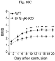
- IFN- ⁇ supports non-encephalitogenic trafficking, it could lead to pathogenicity in the presence of Th-17 cells, which were barely detectable in the naive choroid plexus. Supporting this notion is the fact that IFN- ⁇ R-KO mice fail to develop classic EAE (Lees et al., 2008). These results are also in line with the reported CCR6 + cell entry through the choroid plexus, linked with the induction of EAE (Reboldi et al., 2009), and might suggest that prior to Th-17 entry, this compartment is activated by IFN- ⁇ .
- the activation of trafficking through the choroid plexus which can potentially be the beginning of an inflammatory disease, does not necessarily portend to it; such an opening can serve as a gate for opportunistic cells, which cannot infiltrate by themselves, but can use the opened port to enter the parenchyma as "Trojan horses", thus induce autoimmune disease.
- results of this study provide experimental support for a dialogue that takes place between the circulating immune system and the choroid plexus epithelium. We found this epithelial-T cell crosstalk to facilitate the activation of the choroid plexus and to enable CNS maintenance and immune trafficking. Moreover, this study emphasizes that the mere activation of the choroid plexus for trafficking is not a sign of a beginning of a disease; the effector phenotype of the immune cells and the state of the parenchyma critically determine the outcome.
- the present inventors also show herein that, even though CNS parenchyma of mSOD1 mice (a model for human amyotrophic lateral sclerosis) shows signs of local inflammation, their choroid plexus is not activated to enable immune cell trafficking; however, the choroid plexus is amenable to activation as inferred by the fact that IFN- ⁇ treatment of choroid plexus cultures elevated the expression of trafficking molecules of the choroid plexus cultures, suggesting that the choroid plexus of mSOD1 mice has the ability to respond to effector T cell-derived cytokines (Examples 12 and 13).
- immunomodulation of the choroid plexus epithelium of mSOD mice with a Th1- adjuvant, CpG causes upregulation of all trafficking molecules tested (Example 14).
- the upregulation of the trafficking molecules enables the transfer of the beneficial T cells from the circulation to the CSF and the CNS parenchyma.
- MOG myelin oligodendrocyte glycoprotein
- tuberculosis induced the expression of the trafficking molecule ICAM-1 by the choroid plexus epithelium and infiltration of CD4 + T cells to the CSF (Example 15) and the ventral horn gray matter, the area where motor neurons reside (Example 16).
- the level of Treg cells in the choroid plexus is a function of the level of this cell type in the circulating immune system, such as the spleen and is critical for its function.
- depletion of Fox3P + Treg cells in mice causes the activation of the choroid plexus for trafficking (Example 10).
- Example 18 daily injection of the immune-modulator Copolymer 1 to 5XFAD mice (a mouse model for human Alzheimer's disease), causes enrichment of FoxP3+ Treg cells in the spleen and does not improve the cognitive function of the mice.
- Copolymer 1 is a random non-pathogenic synthetic copolymer, a heterogeneous mix of polypeptides containing the four amino acids L-glutamic acid (E), L-alanine (A), L-tyrosine (Y) and L-lysine (K) in an approximate ratio of 1.5:4.8:1:3.6, but with no uniform sequence.
- Copolymer 1 acts as a low-affinity antigen that activates a wide range of self-reacting T cells, resulting, when given at low frequency, in neuroprotective autoimmunity that is effective against both CNS white matter and grey matter degeneration.
- the present invention provides a method for treating a disease, disorder, condition or injury of the Central Nervous System (CNS) in a subject in need thereof, comprising administering to said subject a therapeutically effective amount of an active ingredient selected from the group consisting of (i) a non-encephalitogenic or weakly encephalitogenic combination of a Th1 adjuvant and an agent selected from a CNS-specific antigen, a peptide derived from a CNS-specific antigen or from an analog thereof, or an analog or derivative of said peptide; (ii) a non-encephalitogenic or weakly encephalitogenic combination of a Th1 adjuvant and CNS-reactive T cells; (iii) CNS-reactive T cells having a Th1 phenotype as sole active ingredient; or (iv) a Th1 adjuvant as a sole active ingredient provided that when said subject is afflicted with Alzheimer's disease, said Th1 adjuvant is not CpG, thereby activating
- an active ingredient
- a Th1 adjuvant as a sole active ingredient is administered provided that when said subject is afflicted with a disorder characterized by the presence of a pathological protein aggregate or neo-epitope, said Th1 adjuvant is not CpG.
- Such disorders include neurodegenerative diseases, such as Alzheimer Disease, Down's syndrome, cerebral amyloid angiopathy, mixed dementia, or inclusion body myositis, glaucoma, or arteriosclerosis associated amyloidoses, or other forms of amyloidoses comprising fibrillaric proteins derived from at least one of the following precursor proteins SAA (Serum-Amyloid-Protein A), AL (k or I-light chains ofImmunoglobulins), AH (gl Ig-heavy chains), ATTR (Transthyretin, Serum-Prealbumin), AApo-A-1 (Apolipoprotein AI), AApoA2 (Apolipoprotein A2), AGel (Gelsolin), ACys (Cystatin C), ALys (Lysozyme), AFib (Fibrinogen), Beta-amyloid (Amyloid precursor protein), Beta-amyloid2M (beta2-microglobul
- SAA Serum-Amyloid-Prot
- the level of the immunosuppression measured in the individual can be compared with a reference that may be, but is not limited to, the level of immunosuppression in the individual before the last administration of the active ingredient, or it may be a predetermined reference based on the level of immunosuppression in a population of individuals in need of treatment according to the present invention that are responding well to the treatment, or the level of immunosuppression in a population of healthy individuals.
- a reference may be, but is not limited to, the level of immunosuppression in the individual before the last administration of the active ingredient, or it may be a predetermined reference based on the level of immunosuppression in a population of individuals in need of treatment according to the present invention that are responding well to the treatment, or the level of immunosuppression in a population of healthy individuals.
- a decision can be made whether to repeat the administration of the active ingredient or wait and continue the monitoring.
- the present invention provides a method for treating a disease, disorder, condition or injury of the CNS in a subject in need thereof having a certain level of immunosuppression in the circulation, said method comprising administering to the subject a therapeutically effective amount of an active ingredient selected from the group consisting of (i) a non-encephalitogenic or weakly encephalitogenic combination of a Th1 adjuvant and an agent selected from a CNS-specific antigen, a peptide derived from a CNS-specific antigen or from an analog thereof, or an analog or derivative of said peptide; (ii) a non-encephalitogenic or weakly encephalitogenic combination of a Th1 adjuvant and CNS-reactive T cells; (iii) CNS-reactive T cells having a Th1 phenotype as sole active ingredient; or (iv) a Th1 adjuvant as a sole active ingredient, wherein said administering is performed according to a regimen causing reduction of the level of said immunos
- the invention further contemplates combining the administration of the agents in (i) to (iv) in the above described methods with administration of IFN- ⁇ .
- the method comprises: (a) administering the active ingredient of (i), (ii) or (iii) to the subject in need; and (b) determining said regimen by: (i) monitoring immunosuppression and/or Th1/Th2 balance in the subject by measuring in a blood sample obtained from the subject, within a predetermined time-period following the administering, one or more parameters reflecting a degree of immunosuppression and/or Th1/Th2 balance in the choroid plexus in the subject; and (ii) comparing the one or more parameters measured in (b)(i) with the reference and determining whether the one or more parameters is different from the reference; and (c) deciding, based on the relation of the one or more parameters measured in (b)(i) to the reference, whether to repeat treatment and monitoring by repeating steps (a) and (b) or to continue monitoring by repeating only step (b).
- the level of immunosuppression in the choroid plexus is reflected in the level of Treg cells in the circulation of the subject.
- This level is understood to undergo variations in response to events that affect the immune system. For example, disease may increase the level of Treg cells in the circulation and activation of the immune system by immunization may reduce the level of immunosuppression relative to the situation in disease.
- the level of Treg cells in the circulation may easily be measured by determining the level of CD4 + CD25 + FoxP3 + cells or CD4 + CD25 + FoxP3 - cells by conventional methods such as, but not limited to fluorescence activated cell sorting (FACS), flow cytometry or real-time PCR.
- FACS fluorescence activated cell sorting
- the Treg cells inhabiting the choroid plexus, the CNS or the circulation are CD4 + CD25 + FoxP3 + cells or CD4 + CD25 + FoxP3 - cells.
- the anti-inflammatory immune cells or immune cells which acquire a healing phenotype are IL-10 producing cells, such as activated M2-machrophages, CD4 + CD25 + Foxp3 + Treg cells or CD4 + CD25 + FoxP3 - Treg cells (see Example 17, Fig. 17D ).
- Th1/Th2 balance refers to the predominant immune response or immune status in terms of Th1-type cells (characterized by production of cytokines typical for Th1 response, such as IFN- ⁇ ) or cytokine milieu (IFN- ⁇ ) or Th2-type cells (characterized by production of cytokines typical for Th2 response, such as IL-4) or cytokine milieu (IL-4).
- Th1-type cells characterized by production of cytokines typical for Th1 response, such as IFN- ⁇
- IFN- ⁇ cytokine milieu
- Th2-type cells characterized by production of cytokines typical for Th2 response, such as IL-4
- IL-4 cytokine milieu
- Th1 cells express both components of IL-12 receptor chains (beta 1 and alpha.
- Th2 cells Only Th2 cells appear to express a fully functional IL-1 receptor, and ST2L/T1, a newly discovered IL-1 RI-like molecule, is found on Th2 cells only.
- Chemokine receptors CXCR3 and CCR5 are characteristic of Th1 cells, while CXCR4, CCR3, CCR4, CCR7 and CCR8 are associated with Th2 cells.
- CD30 a member of the TNF superfamily, is associated with Th2 cells.
- the term "circulation” as used herein refers to the circulating immune system, i.e. the blood, spleen and lymph nodes. It is a well-known fact in the field of immunology that the cell population profile in the spleen is reflected in the cell population profile in the blood (Zhao et al., 2007). Furthermore, daily administration of the immune-modulating drug Copolymer 1 raises the level of Treg cells in the blood of the subjects receiving the daily injections (Lalive et al., 2011). It has also been shown by the inventors of the present invention that daily administration of Copolymer 1 to mice raises the level of Treg cells in the spleen of the mice receiving the daily injections (Example 18). Therefore, data showing changes in the immune cell population in the spleen in response to immunization or disease reflect the changes occurring in the immune cell population in the blood, which in turn indicates the changes taking place in the choroid plexus.
- the parameter measured in a blood sample from the subject is: (i) a ratio of CD4 + Treg cells to CD4 + effector T cells; (ii) a level of CD4 + Treg cells; (iii) a level of IFN- ⁇ producing CD4 + cells; (iv) a ratio of CD4 + Treg cells to IFN- ⁇ producing CD4 + T cells; (v) a proliferative response of peripheral mononuclear cells to a CNS-specific antigen, a peptide derived from a CNS-specific antigen or from an analog thereof, or an analog or derivative of said peptide; or (vi) any combination of (i), (ii), (iii), (iv) and (v).
- the effector T cells measured are CD4 + cells that may be T-Bet + thus producing IFN- ⁇ or they may be T-Bet - that do not produce IFN- ⁇ .
- the IFN- ⁇ producing CD4 + cells may be CNS-specific as it has been shown in accordance with the present invention that the CD4+ cells at the choroid plexus are predominantly CNS-specific (Example 2, Fig. 2 ; Example 4, Figs. 4 and 5 ), and that leukocyte trafficking through the choroid plexus is achieved following immunization with a CNS-specific antigen, but is not achieved following immunization with an irrelevant antigen, thereby identifying the choroid plexus as a novel target for immunomodulation with CNS-specific antigen.
- the parameter indicating a Th1-type immune response may be a predominating level of IFN- ⁇ producing CD4 + cells, while the parameter indicating a Th2-type immune response may be a predominating level of IL-4 producing CD4 + cells.
- the reference is selected from the group consisting of (a) a parameter measured in the most recent blood sample obtained from said subject before said administering, said parameter indicating a degree of immunosuppression and/or Th1/Th2 status in the choroid plexus in said subject.
- the reference is the parameter measured before initiation of treatment of the subject, i.e.
- a base-line degree of immunosuppression and/or Th1/Th2 status in the subject that is in need of treatment and before the treatment starts (b) a parameter indicating the degree of immunosuppression and/or Th1/Th2 status in the choroid plexus characteristic of a population of subjects afflicted with a disease, disorder, condition or injury of the CNS responding well to said administering, wherein said parameter is measured in blood samples obtained from said subjects; or (c) a parameter indicating the degree of immunosuppression and/or Th1/Th2 status in the choroid plexus characteristic of a population of healthy subjects, wherein said parameter is measured in blood samples obtained from said subjects.
- the reference is measured in a blood sample obtained from said subject before said administering in (a).
- the monitoring is thus used to determine whether treatment and monitoring should be repeated or only monitoring should be continued/repeated. For example, but limited thereby, if the reference is the base-line level of ratio of Treg to effector T cells of the subject in need of treatment before treatment has been initiated (measured at the first instance of monitoring), and the subject has been administered the active ingredient once (the first administration), then treatment and monitoring will be repeated if the parameter measured at the second instant of monitoring (measured after the first administering of the active ingredient) is similar to or higher than the base-line reference. The subsequent monitoring will compare the parameter measured after the second administration of the active ingredient with the last parameter measured, which is now the new reference. This cycle is repeated until the treatment is discontinued.
- the subject will not receive an additional dose of the active ingredient but will only be monitored a second time, and then a new decision is made.
- the subsequent monitoring will compare the parameter with the base-line reference. It is also contemplated that the subsequent monitoring will compare the parameter measured at the second monitoring with the parameter measured at the previous monitoring (that was made without treatment), which is now the new reference. The aim is to keep the parameter below the base-line reference.
- the reference used is a parameter indicating the degree of immunosuppression/Th-phenotype in the choroid plexus characteristic of a population of subjects afflicted with a disease, disorder, condition or injury of the CNS responding well to said administering, wherein said parameter is measured in blood samples obtained from said subjects; or a parameter indicating the degree of immunosuppression, Th-phenotype, or both, in the choroid plexus characteristic of a population of healthy subjects, wherein said parameter is measured in blood samples obtained from said subjects, then treatment and monitoring will be repeated if the parameter measured subsequent to treatment in a blood sample obtained from the subject in need of treatment is above said reference. If, on the other hand, the parameter measured after administration of the active ingredient to the subject is equal to or lower than this reference, then only monitoring is repeated and no administration of the active ingredient is performed.
- the parameter measured is the ratio of Treg cells to effector CD4 + T cells in a blood sample obtained from said subject
- said reference is the most recent ratio measured in said subject before said administering the active ingredient
- said treatment and monitoring is repeated when the ratio is substantially similar to or higher than the reference; or (ii) said monitoring is repeated when the parameter is lower than the reference value.
- the predetermined time-period between monitoring and administration of the active ingredient is between 1 and 16 weeks.
- the predetermined time-period may be 2, 3, 4, 5 or six days; or 1, 2, 3, 4, 5, 6, 7, 8, 9, 10, 11, 12, 13, 14, 15 or 16 weeks.
- the Th1 adjuvant augments the choroid plexus's ability to let beneficial Th1 type CD4+ T cells and other anti-inflammatory immune cells or immune cells which acquire a healing phenotype at the cerebrospinal fluid pass through and enter the CNS parenchyma, home to the injured site and assist in the healing of the injured tissue.
- the Th1 adjuvant comprises an agonist of TLR3, 4, 5, 7, 8 or 9; or an antagonist of TLR2 or neutralizing antibody directed to TLR2.
- the TLR agonist may be an agonist of TLR9, such as a CpG or stabilized immune modulatory RNA (SIMRA).
- the CpG may be of class B, for example ODN1826.
- the Th1 adjuvant of the present invention may also be a CpG in combination with a cationic peptide of the sequence KLKL 5 KLK (SEQ ID NO: 2).
- the TLR agonist may be an agonistic antibody, i.e. an antibody that binds to and activates the TLR.
- CpG CpG oligodeoxynucleotides
- CpG ODN CpG ODN
- p refers to the phosphodiester link between consecutive nucleotides, although some ODN have a modified phosphorothioate (PS) backbone instead.
- the TLR3 agonists may be selected from e.g. double-stranded RNA (dsRNA), a molecular pattern associated with viral infection, or a synthetic analog thereof, such as polyinosine-polycytidylic acid (poly(I:C)).
- dsRNA double-stranded RNA
- poly(I:C) polyinosine-polycytidylic acid
- the agonist of TLR 4 may be MPL (3-O-desacyl-4'-monophosphoryl lipid) optionally in combination with alum (AS04).
- the TLR5 agonist may be flagellin, e.g. flagellin purified from B.subtilis (Gram+) or S.typhimurium (Gram-) bacteria.
- the TLR 7/8 agonist may be a single stranded RNA or a small synthetic molecule such as imidazoquinoline or a nucleoside analog, e.g. Gardiquimod, Imiquimod and R848 (resiquimod).
- the Th1 adjuvant comprises a combination of an agonist of TLR 3 with an agonist of TLR 8, or a combination of an agonist of TLR 4 with an agonist of TLR 8.
- the Th1 adjuvant of the present invention may be an agonist of retinoic acid inducible gene I (RIGI) or an agonist of dectin-1.
- central nervous system (CNS)-specific antigen refers to an antigen of the central nervous system that specifically activates T cells such that following activation the activated T cells accumulate at a site of injury or disease in the CNS of a patient, or at the choroid plexus.
- the CNS-specific antigens and analogs thereof, the peptides derived from CNS-specific antigens or from analogs thereof, and analogs or derivatives of said peptides include, but are not limited to, the agents described in US Patent No. 7,560,102 and PCT Publications WO 99/60021 and WO 02/055010 of the applicant, all these applications being herewith incorporated by reference as if fully disclosed herein.
- the active ingredient is a non-encephalitogenic or weakly encephalitogenic combination of a Th1 adjuvant and an agent from a CNS-specific antigen of mammal, preferably human, origin, selected from the group consisting of myelin basic protein (MBP; SEQ ID NO: 3); myelin oligodendrocyte glycoprotein (MOG; SEQ ID NO: 5); proteolipid protein (PLP; SEQ ID NO: 4); myelin-associated glycoprotein (MAG); S-100; ⁇ -amyloid; Thy-1; a peripheral myelin protein including P0, P2 and PMP22; neurotransmitter receptors including acetylcholine receptor; and the protein Nogo including Nogo-A, Nogo-B and Nogo-C and the Nogo receptor.
- MBP myelin basic protein
- MOG myelin oligodendrocyte glycoprotein
- PGP proteolipid protein
- MAG myelin-associated glycoprotein
- S-100
- the active ingredient is a non-encephalitogenic or weakly encephalitogenic combination of a Th1 adjuvant and a peptide derived from a CNS-specific antigen or from an analog thereof, or an analog or derivative of said peptide.
- the peptide may be an immunogenic epitope or a cryptic epitope derived from said CNS-specific antigen.
- a peptide derived from an CNS-specific antigen relates to a peptide that has a sequence comprised within the CNS-antigen sequence.
- the peptide derived from a CNS-specific antigen may be selected from MBP 11-30 , MBP 51-70 , MBP 83-99 , MBP 87-99 , MBP 91-110 , MBP 131-150 , MBP 151-170 or MBP 84-104 , MOG 35-55 or MOG 92-106 , or PLP 139-151 , or PLP 178-191 .
- the peptide is MOG 35-55 .
- the agent is an analog of a CNS-specific antigen peptide obtained by modification of a self-peptide derived from a CNS-specific antigen, which modification consists in the replacement of one or more amino acid residues of the self-peptide by different amino acid residues or by deletion or addition of one or more amino acid residues, said modified CNS peptide still being capable of recognizing the T-cell receptor recognized by the self-peptide but with less affinity (hereinafter "modified CNS peptide", “altered peptide” or "analog of CNS-specific peptide”).
- the invention also comprises chemical derivatives of the peptides of the invention including, but not being limited to, esters of both carboxylic and hydroxy groups, amides, and the like.
- the modified CNS peptide may be immunogenic but not encephalitogenic.
- the most suitable peptides for this purpose are those in which an encephalitogenic self-peptide is modified at the T cell receptor (TCR) binding site and not at the MHC binding site(s), so that the immune response is activated but not anergized.
- TCR T cell receptor
- the invention also encompasses peptides derived from any encephalitogenic epitopes in which critical amino acids in their TCR binding site but not MHC binding site are altered as long as they are non-encephalitogenic and still recognize the T-cell receptor.
- the modified peptide is derived from the residues 86 to 99 of human MBP by alteration of positions 91, 95 or 97 as disclosed in US 5,948,764 .
- the modified peptide is a peptide disclosed in WO 02/055010 , derived from the residues 87-99 of human MBP, in which the lysine residue 91 is replaced by glycine (G91) or alanine (A91), or the proline residue 96 is replaced by alanine (A96), or the serine residue 45 of the peptide MOG 35-55 is replaced with aspartic acid (MOG-45D).
- G91 glycine
- A91 alanine
- A96 proline residue 96
- serine residue 45 of the peptide MOG 35-55 is replaced with aspartic acid (MOG-45D).
- MOG-45D aspartic acid
- the modified CNS-peptide is selected from the peptides MBP 87-99 (G91), MBP 87-99 (A91), MBP 87-99 (A96) and MOG 35-55 (D45).
- the Th1 adjuvant is a CpG.
- the active ingredient comprises the peptide MOG 35-55 and the Th1 adjuvant CpG.
- the active ingredient is a non-encephalitogenic or weakly encephalitogenic combination of a Th1 adjuvant and CNS-reactive T cells generated by ex vivo expansion of T cells in the presence of a CNS-specific antigen; or CNS-reactive T cells generated by expansion of T cells in the presence of a CNS-specific antigen and activated towards a Th1 phenotype ex vivo, as a sole active ingredient.
- the CD4+ T cells of Th1 phenotype may be generated by contacting the CD4 + T cells with CNS-specific activated antigen-present cells, IFN- ⁇ or other relevant cytokines or relevant TLR agonists or antagonists.
- the CD4+ T cells administered to the subject in need thereof are syngeneic or autologous.
- the induction of homeostatic-driven proliferation of T cells can be accomplished by irradiation and bone marrow transplantation or administration of chemotherapeutic drugs such as alkylating agents, antimetabolites, anthracyclines, plant alkaloids, or topoisomerase inhibitors.
- chemotherapeutic drugs such as alkylating agents, antimetabolites, anthracyclines, plant alkaloids, or topoisomerase inhibitors.
- non-encephalitogenic Th1 adjuvant refers to an adjuvant that stimulates the immune system and directs the differentiation of the immune cells towards the Th1 phenotype, but excluding the Th17 phenotype. Thus, this agent stimulates the cellular immune response without causing encephalitis.
- the agent may be a CNS specific antigen, a peptide derived from a CNS-specific antigen or from an analog thereof, or an analog or derivative of said peptide, or CNS-reactive T cells.
- weakly encephalitogenic refers to encephalitogenic activity that causes monophasic encephalomyelitis, i.e.
- transient encephalitis that spontaneously resolves, or that causes weakly symptomatic disease as measured by time of onset or mean maximum disease score.
- An agent may easily be shown to be weakly encephalitogenic by showing that the induced encephalomyelitis is monophasic, or by measuring time of onset or mean maximum disease score by using methods well known in the art (e.g. Oliver et al., 2003).
- time of onset may be assessed by comparing the time of onset of EAE caused by the agent suspected as "weakly encephalitogenic" with the time of onset of EAE caused by agents known to be highly encephalitogenic or weakly encephalitogenic and showing that the time of onset is after that of the highly encephalitogenic agent and approximately simultaneously with that of the weakly encephalitogenic agent.
- a weakly encephalitogenic agent may cause the onset of EAE to occur on day 18 on average with a range of 11 to 28 days after initial immunization with the agent.
- the mean maximum disease score of the allegedly weakly encephalitogenic agent can similarly be assessed by comparing it with mean maximum disease score of known highly encephalitogenic or weakly encephalitogenic agents and showing that the mean maximum disease score of the allegedly weakly encephalitogenic agent is lower than that of the known highly encephalitogenic agent and similar to the known weakly encephalitogenic agent.
- the mean maximum disease score of the allegedly weakly encephalitogenic agent may be around 1.7 with a range of 0.5-2.5.
- reestablishment of a Th1/Th2 phenotype typical for young animals at the choroid plexus by injection of a non-encephalitogenic Th1 adjuvant (in this case CpG) into aging healthy animals improves their cognitive abilities.
- CpG non-encephalitogenic Th1 adjuvant
- administration of CpG augments the immune system's natural capability of treating injury and neurodegeneration in the CNS by upregulating both trafficking and neurotropic molecule at the choroid plexus and thus increasing leukocyte recruitment to the injured spinal cord or neurodegenerative brain in ALS.
- the vaccination with the NS-specific antigen elicited a Th1 immune response that induced the expression of trafficking molecules by the CP (Example 17) enabling infiltration of leukocytes to the cerebrospinal fluid (CSF), and initiated an active programmed immune response that began with inflammation, and was followed by an anti-inflammatory resolution process.
- This response was manifested by transient symptoms of encephalomyelitis that were correlated spatially and temporally with an increase in regulatory T cells in the spinal cord of the immunized mSOD1 mice, leading to a significant increase in survival of these mice compared with unimmunized mSOD1 mice.
- the method of the present invention is beneficial for treating a disease, disorder or condition of the CNS selected from the group consisting of a neurodegenerative disease, disorder or condition selected from the group consisting of amyotrophic lateral sclerosis, Alzheimer's disease, Parkinson's disease and Huntington's disease; primary progressive multiple sclerosis; secondary progressive multiple sclerosis; a retinal degeneration disorder selected from the group consisting of age-related macular degeneration and retinitis pigmentosa; anterior ischemic optic neuropathy; glaucoma; uveitis; depression; stress; and Rett syndrome.
- the present invention is beneficial for amyotrophic lateral sclerosis.
- the method of the present invention may also increase the life span of a subject with amyotrophic lateral sclerosis
- the method of the present invention is useful for treating an injury of the CNS selected from spinal cord injury, closed head injury, blunt trauma, penetrating trauma, hemorrhagic stroke, ischemic stroke, cerebral ischemia, optic nerve injury, myocardial infarction, organophosphate poisoning and injury caused by tumor excision.
- an injury of the CNS selected from spinal cord injury, closed head injury, blunt trauma, penetrating trauma, hemorrhagic stroke, ischemic stroke, cerebral ischemia, optic nerve injury, myocardial infarction, organophosphate poisoning and injury caused by tumor excision.
- the method of the present invention is beneficial for improving CNS motor and/or cognitive function, for example for alleviating age-associate loss of cognitive function, either in diseased subjects or subjects free of a diagnosed disease.
- the method is also efficacious for alleviating loss of cognitive function resulting from acute stress.
- the cognitive function is learning, memory or both.
- the memory is hippocampal-dependent memory, explicit memory, episodic free recall, recollection of source of remembered fact, memory of details of context in which an event occurred, or working memory.
- the method of the present invention is also useful for inhibiting neuronal degeneration in the central nervous system (CNS), protecting neurons from glutamate toxicity or promoting nerve regeneration in nerve tissue damaged by injury to the CNS or by a disease, disorder or condition of the CNS.
- CNS central nervous system
- the nerve regeneration is associated with hippocampal plasticity.
- CNS function refers to CNS motor functions, such as controlling motor function and auditory and visual responses, maintaining balance and equilibrium, movement coordination, the conduction of sensory information and controlling such autonomic functions as breathing, heart rate, and digestion, and to CNS cognitive functions, inter alia, receiving and processing sensory information, thinking, learning, memorizing, perceiving, producing and understanding language.
- the present invention provides a vaccine for use in a method of therapeutic immunization of a mammal comprising an active ingredient selected from (i) a non-encephalitogenic or weakly encephalitogenic combination of a Th1 adjuvant and an agent selected from a CNS-specific antigen, a peptide derived from a CNS-specific antigen or from an analog thereof, or an analog or derivative of said peptide; (ii) a non-encephalitogenic or weakly encephalitogenic combination of a Th1 adjuvant and CNS-reactive T cells; (iii) CNS-reactive T cells having a Th1 phenotype as sole active ingredient; or (iv) a non-encephalitogenic Th1 adjuvant as a sole active ingredient provided that when said subject is afflicted with Alzheimer's disease, said non-encephalitogenic Th1 adjuvant is not CpG, wherein the vaccine is to be administered according to a regimen to thereby confer reduction of the level of
- the present invention provides a vaccine for use in a method of therapeutic immunization of a mammal comprising an active ingredient selected from: (i) a non-encephalitogenic or weakly encephalitogenic combination of a Th1 adjuvant and an agent selected from a CNS-specific antigen, a peptide derived from a CNS-specific antigen or from an analog thereof, or an analog or derivative of said peptide; (ii) a non-encephalitogenic or weakly encephalitogenic combination of a Th1 adjuvant and CNS-reactive T cells; (iii) CNS-reactive T cells having a Th1 phenotype as sole active ingredient; or (iv) a Th1 adjuvant as a sole active ingredient provided that when said subject is afflicted with Alzheimer's disease, said Th1 adjuvant is not CpG, wherein the vaccine is to be administered according to a regimen to thereby confer reduction of the level of immunosuppression in the circulation of said mammal comprising
- the present invention relates to a pharmaceutical composition
- a pharmaceutical composition comprising a pharmaceutically acceptable carrier and a combination of agents selected from the group consisting of (i) a non-encephalitogenic or weakly encephalitogenic combination of a Th1 adjuvant and an agent selected from a CNS-specific antigen, a peptide derived from a CNS-specific antigen or from an analog thereof, or an analog or derivative of said peptide; (ii) a non-encephalitogenic or weakly encephalitogenic combination of a Th1 adjuvant and CNS-reactive T cells; (iii) IFN- ⁇ and a Th1 adjuvant; (iv) a combination of (i) with IFN- ⁇ ; and (v) a combination of (ii) with IFN- ⁇ .
- agents selected from the group consisting of (i) a non-encephalitogenic or weakly encephalitogenic combination of a Th1 adjuvant and an agent selected from a CNS-specific antigen
- the present invention further contemplates methods for determining a need for administration of an active ingredient to a subject in need of treatment of a disease, disorder, condition or injury of the CNS by activating the choroid plexus of said subject and maintaining activation thereof by reducing immune-suppression and establishing Th1-type immune response at the choroid plexus, thereby allowing Treg cells and macrophages to pass through the choroid plexus and accumulate at a site of damage in the CNS caused by said disease, disorder, condition or injury, wherein said active ingredient is selected from the group consisting of (i) a non-encephalitogenic or weakly encephalitogenic combination of a Th1 adjuvant and an agent selected from a CNS-specific antigen, a peptide derived from a CNS-specific antigen or from an analog thereof, or an analog or derivative of said peptide; (ii) a non-encephalitogenic or weakly encephalitogenic combination of a Th1 adjuvant and CNS-reactive
- treating refers to means of obtaining a desired physiological effect.
- the effect may be therapeutic in terms of partially or completely curing a disease and/or symptoms attributed to the disease.
- the term refers to inhibiting the disease, i.e. arresting its development; or ameliorating the disease, i.e. causing regression of the disease.
- Methods of administration include, but are not limited to, parenteral, e.g., intravenous, intraperitoneal, intramuscular, subcutaneous, mucosal (e.g., oral, intranasal, buccal, vaginal, rectal, intraocular), intrathecal, topical and intradermal routes. Administration can be systemic or local.
- parenteral e.g., intravenous, intraperitoneal, intramuscular, subcutaneous, mucosal (e.g., oral, intranasal, buccal, vaginal, rectal, intraocular), intrathecal, topical and intradermal routes.
- parenteral e.g., intravenous, intraperitoneal, intramuscular, subcutaneous, mucosal (e.g., oral, intranasal, buccal, vaginal, rectal, intraocular), intrathecal, topical and intradermal routes.
- mucosal e.g., oral, intranasal, buccal
- carrier refers to a diluent, adjuvant, excipient, or vehicle with which the active agent is administered.
- the carriers in the pharmaceutical composition may comprise a binder, such as microcrystalline cellulose, polyvinylpyrrolidone (polyvidone or povidone), gum tragacanth, gelatin, starch, lactose or lactose monohydrate; a disintegrating agent, such as alginic acid, maize starch and the like; a lubricant or surfactant, such as magnesium stearate, or sodium lauryl sulphate; and a glidant, such as colloidal silicon dioxide.
- a binder such as microcrystalline cellulose, polyvinylpyrrolidone (polyvidone or povidone), gum tragacanth, gelatin, starch, lactose or lactose monohydrate
- a disintegrating agent such as alginic acid, maize starch and the like
- a lubricant or surfactant such as
- the determination of the doses of the active ingredient to be used for human use is based on commonly used practices in the art, and will be finally determined by physicians in clinical trials.
- An expected approximate equivalent dose for administration to a human can be calculated based on the in vivo experimental evidence disclosed herein below, using known formulas (e.g. Reagan-Show et al. (2007) Dose translation from animal to human studies revisited. The FASEB Journal 22:659-661 ). According to this paradigm, the adult human equivalent dose (mg/kg body weight) equals a dose given to a mouse (mg/kg body weight) multiplied with 0.081.
- mice Inbred male 3 month old C57BL/6 mice were supplied by the Animal Breeding Center of the Weizmann Institute of Science. Inbred male 17-24 months old C57BL/6 mice were supplied by the National Institute on Aging (NIA; Baltimore, MD). IPN ⁇ K1-KO mice on the B6 background were purchased from the Jackson Laboratory (Bar Harbor, Maine). Aged mice were allowed a 1-month adaptation period following shipment from the NIA to our laboratory. The cages were placed in a light- and temperature-controlled room, and all behavioural tests were conducted during the dark hours. All animals were handled according to the regulations formulated by the Weizmann Institute's Animal Care and Use Committee, and maintained in a pathogen free environment.
- Bone marrow (BM) chimeras were prepared by subjecting gender-matched recipient mice to lethal whole-body ⁇ -irradiation (950 rad) while shielding the head. The mice were then reconstituted with 5 ⁇ 10 6 BM cells. In this method, 70% chimerism is achieved. In order to attain full chimerism (99%), mice were subjected to a split-dose irradiation, with sub-lethal whole-body ⁇ -irradiation (300 rad) without head shielding 3 days prior to the lethal (950 rad) irradiation. Chimeric mice were used 6-10 weeks after BM transplantation.
- mice on a C57BL/6J background were supplied by Harlan Biotech (Jerusalem, Israel), and the Animal Breeding Center of The Weizmann Institute of Science. All experiments conformed to the regulations formulated by the Institutional Animal Care and Use Committee of the Weizmann Institute of Science.
- IFN- ⁇ or IL-17 For intracellular staining of IFN- ⁇ or IL-17, the cells were incubated with PMA (10 ng/ml; Sigma-Aldrich) and ionomycin (250 ng/ml; Sigma-Aldrich) for 6 h, and Brefeldin-A (10 ⁇ g/ml; Sigma-Aldrich) was added for the last 4 h. Intracellular labeling of cytokines was done with BD Cytofix/Cytoperm Plus Fixation/Permeabilization kit (cat. no. 555028) according to the manufacturer's protocol. Intracellular staining of FoxP3 was performed using the FoxP3 staining kit (eBioscience) according to the manufacturer's protocol.
- fluorochrome-labeled mAbs were used according to the manufacturers' protocols: FITC-conjugated anti-TCR ⁇ , FITC-conjugated anti-CD44, PE-conjugated anti-CD4, PE-conjugated anti-IL-17A, APC-conjugated anti-IFN-y, FITC-conjugated anti-CD45.2, Alexa-700-conjugated anti-CD45.2, Percp-Cy5.5-conjugated anti-TCR ⁇ (all from BioLegend), and APC-conjugated anti-FoxP3 (eBioscience).
- Flow cytometry analysis was performed on each sample using a BD Biosciences LSRII flow cytometer, and the acquired data were analyzed using FlowJo software (Tree star). In each experiment, relevant negative control groups and single stained samples for each tissue were used to determine the populations of interest and to exclude others.
- SCH spinal cord homogenate
- OVA ovalbumin
- SCH was prepared by manually homogenizing the spinal cord of young C57BL/6 mice in PBS. Mice were intracardially perfused with PBS 7 days after immunization, and their spleens, bone marrow and choroid plexus (CP) were extracted and processed to single cell suspensions as described above.
- CD4 + cells were isolated by magnetic depletion of CD4 + T cells on a MACS column (Milteney Biotec), according to the manufacturer's protocol.
- MOG 35-55 amino acid sequence - MEVGWYRSPFSRVVHLYRNGK; SEQ ID NO: 1
- ovalbumin peptides 323-339 GL Biochem (Shanghai) Ltd.
- mice were subcutaneously injected with 200 ⁇ g (300 ⁇ l) of MOG 35-55 (GL Biochem (Shanghai) Ltd.) in incomplete Freund's adjuvant containing 2.5 mg/ml M. tuberculosis (strain H37Ra; BD Diagnostics).
- Pertussis toxin 300 ng; Sigma-Aldrich was injected on the day of the immunization and again 2 days later.
- Clinical signs were evaluated in a blinded fashion by at least 2 investigators and recorded daily (0, healthy; 1, limb tail paralysis; 2, ataxia and/or paresis of hind limbs; 3, paralysis of hind limbs and/or paresis of forelimbs; 4, tetraparalysis; 5, moribund state or death).
- CSF collection was collected by the cisterna magna puncture technique.
- mice were anesthetized and placed on a stereotactic instrument so that the head formed a 135° angle with the body.
- a sagittal incision of the skin was made inferior to the occiput and the subcutaneous tissue and muscle were separated, and a capillary was inserted into the cisterna magna through the dura matter lateral to the arteria dorsalis spinalis.
- Approximately 15 ⁇ l CSF could be aspirated from an individual mouse. The collected CSF was taken for analysis by flow cytometry.
- cDNA was then used as template for high fidelity PCR amplification (Phusion, Finnzymes) using a pool of 23 V ⁇ -specific primers, divided into five primer groups, to minimize potential for cross hybridization.
- Each V ⁇ primer was linked to a restriction site sequence for the ACUI restriction enzyme (NEB).
- PCR reactions were performed in duplicates, and PCR products were then pooled and cleaned using QIAquick PCR purification kit (Qiagen), followed by enzymatic digestion, in accordance with the ACUI protocol (NEB).
- the ACUI enzyme was used to cleave the 14 bp downstream of its binding site, enabling positioning of the Illumina sequencing primer in close proximity to the junction region and assuring sequencing of the entire variable CDR3 region.
- the clustering procedure identified unique CDR3 ⁇ clonotypes - defined as the most prevalent read found in each cluster.
- the clonotype sequences were then translated, and those clonotypes that lacked a stop codon in frame with the V/D/J sequences were considered for further analysis.
- This analysis computed statistics of V/D/J usage, statistical properties of the number of deletions and insertions of nucleotides at both VD and DJ junctions, as well as distributions of CDR3 lengths. The analysis was done using the R statistical software package (R Development Core Team, R). Frequencies of V ⁇ and J ⁇ segment usage were measured for all samples in each treatment group.
- Correlation coefficients were calculated, based on the sample mean of combined V ⁇ and J ⁇ usage in each group, using the data analysis program MATLAB TM (Mathworks, Natick, MA). Hierarchical clustering was performed, based on combined V ⁇ and J ⁇ usage, in all groups (clustergram, MATLAB TM).
- mice Primary culture of choroid plexus cells. Mice were perfused from the left ventricle of the heart with PBS, and their CPs were removed under a dissecting microscope (Stemi DV4; Zeiss) in PBS into tubes containing 0.25% trypsin, and kept on ice. The tubes were shaken for 20 minutes at 37°C and the tissue was dissociated by pipetting.
- the cell suspension was washed in culture medium for epithelial cells (DMEM/HAM's F12 (Invitrogen Corp)) supplemented with 10% FCS (Sigma-Aldrich), 1 mM 1-glutamine, 1 mM sodium pyruvate, 100 U/ml penicillin, 100 mg/ml streptomycin, 5 ⁇ g/ml Insulin, 20 ⁇ M Ara-C, 5ng/ml sodium selenite, 10ng/ml EGF, and cultured (-250,000 cells/well) at 37°C, 5% CO2 in 24-well plates (Corning Incorporated) coated with poly-L-lysine (PLL; Sigma-Aldrich).
- DMEM/HAM's F12 Invitrogen Corp
- FCS Sigma-Aldrich
- cytokines All purchased from PeproTech: IFN- ⁇ , IL-4, IL-10, IL-17A, GM-CSF, IL-6, IL-1 ⁇ or TNF- ⁇ , or their combinations for 24 h.
- Cell viability was quantified by Trypan blue staining after detachment of the cells with 0.25% trypsin for 10 minutes at 37°C.
- Neutralizing antibodies to TNF-R1 or TNF-R2 (20 ⁇ g/ml, BioLegend) were added 1 h prior to the addition of TNF- ⁇ , (100 ng/ml).
- RNA was isolated using the ZR RNA microprep kit (Zymo Research) according to the manufacturer's protocol.
- Transepithelial migration assay For transepithelial migration assay, CP cells were isolated as above and plated on 5- ⁇ m pore size, 6.5 mm diameter polycarbonate filters in 24-well Transwell chambers (Costar, Corning) and grown for 7 days. The medium of the upper chamber was then replaced with 100 ⁇ l of freshly isolated CD115 + monocytes (1.5 x 10 6 cells/ml in RPMI 1640 supplemented with 0.5% BSA) and the inserts were transferred to new wells containing RPMI 1640 supplemented with 0.5% BSA and 100ng/ml CCL2 (PeproTech). The Transwells were then incubated at 37°C, 5% CO 2 . After 24 h the migrating cells at the lower chamber were removed using cell scraper and CD11b + cells were quantified by flow cytometry.
- GVA Glycerol Vinyl Alcohol
- rat anti-CD3 (Abcam); rat anti-ICAM-1 (1:100; Abcam), chicken anti-vimentin (1:500, Millipore), rabbit anti-Ibal (1:300; Wako), goat anti-IL-10 (1:20; R&D), mouse anti-MHC-II (Abcam); mouse anti-Arginase-1 (BD Biosciences); mouse anti-Cytokeratin (Covance); rat anti-CCL11 (R&D Systems); rabbit anti-ZO-1 (Invitrogen); and rabbit anti-Claudin-1 (Invitrogen).
- rat anti-CD3 (Abcam); rat anti-ICAM-1 (1:100; Abcam), chicken anti-vimentin (1:500, Millipore), rabbit anti-Ibal (1:300; Wako), goat anti-IL-10 (1:20; R&D), mouse anti-MHC-II (Abcam); mouse anti-Arginase-1 (BD Biosciences); mouse anti-Cytokeratin (Covance); rat anti
- Secondary antibodies included: Cy2/Cy3-conjugated donkey anti-rat, mouse or rabbit antibody and Cy3 conjugated donkey anti-mouse, goat, rat, chicken or rabbit (1:200; all from Jackson Immuno Research).
- the slides were exposed to Hoechst stain (1:2000; Invitrogen) for 1 min.
- Two negative controls were routinely used in immunostaining procedures, staining with isotype control antibody followed by secondary antibody, and staining with secondary antibody alone.
- CP were isolated from lateral, 3rd and 4th ventricles and pooled in groups of four, due to the limited amount of protein extracted from a single CP.
- the excised tissues were homogenized in PBS containing protease inhibitors (1:100; P8340, Sigma). Four freeze-thaw cycles (3 minutes each) were performed to break the cell membranes. Homogenates were then centrifuged for 10 min at 500g, and the total protein quantities in supernatants were determined by Bradford reagent.
- Frozen supernatants were assayed in duplicate using Multiplex bead-based Luminex Assay (MILLIPLEX mouse cytokine/chemokine panel; Millipore), performed by outsourcing (American Medical Laboratories) according to the manufacturer's instructions. Results are expressed as picograms of protein per milligram of total tissue protein.
- RNA purification, cDNA synthesis and real-time quantitative PCR Total RNA of the hippocampus was extracted with TRI reagent (MRC, Cincinnati, OH) and was purified from the lysates and spinal cord using the RNeasy kit (Qiagen, Hilden, Germany). Total RNA of the choroid plexus was extracted using the RNA MicroPrep kit (Zymo Research). mRNA (1 ⁇ g) was converted to cDNA using High Capacity cDNA Reverse Transcription Kit (Applied Biosystems). The expression of specific mRNAs was assayed using fluorescence based real-time quantitative PCR (qPCR). qPCR reactions were performed using Power SYBR Green PCR Master Mix (Applied Biosystems).
- Glyceraldehyde 3-phosphate dehydrogenase Glyceraldehyde 3-phosphate dehydrogenase (GAPDH), Peptidylprolyl isomerase A (PPIA), and hypoxanthine guanine phosphoribosyltransferase (HPRT) were chosen as reference genes according to their stability in the target tissue.
- the amplification cycles were 95°C for 5 sec, 60°C for 20 sec, and 72°C for 15 sec.
- a melting curve was constructed to evaluate the specificity of the reaction.
- the cDNA was pre-amplified in 14 PCR cycles with non-random PCR primers, thus increasing the sensitivity of the subsequent real-time PCR analysis (PreAmp Master Mix Kit, Applied Biosystems).
- expression was determined using TaqMan Real-Time PCR, according to manufacturer's instructions (Applied Biosystems). All quantitative real-time PCR reactions were performed and analyzed using the 7500 Real-Time PCR System (Applied Biosystems).
- Transplanted BM was comprised of either whole BM, or BM depleted of T-cells, which was prepared by magnetic depletion of CD3 + cells on a MACS column (Milteney Biotec), according to the manufacturer's protocol.
- BM transplantation was performed on the following day, by i.v. injection of 5x10 6 bone-marrow cells suspended in PBS (total volume 0.15 ml).
- BM cells were prelabeled with carboxyfluorescein succinimidyl ester (CFSE, Molecular Probes) prior to transplantation.
- CFSE carboxyfluorescein succinimidyl ester
- 5x10 6 BM cells (20x10 6 cells/mL) were incubated in PBS (without Ca 2+ /Mg 2+ ) supplemented with 5 ⁇ M CFSE (Molecular Probes) for 8 min at 25°C. Following incubation, the cells were washed with RPMI containing 8% FBS.
- RAWM Radial arm water maze
- mice were given three additional trials without the platform to extinguish their initial memory of the platform's position.
- the platform was placed in a new location in the pool (opposite to where it was located in the acquisition phase) and the mice were given three trials per day on 2 consecutive days, conducted in a similar manner to the initial acquisition. Position and movement of the mice were recorded using an EthoVision automated tracking system (Noldus).
- CD115 + monocytes were isolated as previously reported (Shechter et al. , 2009). Briefly, BM cells were harvested from the femur and tibiae of naive mice, and enriched for mononuclear cells on a Ficoll density gradient. The CD115 + BM monocyte population was isolated by MACS enrichment using biotinylated anti-CD115 antibodies and streptavidin-coupled magnetic beads (Miltenyi Biotec) according to the manufacturer's protocols. Monocyte purity was checked by flow cytometry based on CD11b reactivity and was 90%.
- CD44 high /CD62L high are central memory T cells (T CM ) that home to secondary lymphoid organs and generally have little to no effector function
- CD44 high /CD62L -/low are effector memory T cells (T EM ) that survey peripheral tissues, and can be locally activated to an immediate effector function upon exposure to their cognate antigen.
- T CM central memory T cells
- T EM effector memory T cells
- T cells were co-localized with the CP epithelium and adjacent to MHC-II expressing APCs (data not shown), indicating their interactions with APCs in the tissue.
- T EM cells that reside in the CP are constitutively activated by the APCs that present their cognate antigens. If this is the case, these T cells are likely to be specific for brain antigens.
- Example 2 The choroid plexus CD4 + TCR repertoire is enriched with CNS-specific clones.
- TCR-seq a high throughput sequencing procedure of the TCR ⁇ CDR3 region
- the TCR ⁇ repertoire composition of the above groups was further compared using different parameters. We observed a high level of similarity in V ⁇ usage between the TCR ⁇ repertoire found in CP of naive animals and that found in the spleens of the animals immunized with CNS antigens (SCH) (data not shown). In contrast, the CP repertoire had a much lower correlation with the repertoire found in spleens of naive mice or from animals immunized with OVA. The J ⁇ usage in the repertoire of the naive CP was also very similar to that of the SCH-immunized spleen cells, and less similar to the OVA-immunized or naive spleen (data not shown).
- the naive CP formed a distinct cluster with the SCH-immunized spleen, while the OVA-immunized spleen clustered with the naive spleen (data not shown).
- Each cluster had specific enriched V ⁇ and J ⁇ segments associated with it, forming organ and immune state-specific repertoire signatures.
- Example 3 Th2-mediated inflammation of the CP epithelium during the aging process.
- IL-4 was shown to induce the elevation of CCL11 (Bloemen K, et al. (2007)), a chemokine recently shown to play a part in age-related cognitive decline and to be elevated in the CSF and plasma of aged mice and humans (Villeda SA, et al. (2011)). Accordingly, we hypothesized that the age-related elevation of CCL11 found in the CSF, might be a product of the CP epithelium, resulting from Th2-mediated inflammation that develops in this compartment with aging.
- the cultured young CP epithelium When subjected to IL-4, the cultured young CP epithelium expressed high levels of CCL11 ( Fig. 3E ).
- CCL11 immunoreactivity following Brefeldin-A treatment to block protein secretion and to thereby allow identification of intracellular cytokines, showed that the CP epithelium produced CCL11 in response to IL-4, and co-incubation with IFN- ⁇ reversed this induction (data not shown).
- arginase-1 an enzyme that is strongly linked to epithelial Th2-mediated inflammation; in cases of asthma, chronic obstructive pulmonary disease (COPD) and cystic fibrosis, increased arginase activity in the airways was shown to have detrimental effects on the airway epithelium. Moreover, chronic Th2-mediated inflammation of the airways can induce remodeling of the epithelium and contribute to a progressive decline in lung function.
- arginase-1 arg1 mRNA and protein levels to be strongly upregulated in the aged CP ( Fig. 3H ).
- CP epithelial cell cultures upregulated arginase-1 in response to IL-4, mimicking the situation in the aged CP compartment, and the epithelial tight junctions were dysregulated (data not shown), as observed here by staining for ZO-1 (not shown), and as previously reported in the case of human lung epithelial cells. Histological examination of the CP revealed signs of hypertrophism in the aged CP epithelium ( Fig. 3I ), a phenomenon described in the murine lung epithelium in response to IL-4 (Rankin JA, et al. (1996)).
- BM from aged mice contained significantly higher levels of CD4 + and CD4 + /CD44 high cells relative to young animals ( FIG. 4A ).
- Testing the clonotypic repertoire revealed that T cells in the BM were significantly enriched for CNS specificity ( FIG. 4B ).
- T cells from the aged BM repopulated the lymphoid organs.
- the donor BM cells were labeled with the intracellular fluorescent dye, CFSE, prior to transplantation.
- CFSE intracellular fluorescent dye
- mice were tested again, 8 weeks later, in the Morris Water Maze (MWM) for their hippocampus-dependent spatial learning/memory, relative to young mice.
- MMM Morris Water Maze
- Fig. 5 In both the acquisition ( Fig. 5A ) and the reversal phases of the test ( Figs . 5B , C ), the performance of aged mice transplanted with aged-BM depleted of T-cells (aged-BMT-T) was significantly impaired compared to young mice, whereas the performance of aged mice transplanted with whole BM (aged-BMT) was similar to that of the young mice.
- the conclusion is that old mice receiving BM depleted of T cells failed to re-establish learning/memory skills.
- the percentage of the Th1 and Th2 cell populations was greater in the CP than in the spleen, whereas the Treg population in the CP had the same abundance as in the lymphoid organs.
- Example 6 Choroid plexus epithelial cells upregulate trafficking molecules in response to a specific cytokine milieu.
- CP Since we found that the CP is constitutively exposed to T cell-derived cytokines, we hypothesized that such cytokines might have the potential to regulate the expression of trafficking molecules by the CP epithelium. We therefore established an in vitro model using primary cultures of murine CP cells. To verify the purity of the epithelial cultures, we isolated CP cells from mice that express GFP on their myeloid cells (CX 3 CR1 GFP/+ mice (Jung et al., 2000)), enabling us to detect any residual myeloid cell population within the epithelial cell cultures.
- Pro-inflammatory cytokines such as IL-6, IL-1 ⁇ , and tumor necrosis factor (TNF)- ⁇
- TNF tumor necrosis factor
- cytokines released at the injury site would affect the CP in addition to the T-cell derived cytokines.
- IFN- ⁇ intercellular adhesion molecule
- VCAM vascular cell adhesion molecule
- MHC major histocompatibility complex
- chemokines CCL2, Fractalkine (CX 3 CL1) and macrophage colony-stimulating factor (M-CSF) were also upregulated by the CP in response to IFN- ⁇ ( Fig. 7C ).
- IFN- ⁇ was combined with TNF- ⁇ , a strong synergistic effect was observed with respect to most of the tested genes.
- TNF- ⁇ was the only cytokine that upregulated, in an IFN- ⁇ independent manner, mucosal vascular addressin cell adhesion molecule (MADCAM)-1 expression ( Fig. 7A ), as previously reported.
- MADCAM mucosal vascular addressin cell adhesion molecule
- TNF- ⁇ and INF-y reciprocally control the expression of their receptors by the CP.
- TNF- ⁇ , receptors are differentially expressed in various tissues, and were shown to mediate both inflammation and repair of the CNS.
- TNF-R1 signaling we found that the induction of IFN- ⁇ R by the CP was mediated by TNF-R1 signaling, as only neutralizing antibodies directed against TNF-R1 impaired IFN- ⁇ R expression ( Fig. 8B ).
- immunostaining confirmed the expression of TNF-R1 on the epithelial cells (cytokeratin + ) of the CP of naive mice (data not shown).
- IFN- ⁇ elevated the expression of TNF-R1 ( Fig. 8C ), suggesting that the synergistic effect between TNF- ⁇ , and IFN- ⁇ by the CP epithelium is bidirectional, with each cytokine inducing the expression of its reciprocal cytokine receptor.
- Example 8 IFN- ⁇ signaling is needed to maintain immune surveillance of the healthy CNS
- IFN-yR-KO mice IFN- ⁇ R knockout mice.
- Basal expression levels of a wide array of trafficking molecules to be lower in the CP of IFN- ⁇ R deficient mice, relative to WT animals ( Fig. 9A ).
- the basal expression level of Arg1 which was shown above to be induced by the CP epithelium in response to IL-4 ( Fig. 7D , H ), was significantly elevated in the CP of IFN- ⁇ R-KO mice.
- mice Following total body irradiation while protecting the head (Shechter et al., 2009); in these mice, the bone marrow (BM) of the IFN- ⁇ R-KO mice was replaced by WT BM. The same pattern of down-regulation of trafficking molecules by the CP was noticed ( Fig. 9B ). We also created chimeric mice lacking expression of the cytokine IFN- ⁇ in circulating immune cells.
- mice were created by replacing the BM of WT mice with IFN- ⁇ knockout (IFN- ⁇ -KO) BM, following a split-dose irradiation protocol, to allow total depletion of the host hematopoietic cells (achieving 99% chimerism). Strikingly, after 2 months of BM-chimerism, the CP of these animals showed reduced levels of CP-expressed trafficking molecules, relative to their controls that received WT BM ( Fig. 9C ), further supporting the need for IFN- ⁇ signaling for the expression of trafficking molecules by the CP. These results prompted us to examine whether the outcome of poor trafficking molecule expression by the CP of IFN- ⁇ R-KO mice might result in reduced immune surveillance of the CNS.
- IFN- ⁇ -KO mice IFN- ⁇ knockout mice
- Example 9 IFN- ⁇ R-KO mice have impaired recovery following SCI, which is associated with failure of CP activation for leukocyte trafficking
- the lack of activation of the CP was correlated with impaired trafficking of both CD4 + T cells and monocytes to the CSF of the injured IFN- ⁇ R-KO mice relative to WT mice after SCI ( Fig. 10E ).
- the IFN- ⁇ -dependent trafficking of monocytes through the CP was further verified in vitro using a transepithelial migration assay; TNF- ⁇ , in combination with IFN- ⁇ stimulated the CP to mediate monocyte trafficking towards CCL2 (data not shown).
- Example 10 Foxp3 + Tregs depletion activates the CP for trafficking.
- DEREG mice carry a DTR-eGFP transgene under the control of an additional Foxp3 promoter, thereby allowing specific depletion of Treg by application of diphtheria toxin (DTX), as described in Lahl and Sparwasser, 2011, Methods Mol. Biol. 707:157-72 .
- Foxp3-DTR -/- and Foxp3-DTR -/+ were injected i.p. with 8ng/g animal weight DTX per day for 4 constitutive days.
- mice were perfused with PBS and their spleen, CP and CSF were analyzed by flow cytometry for their cellular composition.
- Example 11 Immunomodulation of the choroid plexus epithelium with CpG.
- mice C57B1/6J male mice were injected intraperitoneally (i.p.) with CpG (20ug).
- mice were intracardially perfused with PBS and choroid plexus tissues were isolated from the lateral, third and fourth ventricles of the brain. Tissues were immediately placed on dry ice, and were later processed for RNA isolation. Total RNA of the choroid plexus was extracted and converted to cDNA. The expression of specific mRNAs was assayed using fluorescence based real-time quantitative PCR (qPCR). As seen in Fig.
- the CP upregulated both genes that are required for immune cells trafficking in this compartment (IFNyR, ICAM-1, MadCAM-1) and neurotropic factors needed for central nervous system (CNS) repair (IGF-1, BDNF).
- CpG immunomodulation we injected CpG to animals 1 day before (day -1) preforming spinal cord injury (SCI). The animals were intracardially perfused with PBS and choroid plexus tissues were isolated one day after the spinal cord injury. CpG significantly upregulated both trafficking and neurotropic molecule, pointing to beneficial activation of this compartment in an effort to increase leukocyte recruitment to the injured spinal cord ( Fig. 13 ).
- Example 12 The choroid plexus (CP) of mutant superoxide dismutase 1 G93A (mSOD1) mice is not spontaneously activated to enable leukocyte trafficking.
- ALS Amyotrophic lateral sclerosis
- ALS is a fatal neurodegenerative disease in which motor neurons progressively die, leading to paralysis and death within 1 to 5 years after diagnosis. While the etiology of motor neuron degeneration in ALS remains elusive, it is considered a non-cell-autonomous disease.
- ALS is associated with a local innate-based inflammatory response, the nature of which is not fully understood, and the mechanism underlying its effect on disease onset and progression has not been fully elucidated.
- mutant superoxide dismutase 1 G93A (mSOD1) mice a murine model of ALS, spontaneously develop immune deficiency along disease progression (Banerjee et al., 2008).
- Increased survival of mSOD1 mice has been correlated with enhanced infiltration of CD4 + T cells to the spinal cord, suggesting that the spontaneous infiltration of CD4 + T cells to the spinal cord of mSOD1 mice at early stages of the disease is not sufficient, and that boosting this process may delay disease progression.
- ICAM-1 intercellular adhesion molecule 1
- VCAM-1 vascular cell adhesion molecule 1
- Example 13 IFN- ⁇ -dependent activation of the choroid plexus in mSOD1 mice.
- the CP of healthy WT animals is populated by effector memory CD4 + T cells, capable of producing IFN- ⁇ and IL-4; of these cytokines, IFN- ⁇ was found to be essential to promote trafficking molecule expression by the CP epithelium. We therefore quantitated the levels of these cytokines in the CP of mSOD1 mice. We found a general decrease, relative to WT, in the levels of both cytokines; IFN- ⁇ levels showed an earlier reduction starting from day 70 ( Fig. 15A ).
- Example 14 Immunomodulation of the choroid plexus epithelium of mSOD mice with CpG.
- mice were injected intraperitoneally (i.p.) with CpG (20ug) at age 80 and 83 days, the time period of the beginning of clinical signs. Seven days following the first injection, mice were intracardially perfused with PBS and choroid plexus tissues were isolated from the lateral, third and fourth ventricles of the brain. Tissues were immediately placed on dry ice, and were later processed for RNA isolation. Total RNA of the choroid plexus was extracted and converted to cDNA. The expression of specific mRNAs was assayed using fluorescence based real-time quantitative PCR (qPCR). As seen in Fig. 16 , all trafficking molecules tested were upregulated following CpG injection. These results point to CpG immunomodulation to be potentially beneficial in treating ALS.
- qPCR fluorescence based real-time quantitative PCR
- Example 15 Induction of transient autoimmune encephalomyelitis activates the choroid plexus of WT mice for leukocytes trafficking.
- EAE Monophasic experimental autoimmune encephalomyelitis
- Example 16 Induction of transient autoimmune encephalomyelitis in mSOD1 mice activates the choroid plexus for trafficking of leukocytes to the ventral horn gray matter.
- the activation of the CP was accompanied by an increase in total leukocyte numbers, including T cells and CD4 + T cells, in the CSF of the immunized animals ( FIG. 18C ).
- Flow cytometry analysis of the excised spinal cord parenchyma 14 days post immunization revealed that the immunization caused increased infiltration of CD4 + T cells to the spinal cord of WT and mSOD1 mice compared to non-immunized mice ( Fig. 18D ).
- infiltrating T cells were found in the spinal cord parenchyma in association with central canal ependymal cells (data not shown), previously suggested to provide a migratory pathway for leukocytes that traffic via the CP to the parenchyma.
- T cells were found in association with parenchymal blood vessels (data not shown).
- T cell numbers remained elevated in the spinal cord parenchyma at 28 days post immunization, after the autoimmune encephalomyelitis symptoms were resolved ( Fig. 18E ).
- Examination of the spatial distribution of the infiltrating T cells at this time point revealed that in the immunized mSOD1 mice, T cells were found in the gray matter of the spinal cord ventral horn, the area where motor neurons reside (data not shown); in ALS, these diseased parenchymal regions were shown to express chemoattractive molecules associated with local innate neuroinflammation (Appel et al., 2010).
- T cells were hardly detectable in the gray matter of either WT or mSOD1 mice.
- Example 17 Induction of transient autoimmune encephalomyelitis in mSOD1 mice facilitates accumulation of Foxp3 + regulatory T cells in the spinal cord and increases life expectancy.
- Tregs in the spinal cord were associated with increased local expression of IL-10 ( FIG 19D ), the hallmark cytokine of anti-inflammatory cells.
- Assessment of tissue distribution of Tregs and cells expressing IL-10 revealed accumulation of both IL-10 + and of Foxp3 + cells at the site of motor neuron loss in mSOD1 mice, in association with inflammatory Iba1 + microglia, known to be activated as part of the microglial toxicity in ALS (data not shown).
- Example 18 Infrequent vaccination with Copolymer 1 (GA) rescues cognitive decline and reduces the incidence of peripheral FoxP3 + T cells in 5XFAD mice.
- AD Alzheimer's disease
- AD is an irreversible, progressive brain disorder that occurs gradually and results in memory loss, behavioral and personality changes, and a decline in mental abilities. These losses are related to the death of brain cells and the breakdown of the connections between them. The course of this disease varies from person to person, as does the rate of decline. On average, AD patients live for 8 to 10 years after they are diagnosed, though the disease can last up to 20 years.
- AD advances by stages, from early, mild forgetfulness to a severe loss of mental function. At first, AD destroys neurons in parts of the brain that control memory, especially in the hippocampus and related structures. As nerve cells in the hippocampus stop functioning properly, short-term memory fails. AD also attacks the cerebral cortex, particularly the areas responsible for language and reasoning. Eventually, many other areas of the brain are involved.
- Copolymer 1 also called Cop 1, GA or Glatiramer Acetate
- Cop 1 is a random non-pathogenic synthetic copolymer, a heterogeneous mix of polypeptides containing the four amino acids L-glutamic acid (E), L-alanine (A), L-tyrosine (Y) and L-lysine (K) in an approximate ratio of 1.5:4.8:1:3.6, but with no uniform sequence.
- E L-glutamic acid
- A L-alanine
- Y L-tyrosine
- K L-lysine
- Cop 1 clearly helps retard the progression of human multiple sclerosis (MS) and of the related autoimmune condition studied in mice, EAE.
- MS human multiple sclerosis
- glatiramer acetate has been approved in several countries for the treatment of multiple sclerosis under the trademark Copaxone® (Teva Pharmaceutical Industries Ltd., Petach Tikva, Israel).
- Vaccination with Cop 1 or with Cop 1-activated T cells have been shown by the present inventors to boost the protective autoimmunity, after traumatic CNS insult, thereby reducing further injury-induced damage, and can further protect CNS cells from glutamate toxicity.
- PNS peripheral nervous system
- Cop 1 acts as a low-affinity antigen that activates a wide range of self-reacting T cells, resulting in neuroprotective autoimmunity that is effective against both CNS white matter and grey matter degeneration.
- the neuroprotective effect of Cop 1 vaccination was demonstrated by the inventors in animal models of acute and chronic neurological disorders such as optic nerve injury, glaucoma, and amyotrophic lateral sclerosis as disclosed in the applicant's patent applications WO 01/52878 , WO 01/93893 and WO 03/047500 .
- Copolymer 1 for treatment of prion-related diseases is disclosed in WO 01/97785 .
- Gendelman and co-workers disclose that passive immunization with splenocytes of mice immunized with Cop 1 confers dopaminergic neuroprotection in MPTP-treated mice (Benner et al., 2004).
- 5XFAD females were vaccinated subcutaneously (S.C.) with 100 ⁇ g Cop 1 dissolved in in 200 ⁇ l PBS.
- Three different regimens were compared: 5XFAD+2GA were vaccinated twice, with the second injection given three days after the first; 5XFAD+5GA were vaccinated five times, twice in the first week and once a week for another three weeks, and 5XFAD+daily GA were vaccinated every day for 28 days ( 19B ).
- mice were sacrificed and their spleens were analyzed by FACS and compared to WT and untreated 5XFAD littermates.
- CP sections from six months old untreated non-transgenic (WT), and 5XFAD mice, and 5XFAD+5GA mice were stained for CD3 and FoxP3.
- WT non-transgenic
- 5XFAD+5GA mice were stained for CD3 and FoxP3.
- the incidence of FoxP3 + CD3 + T cells in the CP of untreated 5XFAD mice as compared to WT is significantly increased.
- the incidence of these cells was decreased in the 5XFAD+5GA (indicated as 5XFAD+GA in the figure) mice as compared to untreated 5XFAD littermates.
- Example 19 Active immunization with MOG and monitoring levels of T regulatory and T helper 1 (Th1) cells in the blood.
- mice 200 ⁇ g of MOG 35-55 (GL Biochem Ltd., Shanghai) are emulsified in 200 ⁇ l incomplete Freund's adjuvant (IFA) containing 0.5 mg/ml M. tuberculosis (strain H37Ra; BD Diagnostics), and 200 ⁇ l is injected subcutaneously to three groups of mice: WT, mSOD and 5XFAD. Control mice for each group are injected subcutaneously with PBS. At days 7, 14 and 28 post immunization, the mice are anesthetized, blood is collected into heparin containing tubes and 50ml of each sample are treated with ACK lysing buffer (Life Technologies) to remove erythrocytes.
- IFA incomplete Freund's adjuvant
- IFN- ⁇ corresponding to Th1 cells
- the cells are incubated with PMA (10 ng/ml; Sigma-Aldrich) and ionomycin (250 ng/ml; Sigma-Aldrich) for 6 h, and Brefeldin-A (10 mg/ml; Sigma-Aldrich) was added for the last 4 h.
- Intracellular labeling is done with BD Cytofix/Cytoperm Plus Fixation/Permeabilization kit (cat. no. 555028) according to the manufacturer's protocol.
- Regs the cells are stained according to manufacturer's protocol (Mouse Regulatory T Cell Staining Kit, eBioscience, cat.
- Fluorochrome-labeled mAbs are used according to the manufacturers' protocols. Flow cytometry analysis is performed on each sample using a BD Biosciences LSRII flow cytometer, and the acquired data is analyzed using FlowJo software (Tree star). The ratio between Tregs and Th1 cells is calculated for each sample.
- the ratio of Tregs to Th1 cells will be lower in treated mice 7 or 14 days following immunization, than in the control group, reflecting the reduction of immunosuppression and activation of the choroid plexus. It is also expected that 28 days following immunization, this ratio will increase relatively to the ratio calculated 7 or 14 days following immunization. The increased ratio would correspond to re-establishment of immunosuppression and closing of the choroid plexus for transfer of healing cells.
- the peripheral blood mononuclear cells isolated at the indicated time points are tested for their proliferative response by standard methods, such as 3 H-Thymidine uptake, in the presence or absence of MOG.
- a positive response indicates successful vaccination and would correspond to reduction of immunosuppression and activation of the choroid plexus.
- Example 20 Treatment of Alzheimer's disease in a mouse model.
- Alzheimer's disease or aged mice are first tested and scored for their cognitive ability by the Moris Water Maze test. Acquisition and probe trial are performed as previously described ( Ron-Harel et al., 2008, Rejuvenation research 11(5):903-913 ). Following the probe trial, mice are given three additional trials without the platform to extinguish their initial memory of the platform's position. In the reversal phase, the platform is placed in a new location in the pool (opposite to where it was located in the acquisition phase) and the mice are given three trials per day on 2 consecutive days, conducted in a similar manner to the initial acquisition. Position and movement of the mice are recorded using an EthoVision automated tracking system (Noldus). According to their cognitive ability, mice are divided into aged mice with "intact” or “impaired” memory. The group of impaired memory is divided into two groups: “treated” vs. untreated.
- mice are subjected to induction of homeostatic-driven proliferation of T cells and/or administered a TLR agonist of TLR3, 4, 5, 7 or 9 (but not including CpG), or a neutralizing antibody directed to TLR2, adoptive transfer of CNS+ specific T-cells, optionally activated ex vivo by CNS-specific antigenic peptides or altered peptide ligands (APLs) of the peptides, cytokines such as IFN- ⁇ or contact with CNS-specific APCs.
- Appropriate controls are performed on a group of mice with impaired memory. Following the immunomodulatory treatment, mice are examined again for their cognitive scoring, this time by using a different hippocampal-dependent test (for example: object recognition task) to evaluate the effects of the treatment.
- Example 21 Treatment of other neurogenerative diseases in a mouse models.
- mice expressing the phenotype of Parkinson's disease, Huntington's disease, amyotrophic lateral sclerosis, age-related macular degeneration, retinitis pigmentosa, anterior ischemic optic neuropathy, glaucoma, uveitis, depression or stress are used.
- Diseased or aged mice afflicted with disease affecting cognitive abilities are first tested and scored for their cognitive ability by the Moris Water Maze test. Acquisition and probe trial are performed as previously described ( Ron-Harel N, et al., 2008, Rejuvenation research 11(5):903-913 ). Following the probe trial, mice are given three additional trials without the platform to extinguish their initial memory of the platform's position.
- mice are divided into aged mice with "intact” or “impaired” memory.
- mice afflicted with disease affecting motor function are tested for motor function using accepted tests such as the rotarod test.
- the group of impaired memory is divided into two groups: "treated” vs. untreated.
- mice are subjected to induction of homeostatic-driven proliferation of T cells and/or administered a TLR agonist of TLR3, 4, 5, 7 or 9 (for example CpG), or a neutralizing antibody directed to TLR2, adoptive transfer of CNS+ specific T-cells, optionally activated ex vivo by CNS-specific antigenic peptides or APLs of the peptides, cytokines such as IFN- ⁇ or contact with CNS-specific APCs.
- Appropriate controls are performed on a group of mice with impaired memory. Following the immunomodulatory treatment, mice are examined again for their cognitive scoring and/or motor function, this time by using a different (hippocampal-dependent) test (for example: object recognition task) to evaluate the effects of the treatment.
- the present invention further relates to the following items:
Landscapes
- Health & Medical Sciences (AREA)
- Life Sciences & Earth Sciences (AREA)
- Engineering & Computer Science (AREA)
- Chemical & Material Sciences (AREA)
- Biomedical Technology (AREA)
- Immunology (AREA)
- General Health & Medical Sciences (AREA)
- Organic Chemistry (AREA)
- Bioinformatics & Cheminformatics (AREA)
- Genetics & Genomics (AREA)
- Medicinal Chemistry (AREA)
- Zoology (AREA)
- Public Health (AREA)
- Animal Behavior & Ethology (AREA)
- Veterinary Medicine (AREA)
- Pharmacology & Pharmacy (AREA)
- Biotechnology (AREA)
- Epidemiology (AREA)
- Wood Science & Technology (AREA)
- Microbiology (AREA)
- Cell Biology (AREA)
- Neurosurgery (AREA)
- Neurology (AREA)
- Biochemistry (AREA)
- Molecular Biology (AREA)
- General Engineering & Computer Science (AREA)
- Hematology (AREA)
- Proteomics, Peptides & Aminoacids (AREA)
- Biophysics (AREA)
- Mycology (AREA)
- Gastroenterology & Hepatology (AREA)
- Virology (AREA)
- Developmental Biology & Embryology (AREA)
- Hospice & Palliative Care (AREA)
- Chemical Kinetics & Catalysis (AREA)
- General Chemical & Material Sciences (AREA)
- Nuclear Medicine, Radiotherapy & Molecular Imaging (AREA)
- Psychiatry (AREA)
- Plant Pathology (AREA)
- Physics & Mathematics (AREA)
Applications Claiming Priority (4)
| Application Number | Priority Date | Filing Date | Title |
|---|---|---|---|
| US201261699071P | 2012-09-10 | 2012-09-10 | |
| US201361779440P | 2013-03-13 | 2013-03-13 | |
| EP13836112.6A EP2892353A4 (fr) | 2012-09-10 | 2013-09-10 | Thérapie d'immunomodulation individualisée pour des troubles neurodégénératifs, une lésion du snc et la démence liée à l'âge |
| PCT/IL2013/050761 WO2014037952A1 (fr) | 2012-09-10 | 2013-09-10 | Thérapie d'immunomodulation individualisée pour des troubles neurodégénératifs, une lésion du snc et la démence liée à l'âge |
Related Parent Applications (1)
| Application Number | Title | Priority Date | Filing Date |
|---|---|---|---|
| EP13836112.6A Division EP2892353A4 (fr) | 2012-09-10 | 2013-09-10 | Thérapie d'immunomodulation individualisée pour des troubles neurodégénératifs, une lésion du snc et la démence liée à l'âge |
Publications (2)
| Publication Number | Publication Date |
|---|---|
| EP3320914A1 true EP3320914A1 (fr) | 2018-05-16 |
| EP3320914B1 EP3320914B1 (fr) | 2020-12-30 |
Family
ID=50236621
Family Applications (2)
| Application Number | Title | Priority Date | Filing Date |
|---|---|---|---|
| EP13836112.6A Withdrawn EP2892353A4 (fr) | 2012-09-10 | 2013-09-10 | Thérapie d'immunomodulation individualisée pour des troubles neurodégénératifs, une lésion du snc et la démence liée à l'âge |
| EP17209271.0A Active EP3320914B1 (fr) | 2012-09-10 | 2013-09-10 | Adjuvant t-helper 1 pour le traitement de la sclérose latérale amyotrophique |
Family Applications Before (1)
| Application Number | Title | Priority Date | Filing Date |
|---|---|---|---|
| EP13836112.6A Withdrawn EP2892353A4 (fr) | 2012-09-10 | 2013-09-10 | Thérapie d'immunomodulation individualisée pour des troubles neurodégénératifs, une lésion du snc et la démence liée à l'âge |
Country Status (3)
| Country | Link |
|---|---|
| US (2) | US20150238582A1 (fr) |
| EP (2) | EP2892353A4 (fr) |
| WO (1) | WO2014037952A1 (fr) |
Families Citing this family (8)
| Publication number | Priority date | Publication date | Assignee | Title |
|---|---|---|---|---|
| US10618963B2 (en) | 2014-03-12 | 2020-04-14 | Yeda Research And Development Co. Ltd | Reducing systemic regulatory T cell levels or activity for treatment of disease and injury of the CNS |
| US10519237B2 (en) | 2014-03-12 | 2019-12-31 | Yeda Research And Development Co. Ltd | Reducing systemic regulatory T cell levels or activity for treatment of disease and injury of the CNS |
| CA2942245C (fr) * | 2014-03-12 | 2021-11-02 | Yeda Research And Development Co. Ltd. | Reduction des niveaux ou de l'activite systemiques des lymphocytes t regulateurs en vue du traitement de maladies et de lesions touchant le snc |
| WO2016009430A1 (fr) | 2014-07-14 | 2016-01-21 | Yeda Research And Development Co. Ltd | Réduction de la signalisation de l'interféron type i induite par le vieillissement au niveau du plexus choroïde du cerveau ou dans le snc pour le traitement d'une maladie ou d'une lésion du snc |
| CA3017346A1 (fr) * | 2016-03-11 | 2017-09-14 | Rush University Medical Center | Compositions et methodes de traitement de la maladie de parkinson |
| US20190008812A1 (en) | 2017-07-07 | 2019-01-10 | Washington University | Methods for treating microglial dysfunction |
| US20190388548A1 (en) * | 2018-06-26 | 2019-12-26 | Tzu Chi University | Method for providing ocular neuroprotection or for preventing, treating or alleviating the effects of, an ocular disease associated with retinal ganglion cell death |
| EP3725370A1 (fr) | 2019-04-19 | 2020-10-21 | ImmunoBrain Checkpoint, Inc. | Anticorps anti-pd-l1 modifiés et procédés et utilisations pour le traitement d'une maladie neurodégénérative |
Citations (14)
| Publication number | Priority date | Publication date | Assignee | Title |
|---|---|---|---|---|
| US5948764A (en) | 1995-03-09 | 1999-09-07 | Neurocrine Biosciences, Inc. | Methods for treatment of multiple sclerosis utilizing peptide analogues of human myelin basic protein |
| WO1999060021A2 (fr) | 1998-05-19 | 1999-11-25 | Yeda Research And Development Co. Ltd. | Lymphocytes t actives, antigenes specifiques du systeme nerveux et leur utilisation |
| WO2001052878A2 (fr) | 2000-01-20 | 2001-07-26 | Yeda Research And Development Co. Ltd. | Utilisation de copolymere 1 et de peptides et polypeptides connexes et cellules t traitees a l'aide de ces produits en therapie neuroprotective |
| WO2001093893A2 (fr) | 2000-06-07 | 2001-12-13 | Yeda Research And Development Co. Ltd. | Utilisation du copolymere 1 et des peptides et polypeptides associes, et lymphocytes t traites par ces derniers pour une therapie neuroprotectrice |
| WO2001097785A2 (fr) | 2000-06-20 | 2001-12-27 | Caprion Pharmaceuticals Inc. | Copolymeres et methodes de traitement des maladies a prions |
| WO2002055010A2 (fr) | 2001-01-14 | 2002-07-18 | Yeda Res & Dev | Compositions pharmaceutiques comprenant des peptides modifies derives du snc permettant de promouvoir la regenerescence nerveuse et la prevention de la degenerescence nerveuse |
| WO2003047500A2 (fr) | 2001-12-06 | 2003-06-12 | Yeda Research And Development Co. Ltd | Vaccin et methode de traitement de maladies du motoneurone |
| WO2006080596A1 (fr) * | 2005-01-28 | 2006-08-03 | Hyung-Joo Kwon | Oligonucleotides derives de mycobacteries pour stimuler la fonction immunitaire, pour traiter des maladies immunitaires et une dermatite atopique et/ou pour proteger des cellules immunitaires normales |
| WO2006109195A2 (fr) * | 2005-04-14 | 2006-10-19 | The Provost, Fellows And Scholars Of The College Of The Holy And Undivided Trinity Of Queen Elizabeth | Modulation de reponse immunitaire par hemaglutinine filamenteuse |
| US20090010873A1 (en) * | 2004-11-29 | 2009-01-08 | Yeda Research And Development Co. Ltd. | Methods of Cell Therapy, Neurogenesis and Oligodendrogenesis |
| US7560102B2 (en) | 1998-05-19 | 2009-07-14 | Yeda Research And Development Co., Ltd | Method for reducing neuronal degeneration so as to ameliorate the effects of injury or disease |
| US20100297108A1 (en) | 2007-08-31 | 2010-11-25 | Karsten Henco | Method of providing patient specific immune response in amyloidoses and protein aggregation disorders |
| US9765644B2 (en) | 2015-01-20 | 2017-09-19 | United Technologies Corporation | Deoiler debris baffle |
| US9765301B2 (en) | 2010-07-29 | 2017-09-19 | Koninklijke Nederlandse Akademie Van Wetenschappen | Liver organoid, uses thereof and culture method for obtaining them |
Family Cites Families (6)
| Publication number | Priority date | Publication date | Assignee | Title |
|---|---|---|---|---|
| US7964192B1 (en) * | 1997-12-02 | 2011-06-21 | Janssen Alzheimer Immunotherapy | Prevention and treatment of amyloidgenic disease |
| EP2170931A4 (fr) * | 2007-06-15 | 2010-06-30 | Immurx Inc | Utilisation d'agonistes des tlr et/ou d'interferons du type 1 pour attenuer la toxicite des regimes therapeutiques d'agonistes des tnf-r |
| US20110136897A1 (en) * | 2008-08-14 | 2011-06-09 | Yissum Research Development Company Of The Hebrew University Of Jerusalem Ltd. | Toll-like receptor 9 agonists for the treatment of anxiety-related disorders and inflammatory disorders |
| BR112012028522A2 (pt) * | 2010-05-07 | 2016-07-19 | Baylor Res Inst | sensibilização cruzada mediada por imunorreceptores de células dendríticas (dcir) de células t humanas cd8+ |
| JP2013530949A (ja) * | 2010-05-20 | 2013-08-01 | エル ラウンド ジューン | 抗原特異的Treg並びに関連する組成物、方法及びシステム |
| EP2659906A1 (fr) * | 2012-05-01 | 2013-11-06 | Affiris AG | Compositions |
-
2013
- 2013-09-10 WO PCT/IL2013/050761 patent/WO2014037952A1/fr not_active Ceased
- 2013-09-10 US US14/427,089 patent/US20150238582A1/en not_active Abandoned
- 2013-09-10 EP EP13836112.6A patent/EP2892353A4/fr not_active Withdrawn
- 2013-09-10 EP EP17209271.0A patent/EP3320914B1/fr active Active
-
2018
- 2018-09-28 US US16/145,641 patent/US20190022198A1/en not_active Abandoned
Patent Citations (14)
| Publication number | Priority date | Publication date | Assignee | Title |
|---|---|---|---|---|
| US5948764A (en) | 1995-03-09 | 1999-09-07 | Neurocrine Biosciences, Inc. | Methods for treatment of multiple sclerosis utilizing peptide analogues of human myelin basic protein |
| US7560102B2 (en) | 1998-05-19 | 2009-07-14 | Yeda Research And Development Co., Ltd | Method for reducing neuronal degeneration so as to ameliorate the effects of injury or disease |
| WO1999060021A2 (fr) | 1998-05-19 | 1999-11-25 | Yeda Research And Development Co. Ltd. | Lymphocytes t actives, antigenes specifiques du systeme nerveux et leur utilisation |
| WO2001052878A2 (fr) | 2000-01-20 | 2001-07-26 | Yeda Research And Development Co. Ltd. | Utilisation de copolymere 1 et de peptides et polypeptides connexes et cellules t traitees a l'aide de ces produits en therapie neuroprotective |
| WO2001093893A2 (fr) | 2000-06-07 | 2001-12-13 | Yeda Research And Development Co. Ltd. | Utilisation du copolymere 1 et des peptides et polypeptides associes, et lymphocytes t traites par ces derniers pour une therapie neuroprotectrice |
| WO2001097785A2 (fr) | 2000-06-20 | 2001-12-27 | Caprion Pharmaceuticals Inc. | Copolymeres et methodes de traitement des maladies a prions |
| WO2002055010A2 (fr) | 2001-01-14 | 2002-07-18 | Yeda Res & Dev | Compositions pharmaceutiques comprenant des peptides modifies derives du snc permettant de promouvoir la regenerescence nerveuse et la prevention de la degenerescence nerveuse |
| WO2003047500A2 (fr) | 2001-12-06 | 2003-06-12 | Yeda Research And Development Co. Ltd | Vaccin et methode de traitement de maladies du motoneurone |
| US20090010873A1 (en) * | 2004-11-29 | 2009-01-08 | Yeda Research And Development Co. Ltd. | Methods of Cell Therapy, Neurogenesis and Oligodendrogenesis |
| WO2006080596A1 (fr) * | 2005-01-28 | 2006-08-03 | Hyung-Joo Kwon | Oligonucleotides derives de mycobacteries pour stimuler la fonction immunitaire, pour traiter des maladies immunitaires et une dermatite atopique et/ou pour proteger des cellules immunitaires normales |
| WO2006109195A2 (fr) * | 2005-04-14 | 2006-10-19 | The Provost, Fellows And Scholars Of The College Of The Holy And Undivided Trinity Of Queen Elizabeth | Modulation de reponse immunitaire par hemaglutinine filamenteuse |
| US20100297108A1 (en) | 2007-08-31 | 2010-11-25 | Karsten Henco | Method of providing patient specific immune response in amyloidoses and protein aggregation disorders |
| US9765301B2 (en) | 2010-07-29 | 2017-09-19 | Koninklijke Nederlandse Akademie Van Wetenschappen | Liver organoid, uses thereof and culture method for obtaining them |
| US9765644B2 (en) | 2015-01-20 | 2017-09-19 | United Technologies Corporation | Deoiler debris baffle |
Non-Patent Citations (78)
| Title |
|---|
| ALAMED ET AL., NAT PROTOC., vol. 1, no. 4, 2006, pages 1671 - 1679 |
| ALBERTI S ET AL.: "Age-dependent modifications of Type 1 and Type 2 cytokines within virgin and memory CD4+ T cells in humans", MECHANISMS OF AGEING AND DEVELOPMENT, vol. 127, no. 6, 2006, pages 560 - 566, XP025071546, DOI: doi:10.1016/j.mad.2006.01.014 |
| ANANDASABAPATHY N ET AL.: "Flt3L controls the development of radiosensitive dendritic cells in the meninges and choroid plexus of the steady-state mouse brain", THE JOURNAL OF EXPERIMENTAL MEDICINE, vol. 208, no. 8, 2011, pages 1695 - 1705 |
| BANERJEE, R.; R.L. MOSLEY; A.D. REYNOLDS; A. DHAR; V. JACKSON-LEWIS; P.H. GORDON; S. PRZEDBORSKI; H.E. GENDELMAN: "Adaptive immune neuroprotection in G93A-SOD1 amyotrophic lateral sclerosis mice", PLOS ONE, vol. 3, 2008, pages e2740 |
| BARRETT, E.G.; C. JOHNSTON; G. OBERDORSTER; J.N. FINKELSTEIN: "Silica-induced chemokine expression in alveolar type II cells is mediated by TNF-alpha", AM J PHYSIOL, vol. 275, 1998, pages L1110 - 1119 |
| BASSO ET AL., J. NEUROTRAUMA, vol. 23, no. 5, 2006, pages 635 - 59 |
| BEERS, D.R. ET AL.: "Endogenous regulatory T lymphocytes ameliorate amyotrophic lateral sclerosis in mice and correlate with disease progression in patients with amyotrophic lateral sclerosis", BRAIN : A JOURNAL OF NEUROLOGY, vol. 134, 2011, pages 1293 - 1314, XP055044180, DOI: doi:10.1093/brain/awr074 |
| BEERS, D.R.; J.S. HENKEL; W. ZHAO; J. WANG; S.H. APPEL: "CD4+ T cells support glial neuroprotection, slow disease progression, and modify glial morphology in an animal model of inherited ALS", PROCEEDINGS OF THE NATIONAL ACADEMY OF SCIENCES OF THE UNITED STATES OF AMERICA, vol. 105, 2008, pages 15558 - 15563 |
| BENNER, E.J.; R.L. MOSLEY; C.J. DESTACHE; T.B. LEWIS; V. JACKSON-LEWIS; S. GORANTLA; C. NEMACHEK; S.R. GREEN; S. PRZEDBORSKI; H.E.: "Therapeutic immunization protects dopaminergic neurons in a mouse model of Parkinson's disease", PROCEEDINGS OF THE NATIONAL ACADEMY OF SCIENCES OF THE UNITED STATES OF AMERICA, vol. 101, 2004, pages 9435 - 9440, XP001205934, DOI: doi:10.1073/pnas.0400569101 |
| BLOEMEN K ET AL.: "The allergic cascade: review of the most important molecules in the asthmatic lung", IMMUNOLOGY LETTERS, vol. 113, no. 1, 2007, pages 6 - 18, XP022278011, DOI: doi:10.1016/j.imlet.2007.07.010 |
| BUTCHI NIRANJAN B ET AL: "TLR7 and TLR9 Trigger Distinct Neuroinflammatory Responses in the CNS", AMERICAN JOURNAL OF PATHOLOGY, vol. 179, no. 2, 17 June 2011 (2011-06-17), pages 783 - 794, XP002757119 * |
| CAO C ET AL.: "Abeta-specific Th2 cells provide cognitive and pathological benefits to Alzheimer's mice without infiltrating the CNS", NEUROBIOLOGY OF DISEASE, vol. 34, no. 1, 2009, pages 63 - 70 |
| DANTZER R; O'CONNOR JC; FREUND GG; JOHNSON RW; KELLEY KW: "From inflammation to sickness and depression: when the immune system subjugates the brain", NATURE REVIEWS. NEUROSCIENCE, vol. 9, no. 1, 2008, pages 46 - 56 |
| DE GRAAF MT ET AL.: "Central memory CD4+ T cells dominate the normal cerebrospinal fluid", CYTOMETRY. PART B, CLINICAL CYTOMETRY, vol. 80, no. 1, 2011, pages 43 - 50, XP055406980, DOI: doi:10.1002/cyto.b.20542 |
| DERECKI NC ET AL.: "Regulation of learning and memory by meningeal immunity: a key role for IL-4", THE JOURNAL OF EXPERIMENTAL MEDICINE, vol. 207, no. 5, 2010, pages 1067 - 1080 |
| DERECKI, N.C.; J.C. CRONK; Z. LU; E. XU; S.B. ABBOTT; P.G. GUYENET; J. KIPNIS: "Wild-type microglia arrest pathology in a mouse model of Rett syndrome", NATURE, vol. 484, 2012, pages 105 - 109 |
| EKDAHL CT; CLAASEN JH; BONDE S; KOKAIA Z; LINDVALL O: "Inflammation is detrimental for neurogenesis in adult brain", PROCEEDINGS OF THE NATIONAL ACADEMY OF SCIENCES OF THE UNITED STATES OF AMERICA, vol. 100, no. 23, 2003, pages 13632 - 13637 |
| EMERICH, D.F.; S.J. SKINNER; C.V. BORLONGAN; A.V. VASCONCELLOS; C.G. THANOS: "The choroid plexus in the rise, fall and repair of the brain", BIOESSAYS : NEWS AND REVIEWS IN MOLECULAR, CELLULAR AND DEVELOPMENTAL BIOLOGY, vol. 27, 2005, pages 262 - 274 |
| ENGELHARDT B; RANSOHOFF RM: "The ins and outs of T-lymphocyte trafficking to the CNS: anatomical sites and molecular mechanisms", TRENDS IN IMMUNOLOGY, vol. 26, no. 9, 2005, pages 485 - 495, XP005027888, DOI: doi:10.1016/j.it.2005.07.004 |
| FISH, S.M.; R. PROUJANSKY; W.W. REENSTRA: "Synergistic effects of interferon gamma and tumour necrosis factor alpha on T84 cell function", GUT, vol. 45, 1999, pages 191 - 198 |
| FREEMAN JD; WARREN RL; WEBB JR; NELSON BH; HOLT RA: "Profiling the T-cell receptor beta-chain repertoire by massively parallel sequencing", GENOME RESEARCH, vol. 19, no. 10, 2009, pages 1817 - 1824, XP002636496, DOI: doi:10.1101/gr.092924.109 |
| HARRINGTON JF; MESSIER AA; LEVINE A; SZMYDYNGER-CHODOBSKA J; CHODOBSKI A: "Shedding of tumor necrosis factor type 1 receptor after experimental spinal cord injury", JOURNAL OF NEUROTRAUMA, vol. 22, no. 8, August 2005 (2005-08-01), pages 919 - 28 |
| HATTIANGADY B; RAO MS; SHETTY GA; SHETTY AK: "Brain-derived neurotrophic factor, phosphorylated cyclic AMP response element binding protein and neuropeptide Y decline as early as middle age in the dentate gyrus and CA1 and CA3 sub fields of the hippocampus", EXPERIMENTAL NEUROLOGY, vol. 195, no. 2, 2005, pages 353 - 371 |
| HAUBEN, E.; O. BUTOVSKY; U. NEVO; E. YOLES; G. MOALEM; E. AGRANOV; F. MOR; R. LEIBOWITZ-AMIT; E. PEVSNER; S. AKSELROD: "Passive or active immunization with myelin basic protein promotes recovery from spinal cord contusion", THE JOURNAL OF NEUROSCIENCE : THE OFFICIAL JOURNAL OF THE SOCIETY FOR NEUROSCIENCE, vol. 20, 2000, pages 6421 - 6430 |
| HEYER LJ; KRUGLYAK S; YOOSEPH S: "Exploring expression data: identification and analysis of coexpressed genes", GENOME RESEARCH, vol. 9, no. 11, 1999, pages 1106 - 1115, XP002149915, DOI: doi:10.1101/gr.9.11.1106 |
| HORI, S.; T. NOMURA; S. SAKAGUCHI: "Control of regulatory T cell development by the transcription factor Foxp3", SCIENCE, vol. 299, 2003, pages 1057 - 1061, XP002430366, DOI: doi:10.1126/science.1079490 |
| JUNG, S.; J. ALIBERTI; P. GRAEMMEL; M.J. SUNSHINE; G.W. KREUTZBERG; A. SHER; D.R. LITTMAN: "Analysis of fractalkine receptor CX(3)CR1 function by targeted deletion and green fluorescent protein reporter gene insertion", MOLECULAR AND CELLULAR BIOLOGY, vol. 20, 2000, pages 4106 - 4114, XP002272343, DOI: doi:10.1128/MCB.20.11.4106-4114.2000 |
| KESSLAK JP; SO V; CHOI J; COTMAN CW; GOMEZ-PINILLA F: "Learning upregulates brain-derived neurotrophic factor messenger ribonucleic acid: a mechanism to facilitate encoding and circuit maintenance?", BEHAVIORAL NEUROSCIENCE, vol. 112, no. 4, 1998, pages 1012 - 1019 |
| KIVISAKK, P.; D.J. MAHAD; M.K. CALLAHAN; C. TREBST; B. TUCKY; T. WEI; L. WU; E.S. BAEKKEVOLD; H. LASSMANN; S.M. STAUGAITIS: "Human cerebrospinal fluid central memory CD4+ T cells: evidence for trafficking through choroid plexus and meninges via P-selectin", PROCEEDINGS OF THE NATIONAL ACADEMY OF SCIENCES OF THE UNITED STATES OF AMERICA, vol. 100, 2003, pages 8389 - 8394, XP055248930 |
| KORN, T.; E. BETTELLI; M. OUKKA; V.K. KUCHROO: "IL-17 and Thl7 Cells", ANNUAL REVIEW OF IMMUNOLOGY, vol. 27, 2009, pages 485 - 517 |
| LAHL; SPARWASSER, METHODS MOL. BIOL., vol. 707, 2011, pages 157 - 72 |
| LALIVE P.H.; OLIVER NEUHAUS; MAHDIA BENKHOUCHA; DANIELLE BURGER; REINHARD HOHLFELD; SCOTT S. ZAMVIL; MARTIN S. WEBE: "Glatiramer Acetate in the Treatment of Multiple Sclerosis", EMERGING CONCEPTS REGARDING ITS MECHANISM OF ACTION CNS DRUGS, vol. 25, no. 5, 2011, pages 401 - 414 |
| LEES, J.R.; P.T. GOLUMBEK; J. SIM; D. DORSEY; J.H. RUSSELL: "Regional CNS responses to IFN-gamma determine lesion localization patterns during EAE pathogenesis", THE JOURNAL OF EXPERIMENTAL MEDICINE, vol. 205, 2008, pages 2633 - 2642 |
| LEFRANC MP ET AL.: "IMGT, the international ImMunoGeneTics information system", NUCLEIC ACIDS RESEARCH, vol. 37, 2009, pages D1006 - 1012, XP055339961, DOI: doi:10.1093/nar/gkn838 |
| LIANG ZHAO; LIGUANG SUN; HONGJUN WANG; HAIXIA MA; GUANGWEI LIU; YONG ZHAOL: "Changes of CD4_CD25_Foxp3 regulatory T cells in aged Balb/c mice", JOURNAL OF LEUKOCYTE BIOLOGY, vol. 81, 2007, pages 1386 - 1394 |
| LINTON PJ; DORSHKIND K: "Age-related changes in lymphocyte development and function", NATURE IMMUNOLOGY, vol. 5, no. 2, 2004, pages 133 - 139 |
| LIU, Y.; TEIGE, I.; BIRNIR, B.; ISSAZADEH-NAVIKAS, S.: "Neuron-mediated generation of regulatory T cells from encephalitogenic T cells suppresses EAE", NATURE MEDICINE, vol. 12, 2006, pages 518 - 525, XP055191958, DOI: doi:10.1038/nm1402 |
| MILLER, L.A.; E.C. BUTCHER: "Human airway epithelial monolayers promote selective transmigration of memory T cells: a transepithelial model of lymphocyte migration into the airways", AM J RESPIR CELL MOL BIOL, vol. 19, 1998, pages 892 - 900 |
| MOALEM, G.; R. LEIBOWITZ-AMIT; E. YOLES; F. MOR; I.R. COHEN; M. SCHWARTZ: "Autoimmune T cells protect neurons from secondary degeneration after central nervous system axotomy", NATURE MEDICINE, vol. 5, no. 1, 1999, pages 49 - 55 |
| MOHRS, M.; K. SHINKAI; K. MOHRS; R.M. LOCKSLEY: "Analysis of type 2 immunity in vivo with a bicistronic IL-4 reporter", IMMUNITY, vol. 15, 2001, pages 303 - 311 |
| MONJE ML; TODA H; PALMER TD: "Inflammatory blockade restores adult hippocampal neurogenesis", SCIENCE, vol. 302, no. 5651, 2003, pages 1760 - 1765, XP003003658, DOI: doi:10.1126/science.1088417 |
| MOSLEY, R.L.; P.H. GORDON; C.M. HASIAK; F.J. VAN WETERING; H. MITSUMOTO; H.E. GENDELMAN: "Glatiramer acetate immunization induces specific antibody and cytokine responses in ALS patients", AMYOTROPHIC LATERAL SCLEROSIS : OFFICIAL PUBLICATION OF THE WORLD FEDERATION OF NEUROLOGY RESEARCH GROUP ON MOTOR NEURON DISEASES, vol. 8, 2007, pages 235 - 242 |
| MURUGESAN NIVETHA ET AL: "Active induction of experimental autoimmune encephalomyelitis by MOG35-55 peptide immunization is associated with differential responses in separate compartments of the choroid plexus.", FLUIDS AND BARRIERS OF THE CNS 2012, vol. 9, no. 1, 7 September 2012 (2012-09-07), pages 15, XP021123091, ISSN: 2045-8118 * |
| MURUGESAN, N.; PAUL, D.; LEMIRE, Y.; SHRESTHA, B.; GE, S.; PACHTER, J.S.: "Active induction of experimental autoimmune encephalomyelitis by MOG35-55 peptide immunization is associated with differential responses in separate compartments of the choroid plexus", FLUIDS BARRIERS CNS, vol. 9, 2012, pages 15, XP021123091, DOI: doi:10.1186/2045-8118-9-15 |
| NICOLLE MM ET AL.: "Signatures of hippocampal oxidative stress in aged spatial learning-impaired rodents", NEUROSCIENCE, vol. 107, no. 3, 2001, pages 415 - 431 |
| OAKLEY ET AL., JNEUROSCI, vol. 26, no. 40, 2006, pages 10129 - 40 |
| O'CONNOR, R.A.; MALPASS, K.H.; ANDERTON, S.M.: "The inflamed central nervous system drives the activation and rapid proliferation of Foxp3+ regulatory T cells", JOURNAL OF IMMUNOLOGY, vol. 179, 2007, pages 958 - 966 |
| OLIVER A.R.; GEOFFREY M. LYON; NANCY H. RUDDLE: "Rat and Human Myelin Oligodendrocyte Glycoproteins Induce Experimental Autoimmune Encephalomyelitis by Different Mechanisms in C57BL/6 Mice", J IMMUNOL, vol. 171, 2003, pages 462 - 468, XP055430200, DOI: doi:10.4049/jimmunol.171.1.462 |
| OLSSON T; LIDMAN O; PIEHL F: "Harm or heal--divergent effects of autoimmune neuroinflammation?", TRENDS IN IMMUNOLOGY, vol. 24, no. 1, 2003, pages 5 - 6, XP004398533, DOI: doi:10.1016/S1471-4906(02)00003-0 |
| PINEAU, I.; S. LACROIX: "Proinflammatory cytokine synthesis in the injured mouse spinal cord: multiphasic expression pattern and identification of the cell types involved", THE JOURNAL OF COMPARATIVE NEUROLOGY, vol. 500, 2007, pages 267 - 285 |
| PORTER, J.C.; A. HALL: "Epithelial ICAM-1 and ICAM-2 regulate the egression of human T cells across the bronchial epithelium", FASEB J, vol. 23, 2009, pages 492 - 502 |
| PORTER, J.C.; M. FALZON; A. HALL: "Polarized localization of epithelial CXCL11 in chronic obstructive pulmonary disease and mechanisms of T cell egression", J IMMUNOL, vol. 180, 2008, pages 1866 - 1877 |
| RANKIN JA ET AL.: "Phenotypic and physiologic characterization of transgenic mice expressing interleukin 4 in the lung: lymphocytic and eosinophilic inflammation without airway hyperreactivity", PROCEEDINGS OF THE NATIONAL ACADEMY OF SCIENCES OF THE UNITED STATES OF AMERICA, vol. 93, no. 15, 1996, pages 7821 - 7825, XP055003565, DOI: doi:10.1073/pnas.93.15.7821 |
| REAGAN-SHOW ET AL.: "Dose translation from animal to human studies revisited", THE FASEB JOURNAL, vol. 22, 2007, pages 659 - 661, XP007916336 |
| REBOLDI, A.; C. COISNE; D. BAUMJOHANN; F. BENVENUTO; D. BOTTINELLI; S. LIRA; A. UCCELLI; A. LANZAVECCHIA; B. ENGELHARDT; F. SALLUS: "C-C chemokine receptor 6-regulated entry of TH-17 cells into the CNS through the choroid plexus is required for the initiation of EAE", NATURE IMMUNOLOGY, vol. 10, 2009, pages 514 - 523 |
| RINK L; CAKMAN I; KIRCHNER H: "Altered cytokine production in the elderly", MECHANISMS OF AGEING AND DEVELOPMENT, vol. 102, no. 2-3, 1998, pages 199 - 209 |
| ROBINS HS ET AL.: "Comprehensive assessment of T-cell receptor beta-chain diversity in alphabeta T cells", BLOOD, vol. 114, no. 19, 2009, pages 4099 - 4107 |
| RON-HAREL ET AL., REJUVENATION RESEARCH, vol. 11, no. 5, 2008, pages 903 - 913 |
| RON-HAREL N ET AL., REJUVENATION RESEARCH, vol. 11, no. 5, 2008, pages 903 - 913 |
| RON-HAREL N ET AL.: "Age-dependent spatial memory loss can be partially restored by immune activation", REJUVENATION RESEARCH, vol. 11, no. 5, 2008, pages 903 - 913 |
| RON-HAREL N ET AL: "Immune senescence and brain aging: can rejuvenation of immunity reverse memory loss?", TRENDS IN NEUROSCIENCES 2009 ELSEVIER LTD GBR, vol. 32, no. 7, July 2009 (2009-07-01), pages 367 - 375, XP002779071, ISSN: 0166-2236 * |
| RON-HAREL N; SCHWARTZ M: "Immune senescence and brain aging: can rejuvenation of immunity reverse memory loss?", TRENDS IN NEUROSCIENCES, vol. 32, no. 7, 2009, pages 367 - 375, XP026250783, DOI: doi:10.1016/j.tins.2009.03.003 |
| RON-HAREL, N.; M. CARDON; M. SCHWARTZ: "Brain homeostasis is maintained by ''danger'' signals stimulating a supportive immune response within the brain's borders", BRAIN, BEHAVIOR, AND IMMUNITY, vol. 25, 2011, pages 1036 - 1043, XP028374654, DOI: doi:10.1016/j.bbi.2010.12.011 |
| SCHWARTZ, M.; R. SHECHTER: "Systemic inflammatory cells fight off neurodegenerative disease", NATURE REVIEWS. NEUROLOGY, vol. 6, 2010, pages 405 - 410 |
| SHEARER GM: "Thl/Th2 changes in aging", MECHANISMS OF AGEING AND DEVELOPMENT, vol. 94, no. 1-3, 1997, pages 1 - 5 |
| SHECHTER, R.; A. LONDON; C. VAROL; C. RAPOSO; M. CUSIMANO; G. YOVEL; A. ROLLS; M. MACK; S. PLUCHINO; G. MARTINO: "Infiltrating blood-derived macrophages are vital cells playing an anti-inflammatory role in recovery from spinal cord injury in mice", PLOS MEDICINE, vol. 6, 2009, pages el000113 |
| SIMARD, A.R.; D. SOULET; G. GOWING; J.P. JULIEN; S. RIVEST: "Bone marrow-derived microglia play a critical role in restricting senile plaque formation in Alzheimer's disease", NEURON, vol. 49, 2006, pages 489 - 502, XP055143620, DOI: doi:10.1016/j.neuron.2006.01.022 |
| SMITH TF; WATERMAN MS: "Identification of common molecular subsequences", JOURNAL OF MOLECULAR BIOLOGY, vol. 147, no. 1, 1981, pages 195 - 197, XP024015032, DOI: doi:10.1016/0022-2836(81)90087-5 |
| STEVENSON NJ ET AL.: "CL11 blocks IL-4 and GM-CSF signaling in hematopoietic cells and hinders dendritic cell differentiation via suppressor of cytokine signaling expression", JOURNAL OF LEUKOCYTE BIOLOGY, vol. 85, no. 2, 2009, pages 289 - 297 |
| SZMYDYNGER-CHODOBSKA, J.; N. STRAZIELLE; J.R. GANDY; T.H. KEEFE; B.J. ZINK; J.F. GHERSI-EGEA; A. CHODOBSKI: "Posttraumatic invasion of monocytes across the blood-cerebrospinal fluid barrier", J CEREB BLOOD FLOW METAB, vol. 32, 2012, pages 93 - 104 |
| TAGUCHI, M.; D. SAMPATH; T. KOGA; M. CASTRO; D.C. LOOK; S. NAKAJIMA; M.J. HOLTZMAN: "Patterns for RANTES secretion and intercellular adhesion molecule 1 expression mediate transepithelial T cell traffic based on analyses in vitro and in vivo", J EXP MED, vol. 187, 1998, pages 1927 - 1940 |
| VENTURI V ET AL.: "A mechanism for TCR sharing between T cell subsets and individuals revealed by pyrosequencing", JOURNAL OF IMMUNOLOGY, vol. 186, no. 7, 2011, pages 4285 - 4294, XP055081487, DOI: doi:10.4049/jimmunol.1003898 |
| VILLEDA SA ET AL.: "The ageing systemic milieu negatively regulates neurogenesis and cognitive function", NATURE, vol. 477, no. 7362, 2011, pages 90 - 94 |
| WHALLEY LJ; DEARY U; APPLETON CL; STARR JM: "Cognitive reserve and the neurobiology of cognitive aging", AGEING RESEARCH REVIEWS, vol. 3, no. 4, 2004, pages 369 - 382, XP004639314, DOI: doi:10.1016/j.arr.2004.05.001 |
| WOLF, S.A.; B. STEINER; A. AKPINARLI; T. KAMMERTOENS; C. NASSENSTEIN; A. BRAUN; T. BLANKENSTEIN; G. KEMPERMANN: "CD4-positive T lymphocytes provide a neuroimmunological link in the control of adult hippocampal neurogenesis", JOURNAL OF IMMUNOLOGY, vol. 182, no. 7, 2009, pages 3979 - 3984 |
| ZHOU, L.; J.E. LOPES; M.M. CHONG; IVANOV, II; R. MIN; G.D. VICTORA; Y. SHEN; J. DU; Y.P. RUBTSOV; A.Y. RUDENSKY: "TGF-beta-induced Foxp3 inhibits T(H) 17 cell differentiation by antagonizing RORgammat function", NATURE, vol. 453, 2008, pages 236 - 240, XP055190401, DOI: doi:10.1038/nature06878 |
| ZIV, Y.; H. AVIDAN; S. PLUCHINO; G. MARTINO; M. SCHWARTZ: "Synergy between immune cells and adult neural stem/progenitor cells promotes functional recovery from spinal cord injury", PROCEEDINGS OF THE NATIONAL ACADEMY OF SCIENCES OF THE UNITED STATES OF AMERICA, vol. 103, no. 35, 2006, pages 13174 - 13179 |
| ZIV, Y.; N. RON; O. BUTOVSKY; G. LANDA; E. SUDAI; N. GREENBERG; H. COHEN; J. KIPNIS; M. SCHWARTZ: "Immune cells contribute to the maintenance of neurogenesis and spatial learning abilities in adulthood", NATURE NEUROSCIENCE, vol. 9, no. 2, 2006, pages 268 - 275 |
Also Published As
| Publication number | Publication date |
|---|---|
| EP2892353A4 (fr) | 2016-10-26 |
| US20150238582A1 (en) | 2015-08-27 |
| EP3320914B1 (fr) | 2020-12-30 |
| WO2014037952A1 (fr) | 2014-03-13 |
| US20190022198A1 (en) | 2019-01-24 |
| EP2892353A1 (fr) | 2015-07-15 |
Similar Documents
| Publication | Publication Date | Title |
|---|---|---|
| US20190022198A1 (en) | Individualized Immunomodulation Therapy for Neurodegenerative Disorders, CNS Injury and Age-Related Dementia | |
| Giles et al. | CNS-resident classical DCs play a critical role in CNS autoimmune disease | |
| Walsh et al. | MHCII-independent CD4+ T cells protect injured CNS neurons via IL-4 | |
| Mayo et al. | The innate immune system in demyelinating disease | |
| Peng et al. | Characterization of IL-17+ interphotoreceptor retinoid-binding protein-specific T cells in experimental autoimmune uveitis | |
| Gratz et al. | Organ-specific and memory treg cells: specificity, development, function, and maintenance | |
| US8673293B2 (en) | Use of modified cells for the treatment of multiple sclerosis | |
| ES2765884T3 (es) | Métodos de expansión y evaluación de linfocitos B y uso de linfocitos B expandidos para el tratamiento de enfermedades | |
| RU2492234C2 (ru) | Композиции для лечения рассеянного склероза | |
| Ben-Yehuda et al. | Key role of the CCR2-CCL2 axis in disease modification in a mouse model of tauopathy | |
| Sarkar et al. | The plasticity of immune cell response complicates dissecting the underlying pathology of multiple sclerosis | |
| CN119307446A (zh) | 抗原特异性调节性t细胞的操纵和用途 | |
| AU2016315752B2 (en) | Novel blood cell biomarker for late onset Alzheimer's disease | |
| US20090010873A1 (en) | Methods of Cell Therapy, Neurogenesis and Oligodendrogenesis | |
| Ricart et al. | Cell therapies for IBD: what works? | |
| da Silva et al. | Stem cells and pain | |
| US20230333091A1 (en) | Novel blood cell biomarker for late onset alzheimer's disease | |
| CN113454208A (zh) | 用于免疫抑制的组合物和方法 | |
| da Silva et al. | WJSC | |
| RU2828590C2 (ru) | Иммунодоминантные белки и фрагменты при рассеянном склерозе | |
| Mittelsteadt | Tissue-specific effects of ICOS signaling on regulatory T cell maintenance and function | |
| Desu | Role of TNFR2 Signaling in Oligodendrocyte Precursor Cells During Neuroinflammation and Remyelination Following CNS Disease | |
| Ghezzi | UNRAVELLING THE ROLE OF MUCOSAL ASSOCIATED INVARIANT T CELLS IN THE PATHOGENESIS OF MULTIPLE SCLEROSIS AND ITS ANIMAL MODEL | |
| Potuznik et al. | The Role of T Cells in Multiple Sclerosis: Molecular Mechanisms, Drivers, and Therapeutic Targets | |
| Benito-Cuesta et al. | Allogeneic MHC-mismatched microglia-like cell replacement as a therapeutic approach for multiple sclerosis |
Legal Events
| Date | Code | Title | Description |
|---|---|---|---|
| PUAI | Public reference made under article 153(3) epc to a published international application that has entered the european phase |
Free format text: ORIGINAL CODE: 0009012 |
|
| STAA | Information on the status of an ep patent application or granted ep patent |
Free format text: STATUS: THE APPLICATION HAS BEEN PUBLISHED |
|
| AC | Divisional application: reference to earlier application |
Ref document number: 2892353 Country of ref document: EP Kind code of ref document: P |
|
| AK | Designated contracting states |
Kind code of ref document: A1 Designated state(s): AL AT BE BG CH CY CZ DE DK EE ES FI FR GB GR HR HU IE IS IT LI LT LU LV MC MK MT NL NO PL PT RO RS SE SI SK SM TR |
|
| STAA | Information on the status of an ep patent application or granted ep patent |
Free format text: STATUS: REQUEST FOR EXAMINATION WAS MADE |
|
| 17P | Request for examination filed |
Effective date: 20181114 |
|
| RBV | Designated contracting states (corrected) |
Designated state(s): AL AT BE BG CH CY CZ DE DK EE ES FI FR GB GR HR HU IE IS IT LI LT LU LV MC MK MT NL NO PL PT RO RS SE SI SK SM TR |
|
| STAA | Information on the status of an ep patent application or granted ep patent |
Free format text: STATUS: EXAMINATION IS IN PROGRESS |
|
| 17Q | First examination report despatched |
Effective date: 20190211 |
|
| REG | Reference to a national code |
Ref country code: DE Ref legal event code: R079 Ref document number: 602013075088 Country of ref document: DE Free format text: PREVIOUS MAIN CLASS: A61K0038210000 Ipc: A61K0039000000 |
|
| GRAP | Despatch of communication of intention to grant a patent |
Free format text: ORIGINAL CODE: EPIDOSNIGR1 |
|
| STAA | Information on the status of an ep patent application or granted ep patent |
Free format text: STATUS: GRANT OF PATENT IS INTENDED |
|
| RIC1 | Information provided on ipc code assigned before grant |
Ipc: C07K 16/28 20060101ALI20200529BHEP Ipc: C12N 5/00 20060101ALI20200529BHEP Ipc: A61K 45/06 20060101ALI20200529BHEP Ipc: A61K 35/17 20150101ALI20200529BHEP Ipc: A61K 39/00 20060101AFI20200529BHEP Ipc: C12N 15/117 20100101ALI20200529BHEP Ipc: A61K 38/21 20060101ALI20200529BHEP Ipc: C12N 5/0783 20100101ALI20200529BHEP Ipc: A61P 25/28 20060101ALI20200529BHEP |
|
| INTG | Intention to grant announced |
Effective date: 20200708 |
|
| GRAS | Grant fee paid |
Free format text: ORIGINAL CODE: EPIDOSNIGR3 |
|
| GRAA | (expected) grant |
Free format text: ORIGINAL CODE: 0009210 |
|
| STAA | Information on the status of an ep patent application or granted ep patent |
Free format text: STATUS: THE PATENT HAS BEEN GRANTED |
|
| AC | Divisional application: reference to earlier application |
Ref document number: 2892353 Country of ref document: EP Kind code of ref document: P |
|
| AK | Designated contracting states |
Kind code of ref document: B1 Designated state(s): AL AT BE BG CH CY CZ DE DK EE ES FI FR GB GR HR HU IE IS IT LI LT LU LV MC MK MT NL NO PL PT RO RS SE SI SK SM TR |
|
| REG | Reference to a national code |
Ref country code: GB Ref legal event code: FG4D |
|
| REG | Reference to a national code |
Ref country code: DE Ref legal event code: R096 Ref document number: 602013075088 Country of ref document: DE |
|
| REG | Reference to a national code |
Ref country code: AT Ref legal event code: REF Ref document number: 1349260 Country of ref document: AT Kind code of ref document: T Effective date: 20210115 |
|
| REG | Reference to a national code |
Ref country code: IE Ref legal event code: FG4D |
|
| PG25 | Lapsed in a contracting state [announced via postgrant information from national office to epo] |
Ref country code: GR Free format text: LAPSE BECAUSE OF FAILURE TO SUBMIT A TRANSLATION OF THE DESCRIPTION OR TO PAY THE FEE WITHIN THE PRESCRIBED TIME-LIMIT Effective date: 20210331 Ref country code: FI Free format text: LAPSE BECAUSE OF FAILURE TO SUBMIT A TRANSLATION OF THE DESCRIPTION OR TO PAY THE FEE WITHIN THE PRESCRIBED TIME-LIMIT Effective date: 20201230 Ref country code: NO Free format text: LAPSE BECAUSE OF FAILURE TO SUBMIT A TRANSLATION OF THE DESCRIPTION OR TO PAY THE FEE WITHIN THE PRESCRIBED TIME-LIMIT Effective date: 20210330 Ref country code: RS Free format text: LAPSE BECAUSE OF FAILURE TO SUBMIT A TRANSLATION OF THE DESCRIPTION OR TO PAY THE FEE WITHIN THE PRESCRIBED TIME-LIMIT Effective date: 20201230 |
|
| REG | Reference to a national code |
Ref country code: AT Ref legal event code: MK05 Ref document number: 1349260 Country of ref document: AT Kind code of ref document: T Effective date: 20201230 |
|
| PG25 | Lapsed in a contracting state [announced via postgrant information from national office to epo] |
Ref country code: SE Free format text: LAPSE BECAUSE OF FAILURE TO SUBMIT A TRANSLATION OF THE DESCRIPTION OR TO PAY THE FEE WITHIN THE PRESCRIBED TIME-LIMIT Effective date: 20201230 Ref country code: BG Free format text: LAPSE BECAUSE OF FAILURE TO SUBMIT A TRANSLATION OF THE DESCRIPTION OR TO PAY THE FEE WITHIN THE PRESCRIBED TIME-LIMIT Effective date: 20210330 Ref country code: LV Free format text: LAPSE BECAUSE OF FAILURE TO SUBMIT A TRANSLATION OF THE DESCRIPTION OR TO PAY THE FEE WITHIN THE PRESCRIBED TIME-LIMIT Effective date: 20201230 |
|
| REG | Reference to a national code |
Ref country code: NL Ref legal event code: MP Effective date: 20201230 |
|
| PG25 | Lapsed in a contracting state [announced via postgrant information from national office to epo] |
Ref country code: HR Free format text: LAPSE BECAUSE OF FAILURE TO SUBMIT A TRANSLATION OF THE DESCRIPTION OR TO PAY THE FEE WITHIN THE PRESCRIBED TIME-LIMIT Effective date: 20201230 |
|
| REG | Reference to a national code |
Ref country code: LT Ref legal event code: MG9D |
|
| PG25 | Lapsed in a contracting state [announced via postgrant information from national office to epo] |
Ref country code: SK Free format text: LAPSE BECAUSE OF FAILURE TO SUBMIT A TRANSLATION OF THE DESCRIPTION OR TO PAY THE FEE WITHIN THE PRESCRIBED TIME-LIMIT Effective date: 20201230 Ref country code: RO Free format text: LAPSE BECAUSE OF FAILURE TO SUBMIT A TRANSLATION OF THE DESCRIPTION OR TO PAY THE FEE WITHIN THE PRESCRIBED TIME-LIMIT Effective date: 20201230 Ref country code: PT Free format text: LAPSE BECAUSE OF FAILURE TO SUBMIT A TRANSLATION OF THE DESCRIPTION OR TO PAY THE FEE WITHIN THE PRESCRIBED TIME-LIMIT Effective date: 20210430 Ref country code: CZ Free format text: LAPSE BECAUSE OF FAILURE TO SUBMIT A TRANSLATION OF THE DESCRIPTION OR TO PAY THE FEE WITHIN THE PRESCRIBED TIME-LIMIT Effective date: 20201230 Ref country code: EE Free format text: LAPSE BECAUSE OF FAILURE TO SUBMIT A TRANSLATION OF THE DESCRIPTION OR TO PAY THE FEE WITHIN THE PRESCRIBED TIME-LIMIT Effective date: 20201230 Ref country code: LT Free format text: LAPSE BECAUSE OF FAILURE TO SUBMIT A TRANSLATION OF THE DESCRIPTION OR TO PAY THE FEE WITHIN THE PRESCRIBED TIME-LIMIT Effective date: 20201230 |
|
| PG25 | Lapsed in a contracting state [announced via postgrant information from national office to epo] |
Ref country code: AT Free format text: LAPSE BECAUSE OF FAILURE TO SUBMIT A TRANSLATION OF THE DESCRIPTION OR TO PAY THE FEE WITHIN THE PRESCRIBED TIME-LIMIT Effective date: 20201230 Ref country code: PL Free format text: LAPSE BECAUSE OF FAILURE TO SUBMIT A TRANSLATION OF THE DESCRIPTION OR TO PAY THE FEE WITHIN THE PRESCRIBED TIME-LIMIT Effective date: 20201230 |
|
| PG25 | Lapsed in a contracting state [announced via postgrant information from national office to epo] |
Ref country code: IS Free format text: LAPSE BECAUSE OF FAILURE TO SUBMIT A TRANSLATION OF THE DESCRIPTION OR TO PAY THE FEE WITHIN THE PRESCRIBED TIME-LIMIT Effective date: 20210430 |
|
| REG | Reference to a national code |
Ref country code: DE Ref legal event code: R097 Ref document number: 602013075088 Country of ref document: DE |
|
| PG25 | Lapsed in a contracting state [announced via postgrant information from national office to epo] |
Ref country code: IT Free format text: LAPSE BECAUSE OF FAILURE TO SUBMIT A TRANSLATION OF THE DESCRIPTION OR TO PAY THE FEE WITHIN THE PRESCRIBED TIME-LIMIT Effective date: 20201230 Ref country code: AL Free format text: LAPSE BECAUSE OF FAILURE TO SUBMIT A TRANSLATION OF THE DESCRIPTION OR TO PAY THE FEE WITHIN THE PRESCRIBED TIME-LIMIT Effective date: 20201230 |
|
| PLBE | No opposition filed within time limit |
Free format text: ORIGINAL CODE: 0009261 |
|
| STAA | Information on the status of an ep patent application or granted ep patent |
Free format text: STATUS: NO OPPOSITION FILED WITHIN TIME LIMIT |
|
| PG25 | Lapsed in a contracting state [announced via postgrant information from national office to epo] |
Ref country code: DK Free format text: LAPSE BECAUSE OF FAILURE TO SUBMIT A TRANSLATION OF THE DESCRIPTION OR TO PAY THE FEE WITHIN THE PRESCRIBED TIME-LIMIT Effective date: 20201230 |
|
| 26N | No opposition filed |
Effective date: 20211001 |
|
| PG25 | Lapsed in a contracting state [announced via postgrant information from national office to epo] |
Ref country code: ES Free format text: LAPSE BECAUSE OF FAILURE TO SUBMIT A TRANSLATION OF THE DESCRIPTION OR TO PAY THE FEE WITHIN THE PRESCRIBED TIME-LIMIT Effective date: 20201230 |
|
| PG25 | Lapsed in a contracting state [announced via postgrant information from national office to epo] |
Ref country code: SI Free format text: LAPSE BECAUSE OF FAILURE TO SUBMIT A TRANSLATION OF THE DESCRIPTION OR TO PAY THE FEE WITHIN THE PRESCRIBED TIME-LIMIT Effective date: 20201230 |
|
| REG | Reference to a national code |
Ref country code: CH Ref legal event code: PL |
|
| REG | Reference to a national code |
Ref country code: BE Ref legal event code: MM Effective date: 20210930 |
|
| PG25 | Lapsed in a contracting state [announced via postgrant information from national office to epo] |
Ref country code: IS Free format text: LAPSE BECAUSE OF FAILURE TO SUBMIT A TRANSLATION OF THE DESCRIPTION OR TO PAY THE FEE WITHIN THE PRESCRIBED TIME-LIMIT Effective date: 20210430 Ref country code: MC Free format text: LAPSE BECAUSE OF FAILURE TO SUBMIT A TRANSLATION OF THE DESCRIPTION OR TO PAY THE FEE WITHIN THE PRESCRIBED TIME-LIMIT Effective date: 20201230 |
|
| PG25 | Lapsed in a contracting state [announced via postgrant information from national office to epo] |
Ref country code: LU Free format text: LAPSE BECAUSE OF NON-PAYMENT OF DUE FEES Effective date: 20210910 Ref country code: IE Free format text: LAPSE BECAUSE OF NON-PAYMENT OF DUE FEES Effective date: 20210910 Ref country code: BE Free format text: LAPSE BECAUSE OF NON-PAYMENT OF DUE FEES Effective date: 20210930 |
|
| PG25 | Lapsed in a contracting state [announced via postgrant information from national office to epo] |
Ref country code: LI Free format text: LAPSE BECAUSE OF NON-PAYMENT OF DUE FEES Effective date: 20210930 Ref country code: CH Free format text: LAPSE BECAUSE OF NON-PAYMENT OF DUE FEES Effective date: 20210930 |
|
| PG25 | Lapsed in a contracting state [announced via postgrant information from national office to epo] |
Ref country code: HU Free format text: LAPSE BECAUSE OF FAILURE TO SUBMIT A TRANSLATION OF THE DESCRIPTION OR TO PAY THE FEE WITHIN THE PRESCRIBED TIME-LIMIT; INVALID AB INITIO Effective date: 20130910 |
|
| PG25 | Lapsed in a contracting state [announced via postgrant information from national office to epo] |
Ref country code: NL Free format text: LAPSE BECAUSE OF NON-PAYMENT OF DUE FEES Effective date: 20201230 Ref country code: CY Free format text: LAPSE BECAUSE OF FAILURE TO SUBMIT A TRANSLATION OF THE DESCRIPTION OR TO PAY THE FEE WITHIN THE PRESCRIBED TIME-LIMIT Effective date: 20201230 |
|
| PG25 | Lapsed in a contracting state [announced via postgrant information from national office to epo] |
Ref country code: SM Free format text: LAPSE BECAUSE OF FAILURE TO SUBMIT A TRANSLATION OF THE DESCRIPTION OR TO PAY THE FEE WITHIN THE PRESCRIBED TIME-LIMIT Effective date: 20201230 |
|
| PG25 | Lapsed in a contracting state [announced via postgrant information from national office to epo] |
Ref country code: MK Free format text: LAPSE BECAUSE OF FAILURE TO SUBMIT A TRANSLATION OF THE DESCRIPTION OR TO PAY THE FEE WITHIN THE PRESCRIBED TIME-LIMIT Effective date: 20201230 |
|
| PG25 | Lapsed in a contracting state [announced via postgrant information from national office to epo] |
Ref country code: TR Free format text: LAPSE BECAUSE OF FAILURE TO SUBMIT A TRANSLATION OF THE DESCRIPTION OR TO PAY THE FEE WITHIN THE PRESCRIBED TIME-LIMIT Effective date: 20201230 |
|
| PG25 | Lapsed in a contracting state [announced via postgrant information from national office to epo] |
Ref country code: MT Free format text: LAPSE BECAUSE OF FAILURE TO SUBMIT A TRANSLATION OF THE DESCRIPTION OR TO PAY THE FEE WITHIN THE PRESCRIBED TIME-LIMIT Effective date: 20201230 |
|
| PGFP | Annual fee paid to national office [announced via postgrant information from national office to epo] |
Ref country code: DE Payment date: 20240918 Year of fee payment: 12 |
|
| PGFP | Annual fee paid to national office [announced via postgrant information from national office to epo] |
Ref country code: GB Payment date: 20240920 Year of fee payment: 12 |
|
| PGFP | Annual fee paid to national office [announced via postgrant information from national office to epo] |
Ref country code: FR Payment date: 20240925 Year of fee payment: 12 |