EP3321371A1 - Neuartige verfahren zur schnellen arzneimittelanfälligkeitsprüfung und arzneimittel-screens für wachsende und nichtwachsende zellen von prokaryotischem und eukaryotischem ursprung - Google Patents
Neuartige verfahren zur schnellen arzneimittelanfälligkeitsprüfung und arzneimittel-screens für wachsende und nichtwachsende zellen von prokaryotischem und eukaryotischem ursprung Download PDFInfo
- Publication number
- EP3321371A1 EP3321371A1 EP17159022.7A EP17159022A EP3321371A1 EP 3321371 A1 EP3321371 A1 EP 3321371A1 EP 17159022 A EP17159022 A EP 17159022A EP 3321371 A1 EP3321371 A1 EP 3321371A1
- Authority
- EP
- European Patent Office
- Prior art keywords
- cells
- drug
- burgdorferi
- culture
- hours
- Prior art date
- Legal status (The legal status is an assumption and is not a legal conclusion. Google has not performed a legal analysis and makes no representation as to the accuracy of the status listed.)
- Withdrawn
Links
- 239000003814 drug Substances 0.000 title claims abstract description 409
- 229940079593 drug Drugs 0.000 title claims abstract description 408
- 238000000034 method Methods 0.000 title claims abstract description 244
- 238000012360 testing method Methods 0.000 title claims abstract description 214
- XJMOSONTPMZWPB-UHFFFAOYSA-M propidium iodide Chemical compound [I-].[I-].C12=CC(N)=CC=C2C2=CC=C(N)C=C2[N+](CCC[N+](C)(CC)CC)=C1C1=CC=CC=C1 XJMOSONTPMZWPB-UHFFFAOYSA-M 0.000 claims abstract description 239
- 239000003795 chemical substances by application Substances 0.000 claims abstract description 209
- CGNLCCVKSWNSDG-UHFFFAOYSA-N SYBR Green I Chemical compound CN(C)CCCN(CCC)C1=CC(C=C2N(C3=CC=CC=C3S2)C)=C2C=CC=CC2=[N+]1C1=CC=CC=C1 CGNLCCVKSWNSDG-UHFFFAOYSA-N 0.000 claims abstract description 156
- 230000012010 growth Effects 0.000 claims abstract description 144
- 150000001875 compounds Chemical class 0.000 claims abstract description 138
- 238000010186 staining Methods 0.000 claims abstract description 99
- 239000000203 mixture Substances 0.000 claims abstract description 68
- 230000002401 inhibitory effect Effects 0.000 claims abstract description 64
- 238000004113 cell culture Methods 0.000 claims abstract description 17
- 210000004027 cell Anatomy 0.000 claims description 668
- 241000589969 Borreliella burgdorferi Species 0.000 claims description 393
- 230000005526 G1 to G0 transition Effects 0.000 claims description 167
- 241000894006 Bacteria Species 0.000 claims description 152
- 239000003242 anti bacterial agent Substances 0.000 claims description 141
- 230000003115 biocidal effect Effects 0.000 claims description 87
- 230000035899 viability Effects 0.000 claims description 84
- 241000588724 Escherichia coli Species 0.000 claims description 54
- 208000016604 Lyme disease Diseases 0.000 claims description 52
- 238000011534 incubation Methods 0.000 claims description 45
- 230000003362 replicative effect Effects 0.000 claims description 41
- 230000004083 survival effect Effects 0.000 claims description 38
- 241000589968 Borrelia Species 0.000 claims description 26
- 229940000406 drug candidate Drugs 0.000 claims description 25
- 230000007423 decrease Effects 0.000 claims description 21
- 206010028980 Neoplasm Diseases 0.000 claims description 17
- 241000588747 Klebsiella pneumoniae Species 0.000 claims description 16
- 208000037265 diseases, disorders, signs and symptoms Diseases 0.000 claims description 16
- 241000588626 Acinetobacter baumannii Species 0.000 claims description 15
- 230000000845 anti-microbial effect Effects 0.000 claims description 15
- 201000011510 cancer Diseases 0.000 claims description 15
- 210000003527 eukaryotic cell Anatomy 0.000 claims description 15
- 241000191967 Staphylococcus aureus Species 0.000 claims description 14
- 241000187479 Mycobacterium tuberculosis Species 0.000 claims description 13
- 208000015181 infectious disease Diseases 0.000 claims description 13
- 244000052769 pathogen Species 0.000 claims description 12
- 210000001236 prokaryotic cell Anatomy 0.000 claims description 12
- 230000010261 cell growth Effects 0.000 claims description 11
- 238000005406 washing Methods 0.000 claims description 10
- 210000001519 tissue Anatomy 0.000 claims description 9
- 241000589291 Acinetobacter Species 0.000 claims description 8
- 244000052616 bacterial pathogen Species 0.000 claims description 8
- 238000002474 experimental method Methods 0.000 claims description 8
- 230000001717 pathogenic effect Effects 0.000 claims description 8
- 241000588722 Escherichia Species 0.000 claims description 7
- 241000191940 Staphylococcus Species 0.000 claims description 7
- 238000012544 monitoring process Methods 0.000 claims description 7
- 244000045947 parasite Species 0.000 claims description 7
- 241000192125 Firmicutes Species 0.000 claims description 6
- 241000588748 Klebsiella Species 0.000 claims description 6
- 241000186359 Mycobacterium Species 0.000 claims description 6
- 244000005700 microbiome Species 0.000 claims description 6
- 241000283973 Oryctolagus cuniculus Species 0.000 claims description 5
- 210000004102 animal cell Anatomy 0.000 claims description 5
- 230000000813 microbial effect Effects 0.000 claims description 5
- 241000283690 Bos taurus Species 0.000 claims description 4
- 241000282693 Cercopithecidae Species 0.000 claims description 4
- 241000283086 Equidae Species 0.000 claims description 4
- 241000700159 Rattus Species 0.000 claims description 4
- 241000282887 Suidae Species 0.000 claims description 4
- 230000002538 fungal effect Effects 0.000 claims description 4
- 210000005253 yeast cell Anatomy 0.000 claims description 4
- 208000032839 leukemia Diseases 0.000 claims description 3
- 210000000441 neoplastic stem cell Anatomy 0.000 claims description 3
- 210000000130 stem cell Anatomy 0.000 claims description 3
- 241000251468 Actinopterygii Species 0.000 claims description 2
- 208000031873 Animal Disease Models Diseases 0.000 claims description 2
- 241000283725 Bos Species 0.000 claims description 2
- 206010006187 Breast cancer Diseases 0.000 claims description 2
- 208000026310 Breast neoplasm Diseases 0.000 claims description 2
- 241000282465 Canis Species 0.000 claims description 2
- 241000283707 Capra Species 0.000 claims description 2
- 241000700199 Cavia porcellus Species 0.000 claims description 2
- 206010009944 Colon cancer Diseases 0.000 claims description 2
- 241000699800 Cricetinae Species 0.000 claims description 2
- 241000283074 Equus asinus Species 0.000 claims description 2
- 241000283073 Equus caballus Species 0.000 claims description 2
- 241000282324 Felis Species 0.000 claims description 2
- 241001272567 Hominoidea Species 0.000 claims description 2
- 241000282596 Hylobatidae Species 0.000 claims description 2
- 241000283953 Lagomorpha Species 0.000 claims description 2
- 241000270322 Lepidosauria Species 0.000 claims description 2
- 206010058467 Lung neoplasm malignant Diseases 0.000 claims description 2
- 241000282553 Macaca Species 0.000 claims description 2
- 241000699666 Mus <mouse, genus> Species 0.000 claims description 2
- 241000699670 Mus sp. Species 0.000 claims description 2
- 241000282579 Pan Species 0.000 claims description 2
- 206010061902 Pancreatic neoplasm Diseases 0.000 claims description 2
- 241001494479 Pecora Species 0.000 claims description 2
- 241000224016 Plasmodium Species 0.000 claims description 2
- 241000282405 Pongo abelii Species 0.000 claims description 2
- 206010060862 Prostate cancer Diseases 0.000 claims description 2
- 208000000236 Prostatic Neoplasms Diseases 0.000 claims description 2
- 241000283984 Rodentia Species 0.000 claims description 2
- 241000251539 Vertebrata <Metazoa> Species 0.000 claims description 2
- 238000011558 animal model by disease Methods 0.000 claims description 2
- 210000004978 chinese hamster ovary cell Anatomy 0.000 claims description 2
- 208000029742 colonic neoplasm Diseases 0.000 claims description 2
- 244000053095 fungal pathogen Species 0.000 claims description 2
- 210000005260 human cell Anatomy 0.000 claims description 2
- 201000005202 lung cancer Diseases 0.000 claims description 2
- 208000020816 lung neoplasm Diseases 0.000 claims description 2
- 208000015486 malignant pancreatic neoplasm Diseases 0.000 claims description 2
- 210000004962 mammalian cell Anatomy 0.000 claims description 2
- 230000001613 neoplastic effect Effects 0.000 claims description 2
- 201000002528 pancreatic cancer Diseases 0.000 claims description 2
- 208000008443 pancreatic carcinoma Diseases 0.000 claims description 2
- 230000002062 proliferating effect Effects 0.000 claims description 2
- 244000000066 protist pathogen Species 0.000 claims description 2
- 210000004881 tumor cell Anatomy 0.000 claims description 2
- 239000000975 dye Substances 0.000 abstract description 13
- 239000007850 fluorescent dye Substances 0.000 abstract description 12
- 230000001419 dependent effect Effects 0.000 abstract description 8
- 230000005789 organism growth Effects 0.000 abstract description 6
- 230000000694 effects Effects 0.000 description 197
- 238000003556 assay Methods 0.000 description 140
- 229940088710 antibiotic agent Drugs 0.000 description 137
- 108010013198 Daptomycin Proteins 0.000 description 136
- 229960005484 daptomycin Drugs 0.000 description 135
- DOAKLVKFURWEDJ-QCMAZARJSA-N daptomycin Chemical compound C([C@H]1C(=O)O[C@H](C)[C@@H](C(NCC(=O)N[C@@H](CCCN)C(=O)N[C@@H](CC(O)=O)C(=O)N[C@H](C)C(=O)N[C@@H](CC(O)=O)C(=O)NCC(=O)N[C@H](CO)C(=O)N[C@H](C(=O)N1)[C@H](C)CC(O)=O)=O)NC(=O)[C@H](CC(O)=O)NC(=O)[C@@H](CC(N)=O)NC(=O)[C@H](CC=1C2=CC=CC=C2NC=1)NC(=O)CCCCCCCCC)C(=O)C1=CC=CC=C1N DOAKLVKFURWEDJ-QCMAZARJSA-N 0.000 description 133
- SGKRLCUYIXIAHR-AKNGSSGZSA-N (4s,4ar,5s,5ar,6r,12ar)-4-(dimethylamino)-1,5,10,11,12a-pentahydroxy-6-methyl-3,12-dioxo-4a,5,5a,6-tetrahydro-4h-tetracene-2-carboxamide Chemical compound C1=CC=C2[C@H](C)[C@@H]([C@H](O)[C@@H]3[C@](C(O)=C(C(N)=O)C(=O)[C@H]3N(C)C)(O)C3=O)C3=C(O)C2=C1O SGKRLCUYIXIAHR-AKNGSSGZSA-N 0.000 description 131
- 229960003722 doxycycline Drugs 0.000 description 131
- 230000001580 bacterial effect Effects 0.000 description 92
- GCFBRXLSHGKWDP-XCGNWRKASA-N cefoperazone Chemical compound O=C1C(=O)N(CC)CCN1C(=O)N[C@H](C=1C=CC(O)=CC=1)C(=O)N[C@@H]1C(=O)N2C(C(O)=O)=C(CSC=3N(N=NN=3)C)CS[C@@H]21 GCFBRXLSHGKWDP-XCGNWRKASA-N 0.000 description 84
- 229960004682 cefoperazone Drugs 0.000 description 84
- 239000000890 drug combination Substances 0.000 description 71
- 229960003022 amoxicillin Drugs 0.000 description 67
- LSQZJLSUYDQPKJ-UHFFFAOYSA-N p-Hydroxyampicillin Natural products O=C1N2C(C(O)=O)C(C)(C)SC2C1NC(=O)C(N)C1=CC=C(O)C=C1 LSQZJLSUYDQPKJ-UHFFFAOYSA-N 0.000 description 67
- LSQZJLSUYDQPKJ-NJBDSQKTSA-N amoxicillin Chemical compound C1([C@@H](N)C(=O)N[C@H]2[C@H]3SC([C@@H](N3C2=O)C(O)=O)(C)C)=CC=C(O)C=C1 LSQZJLSUYDQPKJ-NJBDSQKTSA-N 0.000 description 66
- 238000011282 treatment Methods 0.000 description 65
- 238000009635 antibiotic susceptibility testing Methods 0.000 description 56
- 229960005206 pyrazinamide Drugs 0.000 description 45
- IPEHBUMCGVEMRF-UHFFFAOYSA-N pyrazinecarboxamide Chemical compound NC(=O)C1=CN=CC=N1 IPEHBUMCGVEMRF-UHFFFAOYSA-N 0.000 description 45
- 241000589970 Spirochaetales Species 0.000 description 44
- 229960004287 clofazimine Drugs 0.000 description 41
- WDQPAMHFFCXSNU-BGABXYSRSA-N clofazimine Chemical compound C12=CC=CC=C2N=C2C=C(NC=3C=CC(Cl)=CC=3)C(=N/C(C)C)/C=C2N1C1=CC=C(Cl)C=C1 WDQPAMHFFCXSNU-BGABXYSRSA-N 0.000 description 41
- BLUAFEHZUWYNDE-NNWCWBAJSA-N artemisinin Chemical compound C([C@](OO1)(C)O2)C[C@H]3[C@H](C)CC[C@@H]4[C@@]31[C@@H]2OC(=O)[C@@H]4C BLUAFEHZUWYNDE-NNWCWBAJSA-N 0.000 description 37
- 229960004191 artemisinin Drugs 0.000 description 37
- 229930101531 artemisinin Natural products 0.000 description 37
- MYSWGUAQZAJSOK-UHFFFAOYSA-N ciprofloxacin Chemical compound C12=CC(N3CCNCC3)=C(F)C=C2C(=O)C(C(=O)O)=CN1C1CC1 MYSWGUAQZAJSOK-UHFFFAOYSA-N 0.000 description 36
- 201000008827 tuberculosis Diseases 0.000 description 35
- 238000012216 screening Methods 0.000 description 32
- 239000004599 antimicrobial Substances 0.000 description 31
- 239000002609 medium Substances 0.000 description 31
- 230000001965 increasing effect Effects 0.000 description 29
- 230000002147 killing effect Effects 0.000 description 28
- 231100000747 viability assay Toxicity 0.000 description 28
- 238000003026 viability measurement method Methods 0.000 description 28
- QRXWMOHMRWLFEY-UHFFFAOYSA-N isoniazide Chemical compound NNC(=O)C1=CC=NC=C1 QRXWMOHMRWLFEY-UHFFFAOYSA-N 0.000 description 27
- 229960005404 sulfamethoxazole Drugs 0.000 description 25
- JLKIGFTWXXRPMT-UHFFFAOYSA-N sulphamethoxazole Chemical compound O1C(C)=CC(NS(=O)(=O)C=2C=CC(N)=CC=2)=N1 JLKIGFTWXXRPMT-UHFFFAOYSA-N 0.000 description 25
- 230000000844 anti-bacterial effect Effects 0.000 description 24
- 230000000877 morphologic effect Effects 0.000 description 23
- UCSJYZPVAKXKNQ-HZYVHMACSA-N streptomycin Chemical compound CN[C@H]1[C@H](O)[C@@H](O)[C@H](CO)O[C@H]1O[C@@H]1[C@](C=O)(O)[C@H](C)O[C@H]1O[C@@H]1[C@@H](NC(N)=N)[C@H](O)[C@@H](NC(N)=N)[C@H](O)[C@H]1O UCSJYZPVAKXKNQ-HZYVHMACSA-N 0.000 description 23
- ULGZDMOVFRHVEP-RWJQBGPGSA-N Erythromycin Chemical compound O([C@@H]1[C@@H](C)C(=O)O[C@@H]([C@@]([C@H](O)[C@@H](C)C(=O)[C@H](C)C[C@@](C)(O)[C@H](O[C@H]2[C@@H]([C@H](C[C@@H](C)O2)N(C)C)O)[C@H]1C)(C)O)CC)[C@H]1C[C@@](C)(OC)[C@@H](O)[C@H](C)O1 ULGZDMOVFRHVEP-RWJQBGPGSA-N 0.000 description 22
- 229940124602 FDA-approved drug Drugs 0.000 description 20
- FQVHOULQCKDUCY-OGHXVOSASA-N [(2s,3s,4r,6s)-6-[(2r,3s,4r,5r,6s)-6-[[(1s,3r,7r,8s,9s,10r,12r,14e,16s)-7-acetyloxy-8-methoxy-3,12-dimethyl-5,13-dioxo-10-(2-oxoethyl)-4,17-dioxabicyclo[14.1.0]heptadec-14-en-9-yl]oxy]-4-(dimethylamino)-5-hydroxy-2-methyloxan-3-yl]oxy-4-hydroxy-2,4-dimeth Chemical compound O([C@@H]1[C@@H](C)O[C@H]([C@@H]([C@H]1N(C)C)O)O[C@H]1[C@@H](CC=O)C[C@@H](C)C(=O)/C=C/[C@@H]2O[C@H]2C[C@@H](C)OC(=O)C[C@H]([C@@H]1OC)OC(C)=O)[C@H]1C[C@@](C)(O)[C@@H](OC(=O)CC(C)C)[C@H](C)O1 FQVHOULQCKDUCY-OGHXVOSASA-N 0.000 description 20
- RJURFGZVJUQBHK-UHFFFAOYSA-N actinomycin D Natural products CC1OC(=O)C(C(C)C)N(C)C(=O)CN(C)C(=O)C2CCCN2C(=O)C(C(C)C)NC(=O)C1NC(=O)C1=C(N)C(=O)C(C)=C2OC(C(C)=CC=C3C(=O)NC4C(=O)NC(C(N5CCCC5C(=O)N(C)CC(=O)N(C)C(C(C)C)C(=O)OC4C)=O)C(C)C)=C3N=C21 RJURFGZVJUQBHK-UHFFFAOYSA-N 0.000 description 20
- AVKUERGKIZMTKX-NJBDSQKTSA-N ampicillin Chemical compound C1([C@@H](N)C(=O)N[C@H]2[C@H]3SC([C@@H](N3C2=O)C(O)=O)(C)C)=CC=CC=C1 AVKUERGKIZMTKX-NJBDSQKTSA-N 0.000 description 20
- 229960000723 ampicillin Drugs 0.000 description 20
- 238000000386 microscopy Methods 0.000 description 20
- STQGQHZAVUOBTE-UHFFFAOYSA-N 7-Cyan-hept-2t-en-4,6-diinsaeure Natural products C1=2C(O)=C3C(=O)C=4C(OC)=CC=CC=4C(=O)C3=C(O)C=2CC(O)(C(C)=O)CC1OC1CC(N)C(O)C(C)O1 STQGQHZAVUOBTE-UHFFFAOYSA-N 0.000 description 19
- 229930188120 Carbomycin Natural products 0.000 description 19
- -1 aminoglycosides Substances 0.000 description 19
- PYKYMHQGRFAEBM-UHFFFAOYSA-N anthraquinone Natural products CCC(=O)c1c(O)c2C(=O)C3C(C=CC=C3O)C(=O)c2cc1CC(=O)OC PYKYMHQGRFAEBM-UHFFFAOYSA-N 0.000 description 19
- 229950005779 carbomycin Drugs 0.000 description 19
- 229960003405 ciprofloxacin Drugs 0.000 description 18
- 230000003385 bacteriostatic effect Effects 0.000 description 17
- STQGQHZAVUOBTE-VGBVRHCVSA-N daunorubicin Chemical compound O([C@H]1C[C@@](O)(CC=2C(O)=C3C(=O)C=4C=CC=C(C=4C(=O)C3=C(O)C=21)OC)C(C)=O)[C@H]1C[C@H](N)[C@H](O)[C@H](C)O1 STQGQHZAVUOBTE-VGBVRHCVSA-N 0.000 description 17
- 229960004755 ceftriaxone Drugs 0.000 description 16
- VAAUVRVFOQPIGI-SPQHTLEESA-N ceftriaxone Chemical compound S([C@@H]1[C@@H](C(N1C=1C(O)=O)=O)NC(=O)\C(=N/OC)C=2N=C(N)SC=2)CC=1CSC1=NC(=O)C(=O)NN1C VAAUVRVFOQPIGI-SPQHTLEESA-N 0.000 description 16
- 238000000338 in vitro Methods 0.000 description 16
- 229960000282 metronidazole Drugs 0.000 description 16
- VAOCPAMSLUNLGC-UHFFFAOYSA-N metronidazole Chemical compound CC1=NC=C([N+]([O-])=O)N1CCO VAOCPAMSLUNLGC-UHFFFAOYSA-N 0.000 description 16
- WEAHRLBPCANXCN-UHFFFAOYSA-N Daunomycin Natural products CCC1(O)CC(OC2CC(N)C(O)C(C)O2)c3cc4C(=O)c5c(OC)cccc5C(=O)c4c(O)c3C1 WEAHRLBPCANXCN-UHFFFAOYSA-N 0.000 description 14
- 239000012528 membrane Substances 0.000 description 14
- MGSRCZKZVOBKFT-UHFFFAOYSA-N thymol Chemical compound CC(C)C1=CC=C(C)C=C1O MGSRCZKZVOBKFT-UHFFFAOYSA-N 0.000 description 14
- 229930182566 Gentamicin Natural products 0.000 description 13
- CEAZRRDELHUEMR-URQXQFDESA-N Gentamicin Chemical compound O1[C@H](C(C)NC)CC[C@@H](N)[C@H]1O[C@H]1[C@H](O)[C@@H](O[C@@H]2[C@@H]([C@@H](NC)[C@@](C)(O)CO2)O)[C@H](N)C[C@@H]1N CEAZRRDELHUEMR-URQXQFDESA-N 0.000 description 13
- BYBLEWFAAKGYCD-UHFFFAOYSA-N Miconazole Chemical compound ClC1=CC(Cl)=CC=C1COC(C=1C(=CC(Cl)=CC=1)Cl)CN1C=NC=C1 BYBLEWFAAKGYCD-UHFFFAOYSA-N 0.000 description 13
- 229960002518 gentamicin Drugs 0.000 description 13
- MYPYJXKWCTUITO-LYRMYLQWSA-N vancomycin Chemical compound O([C@@H]1[C@@H](O)[C@H](O)[C@@H](CO)O[C@H]1OC1=C2C=C3C=C1OC1=CC=C(C=C1Cl)[C@@H](O)[C@H](C(N[C@@H](CC(N)=O)C(=O)N[C@H]3C(=O)N[C@H]1C(=O)N[C@H](C(N[C@@H](C3=CC(O)=CC(O)=C3C=3C(O)=CC=C1C=3)C(O)=O)=O)[C@H](O)C1=CC=C(C(=C1)Cl)O2)=O)NC(=O)[C@@H](CC(C)C)NC)[C@H]1C[C@](C)(N)[C@H](O)[C@H](C)O1 MYPYJXKWCTUITO-LYRMYLQWSA-N 0.000 description 13
- 150000003952 β-lactams Chemical class 0.000 description 13
- ZCJHPTKRISJQTN-UHFFFAOYSA-N Nanaomycin Chemical compound O=C1C2=C(O)C=CC=C2C(=O)C2=C1C(C)OC(CC(O)=O)C2 ZCJHPTKRISJQTN-UHFFFAOYSA-N 0.000 description 12
- 229930191665 Nanaomycin Natural products 0.000 description 12
- 108010059993 Vancomycin Proteins 0.000 description 12
- 150000004056 anthraquinones Chemical class 0.000 description 12
- 229960002509 miconazole Drugs 0.000 description 12
- 229960005322 streptomycin Drugs 0.000 description 12
- 229960003165 vancomycin Drugs 0.000 description 12
- MYPYJXKWCTUITO-UHFFFAOYSA-N vancomycin Natural products O1C(C(=C2)Cl)=CC=C2C(O)C(C(NC(C2=CC(O)=CC(O)=C2C=2C(O)=CC=C3C=2)C(O)=O)=O)NC(=O)C3NC(=O)C2NC(=O)C(CC(N)=O)NC(=O)C(NC(=O)C(CC(C)C)NC)C(O)C(C=C3Cl)=CC=C3OC3=CC2=CC1=C3OC1OC(CO)C(O)C(O)C1OC1CC(C)(N)C(O)C(C)O1 MYPYJXKWCTUITO-UHFFFAOYSA-N 0.000 description 12
- 241001049988 Mycobacterium tuberculosis H37Ra Species 0.000 description 11
- HCOLPNRPCMFHOH-UHFFFAOYSA-N Prodigiosin Natural products CCCCCC1C=C(C=C/2N=C(C=C2OC)c3ccc[nH]3)N=C1C HCOLPNRPCMFHOH-UHFFFAOYSA-N 0.000 description 11
- 230000003247 decreasing effect Effects 0.000 description 11
- 229960003276 erythromycin Drugs 0.000 description 11
- 238000000799 fluorescence microscopy Methods 0.000 description 11
- TWFGRJUTAULJPZ-USZBIXTISA-N prodigiosin Chemical compound N1=C(C)C(CCCCC)=C\C1=C/C1=NC(C=2[N]C=CC=2)=C[C]1OC TWFGRJUTAULJPZ-USZBIXTISA-N 0.000 description 11
- SOVUOXKZCCAWOJ-HJYUBDRYSA-N (4s,4as,5ar,12ar)-9-[[2-(tert-butylamino)acetyl]amino]-4,7-bis(dimethylamino)-1,10,11,12a-tetrahydroxy-3,12-dioxo-4a,5,5a,6-tetrahydro-4h-tetracene-2-carboxamide Chemical compound C1C2=C(N(C)C)C=C(NC(=O)CNC(C)(C)C)C(O)=C2C(O)=C2[C@@H]1C[C@H]1[C@H](N(C)C)C(=O)C(C(N)=O)=C(O)[C@@]1(O)C2=O SOVUOXKZCCAWOJ-HJYUBDRYSA-N 0.000 description 10
- GSDSWSVVBLHKDQ-UHFFFAOYSA-N 9-fluoro-3-methyl-10-(4-methylpiperazin-1-yl)-7-oxo-2,3-dihydro-7H-[1,4]oxazino[2,3,4-ij]quinoline-6-carboxylic acid Chemical compound FC1=CC(C(C(C(O)=O)=C2)=O)=C3N2C(C)COC3=C1N1CCN(C)CC1 GSDSWSVVBLHKDQ-UHFFFAOYSA-N 0.000 description 10
- 229920001817 Agar Polymers 0.000 description 10
- 229930186147 Cephalosporin Natural products 0.000 description 10
- 108010092160 Dactinomycin Proteins 0.000 description 10
- IAZDPXIOMUYVGZ-UHFFFAOYSA-N Dimethylsulphoxide Chemical compound CS(C)=O IAZDPXIOMUYVGZ-UHFFFAOYSA-N 0.000 description 10
- AOJJSUZBOXZQNB-TZSSRYMLSA-N Doxorubicin Chemical compound O([C@H]1C[C@@](O)(CC=2C(O)=C3C(=O)C=4C=CC=C(C=4C(=O)C3=C(O)C=21)OC)C(=O)CO)[C@H]1C[C@H](N)[C@H](O)[C@H](C)O1 AOJJSUZBOXZQNB-TZSSRYMLSA-N 0.000 description 10
- 229930192392 Mitomycin Natural products 0.000 description 10
- NWIBSHFKIJFRCO-WUDYKRTCSA-N Mytomycin Chemical compound C1N2C(C(C(C)=C(N)C3=O)=O)=C3[C@@H](COC(N)=O)[C@@]2(OC)[C@@H]2[C@H]1N2 NWIBSHFKIJFRCO-WUDYKRTCSA-N 0.000 description 10
- ZJBMQVPEJHVSQA-OCYVVMCSSA-N Pyrromycin Chemical compound O([C@H]1C[C@]([C@@H](C2=CC=3C(=O)C4=C(O)C=CC(O)=C4C(=O)C=3C(O)=C21)C(=O)OC)(O)CC)[C@H]1C[C@H](N(C)C)[C@H](O)[C@H](C)O1 ZJBMQVPEJHVSQA-OCYVVMCSSA-N 0.000 description 10
- ZJBMQVPEJHVSQA-UHFFFAOYSA-N Rutilantin Natural products C12=C(O)C=3C(=O)C4=C(O)C=CC(O)=C4C(=O)C=3C=C2C(C(=O)OC)C(CC)(O)CC1OC1CC(N(C)C)C(O)C(C)O1 ZJBMQVPEJHVSQA-UHFFFAOYSA-N 0.000 description 10
- RJURFGZVJUQBHK-IIXSONLDSA-N actinomycin D Chemical compound C[C@H]1OC(=O)[C@H](C(C)C)N(C)C(=O)CN(C)C(=O)[C@@H]2CCCN2C(=O)[C@@H](C(C)C)NC(=O)[C@H]1NC(=O)C1=C(N)C(=O)C(C)=C2OC(C(C)=CC=C3C(=O)N[C@@H]4C(=O)N[C@@H](C(N5CCC[C@H]5C(=O)N(C)CC(=O)N(C)[C@@H](C(C)C)C(=O)O[C@@H]4C)=O)C(C)C)=C3N=C21 RJURFGZVJUQBHK-IIXSONLDSA-N 0.000 description 10
- 239000008272 agar Substances 0.000 description 10
- 230000015572 biosynthetic process Effects 0.000 description 10
- 229940124587 cephalosporin Drugs 0.000 description 10
- 150000001780 cephalosporins Chemical class 0.000 description 10
- 229960000640 dactinomycin Drugs 0.000 description 10
- 238000001514 detection method Methods 0.000 description 10
- 229960004857 mitomycin Drugs 0.000 description 10
- 229960001699 ofloxacin Drugs 0.000 description 10
- 230000002085 persistent effect Effects 0.000 description 10
- YGSDEFSMJLZEOE-UHFFFAOYSA-N salicylic acid Chemical compound OC(=O)C1=CC=CC=C1O YGSDEFSMJLZEOE-UHFFFAOYSA-N 0.000 description 10
- 239000000523 sample Substances 0.000 description 10
- 239000011550 stock solution Substances 0.000 description 10
- XOXHILFPRYWFOD-UHFFFAOYSA-N sulfachloropyridazine Chemical compound C1=CC(N)=CC=C1S(=O)(=O)NC1=CC=C(Cl)N=N1 XOXHILFPRYWFOD-UHFFFAOYSA-N 0.000 description 10
- 229950008831 sulfachlorpyridazine Drugs 0.000 description 10
- 229960004089 tigecycline Drugs 0.000 description 10
- 239000001974 tryptic soy broth Substances 0.000 description 10
- 108010050327 trypticase-soy broth Proteins 0.000 description 10
- RHNVLFNWDGWACV-UHFFFAOYSA-N 5,6,8-trihydroxy-2,3-dimethyl-9-(5,6,8-trihydroxy-2,3-dimethyl-4-oxo-2,3-dihydrobenzo[g]chromen-9-yl)-2,3-dihydrobenzo[g]chromen-4-one Chemical compound O=C1C(C)C(C)OC(C=C23)=C1C(O)=C3C(O)=CC(O)=C2C1=C2C=C3OC(C)C(C)C(=O)C3=C(O)C2=C(O)C=C1O RHNVLFNWDGWACV-UHFFFAOYSA-N 0.000 description 9
- QTBSBXVTEAMEQO-UHFFFAOYSA-N Acetic acid Chemical compound CC(O)=O QTBSBXVTEAMEQO-UHFFFAOYSA-N 0.000 description 9
- KFZMGEQAYNKOFK-UHFFFAOYSA-N Isopropanol Chemical compound CC(C)O KFZMGEQAYNKOFK-UHFFFAOYSA-N 0.000 description 9
- 238000002768 Kirby-Bauer method Methods 0.000 description 9
- KGTDRFCXGRULNK-UHFFFAOYSA-N Nogalamycin Natural products COC1C(OC)(C)C(OC)C(C)OC1OC1C2=C(O)C(C(=O)C3=C(O)C=C4C5(C)OC(C(C(C5O)N(C)C)O)OC4=C3C3=O)=C3C=C2C(C(=O)OC)C(C)(O)C1 KGTDRFCXGRULNK-UHFFFAOYSA-N 0.000 description 9
- 108010093965 Polymyxin B Proteins 0.000 description 9
- 230000009471 action Effects 0.000 description 9
- 229960003585 cefmetazole Drugs 0.000 description 9
- SNBUBQHDYVFSQF-HIFRSBDPSA-N cefmetazole Chemical compound S([C@@H]1[C@@](C(N1C=1C(O)=O)=O)(NC(=O)CSCC#N)OC)CC=1CSC1=NN=NN1C SNBUBQHDYVFSQF-HIFRSBDPSA-N 0.000 description 9
- 229930185353 chaetochromin Natural products 0.000 description 9
- 229960000318 kanamycin Drugs 0.000 description 9
- 229930027917 kanamycin Natural products 0.000 description 9
- SBUJHOSQTJFQJX-NOAMYHISSA-N kanamycin Chemical compound O[C@@H]1[C@@H](O)[C@H](O)[C@@H](CN)O[C@@H]1O[C@H]1[C@H](O)[C@@H](O[C@@H]2[C@@H]([C@@H](N)[C@H](O)[C@@H](CO)O2)O)[C@H](N)C[C@@H]1N SBUJHOSQTJFQJX-NOAMYHISSA-N 0.000 description 9
- 229930182823 kanamycin A Natural products 0.000 description 9
- 230000007246 mechanism Effects 0.000 description 9
- KGTDRFCXGRULNK-JYOBTZKQSA-N nogalamycin Chemical compound CO[C@@H]1[C@@](OC)(C)[C@@H](OC)[C@H](C)O[C@H]1O[C@@H]1C2=C(O)C(C(=O)C3=C(O)C=C4[C@@]5(C)O[C@H]([C@H]([C@@H]([C@H]5O)N(C)C)O)OC4=C3C3=O)=C3C=C2[C@@H](C(=O)OC)[C@@](C)(O)C1 KGTDRFCXGRULNK-JYOBTZKQSA-N 0.000 description 9
- 229950009266 nogalamycin Drugs 0.000 description 9
- 229920000024 polymyxin B Polymers 0.000 description 9
- 229960005266 polymyxin b Drugs 0.000 description 9
- IEDVJHCEMCRBQM-UHFFFAOYSA-N trimethoprim Chemical compound COC1=C(OC)C(OC)=CC(CC=2C(=NC(N)=NC=2)N)=C1 IEDVJHCEMCRBQM-UHFFFAOYSA-N 0.000 description 9
- 229960001082 trimethoprim Drugs 0.000 description 9
- JGSARLDLIJGVTE-MBNYWOFBSA-N Penicillin G Chemical compound N([C@H]1[C@H]2SC([C@@H](N2C1=O)C(O)=O)(C)C)C(=O)CC1=CC=CC=C1 JGSARLDLIJGVTE-MBNYWOFBSA-N 0.000 description 8
- 239000004098 Tetracycline Substances 0.000 description 8
- HJLSLZFTEKNLFI-UHFFFAOYSA-N Tinidazole Chemical compound CCS(=O)(=O)CCN1C(C)=NC=C1[N+]([O-])=O HJLSLZFTEKNLFI-UHFFFAOYSA-N 0.000 description 8
- 239000003817 anthracycline antibiotic agent Substances 0.000 description 8
- 210000002421 cell wall Anatomy 0.000 description 8
- 229960005091 chloramphenicol Drugs 0.000 description 8
- WIIZWVCIJKGZOK-RKDXNWHRSA-N chloramphenicol Chemical compound ClC(Cl)C(=O)N[C@H](CO)[C@H](O)C1=CC=C([N+]([O-])=O)C=C1 WIIZWVCIJKGZOK-RKDXNWHRSA-N 0.000 description 8
- 238000009792 diffusion process Methods 0.000 description 8
- 201000010099 disease Diseases 0.000 description 8
- 230000005764 inhibitory process Effects 0.000 description 8
- 239000003120 macrolide antibiotic agent Substances 0.000 description 8
- 230000002688 persistence Effects 0.000 description 8
- 125000002924 primary amino group Chemical group [H]N([H])* 0.000 description 8
- JQXXHWHPUNPDRT-WLSIYKJHSA-N rifampicin Chemical compound O([C@](C1=O)(C)O/C=C/[C@@H]([C@H]([C@@H](OC(C)=O)[C@H](C)[C@H](O)[C@H](C)[C@@H](O)[C@@H](C)\C=C\C=C(C)/C(=O)NC=2C(O)=C3C([O-])=C4C)C)OC)C4=C1C3=C(O)C=2\C=N\N1CC[NH+](C)CC1 JQXXHWHPUNPDRT-WLSIYKJHSA-N 0.000 description 8
- 229960001225 rifampicin Drugs 0.000 description 8
- 230000035945 sensitivity Effects 0.000 description 8
- 235000019364 tetracycline Nutrition 0.000 description 8
- 150000003522 tetracyclines Chemical class 0.000 description 8
- 229960005053 tinidazole Drugs 0.000 description 8
- NWPIUETWDSWOKV-ZUOHIMJMSA-N (7s,9r,10r)-7,10-bis[[(2s,4s,5s,6s)-4-(dimethylamino)-5-hydroxy-6-methyloxan-2-yl]oxy]-9-ethyl-4,6,9,11-tetrahydroxy-8,10-dihydro-7h-tetracene-5,12-dione Chemical compound O([C@H]1C[C@]([C@@H](C2=C(O)C=3C(=O)C4=CC=CC(O)=C4C(=O)C=3C(O)=C21)O[C@@H]1O[C@@H](C)[C@@H](O)[C@H](C1)N(C)C)(O)CC)[C@H]1C[C@H](N(C)C)[C@H](O)[C@H](C)O1 NWPIUETWDSWOKV-ZUOHIMJMSA-N 0.000 description 7
- PTBKFATYSVLSSD-UTCJRWHESA-N (nz)-n-[(5-nitrofuran-2-yl)methylidene]hydroxylamine Chemical compound O\N=C/C1=CC=C([N+]([O-])=O)O1 PTBKFATYSVLSSD-UTCJRWHESA-N 0.000 description 7
- NWPIUETWDSWOKV-UHFFFAOYSA-N Rhodomycin A Natural products C12=C(O)C=3C(=O)C4=C(O)C=CC=C4C(=O)C=3C(O)=C2C(OC2OC(C)C(O)C(C2)N(C)C)C(CC)(O)CC1OC1CC(N(C)C)C(O)C(C)O1 NWPIUETWDSWOKV-UHFFFAOYSA-N 0.000 description 7
- 239000005844 Thymol Substances 0.000 description 7
- 229940045799 anthracyclines and related substance Drugs 0.000 description 7
- 239000003430 antimalarial agent Substances 0.000 description 7
- 230000008901 benefit Effects 0.000 description 7
- 239000003153 chemical reaction reagent Substances 0.000 description 7
- 229960002626 clarithromycin Drugs 0.000 description 7
- AGOYDEPGAOXOCK-KCBOHYOISA-N clarithromycin Chemical compound O([C@@H]1[C@@H](C)C(=O)O[C@@H]([C@@]([C@H](O)[C@@H](C)C(=O)[C@H](C)C[C@](C)([C@H](O[C@H]2[C@@H]([C@H](C[C@@H](C)O2)N(C)C)O)[C@H]1C)OC)(C)O)CC)[C@H]1C[C@@](C)(OC)[C@@H](O)[C@H](C)O1 AGOYDEPGAOXOCK-KCBOHYOISA-N 0.000 description 7
- 238000013537 high throughput screening Methods 0.000 description 7
- 229960002180 tetracycline Drugs 0.000 description 7
- 229930101283 tetracycline Natural products 0.000 description 7
- 229960000790 thymol Drugs 0.000 description 7
- FRASJONUBLZVQX-UHFFFAOYSA-N 1,4-naphthoquinone Chemical compound C1=CC=C2C(=O)C=CC(=O)C2=C1 FRASJONUBLZVQX-UHFFFAOYSA-N 0.000 description 6
- 210000000170 cell membrane Anatomy 0.000 description 6
- 208000035475 disorder Diseases 0.000 description 6
- 238000007877 drug screening Methods 0.000 description 6
- 239000001963 growth medium Substances 0.000 description 6
- 229960004171 hydroxychloroquine Drugs 0.000 description 6
- XXSMGPRMXLTPCZ-UHFFFAOYSA-N hydroxychloroquine Chemical compound ClC1=CC=C2C(NC(C)CCCN(CCO)CC)=CC=NC2=C1 XXSMGPRMXLTPCZ-UHFFFAOYSA-N 0.000 description 6
- 230000006698 induction Effects 0.000 description 6
- 238000002156 mixing Methods 0.000 description 6
- 229930014626 natural product Natural products 0.000 description 6
- 229950009490 nifuroxime Drugs 0.000 description 6
- 241000894007 species Species 0.000 description 6
- RZVHIXYEVGDQDX-UHFFFAOYSA-N 9,10-anthraquinone Chemical compound C1=CC=C2C(=O)C3=CC=CC=C3C(=O)C2=C1 RZVHIXYEVGDQDX-UHFFFAOYSA-N 0.000 description 5
- 241000276440 Borrelia burgdorferi B31 Species 0.000 description 5
- 239000004099 Chlortetracycline Substances 0.000 description 5
- 239000002253 acid Substances 0.000 description 5
- 238000004458 analytical method Methods 0.000 description 5
- 230000001093 anti-cancer Effects 0.000 description 5
- 238000002815 broth microdilution Methods 0.000 description 5
- HJJDBAOLQAWBMH-YCRCPZNHSA-N cefmenoxime Chemical compound S([C@@H]1[C@@H](C(N1C=1C(O)=O)=O)NC(=O)\C(=N/OC)C=2N=C(N)SC=2)CC=1CSC1=NN=NN1C HJJDBAOLQAWBMH-YCRCPZNHSA-N 0.000 description 5
- 229960003791 cefmenoxime Drugs 0.000 description 5
- CYDMQBQPVICBEU-UHFFFAOYSA-N chlorotetracycline Natural products C1=CC(Cl)=C2C(O)(C)C3CC4C(N(C)C)C(O)=C(C(N)=O)C(=O)C4(O)C(O)=C3C(=O)C2=C1O CYDMQBQPVICBEU-UHFFFAOYSA-N 0.000 description 5
- 229960004475 chlortetracycline Drugs 0.000 description 5
- 235000019365 chlortetracycline Nutrition 0.000 description 5
- CYDMQBQPVICBEU-XRNKAMNCSA-N chlortetracycline Chemical compound C1=CC(Cl)=C2[C@](O)(C)[C@H]3C[C@H]4[C@H](N(C)C)C(O)=C(C(N)=O)C(=O)[C@@]4(O)C(O)=C3C(=O)C2=C1O CYDMQBQPVICBEU-XRNKAMNCSA-N 0.000 description 5
- 238000001317 epifluorescence microscopy Methods 0.000 description 5
- 230000005284 excitation Effects 0.000 description 5
- 238000005286 illumination Methods 0.000 description 5
- 229940041033 macrolides Drugs 0.000 description 5
- 230000010534 mechanism of action Effects 0.000 description 5
- 230000028161 membrane depolarization Effects 0.000 description 5
- 229960004889 salicylic acid Drugs 0.000 description 5
- 239000000126 substance Substances 0.000 description 5
- 238000003786 synthesis reaction Methods 0.000 description 5
- ODQHMLBNJHJVIM-UHFFFAOYSA-N 1-hydroxy-4-[2-(2-hydroxyethylamino)ethylamino]anthracene-9,10-dione Chemical compound O=C1C2=CC=CC=C2C(=O)C2=C1C(O)=CC=C2NCCNCCO ODQHMLBNJHJVIM-UHFFFAOYSA-N 0.000 description 4
- 125000000954 2-hydroxyethyl group Chemical group [H]C([*])([H])C([H])([H])O[H] 0.000 description 4
- QYQDKDWGWDOFFU-IUODEOHRSA-N Cefotiam Chemical compound CN(C)CCN1N=NN=C1SCC1=C(C(O)=O)N2C(=O)[C@@H](NC(=O)CC=3N=C(N)SC=3)[C@H]2SC1 QYQDKDWGWDOFFU-IUODEOHRSA-N 0.000 description 4
- PXHVJJICTQNCMI-UHFFFAOYSA-N Nickel Chemical compound [Ni] PXHVJJICTQNCMI-UHFFFAOYSA-N 0.000 description 4
- 229930182555 Penicillin Natural products 0.000 description 4
- 208000037581 Persistent Infection Diseases 0.000 description 4
- 238000000692 Student's t-test Methods 0.000 description 4
- 230000000078 anti-malarial effect Effects 0.000 description 4
- 238000013459 approach Methods 0.000 description 4
- WPYMKLBDIGXBTP-UHFFFAOYSA-N benzoic acid Chemical compound OC(=O)C1=CC=CC=C1 WPYMKLBDIGXBTP-UHFFFAOYSA-N 0.000 description 4
- 239000003782 beta lactam antibiotic agent Substances 0.000 description 4
- 238000004166 bioassay Methods 0.000 description 4
- 229960001242 cefotiam Drugs 0.000 description 4
- 229960001668 cefuroxime Drugs 0.000 description 4
- JFPVXVDWJQMJEE-IZRZKJBUSA-N cefuroxime Chemical compound N([C@@H]1C(N2C(=C(COC(N)=O)CS[C@@H]21)C(O)=O)=O)C(=O)\C(=N/OC)C1=CC=CO1 JFPVXVDWJQMJEE-IZRZKJBUSA-N 0.000 description 4
- 230000004663 cell proliferation Effects 0.000 description 4
- 238000001516 cell proliferation assay Methods 0.000 description 4
- 230000001684 chronic effect Effects 0.000 description 4
- 230000034994 death Effects 0.000 description 4
- 231100000517 death Toxicity 0.000 description 4
- 238000001152 differential interference contrast microscopy Methods 0.000 description 4
- 238000010790 dilution Methods 0.000 description 4
- 239000012895 dilution Substances 0.000 description 4
- 229960004679 doxorubicin Drugs 0.000 description 4
- RHMXXJGYXNZAPX-UHFFFAOYSA-N emodin Chemical compound C1=C(O)C=C2C(=O)C3=CC(C)=CC(O)=C3C(=O)C2=C1O RHMXXJGYXNZAPX-UHFFFAOYSA-N 0.000 description 4
- 230000008029 eradication Effects 0.000 description 4
- 125000001495 ethyl group Chemical group [H]C([H])([H])C([H])([H])* 0.000 description 4
- 238000011156 evaluation Methods 0.000 description 4
- 229960000308 fosfomycin Drugs 0.000 description 4
- YMDXZJFXQJVXBF-STHAYSLISA-N fosfomycin Chemical compound C[C@@H]1O[C@@H]1P(O)(O)=O YMDXZJFXQJVXBF-STHAYSLISA-N 0.000 description 4
- 238000001727 in vivo Methods 0.000 description 4
- 239000000463 material Substances 0.000 description 4
- 238000005259 measurement Methods 0.000 description 4
- 239000002547 new drug Substances 0.000 description 4
- FJKROLUGYXJWQN-UHFFFAOYSA-N papa-hydroxy-benzoic acid Natural products OC(=O)C1=CC=C(O)C=C1 FJKROLUGYXJWQN-UHFFFAOYSA-N 0.000 description 4
- 229940049954 penicillin Drugs 0.000 description 4
- WQVJHHACXVLGBL-GOVYWFKWSA-N polymyxin B1 Polymers N1C(=O)[C@H](CCN)NC(=O)[C@@H](NC(=O)[C@H](CCN)NC(=O)[C@H]([C@@H](C)O)NC(=O)[C@H](CCN)NC(=O)CCCC[C@H](C)CC)CCNC(=O)[C@H]([C@@H](C)O)NC(=O)[C@H](CCN)NC(=O)[C@H](CCN)NC(=O)[C@H](CC(C)C)NC(=O)[C@H]1CC1=CC=CC=C1 WQVJHHACXVLGBL-GOVYWFKWSA-N 0.000 description 4
- CXZRDVVUVDYSCQ-UHFFFAOYSA-M pyronin B Chemical compound [Cl-].C1=CC(=[N+](CC)CC)C=C2OC3=CC(N(CC)CC)=CC=C3C=C21 CXZRDVVUVDYSCQ-UHFFFAOYSA-M 0.000 description 4
- 239000002904 solvent Substances 0.000 description 4
- 230000003068 static effect Effects 0.000 description 4
- VLYWMPOKSSWJAL-UHFFFAOYSA-N sulfamethoxypyridazine Chemical compound N1=NC(OC)=CC=C1NS(=O)(=O)C1=CC=C(N)C=C1 VLYWMPOKSSWJAL-UHFFFAOYSA-N 0.000 description 4
- 230000002195 synergetic effect Effects 0.000 description 4
- XLYOFNOQVPJJNP-UHFFFAOYSA-N water Chemical compound O XLYOFNOQVPJJNP-UHFFFAOYSA-N 0.000 description 4
- 239000002132 β-lactam antibiotic Substances 0.000 description 4
- 229940124586 β-lactam antibiotics Drugs 0.000 description 4
- AZQWKYJCGOJGHM-UHFFFAOYSA-N 1,4-benzoquinone Chemical compound O=C1C=CC(=O)C=C1 AZQWKYJCGOJGHM-UHFFFAOYSA-N 0.000 description 3
- LXXNTYGWFSTRKW-UHFFFAOYSA-N 2-methylsulfanyl-n-prop-2-enyl-[1,3]thiazolo[5,4-d]pyrimidin-7-amine Chemical compound N1=CN=C2SC(SC)=NC2=C1NCC=C LXXNTYGWFSTRKW-UHFFFAOYSA-N 0.000 description 3
- 238000012935 Averaging Methods 0.000 description 3
- VWDXGKUTGQJJHJ-UHFFFAOYSA-N Catenarin Natural products C1=C(O)C=C2C(=O)C3=C(O)C(C)=CC(O)=C3C(=O)C2=C1O VWDXGKUTGQJJHJ-UHFFFAOYSA-N 0.000 description 3
- 239000010282 Emodin Substances 0.000 description 3
- RBLJKYCRSCQLRP-UHFFFAOYSA-N Emodin-dianthron Natural products O=C1C2=CC(C)=CC(O)=C2C(=O)C2=C1CC(=O)C=C2O RBLJKYCRSCQLRP-UHFFFAOYSA-N 0.000 description 3
- 241001302584 Escherichia coli str. K-12 substr. W3110 Species 0.000 description 3
- YOOXNSPYGCZLAX-UHFFFAOYSA-N Helminthosporin Natural products C1=CC(O)=C2C(=O)C3=CC(C)=CC(O)=C3C(=O)C2=C1O YOOXNSPYGCZLAX-UHFFFAOYSA-N 0.000 description 3
- VEXZGXHMUGYJMC-UHFFFAOYSA-N Hydrochloric acid Chemical compound Cl VEXZGXHMUGYJMC-UHFFFAOYSA-N 0.000 description 3
- NTGIIKCGBNGQAR-UHFFFAOYSA-N Rheoemodin Natural products C1=C(O)C=C2C(=O)C3=CC(O)=CC(O)=C3C(=O)C2=C1O NTGIIKCGBNGQAR-UHFFFAOYSA-N 0.000 description 3
- NHUHCSRWZMLRLA-UHFFFAOYSA-N Sulfisoxazole Chemical compound CC1=NOC(NS(=O)(=O)C=2C=CC(N)=CC=2)=C1C NHUHCSRWZMLRLA-UHFFFAOYSA-N 0.000 description 3
- 230000002411 adverse Effects 0.000 description 3
- 229940121375 antifungal agent Drugs 0.000 description 3
- 239000003899 bactericide agent Substances 0.000 description 3
- 230000004071 biological effect Effects 0.000 description 3
- FPPNZSSZRUTDAP-UWFZAAFLSA-N carbenicillin Chemical compound N([C@H]1[C@H]2SC([C@@H](N2C1=O)C(O)=O)(C)C)C(=O)C(C(O)=O)C1=CC=CC=C1 FPPNZSSZRUTDAP-UWFZAAFLSA-N 0.000 description 3
- 229960003669 carbenicillin Drugs 0.000 description 3
- 239000006285 cell suspension Substances 0.000 description 3
- 229940121657 clinical drug Drugs 0.000 description 3
- 230000001010 compromised effect Effects 0.000 description 3
- 229960000975 daunorubicin Drugs 0.000 description 3
- 238000011161 development Methods 0.000 description 3
- VASFLQKDXBAWEL-UHFFFAOYSA-N emodin Natural products OC1=C(OC2=C(C=CC(=C2C1=O)O)O)C1=CC=C(C=C1)O VASFLQKDXBAWEL-UHFFFAOYSA-N 0.000 description 3
- 229940124307 fluoroquinolone Drugs 0.000 description 3
- RAXXELZNTBOGNW-UHFFFAOYSA-N imidazole Natural products C1=CNC=N1 RAXXELZNTBOGNW-UHFFFAOYSA-N 0.000 description 3
- 239000003112 inhibitor Substances 0.000 description 3
- 239000002054 inoculum Substances 0.000 description 3
- 125000001449 isopropyl group Chemical group [H]C([H])([H])C([H])(*)C([H])([H])[H] 0.000 description 3
- 230000000670 limiting effect Effects 0.000 description 3
- 238000012986 modification Methods 0.000 description 3
- 230000004048 modification Effects 0.000 description 3
- 230000004660 morphological change Effects 0.000 description 3
- VZJMIYLTPATHJF-UHFFFAOYSA-N n'-(3-pyridin-2-ylisoquinolin-1-yl)pyridine-2-carboximidamide Chemical compound C=1C=CC=NC=1C(N)=NC(C1=CC=CC=C1C=1)=NC=1C1=CC=CC=N1 VZJMIYLTPATHJF-UHFFFAOYSA-N 0.000 description 3
- 229960000564 nitrofurantoin Drugs 0.000 description 3
- NXFQHRVNIOXGAQ-YCRREMRBSA-N nitrofurantoin Chemical compound O1C([N+](=O)[O-])=CC=C1\C=N\N1C(=O)NC(=O)C1 NXFQHRVNIOXGAQ-YCRREMRBSA-N 0.000 description 3
- 102000039446 nucleic acids Human genes 0.000 description 3
- 108020004707 nucleic acids Proteins 0.000 description 3
- 150000007523 nucleic acids Chemical class 0.000 description 3
- 235000015097 nutrients Nutrition 0.000 description 3
- PKUBGLYEOAJPEG-UHFFFAOYSA-N physcion Natural products C1=C(C)C=C2C(=O)C3=CC(C)=CC(O)=C3C(=O)C2=C1O PKUBGLYEOAJPEG-UHFFFAOYSA-N 0.000 description 3
- 239000003910 polypeptide antibiotic agent Substances 0.000 description 3
- 108090000623 proteins and genes Proteins 0.000 description 3
- 230000002829 reductive effect Effects 0.000 description 3
- 210000002966 serum Anatomy 0.000 description 3
- 235000000346 sugar Nutrition 0.000 description 3
- 229960002673 sulfacetamide Drugs 0.000 description 3
- SKIVFJLNDNKQPD-UHFFFAOYSA-N sulfacetamide Chemical compound CC(=O)NS(=O)(=O)C1=CC=C(N)C=C1 SKIVFJLNDNKQPD-UHFFFAOYSA-N 0.000 description 3
- 229960000654 sulfafurazole Drugs 0.000 description 3
- GPTONYMQFTZPKC-UHFFFAOYSA-N sulfamethoxydiazine Chemical compound N1=CC(OC)=CN=C1NS(=O)(=O)C1=CC=C(N)C=C1 GPTONYMQFTZPKC-UHFFFAOYSA-N 0.000 description 3
- 229960004936 sulfamethoxypyridazine Drugs 0.000 description 3
- 229960002229 sulfametoxydiazine Drugs 0.000 description 3
- 229940124530 sulfonamide Drugs 0.000 description 3
- 150000003456 sulfonamides Chemical class 0.000 description 3
- 230000008685 targeting Effects 0.000 description 3
- 238000002560 therapeutic procedure Methods 0.000 description 3
- 230000014616 translation Effects 0.000 description 3
- 238000010200 validation analysis Methods 0.000 description 3
- 229910001868 water Inorganic materials 0.000 description 3
- XMAYWYJOQHXEEK-OZXSUGGESA-N (2R,4S)-ketoconazole Chemical compound C1CN(C(=O)C)CCN1C(C=C1)=CC=C1OC[C@@H]1O[C@@](CN2C=NC=C2)(C=2C(=CC(Cl)=CC=2)Cl)OC1 XMAYWYJOQHXEEK-OZXSUGGESA-N 0.000 description 2
- SAEMBGFHGROQJZ-UHFFFAOYSA-M (2z)-3-ethyl-2-[3-(3-ethyl-1,3-benzothiazol-3-ium-2-yl)-2-methylprop-2-enylidene]-1,3-benzothiazole;iodide Chemical compound [I-].S1C2=CC=CC=C2[N+](CC)=C1\C=C(/C)\C=C1/N(CC)C2=CC=CC=C2S1 SAEMBGFHGROQJZ-UHFFFAOYSA-M 0.000 description 2
- RNIADBXQDMCFEN-IWVLMIASSA-N (4s,4ar,5s,5ar,12ar)-7-chloro-4-(dimethylamino)-1,5,10,11,12a-pentahydroxy-6-methylidene-3,12-dioxo-4,4a,5,5a-tetrahydrotetracene-2-carboxamide Chemical compound C=C1C2=C(Cl)C=CC(O)=C2C(O)=C2[C@@H]1[C@H](O)[C@H]1[C@H](N(C)C)C(=O)C(C(N)=O)=C(O)[C@@]1(O)C2=O RNIADBXQDMCFEN-IWVLMIASSA-N 0.000 description 2
- ZKQNZTYJCCNVKZ-UHFFFAOYSA-N (5-phenyl-1,3-thiazol-2-yl)methanol Chemical compound S1C(CO)=NC=C1C1=CC=CC=C1 ZKQNZTYJCCNVKZ-UHFFFAOYSA-N 0.000 description 2
- 150000000191 1,4-naphthoquinones Chemical class 0.000 description 2
- UKYDSDKVMRDDSN-UHFFFAOYSA-N 1,5-bis[3-(2-hydroxyethylamino)propylamino]anthracene-9,10-dione;hydrochloride Chemical compound Cl.O=C1C2=C(NCCCNCCO)C=CC=C2C(=O)C2=C1C=CC=C2NCCCNCCO UKYDSDKVMRDDSN-UHFFFAOYSA-N 0.000 description 2
- CNRPDCKHCGUKDK-UHFFFAOYSA-N 1,8-bis(phenylsulfanyl)anthracene-9,10-dione Chemical compound C=12C(=O)C3=C(SC=4C=CC=CC=4)C=CC=C3C(=O)C2=CC=CC=1SC1=CC=CC=C1 CNRPDCKHCGUKDK-UHFFFAOYSA-N 0.000 description 2
- 230000002407 ATP formation Effects 0.000 description 2
- APKFDSVGJQXUKY-KKGHZKTASA-N Amphotericin-B Natural products O[C@H]1[C@@H](N)[C@H](O)[C@@H](C)O[C@H]1O[C@H]1C=CC=CC=CC=CC=CC=CC=C[C@H](C)[C@@H](O)[C@@H](C)[C@H](C)OC(=O)C[C@H](O)C[C@H](O)CC[C@@H](O)[C@H](O)C[C@H](O)C[C@](O)(C[C@H](O)[C@H]2C(O)=O)O[C@H]2C1 APKFDSVGJQXUKY-KKGHZKTASA-N 0.000 description 2
- 241000203069 Archaea Species 0.000 description 2
- NOWKCMXCCJGMRR-UHFFFAOYSA-N Aziridine Chemical compound C1CN1 NOWKCMXCCJGMRR-UHFFFAOYSA-N 0.000 description 2
- 241000223836 Babesia Species 0.000 description 2
- 208000035143 Bacterial infection Diseases 0.000 description 2
- 239000005711 Benzoic acid Substances 0.000 description 2
- 241000216520 Borrelia miyamotoi Species 0.000 description 2
- 241001148604 Borreliella afzelii Species 0.000 description 2
- 241001148605 Borreliella garinii Species 0.000 description 2
- 208000003322 Coinfection Diseases 0.000 description 2
- 230000006820 DNA synthesis Effects 0.000 description 2
- 206010059866 Drug resistance Diseases 0.000 description 2
- 241000196324 Embryophyta Species 0.000 description 2
- CWYNVVGOOAEACU-UHFFFAOYSA-N Fe2+ Chemical compound [Fe+2] CWYNVVGOOAEACU-UHFFFAOYSA-N 0.000 description 2
- 241000233866 Fungi Species 0.000 description 2
- DHMQDGOQFOQNFH-UHFFFAOYSA-N Glycine Chemical compound NCC(O)=O DHMQDGOQFOQNFH-UHFFFAOYSA-N 0.000 description 2
- 101001016186 Homo sapiens Dystonin Proteins 0.000 description 2
- 208000032420 Latent Infection Diseases 0.000 description 2
- 108010028921 Lipopeptides Proteins 0.000 description 2
- MSFSPUZXLOGKHJ-UHFFFAOYSA-N Muraminsaeure Natural products OC(=O)C(C)OC1C(N)C(O)OC(CO)C1O MSFSPUZXLOGKHJ-UHFFFAOYSA-N 0.000 description 2
- 108010053775 Nisin Proteins 0.000 description 2
- NVNLLIYOARQCIX-MSHCCFNRSA-N Nisin Chemical compound N1C(=O)[C@@H](CC(C)C)NC(=O)C(=C)NC(=O)[C@@H]([C@H](C)CC)NC(=O)[C@@H](NC(=O)C(=C/C)/NC(=O)[C@H](N)[C@H](C)CC)CSC[C@@H]1C(=O)N[C@@H]1C(=O)N2CCC[C@@H]2C(=O)NCC(=O)N[C@@H](C(=O)N[C@H](CCCCN)C(=O)N[C@@H]2C(NCC(=O)N[C@H](C)C(=O)N[C@H](CC(C)C)C(=O)N[C@H](CCSC)C(=O)NCC(=O)N[C@H](CS[C@@H]2C)C(=O)N[C@H](CC(N)=O)C(=O)N[C@H](CCSC)C(=O)N[C@H](CCCCN)C(=O)N[C@@H]2C(N[C@H](C)C(=O)N[C@@H]3C(=O)N[C@@H](C(N[C@H](CC=4NC=NC=4)C(=O)N[C@H](CS[C@@H]3C)C(=O)N[C@H](CO)C(=O)N[C@H]([C@H](C)CC)C(=O)N[C@H](CC=3NC=NC=3)C(=O)N[C@H](C(C)C)C(=O)NC(=C)C(=O)N[C@H](CCCCN)C(O)=O)=O)CS[C@@H]2C)=O)=O)CS[C@@H]1C NVNLLIYOARQCIX-MSHCCFNRSA-N 0.000 description 2
- 108010013639 Peptidoglycan Proteins 0.000 description 2
- 101000832669 Rattus norvegicus Probable alcohol sulfotransferase Proteins 0.000 description 2
- 240000004808 Saccharomyces cerevisiae Species 0.000 description 2
- 241000187747 Streptomyces Species 0.000 description 2
- 229940123317 Sulfonamide antibiotic Drugs 0.000 description 2
- 108010046308 Type II DNA Topoisomerases Proteins 0.000 description 2
- 102000007537 Type II DNA Topoisomerases Human genes 0.000 description 2
- 150000007513 acids Chemical class 0.000 description 2
- 230000004913 activation Effects 0.000 description 2
- APKFDSVGJQXUKY-INPOYWNPSA-N amphotericin B Chemical compound O[C@H]1[C@@H](N)[C@H](O)[C@@H](C)O[C@H]1O[C@H]1/C=C/C=C/C=C/C=C/C=C/C=C/C=C/[C@H](C)[C@@H](O)[C@@H](C)[C@H](C)OC(=O)C[C@H](O)C[C@H](O)CC[C@@H](O)[C@H](O)C[C@H](O)C[C@](O)(C[C@H](O)[C@H]2C(O)=O)O[C@H]2C1 APKFDSVGJQXUKY-INPOYWNPSA-N 0.000 description 2
- 229960003942 amphotericin b Drugs 0.000 description 2
- 238000010171 animal model Methods 0.000 description 2
- 230000000843 anti-fungal effect Effects 0.000 description 2
- 239000003429 antifungal agent Substances 0.000 description 2
- 229960004099 azithromycin Drugs 0.000 description 2
- MQTOSJVFKKJCRP-BICOPXKESA-N azithromycin Chemical compound O([C@@H]1[C@@H](C)C(=O)O[C@@H]([C@@]([C@H](O)[C@@H](C)N(C)C[C@H](C)C[C@@](C)(O)[C@H](O[C@H]2[C@@H]([C@H](C[C@@H](C)O2)N(C)C)O)[C@H]1C)(C)O)CC)[C@H]1C[C@@](C)(OC)[C@@H](O)[C@H](C)O1 MQTOSJVFKKJCRP-BICOPXKESA-N 0.000 description 2
- 201000008680 babesiosis Diseases 0.000 description 2
- 208000022362 bacterial infectious disease Diseases 0.000 description 2
- 239000000022 bacteriostatic agent Substances 0.000 description 2
- 235000010233 benzoic acid Nutrition 0.000 description 2
- 239000003781 beta lactamase inhibitor Substances 0.000 description 2
- 229940126813 beta-lactamase inhibitor Drugs 0.000 description 2
- 239000007434 bsk-medium Substances 0.000 description 2
- 239000000872 buffer Substances 0.000 description 2
- HVFLCNVBZFFHBT-ZKDACBOMSA-N cefepime Chemical compound S([C@@H]1[C@@H](C(N1C=1C([O-])=O)=O)NC(=O)\C(=N/OC)C=2N=C(N)SC=2)CC=1C[N+]1(C)CCCC1 HVFLCNVBZFFHBT-ZKDACBOMSA-N 0.000 description 2
- 229960002100 cefepime Drugs 0.000 description 2
- 229960004489 cefonicid Drugs 0.000 description 2
- DYAIAHUQIPBDIP-AXAPSJFSSA-N cefonicid Chemical compound S([C@@H]1[C@@H](C(N1C=1C(O)=O)=O)NC(=O)[C@H](O)C=2C=CC=CC=2)CC=1CSC1=NN=NN1CS(O)(=O)=O DYAIAHUQIPBDIP-AXAPSJFSSA-N 0.000 description 2
- 229960004261 cefotaxime Drugs 0.000 description 2
- AZZMGZXNTDTSME-JUZDKLSSSA-M cefotaxime sodium Chemical compound [Na+].N([C@@H]1C(N2C(=C(COC(C)=O)CS[C@@H]21)C([O-])=O)=O)C(=O)\C(=N/OC)C1=CSC(N)=N1 AZZMGZXNTDTSME-JUZDKLSSSA-M 0.000 description 2
- 238000005119 centrifugation Methods 0.000 description 2
- UBGXTWNFKIDHRB-UHFFFAOYSA-N chembl223007 Chemical compound C1=CC(O)=C2C(=O)C(COC(=O)N)=C(COC(N)=O)C(=O)C2=C1O UBGXTWNFKIDHRB-UHFFFAOYSA-N 0.000 description 2
- 239000013068 control sample Substances 0.000 description 2
- 238000007796 conventional method Methods 0.000 description 2
- 230000002596 correlated effect Effects 0.000 description 2
- 208000031513 cyst Diseases 0.000 description 2
- 230000006378 damage Effects 0.000 description 2
- 230000002950 deficient Effects 0.000 description 2
- 238000007865 diluting Methods 0.000 description 2
- 231100000673 dose–response relationship Toxicity 0.000 description 2
- 238000003255 drug test Methods 0.000 description 2
- 230000037149 energy metabolism Effects 0.000 description 2
- 239000003623 enhancer Substances 0.000 description 2
- XHEFDIBZLJXQHF-UHFFFAOYSA-N fisetin Chemical compound C=1C(O)=CC=C(C(C=2O)=O)C=1OC=2C1=CC=C(O)C(O)=C1 XHEFDIBZLJXQHF-UHFFFAOYSA-N 0.000 description 2
- 238000000684 flow cytometry Methods 0.000 description 2
- 238000001506 fluorescence spectroscopy Methods 0.000 description 2
- 239000003269 fluorescent indicator Substances 0.000 description 2
- OVBPIULPVIDEAO-LBPRGKRZSA-N folic acid Chemical compound C=1N=C2NC(N)=NC(=O)C2=NC=1CNC1=CC=C(C(=O)N[C@@H](CCC(O)=O)C(O)=O)C=C1 OVBPIULPVIDEAO-LBPRGKRZSA-N 0.000 description 2
- 230000014509 gene expression Effects 0.000 description 2
- 239000012678 infectious agent Substances 0.000 description 2
- 230000002458 infectious effect Effects 0.000 description 2
- 229960003350 isoniazid Drugs 0.000 description 2
- 229960004125 ketoconazole Drugs 0.000 description 2
- 239000007788 liquid Substances 0.000 description 2
- 229960001977 loracarbef Drugs 0.000 description 2
- JAPHQRWPEGVNBT-UTUOFQBUSA-M loracarbef anion Chemical compound C1([C@H](C(=O)N[C@@H]2C(N3C(=C(Cl)CC[C@@H]32)C([O-])=O)=O)N)=CC=CC=C1 JAPHQRWPEGVNBT-UTUOFQBUSA-M 0.000 description 2
- 201000004792 malaria Diseases 0.000 description 2
- 238000004519 manufacturing process Methods 0.000 description 2
- 229960000826 meclocycline Drugs 0.000 description 2
- 230000001404 mediated effect Effects 0.000 description 2
- 230000002503 metabolic effect Effects 0.000 description 2
- 235000020938 metabolic status Nutrition 0.000 description 2
- 210000003470 mitochondria Anatomy 0.000 description 2
- 201000009671 multidrug-resistant tuberculosis Diseases 0.000 description 2
- 229910052759 nickel Inorganic materials 0.000 description 2
- 239000004309 nisin Substances 0.000 description 2
- 235000010297 nisin Nutrition 0.000 description 2
- 230000035515 penetration Effects 0.000 description 2
- 229940056360 penicillin g Drugs 0.000 description 2
- 230000004260 plant-type cell wall biogenesis Effects 0.000 description 2
- 230000003389 potentiating effect Effects 0.000 description 2
- 230000008569 process Effects 0.000 description 2
- 238000012342 propidium iodide staining Methods 0.000 description 2
- 125000001436 propyl group Chemical group [H]C([*])([H])C([H])([H])C([H])([H])[H] 0.000 description 2
- 238000001243 protein synthesis Methods 0.000 description 2
- 102000004169 proteins and genes Human genes 0.000 description 2
- 230000005180 public health Effects 0.000 description 2
- 150000007660 quinolones Chemical class 0.000 description 2
- 239000003642 reactive oxygen metabolite Substances 0.000 description 2
- 229960005009 rolitetracycline Drugs 0.000 description 2
- HMEYVGGHISAPJR-IAHYZSEUSA-N rolitetracycline Chemical compound O=C([C@@]1(O)C(O)=C2[C@@H]([C@](C3=CC=CC(O)=C3C2=O)(C)O)C[C@H]1[C@@H](C=1O)N(C)C)C=1C(=O)NCN1CCCC1 HMEYVGGHISAPJR-IAHYZSEUSA-N 0.000 description 2
- NIKFYOSELWJIOF-UHFFFAOYSA-N rosanilin Chemical compound Cl.C1=C(N)C(C)=CC(C(=C2C=CC(=N)C=C2)C=2C=CC(N)=CC=2)=C1 NIKFYOSELWJIOF-UHFFFAOYSA-N 0.000 description 2
- 238000013207 serial dilution Methods 0.000 description 2
- TUPFOYXHAYOHIB-WZGOVNIISA-M sodium;(2s,5r,6r)-6-[[(2s)-2-[(4-ethyl-2,3-dioxopiperazine-1-carbonyl)amino]-2-phenylacetyl]amino]-3,3-dimethyl-7-oxo-4-thia-1-azabicyclo[3.2.0]heptane-2-carboxylate;(2s,3s,5r)-3-methyl-4,4,7-trioxo-3-(triazol-1-ylmethyl)-4$l^{6}-thia-1-azabicyclo[3.2.0]h Chemical compound [Na+].C([C@]1(C)S([C@H]2N(C(C2)=O)[C@H]1C(O)=O)(=O)=O)N1C=CN=N1.O=C1C(=O)N(CC)CCN1C(=O)N[C@@H](C=1C=CC=CC=1)C(=O)N[C@@H]1C(=O)N2[C@@H](C([O-])=O)C(C)(C)S[C@@H]21 TUPFOYXHAYOHIB-WZGOVNIISA-M 0.000 description 2
- 239000000243 solution Substances 0.000 description 2
- 238000005556 structure-activity relationship Methods 0.000 description 2
- 229960001544 sulfathiazole Drugs 0.000 description 2
- JNMRHUJNCSQMMB-UHFFFAOYSA-N sulfathiazole Chemical compound C1=CC(N)=CC=C1S(=O)(=O)NC1=NC=CS1 JNMRHUJNCSQMMB-UHFFFAOYSA-N 0.000 description 2
- 239000000725 suspension Substances 0.000 description 2
- 208000024891 symptom Diseases 0.000 description 2
- 238000012353 t test Methods 0.000 description 2
- LPQZKKCYTLCDGQ-WEDXCCLWSA-N tazobactam Chemical compound C([C@]1(C)S([C@H]2N(C(C2)=O)[C@H]1C(O)=O)(=O)=O)N1C=CN=N1 LPQZKKCYTLCDGQ-WEDXCCLWSA-N 0.000 description 2
- 229960003865 tazobactam Drugs 0.000 description 2
- 229940072172 tetracycline antibiotic Drugs 0.000 description 2
- 238000013518 transcription Methods 0.000 description 2
- 230000035897 transcription Effects 0.000 description 2
- 239000006150 trypticase soy agar Substances 0.000 description 2
- 238000007492 two-way ANOVA Methods 0.000 description 2
- 229940126085 β‑Lactamase Inhibitor Drugs 0.000 description 2
- NAALWFYYHHJEFQ-ZASNTINBSA-N (2s,5r,6r)-6-[[(2r)-2-[[6-[4-[bis(2-hydroxyethyl)sulfamoyl]phenyl]-2-oxo-1h-pyridine-3-carbonyl]amino]-2-(4-hydroxyphenyl)acetyl]amino]-3,3-dimethyl-7-oxo-4-thia-1-azabicyclo[3.2.0]heptane-2-carboxylic acid Chemical compound N([C@@H](C(=O)N[C@H]1[C@H]2SC([C@@H](N2C1=O)C(O)=O)(C)C)C=1C=CC(O)=CC=1)C(=O)C(C(N1)=O)=CC=C1C1=CC=C(S(=O)(=O)N(CCO)CCO)C=C1 NAALWFYYHHJEFQ-ZASNTINBSA-N 0.000 description 1
- PTXMXVNPZGJCAA-UHFFFAOYSA-N (3-cyclopentyloxy-4-methoxyphenyl)boronic acid Chemical compound COC1=CC=C(B(O)O)C=C1OC1CCCC1 PTXMXVNPZGJCAA-UHFFFAOYSA-N 0.000 description 1
- CDUCTRPPOAFSES-SHGUKGLYSA-N (7s,9s)-7-(4-amino-5-hydroxy-6-methyloxan-2-yl)oxy-6,9,11-trihydroxy-9-[(z)-n-hydroxy-c-methylcarbonimidoyl]-4-methoxy-8,10-dihydro-7h-tetracene-5,12-dione;hydrochloride Chemical compound Cl.O([C@H]1C[C@@](O)(CC=2C(O)=C3C(=O)C=4C=CC=C(C=4C(=O)C3=C(O)C=21)OC)C(\C)=N/O)C1CC(N)C(O)C(C)O1 CDUCTRPPOAFSES-SHGUKGLYSA-N 0.000 description 1
- RXZBMPWDPOLZGW-XMRMVWPWSA-N (E)-roxithromycin Chemical compound O([C@@H]1[C@@H](C)C(=O)O[C@@H]([C@@]([C@H](O)[C@@H](C)C(=N/OCOCCOC)/[C@H](C)C[C@@](C)(O)[C@H](O[C@H]2[C@@H]([C@H](C[C@@H](C)O2)N(C)C)O)[C@H]1C)(C)O)CC)[C@H]1C[C@@](C)(OC)[C@@H](O)[C@H](C)O1 RXZBMPWDPOLZGW-XMRMVWPWSA-N 0.000 description 1
- NHZLNPMOSADWGC-UHFFFAOYSA-N 4-amino-N-(2-quinoxalinyl)benzenesulfonamide Chemical compound C1=CC(N)=CC=C1S(=O)(=O)NC1=CN=C(C=CC=C2)C2=N1 NHZLNPMOSADWGC-UHFFFAOYSA-N 0.000 description 1
- HVBSAKJJOYLTQU-UHFFFAOYSA-M 4-aminobenzenesulfonate Chemical compound NC1=CC=C(S([O-])(=O)=O)C=C1 HVBSAKJJOYLTQU-UHFFFAOYSA-M 0.000 description 1
- ALYNCZNDIQEVRV-UHFFFAOYSA-N 4-aminobenzoic acid Chemical compound NC1=CC=C(C(O)=O)C=C1 ALYNCZNDIQEVRV-UHFFFAOYSA-N 0.000 description 1
- QTBSBXVTEAMEQO-UHFFFAOYSA-M Acetate Chemical compound CC([O-])=O QTBSBXVTEAMEQO-UHFFFAOYSA-M 0.000 description 1
- OVCDSSHSILBFBN-UHFFFAOYSA-N Amodiaquine Chemical compound C1=C(O)C(CN(CC)CC)=CC(NC=2C3=CC=C(Cl)C=C3N=CC=2)=C1 OVCDSSHSILBFBN-UHFFFAOYSA-N 0.000 description 1
- 241000606646 Anaplasma Species 0.000 description 1
- WZPBZJONDBGPKJ-UHFFFAOYSA-N Antibiotic SQ 26917 Natural products O=C1N(S(O)(=O)=O)C(C)C1NC(=O)C(=NOC(C)(C)C(O)=O)C1=CSC(N)=N1 WZPBZJONDBGPKJ-UHFFFAOYSA-N 0.000 description 1
- 240000000011 Artemisia annua Species 0.000 description 1
- BSYNRYMUTXBXSQ-UHFFFAOYSA-N Aspirin Chemical compound CC(=O)OC1=CC=CC=C1C(O)=O BSYNRYMUTXBXSQ-UHFFFAOYSA-N 0.000 description 1
- 241000606660 Bartonella Species 0.000 description 1
- 239000002028 Biomass Substances 0.000 description 1
- 241000908522 Borreliella Species 0.000 description 1
- 241001579546 Borreliella americana Species 0.000 description 1
- 241001584047 Borreliella carolinensis Species 0.000 description 1
- 241001478201 Borreliella japonica Species 0.000 description 1
- 241001446608 Borreliella lusitaniae Species 0.000 description 1
- 241000004488 Borreliella sinica Species 0.000 description 1
- 206010048610 Cardiotoxicity Diseases 0.000 description 1
- JGQBYBXYRUCBQY-UHFFFAOYSA-N Cephalochromin Chemical compound O=C1CC(C)OC(C=C23)=C1C(O)=C3C(O)=CC(O)=C2C(C1=C2)=C(O)C=C(O)C1=C(O)C1=C2OC(C)CC1=O JGQBYBXYRUCBQY-UHFFFAOYSA-N 0.000 description 1
- 241000221955 Chaetomium Species 0.000 description 1
- 241000606161 Chlamydia Species 0.000 description 1
- 108010078777 Colistin Proteins 0.000 description 1
- 241001445332 Coxiella <snail> Species 0.000 description 1
- 239000004971 Cross linker Substances 0.000 description 1
- 230000000970 DNA cross-linking effect Effects 0.000 description 1
- 230000005778 DNA damage Effects 0.000 description 1
- 231100000277 DNA damage Toxicity 0.000 description 1
- 230000004543 DNA replication Effects 0.000 description 1
- 108090000204 Dipeptidase 1 Proteins 0.000 description 1
- 241000605314 Ehrlichia Species 0.000 description 1
- 102000004190 Enzymes Human genes 0.000 description 1
- 108090000790 Enzymes Proteins 0.000 description 1
- 241001646716 Escherichia coli K-12 Species 0.000 description 1
- 241001446387 Escherichia coli UTI89 Species 0.000 description 1
- 241000206602 Eukaryota Species 0.000 description 1
- 239000004471 Glycine Substances 0.000 description 1
- 241000606790 Haemophilus Species 0.000 description 1
- 241000282412 Homo Species 0.000 description 1
- 201000008225 Klebsiella pneumonia Diseases 0.000 description 1
- FFEARJCKVFRZRR-BYPYZUCNSA-N L-methionine Chemical compound CSCC[C@H](N)C(O)=O FFEARJCKVFRZRR-BYPYZUCNSA-N 0.000 description 1
- 229940123930 Lactamase inhibitor Drugs 0.000 description 1
- 206010024229 Leprosy Diseases 0.000 description 1
- GSDSWSVVBLHKDQ-JTQLQIEISA-N Levofloxacin Chemical compound C([C@@H](N1C2=C(C(C(C(O)=O)=C1)=O)C=C1F)C)OC2=C1N1CCN(C)CC1 GSDSWSVVBLHKDQ-JTQLQIEISA-N 0.000 description 1
- 208000008551 Lyme Neuroborreliosis Diseases 0.000 description 1
- RJQXTJLFIWVMTO-TYNCELHUSA-N Methicillin Chemical compound COC1=CC=CC(OC)=C1C(=O)N[C@@H]1C(=O)N2[C@@H](C(O)=O)C(C)(C)S[C@@H]21 RJQXTJLFIWVMTO-TYNCELHUSA-N 0.000 description 1
- 241000186367 Mycobacterium avium Species 0.000 description 1
- 241000186364 Mycobacterium intracellulare Species 0.000 description 1
- 241000186363 Mycobacterium kansasii Species 0.000 description 1
- 241000186362 Mycobacterium leprae Species 0.000 description 1
- 241000204031 Mycoplasma Species 0.000 description 1
- OVBPIULPVIDEAO-UHFFFAOYSA-N N-Pteroyl-L-glutaminsaeure Natural products C=1N=C2NC(N)=NC(=O)C2=NC=1CNC1=CC=C(C(=O)NC(CCC(O)=O)C(O)=O)C=C1 OVBPIULPVIDEAO-UHFFFAOYSA-N 0.000 description 1
- 241000588653 Neisseria Species 0.000 description 1
- 206010031252 Osteomyelitis Diseases 0.000 description 1
- 206010035717 Pneumonia klebsiella Diseases 0.000 description 1
- 208000031732 Post-Lyme Disease Syndrome Diseases 0.000 description 1
- 229940123573 Protein synthesis inhibitor Drugs 0.000 description 1
- 241000589516 Pseudomonas Species 0.000 description 1
- 241000589517 Pseudomonas aeruginosa Species 0.000 description 1
- OOPDAHSJBRZRPH-UHFFFAOYSA-L Pyrvinium pamoate Chemical compound C1=CC2=CC(N(C)C)=CC=C2[N+](C)=C1C=CC(=C1C)C=C(C)N1C1=CC=CC=C1.C1=CC2=CC(N(C)C)=CC=C2[N+](C)=C1C=CC(=C1C)C=C(C)N1C1=CC=CC=C1.C12=CC=CC=C2C=C(C([O-])=O)C(O)=C1CC1=C(O)C(C([O-])=O)=CC2=CC=CC=C12 OOPDAHSJBRZRPH-UHFFFAOYSA-L 0.000 description 1
- 230000006819 RNA synthesis Effects 0.000 description 1
- URWAJWIAIPFPJE-UHFFFAOYSA-N Rickamicin Natural products O1CC(O)(C)C(NC)C(O)C1OC1C(O)C(OC2C(CC=C(CN)O2)N)C(N)CC1N URWAJWIAIPFPJE-UHFFFAOYSA-N 0.000 description 1
- 241000606701 Rickettsia Species 0.000 description 1
- 229930189077 Rifamycin Natural products 0.000 description 1
- 241000607715 Serratia marcescens Species 0.000 description 1
- 229930192786 Sisomicin Natural products 0.000 description 1
- 241000122971 Stenotrophomonas Species 0.000 description 1
- 108010053950 Teicoplanin Proteins 0.000 description 1
- 241000223996 Toxoplasma Species 0.000 description 1
- 241000589886 Treponema Species 0.000 description 1
- 241000589884 Treponema pallidum Species 0.000 description 1
- KCZFREYKTLNWQR-UHFFFAOYSA-O acetic acid;5,11-dimethyl-2-(2-piperidin-1-ylethyl)-6h-pyrido[4,3-b]carbazol-2-ium-9-ol Chemical compound CC(O)=O.C=1C=C2C(C)=C3NC4=CC=C(O)C=C4C3=C(C)C2=C[N+]=1CCN1CCCCC1 KCZFREYKTLNWQR-UHFFFAOYSA-O 0.000 description 1
- 229960001138 acetylsalicylic acid Drugs 0.000 description 1
- 230000002730 additional effect Effects 0.000 description 1
- 230000004075 alteration Effects 0.000 description 1
- 229910052782 aluminium Inorganic materials 0.000 description 1
- XAGFODPZIPBFFR-UHFFFAOYSA-N aluminium Chemical compound [Al] XAGFODPZIPBFFR-UHFFFAOYSA-N 0.000 description 1
- 229940126575 aminoglycoside Drugs 0.000 description 1
- 229960001444 amodiaquine Drugs 0.000 description 1
- RGHILYZRVFRRNK-UHFFFAOYSA-N anthracene-1,2-dione Chemical compound C1=CC=C2C=C(C(C(=O)C=C3)=O)C3=CC2=C1 RGHILYZRVFRRNK-UHFFFAOYSA-N 0.000 description 1
- 230000000340 anti-metabolite Effects 0.000 description 1
- 230000002141 anti-parasite Effects 0.000 description 1
- 230000000842 anti-protozoal effect Effects 0.000 description 1
- 229940100197 antimetabolite Drugs 0.000 description 1
- 239000002256 antimetabolite Substances 0.000 description 1
- 238000011203 antimicrobial therapy Methods 0.000 description 1
- 239000002246 antineoplastic agent Substances 0.000 description 1
- 239000003972 antineoplastic antibiotic Substances 0.000 description 1
- 229940041181 antineoplastic drug Drugs 0.000 description 1
- 239000003096 antiparasitic agent Substances 0.000 description 1
- 239000003904 antiprotozoal agent Substances 0.000 description 1
- 150000001491 aromatic compounds Chemical class 0.000 description 1
- 206010003246 arthritis Diseases 0.000 description 1
- 125000003118 aryl group Chemical group 0.000 description 1
- 125000004069 aziridinyl group Chemical group 0.000 description 1
- WZPBZJONDBGPKJ-VEHQQRBSSA-N aztreonam Chemical compound O=C1N(S([O-])(=O)=O)[C@@H](C)[C@@H]1NC(=O)C(=N/OC(C)(C)C(O)=O)\C1=CSC([NH3+])=N1 WZPBZJONDBGPKJ-VEHQQRBSSA-N 0.000 description 1
- 229960003644 aztreonam Drugs 0.000 description 1
- 230000008956 bacterial persistence Effects 0.000 description 1
- 102000006635 beta-lactamase Human genes 0.000 description 1
- 125000003460 beta-lactamyl group Chemical group 0.000 description 1
- 230000005540 biological transmission Effects 0.000 description 1
- 238000001574 biopsy Methods 0.000 description 1
- 229910052797 bismuth Inorganic materials 0.000 description 1
- JCXGWMGPZLAOME-UHFFFAOYSA-N bismuth atom Chemical compound [Bi] JCXGWMGPZLAOME-UHFFFAOYSA-N 0.000 description 1
- 230000000903 blocking effect Effects 0.000 description 1
- 239000012888 bovine serum Substances 0.000 description 1
- 229940041011 carbapenems Drugs 0.000 description 1
- 231100000259 cardiotoxicity Toxicity 0.000 description 1
- 229960000603 cefalotin Drugs 0.000 description 1
- 229960003012 cefamandole Drugs 0.000 description 1
- OLVCFLKTBJRLHI-AXAPSJFSSA-N cefamandole Chemical compound CN1N=NN=C1SCC1=C(C(O)=O)N2C(=O)[C@@H](NC(=O)[C@H](O)C=3C=CC=CC=3)[C@H]2SC1 OLVCFLKTBJRLHI-AXAPSJFSSA-N 0.000 description 1
- 229960001139 cefazolin Drugs 0.000 description 1
- MLYYVTUWGNIJIB-BXKDBHETSA-N cefazolin Chemical compound S1C(C)=NN=C1SCC1=C(C(O)=O)N2C(=O)[C@@H](NC(=O)CN3N=NN=C3)[C@H]2SC1 MLYYVTUWGNIJIB-BXKDBHETSA-N 0.000 description 1
- RTXOFQZKPXMALH-GHXIOONMSA-N cefdinir Chemical compound S1C(N)=NC(C(=N\O)\C(=O)N[C@@H]2C(N3C(=C(C=C)CS[C@@H]32)C(O)=O)=O)=C1 RTXOFQZKPXMALH-GHXIOONMSA-N 0.000 description 1
- 229960003719 cefdinir Drugs 0.000 description 1
- 229960002129 cefixime Drugs 0.000 description 1
- OKBVVJOGVLARMR-QSWIMTSFSA-N cefixime Chemical compound S1C(N)=NC(C(=N\OCC(O)=O)\C(=O)N[C@@H]2C(N3C(=C(C=C)CS[C@@H]32)C(O)=O)=O)=C1 OKBVVJOGVLARMR-QSWIMTSFSA-N 0.000 description 1
- 229960004292 ceforanide Drugs 0.000 description 1
- SLAYUXIURFNXPG-CRAIPNDOSA-N ceforanide Chemical compound NCC1=CC=CC=C1CC(=O)N[C@@H]1C(=O)N2C(C(O)=O)=C(CSC=3N(N=NN=3)CC(O)=O)CS[C@@H]21 SLAYUXIURFNXPG-CRAIPNDOSA-N 0.000 description 1
- 229960000484 ceftazidime Drugs 0.000 description 1
- ORFOPKXBNMVMKC-DWVKKRMSSA-N ceftazidime Chemical compound S([C@@H]1[C@@H](C(N1C=1C([O-])=O)=O)NC(=O)\C(=N/OC(C)(C)C(O)=O)C=2N=C(N)SC=2)CC=1C[N+]1=CC=CC=C1 ORFOPKXBNMVMKC-DWVKKRMSSA-N 0.000 description 1
- 229960004086 ceftibuten Drugs 0.000 description 1
- UNJFKXSSGBWRBZ-BJCIPQKHSA-N ceftibuten Chemical compound S1C(N)=NC(C(=C\CC(O)=O)\C(=O)N[C@@H]2C(N3C(=CCS[C@@H]32)C(O)=O)=O)=C1 UNJFKXSSGBWRBZ-BJCIPQKHSA-N 0.000 description 1
- 229960001991 ceftizoxime Drugs 0.000 description 1
- NNULBSISHYWZJU-LLKWHZGFSA-N ceftizoxime Chemical compound N([C@@H]1C(N2C(=CCS[C@@H]21)C(O)=O)=O)C(=O)\C(=N/OC)C1=CSC(N)=N1 NNULBSISHYWZJU-LLKWHZGFSA-N 0.000 description 1
- 230000030833 cell death Effects 0.000 description 1
- VUFGUVLLDPOSBC-XRZFDKQNSA-M cephalothin sodium Chemical compound [Na+].N([C@H]1[C@@H]2N(C1=O)C(=C(CS2)COC(=O)C)C([O-])=O)C(=O)CC1=CC=CS1 VUFGUVLLDPOSBC-XRZFDKQNSA-M 0.000 description 1
- 150000001782 cephems Chemical class 0.000 description 1
- 230000008859 change Effects 0.000 description 1
- DDTDNCYHLGRFBM-YZEKDTGTSA-N chembl2367892 Chemical compound CC(=O)N[C@H]1[C@@H](O)[C@H](O)[C@H](CO)O[C@H]1O[C@@H]([C@H]1C(N[C@@H](C2=CC(O)=CC(O[C@@H]3[C@H]([C@H](O)[C@H](O)[C@@H](CO)O3)O)=C2C=2C(O)=CC=C(C=2)[C@@H](NC(=O)[C@@H]2NC(=O)[C@@H]3C=4C=C(O)C=C(C=4)OC=4C(O)=CC=C(C=4)[C@@H](N)C(=O)N[C@H](CC=4C=C(Cl)C(O5)=CC=4)C(=O)N3)C(=O)N1)C(O)=O)=O)C(C=C1Cl)=CC=C1OC1=C(O[C@H]3[C@H]([C@@H](O)[C@H](O)[C@H](CO)O3)NC(C)=O)C5=CC2=C1 DDTDNCYHLGRFBM-YZEKDTGTSA-N 0.000 description 1
- 238000006243 chemical reaction Methods 0.000 description 1
- 238000002512 chemotherapy Methods 0.000 description 1
- 238000011281 clinical therapy Methods 0.000 description 1
- 229960003346 colistin Drugs 0.000 description 1
- 230000001332 colony forming effect Effects 0.000 description 1
- 230000000295 complement effect Effects 0.000 description 1
- 238000012790 confirmation Methods 0.000 description 1
- 238000011443 conventional therapy Methods 0.000 description 1
- 238000012258 culturing Methods 0.000 description 1
- 229960004244 cyclacillin Drugs 0.000 description 1
- HGBLNBBNRORJKI-WCABBAIRSA-N cyclacillin Chemical compound N([C@H]1[C@H]2SC([C@@H](N2C1=O)C(O)=O)(C)C)C(=O)C1(N)CCCCC1 HGBLNBBNRORJKI-WCABBAIRSA-N 0.000 description 1
- 230000009089 cytolysis Effects 0.000 description 1
- 229960003109 daunorubicin hydrochloride Drugs 0.000 description 1
- 230000007812 deficiency Effects 0.000 description 1
- 239000008367 deionised water Substances 0.000 description 1
- 229910021641 deionized water Inorganic materials 0.000 description 1
- 230000000779 depleting effect Effects 0.000 description 1
- 230000001687 destabilization Effects 0.000 description 1
- 238000002405 diagnostic procedure Methods 0.000 description 1
- 235000014113 dietary fatty acids Nutrition 0.000 description 1
- 230000029087 digestion Effects 0.000 description 1
- 238000010494 dissociation reaction Methods 0.000 description 1
- 230000005593 dissociations Effects 0.000 description 1
- 238000009509 drug development Methods 0.000 description 1
- 238000002651 drug therapy Methods 0.000 description 1
- 230000009977 dual effect Effects 0.000 description 1
- 230000001614 effect on membrane Effects 0.000 description 1
- 238000000295 emission spectrum Methods 0.000 description 1
- 206010014665 endocarditis Diseases 0.000 description 1
- 230000007717 exclusion Effects 0.000 description 1
- 208000036984 extensively drug-resistant tuberculosis Diseases 0.000 description 1
- 229930195729 fatty acid Natural products 0.000 description 1
- 239000000194 fatty acid Substances 0.000 description 1
- 230000004136 fatty acid synthesis Effects 0.000 description 1
- 150000004665 fatty acids Chemical class 0.000 description 1
- 238000001914 filtration Methods 0.000 description 1
- 229940041006 first-generation cephalosporins Drugs 0.000 description 1
- 238000001917 fluorescence detection Methods 0.000 description 1
- 238000002073 fluorescence micrograph Methods 0.000 description 1
- 239000011888 foil Substances 0.000 description 1
- 235000019152 folic acid Nutrition 0.000 description 1
- 239000011724 folic acid Substances 0.000 description 1
- 229960000304 folic acid Drugs 0.000 description 1
- 238000009472 formulation Methods 0.000 description 1
- 244000000058 gram-negative pathogen Species 0.000 description 1
- 230000036541 health Effects 0.000 description 1
- 241000411851 herbal medicine Species 0.000 description 1
- 238000012203 high throughput assay Methods 0.000 description 1
- 238000003384 imaging method Methods 0.000 description 1
- 230000001506 immunosuppresive effect Effects 0.000 description 1
- 230000001939 inductive effect Effects 0.000 description 1
- 230000000854 inhibitional effect Effects 0.000 description 1
- 238000011835 investigation Methods 0.000 description 1
- 150000002500 ions Chemical class 0.000 description 1
- 238000002955 isolation Methods 0.000 description 1
- 239000003835 ketolide antibiotic agent Substances 0.000 description 1
- 239000008141 laxative Substances 0.000 description 1
- 229940125722 laxative agent Drugs 0.000 description 1
- 229960003376 levofloxacin Drugs 0.000 description 1
- QDLAGTHXVHQKRE-UHFFFAOYSA-N lichenxanthone Natural products COC1=CC(O)=C2C(=O)C3=C(C)C=C(OC)C=C3OC2=C1 QDLAGTHXVHQKRE-UHFFFAOYSA-N 0.000 description 1
- 238000012417 linear regression Methods 0.000 description 1
- 150000002632 lipids Chemical class 0.000 description 1
- 238000009630 liquid culture Methods 0.000 description 1
- 238000010872 live dead assay kit Methods 0.000 description 1
- 238000011866 long-term treatment Methods 0.000 description 1
- 238000004949 mass spectrometry Methods 0.000 description 1
- 238000001840 matrix-assisted laser desorption--ionisation time-of-flight mass spectrometry Methods 0.000 description 1
- 230000004066 metabolic change Effects 0.000 description 1
- 229930182817 methionine Natural products 0.000 description 1
- 229960003085 meticillin Drugs 0.000 description 1
- 238000001000 micrograph Methods 0.000 description 1
- 210000001700 mitochondrial membrane Anatomy 0.000 description 1
- 229940041009 monobactams Drugs 0.000 description 1
- 229960003702 moxifloxacin Drugs 0.000 description 1
- FABPRXSRWADJSP-MEDUHNTESA-N moxifloxacin Chemical compound COC1=C(N2C[C@H]3NCCC[C@H]3C2)C(F)=CC(C(C(C(O)=O)=C2)=O)=C1N2C1CC1 FABPRXSRWADJSP-MEDUHNTESA-N 0.000 description 1
- 210000002894 multi-fate stem cell Anatomy 0.000 description 1
- 230000035772 mutation Effects 0.000 description 1
- JORAUNFTUVJTNG-BSTBCYLQSA-N n-[(2s)-4-amino-1-[[(2s,3r)-1-[[(2s)-4-amino-1-oxo-1-[[(3s,6s,9s,12s,15r,18s,21s)-6,9,18-tris(2-aminoethyl)-3-[(1r)-1-hydroxyethyl]-12,15-bis(2-methylpropyl)-2,5,8,11,14,17,20-heptaoxo-1,4,7,10,13,16,19-heptazacyclotricos-21-yl]amino]butan-2-yl]amino]-3-h Chemical compound CC(C)CCCCC(=O)N[C@@H](CCN)C(=O)N[C@H]([C@@H](C)O)CN[C@@H](CCN)C(=O)N[C@H]1CCNC(=O)[C@H]([C@@H](C)O)NC(=O)[C@H](CCN)NC(=O)[C@H](CCN)NC(=O)[C@H](CC(C)C)NC(=O)[C@@H](CC(C)C)NC(=O)[C@H](CCN)NC1=O.CCC(C)CCCCC(=O)N[C@@H](CCN)C(=O)N[C@H]([C@@H](C)O)CN[C@@H](CCN)C(=O)N[C@H]1CCNC(=O)[C@H]([C@@H](C)O)NC(=O)[C@H](CCN)NC(=O)[C@H](CCN)NC(=O)[C@H](CC(C)C)NC(=O)[C@@H](CC(C)C)NC(=O)[C@H](CCN)NC1=O JORAUNFTUVJTNG-BSTBCYLQSA-N 0.000 description 1
- HAPFSTJNKFIFJF-UHFFFAOYSA-N n-[1,2-dihydroacenaphthylen-5-yl(phenyl)methylidene]hydroxylamine Chemical compound C=1C=C(C=23)CCC3=CC=CC=2C=1C(=NO)C1=CC=CC=C1 HAPFSTJNKFIFJF-UHFFFAOYSA-N 0.000 description 1
- 239000013642 negative control Substances 0.000 description 1
- IAIWVQXQOWNYOU-FPYGCLRLSA-N nitrofural Chemical class NC(=O)N\N=C\C1=CC=C([N+]([O-])=O)O1 IAIWVQXQOWNYOU-FPYGCLRLSA-N 0.000 description 1
- 231100000252 nontoxic Toxicity 0.000 description 1
- 231100000028 nontoxic concentration Toxicity 0.000 description 1
- 230000003000 nontoxic effect Effects 0.000 description 1
- 229960001180 norfloxacin Drugs 0.000 description 1
- OGJPXUAPXNRGGI-UHFFFAOYSA-N norfloxacin Chemical compound C1=C2N(CC)C=C(C(O)=O)C(=O)C2=CC(F)=C1N1CCNCC1 OGJPXUAPXNRGGI-UHFFFAOYSA-N 0.000 description 1
- 239000003865 nucleic acid synthesis inhibitor Substances 0.000 description 1
- 238000005457 optimization Methods 0.000 description 1
- 150000002959 penams Chemical class 0.000 description 1
- 150000002960 penicillins Chemical class 0.000 description 1
- 230000008823 permeabilization Effects 0.000 description 1
- KPZYYKDXZKFBQU-UHFFFAOYSA-N phenazine-1-carboxamide Chemical compound C1=CC=C2N=C3C(C(=O)N)=CC=CC3=NC2=C1 KPZYYKDXZKFBQU-UHFFFAOYSA-N 0.000 description 1
- 150000002989 phenols Chemical class 0.000 description 1
- 239000013612 plasmid Substances 0.000 description 1
- 101150022921 pncA gene Proteins 0.000 description 1
- XDJYMJULXQKGMM-UHFFFAOYSA-N polymyxin E1 Natural products CCC(C)CCCCC(=O)NC(CCN)C(=O)NC(C(C)O)C(=O)NC(CCN)C(=O)NC1CCNC(=O)C(C(C)O)NC(=O)C(CCN)NC(=O)C(CCN)NC(=O)C(CC(C)C)NC(=O)C(CC(C)C)NC(=O)C(CCN)NC1=O XDJYMJULXQKGMM-UHFFFAOYSA-N 0.000 description 1
- KNIWPHSUTGNZST-UHFFFAOYSA-N polymyxin E2 Natural products CC(C)CCCCC(=O)NC(CCN)C(=O)NC(C(C)O)C(=O)NC(CCN)C(=O)NC1CCNC(=O)C(C(C)O)NC(=O)C(CCN)NC(=O)C(CCN)NC(=O)C(CC(C)C)NC(=O)C(CC(C)C)NC(=O)C(CCN)NC1=O KNIWPHSUTGNZST-UHFFFAOYSA-N 0.000 description 1
- 235000010482 polyoxyethylene sorbitan monooleate Nutrition 0.000 description 1
- 229920001184 polypeptide Polymers 0.000 description 1
- 229920000053 polysorbate 80 Polymers 0.000 description 1
- 239000011148 porous material Substances 0.000 description 1
- 230000001374 post-anti-biotic effect Effects 0.000 description 1
- 108090000765 processed proteins & peptides Proteins 0.000 description 1
- 102000004196 processed proteins & peptides Human genes 0.000 description 1
- 239000000047 product Substances 0.000 description 1
- 239000000007 protein synthesis inhibitor Substances 0.000 description 1
- 229960001077 pyrvinium pamoate Drugs 0.000 description 1
- 238000004445 quantitative analysis Methods 0.000 description 1
- LISFMEBWQUVKPJ-UHFFFAOYSA-N quinolin-2-ol Chemical compound C1=CC=C2NC(=O)C=CC2=C1 LISFMEBWQUVKPJ-UHFFFAOYSA-N 0.000 description 1
- 230000000306 recurrent effect Effects 0.000 description 1
- 230000009467 reduction Effects 0.000 description 1
- 239000013643 reference control Substances 0.000 description 1
- 230000001105 regulatory effect Effects 0.000 description 1
- 230000010076 replication Effects 0.000 description 1
- 238000012827 research and development Methods 0.000 description 1
- 230000035806 respiratory chain Effects 0.000 description 1
- BTVYFIMKUHNOBZ-QXMMDKDBSA-N rifamycin s Chemical class O=C1C(C(O)=C2C)=C3C(=O)C=C1NC(=O)\C(C)=C/C=C\C(C)C(O)C(C)C(O)C(C)C(OC(C)=O)C(C)C(OC)\C=C/OC1(C)OC2=C3C1=O BTVYFIMKUHNOBZ-QXMMDKDBSA-N 0.000 description 1
- 229940081192 rifamycins Drugs 0.000 description 1
- 230000000630 rising effect Effects 0.000 description 1
- 229960005224 roxithromycin Drugs 0.000 description 1
- 101150008822 rpsA gene Proteins 0.000 description 1
- 229940041008 second-generation cephalosporins Drugs 0.000 description 1
- 229930000044 secondary metabolite Natural products 0.000 description 1
- 238000004904 shortening Methods 0.000 description 1
- 229960005456 sisomicin Drugs 0.000 description 1
- URWAJWIAIPFPJE-YFMIWBNJSA-N sisomycin Chemical compound O1C[C@@](O)(C)[C@H](NC)[C@@H](O)[C@H]1O[C@@H]1[C@@H](O)[C@H](O[C@@H]2[C@@H](CC=C(CN)O2)N)[C@@H](N)C[C@H]1N URWAJWIAIPFPJE-YFMIWBNJSA-N 0.000 description 1
- 244000000000 soil microbiome Species 0.000 description 1
- 238000001228 spectrum Methods 0.000 description 1
- 230000002269 spontaneous effect Effects 0.000 description 1
- 238000010561 standard procedure Methods 0.000 description 1
- 238000007619 statistical method Methods 0.000 description 1
- 230000001954 sterilising effect Effects 0.000 description 1
- 239000000758 substrate Substances 0.000 description 1
- 229960003097 sulfaquinoxaline Drugs 0.000 description 1
- 238000003239 susceptibility assay Methods 0.000 description 1
- 229960001608 teicoplanin Drugs 0.000 description 1
- 229940040944 tetracyclines Drugs 0.000 description 1
- 229940124597 therapeutic agent Drugs 0.000 description 1
- 230000001225 therapeutic effect Effects 0.000 description 1
- YRHRIQCWCFGUEQ-UHFFFAOYSA-N thioxanthen-9-one Chemical compound C1=CC=C2C(=O)C3=CC=CC=C3SC2=C1 YRHRIQCWCFGUEQ-UHFFFAOYSA-N 0.000 description 1
- OHKOGUYZJXTSFX-KZFFXBSXSA-N ticarcillin Chemical compound C=1([C@@H](C(O)=O)C(=O)N[C@H]2[C@H]3SC([C@@H](N3C2=O)C(O)=O)(C)C)C=CSC=1 OHKOGUYZJXTSFX-KZFFXBSXSA-N 0.000 description 1
- 229960004659 ticarcillin Drugs 0.000 description 1
- 231100000419 toxicity Toxicity 0.000 description 1
- 230000001988 toxicity Effects 0.000 description 1
- DCXXMTOCNZCJGO-UHFFFAOYSA-N tristearoylglycerol Chemical compound CCCCCCCCCCCCCCCCCC(=O)OCC(OC(=O)CCCCCCCCCCCCCCCCC)COC(=O)CCCCCCCCCCCCCCCCC DCXXMTOCNZCJGO-UHFFFAOYSA-N 0.000 description 1
- 208000019206 urinary tract infection Diseases 0.000 description 1
- 230000000007 visual effect Effects 0.000 description 1
Images

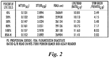

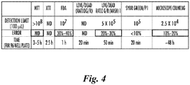

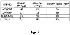


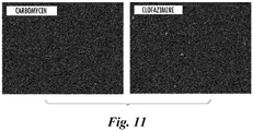
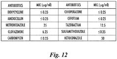


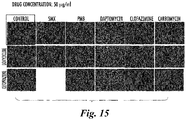


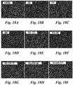
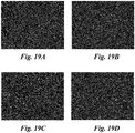

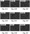

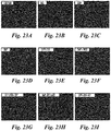

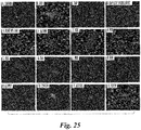
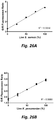

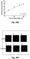
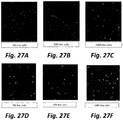


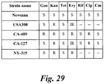


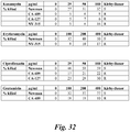
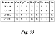


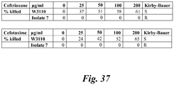

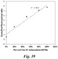


Classifications
-
- G—PHYSICS
- G01—MEASURING; TESTING
- G01N—INVESTIGATING OR ANALYSING MATERIALS BY DETERMINING THEIR CHEMICAL OR PHYSICAL PROPERTIES
- G01N33/00—Investigating or analysing materials by specific methods not covered by groups G01N1/00 - G01N31/00
- G01N33/48—Biological material, e.g. blood, urine; Haemocytometers
- G01N33/50—Chemical analysis of biological material, e.g. blood, urine; Testing involving biospecific ligand binding methods; Immunological testing
- G01N33/5005—Chemical analysis of biological material, e.g. blood, urine; Testing involving biospecific ligand binding methods; Immunological testing involving human or animal cells
- G01N33/5008—Chemical analysis of biological material, e.g. blood, urine; Testing involving biospecific ligand binding methods; Immunological testing involving human or animal cells for testing or evaluating the effect of chemical or biological compounds, e.g. drugs, cosmetics
- G01N33/5014—Chemical analysis of biological material, e.g. blood, urine; Testing involving biospecific ligand binding methods; Immunological testing involving human or animal cells for testing or evaluating the effect of chemical or biological compounds, e.g. drugs, cosmetics for testing toxicity
-
- C—CHEMISTRY; METALLURGY
- C12—BIOCHEMISTRY; BEER; SPIRITS; WINE; VINEGAR; MICROBIOLOGY; ENZYMOLOGY; MUTATION OR GENETIC ENGINEERING
- C12Q—MEASURING OR TESTING PROCESSES INVOLVING ENZYMES, NUCLEIC ACIDS OR MICROORGANISMS; COMPOSITIONS OR TEST PAPERS THEREFOR; PROCESSES OF PREPARING SUCH COMPOSITIONS; CONDITION-RESPONSIVE CONTROL IN MICROBIOLOGICAL OR ENZYMOLOGICAL PROCESSES
- C12Q1/00—Measuring or testing processes involving enzymes, nucleic acids or microorganisms; Compositions therefor; Processes of preparing such compositions
- C12Q1/02—Measuring or testing processes involving enzymes, nucleic acids or microorganisms; Compositions therefor; Processes of preparing such compositions involving viable microorganisms
- C12Q1/18—Testing for antimicrobial activity of a material
-
- Y—GENERAL TAGGING OF NEW TECHNOLOGICAL DEVELOPMENTS; GENERAL TAGGING OF CROSS-SECTIONAL TECHNOLOGIES SPANNING OVER SEVERAL SECTIONS OF THE IPC; TECHNICAL SUBJECTS COVERED BY FORMER USPC CROSS-REFERENCE ART COLLECTIONS [XRACs] AND DIGESTS
- Y02—TECHNOLOGIES OR APPLICATIONS FOR MITIGATION OR ADAPTATION AGAINST CLIMATE CHANGE
- Y02A—TECHNOLOGIES FOR ADAPTATION TO CLIMATE CHANGE
- Y02A50/00—TECHNOLOGIES FOR ADAPTATION TO CLIMATE CHANGE in human health protection, e.g. against extreme weather
- Y02A50/30—Against vector-borne diseases, e.g. mosquito-borne, fly-borne, tick-borne or waterborne diseases whose impact is exacerbated by climate change
Definitions
- AST antibiotic susceptibility tests
- MIC drug's minimum inhibitory concentration
- AST methods utilizing MALDI-TOF mass spectrometry (MS) which was originally used for microorganism identification, has been developed but such methods produced varying AST results and were unable to detect resistance among highly resistant organisms such as E. coli, K. pneumoniae, P. aeruginosa, and also methicillin-resistant S. aureus.
- DST current drug susceptibility test
- MGIT 960 BacT/ALERT systems at pH 6.0 [Aragon et al., 2007], which are unreliable and subject to frequent false resistance problem and thus not routinely performed [Zhang and Mitchison, 2003; Hewlett et al., 1995; Chedore et al., 2010].
- SYBR Green I/Propidium Iodide (PI) staining viability assay The SYBR Green I dye is commonly used in molecular biology to stain nucleic acids and was used for viability assessment for some bacteria by flow cytometry [Barbesti et al., 2000] but has not been commonly used for bacterial viability assessment [Feng et al., 2014] or used for AST. SYBR Green I is a permeable dye that stains all cells green whereas PI is an impermeant dye that stains only dead or damaged cells with a compromised cell membrane [Feng et al., 2014]. Hence, the green/red fluorescence ratio as easily measured by fluorescence microscopy or fluorescence microplate readers represents the viability of a cell population. As the assay is not growth-dependent, it eliminates a bottleneck in current AST.
- the inventors of the present application have developed a novel, growth-independent antibiotic susceptibility test that can produce rapid and robust results less than 24 hours for slow and fast growing organisms.
- the present application shows for the first time, the utility of a SYBR Green I and Propidium Iodide (SYBR Green I/PI) assay for extremely rapid detection of antibiotic resistance in representative major pathogens of serious threats including multidrug resistant Staphylococcus aureus, Escherichia coli, Klebsiella pneumoniae, Acinetobacter baumannii, and Mycobacterium tuberculosis.
- the new SYBR Green/PI AST could identify antibiotic resistance in less than 30 minutes for the fast growing bacteria using different antibiotics for both bactericidal and bacteriostatic agents.
- the optimized SYBR Green I/PI assay generated rapid and robust results in concordance with traditional AST methods when tested on various clinically derived strains and a panel of drugs used for treatment in the clinic.
- the SYBR Green I/PI assay overcomes not only the inefficiency of conventional culture based AST or drug susceptibility testing (DST) but also the limitations and compromised sensitivity of molecular testing methods.
- the SYBR Green I/PI AST testing methodology may revolutionize current drug susceptibility testing as well as drug screens for different organisms or cells of prokaryotic and eukaryotic origin, and may play a major role in saving lives and improving patient outcomes in a timely manner, as well as decreasing transmission of increasing antibiotic resistant organisms.
- the presently disclosed subject matter provides methods, compositions, and kits for extremely rapid assessment of drug susceptibility of growing as well as non-growing cells of prokaryotic and also eukaryotic origins, without the need for organism growth, which is in contrast to the current growth-dependent methods.
- This growth-independent new methodology achieves extremely rapid drug susceptibility testing results previously not possible
- Methods for rapidly identifying compounds with inhibitory activity against both growing and non-growing cells in drug screens using the novel methodology are also provided.
- the presently disclosed subject matter provides methods for assessing the viability or assessing the drug susceptibility of cells, such as antimicrobial susceptibility.
- the cells may be eukaryotic or prokaryotic cells and include, for example, spirochetal organisms, such as from the Borrelia genus, and more preferably, the Borrelia burgdorferi species, and related organisms, and other bacteria, including, but not limited to, Staphylococcus aureus, Escherichia coli, Klebsiella pneumoniae, Acinetobacter baumannii, and Mycobacterium tuberculosis.
- a method for assessing the susceptibility of cells to at least one drug or test agent comprising:
- the methods of the present invention may further comprise the step of contacting or exposing the cells to at least one dose of at least one drug or test agent.
- the methods of the present invention may comprise the step of incubating the cells with a drug or agent that kills or inhibits the growth of the organism or cells.
- the present invention also provides for one or more test agents to be assayed for inhibitory activity, such as in a drug screen which may be optionally be performed in a real-time continuous manner and/or in a high throughput format.
- kits which may be used to carry out the methods of the present invention invention.
- a kit for rapid screening or susceptibility testing of at least one candidate agent that is capable of inhibiting growth or survival of cells of eukaryotic or prokaryotic origin wherein the kit comprises:
- the cells or organisms which are assayed with the methods or kits of the present invention are selected from the group consisting of: all cells of prokaryotic (bacteria and archaea) and also eukaryotic origin, including but not limited to cells of Gram-positive bacteria such as Staphylococcus aureus, and Gram-negative bacteria such as Escherichia coli, Klebsiella pneumoniae, and Acinetobacter baumannii, and mycobacteria such as Mycobacterium tuberculosis, and other microbial species, and eukaryotic cells such as fungal cells, plant cells, parasite cells, animal cells, or cancer cells.
- prokaryotic bacteria and archaea
- eukaryotic origin including but not limited to cells of Gram-positive bacteria such as Staphylococcus aureus, and Gram-negative bacteria such as Escherichia coli, Klebsiella pneumoniae, and Acinetobacter baumannii
- mycobacteria such as Mycobacterium tuber
- the method comprises assessing the drug (e.g. antibiotic) susceptibility of the cells or organism being assayed to at least one agent by incubating the culture of step (a) under suitable conditions without the need for the test organism to grow.
- drug e.g. antibiotic
- the methods of the present invention preferably utilize higher drug concentrations than the normal or usual drug concentrations needed for inhibiting organism or cell growth as used in current conventional drug susceptibility testing.
- the method is performed in a high-throughput format, such as for drug screens.
- the method is performed in a high-throughput format, such as for drug screens.
- the method is performed in a real-time continuous manner optionally in a high-throughput format, such as for drug screens.
- a high-throughput format such as for drug screens.
- the methods of the present invention may be more versatile and efficient than the conventional drug screens typically done at a single time point.
- the presently disclosed subject matter relates to methods for assessing the viability of cells, methods for assessing the susceptibility of cells to drugs or candidate drugs such as candidate antimicrobial agents, methods for screening for at least one agent that inhibits the growth or survival of cells, methods for inhibiting the growth and/or survival of cells, and related compositions and kits that can be used to perform or implement the methods.
- the cells may be of eukaryotic or prokaryotic origin and may, for example, be bacteria which may be replicating or non-replicating cells.
- the cells may be cells of eukaryotic or prokaryotic origin.
- the cells may be from a selected organism which may be a single-celled or multicellular organism.
- the cells may be from a single individual and may for example be cells from a single human (e.g. a cancer patient, such as cells from a biopsy or tumor of said patient), or from a collection or group of individuals (e.g. a group of individuals suffering from a common disease or disorder such as cancer or a disease or disorder mediated by an infectious agent such as bacteria or other microbes).
- Cells from a selected organism may include microbial cells such as bacteria or yeast cells and accordingly in some embodiments the selected organism is bacteria and there is therefore accordingly provided a method for assessing the drug susceptibility of bacterial cells which may optionally be pathogenic bacteria.
- the selected organism may be bacteria of a selected genus such as Staphylococcus, Escherichia, Klebsiella, Acinetobacter or Mycobacterium tuberculosis and the selected organism may, for example, be Staphylococcus aureus, Escherichia coli, Klebsiella pneumoniae, Acinetobacter baumannii, and Mycobacterium tuberculosis.
- the bacteria may be a bacterial isolate or sub-species (e..g from one of the aforementioned species).
- the cells may originate (including be derived from) a single individual or host or from multiple individuals or hosts.
- a cell culture comprising the cells to be tested is obtained or established.
- the cells may be isolated cells such as isolated cells from a selected organism (e.g. bacteria) which may optionally be from a selected species or genus.
- the method is a method for assessing the drug susceptibility of cells from a selected organism wherein the method comprises obtaining or establishing a culture comprising isolated cells from the selected organism e.g. bacterial cells such as the above-mentioned bacterial species and genera, or as otherwise mentioned herein, or other cells of prokaryotic or eukaryotic origin including, for example, yeast cells, fungal cells, parasite cells, or cancer cells.
- the methods of the present invention may further comprise incubating the cells with a staining mixture comprising: (i) a first agent which emits fluorescence of a first color that is indicative of live cells in the culture, and (ii) a second agent which emits fluorescence of a second color that contrasts from the first color and is indicative of dead cells in the culture.
- a staining mixture comprising: (i) a first agent which emits fluorescence of a first color that is indicative of live cells in the culture, and (ii) a second agent which emits fluorescence of a second color that contrasts from the first color and is indicative of dead cells in the culture.
- the first fluorescent compound is, for example, SYBR Green I that identifies live cells
- the second fluorescent compound is, for example, propidium iodide (PI) that identifies dead or non-viable cells.
- the methods of the present invention may be used to assess drug susceptibility or to screen one or more test agents for inhibitory activity. Accordingly, the methods of the present invention may comprise the step of exposing or contacting the cells or organisms to be assayed with at least one dose of at least one drug or test agent. The cells (or organisms e.g. bacteria) are then typically incubated with the at least one drug or test agent.
- the methods of the present invention comprise the step of incubating the cells with a drug or agent that kills or inhibits the growth of the cells / organism for a period of time without the requirement for organism growth.
- the period of time may range from minutes to hours or days depending on the organism.
- the drug(s) or agent(s) being tested may also influence the period of time due to e.g. the rate of drug uptake, the rate of drug action, and the metabolic status of the cell.
- the period of incubation of the cells with the drug or agent is less than about 17 hours, 16 hours, 15 hours, 14 hours, 13 hours, 12 hours, 11 hours, 10 hours, 9 hours, 8 hours, 7 hours, 6 hours, 5 hours, 4 hours, 3 hours, 2 hours or 1 hour but is optionally at least about 5, 10, 15, 20, 25, 30, 35, 40, 45 or 50 minutes.
- the period of incubation may be less than about 55, 50, 45, 40, 35, 30, 25, 20, 15, 10, or 5 minutes. In some embodiments the period of incubation is in the range of about 5 to 10, 5 to 15, 5 to 20, 5 to 25, 5 to 30, 5 to 35, 5 to 40, 5 to 50 or 5 to 60 minutes; about 10 to 15, 10 to 20, 10 to 25, 10 to 30, 10 to 35, 10 to 40, 10 to 50 or 10 to 60 minutes; about 15 to 20, 15 to 25, 15 to 30, 15 to 35, 15 to 40, 15 to 50 or 15 to 60 minutes; about 25 to 30, 25 to 35, 25 to 40, 25 to 50 or 25 to 60 minutes; about 5, 10, 15, 20, 25, 30, 35, 40, 50 or 60 minutes to 1.5 hours; 5, 10, 15, 20, 25, 30, 35, 40, 50 or 60 minutes to 2 hours; 5, 10, 15, 20, 25, 30, 35, 40, 50 or 60 minutes to 3 hours; 5, 10, 15, 20, 25, 30, 35, 40, 50 or 60 minutes to 4 hours; 5, 10, 15, 20, 25, 30, 35, 40, 50 or 60 minutes to 6 hours; 5, 10, 15, 20,
- the period of incubation can be a real-time continuous period, such as from 5 minutes to 16 hours, rather than a limited single time point, for all cells.
- the incubation with the drug / agent may be performed at the same time as incubating the fluorescent agents (i.e. the staining mixture) or afterwards.
- the steps of incubation with the staining mix and the drug / agent may be performed simultaneously, separately or sequentially and may therefore be overlapping or non-overlapping.
- the method further comprises determining a breakpoint (cut off) or range for the at least one drug.
- the methods of the present invention may employ higher drug concentrations than the normal or usual drug concentrations needed for inhibiting organism / cell growth or survival in the current conventional drug susceptibility tests and screens.
- the typical drug concentrations that are found to distinguish susceptible and resistant cells using the novel growth-independent SYBR Green I/PI methodology described herein range from 10-10,000 fold of conventional growth based MIC (minimum inhibitory concentration).
- the high drug concentrations required are necessary to achieve the short time needed to determine the susceptibility or resistance of a particular strain or cell in comparison with sensitive and resistant controls. Multiple drug concentrations within the above range may also be used to rapidly determine the drug susceptibility of the cells being assayed.
- the cells are incubated with the at least one drug or test agent at a concentration range from 10-10,000 fold of conventional growth based MIC.
- concentration range may be from 15-7500 fold; 20-5000; 50-2500 fold of conventional growth based MIC.
- concentration range may be at least 10, 50, 100, 250, 500, 1000, 2500, 5000, 7500 fold of convention growth based MIC.
- the method is a method for assessing the susceptibility of cells to at least one drug wherein step (b) comprises: (b)(i) incubating the cells with at least one dose of said at least one drug for a period of exposure without the need for growth of said cells; and (b)(ii) incubating the drug-treated cells with said staining mixture.
- step (b) comprises: (b)(i) incubating the cells with at least one dose of said at least one drug for a period of exposure without the need for growth of said cells; and (b)(ii) incubating the drug-treated cells with said staining mixture.
- the method is a method for assessing susceptibility of cells to at least one test agent wherein step (b) comprises: (b)(i) incubating the cells with at least one dose of said at least one test agent for a period of exposure without the need for growth of said cells; (b)(ii) incubating the test agent-treated cells with said staining mixture.
- step (b) comprises: (b)(i) incubating the cells with at least one dose of said at least one test agent for a period of exposure without the need for growth of said cells; (b)(ii) incubating the test agent-treated cells with said staining mixture.
- a method may be used as a drug screen, the drug screen may suitably be used to identify compounds or drug candidates (candidate agents) that are capable of inhibiting growth or survival of cultured or growing cells as well as for non-growing dormant or persister cells of prokaryotic or eukaryotic origin.
- the method includes conducting a microscopic counting rescreen to confirm that at least one test agent is a candidate agent for inhibiting growth or survival of the cells.
- incubation of the cells is growth independent and therefore the cells may be incubated under non-growth conditions thereby enabling the methods of the invention to be performed at a much greater speeds than methods which require a period of growth of the cells.
- the steps of incubation with the staining mix and the drug / agent may be performed simultaneously, separately or sequentially and and therefore steps (b)(i) and (b)(ii) may be performed together or separately and may be overlapping or non-overlapping.
- steps (b)(i) and (b)(ii) are performed separately or sequentially then the cells are exposed to the at least one drug or test agent first and then the cells are exposed to the staining mixture.
- the methods may comprise exposing the cells to the at least one drug or test agent first and then after said exposure adding the first and second fluorescent agents (e.g. SYBR Green I and the propidium iodide).
- the first and second fluorescent agents e.g.
- SYBR Green I and the propidium iodide can be used after drug /test agent exposure or together with drug /test agent exposure over a period of time and, as discussed elsewhere herein, optionally in a continuous, real time and/or high throughput manner.
- the methods and drug screens of the present invention can be continuous over a period of time. This is different from conventional drug screens of a defined time- or end-point, e.g. 3 day or 7 day, but with the new methods described herein drug susceptibility or drug action in drug screens can be assessed at multiple or differing time points, in a continuous, real-time and/or high throughput manner.
- a method of the present invention may comprise taking readings of the fluorescence at multiple time points so that drug susceptibility of the cells is assessed at different time points: this may allow for the effects of a drug or test agent (including combinations of drugs or test agents) to be monitored over time as well as allowing for the testing or identification of drug candidates sequentially over a period of time ranging from hours, days and weeks.
- the methodology of the present invention may advantageously be used for real-time continuous monitoring in DST with the present invention allowing one to monitor drug sensitivity or resistance to a test drug or agent in a continuous real-time manner.
- the current DST and drug screens typically use a fixed time point for assessing the results, and the advantage of the novel methodology described herein (e.g. the novel SYBR Green I/PI methodology) allows the DST to be done in a more quantitative and more real-time continuous manner. This enables the identification of susceptible or resistant cells (e.g. strains) in extremely rapid time e.g. in as little as 5 to 30 minutes.
- the methodology of the present invention may be used to provide for real-time continuous monitoring and identification of drug candidates in drug screens.
- the current drug screens are typically done with a fixed time point, which produce very limited information and small number of hits or drug candidates.
- novel methodology described herein e.g. the novel SYBR Green I/PI methodology
- one is able to identify multiple active "hits" without having to terminate the drug screening experiment.
- This enables the identification of different hits at different time points within a defined time period, typically ranging from minutes to hours to days or weeks as indicated above, in a continuous, real time and/or high throughput manner.
- This will be important for identifying drugs that target different cell populations with different metabolic status so as to identify drug candidates that can inhibit or kill the very heterogeneous population of cells of prokaryotic or eukaryotic origin.
- Incubating the cell culture in step (b) preferably comprises incubating the cell culture under suitable conditions. Suitable conditions may comprise incubating the cells at an appropriate cell density.
- An appropriate cell density may be defined by the sensitivity of the fluorescence viability assay (e.g. in the range of 101-9 cells/ml, 102-9 cells/ml, 103-9 cells/ml, 104-9 cells/ml, 105-9 or 106-9 cells/ml, 101-8 cells/ml, 102-8 cells/ml, 103-8 cells/ml, 104-8 cells/ml, or 105-8 cells/ml) for drugs and test agents tested and the fluorescent indicator dyes used.
- the present invention provides growth-independent assays or methods.
- incubating step (b) can be performed without the need for cell growth and such growth may therefore be omitted from the methods of the present invention.
- the growth independent nature of the assays described herein substantially reduces the time from days and weeks needed for current drug susceptibility testing to extremely short time of just minutes or hours.
- the growth independent nature of the assays described herein substantially reduces the time needed for current growth based drug screens to extremely short time of just minutes or hours. Since the methods of the present invention are growth independent it will accordingly be understood that incubation of the cells under suitable conditions need not require growth conditions and therefore the suitable conditions may comprise growth or non-growth conditions.
- the step of incubating the culture with the staining mixture and/or drug or test agent is performed for a period between about 5 to 30 minutes; for a period between about 10 to 20 minutes; or for a period of approximately 15 minutes.
- the step of incubating with the staining mixture and/or drug or test agent is performed in the dark.
- the first color emitted by the first fluorescent agent is green and the second color as emitted by the second fluorescent agent is red or orange.
- the first agent is SYBR Green I and/or the second agent is propidium iodide.
- a non-toxic concentration of the first and second fluorescent agents may be chosen whilst still achieving adequate staining results.
- the first and second fluorescent dyes SYBR Green I/PI can be used at an appropriate concentration that is non-toxic while still staining live cells green for SYBR Green I or dead cells red for PI.
- the fluorescent agents or dyes can be added simultaneously with the drug or test agent together for real-time continuous monitoring of drug susceptibility or drug screens in a dynamic manner to obtain more quantitative data on drug susceptibility and in drug screens.
- the SYBR Green I/PI dyes can be added after drug exposure or incubation for determining the results of sensitivity or resistance of the organism in DST or the activity of the drug or compound in real-time drug screens.
- the SYBR Green I is present in the culture in a concentration range of between about 0.1x and about 100x and propidium iodide is present in the culture in a range of between about 0.1 mM and about 5 mM.
- the concentration of SYBR Green I in the culture is about 10x and the concentration of propidium iodide is about 2mM.
- the concentrations of SYBR Green I and propidium iodide can vary depending on the particular organism or cell types or in DST or in drug screens, or when used simultaneously or sequentially with drugs or test agents.
- the incubation time with the staining mix can vary from typically 10-60 minutes up to hours or days depending on the particular organism or cell types or in DST or in drug screens, or when used simultaneously or sequentially with drugs or test agents.
- the fluorescence intensity of the culture and staining mixture is determined at 535 nm, which measures green fluorescence, and 635 nm, which measures red fluorescence.
- the methodology provided by the present invention is growth-independent which is completely distinct from the current growth based DST.
- the cells may be incubated without the need for the test cells / test organism to grow. This may substantially reduce the time, from days and weeks needed for current drug susceptibility testing, to extremely short times of just minutes or hours with the actual time required depending upon the incubation conditions, the cells used and the drugs tested. Where the effects of multiple drugs or test agents are assessed on a continuous basis then the total incubation time may be increased accordingly so as to allow for the assessment of drug susceptibility or drug action and identification of multiple drug candidates over time in real-time on a continuous or sequential basis.
- non-growing or non-replicating cells such as dormant or persister cells from different pathogenic microbes of prokaryotic and eukaryotic origin, such as persister cells of pathogenic bacteria including mycoplasma, chlamydia, spirochetal organisms, anaplasma, ehrlichia, rickettsia, coxiella, bartonella, fungi, yeasts, parasites (babesia, malaria, toxoplasma etc), as well as other cells which cannot be grown in culture or can only be grown with difficulty or isolated directly from host tissues without culture as in cancer tissues from patients.
- non-growing or non-replicating cells such as dormant or persister cells from different pathogenic microbes of prokaryotic and eukaryotic origin, such as persister cells of pathogenic bacteria including mycoplasma, chlamydia, spirochetal organisms, anaplasma, ehrlichia, rickettsia, coxiella, bartonella
- the methods of the present invention may therefore find particular utility as regards cell types which are typically hard to assay for drug susceptibility or to use in drug screens using conventional methods.
- the methods of the present invention may particularly lend themselves to cells which cannot be grown in culture (or cannot readily be grown in culture) and to slow growing cells such as various Mycobacterium spp. such as M. tuberculosis, M. leprae, and nontuberculous mycobacteria such as M. avium, M. intracellulare, M. kansasii.
- Further examples of cells in this context may include stem cells, pluripotent or multipotent cells, cancer cells, cancer stem cells, and fastidious cells.
- the subject matter of the present disclosure provides a new algorithm of defining resistance and susceptibility using the methods described herein and the ratio of the intensity of emitted fluorescence of the first color to the intensity of emitted fluorescence of the second color (e.g. the ratio of SYBR Green and PI). for rapid drug susceptibility testing and also drug screens in real-time manner that are not case with the conventional and current methods, for all cells of prokaryotic and eukaryotic origin.
- the methods of the present invention may comprise the step of calculating a ratio of the intensity of emitted fluorescence of the first color to the intensity of emitted fluorescence of the second color (e.g. the ratio of SYBR Green and PI). This ratio may then be used to assess the viability of the cells in the culture with the ratio being indicative of the percentage of live and dead cells in the culture. In this way the susceptibility of the cells to a drug or test agent may be assessed, such as in a rapid drug sensitivity test or rapid screen of test agents to identify one or more drug or candidate agents which may exhibit useful inhibitory activity.
- a ratio of the intensity of emitted fluorescence of the first color e.g. the ratio of SYBR Green and PI
- This ratio may then be used to assess the viability of the cells in the culture with the ratio being indicative of the percentage of live and dead cells in the culture.
- the susceptibility of the cells to a drug or test agent may be assessed, such as in a rapid drug sensitivity test or rapid screen of test agents
- a fluorescence reader may be linked to a computer with a software that will calculate the green/red fluorescence ratios of test organism, optionally with sensitive and resistant organisms as controls and drug free controls, to determine the susceptibility and resistance of a test organism.
- the susceptibility (or resistance) of the cells may be assessed by comparing the viability of the cells in the culture in the presence of the at least one drug or test agent with the viability of cells in a control culture which lacks, or has not been exposed to, the at least one drug or test agent.
- the cells are assessed as susceptible or resistant to the at least one drug or test agent according to whether the ratio in step (b) remains the same or decreases or increases after the period of exposure to the at least one dose of the at least one drug or test agent.
- the cells are assessed as susceptible to the at least one drug or test agent if the ratio in step (b) remains the same or decreases after the period of exposure to the at least one dose of the at least one drug or test agent, and wherein the cells are assessed as resistant to the at least one drug or test agent if the ratio in step (b) increases after the period of exposure to the at least one dose of the at least one drug or test agent.
- susceptibility or resistance is assessed by comparison of the ratio with a control, such as known resistant and/or known sensitive cells (e.g. a known resistant or sensitive bacterial strain), or a control which lacks, or has not been exposed to, the at least one drug or test agent.
- a control such as known resistant and/or known sensitive cells (e.g. a known resistant or sensitive bacterial strain), or a control which lacks, or has not been exposed to, the at least one drug or test agent.
- the cells are assessed as resistant or susceptible to the at least one drug or test agent by comparison to sensitive control cells and/or resistant control cells, for example based on a difference between the ratio of the fluorescence of the first agent over the fluorescence of the second agent of the cells and that of sensitive control cells and/or a resistant control cells for the at least one dose of the at least one drug or test agent.
- Susceptibility or resistance may be assessed by comparing the ratios of the control cells of known sensitive and known resistant controls with and without the at least one test agent or drug to define susceptibility or resistance of a particular test strain to a particular test agent or drug.
- test agent or drug may, for example, be any antibiotic currently used in treatment of infections caused by microbes (bacteria, fungi, protozoa, parasites, etc,) or could be any anticancer agent, antimicrobial agent, or antifungal agent etc.
- Cells may be assessed as resistant to the at least one drug or test agent if the ratio in step (b) is increased or does not decrease in comparison with said sensitive control cells after the period of exposure to the at least one dose of the at least one agent, and the cells may be assessed as susceptible to the at least one drug or test agent if the ratio in step (b) is decreased or does not increase in comparison with said resistant control cells after the period of exposure to the at least one dose of the at least one agent.
- a method of the present disclosure is performed in real time and/or in a high-throughput format.
- a high-throughput format is particularly envisaged in the case of drug susceptibility testing or drug screens which may suitably be used to identify agents with activity against growing and non-growing cells of prokaryotic and eukaryotic origin.
- the high-throughput format uses at least one multi-well microplate.
- suitable multi-well microplates include, without limitation, a 6-well microplate, a 24-well microplate, a 96-well microplate, a 384-well microplate, and a 1536-well microplate.
- the multi-well microplate comprises a 96-well microplate.
- the presently disclosed methods can be used without a washing step and for high-throughput screens. That is, the methods can be used to assess viability, susceptibility of cells to drugs and test agents, and in drug screening directly in a high-throughput real-time manner and/or in the absence of washing. Accordingly yet another advantage is that the methods described herein do not require a washing step after drug incubation to obtain results, as multiple readings (e.g. of plates) can be taken directly without a washing step. This not only saves time but also prevents loss of small numbers of cells during the washing step. Thus, in at least some embodiments of the methods described herein readings are taken directly without the incubated cells being first washed to remove drug to obtain results over a period of time in real-time manner
- the cells do not need to be recultured or regrown after drug exposure before results can be obtained and, so consequently, in at least some embodiments of the present invention the cells are not recultured or regrown after drug incubation.
- This novel methodology of the present invention allows more accurate and extremely rapid DST and more drug candidates to be identified in a real-time manner over a period of time without having to stop the experiment or introducing a washing step to remove drugs or test agents to allow for regrowth of the surviving cells in current growth based drug screens.
- the culture comprises a stationary phase culture which may optionally comprise non-replicating persister cells.
- the stationary phase culture may comprise morphological variant forms selected from the group consisting of L-forms or cell wall defective forms, round bodies, planktonic and aggregated biofilm forms.
- the term "cell culture” or "culture” includes a reference to a population of cells in culture. Since the methods of the present invention are growth independent the culture conditions need not be conducive for growth and the cells in the culture need not be growing cells, and may therefore be growing or non-growing cells.
- the cell culture can be found in any type of container, such as a flask, a tube, a microwell plate, array, and the like.
- the culture may be a cell suspension
- the cells may have different phases of growth such as a lag phase, log phase and stationary phase.
- a cell culture in "stationary phase” means that the cells in the culture have an approximately equal growth rate and death rate.
- the term “growing forms” of cells generally refers to cells that are in lag or log phase and not in stationary phase.
- the stationary phase cell culture has been grown for approximately 7 days or more.
- the stationary phase cell culture comprises non-replicating persister cells.
- non-replicating persister cells it is meant cells that enter a state in which they stop replicating and exhibit tolerance to drugs, such as the ability to tolerate antibiotics in the case of bacteria.
- culturable cells includes a reference to cells which are capable of being maintained in culture (but which are not necessarily capable of growing or replicating in culture) and to cells which are capable of growing or replicating in culture; both of these cells types may be employed in the methodology of the present invention.
- the methodology of the present invention is applicable to all prokaryotic and eukaryotic cells.
- the cells used in the methods and kits of the present invention may therefore be selected accordingly.
- the cells may be of prokaryotic (bacteria and archaea) or eukaryotic origin and may be selected from the group consisting of: bacteria; Gram-positive bacteria such as Staphylococcus aureus ; Gram-negative bacteria such as Escherichia coli, Klebsiella pneumoniae, and Acinetobacter baumannii ; mycobacteria such as Mycobacterium tuberculosis ; bacteria from the Borrelia genus (e.g., replicating and/or non-replicating persister forms of B.
- monkeys apes, gibbons, chimpanzees, orangutans, macaques and the like
- human cells cancerous or infected animal cells
- bovine cells e.g., cattle, oxen, and the like
- ovine cells e.g., sheep and the like
- caprine cells e.g.
- porcine cells e.g., pigs, hogs, and the like
- equine cells e.g., horses, donkeys, zebras, and the like
- feline, canine, rodent or lagomorph cells cells from animal disease models (e.g., rats or mice used in experiments, and the like); tumor cells; cells from a human or non-human animal suffering from a cell proliferative disorder e.g.
- a neoplastic disorder cells of a pathogen, e.g., a multicellular pathogen or microorganism such as a pathogenic bacterium, pathogenic fungus, or a pathogenic protist (e.g., a Plasmodium cell); cells derived from a cell line e.g. CHO cells, HeLa cells.
- the cells may be replicating and/or non-replicating persister or dormant forms of any of the aforementioned cells; cancerous (e.g. breast cancer, prostate cancer, colon cancer, pancreatic cancer, lung cancer, leukemia etc), diseased, infected (e.g. with an antibiotic or antimicrobial refractory infection), or precancerous cells of any of the aforementioned cells.
- cell-lines stem cells, pluripotent, multipotent or oligopotent cells, cancer stem cells, cancer cells from cancer tissues of patients, fastidious cells and transformed cells of any of the aforementioned cells or cells otherwise described herein.
- the cells are not cancer cells.
- the cells are not bacterial cells.
- the cells are not microbial cells.
- the cells are not infectious or pathogenic organisms.
- the bacteria are spirochete organisms such as Treponema spp. scuh as T. pallidum and Borrelia spp. such as Borrelia burgdorferi, B. afzelii, B. garinii, B. miyamotoi.
- Borrelia is a genus of bacteria of the Spirochete phylum.
- the Borrelia burgdorferi sensu lato complex includes at least 18 genospecies.
- Non-limiting examples of bacteria in this genus include B. burgdorferi, B. garinii, B. afzelii, B. americana, B. carolinensis, B. lusitaniae, B. japonica, B.
- the bacteria are Borrelia burgdorferi.
- the bacteria comprise replicating forms of Borrelia burgdorferi, non- replicating persister forms of Borrelia burgdorferi, and combinations of replicating forms of Borrelia burgdorferi, non-replicating persister forms of Borrelia burgdorferi.
- the Borrelia burgdorferi comprise a morphological form selected from the group consisting of round bodies, planktonic, spirochete form, a spheroplast form (L-forms), a cystic or round body form, a microcolony form, a biofilm-like and biofilm form, and combinations thereof.
- the cells are preferably not growing Borrelia burgdorferi cells, and more preferably are not growing Borrelia cells. In other embodiments of the invention the cells are preferably not Borrelia burgdorferi cells, and more preferably are not Borrelia cells.
- the presently disclosed subject matter provides a method for assessing the viability or antimicrobial susceptibility of bacteria from a selected genus, the method comprising: (a) establishing a bacterial culture comprising isolated bacteria from the selected genus; (b) incubating the bacterial culture with a staining mixture comprising: (i) a first agent which emits fluorescence of a first color that is indicative of live bacterial cells in the culture, and (ii) a second agent which emits fluorescence of a second color that contrasts from the first color and is indicative of dead bacterial cells in the culture; and (c) calculating a ratio of the intensity of emitted fluorescence of the first color to the intensity of emitted fluorescence the second color; and (d) assessing the viability of the bacteria in the culture, wherein the ratio calculated in (c) is indicative of the percentage of live bacteria in the culture.
- the method may further comprise:
- the presently disclosed subject matter and methods provide a new SYBR Green I/propidium iodide (PI) (also termed SYBR Green/PI) assay based on a green fluorescence to red fluorescence ratio for rapid viability assessment of eukaryotic or prokaryotic cells such as bacteria, such as those from the Borrelia genus or a genus selected from the group consisting of Staphylococcus, Escherichia, Klebsiella, Acinetobacter, and Mycobacterium.
- This assay is superior to other assays commonly used for measuring the viability and for rapid drug susceptibility testing of B.
- viability assay refers to an assay to determine the ability of cells to maintain or recover viability, such as the ability to grow.
- a method of the present invention may comprise the following steps: (a) obtaining a culture comprising the cells to be assayed (which as discussed herein may be eukaryotic or prokaryotic, growing or non-growing etc); (b) performing a viability assay of the cells in the culture by using a SYBR Green I/Propidium Iodide assay based on a ratio of green fluorescence, indicative of live cells, to red fluorescence, indicative of dead cells, comprising: (i) mixing the culture with a staining mixture comprising SYBR Green I and propidium iodide; (ii) allowing the culture and staining mixture to incubate in the dark; (iii) determining the fluorescence intensity of the culture and staining mixture at 535 nm, which measures green fluorescence, and 635 nm, which measures red fluorescence; and (iv) calculating the ratio of green fluorescence to red fluorescence; and wherein a ratio
- the presently disclosed subject matter provides a method for assessing the viability of bacteria from a selected genus, the method comprising: (a) obtaining a culture comprising bacterial cells from the selected genus; (b) performing a viability assay of the bacterial cells in the culture by using a SYBR Green I/Propidium Iodide assay based on a ratio of green fluorescence, indicative of live bacterial cells, to red fluorescence, indicative of dead bacterial cells, comprising:
- the first color emitted by the first fluorescent agent is green and the second color as emitted by the second fluorescent agent is red or orange.
- the first agent is SYBR Green I and the second agent is propidium iodide.
- the SYBR Green I is present in the culture in a concentration range of between about 0.1x and about 100x and propidium iodide is present in the culture in a range of between about 0.1 mM and about 5 mM.
- the concentration of SYBR Green I in the culture is 10x and the concentration of propidium iodide is 2mM.
- the culture in the methods and kits of the present invention may comprise a BSK-H medium.
- the step of incubating the culture with the mixture is performed for about 5, 10, 15, 25, or 30 minutes. In further embodiments, the step of incubating is performed in the dark.
- the method is performed in a high-throughput format, e.g., for drug screening.
- a high-throughput format e.g., for drug screening.
- the presently disclosed methods can be used without a washing step and the need for subsequent cell growth for high-throughput screens. That is, the methods can be used to assess viability, susceptibility of bacteria to antimicrobials, and in drug screening directly in a high-throughput manner in the absence of washing.
- the presently disclosed subject matter provides a method for assessing the susceptibility of cells to at least one drug (e.g. antimicrobial agent), the method comprising: (a) establishing a culture comprising isolated cells to be tested; (b) incubating the culture, optionally under suitable conditions for growth of the cells to occur, with: (i) at least one dose of at least one drug (e.g.
- a staining mixture comprising a first agent which emits fluorescence of a first color that is indicative of live cells in the culture, and a second agent which emits fluorescence of a second color that contrasts from the first color and is indicative of dead cells in the culture, wherein a ratio of the intensity of emitted fluorescence of the first color to the intensity of emitted fluorescence of the second color is indicative of the percentage of live cells in the culture; and (c) assessing the susceptibility of the cells to at least one drug (e.g. antimicrobial agent) by calculating the ratio in (b)(ii) without the need for cell growth, after a period of exposure to at least one dose of at least one drug (e.g.
- a drug e.g. antimicrobial agent
- the cells are assessed as susceptible to at least one drug (e.g. antimicrobial agent) if the ratio in (b)(ii) remains the same or decreases after the period of exposure to at least one dose of at least one drug (e.g. antimicrobial agent), and wherein the cells are assessed as resistant to at least one drug (e.g. antimicrobial agent) if the ratio in (b)(ii) increases after the period of exposure to at least one dose of at least one drug (e.g. antimicrobial agent).
- the method may be performed with no growth, or substantially no growth, of the cells during the incubation step with the at least one dose of at least drug.
- the presently disclosed subject matter provides a method for assessing the susceptibility of bacteria from a selected genus to at least one antimicrobial agent, the method comprising: (a) establishing a bacterial culture comprising isolated bacteria from the selected genus; (b) incubating the culture, optionally under suitable conditions for bacterial growth to occur, with: (i) at least one dose of at least one antimicrobial agent; and (ii) a staining mixture comprising a first agent which emits fluorescence of a first color that is indicative of live bacteria in the culture, and a second agent which emits fluorescence of a second color that contrasts from the first color and is indicative of dead bacteria in the culture, wherein a ratio of the intensity of emitted fluorescence of the first color to the intensity of emitted fluorescence of the second color is indicative of the percentage of live bacteria in the culture; and (c) assessing the susceptibility of bacteria from the selected genus to at least one antimicrobial agent by calculating the ratio in (b
- the presently disclosed subject matter provides a method for screening for a compound that is capable of inhibiting cells which cells may be prokaryotic or eukaryotic cells, the method comprising: (a) obtaining a stationary phase culture that comprises said prokaryotic or eukaryotic cells; (b) contacting the culture with a test compound; (c) performing a viability assay of the cells in the culture by using a SYBR Green I/Propidium Iodide assay based on a ratio of green fluorescence, indicative of live cells, to red fluorescence, indicative of dead cells, comprising: (i) mixing the culture with a staining mixture comprising SYBR Green I and propidium iodide; (ii) allowing the culture and staining mixture to incubate in the dark; (iii) determining the fluorescence intensity of the culture and staining mixture at 535 nm, which measures green fluorescence, and 635 nm, which measures red fluorescence; (i
- contacting refers to any action that results in at least one compound of the presently disclosed subject matter physically contacting at least one cell (e.g. bacterial cell) or the environment in which at least one cell (e.g. bacterial cell) resides (e.g., a culture medium).
- at least one cell e.g. bacterial cell
- the environment in which at least one cell e.g. bacterial cell
- a culture medium e.g., a culture medium
- the presently disclosed subject matter provides a method for screening for a compound that is capable of inhibiting bacteria from a selected genus, the method comprising: (a) obtaining a stationary phase bacterial culture that comprises bacterial cells from the selected genus; (b) contacting the culture with a test compound; (c) performing a viability assay of the bacterial cells in the culture by using a SYBR Green I/Propidium Iodide assay based on a ratio of green fluorescence, indicative of live bacterial cells, to red fluorescence, indicative of dead bacterial cells, comprising: (i) mixing the culture with a staining mixture comprising SYBR Green I and propidium iodide; (ii) allowing the culture and staining mixture to incubate in the dark; (iii) determining the fluorescence intensity of the culture and staining mixture at 535 nm, which measures green fluorescence, and 635 nm, which measures red fluorescence; (iv)
- the method comprises determining a minimum inhibitory concentration breakpoint or cut-off for at least one drug or agent (e.g at least one antimicrobial agent).
- the first color is green and the second color is red or orange.
- the first agent is SYBR Green I and the second agent is propidium iodide.
- the concentration of SYBR Green I in the culture is about 10x and the concentration of propidium iodide is about 2mM.
- the culture further comprises a BSK-H medium.
- the presently disclosed subject matter provides a method for identifying a candidate agent that is capable of inhibiting growth or survival of cells which cells may be prokaryotic or eukaryotic cells, the method comprising: (a) establishing a culture comprising isolated cells to be tested; (b) contacting the culture with a test agent; (c) assessing a viability of the cells in the culture in the presence of the test agent as compared to the viability of the cells in a control culture which lacks the test agent for drug screens on non-growing cells in continuous time lapse manner over different time span and rapid drug susceptibility testing in a growth independent manner such that susceptibility to a test agent can be rapidly determined without relying on cell growth as in conventional drug or antibiotic susceptibility testing, wherein assessing the viability of the cells in the culture comprises: (i) incubating the culture with a staining mixture comprising a first agent which emits fluorescence of a first color that is indicative of live cells, and a second agent which emits fluorescence of
- the presently disclosed subject matter provides a method for identifying a candidate agent that is capable of inhibiting growth or survival of bacteria from a selected genus, the method comprising: (a) establishing a culture comprising isolated bacteria from the selected genus; (b) contacting the culture with a test agent; (c) assessing a viability of the bacteria in the culture in the presence of the test agent as compared to the viability of the bacteria in a control culture which lacks the test agent for drug screens on non-growing persisters in continuous time lapse manner over different time span and rapid antimicrobial susceptibility testing in a growth independent manner such that susceptibility to a test agent can be rapidly determined without relying on bacterial growth as in conventional antibiotic susceptibility testing, wherein assessing the viability of the bacteria in the culture comprises: (i) incubating the culture with a staining mixture comprising a first agent which emits fluorescence of a first color that is indicative of live bacteria, and a second agent which emits fluorescence of a second
- the presently disclosed subject matter provides a method for identifying a candidate agent that is capable of inhibiting growth or survival of of cells which cells may be prokaryotic or eukaryotic cells, the method comprising: (a) establishing a culture comprising isolated cells to be tested; (b) contacting the culture with a test agent; (c) assessing a viability of the cells in the culture in the presence of the test agent as compared to the viability of the cells in a control culture which lacks the test agent for drug screens on non-growing cells in continuous time lapse manner over different time span and rapid drug susceptibility testing in a growth independent manner such that susceptibility to a test agent can be rapidly determined without relying on cell growth as in conventional drug or antibiotic susceptibility testing, wherein assessing the viability of the cells in the culture comprises: (i) incubating the culture with a staining mixture comprising a first agent which emits fluorescence of a first color that is indicative of live cells, and a second agent which emits fluorescence
- the presently disclosed subject matter provides a method for identifying a candidate agent that is capable of inhibiting growth or survival of bacteria from a selected genus, the method comprising: (a) establishing a culture comprising isolated bacteria from the selected genus; (b) contacting the culture with a test agent; (c) assessing a viability of the bacteria in the culture in the presence of the test agent as compared to the viability of the bacteria in a control culture which lacks the test agent for drug screens on non-growing persisters in continuous time lapse manner over different time span and rapid antimicrobial susceptibility testing in a growth independent manner such that susceptibility to a test agent can be rapidly determined without relying on bacterial growth as in conventional antibiotic susceptibility testing, wherein assessing the viability of the bacteria in the culture comprises: (i) incubating the culture with a staining mixture comprising a first agent which emits fluorescence of a first color that is indicative of live bacteria, and a second agent which emits fluorescence of a second
- the method includes conducting a microscopic counting rescreen to confirm that at least one test agent is a candidate agent for inhibiting growth or survival of bacteria.
- the terms “inhibit”, “inhibits”, or “significant decrease” means to decrease, suppress, attenuate, diminish, or arrest, for example the growth and/or survival of cells (e.g. bacteria) in a culture or in a subject, by at least 10%, 20%, 30%, 40%, 50%, 60%, 70%, 80%, 90%, 95%, 98%, 99%, or even 100% compared to an untreated control culture or subject.
- Inhibiting "survival" of cells (e.g. bacteria) in this context refers to killing cells or reducing live cell count. In some embodiments, the growth of the cells is inhibited by more than approximately 50%.
- the percentage of live cells in the culture after the treatment with the test compound is less than approximately 50% compared to the percentage of live cells in the control under identical conditions, but in the absence of the test compound.
- the stationary phase culture comprises non-growing cells such as non-replicating persister cells.
- the term "significant increase” means an increase by at least 10%, 20%, 30%, 40%, 50%, 60%, 70%, 80%, 90%, 95%, 98%, 99%, or even 100%.
- the term "drug” and like expressions as used herein generally includes a reference to compounds or agents (the terms may be used synonymously) which are capable of inhibiting growth and/or survival of cultured or growing cells and/or inhibiting growth and/or survival of non-growing cells such as dormant or persister cells. Accordingly, it will be understood that the methods and kits of the present disclosure relate to static agents and cidal agents and the present invention provides methods for screening or identifying such activity and compounds, and for assessing susceptibility of cells to such agents. As will be understood from the disclosures herein, inhibitory activity, such as static or cidal activity, may be in relation to cultured or growing cells or in relation to growth non-growing dormant or persister cells.
- the term "drug” and similar expressions may include test compounds or agents (see below) and candidate agents. Accordingly, the term may refer to a variety or agents or compounds which may be assayed using the methods described herein such as in the the DSTs and screening methods of the present invention.
- the methods of the present invention may comprise assaying the effects of a combination of drugs or test compounds / agents.
- the terms "at least one drug” and “at least one test agent / compound” may include a reference to where a combination of drugs / agents is being tested. In this way synergistic or effective combinations of drugs or test agents may be identified by the methods of the present invention.
- the term "synergistic" includes a reference to circumstances under which the biological activity of a combination of a compound and at least one additional therapeutic agent is greater than the sum of the biological activities of the respective agents when administered individually.
- the methodology of the present invention can be used for drug combination screens, such as shown in Example 7. Accordingly, in some embodiments of the methods of the present invention may be used to identify one or more drug candidates or agents that synergize with one or more known drugs or other agents.
- drug drug
- antibiotic antibiotic
- antifungal antiparasitic
- agent agent
- test compound test compound
- candidate drug drug
- candidate agent candidate compound
- inhibitor and the like (and for the avoidance of doubt, grammatical variants thereof) may be used interchangeably herein.
- test compound (used synonymously with agent herein) as used herein can be any compound or drug that is desirable to test for inhibitory activity such as its capability of inhibiting growth and/or survival of cultured or growing cells and/or inhibiting growth and/or survival of non-growing cells such as dormant or persister cells, e.g. anti-persister activity.
- inhibitory activity such as its capability of inhibiting growth and/or survival of cultured or growing cells and/or inhibiting growth and/or survival of non-growing cells such as dormant or persister cells, e.g. anti-persister activity.
- anti-persister activity means that the compound has inhibitory activity against those cells that might still persist in a culture or subject after contact or administration with a course of antibiotics. The persistent cells may evade host immune clearance and result in chronic persistent infection in a subject.
- the methodology of the present invention works for both cidal and static agents and the methods of the present invention may accordingly be used to screen for, or identify susceptibility to, cidal and static.
- the test compound may be a known compound, such as an identified drug found to be effective in at least one disease or disorder, or may be an unknown compound that is not known to be effective in any disease or disorder.
- the test compound is a known compound that has been approved by a health regulatory agency (e.g., FDA or EMA) for an indication other than treating chronic persistent Lyme disease.
- the test compound is a compound that is known to exhibit antibiotic activity against bacteria other than those from the Borrelia genus.
- the test compound is a known compound that has not previously been reported to exhibit inhibitory activity against non-growing or non-replicating forms of the cells. In some embodiments, the test compound is a known compound that has not previously been reported to exhibit inhibitory activity against non-growing or dormant forms of the cells. In some embodiments, the test compound is a known compound that has not previously been reported to exhibit antibiotic activity against non-replicating persister forms of bacteria.
- an antimicrobial refers to a compound that has the ability to kill or inhibit the growth of bacteria.
- the term “antibiotic” refers to a compound that has the ability to kill or inhibit the growth of bacteria particularly bacteria selected from a genus including, but not limited to, Borrelia, Staphylococcus, Escherichia, Klebsiella, Acinetobacter, and Mycobacterium. More generally, an antimicrobial is an agent that kills microorganisms or inhibits their growth. Antimicrobials medicines can be grouped according to the microorganisms they act primarily against. For example, antibiotics are used against bacteria.
- beta-lactam or "beta-lactam antibiotic” refers to an antibiotic with a beta-lactam ring as part of its core structure, such as penicillin and penicillin derivatives (penams), cephalosporins (cephems), monobactams, and carbapenems. While these antibiotics work by inhibiting bacterial cell wall biosynthesis, other antibiotics such as protein synthesis inhibitors (tetracyclines, aminoglycosides, macrolides, etc), nucleic acid synthesis inhibitors (fluoroquinolones, rifamycins etc) and antimetabolite sulfa drugs can also be used similarly for antibiotic susceptibility testing with the methodology described herein.
- protein synthesis inhibitors tetracyclines, aminoglycosides, macrolides, etc
- nucleic acid synthesis inhibitors fluoroquinolones, rifamycins etc
- antimetabolite sulfa drugs can also be used similarly for antibiotic susceptibility testing with the
- the term "cell culture” or "culture” includes a reference to a population of cells in culture. Since the methods of the present invention are growth independent the culture conditions need not be conducive for growth and the cells in the culture need not be growing cells, and may therefore be growing or non-growing cells.
- a cell culture or culture may refer to the cells being in a medium conducive for growth of the cells and to optionally further the cells growing in the medium.
- the term "bacterial culture” or “culture” may refer to bacteria growing in a medium conducive for growth of those bacteria.
- the bacterial culture can be found in any type of container, such as a flask, a tube, a microwell plate, or array, and the like.
- bacteria have different phases of growth.
- the first phase of growth is the lag phase, a period of slow growth when the cells are adapting to the high-nutrient environment and preparing for fast growth.
- the lag phase has high biosynthesis rates, as proteins necessary for rapid growth are produced.
- the second phase of growth is the log phase, also known as the logarithmic or exponential phase, in which the bacteria undergo rapid exponential growth.
- the third phase of growth is the stationary phase and is caused by depleted nutrients.
- a bacterial culture in "stationary phase” means that the bacteria in the culture have an approximately equal growth rate and death rate.
- the term “growing forms” of bacteria generally refers to bacteria that are in lag or log phase and not in stationary phase.
- the stationary phase bacterial culture has been grown for approximately 7 days.
- the stationary phase bacterial culture comprises non-replicating persister cells.
- non-replicating persister cells it is meant bacterial cells that enter a state in which they stop replicating and are able to tolerate antibiotics.
- the methodology of the present invention enables rapid drug susceptibility testing and drug screening which considerably shortens the time required by current methods.
- Various features and advantages that may be achieved by the present invention will be appreciated from the discussion herein and may include one or more of the following: (1) suitability for use with all cells- prokaryotic and eukaryotic cells which may or may not be capable of being grown in culture; (2) a new algorithm of defining resistance and susceptibility using this method by the ratio of SYBR Green and PI; (3) use of much higher drug concentrations than the normal or usual drug concentrations needed for inhibiting organism growth as used in the current conventional drug susceptibility tests (DST) which is growth-dependent and time-consuming depending upon the growth speed of the test organism; (4) the new test is growth-independent which is completely distinct from the current growth based DST; (5) the new test can be used for drug screens for all cells of prokaryotic and eukaryotic origin to identify new drug candidates in record time without the requirement for growth of cells or the organisms, especially for slow
- tuberculosis which significantly shortens the time needed for results of the screen from weeks or hours;
- the organisms or cells used in the drug screens can be either growing cells or non-growing dormant cells or persister cells;
- the new test is much more versatile and can be used for continuous real-time drug screens to identify different drug candidates sequentially over a period of time, ranging from hours, days and weeks without having to stop the screen, in contrast to the current drug screen which is at a fixed single time point.
- kits for practising the methods of the presently disclosed subject matter also relates to kits for practising the methods of the presently disclosed subject matter.
- one aspect of the present invention provides a kit for use in a method of the present invention.
- the invention provides a method of the present invention which is performed using a kit as described herein.
- kits contains some or all of the components, reagents, supplies, and the like to practice a method according to the presently disclosed subject matter.
- the term "kit” refers to any intended any article of manufacture (e.g., a package or a container) comprising an effective amount of reagents (e.g. drugs or antibiotics and indicator fluorescent dyes at suitable concentrations) for performing a presently disclosed assay.
- the kit may optionally include the cells to be assayed (e.g. bacteria from the selected genus) and/or one or more control cells e.g. sensitive and/or resistant control cells for testing.
- kits of the present invention may optionally include a set of particular instructions for practising the methods of the presently disclosed subject matter.
- the presently disclosed subject matter provides a kit for screening for a compound that is capable of inhibiting or killing the cells of interest (e.g. bacteria from a selected genus or cancer cells), the kit comprising cells of the type to be screened (e.g. bacterial cells from the selected genus) and reagents for performing a presently disclosed assay.
- the kit is for rapid screening or susceptibility testing of at least one candidate agent that is capable of inhibiting growth or survival of cells of eukaryotic or prokaryotic origin.
- a kit of the present invention comprises a population of cells to be assayed.
- the population of cells may be provided in the form of a culture and the cells may be selected from all cells of prokaryotic or eukaryotic origin.
- the kit does not comprise the cells to be assayed and in such instances the cells to be assayed may be provided by the user of the kit.
- a staining mixture may suitably be comprised in the kits of the present invention wherein the staining mixture comprises: (i) a first agent which emits fluorescence of a first color that is indicative of live cells(e.g. SYBR Green I), and (ii) a second agent (e.g. propidium iodide) which emits fluorescence of a second color that contrasts from the first color and is indicative of dead cells, wherein when the staining mixture is incubated with the cells or culture thereof a calculated ratio of the intensity of emitted fluorescence of the first color to the intensity of emitted fluorescence the second color is indicative of the percentage of live cells in the population or culture thereof.
- a first agent which emits fluorescence of a first color that is indicative of live cells
- a second agent e.g. propidium iodide
- kits for rapid screening or susceptibility testing of at least one candidate agent that is capable of inhibiting growth or survival of cells of eukaryotic or prokaryotic origin comprising: (a) optionally a population of the cells of eukaryotic or prokaryotic origin or a culture thereof (alternatively the cells may be provided separately from the kit, such as by the user of the kit); (b) a staining mixture comprising: (i) a first agent which emits fluorescence of a first color that is indicative of live cells(e.g. SYBR Green I), and (ii) a second agent (e.g.
- propidium iodide which emits fluorescence of a second color that contrasts from the first color and is indicative of dead cells, wherein when the staining mixture is incubated with the cells or culture thereof a calculated ratio of the intensity of emitted fluorescence of the first color to the intensity of emitted fluorescence the second color is indicative of the percentage of live cells in the population or culture thereof; and (c) optionally instructions, such as instructions for using the cells in (a) and the staining mixture in (b) to screen for at least one candidate agent that is capable of inhibiting growth or survival of the cells / for said rapid screening or drug susceptibility testing.
- kit of the present invention may comprise instructions for one or more of the following:
- kits of the present invention comprises a culture of cells
- the culture optionally comprises a stationary phase culture or morphological variant forms such as L-forms (cell wall defective forms), round bodies, microcolonies, or biofilms.
- the stationary phase culture may comprise non-growing cells, such as non-replicating dormant or persister cells.
- Kits of the present invention may optionally comprise one or or of the following:
- kits of the present invention may comprise drugs in different concentrations optionally in a microplate format (e.g. 96-well, 384-well, or 1536-well microplate format).
- a microplate format e.g. 96-well, 384-well, or 1536-well microplate format.
- the presently disclosed subject matter provides a kit for screening for at least one agent that is capable of inhibiting growth or survival of bacteria from a selected genus, the kit comprising isolated non-replicating persister forms of bacteria from the selected genus and reagents for performing a SYBR Green I/Propidium iodide viability assay.
- the presently disclosed subject matter provides a kit for screening or antimicrobial susceptibility testing of at least one candidate agent that is capable of inhibiting growth or survival of bacteria from a selected genus, the kit comprising: (a) a population of isolated bacteria comprising bacteria from the selected genus (e.g.
- a staining mixture comprising: (i) a first agent which emits fluorescence of a first color that is indicative of live bacterial cells, and (ii) a second agent which emits fluorescence of a second color that contrasts from the first color and is indicative of dead bacterial cells, wherein when the staining mixture is incubated with the bacteria population or culture thereof a calculated ratio of the intensity of emitted fluorescence of the first color to the intensity of emitted fluorescence the second color is indicative of the percentage of live bacteria in the population or culture thereof; and (c) instructions for using the bacteria in (a) and the staining mixture in (b) to screen for at least one candidate agent that is capable of inhibiting growth or survival of bacteria from the selected genus.
- the kit further comprises one or more of the following:
- the calculated intensity ratio in is indicative of the percentage of live bacteria in the population or culture thereof after a period of exposure to at least one test agent.
- the bacteria are Borrelia burgdorferi.
- the bacteria are not Borrelia burgdorferi and may optionally be selected from the group consisting of Staphylococcus aureus, Escherichia coli, Klebsiella pneumoniae, Acinetobacter baumannii, and Mycobacterium tuberculosis.
- the culture comprises a stationary phase culture comprising non-replicating persister cells.
- the stationary phase culture comprises at least one morphological form selected from the group consisting of round bodies, planktonic, biofilm and combinations thereof.
- a kit of the present invention may comprise one or more drugs or test agents to be tested in different concentrations e.g. in a 96-well, 384-well, or 1536-well microplate format; a staining mixture as described herein; and/or one or more buffers e.g. to dilute the cells to appropriate density for testing.
- the kit may comprise the cells to be tested; in other embodiments the kit does not comprise the cells to be the tested.
- the user may provide their own control cells, such as sensitive and/or resistant control cells or cells as controls for testing, whilst in other embodiments the kit may contain the sensitive and/or resistant control cells and/or cells as controls for testing.
- a kit of the present invention is a kit for assessing the viability and sensitivity of cultures of bacteria from a selected genus for at least one agent (e.g., current Lyme disease antibiotics or any new agents) that is capable of inhibiting growth or survival of bacteria from the selected genus, the kit comprising cultures of bacteria from the selected genus, reagents for performing a SYBR Green I/Propidium iodide assay, and optionally at least one test agent.
- agent e.g., current Lyme disease antibiotics or any new agents
- the term "about,” when referring to a value can be meant to encompass variations of, in some embodiments, ⁇ 100% in some embodiments ⁇ 50%, in some embodiments ⁇ 20%, in some embodiments ⁇ 10%, in some embodiments ⁇ 5%, in some embodiments ⁇ 1%, in some embodiments ⁇ 0.5%, and in some embodiments ⁇ 0.1% from the specified amount, as such variations are appropriate to perform the disclosed methods or employ the disclosed compositions.
- Borrelia burgdorferi strain B31 was obtained from the American Type Tissue Collection. Borrelia burgdorferi was cultured in BSK-H medium (HiMedia Laboratories Pvt. Ltd.), with 6% rabbit serum (Sigma-Aldrich, St. Louis, MO). All culture media were filter-sterilized by 0.2- ⁇ m filter. Cultures were incubated in sterile 50-mL closed conical tubes (BD Biosciences, San Diego, CA) at 33 °C without antibiotics. After 7 days, 100- ⁇ L stationary-phase B. burgdorferi cultures (1 ⁇ 10 6 cells) were transferred to 96-well tissue culture microplates for drug screening.
- BSK-H medium HiMedia Laboratories Pvt. Ltd.
- rabbit serum Sigma-Aldrich, St. Louis, MO
- All culture media were filter-sterilized by 0.2- ⁇ m filter. Cultures were incubated in sterile 50-mL closed conical tubes (BD Biosciences, San Diego, CA) at 33 °
- Microscopy techniques Specimens were examined on a Nikon Eclipse E800 microscope equipped with differential interference contrast (DIC) and epi-fluorescent illumination, and recorded with a Spot slider color camera.
- Cell proliferation assays were performed by directly counting using a bacterial counting chamber (Hausser Scientific Partnership, Horsham, PA) and DIC microscopy.
- DIC differential interference contrast
- Antibiotics and FDA-approved drug library Doxycycline, amoxicillin, metronidazole, clofazimine, ketoconazole, miconazole, aspirin, polymyxin B (PMB), and sulfamethoxazole (SMX) (all purchased from Sigma-Aldrich) were dissolved in appropriate solvents (Clinical and Laboratory Standards Institute, 2007) to form stock solutions. All the antibiotic stocks were filter-sterilized by a 0.2- ⁇ m filter. The stocks then were pre-diluted into 500- ⁇ M pre-diluted stocks and stored at -20 °C.
- Each drug in the Johns Hopkins Clinical Compound Library (JHCCL) (Chong et al., 2006) was made into 10-mM stock solutions with DMSO. The stock solutions were arrayed in a total of 24 96-well plates, leaving the first and the last columns in each plate for controls. Each solution in these master plates was diluted with PBS to make 500- ⁇ M pre-diluted plates. The first and the last columns in each pre-diluted plate were included as blank controls, doxycycline control, and amoxicillin control. The pre-diluted drug plates were sealed and stored at -20 °C.
- Antibiotic susceptibility test To qualitatively determine the effect of the antibiotics, 10 ⁇ L of each compound from the pre-diluted plate or pre-diluted stock was added to the B. burgdorferi culture in the screening plate. The final volume per well was adjusted to 100 ⁇ L. The plates were sealed and placed in a 33 °C incubator for 7 days.
- SYBR Green I/PI assay was used as described in a previous study (Feng, Wang, Shi, et al., 2014).
- SYBR Green I 10,000 ⁇ stock, Invitrogen, Carlsbad, CA
- propidium iodide (20 mM, Sigma-Aldrich)
- Staining mixture (10 ⁇ L) was added to each well and mixed thoroughly. The plates at room temperature were incubated in the dark for 15 minutes.
- the fluorescence intensities at 535 nm (green emission) and 635 nm (red emission) were measured for each well of the screening plate using a HTS 7000 plus Bio Assay Reader (PerkinElmer Inc., Waltham, MA). Meanwhile the B. burgdorferi suspensions (live and 70% isopropyl alcohol-killed) were mixed with five different proportions of live:dead cells (0:10, 2:8, 5:5, 8:2, 10:0) the mixture was added in wells of a 96-well plate. SYBR Green I/PI reagent was then added to each of the five samples and the green/red fluorescence ratios for each proportion of live/dead B.
- MIC determination The standard microdilution method was used to determine the antibiotic minimum inhibitory concentration (MIC) that would inhibit visible growth of B. burgdorferi after a 72-hour incubation period (Sapi et al., 2011; Dever et al., 1992; Boerner et al., 1995).
- B. burgdorferi cells (1 ⁇ 10 5 ) were inoculated into each well of a 96-well microplate containing 90 ⁇ L fresh BSK-H medium per well. Each diluted antibiotic (10 ⁇ L) was added to the culture. All experiments were run in triplicate. The 96-well plate was sealed and placed in the incubator at 33 °C for 5 days. Cell proliferation was assessed using a SYBR Green I/PI assay and a bacterial counting chamber after the incubation.
- FIG. 1 Representative morphological changes of Borrelia cells following treatment with some currently used antibiotics is shown in FIG. 1 .
- the Borrelia cells treated with amoxicillin and doxycycline adopt cystic or round body forms.
- the Borrelia cells treated with the same antibiotic adopt a spirochete form.
- FIG. 4 shows the B. burgdorferi B31 strain with commonly used viability assays MTT, XTT, fluoroscein diacetate assay (FDA), the commercially available LIVE/DEAD BacLight assay, and the presently disclosed SYBR Green I/PI assay.
- the SYBR Green I/PI assay had a less than 10% error and could be completed in approximately 20 minutes.
- FIG. 5A, FIG. 5B, FIG. 5C, and FIG. 5D show the B. burgdorferi B31 strain observed with: ( FIG. 5A ) the fluorescent microscopy LIVE/DEAD BacLight stain; ( FIG. 5B ) SYBR Green/PI stain; ( FIG. 5C ) FDA stain; and ( FIG. 5D ) the B. burgdorferi biofilm stained by SYBR Green/PI.
- the SYBR Green/PI assay showed correlation with direct microscope counting with antibiotic exposure ( FIG. 6 ).
- FIG. 7 shows a representative drawing of the Yin-Yang model of bacterial persisters and latent infections (Zhang, 2014).
- the Yin-Yang model depicts a dynamic and complex bacterial population consisting of growing (Yang, in red) and non-growing populations (Yin, in black) which are in varying metabolic states in continuum and can interconvert.
- growing population Yang
- non-growing populations Yin, in black
- the persister population is again heterogeneous and composed of various sub-populations in continuum and includes a varying hierarchy of persisters.
- isoniazid kills growing bacteria (Yang)
- rifampin RAF
- PZA pyrazinamide
- Persisters not killed by antibiotics could revert to replicating forms (reverters) and cause relapse.
- the current Lyme disease antibiotics doxycycline and amoxicillin or ceftriaxone only kill the growing Borrelia burgdorferi bacteria (Yang) and have little or no activity for dormant non-growing Borrelia burgdorferi persisters (Yin).
- the Yin-Yang model proposes targeting both replicating and non-replicating cells for better treatment of both persistent bacterial infections, including Lyme disease.
- persister active drugs or antibiotics such as daptomycin, clofazimine, cefoperazone, carbomycin, sulfa drugs, and/or quinolones
- Lyme disease antibiotics such as doxycycline, amoxicillin and/or ceftriaxone, which are active against the growing Borrelia burgdorferi bacteria (Yang) for more effective treatment of all forms of Lyme disease, especially the chronic and persistent forms of the disease.
- B. burgdorferi culture was grown in BSK-H medium for 7 days, and cell number was determined by microscope counting at different time points.
- the B. burgdorferi growth reached peaks (5 ⁇ 10 7 spirochetes/mL) after 5-6 days.
- the microscope counts of cell density remained relatively constant until 11 days of incubation ( FIG. 8A ).
- FIG. 10A, FIG. 10B, FIG. 10C, and FIG. 10D Representative images are shown of the stationary phase of B. burgdorferi strain B31treated by daptomycin, cefoperazone, and tetracycline ( FIG. 10A, FIG. 10B, FIG. 10C, and FIG. 10D ), as well as carbomycin and clofazimine ( FIG. 11 ).
- Some of these compounds may block protein synthesis.
- macrolides have greater activity than penicillin or ceftriaxone for B. burgdorferi persisters.
- Carbomycin, a macrolide showed higher activity against B. burgdorferi than other macrolides, such as erythromycin.
- Other compounds may damage DNA and/or energy, such as clofazimine.
- Compounds also might block DNA synthesis.
- sulfa drugs such as sulfameter and sulfisoxazole, were effective against stationary phase B. burgdorferi , while sulfamethoxazole also exhibited a low MIC ( ⁇ 0.2 ⁇ g/mL).
- the presently disclosed subject matter provides a rapid and convenient viability assay (e.g., SYBR Green I/PI) that is suitable for high-throughput screening for identifying new drugs and for rapidly evaluating antibiotic susceptibility of B. burgdorferi (Feng, Wang, Shi, et al., 2014).
- a FDA-approved compound library was screened for activity against non-replicating persisters of B. burgdorferi. A number of drug candidates were identified that have activity for Borrelia persisters.
- Daptomycin is a lipopeptide antibiotic used in the treatment of infections caused by Gram-positive organisms.
- the presently disclosed data showed daptomycin had the highest activity against stationary phase B. burgdorferi persisters among all the active hits. Daptomycin could disrupt multiple aspects of bacterial cell membrane function. It inserts into the membrane, and creates pores that allow cells to leak ions, which causes rapid depolarization, resulting in a loss of membrane potential and bacterial cell death (Pogliano et al., 2012).
- the B. burgdorferi cells treated by daptomycin showed almost all red fluorescence as spirochetes ( FIG. 10A ) after staining. This result indicated the daptomycin could disrupt the cell membrane of B.
- Macrolides and ketolides were chosen as candidate antibiotics for clinical therapy of Lyme disease in previous studies (Hunfeld and Brade, 2006). Here, it has been found that carbomycin, a 16-membered macrolide, showed higher bactericidal activity against stationary phase B. burgdorferi than the classic macrolides, such as erythromycin and roxithromycin. The MIC data ( FIG. 12 ) showed carbomycin also was effective against multiplying B. burgdorferi.
- Beta-lactams might induce round-body propagules of B. burgdorferi by disrupting the synthesis of the peptidoglycan layer of cell walls (Kersten et al., 1995). Microscopic examination also showed that round-body propagules (Brorsen et al., 2009) were the majority in the cefoperazone treated stationary phase B. burgdorferi ( FIG. 10B ). According to the MICs measured by previous studies, all beta-lactams showed good activity against multiplying B. burgdorferi. The presently disclosed subject matter shows, however, that a difference exists in the effects of beta-lactam antibiotics on stationary phase B. burgdorferi cells.
- Cefoperazone a third generation cephalosporin, appears to be the best beta-lactam antibiotic against stationary phase B. burgdorferi, followed by some second generation cephalosporins, such as cefotiam, cefmetazole and cefonicid.
- first generation cephalosporins showed very limited activity against stationary phase B. burgdorferi. It has been found that the activities of cephalosporins against stationary phase B. burgdorferi did not completely fit with the classic generation grouping of these antibiotics according to their spectrum of activity against Gram-negative and Gram-positive bacteria. This observation is probably related to the differences of B.
- beta-lactamase inhibitor tazobactam had a limited effect on multiplying B. burgdorferi
- piperacillin-tazobactam was active against stationary phase B. burgdorferi.
- Genomic data showed B. burgdorferi possessed a gene coding putative beta-lactamase PhnP. Therefore, a beta-lactamase inhibitor might be helpful in reducing the resistance rates of B. burgdorferi to beta-lactams.
- the optimized combination of beta-lactams and lactamase inhibitor has good activity against B. burgdorferi persistence.
- Tetracycline antibiotics are used in clinic settings as frontline drugs for Lyme disease. These antibiotics have lower MIC values (Hunfeld and Brade, 2006) and have good activity on multiplying B. burgdorferi. Interestingly, it was found that tetracycline had higher activity against stationary phase B. burgdorferi than doxycycline. It was observed that most cells were round-body propagules in the stationary phase B. burgdorferi treated by tetracycline ( FIG. 10C ). B. burgdorferi could form different morphological shapes in stationary phase or under adverse conditions, while tetracycline antibiotics might be ineffective on some morphological cells, such as round bodies (cysts) cells (Sapi et al., 2011).
- Clofazimine was originally developed for the treatment of tuberculosis, although now it is commonly used for the treatment of leprosy (Arbiser and Moschella, 1995). The presently disclosed data showed that clofazimine was effective on the stationary phase B. burgdorferi , although the MIC of clofazimine is relatively high (6.25 ⁇ g/mL). Also, some sulfonamides, such as sulfameter and sulfisoxazole, were found to be effective against stationary phase B. burgdorferi , while sulfamethoxazole exhibited low values for MIC ( ⁇ 0.2 ⁇ g/mL). These effective antibiotics may be regarded as candidates for further drug combination studies in animal models and clinical investigations.
- a FDA-approved drug library consisting of about 2,000 compounds was screened on stationary phase B. burgdorferi enriched in non-replicating persisters using a newly developed SYBR Green I/propidium iodide (PI) assay.
- PI propylene glycol
- a number of drug candidates that have excellent activity against B. burgdorferi persisters were identified from existing drugs used for treating other diseases or conditions. These drugs include daptomycin, clofazimine, carbomycin, sulfa drugs like sulfamethoxazole and certain cephalosporins, such as cefoperazone.
- the presently disclosed subject matter provides methods that can be used to identify novel anti-persister activity against bacteria in the Borrelia genus.
- strain , media and culture The strain, media, and culture were obtained and used as in Example I.
- Antibiotics Doxycycline (Dox), amoxicillin (Amo), cefoperazone (CefP), clofazimine (Cfz), miconazole (Mcz), polymyxin B (Pmb), sulfamethoxazole (Smx), daptomycin (Dap), carbomycin (magnamycin A), vancomycin, nisin, carbencillin, ofloxacin, tigecycline, hydroxychloroquine, rifampin, and clarithromycin (Sigma-Aldrich) were dissolved in suitable solvents (Clinical and Laboratory Standards Institute, 2007) to obtain stock solutions. The antibiotic stocks were filter-sterilized by 0.2- ⁇ m filter except clofazimine, which was dissolved in DSMO (dimethylsulfoxide) and not filtered. Then the stocks were stored at -20°C.
- DSMO dimethylsulfoxide
- Microscopy techniques Specimens were examined on a Nikon Eclipse E800 microscope equipped with differential interference contrast (DIC) and epi-fluorescence illumination, and recorded with a Spot slider color camera.
- Cell proliferation assays were performed by direct counting using a bacterial counting chamber (Hausser Scientific Partnership) and DIC microscopy.
- SYBR Green I/PI assay was performed to assess the viability of B. burgdorferi.
- the ratio of live (green) and dead (red) B. burgdorferi was calculated by counting these cells using a bacterial counting chamber and epi-fluorescence microscopy.
- the three representative images of every sample were captured for quantitative analysis.
- Image Pro-Plus software was applied for measuring the biomass (fluorescence intensity) of different forms (spirochetes, round body, and microcolony) of B. burgdorferi as previously described (Shopov and Williams, 2000).
- Antibiotic exposure assay To qualitatively determine the effect of antibiotics, 10 ⁇ L of each compound from the pre-diluted plate or pre-diluted stock was added to stationary phase B. burgdorferi culture in the 96-well plate. The final volume per well was adjusted to 100 ⁇ L at a concentration of 10 ⁇ g/mL for each antibiotic. Plates were sealed and placed in a 33°C incubator for 7 days. The SYBR Green I/PI viability assay was used to assess the live and dead cells after antibiotic exposure as described (Feng, Wang, Shi, et al., 2014).
- Subculture of antibiotic-treated B. burgdorferi to assess viability of the organisms Seven-day-old B. burgdorferi culture (1 ⁇ 10 7 spirochetes/mL) (500 ⁇ L) was treated with drugs or drug combinations in Eppendorf tubes. After incubation at 33 °C for 7 days without shaking, the cells were collected by centrifugation and rinsed with 1 mL fresh BSK-H medium followed by resuspension in 500 ⁇ L fresh BSK-H medium without antibiotics. Then 50 ⁇ L of cell suspension was transferred to 1 mL fresh BSK-H medium for subculture at 33 °C for 2 weeks. Cell proliferation was assessed using SYBR Green I/PI assay and bacterial counting chamber (Hausser Scientific Partnership) by microscopy as described above.
- B. burgdorferi culture possesses different proportions of morphological variants including round body and microcolony forms as the culture ages: As shown in a previous study (Feng, Wang, Shi, et al., 2014), the stationary phase culture was enriched with morphological variants, such as round body form and biofilm-like aggregated microcolony, form in increasing proportions in contrast to individual spirochetes found in log phase culture ( FIG. 13 ). To more accurately assess the proportion of different morphological variant forms, representative images of each sample taken from cultures of different ages were examined to measure the percentage of different morphological forms of B. burgdorferi (Table 2). It was found that the log phase (3-day-old) B.
- Persister frequencies in log phase and stationary phase cultures Because B. burgdorferi does not form colonies easily on agar plates, the conventional method to assay persister frequency after antibiotic exposure by calculating the percentage of bacteria killed by a bacteriocidal antibiotic cannot be applied to B. burgdorferi. Therefore, the frequency of B. burgdorferi persisters in log phase and stationary phase cultures was assessed using the SYBR Green I/PI viability assay after exposure of the cultures to antimicrobials. E. coli culture was used as a control after exposure to antibiotics to validate the SYBR Green I/PI viability assay for persister frequency assessment. The persister frequency of the log phase E.
- Microcolony form is more tolerant to antibiotics than free-living spirochetal and round body forms: Previous studies showed that the stationary phase B. burgdorferi was more resistant or tolerant to antibiotics than the log phase culture (Feng, Wang, Shi, et al., 2014). In view of the heterogeneity of the morphological variants of the stationary phase culture ( FIG. 13B and FIG. 13C ), the susceptibility of different variant forms of B. burgdorferi to commonly employed antibiotics for Lyme disease (doxycycline, amoxicillin, and ceftriaxone) was determined in a more quantitative manner. Interestingly, it was found that different variant forms had differing susceptibilities to these antibiotics (Table 2).
- FIG. 14 shows the effect of drugs (50 ⁇ g/mL) and combinations on stationary phase Borrelia.
- FIG. 15 shows some promising drug combinations against Borrelia biofilm.
- FIG. 16 shows the activity of drug combinations against Borrelia biofilm.
- burgdorferi persisters as shown by the presence of significant numbers of red cells (dead cells) mixed with some green cells (viable cells) in the microcolony ( FIG. 17A , Panel h).
- the other persister active drug cefoperazone (Feng, Wang, Shi, et al., 2014) had weaker activity than daptomycin since it had some activity for the planktonic form cells (52% cells were green cells), but little activity for the microcolony form of persisters where most of the microcolony cells remained as green (live) cells ( FIG. 17A , Panel e).
- doxycycline had the least activity against stationary phase B.
- daptomycin in combination with doxycycline or cefoperazone produced better bacteriocidal activity for the microcolony form than either of these agents alone or drug combinations without daptomycin, such as doxycycline + cefoperazone or even doxycycline + cefoperazone + sulfamethoxazole, as shown by more red cells (dead cells) being produced after daptomycin drug combinations ( FIG. 17B , Panels f, g, i, j, k and 1). Nevertheless, daptomycin used as part of two drug combinations did not completely eradicate microcolony form of persisters ( FIG. 17B , Panels f and g).
- daptomycin as part of a three drug combination using doxycycline and cefoperazone eradicated all microcolonies with only few traces of red (dead) cells left ( FIG. 17B , Panels h, i, j, k and 1), whereas other daptomycin-containing three drug combinations using cefoperazone + either carbenicillin or carbomycin or clofazimine still had some red microcolonies remaining after treatment.
- burgdorferi persisters were Dox + either CefP or miconazole or sulfamethoxazole (Table 3).
- clofazimine showed good activity against stationary phase B. burgdorferi persisters when combined with doxycycline and cefoperazone (Table 3). It is worth noting that the activity of carbenicillin, vancomycin, ofloxacin, clarithromycin, tigecycline, nisin, and hydroxychloroquine when combined with doxycycline only marginally enhanced doxycycline activity and their anti-persister activities were not as effective as when they were combined with daptomycin (Table 3).
- this drug combination could eliminate not only planktonic stationary phase B. burgdorferi persisters (spirochetal and round body forms), but also the more resistant biofilm-like microcolonies (Table 4, FIG. 18 ). Subculturing the sample treated with this drug combination showed no sign of any detectable organisms by microscopy (detection limit ⁇ 10 5 ) even after 15 days of subculture (Table 4, FIG. 18i ). These findings indicate that the microcolony structures are not eliminated by doxycycline, amoxicillin, persister active drugs alone, two drug combinations or even some three drug combinations, but could be eradicated by the drug combination of doxycycline, cefoperazone and daptomycin. Table 2. Varying degrees of susceptibility of different forms of B.
- Percentages of residual viable B. burgdorferi relative to drug-free control after drug treatment were calculated according to the regression equation and ratio of Green/Red fluorescence obtained by SYBR Green I/PI assay as described (Feng, Wang, Shi, et al., 2014). The samples were treated with antibiotics for 7 days before viability was assessed by the SYBR Green I/PI assay. c. Values in brackets indicate persister frequencies (percentage of live cells after antibiotic treatment). The number of B. burgdorferi assayed by epi-fluorescence microscope counting was calibrated using E. coli as a control. d. The log phase culture was obtained by subculture of a stationary phase culture at 1:50 dilution for 3 days in BSK medium. e.
- the first in vitro drug combination study using persister active drugs was conducted (Feng, Wang, Shi, et al., 2014) in combination with the currently recommended Lyme antibiotics, such as doxycycline or amoxicillin or other antibiotics, to achieve more effective eradication of B. burgdorferi persisters. It was found that it is more effective to kill B. burgdorferi persisters by drug combination than single antibiotic, but bacteriocidal activity depended on the particular antibiotics used (Table 3). It is interesting to note that despite the persister active antibiotics, such as the lipopeptide daptomycin and beta-lactam cefoperazone themselves, were quite active against planktonic B.
- burgdorferi persisters both spirochetal and round body forms
- they were unable to eradicate the more resistant microcolony form when used alone or even in combination FIG. 17 .
- tigecycline was the most active antibiotic against the round body form compared with tinadazole and metronidazole in that study (Sapi et al., 2011), it was found that by itself tigecycline was not very effective at killing the biofilm-like microcolonies (Table 3).
- daptomycin in combination with doxycycline and cefoperazone or carbencillin was able to completely eradicate the most resistant microcolonies ( FIG. 17 ), and this was further confirmed by subculture studies, which showed lack of any growth ( FIG. 18 ). While various drug combinations showed improved activity against stationary phase B. burgdorferi persisters, daptomycin combinations had the best activity among drug combinations against persisters (Table 3). The only non-daptomycin regimens that were close to daptomycin combinations contained cefoperazone ( FIG. 17 , Table 3). Unexpectedly, other antibiotics, such as sulfamethoxazole, clofazimine and miconazole, also had more activity against stationary phase B.
- burgdorferi persisters in combination with doxycycline and cefoperazone. These drugs are not currently used as antibiotics for treatment of Lyme disease clinically (CDC, Post-Treatment Lyme Disease Syndrome, 2014; Hunfeld and Brade, 2006). Although sulfa drugs are bacteriostatic when used alone for growing bacteria, they could kill non-growing round body or aggregated microcolony form of B. burgdorferi during long-term treatment. Clofazimine with high anti-persister activity improved the combination with daptomycin or daptomycin plus doxycycline (Table 3), which may be due to its multiple mechanisms of action including membrane destabilization, reactive oxygen species production, and inhibition of membrane energy metabolism in M.
- mice tuberculosis (Xu et al., 2012). It also was found that miconazole, an imidazole antifungal drug, had high activity against B. burgdorferi persisters when combined with doxycycline and cefoperazone (Table 2). Miconazole has been shown to alter the integrity of lipid membrane (Vanden Bossche et al., 1989) and therefore may facilitate the penetration of other drugs, such as doxycycline and cefoperazone, for improved activity against B. burgdorferi persisters (Table 3).
- burgdorferi persisters could be due to its effect on membrane disruption or depolarization, resulting in a loss of membrane potential and inhibition of energy metabolism (Feng, Wang, Shi, et al., 2014; Pogliano et al., 2012), which is required for persister survival (Zhang, 2014).
- Prior studies have suggested that the combination of beta-lactams plus daptomycin increase effectiveness even with daptomycin resistant Gram-positive infections (Dhand et al., 2011). While drugs traditionally active against Gram-positive organisms are not thought to have activity against B.
- B. burgdorferi spirochetes could develop morphological variants as in vitro cultures age or are subjected to adverse conditions (Feng, Wang, Shi, et al., 2014; Brorson et al., 2009; Alban et al., 2000; Sapi et al., 2011; Murgia and Cinco, 2004).
- the proportions of these variants have not been well characterized over time in culture conditions.
- the percentages of morphological variants were determined as they transitioned from spirochetes to progressively round body form to then microcolony form as log phase culture grew to stationary phase (7-10 days) ( FIG. 13 ).
- daptomycin-containing drug combinations were the most effective at killing B. burgdorferi persisters.
- daptomycin was the common element in the most active regimens against persisters when used with doxycycline plus either beta-lactams (cefoperazone or carbenicillin) or energy inhibitor (clofazimine).
- Daptomycin plus doxycycline and cefoperazone eradicated the most resistant microcolony form of B. burgdorferi persisters and did not yield viable spirochetes upon subculturing, suggesting durable killing of these persisting forms, which was not achieved by any other two or three drug combinations.
- B. burgdorferi strain B31 was obtained from American Type Tissue Collection. B. burgdorferi and was cultured in BSK-H media (HiMedia Laboratories Pvt. Ltd.), with 6% rabbit serum (Sigma-Aldrich, Co). All culture media were filter-sterilized by 0.2 ⁇ M filter. Cultures were incubated in sterile 50 mL closed conical tubes (BD Biosciences, California, USA) at 33°C without antibiotics.
- B. burgdorferi spirochetes (1 ⁇ 10 5 spirochetes/mL) were cultured in BKS-H medium for 6 days without shaking. After the 6-day incubation, amoxicillin at a final concentration of 50 ⁇ g/mL was added to the culture for round body form induction. After 72 h induction at 33°C, the round body forms of B. burgdorferi were examined by the microscopy. The round body cells (100 ⁇ L) were transferred to 96-well tissue culture microplates for evaluation of the effects of antibiotic treatment.
- Microscopy techniques Specimens were examined on a Nikon Eclipse E800 microscope equipped with differential interference contrast (DIC) and epi-fluorescence illumination, and recorded with a Spot slider color camera.
- Cell proliferation assays were performed by direct counting using a bacterial counting chamber (Hausser Scientific Partnership, PA, USA) and DIC microscopy.
- DIC differential interference contrast
- the ratio of live (green) and dead (red) B. burgdorferi was calculated by counting the cells using a bacterial counting chamber and epi-fluorescence microscopy.
- Antibiotics and FDA drug library Doxycycline, metronidazole, cefmetazole, roliteracycline, sulfachlorpyridazine, artemisinin, cefoperazone, daptomycin (Sigma-Aldrich) were dissolved in suitable solvents (Wikler and Ferraro, 2008) to form stock solutions.
- the antibiotic stocks were filter-sterilized by 0.2 ⁇ m filter. Then the stocks were pre-diluted into 500 ⁇ M pre-diluted stocks and stored at -20°C.
- Each drug in the JHCCL FDA-approved drug library was made to 10 mM stock solutions with DMSO.
- the stock solutions were arrayed in a total of 24 96-well plates, leaving the first and the last columns in each plate as controls.
- Each solution in these master plates was diluted with PBS to make 500 ⁇ M pre-diluted working stock plates.
- the first and the last columns in each pre-diluted plate were set as blank controls, doxycycline control, and amoxicillin control.
- the pre-diluted drug stock plates were sealed and stored at -20°C.
- Antibiotic susceptibility test To qualitatively determine the effect of antibiotics, 10 ⁇ L of each compound (final concentration 50 ⁇ M) from the pre-diluted plate or pre-diluted stock was added to round body form or stationary phase B. burgdorferi culture in the 96-well plate. The final volume per well was adjusted to 100 ⁇ L. Plates were sealed and placed in a 33°C incubator for 7 days. The SYBR Green I/PI viability assay was used to assess the live and dead cells after antibiotic exposure as described (Feng, Wang, Shi, et al., 2014).
- Beta-lactam antibiotics are the most commonly used frontline drugs for the treatment of Lyme disease, but intriguingly could induce spirochetal B. burgdorferi to form round bodies which are resistant to Lyme antibiotics (Brorson et al., 2009; Sapi et al., 2011).
- the optimal conditions for induction of round body form were assessed. It was found that 6-day or older culture could not be induced to round body form completely with even 100 ⁇ g/mL amoxicillin ( FIG. 19D ).
- the effective hits were selected as having residual viable cell ratios below that of the amoxicillin control. Hit compounds were selected for further rescreens, followed by microscope counting to verify the screening results. Epi-fluorescence microscope counting further validated the effective drug candidates by the SYBR Green I/PI assay (data not shown). Of the 1582 FDA-approved drugs tested, 23 drugs were found to have higher activity against the round body form of B. burgdorferi than doxycycline (Table 5).
- burgdorferi from 7-day-old culture was treated with FDA-approved drugs (50 ⁇ M) for 7 days.
- the line above tinidazole refers to antibiotics used to treat Lyme disease.
- Residual viable B. burgdorferi was calculated according to the regression equation and ratios of Green/Red fluorescence obtained by SYBR Green I/PI assay.
- c p-values of standard t -test were calculated for the select antibiotic treated samples in comparison with the amoxicillin treated sample as a control.
- d Bold type indicates the 11 drug candidates that had better activity against the round body forms than metronidazole or tinidazole in order of decreasing activity.
- antimalarial drug artemisinin highlighted itself as having among the best activity against the stationary phase B. burgdorferi persisters when combined with other drugs.
- artemisinin in combination with doxycycline and cefoperazone showed remarkable activity against the stationary phase B. burgdorferi persisters (Table 6, FIG. 22o ).
- Cefmenoxime and cefmetazole were the most effective of the few cephalosporin drugs tested against the round body form of B. burgdorferi.
- the sulfa drug sulfachlorpyridazine when combined with daptomycin and doxycycline showed remarkable activity against B. burgdorferi persisters ( FIG. 22n ).
- Residual viable B. burgdorferi was calculated according to the regression equation and ratio of Green/Red fluorescence obtained by SYBR Green I/PI assay as described (Feng, Wang, Shi, et al., 2014). Direct microscopy counting was employed to rectify the results of the SYBR Green I/PI assay. Residual viable percentages less than 30% are shown in bold text and the best drug combinations without daptomycin are underlined.
- Dox doxcycline
- CefP cefoperazone
- DAP daptomycin
- CefM cefmetazole
- Scp sulfachlorpyridazine
- Art artemisinin
- Nft nitrofurantoin.
- burgdorferi culture 500 ⁇ L was treated with 10 ⁇ g/mL drugs alone or drug combinations for 7 days. Then, 50 ⁇ L of washed bacterial cells was subcultured in 1 mL fresh BSK-H medium for 10 days and 20 days, respectively and examined by microscopy.
- Green/Red fluorescence ratios were obtained by microplate reader after SYBR Green I/PI staining. Each value is the mean of three replicates.
- burgdorferi round body form shows lower metabolic activity and is tolerant to antibiotics (Brorson et al., 2009; Kersten et al., 1995; Barthold et al., 2010).
- metronidazole, tinidazole and tigecycline were reported to have certain activity against the round body form, they were not able to completely eradicate these persister forms (Sapi et al., 2011).
- no good antibiotics against the round body form of B. burgdorferi are available (Brorson et al., 2009; Barthold et al., 2010).
- daptomycin still remains the most active drug against the round body form of B. burgdorferi persisters. Daptomycin killed most planktonic round body form of B. burgdorferi ( FIG. 21d ). It is possible that daptomycin preferentially acts on the membrane of the round body form of B. burgdorferi that is different from the membrane of actively growing spirochetal form and thus making it particularly active for the persister forms. Daptomycin is known to disrupt the membrane structure and cause rapid depolarization thus depleting membrane energy that may be required for viability of the persisters.
- the antimalarial activity of artemisinin might involve endoperoxide activation by free ferrous iron from haemoglobin digestion by malaria parasites (Wells et al., 2009).
- the content of ferrous iron or haemoglobin is very low in the B. burgdorferi culture, so the activation of endoperoxide might not be the main mechanism of artemisinin activity against B. burgdorferi round body forms.
- the study in yeast is noted in which artemisinin impairs the membrane structure and causes depolarization of the mitochondrial membrane (Wang, Huang, et al., 2010; Li et al., 2005).
- artemisinin may have a similar mechanism of action of disrupting the bacterial membrane as the basis for its high activity against the round body form of B. burgdorferi. It is noteworthy that artemisinin has been used for treating Lyme co-infections and found to be effective clinically. The reason that artemisinin is effective was interpreted to be due to its action against Babesia co-infection, but it is quite likely that the clinical efficacy of artemisinin may at least partly be due to its activity against the round body form of B. burgdorferi persisters as shown in this study.
- sulfonamide antibiotics such as sulfacetamide, sulfamethoxypyridazine and sulfaquinoxaline.
- sulfonamide antibiotics have also been identified in the previous drug screen against stationary phase persisters and showed low MICs ( ⁇ 0.2 ⁇ g/mL) (Feng, Wang, Shi, et al., 2014).
- the sulfonamides inhibit utilization of PABA required for the synthesis of folic acid, which results in the blockade of several enzymes needed for synthesis of DNA and methionine, glycine, and formylmethionyl-transfer-RNA. It is worth noting that sulfachloropyridazine as the analogue of sulfamethoxypyridazine also showed good activity against stationary phase B. burgdorferi (residual viable cells is about 38%) and, when combined with daptomycin and doxycycline, showed remarkable activity against stationary phase B. burgdorferi (residual viable cells is about 8%) (Table 6). It is believed that further studies on metabolic changes of the round body form of B. burgdorferi could help understand the mechanism by which sulfonamide acts against B. burgdorferi persisters.
- Ciprofloxacin as a fluoroquinolone has shown activity against B. burgdorferi in vitro and could kill the inoculum with 16 ⁇ g/mL MBC (41.5 ⁇ M) after 72 h (Kraiczy et al., 2001). It has been presently disclosed that ciprofloxacin was the most active fluoroquinolone against the round body form of B. burgdorferi among other quinolones, but ciprofloxacin was not identified to have activity against B.
- burgdorferi stationary phase persisters in the previous screen (Feng, Wang, Shi, et al., 2014) as it was not in the old version of the FDA drug library.
- ciprofloxacin (50 ⁇ M) alone could not completely kill the round body form after 7 days. This result indicates that the round body form is more resistant or tolerant to antibiotics than multiplying B. burgdorferi.
- some drugs, such as nifuroxime and thymol did not show activity in the previous drug screen on stationary phase B. burgdorferi, but showed good activity against the round body form in this study.
- This specific activity against the round body form could be related to the physiological difference of different morphological forms and/or the synergistic activity of these drugs with amoxicillin used to induce round forms used for drug screens. It is of interest to note that chlortetracycline was more active than doxycycline against the round body forms and that nitrofuran derivative nifuroxime was more active than metronidazole or tinidazole (Table 5). These findings could indicate the side chain involved in both cases may have conferred additional activity against the round body persisters.
- burgdorferi using the nifuroxime analogue nitrofurantoin showed that nitrofurantoin combined with cefoperazone was more effective than each drug alone (Table 6).
- natural antimicrobial thymol combined with amoxicillin showed good activity (residual percentage is 34%) in the round body drug screen, but thymol alone did not work on the stationary phase B. burgdorferi (residual live cell percentage is 82%) in the previous drug screen.
- Palaniappan et al. reported that thymol could reduce the resistance in E. coli and S. aureus to ampicillin and penicillin (2010). This synergistic activity between thymol and beta-lactams may explain its activity against the B. burgdorferi round body form.
- Vancomycin is a glycopeptide antibiotic acting on the cell wall rather than acting on the cell membrane like daptomycin. Good activity of vancomycin was not found against amoxicillin treated round bodies, though it showed relatively good activity against stationary phase B. burgdorferi in the previous drug screen (Feng, Wang, Shi, et al., 2014). This might be due to cell wall deficiency of the round body form induced by amoxicillin.
- this study represents the first high-throughput drug screens against the round body form of B. burgdorferi persisters and identified a number of FDA-approved antibiotics that show excellent activity against such forms.
- some interesting drug candidates were identified that are preferentially active against the round body form of persisters, including artemisinin, ciprofloxacin, nifuroxime, fosfomycin, chlortetracycline, and some sulfa drugs which were found to be active against the round body form for the first time.
- These round body effective drugs in appropriate combinations can be used to eliminate the persistence phenomenon and improve the treatment of persistent forms of Lyme disease, including antibiotic refractory Lyme arthritis and PTLDS.
- B31 Bacterial strain, media and culture: Borrelia burgdorferi strain B31 (ATCC 35210) was obtained from American Type Tissue Collection. B. burgdorferi was cultured in BSK-H medium (HiMedia Laboratories Pvt. Ltd.), with 6% rabbit serum (Sigma-Aldrich). All culture media were filter-sterilized by 0.2 ⁇ m filter. Cultures were incubated in sterile 50 mL closed conical tubes (BD Biosciences, California, USA) at 33°C without antibiotics. Based on a previous study that demonstrated the antibiotic tolerance of the stationary phase cultures, 7 day old stationary phase B. burgdorferi cultures enriched in persisters were chosen for drug screens in 96-well microtiter plates as described (Feng, Wang, Shi, et al., 2014).
- Microscopy techniques Specimens were examined on a Zeiss AxioImager M2 microscope equipped with differential interference contrast (DIC) and epifluorescent illumination, and recorded with a Hamamatsu ORCA-R 2 C10600 camera.
- Cell proliferation assay was performed by direct counting using a bacterial counting chamber (Hausser Scientific Partnership, PA, USA) and DIC microscopy.
- SYBR Green I/PI assay was performed to assess the viability of B. burgdorferi as described (Feng, Wang, Zhang, et al., 2014). The ratio of live (green) and dead (red) B. burgdorferi was calculated by counting these cells using a bacterial counting chamber and epi-fluorescence microscopy as previously described (Feng, Wang, Zhang, et al., 2014).
- Antibiotics and the NCI chemical compound library Antibiotics including doxycycline, amoxicillin, and daptomycin were purchased from Sigma-Aldrich and dissolved in appropriate solvents (Clinical and Laboratory Standards Institute, 2007) to form stock solutions. All the antibiotic stocks were filter-sterilized by 0.2 ⁇ m filter. Then the stocks were diluted into 500 ⁇ M pre-diluted stocks and stored at -20°C.
- NCI compound library collection consisting of diversity set V (Moody et al., 1978), mechanistic diversity set II (DTP-Mechanistic Set Information, 2015) and the natural products set III (DTP-Natural Products Set Information, 2015), was kindly supplied by National Cancer Institute Developmental Therapeutic Program's Open Compound Repository.
- These NCI compound libraries were prepared in 1 mM stock solutions with DMSO in 96-well plates leaving the first and the last columns in each plate for controls, which included DMSO blank controls, doxycycline control, and amoxicillin control. The pre-diluted drug plates were sealed and stored at -20°C.
- the fluorescence intensities at 535 nm (green emission) and 615 nm (red emission) were measured for each well of the screening plate using SpectraMax M2 Microplate Reader (Molecular Devices Inc., USA). Some effective candidates were further confirmed by epifluorescence microscopy as described (Feng, Wang, Zhang, et al., 2014).
- MIC determination The standard microdilution method was used to determine the minimum inhibitory concentration (MIC) that would inhibit visible growth of B. burgdorferi after a 72 hours incubation period (Sapi et al., 2011; Dever et al., 1992; Boerner et al., 1995).
- B. burgdorferi cells (1 ⁇ 10 5 ) were inoculated into each well of a 96-well microplate containing 90 ⁇ L fresh BSK-H medium per well. Each diluted compound (10 ⁇ L) was added to the culture. All experiments were run in triplicate. The 96-well plate was sealed and placed in an incubator at 33°C for 5 days. Cell proliferation was assessed using the SYBR Green I/PI assay and a bacterial counting chamber after the incubation as described (Feng, Wang, Shi, et al., 2014).
- daptomycin was found to have the highest activity against B. burgdorferi persisters among all the candidate drugs. Although daptomycin could almost eradicate B. burgdorferi persisters at 50 ⁇ M, this drug concentration is quite high for clinical use, and in addition, daptomycin generally has to be used intravenously, which is not convenient to administer.
- NCI compound library collection has three compound libraries: the diversity set IV compound library (1593 compounds), the mechanistic set II library (816 compounds), and the natural product set III library (117 compounds), for a total of 2526 compounds. These compounds are chosen based on structural diversity from more than 250,000 natural products and synthetic compounds (Open Repository Collection of Synthetics and Pure Natural Products, 2014). By screening this NCI compound library collection, new anti-persister compounds were identified that were not found in the previous screens (Feng, Wang, Shi, et al., 2014). These new persister active hits can be used for a treatment for Lyme disease.
- stationary phase B. burgdorferi was used as a persistence model to screen the NCI compound libraries.
- Lyme disease antibiotics doxycycline and amoxicillin were included as control drugs. Consistent with previous results (Feng, Wang, Shi, et al., 2014), the currently used Lyme antibiotics had poor activity against the stationary phase B. burgdorferi persisters, and the bacteria treated with the two antibiotics still had 75% and 76% viable cells remaining, respectively, compared with 93% viable cells in drug-free control (Table 8).
- NSC343783 residual viable cells 20%
- Prodigiosin (24% residual viable cells)
- NSC637578 NSC 678917
- NSC118832 NSC617570 and NSC96932
- chaetochromin a bis-naphtho- ⁇ -pyrone compound
- Mitomycin an aziridine-containing benzoquinone antitumour drug
- NCS659997 and NCS224124 had relatively good activity against stationary phase B.
- NSC299187 Another anthraquinone compound, NSC299187, showed relatively high MIC (3.26-6.52 ⁇ g/mL) although it had excellent anti-persister activity (residual viable cells 13%). It was also noted that prodigiosin (nitrogen-containing aromatic rings compound), mitomycin (aziridine-containing benzoquinone), nanaomycin (1,4-naphthoquinone) and dactinomycin (polypeptide antibiotic) had very good activity against replicating B.
- burgdorferi persisters with 20 ⁇ M drug concentration (about 10 ⁇ g/mL for most compounds and 32 ⁇ g/mL for daptomycin). Most residual viable percentage of stationary phase B. burgdorferi increased with the decrease of drug concentration (Table 10, FIG. 25 ), but five anthracyclines, dimethyldaunomycin, NCS363998, nogalamycin, pyrromycin and Rhodomycin A, at 20 ⁇ M still showed as strong an activity against stationary phase B. burgdorferi persisters as 50 ⁇ M (Table 10, FIG. 25 ). Other non-anthracycline compounds showed relatively weaker activity than the daptomycin at 20 ⁇ M. Table 10.
- NSC number is a numeric identifier for substances submitted to the National Cancer Institute (NCI).
- anthraquinone also called anthracenedione or dioxoanthracene
- Anthraquinones are a class of naturally occurring phenolic compounds isolated from Streptomyces and have diverse medical uses including anti-cancer, antimalarial, and laxatives.
- Anthracycline antibiotics such as daunomycin, nogalamycin, pyrromycin and rhodomycin A, were used in chemotherapy of some cancers, especially for several specific types of leukemia (Tan et al., 1967). It has been reported that anthracycline drugs have antibacterial activity against S. aureus, and the MICs of daunomycin and doxorubicin are 8-32 ⁇ g/mL and 0.12-0.5 ⁇ g/mL, respectively (Zhu et al., 2005).
- Daunomycin did not show bactericidal activity for Gram-negative bacteria Pseudomonas aeruginosa, Klebsiella pneumoniae and E. coli (Moody et al., 1978). This study is the first to demonstrate the activity of this class of compounds active against both growing and non-growing forms of Gram-negative B. burgdorferi. However, the mechanisms of action of this class of anthraquinone compounds against B. burgdorferi are unclear and remain to be determined. Anthracycline antibiotics could inhibit DNA and RNA synthesis by inserting into base pairs of the DNA/RNA strand (Mizuno et al., 1975).
- anthracycline antibiotics could stabilize the topoisomerase II complex and prevent dissociation of topoisomerase II from its nucleic acid substrate, leading to DNA damage and blocking DNA transcription and replication as well as producing reactive oxygen species, which could damage mitochondria and lead to cardiotoxicity as side effects (Jensen et al., 1993; Pommier et al., 2010).
- the sugar moiety of daunomycin plays a critical role in determining its anticancer activity (Zhu et al., 2005). In this study, it was found that anthracycline antibiotics with sugar structure and anthraquinone compounds without sugar structure (NCS299187 and NCS363998) all had good activity against B. burgdorferi persisters.
- 1,4-naphthoquinones such as nanaomycin, NCS659997 and NCS224124, showed high activity against stationary phase B. burgdorferi persisters.
- 1,4-naphthoquinone has an analogue molecular skeleton similar to anthraquinone. Nanaomycin may interfere with the function of the bacterial cell membrane and interact with the respiratory chain of bacteria (Hayashi et al., 1982), and such mode of action may explain its activity against B. burgdorferi persisters.
- chaetochromin a bis-naphtho- ⁇ -pyrone produced by several species of chaetomium, also showed high activity against stationary phase B. burgdorferi.
- Bis-naphtho- ⁇ -pyrones have a broad-range of biological activities such as inhibition of ATP synthesis in mitochondria, cells proliferation inhibition, triacylglycerol synthesis inhibition, and antimicrobial activity (Lu et al., 2014).
- Bis-naphtho- ⁇ -pyrones were active against various bacteria such as S. aureus, E. coli and M. tuberculosis, with MIC values ranging from 2 to 50 ⁇ g/mL (Lu et al., 2014).
- prodigiosin is a secondary metabolite of Serratia marcescens and is well known to have antibacterial, antifungal, antiprotozoal, antimalarial, immunosuppressive and anticancer activities (Williamson et al., 2007).
- Mitomycin shows its activity as a DNA crosslinker through its aziridine functional group and crosslinks the complementary strands of the DNA double helix to cause the death of a bacterial cell (Szybalski and Iyer, 1964; Tomasz, 1995). The activity of mitomycin against B. burgdorferi persisters may also be due to its DNA crosslinking activity.
- Dactinomycin is a polypeptide antitumor antibiotic isolated from soil bacteria Streptomyces (Hollstein, 1974) and is known to bind DNA and interfere with DNA replication (Hollstein, 1974), and also inhibit RNA transcription (Sobell, 1985).
- anthracycline class of compounds and antibiotics along with some other compounds, including prodigiosin, mitomycin, nanaomycin and dactinomycin, have been identified as having excellent activity against B. burgdorferi persisters.
- the structure activity relationship and mechanisms of action of the anthracycline/anthraquinone class of compounds against B. burgdorferi persisters should be addressed in future studies.
- Drug combination studies with the anthracycline/anthraquinone class of compounds and the current Lyme antibiotics to eradicate B. burgdorferi persisters in vitro and in animal models should be of value for improved treatment of Lyme disease.
- Pathogens isolated from clinical specimens undergo antimicrobial drug susceptibility testing to help clinicians provide better antimicrobial therapy. It is important to identify drugs that can be used most effectively to cure the patient especially those who are infected with antibiotic resistant pathogens. For patients who are infected with bacteria that can cause life-threatening infections ( Staphylococcus, Neisseria, Acinetobacter, Pseudomonas, Stenotrophomonas, Haemophilus, Escherichia ), it is crucial that antimicrobial drug susceptibility tests be performed in a quick manner.
- Antimicrobial activity is measured by determining the minimum inhibitory concentration (MIC), the lowest amount of antibiotics that is needed to inhibit the growth of the organism. Hence, the current tests are lengthy in time and depend on the growth of the pathogen.
- the disk diffusion (Kirby-Bauer Disk Diffusion) test consists of inoculating an agar plate with a known organism and then adding antibiotics (with concentrations recommended by the Clinical and Laboratory Standards Institute) onto filter-paper disks that are then placed onto the surface of the agar (CLSI, 2016). During incubation, which can take up to at least 16 hours (one day) even for fast growing organisms, the antibiotic will diffuse from the disk and into the agar.
- a zone of inhibition will form once the bacteria are grown up and the diameter of the zone of inhibition will be measured and used to extrapolate susceptibility categories based on zone size.
- the standards for comparison are provided by the Clinical and Laboratory Standards Institute (CLSI) (CLSI, 2016; CLSI, 2011).
- CLSI Clinical and Laboratory Standards Institute
- the MIC determination procedure in liquid culture broth also takes a long time and is dependent on how fast the organism grows. Tubes or wells containing serial dilutions of drugs are inoculated with bacteria and growth in the presence of drug is observed by the turbidity of the culture, which also takes at least 16 hours for fast growing organisms to several weeks for slow growing organisms.
- the presently disclosed methods use SYBR Green/Propidium Iodide (PI) staining viability assay, which was initially developed for quantitating the viability of Borrelia burgdorferi (Feng, et al., 2014) and has since been adapted for viability assessment and antibiotic susceptibility testing for different bacteria as described herein.
- the SYBR Green I dye is commonly used in molecular biology to stain nucleic acids and was used for viability assessment for some bacteria by flow cytometry, (Barbesti, et al., 2000) but has not been commonly used for bacterial viability assessment, until it was used for drug susceptibility testing for B.
- SYBR Green I is a permeable dye that stains all cells green and PI is an impermeant dye that stains only dead or damaged cells with a compromised cell membrane (Feng, et al., 2014). Hence, the live cells will be stained green by SYBR Green I while dead cells stained red by PI.
- the SYBR Green/PI stain conveniently measures the presence of live and dead cells by green versus red fluorescence ratio, this assay is not growth-dependent and eliminates a bottleneck in current antibiotic susceptibility tests - which depend on the lag period waiting for bacteria to grow as well as bacterial growth to certain numbers for detection by sensors in automated systems or visual measurements. It is important to note that aside from producing rapid results, the SYBR Green/PI staining assay also has other advantages. Compared to other colorimetric viability assays such as MTT and Alamar Blue, the SYBR Green/PI assay is more rapid and has lower detection limit, background, rate of error, is extremely cost-effective, and can be used in a high-throughput manner in 96-well or 384 well plates. In addition, the SYBR Green I/PI assay has been used for drug screens with compound libraries (Feng, et al., 2014; Feng, et al., Emerg Microbes Infect, 2014).
- the SYBR Green/PI stain can successfully stain both live and dead populations of five different pathogens, Gram-positive Staphylococcus aureus (USA300), Gram-negative Escherichia coli (W3110), Klebsiella pneumoniae (Isolate 7), Acinetobacter baumannii and acid-fast Mycobacterium tuberculosis (H37Ra), as representatives for other bacteria, to demonstrate the applicability of this method for antibiotic susceptibility testing of different bacterial species.
- the SYBR Green/PI assay can detect the MIC of antibiotics that is concordant with results from the traditional broth dilution method and also, detect the amount of drug tolerant persisters that is concordant with results from traditional cell forming unit (CFU) enumeration. Further, the SYBR Green/PI assay also has been optimized to assign different strains of S. aureus, E. coli, and K. pneumoniae with different antibiotic sensitivity and resistance profiles into their respective drug susceptibility categories (as defined by CLSI standards) under 2 hours.
- Staphylococcus aureus, Escherichia coli, Klebsiella pneumoniae, Acinetobacter baumannii cultures were cultivated in tryptic soy broth (TSB) and tryptic soy agar (TSA) (Becton Dickinson) and Mycobacterium tuberculosis cultures were cultivated in 7H9 medium at 37°C with the appropriate antibiotics.
- tuberculosis H37Ra tuberculosis H37Ra, were obtained from ATCC (Manassas, Virginia).
- Stock solutions of the antibiotics ampicillin, chloramphenicol, gentamicin, erythromycin, trimethoprim, ciprofloxacin, streptomycin, ceftriaxone, and cefotaxime (Sigma-Aldrich Co.) were prepared and sterilized through filtration or autoclaved, if necessary, and used at indicated concentrations.
- Antibiotic susceptibility assays for different bacteria were performed using Mueller-Hinton broth and agar per CLSI methods as described below.
- the Kirby-Bauer disk diffusion assay was used to determine the susceptibility of the various strains to antibiotics. Instructions and also interpretation of zone diameters were made using the established standards as listed in the Clinical and Laboratory Standards Institute (CLSI). Briefly, bacterial cultures were adjusted to a 0.5 McFarland turbidity standard and spread onto Mueller-Hinton agar. Paper Disks were impregnated onto the agar and the antibiotics at concentrations recommended by CLSI were pipetted onto each blank disk and allowed to air dry for 10 minutes. The plates were incubated at 37°C overnight before the zone of inhibition was measured in millimeters. The protocol used to test for antibiotic susceptibility using a broth dilution method was based on the recommendations of CLSI.
- bacterial cultures were adjusted to a 0.5 McFarland turbidity standard in Mueller-Hinton broth and drug concentrations of 2-fold dilutions beginning with 32 ⁇ g/mL down to 1 ⁇ g/mL were tested.
- the minimum inhibitory concentration was defined as the lowest drug concentration that completely inhibited visible bacterial growth.
- SYBR Green I (10,000X stock, Invitrogen) was mixed with propidium iodide (20 mM, Sigma) in distilled H 2 O.
- the staining dye for Staphylococcus aureus, Escherichia coli, Acinetobacter baumannii, and Mycobacterium tuberculosis were made by mixing SYBR Green I to propidium iodide (1:3) in 100 ⁇ l distilled H 2 O.
- the staining dye for Klebsiella pneumoniae was made by mixing SYBR Green I to propidium iodide (3:1) in 100 ⁇ l distilled H 2 O.
- the SYBR Green/PI staining mix (10 ⁇ l) was added to each 100 ⁇ l of each sample.
- the sample was vortexed and incubated at room temperature in the dark for 20 minutes.
- the green and red fluorescence intensity was detected using a Synergy H1 microplate reader by Bio Tek Instruments (VT, USA) at excitation wavelength of 485 nm, 538 nm (green emission), and 612 nm (red emission).
- the percentage of live cells in each sample was determined by a regression equation generated by a standard curve. To generate a standard curve, different proportions of live and isopropyl alcohol killed cells were made. The staining mixture was added to each sample and the green/red fluorescent ratios were measured as described above. The regression equation was generated using the least-square fitting analysis. Specimens of S. aureus and K.
- Mycobacterium tuberculosis H37Ra was grown in 7H9 liquid medium (Difco) supplemented with 0.05% Tween 80 and 10% bovine serum albumin-dextrose-catalase (ADC) enrichment at 37°C for approximately 10-14 days (mid- to late-log phase).
- Pyrazinamide (PZA) Sigma-Aldrich Co.
- INH bovine serum albumin-dextrose-catalase
- the plate was sealed by aluminum seal foil and incubated at 37°C overnight (15 h), followed by the addition of 10 ⁇ l SYBR green I/PI staining mixture to the above treated and untreated cultures.
- the plate was incubated at room temperature in the dark for 45 minutes followed by fluorescence detection with a microplate reader as described above.
- the viability of INH treated H37Ra WT and INH resistant mutants were calculated according to the green and red fluorescence ratio. The difference of residual viability between H37Ra WT and INH resistant mutants were compared.
- PZA susceptibility testing in M. tuberculosis 100 ⁇ l of 20 day old cultures of M.
- tuberculosis H37Ra and PZA-resistant mutants (approx. 5x10 6 ) was mixed with 4 ⁇ l PZA (50 mg/ml) alone or with enhancer (salicylic acid (40 ⁇ g/ml) or acetic acid (8 ⁇ M)). After overnight incubation, 10 ⁇ l SYBR green I/PI staining mixture was added to 100 ⁇ l PZA treated and untreated cultures. The plate was incubated at room temperature in the dark for 45 minutes and the percentage of viable cells was determined as described above. All untreated groups were used as controls. Statistical analyses were performed using Student's t-test. All experiments were performed in triplicates.
- Stationary phase cultures were diluted to indicated concentrations before treatment with varying concentrations of antibiotics (0 ⁇ g/mL, 5 ⁇ g/mL, 30 ⁇ g/mL, 50 ⁇ g/mL, and 100 ⁇ g/mL). After 1.5 hr of incubation at 37 °C, the cultures were stained with SYBR Green/PI and the percentage of viable cells was determined as described above.
- SYBR Green / PI assay can be used for assessing the viability of various bacterial pathogens
- SYBR Green/PI staining can assay the viability for a range of organisms, especially those considered by the CDC as serious or urgent threats (CDC).
- CDC serious or urgent threats
- the SYBR Green/PI assay was applied to generate a standard curve for Gram-positive bacteria using S. aureus as an example, Gram-negative bacteria using K. pneumoniae, Acinetobacter baumannii, and E. coli as examples, and acid-fast using using M. tuberculosis as an example.
- the SYBR Green/PI assay which was developed for assessing the viability of Borrelia burgdorferi (Feng, et al., 2014), was adapted.
- the bacteria were grown in their normal culture media, and to generate dead cells, the bacteria were killed by treating the cells with 70% isopropyl alcohol for one hour. Mixtures were then made of live and dead cells in known proportions. After staining the cells with SYBR Green/PI, the green and red fluorescence intensities of the different samples were measured using a fluorescence microplate reader (BioTek Synergy HT) and a standard curve was generated.
- a fluorescence microplate reader BioTek Synergy HT
- FIG. 26A, FIG. 26B , FIG. 26C, FIG. 26D , and FIG. 26E were generated ( FIG. 26A, FIG. 26B , FIG. 26C, FIG. 26D , and FIG. 26E ).
- SYBR Green/PI can successfully act as a viability stain for S. aureus, E. coli, K. pneumoniae, A. baumannii, and M. tuberculosis. Additionally, staining of varying viable proportions (0%, 50%, and 100%) of Gram-positive S. aureus ( Fig. 26F ) and Gram-negative K. pneumoniae samples ( Fig. 26F ) with SYBR Green/PI revealed that there is a good correlation between the amount of viable cells in the samples from fluorescence microscopy imaging and the actual known viable percentages ( FIG. 27A, FIG. 27B, FIG. 27C, FIG. 27D, FIG. 27E, and FIG. 27F ).
- the SYBR Green/PI assay was tested to detect the MIC of chloramphenicol and ampicillin for S. aureus and E. coli, respectively.
- AST antibiotic susceptibility testing
- the bacterial cultures were subjected to 2-fold dilutions of antibiotics ranging from (0 ⁇ g/mL to 32 ⁇ g/mL). After overnight incubation, no signs of turbidity was detected in the sample with 8 ⁇ g/mL of chloramphenicol for S. aureus ( FIG.
- SYBR Green/PI assay will be able to distinguish resistant and susceptible categories based on the amount of residual viable cells in the culture after drug treatment. Since the SYBR Green/PI assay detects viability and killing (Feng, et al., 2014), this assay will eliminate the lag time needed for growth to occur, and produce antimicrobial susceptibility results in a shorter time than the traditional growth based methods.
- the strain USA300 showed equal to or a lower percentage of viable cells than Newman across the increasing concentrations of gentamicin, suggesting that USA300 is susceptible to gentamicin ( FIG. 30 ).
- the antibiotic susceptibility determined by the SYBR Green/PI stain matched completely with the susceptibility results of the traditional Kirby-Bauer disk diffusion test but was performed in only 2 hours ( FIG. 29 and FIG. 30 ).
- cut-offs distinguishing resistant and susceptible strains for the different antibiotics tested were established by averaging all the values (i.e., proportion of killed cells) from the respective susceptibility categories ( FIG. 31 ). The cut-off for S.
- aureus strains susceptible and resistant to erythromycin is -0.4 and -0.1, respectively; the cut-off for ciprofloxacin (100 ⁇ g/mL) susceptibility and resistance is -0.3 and -0.1, respectively; the cut-off for gentamicin (100 ⁇ g/mL) susceptibility and resistance is -0.3 and -0.2, respectively; the cut-off for kanamycin (100 ⁇ g/mL) susceptibility and resistance is -0.2 and -0.01, respectively; the cut-off for chloramphenicol (100 ⁇ g/mL) susceptibility and resistance is -0.5 and -0.1, respectively ( FIG. 31 ).
- the inoculum size of the bacteria was increased before drug treatment. As opposed to diluting overnight cultures of bacteria to TSB medium 1:500 in the rapid 1.5 hr assay, overnight cultures were diluted to TSB 1:25. The data suggests that increasing the inoculum size of the culture can decrease the time of the AST down to 30 minutes. By generating a cut-off of 20% in proportion of killed cells, the presently disclosed AST can distinguish sensitive and resistant strains as determined by traditional Kirby-Bauer methods.
- kanamycin 25 ⁇ g/mL killed only 5% of resistant strains CA-409, CA-127, NY-315 as opposed to 27% killing of the sensitive Newman strain.
- resistant strains NY-315 had 17% killed as opposed to 50% killing in the sensitive Newman strain.
- Ciprofloxacin 50 ⁇ g/mL killed 21% and 28% in resistant strains CA-409 and CA-127, respectively, while 58% was killed in sensitive Newman strain.
- gentamicin 400 ⁇ g/mL killed 10% in the resistant strain
- the antimicrobial susceptibility testing was performed for various E. coli strains including K12- strain W3110, and uropathogenic clinical strains UTI89, CFT073, KTE181 and tested their susceptibility categories against bactericidal and bacteriostatic drugs using the traditional Kirby-Bauer disk diffusion assay as a reference control method ( FIG. 33 ).
- the strains KTE181 and CFT073 showed a higher percentage of live cells throughout increasing concentrations of ampicillin, trimethoprim, and streptomycin compared to W3110, the laboratory strain used as a susceptible control strain, suggesting that KTE181 is resistant to ampicillin, trimethoprim, and streptomycin ( FIG. 34 ).
- the strain UTI89 have an amount of live cells that was comparable to W3110, which indicates that KTE181 is sensitive to ampicillin ( FIG. 34 ).
- the same formula as described above was used to establish cut-offs for susceptibility and resistance for E. coli AST.
- the cut-off for E. coli strains susceptible and resistant to ampicillin (100 ⁇ g/mL) is -0.5 and -0.1, respectively;
- the cut-off for trimethoprim (5 ⁇ g/mL) susceptibility and resistance is -0.5 and -0.2, respectively;
- the cut-off for streptomycin (100 ⁇ g/mL) susceptibility, intermediate resistance and resistance is -0.6, -0.5 and -0.1, respectively ( FIG. 34 and FIG. 35 ).
- AST can be performed in 30 minutes in E. coli was tested, as well. Using the same conditions of diluting overnight cultures to fresh TSB media 1:25, similar AST results as traditional Kirby-Bauer using the SYBR Green/ PI assay were observed. Upon ampicillin treatment (100 ⁇ g/mL), sensitive strains W3110 and UTI89 were killed 38% and 23%, respectively, while resistant strain KTE181 was killed only 1%. Trimethoprim treatment (25 ⁇ g/mL) killed 47% of sensitive strain W3110 but 22% and 1% of resistant strains CFT073 and KTE181, respectively.
- Streptomycin treatment (25 ⁇ g/mL) killed 29% of sensitive strain W3110 but 0% in resistant strain KTE181.
- the presently disclosed assay was tested in another Gram-negative organism, K. pneumoniae.
- sensitive strain W3110 was killed 37% and 24%, but the resistant K. pneumoniae strain was not killed at all.
- the presently disclosed AST produced findings in concordance with results from the Kirby-Bauer disk diffusion test for both E. coli and K. pneumoniae. ( FIG. 36 and FIG. 37 ).
- bactericidal drugs kill the bacteria whereas bacteriostatic drugs inhibit the growth of the bacteria.
- SYBR Green I/PI assay would also work with bacteristatic drugs in addition to bactericidal drugs in detecting sensitive strains versus resistant strains.
- Our SYBR Green I/PI assay revealed that treatment with bactericidal drug kanamycin (100, 200, 400, and 800 ⁇ g/ml) for 30 minutes consistently killed 40% of the cells in the sensitive S.
- aureus Newman strain but not the resistant NY315 strain, as expected ( FIG. 42A ).
- the percentage of killed cells after treatment with bactericidal drug ampicillin (100, 200, 400, and 800 ⁇ g/ml) increased in a dose-dependent manner.
- a statistical significant difference between the sensitive strain W3110 and resistant strain KTE181 was evident using 100 ⁇ g/ml of ampicillin but a 10% higher of cells were killed using 200 ⁇ g/ml.
- Using 400 and 800 ⁇ g/ml of ampicillin resulted in killing activity above the limit of detection for the sensitive strain ( Fig. 42B ).
- bacteriostatic agent erythromycin 200, 400, 800, and 1000 ⁇ g/ml
- bacteriostatic drug trimethoprim 50, 100, 150, and 300 ⁇ g/ml
- E. coli strains Fig. 42D
- the killing activity increased in a dose-dependent manner.
- a larger (and even above the detection limit) amount of killed cells were detected as the concentrations of drugs increased.
- the presently disclosed subject matter provides an ultra-rapid antibiotic susceptibility testing (AST) method using SYBR Green I/PI viability assay that is growth independent and can be performed in hours rather than days and weeks for the current AST.
- the current conventional AST relies on the growth of the test organism in the presence of the antibiotic, which can be time consuming depending the growth rate of the test organism, ranging from days with fast growing bacteria to weeks for slow growing bacteria.
- the presently disclosed subject matter demonstrates that by using an optimized SYBR Green I/PI assay, the AST for representative Gram-positive, Gram-negative and acid fast bacteria using S. aureus, E. coli could be determined in less than 2 hrs, and for and for slow growing mycobacteria, M .
- the presently method takes advantage of the rapid SYBR Green I/PI viability assay which was developed initially for rapid assessment of viability of B. burgdorferi (Feng, et al., 2014) and was used for high throughput drug screens against B. burgdorferi persisters (Feng, et al., Emerg Microbes Infect, 2014; Feng, et al., 2015; Feng, et al., 2016).
- this SYBR Green I/PI viability assay was adapted and found to work very well for AST for different bacteria ( FIG 29 , FIG. 30 , FIG. 31 , FIG. 32 , FIG. 33 , and FIG. 34 ).
- Antibiotics with different modes of action including cell wall inhibitor ampicillin, protein synthesis inhibitor streptomycin, folate synthesis inhibitor sulfa drug trimethoprim, DNA gyrase inhibitors ciprofloxacin (quinolones) were tested in the SYBR Green I/PI assay and they all worked well and could determine the AST results in less than 2 hr for fast growing bacteria like E. coli and S. aureus.
- SYBR Green I/PI assay not only works for bactericidal antibiotics such as ampicillin, streptomycin, ciprofloxacin, gentamicin, and kanamycin and also bacteriostatic drugs such as sulfa drug trimethoprim, erythromycin, and chloramphenicol ( Fig. 42 ).
- Advantages of the new SYBR Green I/PI assay include: rapid AST results in less than 2 hrs for fast growers and 16 hrs for slow growers, low cost using plate reader without use of expensive instrumentation (mass spectrometry and flow cytometry), and high throughput capability in 96-well or 384 well plate format.
- a rapid PZA drug susceptibility testing (DST) method was developed based on a modified SYBR Green I/PI assay for viability assessment of M. tuberculosis. Using this SYBR Green/PI assay, PZA resistance in resistant mutants could clearly be distinguished from PZA susceptible control strain.
- This SYBR Green/PI assay has allowed the time required for DST results to be reduced from weeks to less than 1 day. This is made possible using the SYBR Green I/PI viability assay by changing the concept of DST through converting a conventional time-consuming growth/culture-dependent DST to a viability based and growth/culture-independent DST.
- the presently disclosed subject matter demonstrates the feasibility and reliability of adapting the SYBR Green I/PI assay for rapidly identifying PZA resistance in M. tuberculosis.
- the SYBR Green I/PI assay overcomes not only the inefficiency of classic microbiology growth/culture based DST relying on growth of slow growing M. tuberculosis (which takes several weeks), but also the limitations of molecular testing methods (with compromised sensitivity compared with phenotype based DST as mutations are not all covered by molecular tests).
- the SYBR Green I/PI assay can be developed as a convenient, accurate and quantitative test for rapid DST for all TB drugs, with capability of high throughput, thus providing timely guidance on clinical treatment of drug resistant TB, with high efficiency and low cost that can be easily adapted for field application.
- Tuberculosis (TB) remains a leading infectious killer worldwide, with 9 million new TB cases and 1.5 million deaths annually (WHO, 2008).
- Drug-resistant tuberculosis (TB), especially multidrug-resistant TB (MDR-TB) (with bacillary resistance to at least isoniazid and rifampicin) and extensively drug-resistant TB (XDR-TB) poses an increasing challenge for TB control (WHO, 2008).
- MDR-TB multidrug-resistant TB
- XDR-TB extensively drug-resistant TB poses an increasing challenge for TB control (WHO, 2008).
- the standard 6 month TB therapy recommended by WHO for treatment of drug susceptible TB consists of 2 months of daily treatment with isoniazid (INH), rifampicin (RIF), pyrazinamide (PZA) and ethambutol, followed by 4 months of daily or intermittent treatment with INH and RIF (WHO, 2008).
- INH isoniazid
- RIF rifampicin
- PZA pyrazinamide
- ethambutol ethambutol
- PZA plays a unique role in shortening of the treatment from previously 9-12 months to 6 months and is used for treating both drug susceptible and drug resistant TB (Mitchison, 1985).
- the powerful sterilizing activity of PZA is due to its ability to kill a population of persister tubercle bacilli that are not killed by other TB drugs (Mitchison, 1985).
- PZA can have potential hepatotoxicity (Chang, et al., 2008), and should be avoided when there is bacillary resistance demonstrated.
- PZA resistance in MDR-TB ranges from 10% in Papua New Guinea (Simpson, et al., 2011), 25% in Turkey (Senol, et al., 2008), 49% in Thailand (Jonmalung, et al., 2010), to 52% in South Africa (Louw, et al., 2006), 53% in Japan (Ando, et al., 2010), 76.6% in Pakistan (Rao, et al., 2010); 85% in India (Shenau, et al., 2009).
- Bacillary resistance to PZA is even higher in XDR-TB cases (Velayati, et al., 2009).
- DST PZA drug susceptibility testing
- the MGIT960 PZA susceptibility testing method showed even less reliable results than the discontinued radioactive BACTEC 460 method causing more false resistance problem presumably due to higher inoculum size used in the MGIT method (Chedore, et al., 2010).
- These phenotype based PZA DST methods rely on growth of slow growing M. tuberculosis, which can take a lengthy period of 2-6 weeks to obtain the DST results.
- a SYBR Green I/PI viability assay for rapid assessment of Borrelia burgdorferi viability was recently developed (Feng, et al., 2014) and this method has successfully been used for high throughput drug screens against B. burgdorferi persisters ( Feng, et al., Emerg Microbes Infect, 2014 ; Feng, et al., 2016).
- the presently disclosed subject matter provides the successful adaptation of this SYBR Green I/PI viability assay for rapid PZA DST in M. tuberculosis in 16 hrs.
- Mycobacterium tuberculosis H37Ra was grown in 7H9 liquid medium (Difco) supplemented with 0.05% Tween 80 and 10% bovine serum albumin-dextrose-catalase (ADC) enrichment at 37°C for approximately 10-14 days (mid- to late-log phase).
- ADC bovine serum albumin-dextrose-catalase
- PZA susceptibility testing of the PZA-resistant mutants was performed on 7H11 agar plates containing 100, 200, 300 ⁇ g/mL PZA (pH6.0) as described (Zhang, et al., 2013).
- Specimens of M. tuberculosis cultures or cells were examined on a Zeiss AxioImager M2 microscope equipped with epifluorescence illumination. Pictures were taken using ORCA-R 2 high resolution digital camera (HAMAMATSU, Japan). A SYBR Green I/PI assay was performed as previously described to assess cell viability using the ratio of green:red fluorescence to determine the live:dead cell ratio (Feng, et al., 2014). This residual cell viability was confirmed by analyzing three representative images of the bacterial culture using epifluorescence microscopy. Image Pro-Plus software was used to quantitatively determine the fluorescence intensity.
- SYBR Green I and PI were used for double staining of nucleic acids.
- SYBR Green I 10,000 ⁇ stock, Invitrogen
- 30 ⁇ L propidium iodide (20 mM, Sigma) into 1.0 mL of sterile dH 2 O and vortexed thoroughly.
- the staining mixture (10 ⁇ L) was added to each well (100 ⁇ L) and mixed thoroughly.
- the plate was incubated at room temperature in the dark for 45 minutes. With excitation wavelength at 485 nm, the fluorescence intensities at 538 nm (green emission) and 612 nm (red emission) were measured for each well of the plate using Synergy H1 microplate reader (BioTek Inc., USA).
- M. tuberculosis suspensions live and 70% isopropyl alcohol killed
- live:dead cells 0.10, 2:8, 5:5, 8:2, 10:0
- SYBR Green I/PI was added to each well and the green/red fluorescence ratios were measured for each proportion of live/dead M. tuberculosis using Synergy H1 microplate reader.
- the regression equation and regression curve of the relationship between percentage of live bacteria and green/red fluorescence ratios were obtained. The regression equation was used to calculate the percentage of live cells in each well.
- H37Ra WT wild type H37Ra
- PZA Pyrazinamide
- PZA Pyrazinamide
- the culture of wild type H37Ra (H37Ra WT) and PZA (Pyrazinamide) resistant mutants were respectively added into a 96-well plate, each well contains 100 ⁇ L culture (20 day old, 5 ⁇ 10 6 ).
- 4 ⁇ L PZA (50 mg/mL) alone or with enhancer (salicylic acid 40 ⁇ g/mL, or acetic acid 8 ⁇ M) was added into 100 ⁇ L culture as PZA treated group; meanwhile untreated group was set as control.
- 10 ⁇ L SYBR green I/PI staining mixture was added to 100 ⁇ L PZA treated and untreated cultures. The plate was incubated at room temperature in the dark for 45 minutes.
- the fluorescence intensities at 538 nm (green emission) and 612 nm (red emission) were measured for each well of the plate using Synergy H1 microplate reader (BioTek Inc., USA). Relative to the untreated group, the viability of PZA treated H37Ra WT and PZA resistant mutants were calculated according to the green and red fluorescence ratio. The difference in residual viability between H37Ra WT and PZA resistant mutants was compared by standard t -test.
- the parent M. tuberculosis strain H37Ra was susceptible to 100 ⁇ g/mL PZA (pH5.9).
- PZA PZA-resistant spontaneous mutants
- early stationary phase cultures of M. tuberculosis H37Ra were plated on 7H11 agar plates containing 200 ⁇ g/mL PZA (pH5.9).
- Four PZA resistant mutants were selected for developing the rapid PZA DST method by SYBR Green/PI assay as below.
- SYBR Green I dye is commonly used for staining nucleic acids in gels and RT-PCR in molecular biology techniques. In previous studies with Borrelia burgdorferi, it was found that SYBR Green I/PI assay showed more sensitivity, more reliability and low background, than other viability assays such as MTT and XTT assays, FDA assay, commercial LIVE/DEAD BacLight kit, and the Sytox Green/Hoechst 33342 assay (Feng, et al., 2014). In the presently disclosed subject matter, the SYBR Green I/PI assay was adapted to be used as a rapid viability assay for M. tuberculosis.
- the SYBR Green I/PI viability assay was optimized with a range of concentrations of SYBR Green I and PI dyes and tested with serial dilutions of M. tuberculosis culture. A linear relationship between the fluorescence ratio and the number of M. tuberculosis cells was found when the concentration of bacteria was higher than 1 ⁇ 10 6 bacteria/mL.
- a mixture of live and dead M. tuberculosis cells in known proportions was used as standards. Live and 70% isopropyl alcohol killed M.
- tuberculosis cells were prepared in five different proportions (10 8 bacteria/mL) in wells of the 96-well plate for the SYBR Green I/PI assay. The ratios of the integrated intensity of the portion of each spectrum at 538 nm (green) and 612 nm (red) for each bacterial suspension were calculated. The results are consistent with the results of fluorescence microscope counting, showing that the percentages of live bacteria correlated well with the ratio of green fluorescence to red fluorescence in a linear relationship for the SYBR Green/PI assay ( FIG. 39 ).
- H37Ra wild type (WT) and the PZA resistant H37Ra mutants were compared after PZA treatment.
- the H37Ra WT and PZA resistant mutants were treated with different PZA concentrations (0.1, 0.5, 1 and 2 mg/mL) in 96-well microtiter plate, the SYBR Green I/PI assay was then used to determine the residual viability of the mycobacteria (see Methods). It was found that the residual viability difference between H37Ra WT and PZA resistant strains increased with higher PZA concentration in overnight (16 hr) treatment. Higher PZA concentration (2 mg/mL PZA) was more effective to kill M.
- tuberculosis H37Ra WT in a short time span while rapidly distinguishing PZA resistant mutants from drug susceptible H37Ra WT.
- the residual viability of H37Ra WT was found to be obviously decreased as compared with the PZA resistant mutants after 2 mg/mL PZA overnight treatment ( FIG. 40 and FIG. 41 ). This result provided the possibility to rapidly identify PZA resistance in less than one day.
- the SYBR Green I/PI assay was adapted for viability assessment of M. tuberculosis and developed a rapid PZA DST, reducing the time required for DST results from previously weeks to less than 1 day. This is made possible using the SYBR Green I/PI viability assay by converting a conventional growth/culture-dependent DST to a viability based and growth/culture-independent DST.
- the presently disclosed subject matter is the first to demonstrate the feasibility and reliability of the adapted SYBR Green I/PI assay for rapidly identifying PZA resistant M. tuberculosis mutants.
- the SYBR Green I/PI assay overcomes the inefficiency of classic microbiology growth/culture based DST relying on growth of slow growing M.
- the SYBR Green I/PI assay can be developed as a convenient and accurate test for rapid TB DST for all TB drugs and thus provide timely feedback for guiding clinical treatment.
- Each drug in the JHCCL FDA-approved drug library version 1.32 was pre-diluted in 1 mM stock solutions with DMSO. The first and the last columns in each pre-diluted plate were set as DMSO drug free controls and PZA alone control. The pre-diluted drug stock plates were sealed and stored at -20°C. PZA was dissolved in dimethylsulfoxide (DMSO) to form 20 mM stock.
- DMSO dimethylsulfoxide
- M. tuberculosis H37Ra was cultured in 7H9 medium with 10% albumin-dextrose-catalase (ADC) at 37°C for 20 days as stationary phase culture in sterile 50 mL closed conical tubes.
- the stationary phase cells were collected by centrifugation and resuspended in pH 5.5 7H9 medium without ADC. Glass beads were added and the tube was vortexed to break up clumps and aggregates. Culture was allowed to settle for 10 minuites and the supernatant containing dispersed cell suspension was kept. Cell numbers were counted using a counting chamber (Hausser Scientific Partnership, PA, USA).
- the culture was diluted to 10 7 cells/mL with pH 5.5 7H9 medium without ADC for the drug screen.
- PZA at a final concentration of 200 ⁇ M was added to the cell suspesion for PZA drug combination screen.
- the culture was transferred to 96-well microplates for high-throughput drug screen.
- the 96 well microplates were directly examined using BZ-X710 All-in-One fluorescence microscope (KEYENCE, Inc.).
- the SYBR Green I/PI viability assay was performed to assess cell viability using the ratio of green/red fluorescence to determine the live:dead cell ratio, respectively, as described previously [Feng et al., 2014]. This residual cell viability reading was confirmed by analyzing representative images of the bacterial culture using epifluorescence microscopy.
- BZ-X Analyzer and Image Pro-Plus software were used to quantitatively determine the fluorescence intensity.
- SYBR Green/PI assay does not require washing of the 96 well-plates after drug incubation to obtain results as the plates can be read directly without the tedious washing step, which not only saves time but also prevents loss of small number of cells during the washing step.
- top 100 hits identified from the drug combination screen (Table 11).
- SYBR Green I/PI ratio Rifamixin, RIFAXIMIN Antibiotic, Antibacterial 0.958 Formylrifamycin, 3-formyl Rifamycin Antibacterial (tuberculostatic) 0.983 CYACETACIDE, Cyacetacide (cyanoacetohydrazide) Antibacterial 1.049 Toltrazuril Antibacterial 1.068 Domiphen bromide Antibacterial 1.098 Cetalkonium Chloride, Cetalkonium chloride (Benzyldimethylhexadecylammonium chloride) Antibacterial 1.099 CHLORTETRACYCLINE, Chlortetracycline Hydrochloride Antibiotic 1.116 NITROXOLINE, Nitroxoline (8-hydroxy 5-nitroquinoline) Antibiotic 1.117 PIROXICAM Antiinflammatory 1.118 NIFUROXAZIDE Antibiotic 1.120938 RIFAMYCIN, Rifamycin sv
Landscapes
- Health & Medical Sciences (AREA)
- Life Sciences & Earth Sciences (AREA)
- Engineering & Computer Science (AREA)
- Chemical & Material Sciences (AREA)
- Immunology (AREA)
- Molecular Biology (AREA)
- Toxicology (AREA)
- Organic Chemistry (AREA)
- Biomedical Technology (AREA)
- Proteomics, Peptides & Aminoacids (AREA)
- Bioinformatics & Cheminformatics (AREA)
- Microbiology (AREA)
- Zoology (AREA)
- Urology & Nephrology (AREA)
- Biotechnology (AREA)
- Wood Science & Technology (AREA)
- Hematology (AREA)
- Physics & Mathematics (AREA)
- Analytical Chemistry (AREA)
- Biochemistry (AREA)
- General Health & Medical Sciences (AREA)
- General Physics & Mathematics (AREA)
- Pathology (AREA)
- Biophysics (AREA)
- Tropical Medicine & Parasitology (AREA)
- Medicinal Chemistry (AREA)
- Food Science & Technology (AREA)
- Cell Biology (AREA)
- General Engineering & Computer Science (AREA)
- Genetics & Genomics (AREA)
- Pharmaceuticals Containing Other Organic And Inorganic Compounds (AREA)
- Measuring Or Testing Involving Enzymes Or Micro-Organisms (AREA)
Applications Claiming Priority (1)
| Application Number | Priority Date | Filing Date | Title |
|---|---|---|---|
| US15/347,285 US20170058314A1 (en) | 2014-10-31 | 2016-11-09 | Novel methodology for identifying anti-persister activity and antimicrobial susceptibility for borrelia burgdorferi and other bacteria |
Publications (1)
| Publication Number | Publication Date |
|---|---|
| EP3321371A1 true EP3321371A1 (de) | 2018-05-16 |
Family
ID=58264397
Family Applications (1)
| Application Number | Title | Priority Date | Filing Date |
|---|---|---|---|
| EP17159022.7A Withdrawn EP3321371A1 (de) | 2016-11-09 | 2017-03-02 | Neuartige verfahren zur schnellen arzneimittelanfälligkeitsprüfung und arzneimittel-screens für wachsende und nichtwachsende zellen von prokaryotischem und eukaryotischem ursprung |
Country Status (1)
| Country | Link |
|---|---|
| EP (1) | EP3321371A1 (de) |
Cited By (5)
| Publication number | Priority date | Publication date | Assignee | Title |
|---|---|---|---|---|
| CN110305933A (zh) * | 2019-08-20 | 2019-10-08 | 郑州安图生物工程股份有限公司 | 一种快速检测药敏的方法 |
| CN112461630A (zh) * | 2020-11-09 | 2021-03-09 | 深圳市梓健生物科技有限公司 | 一种荧光染色液及其应用 |
| CN113599398A (zh) * | 2021-09-14 | 2021-11-05 | 中国农业科学院兰州畜牧与兽药研究所 | 一种防治犊牦牛肠型大肠杆菌病的药物组合物及制备方法 |
| CN118948860A (zh) * | 2024-07-31 | 2024-11-15 | 首都医科大学附属北京胸科医院 | 吡啶硫酮锌及衍生物的应用 |
| CN119242592A (zh) * | 2024-10-16 | 2025-01-03 | 江南大学 | 一种分泌甘宝素单克隆抗体的杂交瘤细胞株及其应用 |
Citations (2)
| Publication number | Priority date | Publication date | Assignee | Title |
|---|---|---|---|---|
| WO1992015700A1 (en) * | 1991-02-28 | 1992-09-17 | Anticancer, Inc. | A native-state histoculturing method for skin |
| WO2011033290A2 (en) * | 2009-09-16 | 2011-03-24 | Aberystwyth University | Dual-fluorescence assay |
-
2017
- 2017-03-02 EP EP17159022.7A patent/EP3321371A1/de not_active Withdrawn
Patent Citations (2)
| Publication number | Priority date | Publication date | Assignee | Title |
|---|---|---|---|---|
| WO1992015700A1 (en) * | 1991-02-28 | 1992-09-17 | Anticancer, Inc. | A native-state histoculturing method for skin |
| WO2011033290A2 (en) * | 2009-09-16 | 2011-03-24 | Aberystwyth University | Dual-fluorescence assay |
Non-Patent Citations (131)
Cited By (6)
| Publication number | Priority date | Publication date | Assignee | Title |
|---|---|---|---|---|
| CN110305933A (zh) * | 2019-08-20 | 2019-10-08 | 郑州安图生物工程股份有限公司 | 一种快速检测药敏的方法 |
| CN112461630A (zh) * | 2020-11-09 | 2021-03-09 | 深圳市梓健生物科技有限公司 | 一种荧光染色液及其应用 |
| CN113599398A (zh) * | 2021-09-14 | 2021-11-05 | 中国农业科学院兰州畜牧与兽药研究所 | 一种防治犊牦牛肠型大肠杆菌病的药物组合物及制备方法 |
| CN113599398B (zh) * | 2021-09-14 | 2024-01-26 | 中国农业科学院兰州畜牧与兽药研究所 | 一种治疗犊牦牛肠型大肠杆菌病的药物组合物及制备方法 |
| CN118948860A (zh) * | 2024-07-31 | 2024-11-15 | 首都医科大学附属北京胸科医院 | 吡啶硫酮锌及衍生物的应用 |
| CN119242592A (zh) * | 2024-10-16 | 2025-01-03 | 江南大学 | 一种分泌甘宝素单克隆抗体的杂交瘤细胞株及其应用 |
Similar Documents
| Publication | Publication Date | Title |
|---|---|---|
| Ramón-García et al. | Repurposing clinically approved cephalosporins for tuberculosis therapy | |
| Feng et al. | Identification of novel activity against Borrelia burgdorferi persisters using an FDA approved drug library | |
| EP3321371A1 (de) | Neuartige verfahren zur schnellen arzneimittelanfälligkeitsprüfung und arzneimittel-screens für wachsende und nichtwachsende zellen von prokaryotischem und eukaryotischem ursprung | |
| US20170058314A1 (en) | Novel methodology for identifying anti-persister activity and antimicrobial susceptibility for borrelia burgdorferi and other bacteria | |
| Reens et al. | A cell-based infection assay identifies efflux pump modulators that reduce bacterial intracellular load | |
| EP3607084B1 (de) | Verfahren zum testen der antimikrobiellen empfindlichkeit | |
| Cory et al. | Alterations in P-Glycoprotein Expression and Function Between Macrophage Subsets: Cory et al. | |
| Beebout et al. | Cytochrome bd promotes Escherichia coli biofilm antibiotic tolerance by regulating accumulation of noxious chemicals | |
| Feng et al. | A rapid growth-independent antibiotic resistance detection test by SYBR green/propidium iodide viability assay | |
| CA2914708A1 (en) | Method for the rapid determination of susceptibility or resistance of bacteria to antibiotics | |
| Yang et al. | A fluorescent probe for detecting Mycobacterium tuberculosis and identifying genes critical for cell entry | |
| Chen et al. | Resensitizing multidrug-resistant Gram-negative bacteria to carbapenems and colistin using disulfiram | |
| De Groote et al. | Optimization and lead selection of benzothiazole amide analogs toward a novel antimycobacterial agent | |
| EP3149190A1 (de) | Identifizierung von neuartiger antipersister aktivität für borrelia burgdorferi | |
| Olivença et al. | Ethambutol and meropenem/clavulanate synergy promotes enhanced extracellular and intracellular killing of Mycobacterium tuberculosis | |
| El-Banna et al. | Modulation of antibiotic efficacy against Klebsiella pneumoniae by antihistaminic drugs | |
| Roquet-Banères et al. | In vitro and in vivo efficacy of NITD-916 against Mycobacterium fortuitum | |
| Patel et al. | Furamidine-induced autophagy exerts an anti-mycobacterial effect in a SIRT1-pAMPK-FOXO3a-dependent manner by elevation of intracellular Ca2+ level expression | |
| Hu et al. | Detection of “hidden” antimicrobial drug resistance | |
| LaGree et al. | Stimulating transcription in antibiotic-tolerant Escherichia coli sensitizes it to fluoroquinolone and nonfluoroquinolone topoisomerase inhibitors | |
| Taha | Bacteriological profile, antibiotic susceptibility, and biofilm formation in children with chronic suppurative otitis media | |
| Gustine et al. | Cell wall hydrolytic enzymes enhance antimicrobial drug activity against mycobacterium | |
| Liao et al. | A high-throughput, whole cell assay to identify compounds active against carbapenem-resistant Klebsiella pneumoniae | |
| Kojima et al. | Single-dose treatment of male patients with gonococcal urethritis using 2 g spectinomycin: microbiological and clinical evaluations | |
| Gao et al. | Identification of 6, 8-ditrifluoromethyl halogenated phenazine as a potent bacterial biofilm-eradicating agent |
Legal Events
| Date | Code | Title | Description |
|---|---|---|---|
| PUAI | Public reference made under article 153(3) epc to a published international application that has entered the european phase |
Free format text: ORIGINAL CODE: 0009012 |
|
| STAA | Information on the status of an ep patent application or granted ep patent |
Free format text: STATUS: THE APPLICATION HAS BEEN PUBLISHED |
|
| AK | Designated contracting states |
Kind code of ref document: A1 Designated state(s): AL AT BE BG CH CY CZ DE DK EE ES FI FR GB GR HR HU IE IS IT LI LT LU LV MC MK MT NL NO PL PT RO RS SE SI SK SM TR |
|
| AX | Request for extension of the european patent |
Extension state: BA ME |
|
| STAA | Information on the status of an ep patent application or granted ep patent |
Free format text: STATUS: THE APPLICATION IS DEEMED TO BE WITHDRAWN |
|
| 18D | Application deemed to be withdrawn |
Effective date: 20181117 |