EP3326553A1 - Appareil d'ablation vasculaire - Google Patents
Appareil d'ablation vasculaire Download PDFInfo
- Publication number
- EP3326553A1 EP3326553A1 EP17207507.9A EP17207507A EP3326553A1 EP 3326553 A1 EP3326553 A1 EP 3326553A1 EP 17207507 A EP17207507 A EP 17207507A EP 3326553 A1 EP3326553 A1 EP 3326553A1
- Authority
- EP
- European Patent Office
- Prior art keywords
- distal end
- intraluminal member
- vein
- intraluminal
- wire
- Prior art date
- Legal status (The legal status is an assumption and is not a legal conclusion. Google has not performed a legal analysis and makes no representation as to the accuracy of the status listed.)
- Ceased
Links
Images




Classifications
-
- A—HUMAN NECESSITIES
- A61—MEDICAL OR VETERINARY SCIENCE; HYGIENE
- A61B—DIAGNOSIS; SURGERY; IDENTIFICATION
- A61B17/00—Surgical instruments, devices or methods
- A61B17/00008—Vein tendon strippers
-
- A—HUMAN NECESSITIES
- A61—MEDICAL OR VETERINARY SCIENCE; HYGIENE
- A61B—DIAGNOSIS; SURGERY; IDENTIFICATION
- A61B17/00—Surgical instruments, devices or methods
- A61B17/32—Surgical cutting instruments
- A61B17/3205—Excision instruments
- A61B17/3207—Atherectomy devices working by cutting or abrading; Similar devices specially adapted for non-vascular obstructions
-
- A—HUMAN NECESSITIES
- A61—MEDICAL OR VETERINARY SCIENCE; HYGIENE
- A61B—DIAGNOSIS; SURGERY; IDENTIFICATION
- A61B17/00—Surgical instruments, devices or methods
- A61B17/32—Surgical cutting instruments
- A61B17/3205—Excision instruments
- A61B17/3207—Atherectomy devices working by cutting or abrading; Similar devices specially adapted for non-vascular obstructions
- A61B17/320725—Atherectomy devices working by cutting or abrading; Similar devices specially adapted for non-vascular obstructions with radially expandable cutting or abrading elements
-
- A—HUMAN NECESSITIES
- A61—MEDICAL OR VETERINARY SCIENCE; HYGIENE
- A61B—DIAGNOSIS; SURGERY; IDENTIFICATION
- A61B17/00—Surgical instruments, devices or methods
- A61B17/32—Surgical cutting instruments
- A61B17/3205—Excision instruments
- A61B17/3207—Atherectomy devices working by cutting or abrading; Similar devices specially adapted for non-vascular obstructions
- A61B17/320758—Atherectomy devices working by cutting or abrading; Similar devices specially adapted for non-vascular obstructions with a rotating cutting instrument, e.g. motor driven
-
- A—HUMAN NECESSITIES
- A61—MEDICAL OR VETERINARY SCIENCE; HYGIENE
- A61B—DIAGNOSIS; SURGERY; IDENTIFICATION
- A61B17/00—Surgical instruments, devices or methods
- A61B2017/00743—Type of operation; Specification of treatment sites
- A61B2017/00778—Operations on blood vessels
-
- A—HUMAN NECESSITIES
- A61—MEDICAL OR VETERINARY SCIENCE; HYGIENE
- A61B—DIAGNOSIS; SURGERY; IDENTIFICATION
- A61B17/00—Surgical instruments, devices or methods
- A61B17/32—Surgical cutting instruments
- A61B2017/320004—Surgical cutting instruments abrasive
-
- A—HUMAN NECESSITIES
- A61—MEDICAL OR VETERINARY SCIENCE; HYGIENE
- A61B—DIAGNOSIS; SURGERY; IDENTIFICATION
- A61B17/00—Surgical instruments, devices or methods
- A61B17/32—Surgical cutting instruments
- A61B2017/320004—Surgical cutting instruments abrasive
- A61B2017/320012—Brushes
-
- A—HUMAN NECESSITIES
- A61—MEDICAL OR VETERINARY SCIENCE; HYGIENE
- A61B—DIAGNOSIS; SURGERY; IDENTIFICATION
- A61B17/00—Surgical instruments, devices or methods
- A61B17/32—Surgical cutting instruments
- A61B17/3205—Excision instruments
- A61B17/3207—Atherectomy devices working by cutting or abrading; Similar devices specially adapted for non-vascular obstructions
- A61B2017/320733—Atherectomy devices working by cutting or abrading; Similar devices specially adapted for non-vascular obstructions with a flexible cutting or scraping element, e.g. with a whip-like distal filament member
Definitions
- the invention relates to a vascular ablation apparatus and method. More particularly, the invention relates to a vascular ablation apparatus that delivers a sclerosing solution during the disruption and/or irritation of a vessel wall. The invention further relates to a vascular ablation method wherein disruption and/or irritation of the vessel wall is combined with the application of sclerosant.
- Venous stasis is a common condition in which the flow of blood from the legs to the heart is abnormal. Most people assume that the heart pumps blood out to the legs and then pumps it back. That's only half right. Actually, the heart only pumps the blood out. Leg muscles pump it back. Every time a leg muscle tightens (called contraction), it squeezes the leg veins flat. Blood is pushed through the veins like toothpaste being squeezed from a tube. When everything is working normally, a series of one-way gates (called valves) make sure the blood can only move one direction; that is, toward the heart. However, when the valves are damaged, the "muscle pump” doesn't work (imagine a busy intersection with no traffic signals). This condition is called reflux and most often involves a large leg vein called the saphenous vein. When saphenous reflux is present, blood simply pools in the legs, causing everything from unsightly varicose veins to severe pain and ulceration of the skin.
- saphenous reflux is present, blood simply pools in the legs, causing everything from unsight
- the treatment options for venous stasis are currently limited.
- the traditional therapy used to treat venous reflux in the saphenous vein is surgical stripping and ligation. This procedure is performed by surgeons and often involves general anesthesia in a hospital setting.
- Vein stripping and ligation begins with an incision in the groin region to expose the saphenous vein; once identified the surgeon "ligates” or ties off the saphenous vein and small veins with sutures. A second incision is made either just below the knee or at the ankle for the same purpose. Once both ends of the vein are free, a thin wire-like instrument called the stripping tool is threaded through the vein from the groin to the second incision.
- the stripping tool is tied to the vein and the vein is pulled out, or stripped, and removed from the leg.
- Some common side effects from vein stripping and ligation surgery may include temporary pain or discomfort, bruising, hematoma, numbness, and less frequently wound infection.
- Endovenous laser treatment allows delivery of laser energy directly into the blood vessel lumen in order to produce endothelial and vein wall damage with subsequent fibrosis. It is presumed that destruction of the greater saphenous vein with a laser is a function of thermal destruction. The presumed target is intravascular red blood cell absorption of laser energy. However, thermal damage with resorbtion of the greater saphenous vein has also been seen in veins emptied of blood. Therefore, direct thermal effects on the vein wall probably occur. The extent of tissue thermal injury is strongly dependent on the amount and duration of heat exposure.
- Radio-Frequency treatments are also used.
- the VNUS Closure procedure (www.vnus.com) is a minimally invasive option for patients with superficial venous reflux and varicose veins.
- RF radiofrequency
- laser and RF include, but are not limited to the following: vessel perforation, thrombosis, pulmonary embolism, phlebitis, hematoma, infection, paresthesia and skin burn.
- vessel perforation thrombosis
- pulmonary embolism pulmonary embolism
- phlebitis hematoma
- infection paresthesia and skin burn.
- Paresthesia may occur from thermal damage to adjacent sensory nerves. The risk of paresthesia is higher with treatment at or below the calf.
- Vein sclerotherapy has been a treatment in Europe since the 1960's. It is an injection technique for treating various vein conditions such as; varicose veins, reticular veins, spider veins of the leg, and also some fine facial veins. Varicose and spider veins are predominant in women and affect about 40% of the population.
- Sclerotherapy is the treatment of choice for the smaller spider or thread veins, which are so common on the face and legs. Sclerotherapy is a 30-minute, virtually painless procedure with huge advantages over surgery, including no general anesthetics, no stitches, no hospitalization and no time off work.
- the mechanism of sclerotherapy is as follows. Vascular fibrosis and obliteration only occurs in response to irreversible endothelial cellular destruction and exposure of the underlying subendothelial cell layer. If an injected sclerosant is too weak, there may be no endothelial injury at all. If the sclerosant is a little stronger, the varicose vessel is damaged, but recanalization occurs and an incompetent pathway for retrograde blood flow persists. If the injected sclerosant is too strong, the varicose vessel endothelium is destroyed, but the sclerosant may flow into adjacent normal vessels and causes damage there as well.
- the key goal is to deliver a minimum volume and concentration of sclerosant that will cause irreversible damage to the endothelium of the abnormal vessel to be sclerosed, while leaving adjacent normal vessels untouched. It is important to protect normal superficial vessels, and it is critically important to avoid injuring the endothelium of deep veins, because deep vein thrombosis places patients at risk of death from thromboembolism, as well as causing permanent disability from chronic deep venous valvular insufficiency.
- the rational treatment of varicosities and telangiectasias by chemical sclerosis depends upon the ability to produce vascular endothelial damage that is irreversible in the area under treatment, but that does not extend to adjacent normal vessels.
- Sclerosant is diluted with blood as it diffuses away from the site of injection.
- vascular endothelium is irreversibly injured; the vessel will be fully sclerosed and eventually will be completely replaced by a fibrous tissue.
- vascular endothelium is injured, and the vessel will be partially or completely thrombosed but will eventually recanalize.
- the sclerosant will be diluted below its injurious concentration, and there will be no endothelial injury.
- the original injected concentration is of no real importance. What is important is the diluted concentration of sclerosant at the surface of the endothelium.
- An injected concentration that is perfectly effective in a spider vein may be ineffective in a reticular feeding vein or a truncal varix simply because dilution reduces the final concentration so low that there will be no endothelial injury whatsoever. If the injected concentration is too high, dilution will leave the final concentration so high that endothelial damage will occur where it is not wanted.
- Vascular spasm is usually and unwanted phenomena of vessels in general and veins in particular. Spasm of the greater saphenous vein can occur during a cardiac bypass surgery and after graft implantation. Arterial spasm can also occur and can result in an infarction. Vascular spasm also occurs with excessive manipulation of vessels with catheters or guide wires. Without any additional irritation venous spasm usually resolves spontaneously. Because of the cylindrical geometry of blood vessels, the volume contained in a vessel depends on the square of the vessel radius: the volume of any cylinder is calculated as (pi)(r2)(L) (where r is the radius and L is the length of the vessel). During spasm the diameter of the vessel is reduced dramatically, up to 5 to 10 fold smaller, decreasing the volume of blood within the vein by a magnitude of 25 to 100.
- the apparatus includes an elongated intraluminal member shaped and dimensioned for passage through vessels of a subject.
- the intraluminal member includes a proximal end and a distal end.
- a conduit extends from proximate the proximal end of the intraluminal member to proximate the distal end of the intraluminal member.
- the conduit is shaped and dimensioned for fluid communication between the proximal end of the intraluminal member and the distal end of the intraluminal member.
- the distal end of the intraluminal member includes disruption means proximate thereto for disrupting or irritating a predetermined vessel wall.
- the method is achieved by advancing an elongated intraluminal member through a vein to a treatment site, activating the intraluminal member for disruption or irritation of the treatment site and injecting sclerosant into the vein at the treatment site.
- an apparatus 10 and method for the treatment of venous stasis for example, varicose veins
- venous stasis for example, varicose veins
- present apparatus 10 and method are described with reference to its use in treating varicose veins, the present apparatus and method may be employed in other applications without departing from the spirit of the present invention.
- various embodiments of the present apparatus are disclosed below and the same reference numerals are used in designating similar parts within these different embodiments.
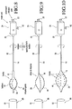
- the apparatus 10 includes a wire 12 (see Figures 1 , 2, 3, 4, 5, 6, 7a and 7b ), balloon catheter 14 (see Figures 8, 9 and 10 ), multi-prong assembly 15 (see Figures 11 and 12 ) or multi-infusion wire assembly 60 (see Figure 13 ) capable of delivering a sclerosant material while irritating and/or disrupting the vessel wall 16 of the treatment site. While the terms irritating and/or disrupting are utilized in describing the present invention, those skilled in the art will understand that these terms may be used substantially interchangeably without varying the scope of the present invention.
- the present apparatus 10 treats varicose veins by disrupting and/or irritating a vessel wall 16 in conjunction with the direct administration of a sclerosant material.
- This direct delivery of the sclerosant to the irritated or disrupted vessel provides for improved success rates in the treatment of varicose veins when compared with the limited success rates for traditional sclerotherapy as discussed above with reference to prior art techniques.
- the vein spasms produced as a result of the apparatus 10 will further enhance the efficacy of the procedure.
- delivery of sclerosant can be done either before, during or after vascular irritation and/or disruption.
- the location and method of delivery of sclerosant can be done either at the sclerosing site, proximal to it, distal to it, as well as at the vascular entry site through a vascular sheath.
- the delivery of the sclerosant may be achieved at a variety of locations, including system administration.
- the invention is primarily directed to the use of mechanical disruption devices, it is contemplated that other disrupting mechanisms (for example, heat, cold, electrical, laser, Radiofrequency, etc.) may be employed within the spirit of the present invention.
- the wire 12, balloon catheter 14, multi-prong assembly 15 or multi-infusion wire assembly 60 is moved to create the disruption and/or irritation in the vessel wall.
- the disruption and/or irritation is caused by a mechanical device, which when positioned in the vessel provides an outwardly directed radial force to engage, irritate and damage but not break through the inner vessel wall.
- the mechanical device may take the form of a wire, catheter, balloon, spring, brush or a combination thereof. Movement may either be created by rotation of the apparatus 10, by linear movement of the apparatus 10 or by a combination of both rotational and linear movement of the apparatus 10. In addition, the movement may be achieved via either manual actuation or via the implementation of a power system.
- the apparatus 10 functions by disrupting and/or irritating the lining of the vessel wall 16 and delivering a sclerosing solution directly to the vessel wall 16 for safe and easy ablation.
- the apparatus 10 includes an infusion wire 12 having a distal end 18 and a proximal end 20. While the term "wire" is used in the present description it is contemplated that various materials (for example, metals, plastics, etc.) may be used without departing from the spirit of the present invention.
- the infusion wire might take the general form of a catheter shaped and dimensioned to function in the manner detailed herein.
- the structure of the wire itself may be varied to suit specific needs; for example, the distal end of the wire may be solid or incorporate a spring structure to provide for desired rigidity.
- the infusion wire At its proximal end 20, the infusion wire includes a sclerosant infusion port 22 for supply of the sclerosing agent to the infusion wire 12.
- a motor 24 is coupled to the proximal end 20 for the controlled rotation of the infusion wire 12 in a manner which will be discussed below in greater detail.
- the distal end 18 of the infusion wire 12 is provided with a series of sinusoidal waves 26 shaped and dimensioned for engaging the vessel wall 16 of the vessel 28 into which the infusion wire 12 is placed.
- the infusion wire 12 is provided with application ports 34 through which the sclerosing agent is administered within the vessel 28.
- the application ports 34 are in fluid communication with the infusion port 22 via a conduit 35 extending through the wire 12.
- Controlled administration of the sclerosing agent through the application ports 34 is enhanced by closing off the distal tip 36 of the infusion wire 12 so as to ensure the sclerosing material is administered only though the application ports 34 at the upper and lower extents 30, 32 of the infusion wire 12.
- proximal to the disrupting and/or irritating end the infusion wire may be enclosed in a sheath.
- Figures 2, 3, 4, 5 and 6 are disclosed with reference to Figures 2, 3, 4, 5 and 6 . These embodiments are substantially similar to those disclosed above with reference to Figure 1 , however, the distal end 18 is shaped differently.
- Figure 2 provides an infusion wire 12 with a substantially V-shaped distal end
- Figure 3 provides an infusion wire 12 with a sharp tip 38 having an application port 34 therein
- Figure 4 provides an infusion wire 12 with a substantially J-shaped distal end
- Figure 5 provides an infusion wire 12 with a circular distal end 18
- Figure 6 provides an infusion wire 12 with a twisted circular distal end 18.
- a wide variety of shapes may be employed without departing from the spirit of the present invention.
- the shape of the infusion wire 12 prior to insertion of the apparatus 10 within the vessel 28. It is contemplated this may be accomplished via the use of a stiffener which holds the infusion wire 12 in a desired configuration prior to insertion or through the fabrication of the infusion wire 12 from shape memory materials trained to assume the desired shaped when exposed to elevated temperatures within the body. This may also be accomplished by containment of the wire 12 in a sheath.
- FIG. 7a and 7b an embodiment of the infusion wire 12 utilizing a stiffener 39 is shown.
- the stiffener 39 is positioned within the infusion wire 12.
- the stiffener 39 holds the infusion wire 12 in a straightened configuration allowing insertion through a needle or sheath (not shown).
- the stiffener 39 is withdrawn and the infusion wire 12 resumes its desired configuration (see Figure 7b ) permitting disruption and/or irritation of the vessel wall 16 while a sclerosing agent is applied to the vessel wall 16.
- a stiffener as disclosed in conjunction with the present embodiment could also be used in conjunction with infusion wires of various shapes, for example, those disclosed in the embodiments of Figures 1 , 2, 3 and 4 .
- the infusion wire 12 may be replaced with a balloon catheter 14 shaped and dimensioned specifically for use in accordance with the present invention. More specifically, and as with the infusion wire 12, the balloon catheter 14 includes a proximal end 20 and a distal end 18 with the balloon 40 itself positioned adjacent the distal end 18 of the catheter 14.
- the catheter 14 includes a sclerosant infusion port 22 for administration of the sclerosing agent to the balloon catheter 14.
- the sclerosant may also be used in the inflation of the balloon 40 and will leach into the surrounding area via application ports 34 directly after inflation of the balloon 40 (see Figure 9 ).
- the application ports 34 are in fluid communication with the infusion port 22 via a conduit 35 extending through the balloon catheter 14.
- the catheter 14 is provided with a balloon port 42 at the proximal end 20 (see Figures 8 and 10 ).
- the balloon port 42 provides for the administration of fluid used in controlling inflation and deflation of the balloon 40 in a convention manner.
- the sclerosant infusion port 22 is directly linked with the application ports 34 for the injection of the sclerosant within the vessel 28.
- a conduit is provided in accordance with a preferred embodiment of the present invention, sclerosant delivery may also be accomplished by containment of the wire proximal to the balloon in a sheath and delivering the sclerosant fluid or foam through the annular space between the wire shaft and sheath to exit near the distal end of the sheath into the vein.
- Manual manipulations maybe used or a motor 24 may be coupled to the proximal end 20 for the controlled rotation of the balloon catheter 14 in a manner that will be discussed below in greater detail.
- the distal end 18 of the catheter 14 is provided with a balloon 40 having a series of sharp or rugged bumps or edges or hooks 44 shaped and dimensioned for scraping and injuring the surface or lining of the vessel wall 16.
- a series of application ports 34 are formed in the wall of the balloon 40 for the delivery of the sclerosing agent to the vessel wall 16.
- the application ports 34 are linked to the sclerosing agent supply port 22 at the proximal end 20 of the catheter 14.
- a variety of balloon catheter lumen structures are known for the delivery of medicinal agents to the surface of the balloon and these known catheter lumen structures may be employed within the spirit of the present invention.
- Sclerosant delivery may also be accomplished by containment of the wire proximal to the balloon in a sheath and delivering the sclerosant fluid or foam through the annular space between the wire shaft and sheath to exit near the distal end of the sheath into the vein.
- the present ablation apparatus 10 will produce spasms within the vessel 28 being treated.
- Vessels 28, and in particular veins develop spasms upon irritation. Spasm is generally a non-desirable situation in vascular procedures and is commonly avoided.
- the apparatus 10 described in accordance with the present invention purposefully creates spasms at the treatment site.
- the creation of spasms in accordance with the present invention results in a decrease in vein size that markedly limits flow in the vein. This effect, together with the mechanical irritation and destruction of the vein, makes the vein very susceptible to the sclerosing agent and increases the likelihood for successful treatment.
- Spasm of the vein will reduce the size of larger veins to that of smaller veins and limit flow and volume. This will dramatically increase the likelihood of successful treatment when employed in accordance with the present invention. Success rates associated with prior art sclerotherapy techniques are limited. The bigger the vein, and the more flow and blood are present in the vein, the less likely the sclerosant will achieve complete destruction. In accordance with the present invention, the mechanical disruption in conjunction with sclerotherapy increases the success rate of the procedure. This is achieved by delivery of the sclerosant to the vessel, by causing spasm that limits the flow in the vessel and by mechanical destruction and disruption of the endothelium.
- the spasm will be created within a blood vessel, either an artery or a vein.
- the spasm can be created by mechanical irritation or any other way, including, but not limited to, electrical, chemical, thermal treatment.
- the spasm is preferably created by disruption of the endothelial layer of a blood vessel, without perforation of the vessel wall. It is further contemplated that the creation of spasm within the vessel may be achieved for the purpose of increasing its susceptibility to subsequent injury, whether chemical or other.
- treatment is achieved in the following manner. Regardless of which of the various embodiments are employed, the apparatus 10 is first advanced through the vein 28 to the treatment area. The apparatus 10 is then activated for disruption and/or irritation of the treatment site. As discussed above, movement of the apparatus 10 to accomplish disruption and/or irritation may be achieved either manually or mechanically.
- sclerosant is injected into the vein 28 at the treatment site.
- the sclerosant may be administered either manually (for example, a manually actuated syringe) or automatically (for example, a controlled pumping system linked to the apparatus 10).
- the apparatus may be pulled through the vein 28 toward the access site.
- the apparatus 10 is removed from the vein 28.
- the sinusoidal distal end 18 of the infusion wire 12 is inserted to a desired site within the vessel 28.
- the motor 24 is then activated, rotating the sinusoidal infusion wire 12 in a manner which disrupts and irritates the vessel wall 16.
- the sclerosing agent is pumped into the infusion wire 12 and is dispensed at the treatment site via the application ports 34 located at the upper and lower extents 30, 32 of the sinusoidal waves 26 of the infusion wire 12.
- the infusion wire 12 is angled with a port or ports 34 that deliver a sclerosing agent directly to the vessel 16.
- a port or ports 34 that deliver a sclerosing agent directly to the vessel 16.
- Each of these designs also provide for a sharp edge or edges that irritate the vessel wall 16 at the same time. It is contemplated the wire 12 can be inserted through a needle or a small sheath.
- a balloon catheter 14 may also be employed in accordance with the spirit of the present invention.
- the balloon 40 is provided with sharp or rugged bumps or edges or hooks 44 that will scrape and injure the surface or lining of the vessel wall 16.
- This apparatus 10 will block flow in the vein and simultaneously deliver the sclerosant to the vessel wall 16. This injury will facilitate the destruction of the vein.
- the balloon 40 and the destructing edges 44 may have variable diameters and shapes, based on the size of the vessel being treated.
- the balloon 40 which is inserted in a deflated state, is inflated under the control of applied fluid, preferably but not necessarily the sclerosing solution.
- the sclerosing solution is used in expanding the balloon or not, the sclerosing solution is injected into the balloon 40 or a chamber within the balloon (not shown) while the balloon 40 is in the vein 28 to be ablated.
- the balloon 40 will be pulled back with or without rotation within the vein 28.
- this movement may be done by hand or with a mechanical device (for example, a motor 24) rapidly rotating the balloon catheter apparatus 10.
- a mechanical device may also control the rate and volume of delivery of the sclerosant.
- the sclerosing solution will leach out of the balloon 40 directly to the disrupted vein wall. The effect of the sclerosing solution will be enhanced by the disruption or irritation of the vessel wall 16.
- the balloon catheter may be modified with a wire or wires over the balloon.
- the wire can have sharp edges that will disrupt the vessel.
- the wire can rotate with or without the balloon.
- an ablation apparatus 10 includes a multi-prong assembly 15 composed of a delivery member 50 housed within a sheath 52 adapted for movement relative to the delivery member 50.
- the delivery member 50 includes a proximal end 54 and a distal end 56.
- the proximal end 54 is a generally cylindrical member having an injection port 22 for the application of sclerosing agent toward the distal end 56 of the delivery member 50.
- the distal end 56 of the delivery member 50 includes a series of outwardly biased prongs 58.
- the prongs 58 are biased such that when the sheath 52 is moved toward the proximal end 54 of the delivery member 50 and the prongs 58 are exposed (see Figure 11 ), they extend outwardly into contact with the vessel wall 16. As such, the prongs 58 contact the vessel wall 16 permitting disruption and/or irritation of the vessel wall 16.
- Sclerosing agent is delivered to the vessel wall 16 via ports 34 formed within the prongs 58.
- the ports 34 are formed so as to be in fluid communication with the injection port 22 located at the proximal end 54 of the delivery member 50.
- the multi-prong assembly 15 is delivered to the treatment site in its storage configuration (see Figure 12 ) with the sheath 52 covering the prongs 58.
- the sheath 52 is moved relative to the delivery member 50 revealing the prongs 58 (see Figure 11 ) and permitting the prongs 58 to extend outwardly into engagement with the vessel wall 16.
- the prongs 58 are moved to disrupt and/or irritate the vessel wall 16 either before, during or after a sclerosing agent is delivered.
- the present ablation apparatus 10 may be either manually or automatically manipulated for both rotational and longitudinal movement during the disruption and/or irritation of the vessel wall 16.
- an ablation apparatus 10 includes a multi-infusion wire assembly 60.
- the multi-infusion wire assembly 60 includes a proximal end 62 and a distal end 64.
- the proximal end 62 is a generally cylindrical member having an injection port 22 for the application of sclerosing agent toward the distal end 64 of the assembly 60.
- the distal end 64 of the multi-infusion wire assemble 62 includes a series of helically wound wires 66 forming an elongated structure with an enlarged central region 68.
- the enlarged central region 68 is oriented outwardly such that the wires 66 may contact the vessel wall 16 for disruption and/or irritation in accordance with the present invention.
- Sclerosing agent is delivered to the vessel wall 16 via ports 34 formed within the wires 66.
- the ports 34 are formed so as to be in fluid communication with the injection port 22 located at the proximal end 62 of the assembly 60.
- the multi-infusion wire assembly 60 functions in much the same manner as the multi-prong assembly 15 disclosed with reference to Figures 11 and 12 , and consequently includes a sheath 70 for delivery of the assembly 60.
- Sclerosant delivery may also be accomplished by delivering the sclerosant fluid or foam through the annular space between the wire shaft and sheath 70 to exit near the distal end of sheath 70 into the vein.
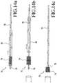
- the ablation apparatus 72 is formed with a brush 74 at its distal end 76.
- the bush 74 may be positioned at the very end of the apparatus 72 (see Figure 14c ) or slightly removed from the end of the apparatus 72 (see Figures 14a and 14b ).
- the apparatus 72 includes a conduit 78 for the application of sclerosing agent to the treatment site.
- the apparatus 72 is further provided with a sheath 80 facilitating position of the apparatus 72 at a desired treatment site.
- the sheath 80 is utilized in a conventional manner to cover the distal end 76 of the apparatus 72 until it is properly positioned (see Figure 14a ), at which time it is withdrawn for treatment of the site (see Figure 14b ).
- the brush 74 may be manually or automatically moved to created disruption at the treatment site.
- the sheath may function as a conduit providing a guiding tube for the application the sclerosing agent to the treatment.
- the present invention combines mechanical disruption and/or irritation of the treatment site with the application of a sclerosing agent.
- a further embodiment for achieving this goal is disclosed.
- the disclosed embodiment achieves mechanical disruption and/or irritation of the vessel wall through the application of fluid jets.
- the apparatus 110 includes a conduit 112 having a proximal end 114 and a distal end 116.
- the proximal end 114 is coupled to a pressurized fluid source 118 and a source of sclerosing agent 120, both of which are transmitted to the distal end 116 for application at the treatment site.
- the distal end 116 includes a series of apertures 122 for the application of fluid jets 124 and sclerosing agent at the treatment site.
- the jet(s) 124 will cause disruption and/or irritation of the vessel by mechanical means and the sclerosing fluid will cause the chemical destruction of the vessel.
- the fluid of the jets may be the sclerosing agent or the fluid jets may be applied with another fluid and combined with the separate application of the sclerosing agent.
- the fluid jets may point at the vessel wall and may have various shapes and intensities dictated by the specific needs of the treatment site. It is further contemplated the fluid can be hot causing heat damage or very cold causing cold damage.
- sclerosing agents may be employed. Those skilled in the art will certainly appreciate the pros and cons of the known sclerosing agents and choose the sclerosing agent most appropriate for specific applications.
- foamed sclerotherapy may be employed in accordance with an alternate embodiment of the present invention.
- foam sclerotherapy involves mixing the sclerosing solution with air or other gas, for example, oxygen, carbon dioxide and/or nitrogen to create foam. The idea behind this technique is to deliver a higher concentration of sclerosant to the vein wall.
- foam sclerotherapy employs a mixture of the sclerosant with gas creating microbubbles.
- the microbubbles displace the blood and ensure contact between the sclerosant and the vain wall.
- One potential disadvantage with the foam is that it can leach into the deep venous system and damage the endothelium there. This will cause deep venous thrombosis (DVT), a known complication of varicose vein treatment. DVT is less likely to occur when using liquid sclerosant. The liquid sclerosant gets diluted very quickly once it is flowing into the deep venous system and is therefore much less likely to cause endothelial damage and DVT.
- the present invention is directed toward ablation of veins, especially the greater sephanous vein for treatment varicose veins.
- the present invention may be employed in the sclerosis and disruption and/or irritation of other hollow structures such as arteries, bronchi and any other organs, without departing from the spirit of the present invention.
- the present invention further offers many cost advantages when compared with prior art techniques for the treatment of varicose veins.
- the currently available methods of large vein ablation include laser ablation, radio-frequency ablation and stripping.
- the present sclerotherapy wire method is less expensive as there is no need for a power generator.
- prior art techniques are based on energy emission to "burn" the vein.
- the energy usually extends beyond the thin wall of the vein and can cause damage to the adjacent structures, including nerve damage and skin burns. It can also cause bleeding secondary to vascular disruption.
- the direct disruption-sclerotherapy method of the present invention either with a wire or with a balloon catheter, will direct treatment to the vein wall and not beyond it.
- Sclerotherapy has been used since the 1960's for ablation of veins. It is a safe and proven treatment. It is less effective in large veins because of the high flow and high volume causing rapid dilution of the sclerosant solution.
- the wire or balloon disruption delivery in accordance with the present invention ensures that the vein wall will be damaged and in contact with the appropriate concentration of sclerosant, regardless of the flow and blood volume in the vein.
- tumescent anesthesia Because of the risk of damage to adjacent tissue, before ablation with laser or RF, the operator needs to inject a large amount of local anesthetic, called tumescent anesthesia. This is very time consuming and might take up to 45 minutes depending upon the length of vein being treated.
- the present invention eliminates the need to use tumescent anesthesia.
Landscapes
- Health & Medical Sciences (AREA)
- Surgery (AREA)
- Life Sciences & Earth Sciences (AREA)
- Medical Informatics (AREA)
- Animal Behavior & Ethology (AREA)
- Engineering & Computer Science (AREA)
- Biomedical Technology (AREA)
- Heart & Thoracic Surgery (AREA)
- Veterinary Medicine (AREA)
- Molecular Biology (AREA)
- Nuclear Medicine, Radiotherapy & Molecular Imaging (AREA)
- General Health & Medical Sciences (AREA)
- Public Health (AREA)
- Vascular Medicine (AREA)
- Rheumatology (AREA)
- Surgical Instruments (AREA)
- Infusion, Injection, And Reservoir Apparatuses (AREA)
Applications Claiming Priority (4)
| Application Number | Priority Date | Filing Date | Title |
|---|---|---|---|
| US47204803P | 2003-05-21 | 2003-05-21 | |
| US50142303P | 2003-09-10 | 2003-09-10 | |
| US10/736,535 US7862575B2 (en) | 2003-05-21 | 2003-12-17 | Vascular ablation apparatus and method |
| EP04776062.4A EP1638479B1 (fr) | 2003-05-21 | 2004-05-20 | Appareil d'ablation vasculaire |
Related Parent Applications (1)
| Application Number | Title | Priority Date | Filing Date |
|---|---|---|---|
| EP04776062.4A Division EP1638479B1 (fr) | 2003-05-21 | 2004-05-20 | Appareil d'ablation vasculaire |
Publications (1)
| Publication Number | Publication Date |
|---|---|
| EP3326553A1 true EP3326553A1 (fr) | 2018-05-30 |
Family
ID=33545311
Family Applications (2)
| Application Number | Title | Priority Date | Filing Date |
|---|---|---|---|
| EP04776062.4A Expired - Lifetime EP1638479B1 (fr) | 2003-05-21 | 2004-05-20 | Appareil d'ablation vasculaire |
| EP17207507.9A Ceased EP3326553A1 (fr) | 2003-05-21 | 2004-05-20 | Appareil d'ablation vasculaire |
Family Applications Before (1)
| Application Number | Title | Priority Date | Filing Date |
|---|---|---|---|
| EP04776062.4A Expired - Lifetime EP1638479B1 (fr) | 2003-05-21 | 2004-05-20 | Appareil d'ablation vasculaire |
Country Status (4)
| Country | Link |
|---|---|
| US (2) | US7862575B2 (fr) |
| EP (2) | EP1638479B1 (fr) |
| CA (1) | CA2526421C (fr) |
| WO (1) | WO2004112569A2 (fr) |
Cited By (1)
| Publication number | Priority date | Publication date | Assignee | Title |
|---|---|---|---|---|
| US12207807B2 (en) | 2018-05-18 | 2025-01-28 | University of Galway | Devices and methods for treating a vessel in a subject |
Families Citing this family (76)
| Publication number | Priority date | Publication date | Assignee | Title |
|---|---|---|---|---|
| US7819887B2 (en) | 2004-11-17 | 2010-10-26 | Rex Medical, L.P. | Rotational thrombectomy wire |
| US8623009B2 (en) * | 2005-04-05 | 2014-01-07 | The Brigham And Women's Hospital, Inc. | Endoscopic gastric bypass defect repair |
| US20080243068A1 (en) * | 2005-12-29 | 2008-10-02 | Kamal Ramzipoor | Methods and apparatus for treatment of venous insufficiency |
| EP2061385B1 (fr) * | 2006-09-13 | 2014-06-04 | Vascular Insights LLC | Dispositif de traitement vasculaire |
| US8448644B2 (en) * | 2006-11-02 | 2013-05-28 | Cooltouch Incorporated | Sonic endovenous catheter |
| US20080140002A1 (en) * | 2006-12-06 | 2008-06-12 | Kamal Ramzipoor | System for delivery of biologically active substances with actuating three dimensional surface |
| EP2120737B1 (fr) | 2007-02-05 | 2020-04-01 | Boston Scientific Limited | Appareil de thrombectomie |
| US20170136158A1 (en) | 2015-10-16 | 2017-05-18 | Angiodynamics, Inc. | Systems and Methods for Removing Undesirable Material Within a Circulatory System |
| US11589880B2 (en) | 2007-12-20 | 2023-02-28 | Angiodynamics, Inc. | System and methods for removing undesirable material within a circulatory system utilizing during a surgical procedure |
| US10517617B2 (en) | 2007-12-20 | 2019-12-31 | Angiodynamics, Inc. | Systems and methods for removing undesirable material within a circulatory system utilizing a balloon catheter |
| WO2009109967A1 (fr) | 2008-03-02 | 2009-09-11 | V.V.T. Medical Ltd. | Procédé et dispositif pour une ablation de veine |
| US9510854B2 (en) | 2008-10-13 | 2016-12-06 | Boston Scientific Scimed, Inc. | Thrombectomy catheter with control box having pressure/vacuum valve for synchronous aspiration and fluid irrigation |
| US9099738B2 (en) * | 2008-11-03 | 2015-08-04 | Basvah Llc | Lithium secondary batteries with positive electrode compositions and their methods of manufacturing |
| ES2738478T3 (es) * | 2010-05-07 | 2020-01-23 | Care Fusion 2200 Inc | Diseño de catéter mejorado para uso en el tratamiento de enfermedades pleurales |
| US9795406B2 (en) | 2010-05-13 | 2017-10-24 | Rex Medical, L.P. | Rotational thrombectomy wire |
| US8663259B2 (en) | 2010-05-13 | 2014-03-04 | Rex Medical L.P. | Rotational thrombectomy wire |
| US9023070B2 (en) | 2010-05-13 | 2015-05-05 | Rex Medical, L.P. | Rotational thrombectomy wire coupler |
| US8764779B2 (en) | 2010-05-13 | 2014-07-01 | Rex Medical, L.P. | Rotational thrombectomy wire |
| US10039900B2 (en) * | 2010-09-07 | 2018-08-07 | Angiodynamics, Inc. | Fluid delivery and treatment device and method of use |
| US8685050B2 (en) | 2010-10-06 | 2014-04-01 | Rex Medical L.P. | Cutting wire assembly for use with a catheter |
| US8685049B2 (en) | 2010-11-18 | 2014-04-01 | Rex Medical L.P. | Cutting wire assembly for use with a catheter |
| US9282991B2 (en) | 2010-10-06 | 2016-03-15 | Rex Medical, L.P. | Cutting wire assembly with coating for use with a catheter |
| US9585667B2 (en) * | 2010-11-15 | 2017-03-07 | Vascular Insights Llc | Sclerotherapy catheter with lumen having wire rotated by motor and simultaneous withdrawal from vein |
| AU2015203121B2 (en) * | 2010-11-15 | 2017-04-13 | Merit Medical Systems, Inc. | Direction reversing vascular treatment device |
| US8702736B2 (en) | 2010-11-22 | 2014-04-22 | Rex Medical L.P. | Cutting wire assembly for use with a catheter |
| WO2012071058A1 (fr) * | 2010-11-23 | 2012-05-31 | William Joseph Drasler | Cathéter d'ablation veineuse chauffé |
| US20130030410A1 (en) * | 2010-11-23 | 2013-01-31 | William Joseph Drasler | Venous heated ablation catheter |
| US9055964B2 (en) | 2011-03-15 | 2015-06-16 | Angio Dynamics, Inc. | Device and method for removing material from a hollow anatomical structure |
| US12245788B2 (en) | 2011-03-15 | 2025-03-11 | Angiodynamics, Inc. | Device and method for removing material from a hollow anatomical structure |
| US20120109191A1 (en) * | 2011-12-13 | 2012-05-03 | Vascular Insights Llc | Adhesive-based varicose vein treatment |
| US9398905B2 (en) | 2012-12-13 | 2016-07-26 | Ethicon Endo-Surgery, Llc | Circular needle applier with offset needle and carrier tracks |
| AU2015203354B2 (en) * | 2012-12-13 | 2016-05-12 | Ethicon Endo-Surgery, Inc. | Circular needle applier |
| US9358042B2 (en) | 2013-03-13 | 2016-06-07 | The Spectranetics Corporation | Expandable member for perforation occlusion |
| US9788830B2 (en) | 2014-06-06 | 2017-10-17 | Ethicon Llc | Needle cartridge with cage |
| EP3055050B1 (fr) | 2013-10-13 | 2020-08-05 | V.V.T. Med Ltd. | Dispositif et procédé de formation de mousse |
| PL3054856T3 (pl) | 2013-10-13 | 2021-06-14 | V.V.T. Med Ltd. | Urządzenie do zsynchronizowanego wstrzykiwania i zasysania |
| EP3054857B1 (fr) | 2013-10-13 | 2020-09-09 | V.V.T. Med Ltd. | Dispositif de résection d'une veine |
| US10639061B2 (en) | 2013-11-11 | 2020-05-05 | Cook Medical Technologies Llc | Devices and methods for modifying veins and other bodily vessels |
| US20150250523A1 (en) * | 2014-03-10 | 2015-09-10 | Terumo Kabushiki Kaisha | Method for treating varicose veins and intraluminal device used in such method |
| US20150250536A1 (en) * | 2014-03-10 | 2015-09-10 | Terumo Kabushiki Kaisha | Method for treating varicose veins and intraluminal device used in such method |
| US20150250522A1 (en) * | 2014-03-10 | 2015-09-10 | Terumo Kabushiki Kaisha | Method for treating varicose veins and intraluminal device used in such method |
| US9883877B2 (en) | 2014-05-19 | 2018-02-06 | Walk Vascular, Llc | Systems and methods for removal of blood and thrombotic material |
| US9526495B2 (en) | 2014-06-06 | 2016-12-27 | Ethicon Endo-Surgery, Llc | Articulation control for surgical instruments |
| US20160030068A1 (en) * | 2014-07-31 | 2016-02-04 | Terumo Kabushiki Kaisha | Method for treating varicose veins and intraluminal device used in such method |
| US20160030719A1 (en) * | 2014-07-31 | 2016-02-04 | Terumo Kabushiki Kaisha | Method for treating varicose veins and intraluminal device used in such method |
| US9457153B2 (en) | 2014-08-29 | 2016-10-04 | Vascular Insights Llc | Syringe accessory |
| JP6732403B2 (ja) | 2014-10-03 | 2020-07-29 | テルモ株式会社 | 医療デバイス |
| PL228142B1 (pl) * | 2014-10-17 | 2018-02-28 | Balton Społka Z Ograniczoną Odpowiedzialnością | Kateter zawierajacy element tnacy |
| US10398847B2 (en) | 2014-12-12 | 2019-09-03 | Wake Forrest University Health Sciences | Incremental syringe |
| GB201511595D0 (en) * | 2014-12-23 | 2015-08-19 | Whiteley Mark | Medical device for treating a vein |
| CN107205750B (zh) | 2015-01-08 | 2020-08-21 | 西纳塞弗医疗有限公司 | 鼻旁窦医疗装置及其用途 |
| US10022120B2 (en) | 2015-05-26 | 2018-07-17 | Ethicon Llc | Surgical needle with recessed features |
| CN105105863B (zh) * | 2015-07-28 | 2017-11-24 | 中国医学科学院北京协和医院 | 实验动物血管内膜损伤实验装置 |
| US10499892B2 (en) | 2015-08-11 | 2019-12-10 | The Spectranetics Corporation | Temporary occlusion balloon devices and methods for preventing blood flow through a vascular perforation |
| US10449336B2 (en) | 2015-08-11 | 2019-10-22 | The Spectranetics Corporation | Temporary occlusions balloon devices and methods for preventing blood flow through a vascular perforation |
| EP3135206B1 (fr) | 2015-08-31 | 2020-01-15 | Gefässpraxis Dr. Erpen AG | Dispositif de traitement des varices |
| US10561440B2 (en) | 2015-09-03 | 2020-02-18 | Vesatek, Llc | Systems and methods for manipulating medical devices |
| USD800306S1 (en) | 2015-12-10 | 2017-10-17 | Ethicon Llc | Surgical suturing device |
| US10226263B2 (en) | 2015-12-23 | 2019-03-12 | Incuvate, Llc | Aspiration monitoring system and method |
| USD802769S1 (en) | 2016-05-16 | 2017-11-14 | Teleflex Medical Incorporated | Thrombectomy handle assembly |
| CN109661250B (zh) | 2016-07-03 | 2021-10-29 | 西纳塞弗医疗有限公司 | 用于治疗鼻窦和/或耳部的医疗装置及其使用方法 |
| USD865964S1 (en) | 2017-01-05 | 2019-11-05 | Ethicon Llc | Handle for electrosurgical instrument |
| US10580131B2 (en) | 2017-02-23 | 2020-03-03 | Zebra Medical Vision Ltd. | Convolutional neural network for segmentation of medical anatomical images |
| EP3723634B1 (fr) * | 2017-12-12 | 2024-02-28 | Boston Scientific Scimed, Inc. | Dispositif médical rotatif |
| US11678905B2 (en) | 2018-07-19 | 2023-06-20 | Walk Vascular, Llc | Systems and methods for removal of blood and thrombotic material |
| US20200113575A1 (en) * | 2018-10-12 | 2020-04-16 | Merit Medical Systems, Inc. | Vascular treatment apparatus with auto injection device and related methods |
| USD895112S1 (en) | 2018-11-15 | 2020-09-01 | Ethicon Llc | Laparoscopic bipolar electrosurgical device |
| US11648020B2 (en) | 2020-02-07 | 2023-05-16 | Angiodynamics, Inc. | Device and method for manual aspiration and removal of an undesirable material |
| KR102438221B1 (ko) | 2020-05-13 | 2022-09-01 | 유펙스메드 주식회사 | 전자적 제어로 약액을 주입하는 정맥 질환 치료장치 |
| WO2022174175A1 (fr) | 2021-02-15 | 2022-08-18 | Walk Vascular, Llc | Systèmes et procédés d'élimination de sang et de matériau thrombotique |
| US12274458B2 (en) | 2021-02-15 | 2025-04-15 | Walk Vascular, Llc | Systems and methods for removal of blood and thrombotic material |
| US11696793B2 (en) * | 2021-03-19 | 2023-07-11 | Crossfire Medical Inc | Vascular ablation |
| EP4504071A1 (fr) * | 2022-04-07 | 2025-02-12 | Clearstream Technologies Limited | Dispositif d'ablation veineuse |
| EP4565150A4 (fr) | 2022-08-08 | 2025-12-10 | Crossfire Medical Inc | Ablation vasculaire segmentaire |
| AU2023371657A1 (en) | 2022-11-04 | 2025-05-15 | Solvein Inc. | Catheters and related methods for aspiration and controlled delivery of closure agents |
| US11911581B1 (en) | 2022-11-04 | 2024-02-27 | Controlled Delivery Systems, Inc. | Catheters and related methods for the aspiration controlled delivery of closure agents |
Citations (4)
| Publication number | Priority date | Publication date | Assignee | Title |
|---|---|---|---|---|
| FR2651682A1 (fr) * | 1989-09-13 | 1991-03-15 | Cadet Pierre | Appareil destine a la destruction des varices des membres inferieurs chez l'homme. |
| JPH11262527A (ja) * | 1998-03-17 | 1999-09-28 | Buaayu:Kk | 血管内擦過バルーンカテーテル |
| US20020010418A1 (en) * | 2000-07-21 | 2002-01-24 | Syntheon, Llc | Methods and apparatus for sclerosing the wall of a varicose vein |
| US6402745B1 (en) * | 2000-02-23 | 2002-06-11 | Peter J. Wilk | Intravenous whip electrode for vein ablation |
Family Cites Families (35)
| Publication number | Priority date | Publication date | Assignee | Title |
|---|---|---|---|---|
| US4728319A (en) | 1986-03-20 | 1988-03-01 | Helmut Masch | Intravascular catheter |
| DE3830909A1 (de) | 1988-09-10 | 1990-03-22 | Astra Meditec Ab | Krampfadersonde |
| US5087244A (en) | 1989-01-31 | 1992-02-11 | C. R. Bard, Inc. | Catheter and method for locally applying medication to the wall of a blood vessel or other body lumen |
| US5022399A (en) * | 1989-05-10 | 1991-06-11 | Biegeleisen Ken P | Venoscope |
| DE69023515T2 (de) | 1989-06-28 | 1996-04-04 | David S Zimmon | Aufblasbare tamponadevorrichtungen. |
| US5074871A (en) | 1989-12-07 | 1991-12-24 | Evi Corporation | Catheter atherotome |
| CA2081896A1 (fr) | 1990-06-15 | 1991-12-16 | James E. Shapland | Dispositif et methode pour l'administration de medicaments |
| DE69119578T2 (de) | 1991-02-28 | 1996-12-19 | Pierre Cadet | Vorrichtung zum Zerstören von Krampfadern |
| US6733473B1 (en) | 1991-04-05 | 2004-05-11 | Boston Scientific Corporation | Adjustably stiffenable convertible catheter assembly |
| WO1993019679A1 (fr) | 1992-04-07 | 1993-10-14 | The Johns Hopkins University | Systeme de catheter de fragmentation mecanique intradermique |
| US5370653A (en) * | 1993-07-22 | 1994-12-06 | Micro Therapeutics, Inc. | Thrombectomy method and apparatus |
| US5415636A (en) * | 1994-04-13 | 1995-05-16 | Schneider (Usa) Inc | Dilation-drug delivery catheter |
| US5836905A (en) * | 1994-06-20 | 1998-11-17 | Lemelson; Jerome H. | Apparatus and methods for gene therapy |
| US5891108A (en) | 1994-09-12 | 1999-04-06 | Cordis Corporation | Drug delivery stent |
| US6311692B1 (en) | 1996-10-22 | 2001-11-06 | Epicor, Inc. | Apparatus and method for diagnosis and therapy of electrophysiological disease |
| US5882329A (en) | 1997-02-12 | 1999-03-16 | Prolifix Medical, Inc. | Apparatus and method for removing stenotic material from stents |
| US6090118A (en) | 1998-07-23 | 2000-07-18 | Mcguckin, Jr.; James F. | Rotational thrombectomy apparatus and method with standing wave |
| US7037316B2 (en) * | 1997-07-24 | 2006-05-02 | Mcguckin Jr James F | Rotational thrombectomy device |
| US6258087B1 (en) * | 1998-02-19 | 2001-07-10 | Curon Medical, Inc. | Expandable electrode assemblies for forming lesions to treat dysfunction in sphincters and adjoining tissue regions |
| US6159196A (en) * | 1998-03-09 | 2000-12-12 | Ruiz; Carlos | Methods and apparatus for transvascular muscular revascularization and drug delivery |
| US6231518B1 (en) | 1998-05-26 | 2001-05-15 | Comedicus Incorporated | Intrapericardial electrophysiological procedures |
| US6369039B1 (en) | 1998-06-30 | 2002-04-09 | Scimed Life Sytems, Inc. | High efficiency local drug delivery |
| US6048332A (en) * | 1998-10-09 | 2000-04-11 | Ave Connaught | Dimpled porous infusion balloon |
| US6663613B1 (en) * | 2000-01-25 | 2003-12-16 | Bacchus Vascular, Inc. | System and methods for clot dissolution |
| WO2001076458A2 (fr) | 2000-04-07 | 2001-10-18 | Bacchus Vascular, Inc. | Procedes et dispositif d'endarteriectomie percutanee a distance |
| USD450843S1 (en) | 2000-05-30 | 2001-11-20 | Boston Scientific Corporation | Thrombectomy handpiece |
| US7285126B2 (en) | 2000-06-29 | 2007-10-23 | Concentric Medical, Inc. | Systems, methods and devices for removing obstructions from a blood vessel |
| US20030120256A1 (en) | 2001-07-03 | 2003-06-26 | Syntheon, Llc | Methods and apparatus for sclerosing the wall of a varicose vein |
| US6544221B1 (en) | 2000-08-30 | 2003-04-08 | Advanced Cardiovascular Systems, Inc. | Balloon designs for drug delivery |
| EP1399213A4 (fr) * | 2000-10-11 | 2008-03-19 | Micro Therapeutics Inc | Methodes de traitement d'anevrismes |
| DE10059742A1 (de) | 2000-12-01 | 2002-06-13 | Angiolas Medical Gmbh | System zum Behandeln vaskulärer Verwachsungen |
| US6623452B2 (en) | 2000-12-19 | 2003-09-23 | Scimed Life Systems, Inc. | Drug delivery catheter having a highly compliant balloon with infusion holes |
| AU2002340749A1 (en) * | 2001-05-04 | 2002-11-18 | Concentric Medical | Coated combination vaso-occlusive device |
| US6726674B2 (en) | 2001-09-04 | 2004-04-27 | Jomed Gmbh | Methods for minimally invasive, localized delivery of sclerotherapeutic agents |
| EP1644077A4 (fr) | 2003-03-18 | 2012-01-04 | Catharos Medical Systems Inc | Procedes et dispositifs pour recuperer un agent medical d'un site de collecte de fluides physiologiques efferents |
-
2003
- 2003-12-17 US US10/736,535 patent/US7862575B2/en active Active
-
2004
- 2004-05-20 CA CA2526421A patent/CA2526421C/fr not_active Expired - Lifetime
- 2004-05-20 EP EP04776062.4A patent/EP1638479B1/fr not_active Expired - Lifetime
- 2004-05-20 WO PCT/US2004/015858 patent/WO2004112569A2/fr not_active Ceased
- 2004-05-20 EP EP17207507.9A patent/EP3326553A1/fr not_active Ceased
-
2007
- 2007-08-02 US US11/882,474 patent/US8465508B2/en not_active Expired - Lifetime
Patent Citations (4)
| Publication number | Priority date | Publication date | Assignee | Title |
|---|---|---|---|---|
| FR2651682A1 (fr) * | 1989-09-13 | 1991-03-15 | Cadet Pierre | Appareil destine a la destruction des varices des membres inferieurs chez l'homme. |
| JPH11262527A (ja) * | 1998-03-17 | 1999-09-28 | Buaayu:Kk | 血管内擦過バルーンカテーテル |
| US6402745B1 (en) * | 2000-02-23 | 2002-06-11 | Peter J. Wilk | Intravenous whip electrode for vein ablation |
| US20020010418A1 (en) * | 2000-07-21 | 2002-01-24 | Syntheon, Llc | Methods and apparatus for sclerosing the wall of a varicose vein |
Cited By (1)
| Publication number | Priority date | Publication date | Assignee | Title |
|---|---|---|---|---|
| US12207807B2 (en) | 2018-05-18 | 2025-01-28 | University of Galway | Devices and methods for treating a vessel in a subject |
Also Published As
| Publication number | Publication date |
|---|---|
| US7862575B2 (en) | 2011-01-04 |
| EP1638479A4 (fr) | 2012-10-31 |
| US20070282359A1 (en) | 2007-12-06 |
| CA2526421C (fr) | 2011-09-20 |
| EP1638479B1 (fr) | 2018-01-10 |
| US20050055040A1 (en) | 2005-03-10 |
| CA2526421A1 (fr) | 2004-12-29 |
| EP1638479A2 (fr) | 2006-03-29 |
| WO2004112569A2 (fr) | 2004-12-29 |
| WO2004112569A3 (fr) | 2005-12-29 |
| US8465508B2 (en) | 2013-06-18 |
Similar Documents
| Publication | Publication Date | Title |
|---|---|---|
| EP1638479B1 (fr) | Appareil d'ablation vasculaire | |
| US9782562B2 (en) | Venous insufficiency treatment method | |
| JP6035244B2 (ja) | 血管処置装置および方法 | |
| EP2257224B1 (fr) | Dispositif pour une ablation de veine | |
| US9814513B2 (en) | Endovascular plasma treatment device and method of use | |
| US5893858A (en) | Method for removing veins | |
| JP6578207B2 (ja) | 脈管を閉鎖する装置および方法 | |
| US20070135791A1 (en) | Method for Infusing the Interior of a Blood Vessel | |
| EP2863811B1 (fr) | Dispositifs d'occlusion de vaisseau | |
| JP2016501075A (ja) | 一時的な脈管足場および切り込み入りデバイス | |
| US20150094740A1 (en) | Methods and Systems for Ligating a Blood Vessel | |
| US5611357A (en) | Method and apparatus for treating varicose veins | |
| Bidwai et al. | Balloon control of the saphenofemoral junction during foam sclerotherapy: proposed innovation | |
| US20080243102A1 (en) | Apparatus for segmental varicose sclerosis | |
| TW202421070A (zh) | 分段式血管消融術 | |
| RU2712089C2 (ru) | Медицинское устройство, набор для лечения и способ лечения нарушенных кровеносных сосудов, полостей организма и протоков организма, а также способ размещения медицинского текучего вещества в окружающей их ткани | |
| Bergan | Varicose veins: hooks, clamps, and suction. Application of new techniques to enhance varicose vein surgery | |
| US20180280671A1 (en) | Device for treating venous incompetence, and related methods | |
| US20220047317A1 (en) | Percutaneous ultrasound guided minimally invasive vein ablation method for saphenous veins in the lower extremity | |
| US8709004B2 (en) | Method and device for vascular treatment | |
| US20200129229A1 (en) | Venous ablation catheter and method | |
| US12478399B1 (en) | Method of treating a blood vessel in a subject | |
| Saleh | Duplex ultrasound guided catheter directed foam sclerotherapy for treatment of axial varicose veins of the lower limbs and its preliminary results | |
| Lin et al. | Endovenous catheter ablation of the saphenous vein | |
| US20100234832A1 (en) | Method for vascular treatment |
Legal Events
| Date | Code | Title | Description |
|---|---|---|---|
| PUAI | Public reference made under article 153(3) epc to a published international application that has entered the european phase |
Free format text: ORIGINAL CODE: 0009012 |
|
| STAA | Information on the status of an ep patent application or granted ep patent |
Free format text: STATUS: THE APPLICATION HAS BEEN PUBLISHED |
|
| AC | Divisional application: reference to earlier application |
Ref document number: 1638479 Country of ref document: EP Kind code of ref document: P |
|
| AK | Designated contracting states |
Kind code of ref document: A1 Designated state(s): AT BE BG CH CY CZ DE DK EE ES FI FR GB GR HU IE IT LI LU MC NL PL PT RO SE SI SK TR |
|
| STAA | Information on the status of an ep patent application or granted ep patent |
Free format text: STATUS: REQUEST FOR EXAMINATION WAS MADE |
|
| 17P | Request for examination filed |
Effective date: 20181130 |
|
| RBV | Designated contracting states (corrected) |
Designated state(s): AT BE BG CH CY CZ DE DK EE ES FI FR GB GR HU IE IT LI LU MC NL PL PT RO SE SI SK TR |
|
| STAA | Information on the status of an ep patent application or granted ep patent |
Free format text: STATUS: EXAMINATION IS IN PROGRESS |
|
| 17Q | First examination report despatched |
Effective date: 20211124 |
|
| P01 | Opt-out of the competence of the unified patent court (upc) registered |
Effective date: 20230526 |
|
| STAA | Information on the status of an ep patent application or granted ep patent |
Free format text: STATUS: THE APPLICATION HAS BEEN REFUSED |
|
| 18R | Application refused |
Effective date: 20250325 |