EP3340867B1 - Surveillance respiratoire non-invasive - Google Patents
Surveillance respiratoire non-invasive Download PDFInfo
- Publication number
- EP3340867B1 EP3340867B1 EP16838132.5A EP16838132A EP3340867B1 EP 3340867 B1 EP3340867 B1 EP 3340867B1 EP 16838132 A EP16838132 A EP 16838132A EP 3340867 B1 EP3340867 B1 EP 3340867B1
- Authority
- EP
- European Patent Office
- Prior art keywords
- waveform
- heart rate
- respiration
- rate variability
- data
- Prior art date
- Legal status (The legal status is an assumption and is not a legal conclusion. Google has not performed a legal analysis and makes no representation as to the accuracy of the status listed.)
- Active
Links
Images




Classifications
-
- A—HUMAN NECESSITIES
- A61—MEDICAL OR VETERINARY SCIENCE; HYGIENE
- A61B—DIAGNOSIS; SURGERY; IDENTIFICATION
- A61B5/00—Measuring for diagnostic purposes; Identification of persons
- A61B5/08—Measuring devices for evaluating the respiratory organs
- A61B5/091—Measuring volume of inspired or expired gases, e.g. to determine lung capacity
-
- A—HUMAN NECESSITIES
- A61—MEDICAL OR VETERINARY SCIENCE; HYGIENE
- A61B—DIAGNOSIS; SURGERY; IDENTIFICATION
- A61B5/00—Measuring for diagnostic purposes; Identification of persons
- A61B5/72—Signal processing specially adapted for physiological signals or for diagnostic purposes
- A61B5/7271—Specific aspects of physiological measurement analysis
- A61B5/7278—Artificial waveform generation or derivation, e.g. synthesizing signals from measured signals
-
- A—HUMAN NECESSITIES
- A61—MEDICAL OR VETERINARY SCIENCE; HYGIENE
- A61B—DIAGNOSIS; SURGERY; IDENTIFICATION
- A61B5/00—Measuring for diagnostic purposes; Identification of persons
- A61B5/02—Detecting, measuring or recording for evaluating the cardiovascular system, e.g. pulse, heart rate, blood pressure or blood flow
- A61B5/024—Measuring pulse rate or heart rate
-
- A—HUMAN NECESSITIES
- A61—MEDICAL OR VETERINARY SCIENCE; HYGIENE
- A61B—DIAGNOSIS; SURGERY; IDENTIFICATION
- A61B5/00—Measuring for diagnostic purposes; Identification of persons
- A61B5/02—Detecting, measuring or recording for evaluating the cardiovascular system, e.g. pulse, heart rate, blood pressure or blood flow
- A61B5/024—Measuring pulse rate or heart rate
- A61B5/02405—Determining heart rate variability
-
- A—HUMAN NECESSITIES
- A61—MEDICAL OR VETERINARY SCIENCE; HYGIENE
- A61B—DIAGNOSIS; SURGERY; IDENTIFICATION
- A61B5/00—Measuring for diagnostic purposes; Identification of persons
- A61B5/02—Detecting, measuring or recording for evaluating the cardiovascular system, e.g. pulse, heart rate, blood pressure or blood flow
- A61B5/024—Measuring pulse rate or heart rate
- A61B5/0245—Measuring pulse rate or heart rate by using sensing means generating electric signals, i.e. ECG signals
-
- A—HUMAN NECESSITIES
- A61—MEDICAL OR VETERINARY SCIENCE; HYGIENE
- A61B—DIAGNOSIS; SURGERY; IDENTIFICATION
- A61B5/00—Measuring for diagnostic purposes; Identification of persons
- A61B5/24—Detecting, measuring or recording bioelectric or biomagnetic signals of the body or parts thereof
- A61B5/316—Modalities, i.e. specific diagnostic methods
- A61B5/318—Heart-related electrical modalities, e.g. electrocardiography [ECG]
-
- A—HUMAN NECESSITIES
- A61—MEDICAL OR VETERINARY SCIENCE; HYGIENE
- A61B—DIAGNOSIS; SURGERY; IDENTIFICATION
- A61B6/00—Apparatus or devices for radiation diagnosis; Apparatus or devices for radiation diagnosis combined with radiation therapy equipment
- A61B6/02—Arrangements for diagnosis sequentially in different planes; Stereoscopic radiation diagnosis
- A61B6/03—Computed tomography [CT]
- A61B6/032—Transmission computed tomography [CT]
-
- A—HUMAN NECESSITIES
- A61—MEDICAL OR VETERINARY SCIENCE; HYGIENE
- A61B—DIAGNOSIS; SURGERY; IDENTIFICATION
- A61B6/00—Apparatus or devices for radiation diagnosis; Apparatus or devices for radiation diagnosis combined with radiation therapy equipment
- A61B6/50—Apparatus or devices for radiation diagnosis; Apparatus or devices for radiation diagnosis combined with radiation therapy equipment specially adapted for specific body parts; specially adapted for specific clinical applications
-
- A—HUMAN NECESSITIES
- A61—MEDICAL OR VETERINARY SCIENCE; HYGIENE
- A61B—DIAGNOSIS; SURGERY; IDENTIFICATION
- A61B6/00—Apparatus or devices for radiation diagnosis; Apparatus or devices for radiation diagnosis combined with radiation therapy equipment
- A61B6/52—Devices using data or image processing specially adapted for radiation diagnosis
- A61B6/5258—Devices using data or image processing specially adapted for radiation diagnosis involving detection or reduction of artifacts or noise
- A61B6/5264—Devices using data or image processing specially adapted for radiation diagnosis involving detection or reduction of artifacts or noise due to motion
-
- A—HUMAN NECESSITIES
- A61—MEDICAL OR VETERINARY SCIENCE; HYGIENE
- A61B—DIAGNOSIS; SURGERY; IDENTIFICATION
- A61B6/00—Apparatus or devices for radiation diagnosis; Apparatus or devices for radiation diagnosis combined with radiation therapy equipment
- A61B6/52—Devices using data or image processing specially adapted for radiation diagnosis
- A61B6/5258—Devices using data or image processing specially adapted for radiation diagnosis involving detection or reduction of artifacts or noise
- A61B6/5264—Devices using data or image processing specially adapted for radiation diagnosis involving detection or reduction of artifacts or noise due to motion
- A61B6/527—Devices using data or image processing specially adapted for radiation diagnosis involving detection or reduction of artifacts or noise due to motion using data from a motion artifact sensor
-
- A—HUMAN NECESSITIES
- A61—MEDICAL OR VETERINARY SCIENCE; HYGIENE
- A61N—ELECTROTHERAPY; MAGNETOTHERAPY; RADIATION THERAPY; ULTRASOUND THERAPY
- A61N5/00—Radiation therapy
- A61N5/10—X-ray therapy; Gamma-ray therapy; Particle-irradiation therapy
- A61N5/1048—Monitoring, verifying, controlling systems and methods
- A61N5/1049—Monitoring, verifying, controlling systems and methods for verifying the position of the patient with respect to the radiation beam
-
- A—HUMAN NECESSITIES
- A61—MEDICAL OR VETERINARY SCIENCE; HYGIENE
- A61B—DIAGNOSIS; SURGERY; IDENTIFICATION
- A61B5/00—Measuring for diagnostic purposes; Identification of persons
- A61B5/02—Detecting, measuring or recording for evaluating the cardiovascular system, e.g. pulse, heart rate, blood pressure or blood flow
- A61B5/024—Measuring pulse rate or heart rate
- A61B5/02416—Measuring pulse rate or heart rate using photoplethysmograph signals, e.g. generated by infrared radiation
-
- A—HUMAN NECESSITIES
- A61—MEDICAL OR VETERINARY SCIENCE; HYGIENE
- A61B—DIAGNOSIS; SURGERY; IDENTIFICATION
- A61B5/00—Measuring for diagnostic purposes; Identification of persons
- A61B5/02—Detecting, measuring or recording for evaluating the cardiovascular system, e.g. pulse, heart rate, blood pressure or blood flow
- A61B5/024—Measuring pulse rate or heart rate
- A61B5/02416—Measuring pulse rate or heart rate using photoplethysmograph signals, e.g. generated by infrared radiation
- A61B5/02422—Measuring pulse rate or heart rate using photoplethysmograph signals, e.g. generated by infrared radiation within occluders
-
- A—HUMAN NECESSITIES
- A61—MEDICAL OR VETERINARY SCIENCE; HYGIENE
- A61B—DIAGNOSIS; SURGERY; IDENTIFICATION
- A61B5/00—Measuring for diagnostic purposes; Identification of persons
- A61B5/02—Detecting, measuring or recording for evaluating the cardiovascular system, e.g. pulse, heart rate, blood pressure or blood flow
- A61B5/024—Measuring pulse rate or heart rate
- A61B5/02438—Measuring pulse rate or heart rate with portable devices, e.g. worn by the patient
-
- A—HUMAN NECESSITIES
- A61—MEDICAL OR VETERINARY SCIENCE; HYGIENE
- A61B—DIAGNOSIS; SURGERY; IDENTIFICATION
- A61B5/00—Measuring for diagnostic purposes; Identification of persons
- A61B5/103—Measuring devices for testing the shape, pattern, colour, size or movement of the body or parts thereof, for diagnostic purposes
- A61B5/11—Measuring movement of the entire body or parts thereof, e.g. head or hand tremor or mobility of a limb
- A61B5/113—Measuring movement of the entire body or parts thereof, e.g. head or hand tremor or mobility of a limb occurring during breathing
-
- A—HUMAN NECESSITIES
- A61—MEDICAL OR VETERINARY SCIENCE; HYGIENE
- A61B—DIAGNOSIS; SURGERY; IDENTIFICATION
- A61B5/00—Measuring for diagnostic purposes; Identification of persons
- A61B5/72—Signal processing specially adapted for physiological signals or for diagnostic purposes
- A61B5/7203—Signal processing specially adapted for physiological signals or for diagnostic purposes for noise prevention, reduction or removal
- A61B5/7207—Signal processing specially adapted for physiological signals or for diagnostic purposes for noise prevention, reduction or removal of noise induced by motion artifacts
- A61B5/721—Signal processing specially adapted for physiological signals or for diagnostic purposes for noise prevention, reduction or removal of noise induced by motion artifacts using a separate sensor to detect motion or using motion information derived from signals other than the physiological signal to be measured
-
- A—HUMAN NECESSITIES
- A61—MEDICAL OR VETERINARY SCIENCE; HYGIENE
- A61N—ELECTROTHERAPY; MAGNETOTHERAPY; RADIATION THERAPY; ULTRASOUND THERAPY
- A61N5/00—Radiation therapy
- A61N5/10—X-ray therapy; Gamma-ray therapy; Particle-irradiation therapy
- A61N5/1048—Monitoring, verifying, controlling systems and methods
- A61N5/1064—Monitoring, verifying, controlling systems and methods for adjusting radiation treatment in response to monitoring
- A61N5/1068—Gating the beam as a function of a physiological signal
Definitions
- the present invention relates to methods, devices and electrocardiograph systems for non-invasive monitoring of respiration.
- Testing and monitoring of respiratory function is important in any situation where a patient presents with symptoms which are indicative of a possible respiratory disorder.
- Such disorders may include, for example, asthma, cystic fibrosis, or any form of pulmonary disorder. It is also relevant in clinical situations where respiratory function is likely to be compromised, for example, where breathing assistance is being provided, or in unconscious or anaesthetised patients.
- Pulmonary function testing plays a significant role in the assessment of individuals suspected of having respiratory disorders. This is often utilised as an extension to the clinical history and physical examination, to help confirm the presence or absence of disease, distinguish between forms of disease and to quantify the severity of disease once a diagnosis is established. From respiratory studies, various values may be determined related to lung capacity [1], for example tidal lung volume (VT), inspiratory reserve volume (IRV), expiratory reserve volume (ERV), inspiratory capacity (IC), vital capacity (VC) can be measured, as shown in Fig 1 .
- VT tidal lung volume
- IMV inspiratory reserve volume
- ERP expiratory reserve volume
- IC inspiratory capacity
- VC vital capacity
- a spirometer This is a device into which the patient breathes, and various parameters are measured. It is designed for episodic use. Further, it requires that the patient is conscious and able to respond to instructions.
- respiration volumes Another way to determine respiration volumes is to use a clinical device to measure rate and lung volumes during respiration.
- Such devices require a mask to be fixed in place so as to provide a seal over the mouth and nose, so that the volume of gas inhaled and exhaled can be measured.
- the mask requirement restricts the range of applications for such device.
- respiration rate It is known to measure respiration rate using heart rate from devices such as pulse oximeters. Whilst measuring respiration rate is an important management tool, it does not provide information about the volume of respiration.
- results can be determined using calibration from a baseline for a relative extent of lung respiration. In other embodiments, results can be provided without calibration from a baseline.
- the disclosed methods and devices provides instruments or devices that include the utilisation of a method of estimating at least one parameter relating to lung volume, including at least the steps of:
- Implementations of the present invention are particularly advantageous, as they allow for monitoring of respiration volume without any additional monitoring beyond ECG or other heartbeat measurement apparatus, which is likely to be deployed in many clinical situations in any case. Further, it allows for respiration volume to be monitored without bulky equipment on an ongoing basis.
- a device wherein the estimation of the parameter related to lung volume is utilised to determine the movement of one or more organs of a person upon inhalation or exhalation.
- a device wherein the estimation of the parameter related to lung volume is utilised to determine the movement of organs of a person upon inhalation or exhalation may be further utilised to maintain a radiation targeted organ in radiation therapy or in a Magnetic Resonance Imaging (MRI) or Computed Tomography (CT) scan.
- MRI Magnetic Resonance Imaging
- CT Computed Tomography
- a device is wearable and certain of the processor steps or the output means are carried out remotely to one another.
- a device includes a processor, and includes software adapted to implement the disclosed method.
- the disclosed methods and devices can provided that the variability between the period of successive beats is measuring the variability between the period of successive beats. Also, the disclosed methods and device can determine the start and finish of successive breaths by the maxima and minima in the period is identifying the start and finish of successive breaths by the maxima and minima in the period. Also the disclosed methods and devices can determine the amplitude of variability of period between successive breaths is identifying the amplitude of variability of period between successive breaths.
- heart beat measurement techniques for example pressure based systems (such as wrist, finger and body worn pulse measurement devices), acoustic systems, imaging, or any other technique for collecting heart rate data could be used to gather the required cardiac data.
- pressure based systems such as wrist, finger and body worn pulse measurement devices
- acoustic systems such as imaging, or any other technique for collecting heart rate data
- imaging or any other technique for collecting heart rate data
- an electrocardiogram is used to assess the electrical activity of the heart, including the rate and rhythm.
- Heart rate variability is a physiological phenomenon seen in normal healthy individuals without significant conduction system disorders. This is reflected by beat-to-beat variation of the heart rate, measurable using the R-R interval on an ECG. HRV is influenced by the autonomic nervous system and the degree of variation provides a gauge of autonomic modulation.
- Respiratory sinus arrhythmia refers to HRV synchronous with respiration, primarily through inhibition of vagal tone during inspiration [4].
- RSA Respiratory sinus arrhythmia
- each breath is identified by the peak and troughs of the waveform, where each trough is due to a minimum heart rate caused by expiration and each peak caused by inspiration.
- RSA analysis can be extended to include the measurements of lung volumes and capacities. This will permit a continuous measurement of respiration rate and volume by fitting an ECG telemetry device, as is the current practice with cardiac studies. It is envisaged that in one commercial application, the present invention may be used to produce an additional parameter display on an ECG device, to which a patient is already connected and which is already gathering ECG data.
- the examples below represent the collection of data in order to demonstrate the correlation between the ECG derived inhalation and exhalation data.
- the subjects at the same time as the ECG data was collected, were monitored using a clinical device, with a sealed mask over the airways, to provide direct breath by breath analysis for comparison purposes.
- This example used the Cosmed K4b 2 , an indirect calorimetry device which provides breath by breath analysis of respiration and VT and can be used for a spirometry exam [5].
- the Cosmed K4b 2 allows for measurement over extended periods, however the device requires a mask to be placed over the nose and mouth restricting the ability to use the device for extended studies without inhibiting the patient's activity.
- a specific ECG device based upon the TI ADS1298 analogue front end was used [6].
- the device design for this application is preferably unobtrusive and wearable.
- the ECG electrode positions on the chest with the ECG electrodes are placed in modified LEAD I position [7] with the right leg electrode placed on the right waist.
- the device is configured to sample the ECG at 500Hz, and logged to a CSV file on a micro SD card (flash memory).
- the protocol applied in the illustrative example directs the participant to breathe at specified breathing rates, following an instructional video.
- participants wear the Cosmed K4b 2 indirect calorimetry device, along with a single lead ECG.
- the Cosmed K4b 2 and ECG recordings start when the participant has finished a normal breath exhale.
- the participant views the instructional video, with the purpose of encouraging all participants to breathe at known breathing rates of 15, 10, 7 and 5 breaths per minute for 1 minute each. Participants are instructed that if they are unable to maintain the breathing rates for the deep breaths, to match the breathing rate as close as possible.
- This protocol was developed to allow for short fast changes in HRV through to long large changes in HRV and also ensures a full range of respiration volumes, to validate the algorithm.
- the analysis of the ECG waveform to determine respiration is performed over multiple stages.
- the QRS complexes are detected utilising a waveform decomposition strategy developed by G. Gargiulo et. al [9], [10].
- This algorithm originally developed to detect QRS in cardiac stress test ECGs utilises a 2-poles 2-zeros resonator filter centred at 17 Hz with a bandwidth of 6 Hz to highlight the sharp edges of the QRS and smooth out the other ECG waveforms.
- a simple adaptive threshold filter is used to select the QRS fiducial point utilised by the second stage of the respiration detection algorithm.
- the heart rate variability is calculated over the duration of the sampled ECG data, allocating an x- value equal to the midpoint between the R peaks. This heart rate variability is then inverted and resampled at 1000 Hz.
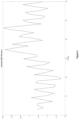
- the heart rate variability is considered to be the respiration waveform, where the local minima are considered the point of maximum expiration due to the decreased heart rate, and the local maxima are the points of maximum inspiration for each breath.
- the HRV derived respiration waveforms for the six participants, with each detected maxima and minima labelled may be obtained.
- the y axis represents the time difference between R peaks in seconds; the x axis is time.
- HRV is responsive to physiological signals other than respiration, including sensitivity to swallowing and movement.
- the Cosmed K4b 2 has detected 40 breaths, and the algorithm has detected 42.
- the data points that are circled are possibly caused by physiological causes other than RSA.
- the two points circled are the correlating breaths between the HRV and Cosmed. Accordingly, it can be seen that the waveforms (without correction) are generally consistent.
- Participant 2's inspirations from HRV and inspired volumes from the Cosmed K4b 2 are obtained.
- the data is well synchronised the data between the HRV derived waveform and the Cosmed K4b 2 , resulting in a plot showing an apparent correlation and definite comparable trending.
- Key trends are able to be visualised between the two waveforms, including the maximum value of each waveform, as well as the local variations in volumes.
- the circled data points are those identified as key points showing the trend between the two datasets.
- Assessing the HRV waveforms key features can be identified as consistent with the measurement of respiration volumes from HRV, including each waveform showing an increase in amplitude corresponding with the change in breathing rate.
- the variation in amplitude between small volumes is a key requirement of using HRV to infer respiration volume, the small variations in inspired volume measured by the Cosmed K4b 2 trend with the amplitude changes of the HRV waveform.
- the data is presented as a linear scaling of the inspirations from the HRV waveform. This represents a useful correlation to provide an estimate of variances in respiration volume, which can be converted to estimates of the respiration volume associated with each breath with appropriate baseline data.
- heart rate variability is mainly caused by respiratory sinus arrhythmia (RSA) [12]
- RSA respiratory sinus arrhythmia
- other physiological impacts which affect heart rate variability including blood pressure, myocardial infarction, disorders of the nervous system, cardiac arrhythmia, diabetes, renal failure, medication, smoking, sleep, gender and age [11].
- the impact of beta-blocker medication and heart conditions to HRV is easily identified with participant 5, where the HRV waveform is irregular, and shows no clear increase in amplitude. Further studies with participants taking cardiac medication are required to determine whether the HRV due to respiration can be recovered. Thus, the present invention may not be applicable to all patients, or may have reduced accuracy, due to medication, pathology, or other factors.
- RSA is the main cause of HRV, therefore herein it is proposed that with further analysis, the RSA data may be extracted from the HRV and recover the true respiration waveform including amplitude variations, which has been shown has a correlation to respiration volume.
- DEEM reliable energy expenditure monitoring
- the main PCB incorporates the processor (LPC1768, a triaxial accelerometer (MMA7660FC, a micro SD card slot, and the required support circuitry.
- the main PCB is a stand-alone device capable of accomplishing the task of activity monitoring and storing the data directly onto to the SD card.
- Activity monitoring is implemented by sampling the 6-bit triaxial accelerometer in a range of ⁇ 1.5 g at 120 Hz. This range and sample rate are known to be sufficient to correctly distinguish between walking, running and other physical activities and enable direct caloric expenditure estimation.
- Direct interfacing of the SD card via a dedicated SPI channel on the same board allows for a standard FAT32 storage system which is directly readable by a PC.
- the main board also includes a dedicated connection to the analogue (daughter) board to enable biopotential monitoring.
- the daughter board is based around the Texas Instruments ADS1298 analogue front end for biopotential measurements. Configured is the daughter board to enable scaling of the ECG leads from 1 lead to a 12-lead diagnostic ECG acquisition.
- the connection interface between the two boards comprises an SPI interface and 8 general-purpose input/output (GPIO).
- GPIO general-purpose input/output
- the communication interface and the power-supply connection are strategically placed at different extremities of the board.
- the digital and analogue signal pathways are separate maintaining the logical organization of the ADS1298.
- the patient interface board connects to the daughter board via ribbon cable allowing acquisition of a full 12-lead ECG.
- the illustrative firmware is optimized to acquire analogue data at a frequency of 500Hz; accelerometer data are acquired in parallel via the I2C digital interface.
- the incoming data stream fills a buffer implemented using 3.6kB of the processor's memory. Data is dumped to the mass memory storage every 300ms.
- the device is housed in a suitable plastic housing and powered by 4 LR6 non rechargeable batteries.
- This device is fitted to a patient by a belt around the waist, and the ECG electrodes connected.
- the patient leads are connected via MC touch-proof 2mm connectors, and commercially available ECG leads with snap connectors.
- the firmware starts recording data as soon as the device is turned on.
- Each file is formatted reporting the current time stamp (as flagged by the LPC 1768 RTC) formatted as a string ("ddd mmm dd hh:mm:ss yyyy").
- Raw data is saved as a CSV file (ADC Units, converted to mV using a MATLAB script) and directly readable by any system supporting FAT32.
- user feedback provides some useful information.
- One red LED confirms that the device is correctly powered and an orange LED flashes when accessing the SD card.
- 1.5V alkaline batteries ensure continuous acquisition for up to 150 hours.
- Activity level evaluation, caloric expenditure and heart-rate (user feedback) are currently given via a MATLAB script, which extracts and reads the data directly from the SD-card. ECG traces are assessed by cardiologists who also have access to the mentioned activity data to draw diagnosis.
- these parameters be determined as a first step using conventional techniques, for example those discussed above in relation to the examples. Once these parameters are measured, they provide a predetermined basic value to which the relative measurements can be applied to provide on-going data. For example, a patient specific correlation function could be used, or a standard function working from the patient specific lung volume data.
- methods for measuring respiration include discrete measurements using a spirometer, or significantly impact a person's ability to perform their daily activity by placing a mask over a patient's face, as required with the Cosmed K4b 2 or similar devices.
- the inventive method for measuring respiratory volumes will allow for the continuous measurement of a patients respiratory data with ECG acquisition, allowing for telemetry monitoring without providing further discomfort or invasion of a patient's daily activities.
- results can be determined using calibration from a baseline for a relative extent of lung respiration. In other embodiments, results can be provided without calibration from a baseline. Given the variety of utilisations in devices and instruments where lung capacity can be determinative, some are described here, however, one skilled in the art could envisage others that are within the scope of this discussion.
- Figure 1 is a flow chart of a process that can be utilised in any device or instrument described or contemplated by one skilled in the art.
- Figure 1 is arranged so that one or two data streams can be utilised to provide output of a respiration waveform from a heart rate variability (HRV).
- HRV heart rate variability
- the first data stream 701 can provide waveforms that can be correlated to generate output of a respiration waveform with or without the second data stream 703, depending upon the device or instrument utilising the disclosed process.
- the second data stream 703 is calibration data which can be matched with the HRV respiration waveform and is synchronized with measured a respiration waveform.
- FIG 2 there is depicted how calibration data 703 can be matched to collected data 701 in the dotted line box 801. Shifting to match may be required.
- the quality of the input data may need to be high.
- utilising a calibration method in the described devices and instruments may be required.
- calibration may not be required, or calibration of a lesser quality may suffice.
- calibration can be any form. For example, utilising a Body Mass Index (BMI) for calibration is contemplated.
- BMI Body Mass Index
- heart beat data is imported 707 where it can be collected in a variety of manners depending on the use of the device or instrument and/or its configuration.
- electrodes can be used.
- a finger or wrist probe can collect the data.
- the quality of the data may or may not be of importance. For uses involving medical procedures, the quality of the input data is most likely high. For uses such as those of casual sports, for example, where a runner is monitoring his/her lung capacity, the input data may not need be as high as that of a medical device.
- One skilled in the art would be able to make a determination how to collect HRV data.
- the devices and/or instruments which may utilise the described process of arriving at respiration output derived from heart beat data can include medical uses and therapy uses.
- Medical uses may involve radiation treatments and MRI/CT scanning precision. Medical uses may further include monitoring lung volume of pneumonia patients and helping to control sleep apnoea. Any of these medical uses are not part of the invention as defined by the claims.
- radiation beams are directed to a particular area of a patient's body.
- a radiation beam may be directed to a small tumour in a patient's organ.
- the displacement of the organs can be predicted based upon the HRV in accordance with this disclosure.
- the organ movement can be tracked and a radiation dose can be delivered during the movement or the radiation dose can be delivered at a particular point during a breath. In this way, dose timing may be determined so as to provide more accuracy in where a radiation dose is received by a patient. Delivering a radiation dose is not part of the present invention as defined by the claims
- MRI/CT scans patients are often told to hold their breath. In these circumstances, utilisation of a face mask to monitor organ displacement during breathing may be out of the question given the physical constraints of the chamber in which a patient must remain during a scan. However, knowing the lung volume displacement at a particular moment, in real time, can provide information about how far the organs have moved during breathing.
- MRI/CT data may be adjusted to compensate for a patient's breathing during scans were the organ displacement data to be known. The displacement of the patient's organs can be predicted or observed based upon the respiration waveform from HRV in accordance with this disclosure and utilised for the output of the MRI/CRT.
- Pneumonia patients suffer from fluid build up so that their lung capacity is reduced. Other conditions such as chronic obstructive pulmonary disease may require health care professionals to monitor pulmonary distress on a daily basis. A determination of pulmonary distress may be made by monitoring HRV using electrodes in such patients which would avoid the discomfort of a facial mask. Feedback to health care professionals and/or a patient can be in real time, in situ or remotely.
- breathing during sleep is temporarily halted so that the volume of the lungs is unchanged.
- a determination of breathing cessation may be made by monitoring HRV in such patients which would avoid the discomfort of a facial mask.
- Feedback to health care professionals and/or a patient can be in real time.
- cardiac output can be used as described herein.
- Breathing teaching tools to correct breathing problems like shallow breathing and not breathing out entirely or holding one's breath can be provided by tracking HRV.
- a patient or client can wear a heartbeat monitor while going about daily activates to receive real time feedback about lung volume to retrain breathing habits.
- Such devices can be utilised in sports training such as running and swimming. While other stimuli can stimulate heart rate during physical activity other than breathing, those variables can be accounted depending upon the use. Also, the quality of the respiration data may not be critical when utilised in a casual manner for example when utilised by Fitbit or a smart watch.
- HRV in accordance with the present disclosure.
- devices and instruments can utilise HRV in accordance with the present disclosure.
- HRV as disclosed herein can provide solutions.
- the quality of the output may depend upon the quality of calibration and of the electrodes and processor speed.
- the flowchart is intended to depict the process of a device or instrument for characterising measurements of lung respiration volume
- a device or instrument for characterising measurements of lung respiration volume which can include a processor for receiving a detected series of heart beats, measuring variability between a period of successive beats, identifying the start and finish of successive breaths by the maxima and minima in the period, identifying the amplitude of variability of period between successive breaths and thereby determining a value for a measurement of an extent of lung respiration.
- a device may include output means such a display or an audible output for generating the value for the measurement of the extent of lung respiration. Further processing based upon the particular use of the described device is contemplated.
- an HRV waveform 709 heart beat is detected in a manner such as that described herein or in any other suitable manner.
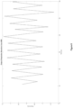
- Beat to Beat (BB) intervals can be calculated 713 and interpolated 715 so that the output may look like that of FIG. 4 . Therefore, an HRV waveform as described herein is determined 709 from the imported heart beat data or the real time collected heat beat data.
- Figure 1 further shows constructing an HRV respiration waveform 717.
- a processor can detect inspirations and expirations 721.
- a suitable threshold filter can be applied 723 to result in an HRV respiration waveform such as that shown in FIG. 5 .
- a measured respiration waveform can be generated 725, such as that shown in FIG. 6 such that the calculated HRV respiration waveform 717 and the generated measured respiration waveform of FIG. 5 are synchronized and correlated 705.
- FIG. 7 depicts an example of such output.
- Data corrections such as calculating coefficients for gain compensation 727 and applying a frequency phase shift filter 729 can be applied before and during application of a suitable correlation equation 731.
- the output 733 which is depicted in FIG. 8 may be used for estimating at least one parameter relating to lung volume can be utilised in disclosed devices and instruments and those contemplated by those having skill in the art.
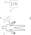
- FIG. 9 depicts a device or instrument as described herein.
- the person may be stationary such as in an MRI/CT scanning position, or moving.
- An electrode or plurality of electrodes can receive the heat beat data and transmit the same 1505, wired or wirelessly so that the data can be received by a processor 1507.
- the processor may be in situ or remote, and the data of the respiration waveform may be processed locally or remotely.
- the process to output such as that depicted in FIG. 8 may be used for estimating at least one parameter relating to lung volume can be utilised in disclosed devices and instruments and those contemplated by those having skill in the art.
- the received raw heart beat data may be in real-time, may be historical or may even be fabricated.
- the received raw heart beat data may be taken under a variety of circumstances, including under various stress levels of the subject.
- the HRV waveform may be derived in any manner, including from a single set of data, or from data obtained in accordance with two or more events that may be under different or the same circumstances, or in any suitable manner.
- the raw heart beat data may be accumulated at a first location, and then may be transmitted to at least one other location for analysis or the entire process may take place at a single location.
- the output means can include any output means including but not limited to displays, audible signals, a data transmission to another device or system including a receiver for data collection and any other device, system or apparatus that can provide an analysis of the output data and/or store the output.
- the respiratory output can be utilised in any suitable manner.
Landscapes
- Health & Medical Sciences (AREA)
- Life Sciences & Earth Sciences (AREA)
- Engineering & Computer Science (AREA)
- Medical Informatics (AREA)
- Biomedical Technology (AREA)
- Veterinary Medicine (AREA)
- General Health & Medical Sciences (AREA)
- Pathology (AREA)
- Public Health (AREA)
- Animal Behavior & Ethology (AREA)
- Physics & Mathematics (AREA)
- Biophysics (AREA)
- Heart & Thoracic Surgery (AREA)
- Molecular Biology (AREA)
- Surgery (AREA)
- Cardiology (AREA)
- Physiology (AREA)
- Nuclear Medicine, Radiotherapy & Molecular Imaging (AREA)
- Radiology & Medical Imaging (AREA)
- Computer Vision & Pattern Recognition (AREA)
- High Energy & Nuclear Physics (AREA)
- Optics & Photonics (AREA)
- Pulmonology (AREA)
- Signal Processing (AREA)
- Psychiatry (AREA)
- Artificial Intelligence (AREA)
- Dentistry (AREA)
- Oral & Maxillofacial Surgery (AREA)
- Theoretical Computer Science (AREA)
- Vascular Medicine (AREA)
- Measurement Of The Respiration, Hearing Ability, Form, And Blood Characteristics Of Living Organisms (AREA)
- Measurement And Recording Of Electrical Phenomena And Electrical Characteristics Of The Living Body (AREA)
Claims (10)
- Dispositif pour estimer un paramètre relatif à un volume respiratoire pulmonaire comprenant : un processeur pour :(a) recevoir (701) une série détectée de battements cardiaques ;(b) mesurer (709) une forme d'onde de variabilité du rythme cardiaque dans une période entre des battements successifs ;(c) identifier (719) des pics et des creux dans la forme d'onde de variabilité du rythme cardiaque pour détecter des inspirations ou des expirations ;(d) appliquer (723) un filtre de seuil pour déterminer une forme d'onde de respiration de variabilité de la fréquence cardiaque ;(e) générer (725) une forme d'onde de respiration mesurée à partir de données de mesure d'étalonnage ;(f) déterminer (705, 731) une forme d'onde de sortie (733) en synchronisant et en corrélant la forme d'onde de respiration de variabilité du rythme cardiaque et la forme d'onde de respiration mesurée générée ; et(g) estimer au moins un paramètre relatif à un volume pulmonaire sur la base de ladite forme d'onde de sortie.
- Dispositif selon la revendication 1, dans lequel le dispositif est configuré pour utiliser l'estimation dudit paramètre à l'étape (g) pour déterminer le mouvement d'un ou plusieurs organes d'une personne lors de l'inspiration ou de l'expiration.
- Dispositif selon la revendication 2, dans lequel le dispositif est configuré pour fournir la détermination du mouvement d'un ou plusieurs organes à d'autres dispositifs pour une radiothérapie ou une imagerie par résonance magnétique (IRM) ou une tomodensitométrie (CT).
- Dispositif selon les revendications 1 ou 2, dans lequel le dispositif est portable.
- Dispositif selon la revendication 1, dans lequel le dispositif est configuré pour traiter certaines des données dérivées des étapes (a) à (f) à distance à partir de données dérivées de l'étape (g).
- Procédé d'estimation d'un paramètre relatif à un volume pulmonaire, comportant au moins les étapes de :(a) détection d'une série de battements cardiaques ;et, en utilisant un dispositif selon l'une quelconque des revendications 1 à 5 :(b) mesure (719) d'une forme d'onde de variabilité du rythme cardiaque dans une période entre des battements successifs :(c) identification (719) de pics et de creux dans la forme d'onde de variabilité du rythme cardiaque pour détecter des inspirations et des expirations ;(d) application (723) d'un filtre de seuil pour déterminer une forme d'onde de respiration de variabilité de la fréquence cardiaque :(e) génération (725) d'une forme d'onde de respiration mesurée à partir de données de mesure d'étalonnage ;(f) détermination (705, 731) d'une forme d'onde de sortie (733) en synchronisant et en corrélant la forme d'onde de respiration de variabilité de la fréquence cardiaque et la forme d'onde de respiration mesurée générée ; et estimation d'au moins un paramètre relatif à un volume pulmonaire sur la base de ladite forme d'onde de sortie.
- Procédé selon la revendication 6, dans lequel l'identification (719) de pics et de creux dans la forme d'onde de variabilité du rythme cardiaque pour détecter des inspirations et des expirations comprend l'identification du début et de la fin des respirations successives par les maxima et minima dans la période.
- Procédé selon la revendication 6 ou 7, dans lequel l'application (723) d'un filtre de seuil pour déterminer une forme d'onde de respiration de variabilité de la fréquence cardiaque comprend l'identification de l'amplitude de la forme d'onde de variabilité de la fréquence cardiaque de la période entre des respirations successives.
- Procédé selon les revendications 6 à 8, mis en œuvre dans un dispositif portable comprenant le dispositif selon la revendication 4.
- Electrocardiographe comprenant le dispositif selon l'une quelconque des revendications 1 à 5, dans lequel le processeur est configuré pour traiter des données selon le procédé de la revendication 6.
Applications Claiming Priority (2)
| Application Number | Priority Date | Filing Date | Title |
|---|---|---|---|
| AU2015903472A AU2015903472A0 (en) | 2015-08-27 | Non-invasive respiratory monitoring | |
| PCT/AU2016/050795 WO2017031547A1 (fr) | 2015-08-27 | 2016-08-26 | Surveillance respiratoire non-invasive |
Publications (4)
| Publication Number | Publication Date |
|---|---|
| EP3340867A1 EP3340867A1 (fr) | 2018-07-04 |
| EP3340867A4 EP3340867A4 (fr) | 2019-04-17 |
| EP3340867C0 EP3340867C0 (fr) | 2023-07-19 |
| EP3340867B1 true EP3340867B1 (fr) | 2023-07-19 |
Family
ID=58099352
Family Applications (1)
| Application Number | Title | Priority Date | Filing Date |
|---|---|---|---|
| EP16838132.5A Active EP3340867B1 (fr) | 2015-08-27 | 2016-08-26 | Surveillance respiratoire non-invasive |
Country Status (8)
| Country | Link |
|---|---|
| US (1) | US11363995B2 (fr) |
| EP (1) | EP3340867B1 (fr) |
| JP (2) | JP2018531764A (fr) |
| KR (1) | KR102574224B1 (fr) |
| CN (1) | CN108348175B (fr) |
| AU (1) | AU2016310411B2 (fr) |
| NZ (1) | NZ740374A (fr) |
| WO (1) | WO2017031547A1 (fr) |
Families Citing this family (5)
| Publication number | Priority date | Publication date | Assignee | Title |
|---|---|---|---|---|
| WO2017031547A1 (fr) | 2015-08-27 | 2017-03-02 | Gemgard Pty Limited | Surveillance respiratoire non-invasive |
| GB201519985D0 (en) * | 2015-11-12 | 2015-12-30 | Respinor As | Ultrasonic method and apparatus for respiration monitoring |
| US12144604B2 (en) | 2018-12-11 | 2024-11-19 | Respinor As | Systems and methods for motion compensation in ultrasonic respiration monitoring |
| KR102742376B1 (ko) * | 2018-12-11 | 2024-12-13 | 레스피노르 에이에스 | 초음파 호흡 모니터링에서의 모션 보상을 위한 시스템들 및 방법들 |
| CN115778394B (zh) * | 2022-11-09 | 2025-04-11 | 鹃湖实验室 | 用于结缔组织体表代谢异常监测的穿戴式装置和方法 |
Family Cites Families (11)
| Publication number | Priority date | Publication date | Assignee | Title |
|---|---|---|---|---|
| JPH0451912U (fr) * | 1990-09-10 | 1992-05-01 | ||
| US7393329B1 (en) * | 1997-05-23 | 2008-07-01 | William Beaumont Hospital | Method and apparatus for delivering radiation therapy during suspended ventilation |
| FI20025029A0 (fi) * | 2002-05-29 | 2002-05-29 | Joni Kettunen | Menetelmä luotettavan hengitysaktiviteetti-informaation saamiseksi sydämen sykemittauksesta |
| EP1587421A2 (fr) * | 2003-01-09 | 2005-10-26 | Koninklijke Philips Electronics N.V. | Moniteur de respiration pour tomographie informatisee |
| WO2005067790A1 (fr) * | 2004-01-16 | 2005-07-28 | Compumedics Ltd | Procede et dispositif permettant de surveiller de detecter et de classifier des troubles respiratoires du sommeil a partir d'un electrocardiogramme |
| EP1814454A2 (fr) * | 2004-11-19 | 2007-08-08 | Vivometrics, Inc. | Methodes et systemes de determination d'un rythme respiratoire en temps reel a l'aide de ressources de traitement limitees |
| CN101198277B (zh) * | 2005-02-22 | 2011-06-15 | 海尔思-斯玛特有限公司 | 用于生理学和心理生理学监控的系统 |
| US20080021300A1 (en) | 2006-06-29 | 2008-01-24 | Allison John W | Four-dimensional target modeling and radiation treatment |
| US20080243007A1 (en) * | 2007-03-28 | 2008-10-02 | Cardiac Pacemakers, Inc. | Pulmonary Artery Pressure Signals And Methods of Using |
| US8430817B1 (en) * | 2009-10-15 | 2013-04-30 | Masimo Corporation | System for determining confidence in respiratory rate measurements |
| WO2017031547A1 (fr) | 2015-08-27 | 2017-03-02 | Gemgard Pty Limited | Surveillance respiratoire non-invasive |
-
2016
- 2016-08-26 WO PCT/AU2016/050795 patent/WO2017031547A1/fr not_active Ceased
- 2016-08-26 NZ NZ740374A patent/NZ740374A/en not_active IP Right Cessation
- 2016-08-26 JP JP2018529688A patent/JP2018531764A/ja active Pending
- 2016-08-26 EP EP16838132.5A patent/EP3340867B1/fr active Active
- 2016-08-26 US US15/755,916 patent/US11363995B2/en active Active
- 2016-08-26 CN CN201680061065.4A patent/CN108348175B/zh active Active
- 2016-08-26 KR KR1020187008610A patent/KR102574224B1/ko active Active
- 2016-08-26 AU AU2016310411A patent/AU2016310411B2/en not_active Ceased
-
2022
- 2022-01-27 JP JP2022010978A patent/JP2022068177A/ja active Pending
Also Published As
| Publication number | Publication date |
|---|---|
| KR102574224B1 (ko) | 2023-09-05 |
| CN108348175B (zh) | 2021-08-10 |
| EP3340867C0 (fr) | 2023-07-19 |
| WO2017031547A1 (fr) | 2017-03-02 |
| KR20180044393A (ko) | 2018-05-02 |
| US20180325465A1 (en) | 2018-11-15 |
| EP3340867A4 (fr) | 2019-04-17 |
| NZ740374A (en) | 2022-07-29 |
| CN108348175A (zh) | 2018-07-31 |
| AU2016310411A1 (en) | 2018-03-22 |
| JP2022068177A (ja) | 2022-05-09 |
| AU2016310411B2 (en) | 2020-04-02 |
| EP3340867A1 (fr) | 2018-07-04 |
| JP2018531764A (ja) | 2018-11-01 |
| US11363995B2 (en) | 2022-06-21 |
Similar Documents
| Publication | Publication Date | Title |
|---|---|---|
| US20200329977A1 (en) | Devices and methods for respiratory variation monitoring by measurement of respiratory volumes, motion and variability | |
| AU2017279693B2 (en) | Devices and methods for respiratory variation monitoring by measurement of respiratory volumes, motion and variability | |
| US8700137B2 (en) | Cardiac performance monitoring system for use with mobile communications devices | |
| JP5944899B2 (ja) | 呼吸の量、運動、および変化を測定することによる呼吸変動モニタリングのための装置 | |
| JP2023153893A (ja) | 換気療法システムおよび方法 | |
| KR20190071808A (ko) | 호흡 조기 경고 점수 계산 시스템 및 방법 | |
| JP4753881B2 (ja) | 心電図から導出された睡眠呼吸障害の監視、検出及び分類のための装置及び信号処理方法 | |
| US20050131288A1 (en) | Flexible, patient-worn, integrated, self-contained sensor systems for the acquisition and monitoring of physiologic data | |
| JP2022068177A (ja) | 非侵襲的な呼吸器モニタリング | |
| Jung et al. | Estimation of tidal volume using load cells on a hospital bed | |
| Estrada et al. | Evaluating respiratory muscle activity using a wireless sensor platform | |
| US20260083390A1 (en) | Wearable diaphragmatic efficiency monitoring devices | |
| Cook et al. | A novel method for non-invasive respiration monitoring | |
| Kantele-Mesimäki | Patient on the move: feasibility of a multi-parameter respiratory rate measurement |
Legal Events
| Date | Code | Title | Description |
|---|---|---|---|
| STAA | Information on the status of an ep patent application or granted ep patent |
Free format text: STATUS: THE INTERNATIONAL PUBLICATION HAS BEEN MADE |
|
| PUAI | Public reference made under article 153(3) epc to a published international application that has entered the european phase |
Free format text: ORIGINAL CODE: 0009012 |
|
| STAA | Information on the status of an ep patent application or granted ep patent |
Free format text: STATUS: REQUEST FOR EXAMINATION WAS MADE |
|
| 17P | Request for examination filed |
Effective date: 20180327 |
|
| AK | Designated contracting states |
Kind code of ref document: A1 Designated state(s): AL AT BE BG CH CY CZ DE DK EE ES FI FR GB GR HR HU IE IS IT LI LT LU LV MC MK MT NL NO PL PT RO RS SE SI SK SM TR |
|
| AX | Request for extension of the european patent |
Extension state: BA ME |
|
| DAV | Request for validation of the european patent (deleted) | ||
| DAX | Request for extension of the european patent (deleted) | ||
| A4 | Supplementary search report drawn up and despatched |
Effective date: 20190315 |
|
| RIC1 | Information provided on ipc code assigned before grant |
Ipc: A61B 5/0402 20060101ALN20190311BHEP Ipc: A61B 5/0245 20060101ALI20190311BHEP Ipc: A61B 6/00 20060101ALI20190311BHEP Ipc: A61B 5/113 20060101ALN20190311BHEP Ipc: A61B 5/091 20060101ALI20190311BHEP Ipc: A61B 5/00 20060101AFI20190311BHEP Ipc: A61B 6/03 20060101ALN20190311BHEP Ipc: A61B 5/055 20060101ALN20190311BHEP Ipc: A61B 5/024 20060101ALI20190311BHEP Ipc: A61N 5/10 20060101ALN20190311BHEP |
|
| STAA | Information on the status of an ep patent application or granted ep patent |
Free format text: STATUS: EXAMINATION IS IN PROGRESS |
|
| 17Q | First examination report despatched |
Effective date: 20220503 |
|
| REG | Reference to a national code |
Ref country code: DE Ref legal event code: R079 Free format text: PREVIOUS MAIN CLASS: A61B0005020000 Ipc: A61B0005000000 Ref country code: DE Ref legal event code: R079 Ref document number: 602016081214 Country of ref document: DE Free format text: PREVIOUS MAIN CLASS: A61B0005020000 Ipc: A61B0005000000 |
|
| GRAP | Despatch of communication of intention to grant a patent |
Free format text: ORIGINAL CODE: EPIDOSNIGR1 |
|
| STAA | Information on the status of an ep patent application or granted ep patent |
Free format text: STATUS: GRANT OF PATENT IS INTENDED |
|
| RIC1 | Information provided on ipc code assigned before grant |
Ipc: A61B 5/318 20210101ALN20230126BHEP Ipc: A61B 5/113 20060101ALN20230126BHEP Ipc: A61B 5/055 20060101ALN20230126BHEP Ipc: A61N 5/10 20060101ALI20230126BHEP Ipc: A61B 6/03 20060101ALI20230126BHEP Ipc: A61B 6/00 20060101ALI20230126BHEP Ipc: A61B 5/091 20060101ALI20230126BHEP Ipc: A61B 5/0245 20060101ALI20230126BHEP Ipc: A61B 5/024 20060101ALI20230126BHEP Ipc: A61B 5/00 20060101AFI20230126BHEP |
|
| INTG | Intention to grant announced |
Effective date: 20230209 |
|
| RIC1 | Information provided on ipc code assigned before grant |
Ipc: A61B 5/318 20210101ALN20230127BHEP Ipc: A61B 5/113 20060101ALN20230127BHEP Ipc: A61B 5/055 20060101ALN20230127BHEP Ipc: A61N 5/10 20060101ALI20230127BHEP Ipc: A61B 6/03 20060101ALI20230127BHEP Ipc: A61B 6/00 20060101ALI20230127BHEP Ipc: A61B 5/091 20060101ALI20230127BHEP Ipc: A61B 5/0245 20060101ALI20230127BHEP Ipc: A61B 5/024 20060101ALI20230127BHEP Ipc: A61B 5/00 20060101AFI20230127BHEP |
|
| GRAS | Grant fee paid |
Free format text: ORIGINAL CODE: EPIDOSNIGR3 |
|
| GRAA | (expected) grant |
Free format text: ORIGINAL CODE: 0009210 |
|
| STAA | Information on the status of an ep patent application or granted ep patent |
Free format text: STATUS: THE PATENT HAS BEEN GRANTED |
|
| AK | Designated contracting states |
Kind code of ref document: B1 Designated state(s): AL AT BE BG CH CY CZ DE DK EE ES FI FR GB GR HR HU IE IS IT LI LT LU LV MC MK MT NL NO PL PT RO RS SE SI SK SM TR |
|
| REG | Reference to a national code |
Ref country code: GB Ref legal event code: FG4D |
|
| REG | Reference to a national code |
Ref country code: CH Ref legal event code: EP |
|
| REG | Reference to a national code |
Ref country code: DE Ref legal event code: R096 Ref document number: 602016081214 Country of ref document: DE |
|
| REG | Reference to a national code |
Ref country code: IE Ref legal event code: FG4D |
|
| U01 | Request for unitary effect filed |
Effective date: 20230816 |
|
| U07 | Unitary effect registered |
Designated state(s): AT BE BG DE DK EE FI FR IT LT LU LV MT NL PT SE SI Effective date: 20230821 |
|
| REG | Reference to a national code |
Ref country code: LT Ref legal event code: MG9D |
|
| PG25 | Lapsed in a contracting state [announced via postgrant information from national office to epo] |
Ref country code: GR Free format text: LAPSE BECAUSE OF FAILURE TO SUBMIT A TRANSLATION OF THE DESCRIPTION OR TO PAY THE FEE WITHIN THE PRESCRIBED TIME-LIMIT Effective date: 20231020 |
|
| PG25 | Lapsed in a contracting state [announced via postgrant information from national office to epo] |
Ref country code: IS Free format text: LAPSE BECAUSE OF FAILURE TO SUBMIT A TRANSLATION OF THE DESCRIPTION OR TO PAY THE FEE WITHIN THE PRESCRIBED TIME-LIMIT Effective date: 20231119 |
|
| PG25 | Lapsed in a contracting state [announced via postgrant information from national office to epo] |
Ref country code: RS Free format text: LAPSE BECAUSE OF FAILURE TO SUBMIT A TRANSLATION OF THE DESCRIPTION OR TO PAY THE FEE WITHIN THE PRESCRIBED TIME-LIMIT Effective date: 20230719 Ref country code: NO Free format text: LAPSE BECAUSE OF FAILURE TO SUBMIT A TRANSLATION OF THE DESCRIPTION OR TO PAY THE FEE WITHIN THE PRESCRIBED TIME-LIMIT Effective date: 20231019 Ref country code: IS Free format text: LAPSE BECAUSE OF FAILURE TO SUBMIT A TRANSLATION OF THE DESCRIPTION OR TO PAY THE FEE WITHIN THE PRESCRIBED TIME-LIMIT Effective date: 20231119 Ref country code: HR Free format text: LAPSE BECAUSE OF FAILURE TO SUBMIT A TRANSLATION OF THE DESCRIPTION OR TO PAY THE FEE WITHIN THE PRESCRIBED TIME-LIMIT Effective date: 20230719 Ref country code: GR Free format text: LAPSE BECAUSE OF FAILURE TO SUBMIT A TRANSLATION OF THE DESCRIPTION OR TO PAY THE FEE WITHIN THE PRESCRIBED TIME-LIMIT Effective date: 20231020 |
|
| U21 | Renewal fee for the european patent with unitary effect paid with additional fee |
Year of fee payment: 8 Effective date: 20240118 |
|
| PG25 | Lapsed in a contracting state [announced via postgrant information from national office to epo] |
Ref country code: PL Free format text: LAPSE BECAUSE OF FAILURE TO SUBMIT A TRANSLATION OF THE DESCRIPTION OR TO PAY THE FEE WITHIN THE PRESCRIBED TIME-LIMIT Effective date: 20230719 |
|
| REG | Reference to a national code |
Ref country code: CH Ref legal event code: PL |
|
| REG | Reference to a national code |
Ref country code: DE Ref legal event code: R097 Ref document number: 602016081214 Country of ref document: DE |
|
| PG25 | Lapsed in a contracting state [announced via postgrant information from national office to epo] |
Ref country code: ES Free format text: LAPSE BECAUSE OF FAILURE TO SUBMIT A TRANSLATION OF THE DESCRIPTION OR TO PAY THE FEE WITHIN THE PRESCRIBED TIME-LIMIT Effective date: 20230719 |
|
| PG25 | Lapsed in a contracting state [announced via postgrant information from national office to epo] |
Ref country code: SM Free format text: LAPSE BECAUSE OF FAILURE TO SUBMIT A TRANSLATION OF THE DESCRIPTION OR TO PAY THE FEE WITHIN THE PRESCRIBED TIME-LIMIT Effective date: 20230719 Ref country code: RO Free format text: LAPSE BECAUSE OF FAILURE TO SUBMIT A TRANSLATION OF THE DESCRIPTION OR TO PAY THE FEE WITHIN THE PRESCRIBED TIME-LIMIT Effective date: 20230719 Ref country code: ES Free format text: LAPSE BECAUSE OF FAILURE TO SUBMIT A TRANSLATION OF THE DESCRIPTION OR TO PAY THE FEE WITHIN THE PRESCRIBED TIME-LIMIT Effective date: 20230719 Ref country code: CZ Free format text: LAPSE BECAUSE OF FAILURE TO SUBMIT A TRANSLATION OF THE DESCRIPTION OR TO PAY THE FEE WITHIN THE PRESCRIBED TIME-LIMIT Effective date: 20230719 Ref country code: CH Free format text: LAPSE BECAUSE OF NON-PAYMENT OF DUE FEES Effective date: 20230831 Ref country code: SK Free format text: LAPSE BECAUSE OF FAILURE TO SUBMIT A TRANSLATION OF THE DESCRIPTION OR TO PAY THE FEE WITHIN THE PRESCRIBED TIME-LIMIT Effective date: 20230719 Ref country code: MC Free format text: LAPSE BECAUSE OF FAILURE TO SUBMIT A TRANSLATION OF THE DESCRIPTION OR TO PAY THE FEE WITHIN THE PRESCRIBED TIME-LIMIT Effective date: 20230719 |
|
| REG | Reference to a national code |
Ref country code: IE Ref legal event code: MM4A |
|
| PLBE | No opposition filed within time limit |
Free format text: ORIGINAL CODE: 0009261 |
|
| STAA | Information on the status of an ep patent application or granted ep patent |
Free format text: STATUS: NO OPPOSITION FILED WITHIN TIME LIMIT |
|
| 26N | No opposition filed |
Effective date: 20240422 |
|
| GBPC | Gb: european patent ceased through non-payment of renewal fee |
Effective date: 20231019 |
|
| PG25 | Lapsed in a contracting state [announced via postgrant information from national office to epo] |
Ref country code: IE Free format text: LAPSE BECAUSE OF NON-PAYMENT OF DUE FEES Effective date: 20230826 |
|
| PG25 | Lapsed in a contracting state [announced via postgrant information from national office to epo] |
Ref country code: GB Free format text: LAPSE BECAUSE OF NON-PAYMENT OF DUE FEES Effective date: 20231019 |
|
| PG25 | Lapsed in a contracting state [announced via postgrant information from national office to epo] |
Ref country code: IE Free format text: LAPSE BECAUSE OF NON-PAYMENT OF DUE FEES Effective date: 20230826 Ref country code: GB Free format text: LAPSE BECAUSE OF NON-PAYMENT OF DUE FEES Effective date: 20231019 |
|
| U20 | Renewal fee for the european patent with unitary effect paid |
Year of fee payment: 9 Effective date: 20240812 |
|
| PG25 | Lapsed in a contracting state [announced via postgrant information from national office to epo] |
Ref country code: CY Free format text: LAPSE BECAUSE OF FAILURE TO SUBMIT A TRANSLATION OF THE DESCRIPTION OR TO PAY THE FEE WITHIN THE PRESCRIBED TIME-LIMIT; INVALID AB INITIO Effective date: 20160826 |
|
| PG25 | Lapsed in a contracting state [announced via postgrant information from national office to epo] |
Ref country code: HU Free format text: LAPSE BECAUSE OF FAILURE TO SUBMIT A TRANSLATION OF THE DESCRIPTION OR TO PAY THE FEE WITHIN THE PRESCRIBED TIME-LIMIT; INVALID AB INITIO Effective date: 20160826 |
|
| U20 | Renewal fee for the european patent with unitary effect paid |
Year of fee payment: 10 Effective date: 20250826 |
|
| PG25 | Lapsed in a contracting state [announced via postgrant information from national office to epo] |
Ref country code: TR Free format text: LAPSE BECAUSE OF FAILURE TO SUBMIT A TRANSLATION OF THE DESCRIPTION OR TO PAY THE FEE WITHIN THE PRESCRIBED TIME-LIMIT Effective date: 20230719 |