EP3366763B1 - Vorrichtung zur freisetzung von zellen - Google Patents
Vorrichtung zur freisetzung von zellen Download PDFInfo
- Publication number
- EP3366763B1 EP3366763B1 EP18167250.2A EP18167250A EP3366763B1 EP 3366763 B1 EP3366763 B1 EP 3366763B1 EP 18167250 A EP18167250 A EP 18167250A EP 3366763 B1 EP3366763 B1 EP 3366763B1
- Authority
- EP
- European Patent Office
- Prior art keywords
- pore size
- cell
- large pore
- cells
- size section
- Prior art date
- Legal status (The legal status is an assumption and is not a legal conclusion. Google has not performed a legal analysis and makes no representation as to the accuracy of the status listed.)
- Active
Links
Images


Classifications
-
- C—CHEMISTRY; METALLURGY
- C12—BIOCHEMISTRY; BEER; SPIRITS; WINE; VINEGAR; MICROBIOLOGY; ENZYMOLOGY; MUTATION OR GENETIC ENGINEERING
- C12M—APPARATUS FOR ENZYMOLOGY OR MICROBIOLOGY; APPARATUS FOR CULTURING MICROORGANISMS FOR PRODUCING BIOMASS, FOR GROWING CELLS OR FOR OBTAINING FERMENTATION OR METABOLIC PRODUCTS, i.e. BIOREACTORS OR FERMENTERS
- C12M41/00—Means for regulation, monitoring, measurement or control, e.g. flow regulation
- C12M41/48—Automatic or computerized control
-
- C—CHEMISTRY; METALLURGY
- C12—BIOCHEMISTRY; BEER; SPIRITS; WINE; VINEGAR; MICROBIOLOGY; ENZYMOLOGY; MUTATION OR GENETIC ENGINEERING
- C12N—MICROORGANISMS OR ENZYMES; COMPOSITIONS THEREOF; PROPAGATING, PRESERVING, OR MAINTAINING MICROORGANISMS; MUTATION OR GENETIC ENGINEERING; CULTURE MEDIA
- C12N5/00—Undifferentiated human, animal or plant cells, e.g. cell lines; Tissues; Cultivation or maintenance thereof; Culture media therefor
- C12N5/06—Animal cells or tissues; Human cells or tissues
- C12N5/0602—Vertebrate cells
- C12N5/0696—Artificially induced pluripotent stem cells, e.g. iPS
-
- C—CHEMISTRY; METALLURGY
- C12—BIOCHEMISTRY; BEER; SPIRITS; WINE; VINEGAR; MICROBIOLOGY; ENZYMOLOGY; MUTATION OR GENETIC ENGINEERING
- C12M—APPARATUS FOR ENZYMOLOGY OR MICROBIOLOGY; APPARATUS FOR CULTURING MICROORGANISMS FOR PRODUCING BIOMASS, FOR GROWING CELLS OR FOR OBTAINING FERMENTATION OR METABOLIC PRODUCTS, i.e. BIOREACTORS OR FERMENTERS
- C12M23/00—Constructional details, e.g. recesses, hinges
- C12M23/28—Constructional details, e.g. recesses, hinges disposable or single use
-
- C—CHEMISTRY; METALLURGY
- C12—BIOCHEMISTRY; BEER; SPIRITS; WINE; VINEGAR; MICROBIOLOGY; ENZYMOLOGY; MUTATION OR GENETIC ENGINEERING
- C12M—APPARATUS FOR ENZYMOLOGY OR MICROBIOLOGY; APPARATUS FOR CULTURING MICROORGANISMS FOR PRODUCING BIOMASS, FOR GROWING CELLS OR FOR OBTAINING FERMENTATION OR METABOLIC PRODUCTS, i.e. BIOREACTORS OR FERMENTERS
- C12M23/00—Constructional details, e.g. recesses, hinges
- C12M23/44—Multiple separable units; Modules
-
- C—CHEMISTRY; METALLURGY
- C12—BIOCHEMISTRY; BEER; SPIRITS; WINE; VINEGAR; MICROBIOLOGY; ENZYMOLOGY; MUTATION OR GENETIC ENGINEERING
- C12M—APPARATUS FOR ENZYMOLOGY OR MICROBIOLOGY; APPARATUS FOR CULTURING MICROORGANISMS FOR PRODUCING BIOMASS, FOR GROWING CELLS OR FOR OBTAINING FERMENTATION OR METABOLIC PRODUCTS, i.e. BIOREACTORS OR FERMENTERS
- C12M27/00—Means for mixing, agitating or circulating fluids in the vessel
-
- C—CHEMISTRY; METALLURGY
- C12—BIOCHEMISTRY; BEER; SPIRITS; WINE; VINEGAR; MICROBIOLOGY; ENZYMOLOGY; MUTATION OR GENETIC ENGINEERING
- C12M—APPARATUS FOR ENZYMOLOGY OR MICROBIOLOGY; APPARATUS FOR CULTURING MICROORGANISMS FOR PRODUCING BIOMASS, FOR GROWING CELLS OR FOR OBTAINING FERMENTATION OR METABOLIC PRODUCTS, i.e. BIOREACTORS OR FERMENTERS
- C12M29/00—Means for introduction, extraction or recirculation of materials, e.g. pumps
- C12M29/04—Filters; Permeable or porous membranes or plates, e.g. dialysis
-
- C—CHEMISTRY; METALLURGY
- C12—BIOCHEMISTRY; BEER; SPIRITS; WINE; VINEGAR; MICROBIOLOGY; ENZYMOLOGY; MUTATION OR GENETIC ENGINEERING
- C12M—APPARATUS FOR ENZYMOLOGY OR MICROBIOLOGY; APPARATUS FOR CULTURING MICROORGANISMS FOR PRODUCING BIOMASS, FOR GROWING CELLS OR FOR OBTAINING FERMENTATION OR METABOLIC PRODUCTS, i.e. BIOREACTORS OR FERMENTERS
- C12M35/00—Means for application of stress for stimulating the growth of microorganisms or the generation of fermentation or metabolic products; Means for electroporation or cell fusion
-
- C—CHEMISTRY; METALLURGY
- C12—BIOCHEMISTRY; BEER; SPIRITS; WINE; VINEGAR; MICROBIOLOGY; ENZYMOLOGY; MUTATION OR GENETIC ENGINEERING
- C12M—APPARATUS FOR ENZYMOLOGY OR MICROBIOLOGY; APPARATUS FOR CULTURING MICROORGANISMS FOR PRODUCING BIOMASS, FOR GROWING CELLS OR FOR OBTAINING FERMENTATION OR METABOLIC PRODUCTS, i.e. BIOREACTORS OR FERMENTERS
- C12M37/00—Means for sterilizing, maintaining sterile conditions or avoiding chemical or biological contamination
- C12M37/02—Filters
-
- C—CHEMISTRY; METALLURGY
- C12—BIOCHEMISTRY; BEER; SPIRITS; WINE; VINEGAR; MICROBIOLOGY; ENZYMOLOGY; MUTATION OR GENETIC ENGINEERING
- C12M—APPARATUS FOR ENZYMOLOGY OR MICROBIOLOGY; APPARATUS FOR CULTURING MICROORGANISMS FOR PRODUCING BIOMASS, FOR GROWING CELLS OR FOR OBTAINING FERMENTATION OR METABOLIC PRODUCTS, i.e. BIOREACTORS OR FERMENTERS
- C12M41/00—Means for regulation, monitoring, measurement or control, e.g. flow regulation
- C12M41/12—Means for regulation, monitoring, measurement or control, e.g. flow regulation of temperature
-
- C—CHEMISTRY; METALLURGY
- C12—BIOCHEMISTRY; BEER; SPIRITS; WINE; VINEGAR; MICROBIOLOGY; ENZYMOLOGY; MUTATION OR GENETIC ENGINEERING
- C12M—APPARATUS FOR ENZYMOLOGY OR MICROBIOLOGY; APPARATUS FOR CULTURING MICROORGANISMS FOR PRODUCING BIOMASS, FOR GROWING CELLS OR FOR OBTAINING FERMENTATION OR METABOLIC PRODUCTS, i.e. BIOREACTORS OR FERMENTERS
- C12M41/00—Means for regulation, monitoring, measurement or control, e.g. flow regulation
- C12M41/30—Means for regulation, monitoring, measurement or control, e.g. flow regulation of concentration
- C12M41/36—Means for regulation, monitoring, measurement or control, e.g. flow regulation of concentration of biomass, e.g. colony counters or by turbidity measurements
-
- C—CHEMISTRY; METALLURGY
- C12—BIOCHEMISTRY; BEER; SPIRITS; WINE; VINEGAR; MICROBIOLOGY; ENZYMOLOGY; MUTATION OR GENETIC ENGINEERING
- C12M—APPARATUS FOR ENZYMOLOGY OR MICROBIOLOGY; APPARATUS FOR CULTURING MICROORGANISMS FOR PRODUCING BIOMASS, FOR GROWING CELLS OR FOR OBTAINING FERMENTATION OR METABOLIC PRODUCTS, i.e. BIOREACTORS OR FERMENTERS
- C12M45/00—Means for pre-treatment of biological substances
- C12M45/02—Means for pre-treatment of biological substances by mechanical forces; Stirring; Trituration; Comminuting
-
- C—CHEMISTRY; METALLURGY
- C12—BIOCHEMISTRY; BEER; SPIRITS; WINE; VINEGAR; MICROBIOLOGY; ENZYMOLOGY; MUTATION OR GENETIC ENGINEERING
- C12M—APPARATUS FOR ENZYMOLOGY OR MICROBIOLOGY; APPARATUS FOR CULTURING MICROORGANISMS FOR PRODUCING BIOMASS, FOR GROWING CELLS OR FOR OBTAINING FERMENTATION OR METABOLIC PRODUCTS, i.e. BIOREACTORS OR FERMENTERS
- C12M47/00—Means for after-treatment of the produced biomass or of the fermentation or metabolic products, e.g. storage of biomass
- C12M47/04—Cell isolation or sorting
-
- C—CHEMISTRY; METALLURGY
- C12—BIOCHEMISTRY; BEER; SPIRITS; WINE; VINEGAR; MICROBIOLOGY; ENZYMOLOGY; MUTATION OR GENETIC ENGINEERING
- C12N—MICROORGANISMS OR ENZYMES; COMPOSITIONS THEREOF; PROPAGATING, PRESERVING, OR MAINTAINING MICROORGANISMS; MUTATION OR GENETIC ENGINEERING; CULTURE MEDIA
- C12N5/00—Undifferentiated human, animal or plant cells, e.g. cell lines; Tissues; Cultivation or maintenance thereof; Culture media therefor
- C12N5/06—Animal cells or tissues; Human cells or tissues
- C12N5/0602—Vertebrate cells
- C12N5/0634—Cells from the blood or the immune system
Definitions
- the present invention relates to cell preservation technology, and particularly to a pluripotent stem cell production system.
- Embryonic stem cells are stem cells established from early embryos of human or mice. ES cells are pluripotent, being capable of differentiating into all cells in the body. At the current time, human ES cells are usable in cell transplantation therapy for numerous diseases including Parkinson's disease, juvenile onset diabetes and leukemia.
- ES cells are usable in cell transplantation therapy for numerous diseases including Parkinson's disease, juvenile onset diabetes and leukemia.
- barriers exist against transplantation of ES cells In particular, transplantation of ES cells can provoke immunorejection similar to the rejection encountered after unsuccessful organ transplantation.
- ethical considerations as well as critical and dissenting opinions against the use of ES cell lines that have been established by destruction of human embryos.
- iPS cells induced pluripotent stem cells
- US2012/0196358A1 describes a device for removing cumulus from oocytes.
- US2012/0258536A1 describes a method and device for dispersion of assemblies of biological material.
- WO2009/106760A2 describes a device and method for isolating and cultivating live cells.
- Induced stem cells such as iPS cells are established by introducing inducing factors such as genes into cells which are then subjected to amplifying culturing and cryopreservation.
- inducing factors such as genes
- GLP or GMP grade for example, GLP or GMP grade
- iPS cells for clinical use must be prepared and stored in a cleanroom kept in a state of very high cleanliness.
- the cost for maintaining the required level of cleanliness, however, is extremely high.
- the preparation of iPS cells is therefore very costly, and this has been a great hindrance against industrialization.
- iPS cells from only a single individual are prepared in the same cleanroom over a prescribed period of time.
- long time periods are necessary to establish iPS cells and evaluate their quality.
- iPS cells are only prepared once for a single individual in the cleanroom, a very long period of time is required to prepare iPS cells for many different individuals.
- a cell mass dissociator comprising a connecting block provided in its interior with a through-hole through which a cell mass-containing culture medium flows, wherein a recess is provided at the first edge of the connecting block and a protrusion is provided at the second edge of the connecting block, in the case of multiple connecting blocks, the protrusions engage with the recesses of the adjacent connecting blocks, and the through-hole has a first large pore size section that connects with the recess, a small pore size section that connects with the first large pore size section and has a smaller pore size than the first large pore size section, and a second large pore size section that connects with the small pore size section, has a larger pore size than the small pore size section and has an opening at the tip of the protrusion.
- the second large pore size sections may be smoothly connecting with the first large pore size sections of adjacent connecting blocks.
- the central axes of the first and second large pore size sections and the central axes of the small pore size sections may be offset.
- the first and second dissociating mechanisms may each further comprise a tip block with a through-hole provided in the interior, a recess may be provided at the first edge of the tip block and a nozzle at the second edge of the tip block, the recess of the tip block may be engaged with the protrusion of the connecting block, and the through-hole may have a large pore size section that connects with the recess, and a small pore size section that connects with the large pore size section, has a smaller pore size than the large pore size section and has an opening at the tip of the nozzle.
- the second large pore size section of the connecting block and the large pore size section of the tip block may be smoothly connecting.
- the cell mass dissociator described above may further comprise a terminal block with a through-hole provided in the interior, a recess may be provided at the first edge of the terminal block and a protrusion at the second edge of the terminal block, and the protrusion of the terminal block may be engaged with the recess of the connecting block.
- the cell mass dissociator described above may further comprise an insertion nozzle that is inserted in the recess of the terminal block, and a suction drainer in connection with the insertion nozzle, that suction drains the cell mass-containing culture medium.
- the first dissociating mechanism 60 may comprise a cell mass dissociator comprising a terminal block 61, a connecting block 62 and a tip block 63.
- the terminal block 61, connecting block 62 and tip block 63 are each provided with a through-hole inside them through which the cell mass-containing culture medium flows.
- the terminal block 61, connecting block 62 and tip block 63 are connected.
- the cell mass dissociator may comprise a single connecting block 62, or it may comprise a plurality of connecting blocks 62.
- a recess 62a at the first edge of the connecting block 62 there is provided a recess 62a, and at the second edge opposite the first edge of the connecting block 62 there is provided a protrusion 62b.
- the protrusion 62b is cylindrical, for example.
- the protrusions 62b engage with the recesses 62a of the adjacent connecting blocks 62.
- the side wall of the protrusion 62b shown in Fig. 24 may be smooth, or a male screw may be provided.
- the inner wall of the recess 62a may be smooth, or a female screw may be provided.
- the through-hole provided in the connecting block 62 has a first large pore size section 62c that connects with the recess 62a, a small pore size section 62d that connects with the first large pore size section 62c and has a smaller pore size than the first large pore size section 62c, and a second large pore size section 62e that connects with the small pore size section 62d, has a larger pore size than the small pore size section 62d, and has an opening at the tip of the protrusion 62b.
- the cross-sectional shapes of the first large pore size section 62c, small pore size section 62d and second large pore size section 62e are circular, for example.
- the pore size of the first large pore size section 62c and the pore size of the second large pore size section 62e are the same, for example.
- the pore sizes of the first and second large pore size sections 62c, 62e shown in Fig. 24 are, for example, between 2.0 mm and 4.0 mm, inclusive, without any particular restriction to this range.
- the pore size of the small pore size section 62d is, for example, between 0.4 mm and 1.2 mm, inclusive, without any particular restriction to this range.
- a step is formed at the section connecting from the first large pore size section 62c to the small pore size section 62d.
- a step is also formed at the section connecting from the small pore size section 62d to the second large pore size section 62e.
- the side walls of the steps may be perpendicular to the central axis of the through-hole, or they may be inclined at less than 90°.
- the central axes of the first and second large pore size sections 62c, 62e and the central axis of the small pore size section 62d in the connecting block 62 may match.
- the central axes of the first and second large pore size sections 62c, 62e and the central axis of the small pore size section 62d in the connecting block 62 may be offset, as shown in Fig. 27 .
- a recess 63a is provided at the first edge of the tip block 63 shown in Fig. 24
- a nozzle solution 63b is provided at the second edge opposite the first edge of the tip block 63.
- the through-hole provided in the tip block 63 has a large pore size section 63c that connects with the recess 63a, and a small pore size section 63d that connects with the large pore size section 63c, has a smaller pore size than the large pore size section 63c, and has an opening at the tip of the nozzle section 63b.
- the cross-sectional shapes of the large pore size section 63c and the small pore size section 63d are circular, for example.
- the pore size of the large pore size section 63c of the tip block 63 and the pore size of the second large pore size section 62e of the connecting block 62 are the same, for example. This will allow the second large pore size section 62e of the connecting block 62 and the large pore size section 63c of the adjacent tip block 63 to smoothly connect when the connecting block 62 and the tip block 63 have been connected, as shown in Fig. 25 and Fig. 26 .
- the pore size of the large pore size section 63c shown in Fig. 24 is, for example, between 2.0 mm and 4.0 mm, inclusive, without any particular restriction to this range.
- the pore size of the small pore size section 63d is, for example, between 0.4 mm and 1.2 mm, inclusive, without any particular restriction to this range.
- a step is formed at the section connecting from the large pore size section 63c to the small pore size section 63d.
- the side walls of the steps may be perpendicular to the central axis of the through-hole, or they may be inclined at less than 90°.
- a recess 61a is provided at the first edge of the terminal block 61, and a protrusion 61b is provided at the second edge opposite the first edge of the terminal block 61.
- the protrusion 61b of the terminal block engages with the recess 62a of the connecting block 62.
- the side wall of the protrusion 61b of the terminal block may be smooth, or a male screw may be provided.
- the through-hole provided in the terminal block 61 has at least a large pore size section 61c that connects with the recess 61a and has an opening at the tip of the protrusion 61b.
- the cross-sectional shapes of the recess 61a and the large pore size section 61c are circular, for example.
- the pore size of the large pore size section 61c of the terminal block 61 and the pore size of the second large pore size section 62e of the connecting block 62 are the same, for example. This will allow the large pore size section 61c of the terminal block 61 and the large pore size section 62c of the adjacent connecting block 62 to smoothly connect when the terminal block 61 and the connecting block 62 have been connected, as shown in Fig. 25 and Fig. 26 .
- the pore size of the large pore size section 61c shown in Fig. 24 is, for example, between 2.0 mm and 4.0 mm, inclusive, without any particular restriction to this range.
- the materials of the terminal block 61, the connecting block 62 and the tip block 63 may be, but are not restricted to, resins such as polypropylene.
- an insertion nozzle 64 is inserted in the recess 61a of the terminal block 61.
- a suction drainer that suction drains the cell mass-containing culture medium, either directly or through a tube or the like, is connected to the insertion nozzle 64.
- the nozzle section 63b of the tip block 63 is thrust into the cell mass-containing culture medium and suction drainage of the culture medium is carried out once or suction drainage of the culture medium is repeated by the suction drainer, the cell mass-containing culture medium is reciprocated in the through-holes in the connecting block 62 and the tip block 63. Because steps are provided in the through-holes of the connecting block 62 and tip block 63, the cell masses in the culture medium are dissociated into small cell masses in an efficient manner.
- Fig. 32(a) the cell mass sizes dissociated by the conventional method have been non-uniform. Moreover, the obtained cell mass sizes have been variable depending on the technician. If the dissociated cell masses are too large, the nutrients and hormones in the culture medium may fail to reach the interior and the cells may differentiate. In addition, if the cell masses are too small and a ROCK inhibitor is not used, cell death or karyotypic abnormalities may occur. In contrast, by using the cell mass dissociator illustrated in Fig. 25 , Fig. 26 and Fig.
- the culture medium may include enzymes such as trypsin, or TrypLE Express R (ThermoFisher SCIENTIFIC), TrypLE Select R (ThermoFisher SCIENTIFIC) or TrypLE Select R (ThermoFisher SCIENTIFIC). Also, by increasing the number of connecting blocks 62 or raising the pressure during suction drainage of the culture medium, it is possible to degrade the cell masses into single cells.
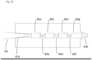
- the cell mass dissociator does not need to be composed of a plurality of blocks.
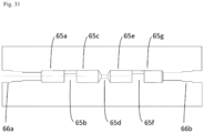
- the cell mass dissociator may have an integral cylindrical shape with a through-hole in the interior, the through-hole through which the cell mass-containing culture medium flows having, in an alternating manner, large pore size sections 65a, 65c, 65e, 65g, and small pore size sections 65b, 65d, 65f that connect with the large pore size sections 65a, 65c, 65e, 65g and have smaller pore sizes than the large pore size sections 65a, 65c, 65e, 65g.
- the central axes of the large pore size sections 65a, 65c, 65e, 65g and the central axes of at least some of the small pore size sections 65b, 65d, 65f may be offset.
- the culture medium may pass through the cell mass dissociator only once to dissociate the cell masses in the culture medium into small cell masses.
- insertion sections 66a, 66b may be provided to allow insertion of a tube or the like at both ends of the cell mass dissociator.
- the culture medium passes from the insertion section 66a through the through-hole and is discharged from the insertion section 66b, during which time the cell masses in the culture medium are dissociated.
- the central axes of the large pore size sections 65a, 65c, 65e, 65g and the central axes of at least some of the small pore size sections 65b, 65d, 65f may be offset.
- Human blood cells were acquired from a healthy adult male. There were also prepared modified mRNA (TriLink), a non-adherent dish, a 15 mL tube, a 50 mL tube, Ficoll, a Cytoflowmeter (BD), anti-CD34 antibody (Miltenyi Biotec), anti-CD3 antibody (Miltenyi Biotec), MACS R buffer (Miltenyi Biotec), T cell culture medium, low serum culture medium (Opti-MEM R , Gibco), siRNA introducing reagent (Lipofectamine R , RNAiMAX, ThermoFisherScience) and anti-TRA-1-60 antibody (BD).
- the T cell (CD3-positive cell) culture medium was a liquid mixture of the following culture medium A and culture medium B.
- Culture medium A as a liquid mixture of 15 mL of X vivo-10 (Lonza, 04-743Q) and IL-2 (10 ⁇ g/mL).
- Culture medium B was prepared by mixing X vivo-10 and 50 ⁇ L of Dynabeads CD3/CD28 (Life Technologies, 111-31D) in a 1.5 mL tube, vortexing the mixture for 5 seconds, allowing spin-down, stationing the mixture in a DynaMag-2 (Thermo fisher Science), and removing the supernatant after one minute of stationing.
- a blood cell culture medium (blood stem/precursor cell medium) by adding 10 ⁇ L of IL-6 (100 ⁇ g/mL), 10 ⁇ L of SCF (300 ⁇ g/mL), 10 ⁇ L of TPO (300 ⁇ g/mL), 10 ⁇ L of Flt3 ligand (300 ⁇ g/mL) and 10 ⁇ L of IL-3 (10 ⁇ g/mL) to 10 mL of serum-free medium (StemSpan H3000, STEMCELL Technologies).
- OCT3/4 mRNA-containing solution SOX2 mRNA-containing solution, KLF4 mRNA-containing solution, c-MYC mRNA-containing solution, LIN28A mRNA-containing solution and green fluorescent protein (GFP) mRNA-containing solution, each to a concentration of 100 ng/ ⁇ L.
- SOX2 mRNA-containing solution SOX2 mRNA-containing solution
- KLF4 mRNA-containing solution c-MYC mRNA-containing solution
- LIN28A mRNA-containing solution LIN28A mRNA-containing solution
- GFP green fluorescent protein
- a centrifuge was set to 18°C. Blood was sampled in amounts from 5 mL to 50 mL, EDTA was added to the blood, and each mixture was gently mixed. Also, medium for human lymphocyte separation Ficoll-Paque PREMIUM, GE Healthcare, Japan) was dispensed into two 15 mL tubes at 5 mL each. After adding 5 mL of PBS to the blood for dilution, 5 mL of each was overlaid onto the human lymphocyte separation medium in the tubes. During this time, the diluted blood was slowly added onto the medium while causing it to slide on the tube wall, so as not to disturb the interface.
- the solutions in the tubes were centrifuged at 400 ⁇ g, 18°C for 30 minutes. Acceleration and deceleration were carried out slowly during the procedure. After centrifugation, a white cloudy intermediate layer appeared in the tube.
- the white cloudy intermediate layer includes mononuclear cells.
- the white cloudy intermediate layer in each tube was slowly collected with a Pipetman and transferred to a new 15 mL tube. The lower layer was not handled during this time. Approximately 1 mL of the white cloudy intermediate layer could be collected from each tube.
- the intermediate layers of two tubes were combined and transferred to a single tube.
- the solution was further centrifuged at 200 ⁇ g, 18°C for 10 minutes.
- an aspirator was used to remove the supernatant of the solution by aspiration, and 3 mL of serum-free hematopoietic cell culture medium of known composition (X-VIVO R 10, Lonza) was added forming a suspension, to obtain a mononuclear cell suspension.
- a 10 ⁇ L portion of the mononuclear cell suspension was stained with Trypan blue and the count was determined with a hemocytometer.
- Reaction was performed between 1 ⁇ 10 7 mononuclear cells and CD34 antibody or CD3 antibody for 15 minutes in 100 ⁇ L of solution at 4°C. Following the reaction, 5 mL of MACS R buffer (Miltenyi Biotec) was added to the solution, and centrifugation was performed at 270 g. After centrifugation, the supernatant was removed and 1 mL of MACS buffer was added. Next, utilizing the separation program of an automatic magnetic cell separator (autoMACS, Miltenyi Biotec), CD34-positive cells and CD3-positive cells were separated from among the mononuclear cells.
- autoMACS automatic magnetic cell separator
- a first mixture was prepared by mixing 100 ⁇ L of low serum culture medium (Opti-MEM R , Gibco) and 25 ⁇ L of initializing factor mixture.
- a second mixture was also prepared by mixing 112.5 ⁇ L of low serum culture medium (Opti-MEM R , Gibco) and 12.5 ⁇ L of siRNA introducing reagent (Lipofectamine R , RNAiMAX, ThermoFisherScience).
- siRNA introducing reagent Lipofectamine R , RNAiMAX, ThermoFisherScience
- the mononuclear cells were then cultured in a feeder-free manner at 37°C for 18 hours.
- the culturing conditions were 5% CO 2 concentration, 19% oxygen concentration, 37°C temperature.
- the mononuclear cell density upon addition of the lipofection reaction mixture was 3 ⁇ 10 6 .
- the mononuclear cells were collected in a 15 mL tube and centrifuged at 300 g, and the supernatant was removed.
- CD34 blood cell culture medium 1.25 mL of CD34 blood cell culture medium was added to a 15 mL tube, the mononuclear cell suspension was returned to the same 12-well plate, and feeder-free culturing of the mononuclear cells was carried out overnight at 37 degrees.
- the culturing conditions were 5% CO 2 concentration and 19% oxygen concentration. The steps described above were repeated once every 2 days for 7 days.
- the density of cells after a total of 4 lipofections was 3 ⁇ 10 6 .
- GFP expression was examined with a fluorescent microscope, expression of GFP was confirmed, as shown in Fig. 35 . This confirmed that mRNA had been transfected in the mononuclear cells, and that protein had been synthesized from the transfected mRNA.
- TRA-1-60 a surface antigen specifically expressed on the iPS cells that had begun to be initialized, the antibody being labeled with Allophycocyanin (APC) fluorescent dye.
- APC Allophycocyanin
- a dot plot was drawn with autologous fluorescence intensity on the x-axis and fluorescent labeled anti-TRA-1-60 antibody fluorescence intensity on the y-axis.
- No TRA-1-60-positive cells were detected in a negative control without gene introduction.
- TRA-1-60-positive cells were detected in Experiments 1, 2 and 3.
- Experiment 1 represents the results of induction from all of the mononuclear cells without separation by markers
- Experiment 2 represents the results of induction from cells separated as CD3-positive
- Experiment 3 represents the results of induction from cells separated as CD34-positive. It was thus demonstrated that iPS cells can be induced by using lipofection of initializing factor RNA to introduce the initializing factor into blood-derived cells.
- a bFGF-containing human iPS culture medium was prepared by mixing 500 mL of Primate ES Cell Medium (ReproCELL) and 0.2 mL of bFGF (Gibco PHG0266) at a 10 ⁇ g/mL concentration.
- deacylated gellan gum (Nissan Chemical Industries, Ltd.) was added to the bFGF-containing human iPS culture medium to a concentration of 0.02 wt%, to prepare a bFGF-containing human iPS gel medium.
- 5 mL of trypsin at 2.5 wt% concentration, 5 mL of collagenase IV at 1 mg/mL concentration, 0.5 mL of CaCl 2 at 0.1 mol/L concentration, 10 mL of KnockOut Serum Replacement R (Invitrogen 10828-028) and 30 mL of purified water were mixed to prepare a dissociation solution, commonly known as CTK solution.
- CTK solution After adding 300 ⁇ L of the CTK solution to a 6-well dish (Thermoscientific 12-556-004) in which iPS cells were being cultured on feeder cells, the mixture was incubated for 3 minutes in a CO 2 incubator. After 3 minutes, the dish was removed from the incubator, detachment of the feeder cells alone was confirmed, and an aspirator was used to remove the CTK solution.
- bFGF-containing human iPS culture medium After suspension of the iPS cells, 4 mL of bFGF-containing human iPS culture medium was added to a 15 mL centrifugation tube, and the iPS cell suspension was centrifuged at 270 g using a centrifuge. After centrifugation, the supernatant was removed, 1 mL of bFGF-containing human iPS culture medium was added to a 15 mL centrifugation tube, and a hemocytometer was used to calculate the cell count. After cell counting, 5 ⁇ 10 5 of iPS cells each were seeded in a 15 mL Falcon tube R (Corning 352096) or a non-adherent dish, and suspension culture was carried out without agitation.
- a 2 mL portion of bFGF-containing human iPS gel medium was used in the 15 mL tube.
- a 2 mL portion of non-gelled bFGF-containing human iPS culture medium was used in the non-adherent dish.
- ROCK inhibitor (Selleck S1049) was added at 10 ⁇ mol/L to each medium.
- 500 ⁇ L of bFGF-containing human iPS gel medium was added each day to the 15 mL tube and non-adherent dish and 500 ⁇ L of bFGF-containing human iPS culture medium was added each day to the non-adherent dish.
- ROCK inhibitor was added to the 15 mL tube and non-adherent dish each day to a final concentration of 10 ⁇ mol/L, and suspension culture was continued for 7 days.
- Fig. 37 The results are shown in Fig. 37 .
- Fig. 37(b) when iPS cells were cultured in the non-adherent dish using non-gelled bFGF-containing human iPS culture medium, notable aggregation of the iPS cell colonies was observed.
- Fig. 37(a) when iPS cells were cultured using bFGF-containing human iPS gel medium in the 15 mL tube, no such conspicuous aggregation was observed.
- Fig. 38(a) is a photograph on the 1st day after culturing of iPS cells using bFGF-containing human iPS gel medium in the 15 mL tube, and Fig.
- FIG. 38(b) is a photograph on the 9th day after culturing of iPS cells using bFGF-containing human iPS gel medium in the 15 mL tube.
- the photographs of Fig. 38(a) and Fig. 38(b) confirmed colony formation without aggregation between iPS cells of different lines.
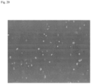
- Fig. 39(a) is a photograph immediately before reseeding of the iPS cell colonies that had been suspension cultured for 7 days in gel medium, onto feeder cells.
- Fig. 39(b) is a photograph taken when confirming the forms of the colonies after 3 days. As shown in Fig. 40 , the results confirmed that at least 95% of the colonies were undifferentiated. It was thus demonstrated that iPS cells can be cultured in gel medium while maintaining their undifferentiated state.
- bFGF-containing human iPS culture medium and bFGF-containing human iPS gel medium were prepared as in Example 2.
- the mixture was incubated for 3 minutes in a CO 2 incubator. After 3 minutes, the dish was removed from the incubator, detachment of the feeder cells alone was confirmed, and an aspirator was used to remove the CTK solution.
- 500 ⁇ L of PBS was added to the dish to rinse the iPS cells, and then the PBS was removed from the dish and 0.3 mL of Accumax was added to the dish, after which the dish was placed in a CO 2 incubator and incubated for 5 minutes.
- 0.7 mL of bFGF-containing iPS culture medium was added to the dish and the iPS cells were suspended until single cells were obtained.
- bFGF-containing human iPS culture medium After suspension of the iPS cells, 4 mL of bFGF-containing human iPS culture medium was added to a 15 mL centrifugation tube, and the iPS cell suspension was centrifuged at 270 g using a centrifuge. After centrifugation, the supernatant was removed, 1 mL of bFGF-containing human iPS culture medium was added to a 15 mL centrifugation tube, and a hemocytometer was used to calculate the cell count. The cells were counted, and then 5 ⁇ 10 5 iPS cells were seeded in each 15 mL tube and suspension culture was carried out without agitation.
- a 2 mL portion of bFGF-containing human iPS gel medium was used in a 15 mL tube.
- ROCK inhibitor was added at 10 ⁇ mol/L to each medium.
- a 500 ⁇ L portion of bFGF-containing human iPS gel medium was added to the 15 mL tube each day thereafter.
- a 500 ⁇ L portion of gel medium includes 0.5 ⁇ L of ROCK inhibitor.
- iPS cells were also suspension cultured for 7 days under the same conditions, but without addition of a ROCK inhibitor.
- Gel medium containing iPS cells was added to each of two dialysis modules (Spectrum G235035) comprising a dialysis tube with a 100 kDa molecular cutoff.
- the dialysis modules were each placed in a 50 mL centrifugation tube, and gel medium was placed around the dialysis tubes in the centrifugation tubes.
- the gel medium containing the iPS cells was also directly placed in a separate 50 mL centrifugation tube.
- a pump was connected to one of the centrifugation tubes of the two centrifugation tubes in which dialysis tubes had been placed, as shown in Fig. 6 , and the gel medium in the centrifugation tube was continuously exchanged for several days.
- the gel medium was stored at 4°C, and set so as to be at 37°C when reaching the centrifugation tube.
- No pump was connected to the other centrifugation tube of the two centrifugation tubes in which a dialysis tube had been placed, and the gel medium in the centrifugation tube was not exchanged.
- the gel medium was also not exchanged in the centrifugation tube in which a dialysis tube had not been placed.
Landscapes
- Health & Medical Sciences (AREA)
- Engineering & Computer Science (AREA)
- Life Sciences & Earth Sciences (AREA)
- Chemical & Material Sciences (AREA)
- Zoology (AREA)
- Bioinformatics & Cheminformatics (AREA)
- Wood Science & Technology (AREA)
- Organic Chemistry (AREA)
- Biotechnology (AREA)
- Biomedical Technology (AREA)
- Genetics & Genomics (AREA)
- Microbiology (AREA)
- Biochemistry (AREA)
- General Engineering & Computer Science (AREA)
- General Health & Medical Sciences (AREA)
- Sustainable Development (AREA)
- Analytical Chemistry (AREA)
- Cell Biology (AREA)
- Molecular Biology (AREA)
- Clinical Laboratory Science (AREA)
- Thermal Sciences (AREA)
- Hematology (AREA)
- Computer Hardware Design (AREA)
- Physics & Mathematics (AREA)
- Immunology (AREA)
- Mechanical Engineering (AREA)
- Developmental Biology & Embryology (AREA)
- Transplantation (AREA)
- Micro-Organisms Or Cultivation Processes Thereof (AREA)
- Apparatus Associated With Microorganisms And Enzymes (AREA)
- Peptides Or Proteins (AREA)
Claims (7)
- Ein Zellmassen-Dissoziator, der einen Verbindungsblock (62) beinhaltet, der in seinem Innern mit einem Durchgangsloch versehen ist, durch das ein zellmassenhaltiges Kulturmedium fließt, wobei:an der ersten Kante des Verbindungsblocks (62) eine Aussparung (62a) bereitgestellt ist und an der zweiten Kante des Verbindungsblocks (62) ein Vorsprung (62b) bereitgestellt ist,im Falle mehrerer Verbindungsblöcke (62) die Vorsprünge (62b) mit den Aussparungen (62a) der benachbarten Verbindungsblöcke (62) in Eingriff stehen unddas Durchgangsloch Folgendes aufweist: einen ersten Abschnitt mit großer Porengröße (62c), der mit der Aussparung (62a) verbunden ist, einen Abschnitt mit kleiner Porengröße (62d), der mit dem ersten Abschnitt mit großer Porengröße (62c) verbunden ist und eine kleinere Porengröße aufweist als der erste Abschnitt mit der großen Porengröße (62c), und einen zweiten Abschnitt mit großer Porengröße (62e), der mit dem Abschnitt mit kleiner Porengröße (62d) verbunden ist, eine größere Porengröße aufweist als der Abschnitt mit der kleinen Porengröße (62d) und eine Öffnung an der Spitze des Vorsprungs (62b) aufweist.
- Zellmassen-Dissoziator nach Anspruch 1, wobei, wenn mehrere Verbindungsblöcke (62) vorhanden sind und die mehreren Verbindungsblöcke (62) miteinander verbunden sind, die zweiten Abschnitte mit großer Porengröße (62e) mit den ersten Abschnitten mit großer Porengröße (62c) von benachbarten Verbindungsblöcken (62) glatt verbunden sind.
- Zellmassen-Dissoziator nach Anspruch 1 oder 2, wobei die Mittelachsen des ersten und zweiten Abschnitts mit großer Porengröße (62c, 62e) und die Mittelachse des Abschnitts mit kleiner Porengröße (62d) versetzt sind.
- Zellmassen-Dissoziator nach einem der Ansprüche 1 bis 3, der ferner einen im Inneren mit einem Durchgangsloch versehenen Spitzenblock (63) beinhaltet, wobei:eine Aussparung (63a) an der ersten Kante des Spitzenblocks (63) und eine Düse (63b) an der zweiten Kante des Spitzenblocks (63) bereitgestellt sind,die Aussparung (63a) des Spitzenblocks (63) mit dem Vorsprung (62b) des Verbindungsblocks (62) in Eingriff steht unddas Durchgangsloch Folgendes aufweist: einen Abschnitt mit großer Porengröße (63c), der mit der Aussparung (63a) verbunden ist, und einen Abschnitt mit kleiner Porengröße (63d), der mit dem Abschnitt mit großer Porengröße (63c) verbunden ist, eine kleinere Porengröße aufweist als der Abschnitt mit großer Porengröße (63c) und eine Öffnung an der Spitze der Düse (63b) aufweist.
- Zellmassen-Dissoziator nach Anspruch 4, wobei, wenn der Verbindungsblock (62) und der Spitzenblock (63) verbunden worden sind, der zweite Abschnitt mit großer Porengröße (62e) des Verbindungsblocks (62) und der Abschnitt mit großer Porengröße (63c) des Spitzenblocks (63) glatt miteinander verbunden sind.
- Zellmassen-Dissoziator nach einem der Ansprüche 1 bis 5, der ferner einen im Inneren mit einem Durchgangsloch versehenen Endblock (61) beinhaltet, wobei eine Aussparung (61a) an der ersten Kante des Endblocks (61) bereitgestellt ist und ein Vorsprung (61b) an der zweiten Kante des Endblocks (61) bereitgestellt ist, und der Vorsprung (61b) des Endblocks (61) in Eingriff mit der Aussparung (62a) des Verbindungsblocks (62) steht.
- Zellmassen-Dissoziator nach Anspruch 6, der ferner Folgendes beinhaltet:eine Einschubdüse (64), die in die Aussparung (61a) des Endblocks (61) eingeschoben wird, undeine Saugdrainagevorrichtung in Verbindung mit der Einschubdüse (64), die das zellmassenhaltige Kulturmedium durch Saugen drainiert.
Applications Claiming Priority (4)
| Application Number | Priority Date | Filing Date | Title |
|---|---|---|---|
| JP2015170797 | 2015-08-31 | ||
| US201662356199P | 2016-06-29 | 2016-06-29 | |
| PCT/JP2016/075540 WO2017038887A1 (ja) | 2015-08-31 | 2016-08-31 | 多能性幹細胞製造システム |
| EP16841917.4A EP3345990A4 (de) | 2015-08-31 | 2016-08-31 | System zur produktion pluripotenter stammzellen |
Related Parent Applications (2)
| Application Number | Title | Priority Date | Filing Date |
|---|---|---|---|
| EP16841917.4A Division EP3345990A4 (de) | 2015-08-31 | 2016-08-31 | System zur produktion pluripotenter stammzellen |
| EP16841917.4A Division-Into EP3345990A4 (de) | 2015-08-31 | 2016-08-31 | System zur produktion pluripotenter stammzellen |
Publications (3)
| Publication Number | Publication Date |
|---|---|
| EP3366763A2 EP3366763A2 (de) | 2018-08-29 |
| EP3366763A3 EP3366763A3 (de) | 2019-02-20 |
| EP3366763B1 true EP3366763B1 (de) | 2023-11-15 |
Family
ID=58188066
Family Applications (4)
| Application Number | Title | Priority Date | Filing Date |
|---|---|---|---|
| EP16842835.7A Pending EP3344755A4 (de) | 2015-08-31 | 2016-08-30 | Herstellungssystem für pluripotente stammzellen und verfahren zur herstellung induzierter pluripotenter stammzellen |
| EP16841917.4A Pending EP3345990A4 (de) | 2015-08-31 | 2016-08-31 | System zur produktion pluripotenter stammzellen |
| EP18167250.2A Active EP3366763B1 (de) | 2015-08-31 | 2016-08-31 | Vorrichtung zur freisetzung von zellen |
| EP18203880.2A Withdrawn EP3473703A3 (de) | 2015-08-31 | 2016-08-31 | System zur produktion pluripotenter stammzellen |
Family Applications Before (2)
| Application Number | Title | Priority Date | Filing Date |
|---|---|---|---|
| EP16842835.7A Pending EP3344755A4 (de) | 2015-08-31 | 2016-08-30 | Herstellungssystem für pluripotente stammzellen und verfahren zur herstellung induzierter pluripotenter stammzellen |
| EP16841917.4A Pending EP3345990A4 (de) | 2015-08-31 | 2016-08-31 | System zur produktion pluripotenter stammzellen |
Family Applications After (1)
| Application Number | Title | Priority Date | Filing Date |
|---|---|---|---|
| EP18203880.2A Withdrawn EP3473703A3 (de) | 2015-08-31 | 2016-08-31 | System zur produktion pluripotenter stammzellen |
Country Status (7)
| Country | Link |
|---|---|
| US (5) | US11286454B2 (de) |
| EP (4) | EP3344755A4 (de) |
| JP (7) | JP7370529B2 (de) |
| CN (5) | CN108138130A (de) |
| CA (2) | CA2996582A1 (de) |
| SG (1) | SG11201801549VA (de) |
| WO (2) | WO2017040548A1 (de) |
Families Citing this family (56)
| Publication number | Priority date | Publication date | Assignee | Title |
|---|---|---|---|---|
| CN108138130A (zh) | 2015-08-31 | 2018-06-08 | 爱平世股份有限公司 | 多能干细胞制造系统和生产诱导多能干细胞的方法 |
| EP3541922A4 (de) | 2016-11-18 | 2020-06-03 | New York Stem Cell Foundation, Inc. | Mikrofluidisches system und verfahren zu dessen verwendung |
| EP3556842A4 (de) * | 2017-01-20 | 2020-01-15 | FUJIFILM Corporation | Zellkulturvorrichtung und zellkulturverfahren |
| CA3054119A1 (en) * | 2017-02-24 | 2018-08-30 | I Peace, Inc. | Cell treatment device, suspension culture vessel, and stem cell induction method |
| CN110352238A (zh) * | 2017-02-24 | 2019-10-18 | 田边刚士 | 诱导性多能干细胞的制作方法 |
| US20190382706A1 (en) * | 2017-02-27 | 2019-12-19 | Koji Tanabe | Somatic cell production system |
| CN115247129A (zh) | 2017-02-27 | 2022-10-28 | 田边刚士 | 细胞处理装置 |
| TWI862477B (zh) * | 2017-05-26 | 2024-11-21 | 美商凱特製藥公司 | 製備和使用胚胎間充質先驅細胞的方法 |
| JP7114693B2 (ja) * | 2017-08-24 | 2022-08-08 | テルモ ビーシーティー、インコーポレーテッド | 細胞増殖 |
| KR102503086B1 (ko) * | 2017-09-08 | 2023-02-23 | 가부시키가이샤 오츠카 세이야쿠 고죠 | 어린 돼지 유래 줄기 세포 및 그 제조 방법 |
| JPWO2019131626A1 (ja) * | 2017-12-28 | 2020-12-10 | オリンパス株式会社 | 細胞培養制御方法、細胞培養制御装置、細胞培養装置および細胞培養システム |
| US11920119B2 (en) | 2018-02-09 | 2024-03-05 | Global Life Sciences Solutions Usa Llc | Systems and methods for bioprocessing |
| US12252682B2 (en) | 2018-02-09 | 2025-03-18 | Global Life Sciences Solutions Usa Llc | System and method for fluid flow management in a bioprocessing system |
| CN112055744A (zh) * | 2018-02-09 | 2020-12-08 | 环球生命科技咨询美国有限责任公司 | 用于在生物处理系统中的流体管线管理的装置 |
| US11932842B2 (en) | 2018-02-09 | 2024-03-19 | Global Life Sciences Solutions Usa Llc | Bioprocessing apparatus |
| US12077743B2 (en) | 2018-02-09 | 2024-09-03 | Global Life Sciences Solutions Usa Llc | Apparatus for fluid line management in a bioprocessing system |
| US10889792B2 (en) | 2018-02-09 | 2021-01-12 | Global Life Sciences Solutions Usa Llc | Cell expansion vessel systems and methods |
| JP6939643B2 (ja) * | 2018-02-26 | 2021-09-22 | 株式会社Ihi | 細胞分割装置及び細胞培養装置 |
| TWI846694B (zh) * | 2018-05-04 | 2024-07-01 | 美商健臻公司 | 具有過濾系統的灌注式生物反應器 |
| US12385001B2 (en) * | 2018-06-19 | 2025-08-12 | Stemcell Technologies Canada Inc. | Systems, methods and apparatus for the automated culture of cells |
| WO2020017411A1 (ja) * | 2018-07-17 | 2020-01-23 | 国立大学法人神戸大学 | 固液界面検出装置及びこれを備えた前処理装置 |
| DK3778860T3 (da) * | 2018-07-24 | 2026-04-20 | Fujifilm Corp | Driftsprogram til cellekulturhjælpeapparat, cellekulturhjælpeapparat og driftsfremgangsmåde til cellekulturhjælpeapparat |
| EP3842513A4 (de) * | 2018-08-20 | 2022-06-29 | I Peace, Inc. | Zellinkubator |
| WO2020040135A1 (ja) * | 2018-08-20 | 2020-02-27 | 剛士 田邊 | 細胞の培養又は誘導方法 |
| WO2020044984A1 (ja) * | 2018-08-27 | 2020-03-05 | 富士フイルム株式会社 | 細胞培養方法及び細胞培養装置 |
| US11692161B2 (en) | 2019-02-05 | 2023-07-04 | Corning Incorporated | Packed-bed bioreactor systems and methods of using the same |
| JP2020167959A (ja) * | 2019-04-04 | 2020-10-15 | オリンパス株式会社 | 培地モニタリング装置 |
| WO2020196490A1 (ja) * | 2019-03-25 | 2020-10-01 | テルモ株式会社 | 細胞培養物を清浄度の高い空間において調製するための容器及びキット |
| JP7483687B2 (ja) * | 2019-03-27 | 2024-05-15 | 株式会社カネカ | 細胞製造装置、細胞製造方法並びにこれに用いられるサーバ、システムおよび装置 |
| CN110066733A (zh) * | 2019-05-29 | 2019-07-30 | 广东唯泰生物科技有限公司 | 干细胞培养机器人 |
| CN113785049B (zh) * | 2019-06-10 | 2025-11-25 | 爱平世股份有限公司 | 细胞培养方法及单核细胞的回收方法 |
| CN113811597B (zh) * | 2019-06-28 | 2024-06-18 | 爱平世股份有限公司 | 细胞培养装置的应用 |
| JP7037141B2 (ja) * | 2019-06-28 | 2022-03-16 | アイ ピース,インコーポレイテッド | 細胞塊分割器、細胞塊分割器の製造方法、及び細胞塊の分割方法 |
| CN110229751A (zh) * | 2019-07-17 | 2019-09-13 | 中国人民解放军军事科学院军事医学研究院 | mRNA制备系统及制备方法 |
| US20220356436A1 (en) * | 2019-08-29 | 2022-11-10 | Fanuc Corporation | Cell production device |
| WO2021038998A1 (ja) * | 2019-08-29 | 2021-03-04 | ファナック株式会社 | 細胞製造装置及びそのシステム |
| WO2021038996A1 (ja) * | 2019-08-29 | 2021-03-04 | ファナック株式会社 | 細胞製造装置及びその製造方法 |
| US11118151B2 (en) | 2019-11-05 | 2021-09-14 | Corning Incorporated | Fixed bed bioreactor and methods of using the same |
| JP7656868B2 (ja) * | 2019-11-06 | 2025-04-04 | アイ ピース,インコーポレイテッド | 細胞培養装置 |
| CN111126329A (zh) * | 2019-12-30 | 2020-05-08 | 杭州原生生物科技有限公司 | 一种自动识别多能干细胞群的方法 |
| JPWO2021166227A1 (de) * | 2020-02-21 | 2021-08-26 | ||
| AU2021273985A1 (en) * | 2020-05-22 | 2022-12-22 | Sqz Biotechnologies Company | Point of care system for automatically processing cells |
| CN111666895B (zh) * | 2020-06-08 | 2023-05-26 | 上海市同济医院 | 基于深度学习的神经干细胞分化方向预测系统及方法 |
| JP7682619B2 (ja) | 2020-10-19 | 2025-05-26 | キヤノンメディカルシステムズ株式会社 | 誘導多能性幹細胞樹立装置及び方法 |
| WO2022139950A1 (en) * | 2020-12-23 | 2022-06-30 | Cedars-Sinai Medical Center | Methods for differentiation of pancreatic exocrine cells from human induced pluripotent stem cells |
| JP7744155B2 (ja) * | 2021-04-19 | 2025-09-25 | キヤノンメディカルシステムズ株式会社 | 多能性幹細胞製造システム |
| WO2022244670A1 (ja) * | 2021-05-18 | 2022-11-24 | 国立大学法人京都大学 | 多能性幹細胞の製造方法 |
| CN113265333A (zh) * | 2021-06-23 | 2021-08-17 | 无锡生基医药科技有限公司 | 一种全封闭、半自动化控制、模块化的质粒瞬时转染系统 |
| WO2023034720A1 (en) * | 2021-08-28 | 2023-03-09 | Cornell University | Compositions and methods for cell reprogramming |
| WO2023053220A1 (ja) * | 2021-09-28 | 2023-04-06 | 公益財団法人京都大学iPS細胞研究財団 | 多能性幹細胞の製造方法 |
| US20230130734A1 (en) * | 2021-10-27 | 2023-04-27 | Regenerative Research Foundation | Cell culture feeding device |
| JP7836751B2 (ja) * | 2022-12-15 | 2026-03-27 | ヤマハ発動機株式会社 | 組換えタンパク質産生細胞の培養方法 |
| WO2024176440A1 (ja) * | 2023-02-24 | 2024-08-29 | 公益財団法人京都大学iPS細胞研究財団 | 人工多能性幹細胞の製造方法 |
| CN116426573A (zh) * | 2023-04-14 | 2023-07-14 | 臻赫医药(杭州)有限公司 | 重编程因子抗衰老表达系统、生物材料及用途 |
| JP7721836B1 (ja) * | 2023-12-04 | 2025-08-12 | 公益財団法人京都大学iPS細胞研究財団 | 人工多能性幹細胞の製造方法 |
| CN118460359B (zh) * | 2024-07-10 | 2024-09-20 | 江苏颐泽生物科技有限公司 | 一种具有细胞活性检测功能的干细胞贮存装置 |
Family Cites Families (71)
| Publication number | Priority date | Publication date | Assignee | Title |
|---|---|---|---|---|
| EP0258795B1 (de) * | 1986-08-27 | 1993-11-03 | Kawasumi Laboratories, Inc. | Verfahren und Vorrichtung zur Kultivierung von Zellen |
| US5399493A (en) * | 1989-06-15 | 1995-03-21 | The Regents Of The University Of Michigan | Methods and compositions for the optimization of human hematopoietic progenitor cell cultures |
| CA2183638C (en) * | 1995-03-20 | 2001-03-20 | Precision System Science Co., Ltd | Liquid processing method making use of pipette device and apparatus for same |
| CN1609593B (zh) | 1996-08-16 | 2012-04-25 | Ge保健尼亚加拉公司 | 用于分析井板、凝胶和斑点的数字成像系统 |
| TWI288779B (en) | 2002-03-28 | 2007-10-21 | Blasticon Biotech Forschung | Dedifferentiated, programmable stem cells of monocytic origin, and their production and use |
| JP4309815B2 (ja) | 2004-07-05 | 2009-08-05 | 株式会社日立製作所 | 細胞培養システム及び細胞培養方法 |
| CN101443444A (zh) | 2004-12-29 | 2009-05-27 | 比奥根艾迪克Ma公司 | 生物反应器过程控制系统及方法 |
| CN100335612C (zh) * | 2005-01-20 | 2007-09-05 | 上海交通大学 | 植物细胞或组织两段连续培养系统 |
| CN1664545A (zh) * | 2005-03-18 | 2005-09-07 | 宁波美生医疗器材有限公司 | 一种细胞薄层涂片的制作方法 |
| JP2007000038A (ja) | 2005-06-22 | 2007-01-11 | Toray Ind Inc | 閉鎖系循環回路型培養装置 |
| CN2841667Y (zh) * | 2005-09-09 | 2006-11-29 | 刘平 | 细胞滤过器 |
| US20070087437A1 (en) * | 2005-10-14 | 2007-04-19 | Jifan Hu | Methods for rejuvenating cells in vitro and in vivo |
| BRPI0619794B8 (pt) | 2005-12-13 | 2022-06-14 | Univ Kyoto | Uso de um fator de reprogramação, agente para a preparação de uma célula-tronco pluripotente induzida a partir de uma célula somática e métodos para preparar uma célula- tronco pluripotente induzida método e para preparar uma célula somática e uso de células-tronco pluripotentes induzidas |
| WO2008136729A1 (en) * | 2007-05-04 | 2008-11-13 | Sundstroem Erik | Slicing device |
| JP2008307007A (ja) * | 2007-06-15 | 2008-12-25 | Bayer Schering Pharma Ag | 出生後のヒト組織由来未分化幹細胞から誘導したヒト多能性幹細胞 |
| US8815585B2 (en) | 2007-06-29 | 2014-08-26 | Cellular Dynamics International, Inc. | Automated method and apparatus for embryonic stem cell culture |
| US7858371B2 (en) * | 2007-09-26 | 2010-12-28 | Weyerhaeuser Nr Company | Method of separating embryo suspension mass |
| US9683232B2 (en) * | 2007-12-10 | 2017-06-20 | Kyoto University | Efficient method for nuclear reprogramming |
| EP2238236B1 (de) | 2008-01-09 | 2017-03-29 | Screencell | Vorrichtung und verfahren zur isolierung und kultivierung lebender zellen auf einem filter oder derer extraktion von genetischem material |
| KR101481164B1 (ko) | 2008-01-30 | 2015-01-09 | 주식회사 미래셀바이오 | 체세포 유래 다능성 줄기세포의 제조 방법 |
| US10047346B2 (en) * | 2008-08-08 | 2018-08-14 | Mayo Foundation For Medical Education And Research | Method of treating heart tissue using induced pluripotent stem cells |
| EP2881461A1 (de) * | 2008-11-21 | 2015-06-10 | Fraunhofer-Gesellschaft zur Förderung der angewandten Forschung e.V. | Umprogrammierung von Zellen zu einem pluripotenten Status |
| WO2010141320A1 (en) * | 2009-06-01 | 2010-12-09 | Mariposa Biotechnology, Inc. | Device for removing cumulus from oocytes |
| DK2438160T3 (en) | 2009-06-05 | 2016-01-11 | Cellular Dynamics Int Inc | Reprogramming of T cells and hematopoietic cells |
| US8927287B2 (en) * | 2009-10-09 | 2015-01-06 | Georgia Tech Research Corporation | Method for dispersion of assemblies of biological material |
| EP3633025B1 (de) * | 2009-11-12 | 2022-09-14 | Technion Research & Development Foundation Ltd. | Kulturmedien, zellkulturen und verfahren zur kultivierung pluripotenter stammzellen in undifferenziertem zustand |
| EP2528687B1 (de) * | 2010-01-29 | 2018-08-22 | Micronics, Inc. | System beinhaltend ein wirtinstrument und eine mikrofluidische kartusche für untersuchungen |
| CA2796464C (en) * | 2010-04-16 | 2021-08-03 | Immune Disease Institute, Inc. | Sustained polypeptide expression from synthetic, modified rnas and uses thereof |
| EP2603517B1 (de) * | 2010-05-12 | 2023-07-19 | Cellectis | Dynamische misch- und elektroporationskammer und system dafür |
| US8048675B1 (en) * | 2010-05-12 | 2011-11-01 | Ipierian, Inc. | Integration-free human induced pluripotent stem cells from blood |
| CA2810488C (en) * | 2010-09-07 | 2025-05-06 | Technion Research & Development Foundation Limited | NEW PROCESSES AND CULTURE MEDIA FOR THE CULTURE OF MULTIPOTENTED STEM CELLS |
| US20160272934A1 (en) * | 2010-10-08 | 2016-09-22 | Cellanyx Diagnostics, Llc | Systems, devices and methods for microfluidic culturing, manipulation and analysis of tissues and cells |
| CN102559587A (zh) * | 2010-12-16 | 2012-07-11 | 中国科学院上海药物研究所 | 诱导性多能干细胞的制备方法以及用于制备诱导性多能干细胞的培养基 |
| WO2012087965A2 (en) * | 2010-12-22 | 2012-06-28 | Fate Therapauetics, Inc. | Cell culture platform for single cell sorting and enhanced reprogramming of ipscs |
| CN102174395B (zh) | 2011-01-30 | 2013-08-21 | 中国科学院广州生物医药与健康研究院 | 诱导多能干细胞自动化扩增与培养系统 |
| WO2012115153A1 (ja) | 2011-02-25 | 2012-08-30 | 株式会社ニコン | 細胞評価方法、細胞培養方法、細胞評価装置、インキュベータ、細胞評価プログラム、コロニー分類プログラム、幹細胞の培養方法、幹細胞評価装置および幹細胞評価プログラム |
| GB201105413D0 (en) * | 2011-03-30 | 2011-05-11 | Cambridge Entpr Ltd | Nuclear reprogramming substrate |
| AU2012243396A1 (en) * | 2011-04-15 | 2013-10-31 | The University Of British Columbia | Method and apparatus for separation of particles |
| CN107058101B (zh) * | 2011-10-17 | 2021-06-01 | 麻省理工学院 | 细胞内传递 |
| WO2013058403A1 (ja) | 2011-10-21 | 2013-04-25 | 国立大学法人京都大学 | 層流による多能性維持単一分散細胞培養法 |
| EP2594632A1 (de) * | 2011-11-18 | 2013-05-22 | Miltenyi Biotec GmbH | Verfahren und Vorrichtung zur Zellmodifizierung |
| US20140329317A1 (en) * | 2011-11-25 | 2014-11-06 | Kyoto University | Method for culturing pluripotent stem cell |
| WO2013082509A1 (en) * | 2011-12-01 | 2013-06-06 | The New York Stem Cell Foundation | Automated system for producing induced pluripotent stem cells or differentiated cells |
| US10428309B2 (en) * | 2011-12-01 | 2019-10-01 | New York Stem Cell Foundation, Inc. | Systems and methods for producing stem cells and differentiated cells |
| TWI582233B (zh) * | 2011-12-15 | 2017-05-11 | 陶氏農業科學公司 | 使用農桿菌改良轉形的方法 |
| JP5274731B1 (ja) | 2011-12-22 | 2013-08-28 | 三洋電機株式会社 | 観察システム、プログラム及び観察システムの制御方法 |
| WO2013136372A1 (ja) | 2012-03-16 | 2013-09-19 | 株式会社日立製作所 | 細胞シート、細胞培養方法および細胞培養装置 |
| WO2013177228A1 (en) * | 2012-05-22 | 2013-11-28 | Loma Linda University | Generation of integration/transgene-free stem cells |
| EP2861612A4 (de) | 2012-06-13 | 2016-02-17 | Stemgent Inc | Verfahren zur herstellung von pluripotenten stammzellen |
| US9664671B2 (en) * | 2012-07-24 | 2017-05-30 | Nissan Chemical Industries, Ltd. | Culture medium composition and method of culturing cell or tissue using thereof |
| IL300460A (en) * | 2012-07-24 | 2023-04-01 | Nissan Chemical Corp | A culture medium preparation, and a method for culturing cells or tissues by using the preparation |
| AU2013343864B2 (en) * | 2012-11-09 | 2019-04-04 | BioNTech SE | Method for cellular RNA expression |
| US9738861B2 (en) | 2013-03-06 | 2017-08-22 | Kyoto University | Culture system for pluripotent stem cells and method for subculturing pluripotent stem cells |
| EP2970881A4 (de) * | 2013-03-14 | 2017-01-25 | Children's Medical Center Corporation | Zusammensetzungen und verfahren zur neuprogrammierung hämatopoietischer stammzelllinien |
| CA2895638A1 (en) | 2013-03-15 | 2014-09-18 | Fluidigm Corporation | Methods and devices for analysis of defined multicellular combinations |
| US10519421B2 (en) | 2013-03-21 | 2019-12-31 | Kyoto University | Induction of motor neurons from pluripotent stem cells |
| WO2014171486A1 (ja) * | 2013-04-17 | 2014-10-23 | 日産化学工業株式会社 | 培地組成物及び当該培地組成物を用いた赤血球の製造方法 |
| US20150159127A1 (en) * | 2013-05-10 | 2015-06-11 | BioCapacity On Demand, LLC | Mobile vessel or carrier for the manufacturing production, purification and sterile fill and finish of biologics |
| EP3008174A4 (de) * | 2013-06-11 | 2017-03-08 | Kyoto University | Verfahren zur effizienten herstellung induzierter pluripotenter stammzellen |
| US9580678B2 (en) * | 2013-06-21 | 2017-02-28 | The Regents Of The University Of California | Microfluidic tumor tissue dissociation device |
| EP3048169B1 (de) | 2013-09-04 | 2019-07-10 | Otsuka Pharmaceutical Factory, Inc. | Verfahren zur herstellung pluripotenter stammzellen |
| JP5751726B2 (ja) | 2013-11-12 | 2015-07-22 | 東京エレクトロン株式会社 | 多能性幹細胞の培養方法及び施設 |
| TWI708846B (zh) | 2014-01-23 | 2020-11-01 | 日商日產化學工業股份有限公司 | 培養基組成物的製造方法 |
| CN112662611B (zh) | 2014-01-23 | 2024-10-29 | 日产化学工业株式会社 | 培养基组合物 |
| US10100286B2 (en) * | 2014-06-20 | 2018-10-16 | The Board Of Trustees Of The University Of Alabama | Systems and methods of dissociating aggregate spheres of cells |
| US20170355950A1 (en) * | 2014-12-04 | 2017-12-14 | The Arizona Board Of Regents On Behalf Of The University Of Arizona | Systems for dissociation of biological tissues |
| JP6291429B2 (ja) | 2015-01-20 | 2018-03-14 | 富士フイルム株式会社 | 細胞培養装置および細胞培養方法 |
| US10350245B2 (en) * | 2015-01-21 | 2019-07-16 | Fred Hutchinson Cancer Research Center | Point-of-care and/or portable platform for gene therapy |
| US10836997B2 (en) | 2015-03-09 | 2020-11-17 | Keio University | Method for differentiating pluripotent stem cells into desired cell type |
| CN108138130A (zh) | 2015-08-31 | 2018-06-08 | 爱平世股份有限公司 | 多能干细胞制造系统和生产诱导多能干细胞的方法 |
| KR102802007B1 (ko) * | 2016-06-08 | 2025-04-28 | 더 리전트 오브 더 유니버시티 오브 캘리포니아 | 조직 및 세포를 프로세싱하기 위한 방법 및 장치 |
-
2016
- 2016-08-30 CN CN201680050145.XA patent/CN108138130A/zh active Pending
- 2016-08-30 US US15/756,029 patent/US11286454B2/en active Active
- 2016-08-30 JP JP2018510722A patent/JP7370529B2/ja active Active
- 2016-08-30 CA CA2996582A patent/CA2996582A1/en active Pending
- 2016-08-30 EP EP16842835.7A patent/EP3344755A4/de active Pending
- 2016-08-30 WO PCT/US2016/049530 patent/WO2017040548A1/en not_active Ceased
- 2016-08-30 CN CN202210349694.4A patent/CN114717183B/zh active Active
- 2016-08-31 CA CA2996988A patent/CA2996988A1/en active Pending
- 2016-08-31 CN CN201810580097.6A patent/CN108823062B/zh not_active Expired - Fee Related
- 2016-08-31 CN CN202211151066.1A patent/CN115478045A/zh active Pending
- 2016-08-31 EP EP16841917.4A patent/EP3345990A4/de active Pending
- 2016-08-31 EP EP18167250.2A patent/EP3366763B1/de active Active
- 2016-08-31 SG SG11201801549VA patent/SG11201801549VA/en unknown
- 2016-08-31 JP JP2017526727A patent/JP6198203B2/ja active Active
- 2016-08-31 US US15/756,031 patent/US11518974B2/en active Active
- 2016-08-31 CN CN201680050144.5A patent/CN108138113A/zh active Pending
- 2016-08-31 EP EP18203880.2A patent/EP3473703A3/de not_active Withdrawn
- 2016-08-31 WO PCT/JP2016/075540 patent/WO2017038887A1/ja not_active Ceased
-
2017
- 2017-05-16 JP JP2017097408A patent/JP2017221187A/ja active Pending
- 2017-05-16 JP JP2017097398A patent/JP6283904B2/ja active Active
- 2017-08-10 JP JP2017155756A patent/JP6253216B2/ja active Active
-
2019
- 2019-03-11 US US16/299,048 patent/US10508260B2/en active Active
- 2019-11-14 JP JP2019206125A patent/JP7351483B2/ja active Active
-
2022
- 2022-01-06 US US17/569,634 patent/US20220119758A1/en not_active Abandoned
- 2022-04-06 JP JP2022063287A patent/JP2022088643A/ja active Pending
- 2022-08-03 US US17/817,287 patent/US11912977B2/en active Active
Also Published As
Similar Documents
| Publication | Publication Date | Title |
|---|---|---|
| EP3366763B1 (de) | Vorrichtung zur freisetzung von zellen | |
| US11959058B2 (en) | Cell processing system and cell processing device | |
| US20160194609A1 (en) | Isolation, expansion and characterization of wharton's jelly mesenchymal stem cells | |
| US20230055044A1 (en) | Method for manufacturing induced pluripotent stem cells | |
| JP2020036608A (ja) | 多能性幹細胞製造システム、幹細胞の誘導方法、幹細胞の浮遊培養方法、幹細胞の浮遊培養器、人工多能性幹細胞の作製方法、及び動物細胞から特定の体細胞を作製する方法 | |
| JP6530577B1 (ja) | 細胞処理システム及び細胞処理装置 | |
| JP6530874B1 (ja) | 細胞処理システム及び細胞処理装置 | |
| HK40067121B (zh) | 多能干细胞制造系统和生产诱导多能干细胞的方法 | |
| HK40078389A (en) | A method for producing an induced pluripotent stem cell | |
| HK40002823B (en) | Cell mass dissociator | |
| HK40002823A (en) | Cell mass dissociator | |
| JP2019088335A (ja) | 細胞処理システム及び細胞処理装置 | |
| HK40078284A (en) | Cell processing device |
Legal Events
| Date | Code | Title | Description |
|---|---|---|---|
| PUAI | Public reference made under article 153(3) epc to a published international application that has entered the european phase |
Free format text: ORIGINAL CODE: 0009012 |
|
| STAA | Information on the status of an ep patent application or granted ep patent |
Free format text: STATUS: REQUEST FOR EXAMINATION WAS MADE |
|
| 17P | Request for examination filed |
Effective date: 20180413 |
|
| AC | Divisional application: reference to earlier application |
Ref document number: 3345990 Country of ref document: EP Kind code of ref document: P |
|
| AK | Designated contracting states |
Kind code of ref document: A2 Designated state(s): AL AT BE BG CH CY CZ DE DK EE ES FI FR GB GR HR HU IE IS IT LI LT LU LV MC MK MT NL NO PL PT RO RS SE SI SK SM TR |
|
| AX | Request for extension of the european patent |
Extension state: BA ME |
|
| PUAL | Search report despatched |
Free format text: ORIGINAL CODE: 0009013 |
|
| AK | Designated contracting states |
Kind code of ref document: A3 Designated state(s): AL AT BE BG CH CY CZ DE DK EE ES FI FR GB GR HR HU IE IS IT LI LT LU LV MC MK MT NL NO PL PT RO RS SE SI SK SM TR |
|
| AX | Request for extension of the european patent |
Extension state: BA ME |
|
| RIC1 | Information provided on ipc code assigned before grant |
Ipc: C12M 1/33 20060101ALI20190114BHEP Ipc: C12M 1/00 20060101AFI20190114BHEP |
|
| STAA | Information on the status of an ep patent application or granted ep patent |
Free format text: STATUS: EXAMINATION IS IN PROGRESS |
|
| 17Q | First examination report despatched |
Effective date: 20221223 |
|
| GRAP | Despatch of communication of intention to grant a patent |
Free format text: ORIGINAL CODE: EPIDOSNIGR1 |
|
| STAA | Information on the status of an ep patent application or granted ep patent |
Free format text: STATUS: GRANT OF PATENT IS INTENDED |
|
| INTG | Intention to grant announced |
Effective date: 20230613 |
|
| P01 | Opt-out of the competence of the unified patent court (upc) registered |
Effective date: 20230824 |
|
| GRAS | Grant fee paid |
Free format text: ORIGINAL CODE: EPIDOSNIGR3 |
|
| GRAA | (expected) grant |
Free format text: ORIGINAL CODE: 0009210 |
|
| STAA | Information on the status of an ep patent application or granted ep patent |
Free format text: STATUS: THE PATENT HAS BEEN GRANTED |
|
| AC | Divisional application: reference to earlier application |
Ref document number: 3345990 Country of ref document: EP Kind code of ref document: P |
|
| AK | Designated contracting states |
Kind code of ref document: B1 Designated state(s): AL AT BE BG CH CY CZ DE DK EE ES FI FR GB GR HR HU IE IS IT LI LT LU LV MC MK MT NL NO PL PT RO RS SE SI SK SM TR |
|
| REG | Reference to a national code |
Ref country code: CH Ref legal event code: EP Ref country code: GB Ref legal event code: FG4D |
|
| REG | Reference to a national code |
Ref country code: DE Ref legal event code: R096 Ref document number: 602016084216 Country of ref document: DE |
|
| REG | Reference to a national code |
Ref country code: IE Ref legal event code: FG4D |
|
| REG | Reference to a national code |
Ref country code: LT Ref legal event code: MG9D |
|
| REG | Reference to a national code |
Ref country code: NL Ref legal event code: MP Effective date: 20231115 |
|
| PG25 | Lapsed in a contracting state [announced via postgrant information from national office to epo] |
Ref country code: GR Free format text: LAPSE BECAUSE OF FAILURE TO SUBMIT A TRANSLATION OF THE DESCRIPTION OR TO PAY THE FEE WITHIN THE PRESCRIBED TIME-LIMIT Effective date: 20240216 |
|
| PG25 | Lapsed in a contracting state [announced via postgrant information from national office to epo] |
Ref country code: IS Free format text: LAPSE BECAUSE OF FAILURE TO SUBMIT A TRANSLATION OF THE DESCRIPTION OR TO PAY THE FEE WITHIN THE PRESCRIBED TIME-LIMIT Effective date: 20240315 |
|
| PG25 | Lapsed in a contracting state [announced via postgrant information from national office to epo] |
Ref country code: LT Free format text: LAPSE BECAUSE OF FAILURE TO SUBMIT A TRANSLATION OF THE DESCRIPTION OR TO PAY THE FEE WITHIN THE PRESCRIBED TIME-LIMIT Effective date: 20231115 |
|
| REG | Reference to a national code |
Ref country code: AT Ref legal event code: MK05 Ref document number: 1631809 Country of ref document: AT Kind code of ref document: T Effective date: 20231115 |
|
| PG25 | Lapsed in a contracting state [announced via postgrant information from national office to epo] |
Ref country code: NL Free format text: LAPSE BECAUSE OF FAILURE TO SUBMIT A TRANSLATION OF THE DESCRIPTION OR TO PAY THE FEE WITHIN THE PRESCRIBED TIME-LIMIT Effective date: 20231115 |
|
| PG25 | Lapsed in a contracting state [announced via postgrant information from national office to epo] |
Ref country code: AT Free format text: LAPSE BECAUSE OF FAILURE TO SUBMIT A TRANSLATION OF THE DESCRIPTION OR TO PAY THE FEE WITHIN THE PRESCRIBED TIME-LIMIT Effective date: 20231115 |
|
| PG25 | Lapsed in a contracting state [announced via postgrant information from national office to epo] |
Ref country code: ES Free format text: LAPSE BECAUSE OF FAILURE TO SUBMIT A TRANSLATION OF THE DESCRIPTION OR TO PAY THE FEE WITHIN THE PRESCRIBED TIME-LIMIT Effective date: 20231115 |
|
| PG25 | Lapsed in a contracting state [announced via postgrant information from national office to epo] |
Ref country code: NL Free format text: LAPSE BECAUSE OF FAILURE TO SUBMIT A TRANSLATION OF THE DESCRIPTION OR TO PAY THE FEE WITHIN THE PRESCRIBED TIME-LIMIT Effective date: 20231115 Ref country code: LT Free format text: LAPSE BECAUSE OF FAILURE TO SUBMIT A TRANSLATION OF THE DESCRIPTION OR TO PAY THE FEE WITHIN THE PRESCRIBED TIME-LIMIT Effective date: 20231115 Ref country code: IS Free format text: LAPSE BECAUSE OF FAILURE TO SUBMIT A TRANSLATION OF THE DESCRIPTION OR TO PAY THE FEE WITHIN THE PRESCRIBED TIME-LIMIT Effective date: 20240315 Ref country code: GR Free format text: LAPSE BECAUSE OF FAILURE TO SUBMIT A TRANSLATION OF THE DESCRIPTION OR TO PAY THE FEE WITHIN THE PRESCRIBED TIME-LIMIT Effective date: 20240216 Ref country code: ES Free format text: LAPSE BECAUSE OF FAILURE TO SUBMIT A TRANSLATION OF THE DESCRIPTION OR TO PAY THE FEE WITHIN THE PRESCRIBED TIME-LIMIT Effective date: 20231115 Ref country code: BG Free format text: LAPSE BECAUSE OF FAILURE TO SUBMIT A TRANSLATION OF THE DESCRIPTION OR TO PAY THE FEE WITHIN THE PRESCRIBED TIME-LIMIT Effective date: 20240215 Ref country code: AT Free format text: LAPSE BECAUSE OF FAILURE TO SUBMIT A TRANSLATION OF THE DESCRIPTION OR TO PAY THE FEE WITHIN THE PRESCRIBED TIME-LIMIT Effective date: 20231115 Ref country code: PT Free format text: LAPSE BECAUSE OF FAILURE TO SUBMIT A TRANSLATION OF THE DESCRIPTION OR TO PAY THE FEE WITHIN THE PRESCRIBED TIME-LIMIT Effective date: 20240315 |
|
| PG25 | Lapsed in a contracting state [announced via postgrant information from national office to epo] |
Ref country code: SE Free format text: LAPSE BECAUSE OF FAILURE TO SUBMIT A TRANSLATION OF THE DESCRIPTION OR TO PAY THE FEE WITHIN THE PRESCRIBED TIME-LIMIT Effective date: 20231115 Ref country code: RS Free format text: LAPSE BECAUSE OF FAILURE TO SUBMIT A TRANSLATION OF THE DESCRIPTION OR TO PAY THE FEE WITHIN THE PRESCRIBED TIME-LIMIT Effective date: 20231115 Ref country code: PL Free format text: LAPSE BECAUSE OF FAILURE TO SUBMIT A TRANSLATION OF THE DESCRIPTION OR TO PAY THE FEE WITHIN THE PRESCRIBED TIME-LIMIT Effective date: 20231115 Ref country code: NO Free format text: LAPSE BECAUSE OF FAILURE TO SUBMIT A TRANSLATION OF THE DESCRIPTION OR TO PAY THE FEE WITHIN THE PRESCRIBED TIME-LIMIT Effective date: 20240215 Ref country code: LV Free format text: LAPSE BECAUSE OF FAILURE TO SUBMIT A TRANSLATION OF THE DESCRIPTION OR TO PAY THE FEE WITHIN THE PRESCRIBED TIME-LIMIT Effective date: 20231115 Ref country code: HR Free format text: LAPSE BECAUSE OF FAILURE TO SUBMIT A TRANSLATION OF THE DESCRIPTION OR TO PAY THE FEE WITHIN THE PRESCRIBED TIME-LIMIT Effective date: 20231115 |
|
| PG25 | Lapsed in a contracting state [announced via postgrant information from national office to epo] |
Ref country code: DK Free format text: LAPSE BECAUSE OF FAILURE TO SUBMIT A TRANSLATION OF THE DESCRIPTION OR TO PAY THE FEE WITHIN THE PRESCRIBED TIME-LIMIT Effective date: 20231115 |
|
| PG25 | Lapsed in a contracting state [announced via postgrant information from national office to epo] |
Ref country code: CZ Free format text: LAPSE BECAUSE OF FAILURE TO SUBMIT A TRANSLATION OF THE DESCRIPTION OR TO PAY THE FEE WITHIN THE PRESCRIBED TIME-LIMIT Effective date: 20231115 |
|
| PG25 | Lapsed in a contracting state [announced via postgrant information from national office to epo] |
Ref country code: SK Free format text: LAPSE BECAUSE OF FAILURE TO SUBMIT A TRANSLATION OF THE DESCRIPTION OR TO PAY THE FEE WITHIN THE PRESCRIBED TIME-LIMIT Effective date: 20231115 |
|
| PG25 | Lapsed in a contracting state [announced via postgrant information from national office to epo] |
Ref country code: SM Free format text: LAPSE BECAUSE OF FAILURE TO SUBMIT A TRANSLATION OF THE DESCRIPTION OR TO PAY THE FEE WITHIN THE PRESCRIBED TIME-LIMIT Effective date: 20231115 Ref country code: SK Free format text: LAPSE BECAUSE OF FAILURE TO SUBMIT A TRANSLATION OF THE DESCRIPTION OR TO PAY THE FEE WITHIN THE PRESCRIBED TIME-LIMIT Effective date: 20231115 Ref country code: RO Free format text: LAPSE BECAUSE OF FAILURE TO SUBMIT A TRANSLATION OF THE DESCRIPTION OR TO PAY THE FEE WITHIN THE PRESCRIBED TIME-LIMIT Effective date: 20231115 Ref country code: IT Free format text: LAPSE BECAUSE OF FAILURE TO SUBMIT A TRANSLATION OF THE DESCRIPTION OR TO PAY THE FEE WITHIN THE PRESCRIBED TIME-LIMIT Effective date: 20231115 Ref country code: EE Free format text: LAPSE BECAUSE OF FAILURE TO SUBMIT A TRANSLATION OF THE DESCRIPTION OR TO PAY THE FEE WITHIN THE PRESCRIBED TIME-LIMIT Effective date: 20231115 Ref country code: DK Free format text: LAPSE BECAUSE OF FAILURE TO SUBMIT A TRANSLATION OF THE DESCRIPTION OR TO PAY THE FEE WITHIN THE PRESCRIBED TIME-LIMIT Effective date: 20231115 Ref country code: CZ Free format text: LAPSE BECAUSE OF FAILURE TO SUBMIT A TRANSLATION OF THE DESCRIPTION OR TO PAY THE FEE WITHIN THE PRESCRIBED TIME-LIMIT Effective date: 20231115 |
|
| REG | Reference to a national code |
Ref country code: DE Ref legal event code: R097 Ref document number: 602016084216 Country of ref document: DE |
|
| PLBE | No opposition filed within time limit |
Free format text: ORIGINAL CODE: 0009261 |
|
| STAA | Information on the status of an ep patent application or granted ep patent |
Free format text: STATUS: NO OPPOSITION FILED WITHIN TIME LIMIT |
|
| 26N | No opposition filed |
Effective date: 20240819 |
|
| PG25 | Lapsed in a contracting state [announced via postgrant information from national office to epo] |
Ref country code: SI Free format text: LAPSE BECAUSE OF FAILURE TO SUBMIT A TRANSLATION OF THE DESCRIPTION OR TO PAY THE FEE WITHIN THE PRESCRIBED TIME-LIMIT Effective date: 20231115 |
|
| PG25 | Lapsed in a contracting state [announced via postgrant information from national office to epo] |
Ref country code: SI Free format text: LAPSE BECAUSE OF FAILURE TO SUBMIT A TRANSLATION OF THE DESCRIPTION OR TO PAY THE FEE WITHIN THE PRESCRIBED TIME-LIMIT Effective date: 20231115 |
|
| REG | Reference to a national code |
Ref country code: CH Ref legal event code: PL |
|
| PG25 | Lapsed in a contracting state [announced via postgrant information from national office to epo] |
Ref country code: LU Free format text: LAPSE BECAUSE OF NON-PAYMENT OF DUE FEES Effective date: 20240831 |
|
| GBPC | Gb: european patent ceased through non-payment of renewal fee |
Effective date: 20240831 |
|
| PG25 | Lapsed in a contracting state [announced via postgrant information from national office to epo] |
Ref country code: MC Free format text: LAPSE BECAUSE OF FAILURE TO SUBMIT A TRANSLATION OF THE DESCRIPTION OR TO PAY THE FEE WITHIN THE PRESCRIBED TIME-LIMIT Effective date: 20231115 Ref country code: CH Free format text: LAPSE BECAUSE OF NON-PAYMENT OF DUE FEES Effective date: 20240831 |
|
| REG | Reference to a national code |
Ref country code: BE Ref legal event code: MM Effective date: 20240831 |
|
| PG25 | Lapsed in a contracting state [announced via postgrant information from national office to epo] |
Ref country code: GB Free format text: LAPSE BECAUSE OF NON-PAYMENT OF DUE FEES Effective date: 20240831 |
|
| PG25 | Lapsed in a contracting state [announced via postgrant information from national office to epo] |
Ref country code: BE Free format text: LAPSE BECAUSE OF NON-PAYMENT OF DUE FEES Effective date: 20240831 |
|
| PG25 | Lapsed in a contracting state [announced via postgrant information from national office to epo] |
Ref country code: FR Free format text: LAPSE BECAUSE OF NON-PAYMENT OF DUE FEES Effective date: 20240831 |
|
| PG25 | Lapsed in a contracting state [announced via postgrant information from national office to epo] |
Ref country code: IE Free format text: LAPSE BECAUSE OF NON-PAYMENT OF DUE FEES Effective date: 20240831 |
|
| PG25 | Lapsed in a contracting state [announced via postgrant information from national office to epo] |
Ref country code: FI Free format text: LAPSE BECAUSE OF FAILURE TO SUBMIT A TRANSLATION OF THE DESCRIPTION OR TO PAY THE FEE WITHIN THE PRESCRIBED TIME-LIMIT Effective date: 20231115 |
|
| PGFP | Annual fee paid to national office [announced via postgrant information from national office to epo] |
Ref country code: DE Payment date: 20250820 Year of fee payment: 10 |
|
| PG25 | Lapsed in a contracting state [announced via postgrant information from national office to epo] |
Ref country code: CY Free format text: LAPSE BECAUSE OF FAILURE TO SUBMIT A TRANSLATION OF THE DESCRIPTION OR TO PAY THE FEE WITHIN THE PRESCRIBED TIME-LIMIT; INVALID AB INITIO Effective date: 20160831 |
|
| PG25 | Lapsed in a contracting state [announced via postgrant information from national office to epo] |
Ref country code: HU Free format text: LAPSE BECAUSE OF FAILURE TO SUBMIT A TRANSLATION OF THE DESCRIPTION OR TO PAY THE FEE WITHIN THE PRESCRIBED TIME-LIMIT; INVALID AB INITIO Effective date: 20160831 |