EP3373008A1 - In-vitro-verfahren und kit zur vorhersage der klinischen resultate in patienten mit membranöser nephropathie - Google Patents
In-vitro-verfahren und kit zur vorhersage der klinischen resultate in patienten mit membranöser nephropathie Download PDFInfo
- Publication number
- EP3373008A1 EP3373008A1 EP18159989.5A EP18159989A EP3373008A1 EP 3373008 A1 EP3373008 A1 EP 3373008A1 EP 18159989 A EP18159989 A EP 18159989A EP 3373008 A1 EP3373008 A1 EP 3373008A1
- Authority
- EP
- European Patent Office
- Prior art keywords
- antibodies
- sod2
- patients
- igg4
- concentration
- Prior art date
- Legal status (The legal status is an assumption and is not a legal conclusion. Google has not performed a legal analysis and makes no representation as to the accuracy of the status listed.)
- Withdrawn
Links
- 201000008350 membranous glomerulonephritis Diseases 0.000 title claims abstract description 100
- 206010018372 Glomerulonephritis membranous Diseases 0.000 title claims abstract description 98
- 231100000855 membranous nephropathy Toxicity 0.000 title claims abstract description 98
- 238000000034 method Methods 0.000 title claims abstract description 58
- 238000000338 in vitro Methods 0.000 title claims abstract description 27
- 201000001474 proteinuria Diseases 0.000 claims abstract description 53
- 102000012288 Phosphopyruvate Hydratase Human genes 0.000 claims abstract description 44
- 108010022181 Phosphopyruvate Hydratase Proteins 0.000 claims abstract description 44
- 238000002560 therapeutic procedure Methods 0.000 claims abstract description 32
- 210000002966 serum Anatomy 0.000 claims abstract description 31
- 230000003907 kidney function Effects 0.000 claims abstract description 24
- 201000010099 disease Diseases 0.000 claims abstract description 20
- 208000037265 diseases, disorders, signs and symptoms Diseases 0.000 claims abstract description 20
- 238000012544 monitoring process Methods 0.000 claims abstract description 19
- 239000000427 antigen Substances 0.000 claims description 41
- 102000036639 antigens Human genes 0.000 claims description 41
- 108091007433 antigens Proteins 0.000 claims description 41
- 239000000523 sample Substances 0.000 claims description 27
- 239000012472 biological sample Substances 0.000 claims description 26
- 239000003153 chemical reaction reagent Substances 0.000 claims description 10
- 238000001574 biopsy Methods 0.000 claims description 8
- 210000004369 blood Anatomy 0.000 claims description 8
- 239000008280 blood Substances 0.000 claims description 8
- 210000005084 renal tissue Anatomy 0.000 claims description 8
- 239000013641 positive control Substances 0.000 claims description 7
- 210000000557 podocyte Anatomy 0.000 claims description 6
- 108010045815 superoxide dismutase 2 Proteins 0.000 claims description 6
- 102000004190 Enzymes Human genes 0.000 claims description 4
- 108090000790 Enzymes Proteins 0.000 claims description 4
- 206010018364 Glomerulonephritis Diseases 0.000 claims description 4
- 230000007423 decrease Effects 0.000 claims description 4
- 201000008383 nephritis Diseases 0.000 claims description 4
- 238000003127 radioimmunoassay Methods 0.000 claims description 4
- 238000001262 western blot Methods 0.000 claims description 4
- 238000004949 mass spectrometry Methods 0.000 claims description 3
- 238000003556 assay Methods 0.000 claims description 2
- 102100032891 Superoxide dismutase [Mn], mitochondrial Human genes 0.000 claims 4
- 239000000284 extract Substances 0.000 claims 1
- 210000003584 mesangial cell Anatomy 0.000 claims 1
- 238000003745 diagnosis Methods 0.000 abstract description 13
- 231100001028 renal lesion Toxicity 0.000 abstract description 5
- 102000019197 Superoxide Dismutase Human genes 0.000 description 32
- 108010012715 Superoxide dismutase Proteins 0.000 description 32
- 238000006243 chemical reaction Methods 0.000 description 13
- 239000012528 membrane Substances 0.000 description 13
- 102000016912 Aldehyde Reductase Human genes 0.000 description 11
- 108010053754 Aldehyde reductase Proteins 0.000 description 11
- 238000004458 analytical method Methods 0.000 description 11
- 238000011282 treatment Methods 0.000 description 11
- 102000004169 proteins and genes Human genes 0.000 description 9
- 108090000623 proteins and genes Proteins 0.000 description 9
- 230000004044 response Effects 0.000 description 9
- 230000035945 sensitivity Effects 0.000 description 9
- 239000003656 tris buffered saline Substances 0.000 description 9
- 108010001336 Horseradish Peroxidase Proteins 0.000 description 8
- 210000003734 kidney Anatomy 0.000 description 8
- 230000001225 therapeutic effect Effects 0.000 description 8
- 102100029392 Secretory phospholipase A2 receptor Human genes 0.000 description 7
- 238000000151 deposition Methods 0.000 description 7
- 230000008021 deposition Effects 0.000 description 7
- QAPSNMNOIOSXSQ-YNEHKIRRSA-N 1-[(2r,4s,5r)-4-[tert-butyl(dimethyl)silyl]oxy-5-(hydroxymethyl)oxolan-2-yl]-5-methylpyrimidine-2,4-dione Chemical compound O=C1NC(=O)C(C)=CN1[C@@H]1O[C@H](CO)[C@@H](O[Si](C)(C)C(C)(C)C)C1 QAPSNMNOIOSXSQ-YNEHKIRRSA-N 0.000 description 6
- 101000654935 Homo sapiens Thrombospondin type-1 domain-containing protein 7A Proteins 0.000 description 6
- 102100032612 Thrombospondin type-1 domain-containing protein 7A Human genes 0.000 description 6
- DDRJAANPRJIHGJ-UHFFFAOYSA-N creatinine Chemical compound CN1CC(=O)NC1=N DDRJAANPRJIHGJ-UHFFFAOYSA-N 0.000 description 6
- 229940079593 drug Drugs 0.000 description 6
- 239000003814 drug Substances 0.000 description 6
- 230000001575 pathological effect Effects 0.000 description 6
- 239000013589 supplement Substances 0.000 description 6
- 230000004083 survival effect Effects 0.000 description 6
- 238000012360 testing method Methods 0.000 description 6
- 210000001519 tissue Anatomy 0.000 description 6
- 241000283074 Equus asinus Species 0.000 description 5
- 239000000020 Nitrocellulose Substances 0.000 description 5
- FJWGYAHXMCUOOM-QHOUIDNNSA-N [(2s,3r,4s,5r,6r)-2-[(2r,3r,4s,5r,6s)-4,5-dinitrooxy-2-(nitrooxymethyl)-6-[(2r,3r,4s,5r,6s)-4,5,6-trinitrooxy-2-(nitrooxymethyl)oxan-3-yl]oxyoxan-3-yl]oxy-3,5-dinitrooxy-6-(nitrooxymethyl)oxan-4-yl] nitrate Chemical compound O([C@@H]1O[C@@H]([C@H]([C@H](O[N+]([O-])=O)[C@H]1O[N+]([O-])=O)O[C@H]1[C@@H]([C@@H](O[N+]([O-])=O)[C@H](O[N+]([O-])=O)[C@@H](CO[N+]([O-])=O)O1)O[N+]([O-])=O)CO[N+](=O)[O-])[C@@H]1[C@@H](CO[N+]([O-])=O)O[C@@H](O[N+]([O-])=O)[C@H](O[N+]([O-])=O)[C@H]1O[N+]([O-])=O FJWGYAHXMCUOOM-QHOUIDNNSA-N 0.000 description 5
- 238000013459 approach Methods 0.000 description 5
- 208000020832 chronic kidney disease Diseases 0.000 description 5
- 229920001220 nitrocellulos Polymers 0.000 description 5
- 229940079938 nitrocellulose Drugs 0.000 description 5
- 230000007170 pathology Effects 0.000 description 5
- 229960004641 rituximab Drugs 0.000 description 5
- 239000000243 solution Substances 0.000 description 5
- 108091003079 Bovine Serum Albumin Proteins 0.000 description 4
- WZUVPPKBWHMQCE-UHFFFAOYSA-N Haematoxylin Chemical compound C12=CC(O)=C(O)C=C2CC2(O)C1C1=CC=C(O)C(O)=C1OC2 WZUVPPKBWHMQCE-UHFFFAOYSA-N 0.000 description 4
- MHAJPDPJQMAIIY-UHFFFAOYSA-N Hydrogen peroxide Chemical compound OO MHAJPDPJQMAIIY-UHFFFAOYSA-N 0.000 description 4
- 101710122046 Secretory phospholipase A2 receptor Proteins 0.000 description 4
- 239000000654 additive Substances 0.000 description 4
- 230000000996 additive effect Effects 0.000 description 4
- 229940098773 bovine serum albumin Drugs 0.000 description 4
- 239000000872 buffer Substances 0.000 description 4
- 210000004027 cell Anatomy 0.000 description 4
- 238000010790 dilution Methods 0.000 description 4
- 239000012895 dilution Substances 0.000 description 4
- MHMNJMPURVTYEJ-UHFFFAOYSA-N fluorescein-5-isothiocyanate Chemical compound O1C(=O)C2=CC(N=C=S)=CC=C2C21C1=CC=C(O)C=C1OC1=CC(O)=CC=C21 MHMNJMPURVTYEJ-UHFFFAOYSA-N 0.000 description 4
- 206010025135 lupus erythematosus Diseases 0.000 description 4
- 239000000203 mixture Substances 0.000 description 4
- 239000013642 negative control Substances 0.000 description 4
- PMATZTZNYRCHOR-CGLBZJNRSA-N Cyclosporin A Chemical compound CC[C@@H]1NC(=O)[C@H]([C@H](O)[C@H](C)C\C=C\C)N(C)C(=O)[C@H](C(C)C)N(C)C(=O)[C@H](CC(C)C)N(C)C(=O)[C@H](CC(C)C)N(C)C(=O)[C@@H](C)NC(=O)[C@H](C)NC(=O)[C@H](CC(C)C)N(C)C(=O)[C@H](C(C)C)NC(=O)[C@H](CC(C)C)N(C)C(=O)CN(C)C1=O PMATZTZNYRCHOR-CGLBZJNRSA-N 0.000 description 3
- 108010036949 Cyclosporine Proteins 0.000 description 3
- 101001126009 Homo sapiens Secretory phospholipase A2 receptor Proteins 0.000 description 3
- 241000283973 Oryctolagus cuniculus Species 0.000 description 3
- 102000003992 Peroxidases Human genes 0.000 description 3
- 229920001213 Polysorbate 20 Polymers 0.000 description 3
- HEMHJVSKTPXQMS-UHFFFAOYSA-M Sodium hydroxide Chemical compound [OH-].[Na+] HEMHJVSKTPXQMS-UHFFFAOYSA-M 0.000 description 3
- 208000035286 Spontaneous Remission Diseases 0.000 description 3
- 230000008901 benefit Effects 0.000 description 3
- 229940109239 creatinine Drugs 0.000 description 3
- 229940088598 enzyme Drugs 0.000 description 3
- 230000006872 improvement Effects 0.000 description 3
- 230000003960 inflammatory cascade Effects 0.000 description 3
- 230000003834 intracellular effect Effects 0.000 description 3
- 238000011862 kidney biopsy Methods 0.000 description 3
- 238000000370 laser capture micro-dissection Methods 0.000 description 3
- 230000003902 lesion Effects 0.000 description 3
- 239000000463 material Substances 0.000 description 3
- 230000007246 mechanism Effects 0.000 description 3
- 108040007629 peroxidase activity proteins Proteins 0.000 description 3
- 239000000256 polyoxyethylene sorbitan monolaurate Substances 0.000 description 3
- 235000010486 polyoxyethylene sorbitan monolaurate Nutrition 0.000 description 3
- 238000012502 risk assessment Methods 0.000 description 3
- 239000000758 substrate Substances 0.000 description 3
- 208000024891 symptom Diseases 0.000 description 3
- 238000005406 washing Methods 0.000 description 3
- YBJHBAHKTGYVGT-ZKWXMUAHSA-N (+)-Biotin Chemical compound N1C(=O)N[C@@H]2[C@H](CCCCC(=O)O)SC[C@@H]21 YBJHBAHKTGYVGT-ZKWXMUAHSA-N 0.000 description 2
- UUUHXMGGBIUAPW-UHFFFAOYSA-N 1-[1-[2-[[5-amino-2-[[1-[5-(diaminomethylideneamino)-2-[[1-[3-(1h-indol-3-yl)-2-[(5-oxopyrrolidine-2-carbonyl)amino]propanoyl]pyrrolidine-2-carbonyl]amino]pentanoyl]pyrrolidine-2-carbonyl]amino]-5-oxopentanoyl]amino]-3-methylpentanoyl]pyrrolidine-2-carbon Chemical compound C1CCC(C(=O)N2C(CCC2)C(O)=O)N1C(=O)C(C(C)CC)NC(=O)C(CCC(N)=O)NC(=O)C1CCCN1C(=O)C(CCCN=C(N)N)NC(=O)C1CCCN1C(=O)C(CC=1C2=CC=CC=C2NC=1)NC(=O)C1CCC(=O)N1 UUUHXMGGBIUAPW-UHFFFAOYSA-N 0.000 description 2
- 102100030988 Angiotensin-converting enzyme Human genes 0.000 description 2
- 241000945470 Arcturus Species 0.000 description 2
- 241000601295 Bairdiella ronchus Species 0.000 description 2
- 238000010824 Kaplan-Meier survival analysis Methods 0.000 description 2
- 206010028980 Neoplasm Diseases 0.000 description 2
- 206010029164 Nephrotic syndrome Diseases 0.000 description 2
- 108090000882 Peptidyl-Dipeptidase A Proteins 0.000 description 2
- 102000007056 Recombinant Fusion Proteins Human genes 0.000 description 2
- 108010008281 Recombinant Fusion Proteins Proteins 0.000 description 2
- 208000001647 Renal Insufficiency Diseases 0.000 description 2
- 230000004075 alteration Effects 0.000 description 2
- 230000003078 antioxidant effect Effects 0.000 description 2
- 201000004995 autoimmune glomerulonephritis Diseases 0.000 description 2
- 230000000903 blocking effect Effects 0.000 description 2
- 201000011510 cancer Diseases 0.000 description 2
- 238000012512 characterization method Methods 0.000 description 2
- 239000003795 chemical substances by application Substances 0.000 description 2
- 229960001265 ciclosporin Drugs 0.000 description 2
- 239000007979 citrate buffer Substances 0.000 description 2
- 230000000295 complement effect Effects 0.000 description 2
- 239000002131 composite material Substances 0.000 description 2
- 229930182912 cyclosporin Natural products 0.000 description 2
- 231100000433 cytotoxic Toxicity 0.000 description 2
- 229940127089 cytotoxic agent Drugs 0.000 description 2
- 239000002254 cytotoxic agent Substances 0.000 description 2
- 231100000599 cytotoxic agent Toxicity 0.000 description 2
- 230000001472 cytotoxic effect Effects 0.000 description 2
- 238000011161 development Methods 0.000 description 2
- 238000002224 dissection Methods 0.000 description 2
- 238000009826 distribution Methods 0.000 description 2
- 230000000694 effects Effects 0.000 description 2
- 208000028208 end stage renal disease Diseases 0.000 description 2
- 201000000523 end stage renal failure Diseases 0.000 description 2
- 238000011156 evaluation Methods 0.000 description 2
- 238000010166 immunofluorescence Methods 0.000 description 2
- 238000003364 immunohistochemistry Methods 0.000 description 2
- 238000011534 incubation Methods 0.000 description 2
- 208000015181 infectious disease Diseases 0.000 description 2
- 230000002757 inflammatory effect Effects 0.000 description 2
- 208000017169 kidney disease Diseases 0.000 description 2
- 201000006370 kidney failure Diseases 0.000 description 2
- 238000012423 maintenance Methods 0.000 description 2
- 238000004519 manufacturing process Methods 0.000 description 2
- 230000003287 optical effect Effects 0.000 description 2
- 239000007800 oxidant agent Substances 0.000 description 2
- 230000001590 oxidative effect Effects 0.000 description 2
- 239000012188 paraffin wax Substances 0.000 description 2
- 230000002688 persistence Effects 0.000 description 2
- 229940012957 plasmin Drugs 0.000 description 2
- 230000009257 reactivity Effects 0.000 description 2
- 238000011160 research Methods 0.000 description 2
- 229920006395 saturated elastomer Polymers 0.000 description 2
- 239000007787 solid Substances 0.000 description 2
- 238000010186 staining Methods 0.000 description 2
- 238000007619 statistical method Methods 0.000 description 2
- 239000000126 substance Substances 0.000 description 2
- 230000009885 systemic effect Effects 0.000 description 2
- XLYOFNOQVPJJNP-UHFFFAOYSA-N water Chemical compound O XLYOFNOQVPJJNP-UHFFFAOYSA-N 0.000 description 2
- FUFLCEKSBBHCMO-UHFFFAOYSA-N 11-dehydrocorticosterone Natural products O=C1CCC2(C)C3C(=O)CC(C)(C(CC4)C(=O)CO)C4C3CCC2=C1 FUFLCEKSBBHCMO-UHFFFAOYSA-N 0.000 description 1
- ZIIUUSVHCHPIQD-UHFFFAOYSA-N 2,4,6-trimethyl-N-[3-(trifluoromethyl)phenyl]benzenesulfonamide Chemical compound CC1=CC(C)=CC(C)=C1S(=O)(=O)NC1=CC=CC(C(F)(F)F)=C1 ZIIUUSVHCHPIQD-UHFFFAOYSA-N 0.000 description 1
- YRNWIFYIFSBPAU-UHFFFAOYSA-N 4-[4-(dimethylamino)phenyl]-n,n-dimethylaniline Chemical compound C1=CC(N(C)C)=CC=C1C1=CC=C(N(C)C)C=C1 YRNWIFYIFSBPAU-UHFFFAOYSA-N 0.000 description 1
- XGWFJBFNAQHLEF-UHFFFAOYSA-N 9-anthroic acid Chemical compound C1=CC=C2C(C(=O)O)=C(C=CC=C3)C3=CC2=C1 XGWFJBFNAQHLEF-UHFFFAOYSA-N 0.000 description 1
- 239000005541 ACE inhibitor Substances 0.000 description 1
- -1 Abcam Proteins 0.000 description 1
- MFYSYFVPBJMHGN-ZPOLXVRWSA-N Cortisone Chemical compound O=C1CC[C@]2(C)[C@H]3C(=O)C[C@](C)([C@@](CC4)(O)C(=O)CO)[C@@H]4[C@@H]3CCC2=C1 MFYSYFVPBJMHGN-ZPOLXVRWSA-N 0.000 description 1
- MFYSYFVPBJMHGN-UHFFFAOYSA-N Cortisone Natural products O=C1CCC2(C)C3C(=O)CC(C)(C(CC4)(O)C(=O)CO)C4C3CCC2=C1 MFYSYFVPBJMHGN-UHFFFAOYSA-N 0.000 description 1
- 108010091326 Cryoglobulins Proteins 0.000 description 1
- CMSMOCZEIVJLDB-UHFFFAOYSA-N Cyclophosphamide Chemical compound ClCCN(CCCl)P1(=O)NCCCO1 CMSMOCZEIVJLDB-UHFFFAOYSA-N 0.000 description 1
- 229930105110 Cyclosporin A Natural products 0.000 description 1
- 238000002965 ELISA Methods 0.000 description 1
- 241000588724 Escherichia coli Species 0.000 description 1
- LFQSCWFLJHTTHZ-UHFFFAOYSA-N Ethanol Chemical compound CCO LFQSCWFLJHTTHZ-UHFFFAOYSA-N 0.000 description 1
- 102000010834 Extracellular Matrix Proteins Human genes 0.000 description 1
- 108010037362 Extracellular Matrix Proteins Proteins 0.000 description 1
- 102000008946 Fibrinogen Human genes 0.000 description 1
- 108010049003 Fibrinogen Proteins 0.000 description 1
- 238000000729 Fisher's exact test Methods 0.000 description 1
- HVLSXIKZNLPZJJ-TXZCQADKSA-N HA peptide Chemical compound C([C@@H](C(=O)N[C@@H](CC(O)=O)C(=O)N[C@@H](C(C)C)C(=O)N1[C@@H](CCC1)C(=O)N[C@@H](CC(O)=O)C(=O)N[C@@H](CC=1C=CC(O)=CC=1)C(=O)N[C@@H](C)C(O)=O)NC(=O)[C@H]1N(CCC1)C(=O)[C@@H](N)CC=1C=CC(O)=CC=1)C1=CC=C(O)C=C1 HVLSXIKZNLPZJJ-TXZCQADKSA-N 0.000 description 1
- 102100036242 HLA class II histocompatibility antigen, DQ alpha 2 chain Human genes 0.000 description 1
- 108010086786 HLA-DQA1 antigen Proteins 0.000 description 1
- 101000836540 Homo sapiens Aldo-keto reductase family 1 member B1 Proteins 0.000 description 1
- 238000012313 Kruskal-Wallis test Methods 0.000 description 1
- 208000005777 Lupus Nephritis Diseases 0.000 description 1
- 241001218256 Mouriri domingensis Species 0.000 description 1
- 101001126003 Mus musculus Secretory phospholipase A2 receptor Proteins 0.000 description 1
- 206010029155 Nephropathy toxic Diseases 0.000 description 1
- 108091093105 Nuclear DNA Proteins 0.000 description 1
- CTQNGGLPUBDAKN-UHFFFAOYSA-N O-Xylene Chemical compound CC1=CC=CC=C1C CTQNGGLPUBDAKN-UHFFFAOYSA-N 0.000 description 1
- 102000015439 Phospholipases Human genes 0.000 description 1
- 108010064785 Phospholipases Proteins 0.000 description 1
- 102000013566 Plasminogen Human genes 0.000 description 1
- 108010051456 Plasminogen Proteins 0.000 description 1
- WDVSHHCDHLJJJR-UHFFFAOYSA-N Proflavine Chemical compound C1=CC(N)=CC2=NC3=CC(N)=CC=C3C=C21 WDVSHHCDHLJJJR-UHFFFAOYSA-N 0.000 description 1
- 244000088415 Raphanus sativus Species 0.000 description 1
- 235000006140 Raphanus sativus var sativus Nutrition 0.000 description 1
- 206010038389 Renal cancer Diseases 0.000 description 1
- 208000006265 Renal cell carcinoma Diseases 0.000 description 1
- 208000025747 Rheumatic disease Diseases 0.000 description 1
- 108010071390 Serum Albumin Proteins 0.000 description 1
- 102000007562 Serum Albumin Human genes 0.000 description 1
- QAOWNCQODCNURD-UHFFFAOYSA-N Sulfuric acid Chemical compound OS(O)(=O)=O QAOWNCQODCNURD-UHFFFAOYSA-N 0.000 description 1
- 102100035604 Synaptopodin Human genes 0.000 description 1
- 101710119889 Synaptopodin Proteins 0.000 description 1
- 239000007983 Tris buffer Substances 0.000 description 1
- 238000002835 absorbance Methods 0.000 description 1
- 239000002253 acid Substances 0.000 description 1
- 230000004913 activation Effects 0.000 description 1
- 239000000853 adhesive Substances 0.000 description 1
- 230000001070 adhesive effect Effects 0.000 description 1
- 229940044094 angiotensin-converting-enzyme inhibitor Drugs 0.000 description 1
- 230000003110 anti-inflammatory effect Effects 0.000 description 1
- 230000000890 antigenic effect Effects 0.000 description 1
- 239000003963 antioxidant agent Substances 0.000 description 1
- 235000006708 antioxidants Nutrition 0.000 description 1
- 238000003491 array Methods 0.000 description 1
- 239000012298 atmosphere Substances 0.000 description 1
- 230000001363 autoimmune Effects 0.000 description 1
- 239000012620 biological material Substances 0.000 description 1
- 239000000090 biomarker Substances 0.000 description 1
- 230000015572 biosynthetic process Effects 0.000 description 1
- 229960002685 biotin Drugs 0.000 description 1
- 235000020958 biotin Nutrition 0.000 description 1
- 239000011616 biotin Substances 0.000 description 1
- 239000007853 buffer solution Substances 0.000 description 1
- 238000011088 calibration curve Methods 0.000 description 1
- 230000001364 causal effect Effects 0.000 description 1
- 239000013592 cell lysate Substances 0.000 description 1
- 238000005119 centrifugation Methods 0.000 description 1
- 239000013068 control sample Substances 0.000 description 1
- 229960004544 cortisone Drugs 0.000 description 1
- 238000005520 cutting process Methods 0.000 description 1
- 229960004397 cyclophosphamide Drugs 0.000 description 1
- 210000000805 cytoplasm Anatomy 0.000 description 1
- 230000006378 damage Effects 0.000 description 1
- 230000003247 decreasing effect Effects 0.000 description 1
- 238000004925 denaturation Methods 0.000 description 1
- 230000036425 denaturation Effects 0.000 description 1
- 238000001514 detection method Methods 0.000 description 1
- 239000000104 diagnostic biomarker Substances 0.000 description 1
- 238000000502 dialysis Methods 0.000 description 1
- 238000010828 elution Methods 0.000 description 1
- 230000002255 enzymatic effect Effects 0.000 description 1
- YQGOJNYOYNNSMM-UHFFFAOYSA-N eosin Chemical compound [Na+].OC(=O)C1=CC=CC=C1C1=C2C=C(Br)C(=O)C(Br)=C2OC2=C(Br)C(O)=C(Br)C=C21 YQGOJNYOYNNSMM-UHFFFAOYSA-N 0.000 description 1
- 210000002744 extracellular matrix Anatomy 0.000 description 1
- 229940012952 fibrinogen Drugs 0.000 description 1
- 231100000854 focal segmental glomerulosclerosis Toxicity 0.000 description 1
- 201000005206 focal segmental glomerulosclerosis Diseases 0.000 description 1
- 230000006870 function Effects 0.000 description 1
- 102000037865 fusion proteins Human genes 0.000 description 1
- 108020001507 fusion proteins Proteins 0.000 description 1
- 230000002068 genetic effect Effects 0.000 description 1
- PCHJSUWPFVWCPO-UHFFFAOYSA-N gold Chemical compound [Au] PCHJSUWPFVWCPO-UHFFFAOYSA-N 0.000 description 1
- 230000005484 gravity Effects 0.000 description 1
- 208000002672 hepatitis B Diseases 0.000 description 1
- VKYKSIONXSXAKP-UHFFFAOYSA-N hexamethylenetetramine Chemical compound C1N(C2)CN3CN1CN2C3 VKYKSIONXSXAKP-UHFFFAOYSA-N 0.000 description 1
- 231100000086 high toxicity Toxicity 0.000 description 1
- 230000001900 immune effect Effects 0.000 description 1
- 230000016784 immunoglobulin production Effects 0.000 description 1
- 238000002991 immunohistochemical analysis Methods 0.000 description 1
- 230000002134 immunopathologic effect Effects 0.000 description 1
- 230000036046 immunoreaction Effects 0.000 description 1
- 238000001727 in vivo Methods 0.000 description 1
- 230000006698 induction Effects 0.000 description 1
- 239000003112 inhibitor Substances 0.000 description 1
- 238000002955 isolation Methods 0.000 description 1
- 230000007774 longterm Effects 0.000 description 1
- 230000008018 melting Effects 0.000 description 1
- 238000002844 melting Methods 0.000 description 1
- 239000002184 metal Substances 0.000 description 1
- 229910052751 metal Inorganic materials 0.000 description 1
- 239000008267 milk Substances 0.000 description 1
- 210000004080 milk Anatomy 0.000 description 1
- 235000013336 milk Nutrition 0.000 description 1
- 238000012986 modification Methods 0.000 description 1
- 230000004048 modification Effects 0.000 description 1
- 239000003607 modifier Substances 0.000 description 1
- 238000011328 necessary treatment Methods 0.000 description 1
- 230000003589 nefrotoxic effect Effects 0.000 description 1
- 231100000381 nephrotoxic Toxicity 0.000 description 1
- 231100000417 nephrotoxicity Toxicity 0.000 description 1
- 230000007694 nephrotoxicity Effects 0.000 description 1
- 238000010606 normalization Methods 0.000 description 1
- 210000000056 organ Anatomy 0.000 description 1
- 230000003647 oxidation Effects 0.000 description 1
- 238000007254 oxidation reaction Methods 0.000 description 1
- 239000002245 particle Substances 0.000 description 1
- 230000008506 pathogenesis Effects 0.000 description 1
- 230000001717 pathogenic effect Effects 0.000 description 1
- 230000008807 pathological lesion Effects 0.000 description 1
- 230000000737 periodic effect Effects 0.000 description 1
- 210000005259 peripheral blood Anatomy 0.000 description 1
- 239000011886 peripheral blood Substances 0.000 description 1
- 230000002085 persistent effect Effects 0.000 description 1
- DHRLEVQXOMLTIM-UHFFFAOYSA-N phosphoric acid;trioxomolybdenum Chemical compound O=[Mo](=O)=O.O=[Mo](=O)=O.O=[Mo](=O)=O.O=[Mo](=O)=O.O=[Mo](=O)=O.O=[Mo](=O)=O.O=[Mo](=O)=O.O=[Mo](=O)=O.O=[Mo](=O)=O.O=[Mo](=O)=O.O=[Mo](=O)=O.O=[Mo](=O)=O.OP(O)(O)=O DHRLEVQXOMLTIM-UHFFFAOYSA-N 0.000 description 1
- 238000002360 preparation method Methods 0.000 description 1
- 238000012545 processing Methods 0.000 description 1
- 239000000047 product Substances 0.000 description 1
- 238000004393 prognosis Methods 0.000 description 1
- 230000002062 proliferating effect Effects 0.000 description 1
- 230000017854 proteolysis Effects 0.000 description 1
- 238000000575 proteomic method Methods 0.000 description 1
- 238000004445 quantitative analysis Methods 0.000 description 1
- 238000010791 quenching Methods 0.000 description 1
- 230000000171 quenching effect Effects 0.000 description 1
- 230000007115 recruitment Effects 0.000 description 1
- 230000009467 reduction Effects 0.000 description 1
- 201000010174 renal carcinoma Diseases 0.000 description 1
- 208000012263 renal involvement Diseases 0.000 description 1
- 229920005989 resin Polymers 0.000 description 1
- 239000011347 resin Substances 0.000 description 1
- 230000004043 responsiveness Effects 0.000 description 1
- 230000000552 rheumatic effect Effects 0.000 description 1
- 101150005399 sod2 gene Proteins 0.000 description 1
- 238000010561 standard procedure Methods 0.000 description 1
- 239000008223 sterile water Substances 0.000 description 1
- 230000008685 targeting Effects 0.000 description 1
- MPLHNVLQVRSVEE-UHFFFAOYSA-N texas red Chemical compound [O-]S(=O)(=O)C1=CC(S(Cl)(=O)=O)=CC=C1C(C1=CC=2CCCN3CCCC(C=23)=C1O1)=C2C1=C(CCC1)C3=[N+]1CCCC3=C2 MPLHNVLQVRSVEE-UHFFFAOYSA-N 0.000 description 1
- 229920001169 thermoplastic Polymers 0.000 description 1
- 239000004416 thermosoftening plastic Substances 0.000 description 1
- 230000009466 transformation Effects 0.000 description 1
- 238000002054 transplantation Methods 0.000 description 1
- LENZDBCJOHFCAS-UHFFFAOYSA-N tris Chemical compound OCC(N)(CO)CO LENZDBCJOHFCAS-UHFFFAOYSA-N 0.000 description 1
- 238000007817 turbidimetric assay Methods 0.000 description 1
- 238000000539 two dimensional gel electrophoresis Methods 0.000 description 1
- 238000002562 urinalysis Methods 0.000 description 1
- 230000002485 urinary effect Effects 0.000 description 1
- 230000003612 virological effect Effects 0.000 description 1
- 230000000007 visual effect Effects 0.000 description 1
- 239000008096 xylene Substances 0.000 description 1
Images












Classifications
-
- G—PHYSICS
- G01—MEASURING; TESTING
- G01N—INVESTIGATING OR ANALYSING MATERIALS BY DETERMINING THEIR CHEMICAL OR PHYSICAL PROPERTIES
- G01N33/00—Investigating or analysing materials by specific methods not covered by groups G01N1/00 - G01N31/00
- G01N33/48—Biological material, e.g. blood, urine; Haemocytometers
- G01N33/50—Chemical analysis of biological material, e.g. blood, urine; Testing involving biospecific ligand binding methods; Immunological testing
- G01N33/53—Immunoassay; Biospecific binding assay; Materials therefor
- G01N33/564—Immunoassay; Biospecific binding assay; Materials therefor for pre-existing immune complex or autoimmune disease, i.e. systemic lupus erythematosus, rheumatoid arthritis, multiple sclerosis, rheumatoid factors or complement components C1-C9
-
- G—PHYSICS
- G01—MEASURING; TESTING
- G01N—INVESTIGATING OR ANALYSING MATERIALS BY DETERMINING THEIR CHEMICAL OR PHYSICAL PROPERTIES
- G01N2333/00—Assays involving biological materials from specific organisms or of a specific nature
- G01N2333/90—Enzymes; Proenzymes
- G01N2333/902—Oxidoreductases (1.)
- G01N2333/90283—Oxidoreductases (1.) acting on superoxide radicals as acceptor (1.15)
-
- G—PHYSICS
- G01—MEASURING; TESTING
- G01N—INVESTIGATING OR ANALYSING MATERIALS BY DETERMINING THEIR CHEMICAL OR PHYSICAL PROPERTIES
- G01N2333/00—Assays involving biological materials from specific organisms or of a specific nature
- G01N2333/90—Enzymes; Proenzymes
- G01N2333/988—Lyases (4.), e.g. aldolases, heparinase, enolases, fumarase
-
- G—PHYSICS
- G01—MEASURING; TESTING
- G01N—INVESTIGATING OR ANALYSING MATERIALS BY DETERMINING THEIR CHEMICAL OR PHYSICAL PROPERTIES
- G01N2800/00—Detection or diagnosis of diseases
- G01N2800/34—Genitourinary disorders
- G01N2800/347—Renal failures; Glomerular diseases; Tubulointerstitial diseases, e.g. nephritic syndrome, glomerulonephritis; Renovascular diseases, e.g. renal artery occlusion, nephropathy
-
- G—PHYSICS
- G01—MEASURING; TESTING
- G01N—INVESTIGATING OR ANALYSING MATERIALS BY DETERMINING THEIR CHEMICAL OR PHYSICAL PROPERTIES
- G01N2800/00—Detection or diagnosis of diseases
- G01N2800/52—Predicting or monitoring the response to treatment, e.g. for selection of therapy based on assay results in personalised medicine; Prognosis
-
- G—PHYSICS
- G01—MEASURING; TESTING
- G01N—INVESTIGATING OR ANALYSING MATERIALS BY DETERMINING THEIR CHEMICAL OR PHYSICAL PROPERTIES
- G01N2800/00—Detection or diagnosis of diseases
- G01N2800/54—Determining the risk of relapse
Definitions
- the present invention is based on the characterization of nephrotoxic antibodies in patients with Membranous Nephropathy performed in the laboratories of the inventors (i.e. anti-SOD2 IgG4, anti-alpha enolase IgG4) and defines, for the first time, the possibility to predict the clinical outcome in the same patients simply on the basis of the presence of these antibodies in serum at the time of diagnosis of the disease and prior any therapeutic approach is started.
- the presence of the antibodies above is additive to other antibodies already described in Mambranous nephropathy (anti-PLA2R1, anti-PLA2R1 epitopes) that can predict to some extent the outcome; the concomitant presence of anti-PLA2R1 (global or epitopes) with anti-SOD2 and anti-alpha Enolase is able to define more than 95% of poor-outcomers.
- anti-PLA2R1 global or epitopes
- anti-SOD2 and anti-alpha Enolase is able to define more than 95% of poor-outcomers.
- an in vitro method for the prediction of the clinical outcome in terms of proteinuria and renal function in patients with Membranous Nephropathy if utilized at the discovery of the disease; an in vitro method that is able to predict the clinical outcome in terms of proteinuria and renal function in patients with Membranous Nephropathy in response to therapies.
- kits for the determination of anti-SOD2 and/or anti-alpha enolase antibodies of IgG4 isotype that allows the prediction at the start of the disease of the clinical outcome in terms of proteinuria and renal function during 1 to 3 years follow-up; a kit for the determination of anti-SOD2 and/or anti-alpha enolase antibodies of IgG4 isotype that allows the prediction of the clinical response to therapies in patients with Membranous Nephropathy.
- Membranous Nephropathy is a clinical-pathological condition characterized by deposition of auto-antibodies in glomeruli that produce proteinuria and renal lesions and that may evolve to renal failure. Implication of an inflammatory cascade following antibody deposition involving free radicals and PKC have been proposed as an evolving mechanism. Therapies are now in progress and are designed to reduce antibody production and/or clinical signs of the disease that are mainly proteinuria and decline of renal function but chances are that blocking the post-antibody inflammatory cascade could be crucial to modify evolution. Biomarkers of the pathology could help to predict outcome and tailor the drug approach.
- Idiopathic Membranous Nephropathy is the most common cause of nephrotic syndrome in adults ⁇ Makker, 2011 #81 ⁇ . It is characterized by a well-defined pattern of sub-epithelial IgG4 deposition that constitutes the basis of the pathologic diagnosis.
- Membranous Nephropathy is considered the basic model for any autoimmune glomerulonephritis ("primary MN") or occurs as a manifestation of systemic conditions, including cancer, infections, reactivity to drugs or lupus erythematosus ("secondary MN" ⁇ Glassock, 2010 #80;Ronco, 2015 #460;Lefaucheur, 2006 #90 ⁇ .
- Circulating antibodies in MN have been extensively investigated in the past however showing variable representation and ability to predict outcome.
- Ruggenenti ⁇ Ruggenenti, 2015 #443 ⁇ in a prospective study showed the same outcomes in patients with and without detectable anti-hPLA2r IgGs antibodies at baseline; Bech ⁇ Bech, 2014 #446 ⁇ and Hofstra ⁇ Hofstra, 2012 #447 ⁇ showed the same percent of both complete and partial remission in patients with different tertiles of antibody titer, high levels only predicting spontaneous remission.
- serum levels of anti-PLA2r and anti-THSD7A antibodies are considered strong diagnostic biomarkers of MN ⁇ Ruggenenti, 2015 #443;Bech, 2014 #446;Hofstra, 2012 #447;Hoxha, 2014 #448 ⁇ that means that their presence suggest the existence of renal lesions typical of MN but they probably do not predict the clinical outcome of the diasese.
- anti-SOD2 antibodies would play a negative role on the balance between first deposition of ant-PLA2r antibodies, the induction of an inflammatory phase (oxidant production) and the anti-inflammatory step represented by SOD2 production.
- anti-alpha enolase antibodies would block the transformation of plasminogen transform into plasmin, that is the active molecule for proteolysis since it is known that plasmin degrades extracellular matrix components and activates, in parallel, other metalloproeases.
- the present invention relates to an in vitro method based upon determining circulating antibodies able to predict the clinical outcome and monitor patients with Membranous Nephropathy (MN).
- MN Membranous Nephropathy
- the second improvement given by the present invention is related to the possibility to predict the clinical response to therapies that are classically utilized, with variable results, in patients with MN.
- the search starting objective which brought to the discovery that is topics of the present patent was the characterization of the target antigens existing in the kidney, seat of the inflammatory lesion, that characterize MN.
- the present invention is based upon the finding that subjects affected by MN, and wherein alterations of the renal structure and function typical of MN are observed, have higher renal and circulating levels than normal controls of some autoantibodies of the IgG4 isotype, antibodies having as antigenic target endogenous proteins expressed by the kidney such as SOD2 and/or ⁇ -enolase.
- the demonstration of the presence in the renal glomerulus of antibodies of IgG4 class versus the above proteins (that is SOD2 and/or ⁇ -enolase), carried out for the first time by the inventors represented the basis of the herein-described application.
- anti-SOD2 in serum of patients with MN is also additive to the presence of other antibodies that are typically high in MN, such as anti-PLA2r IgGs and are, in particular, additive to the presence of anti-epitope antibodies.
- anti-PLA2r IgGs antibodies that are typically high in MN
- anti-epitope antibodies are, in particular, additive to the presence of anti-epitope antibodies.
- anti-SOD2 and/or anti-aenolase IgG4 antibodies can be advantageously used as markers of outcome in patients with MN.
- the determination of serum levels of anti-SOD2 and/or anti-aenolase IgG4 thereof as markers it is here demonstrated is a highly sensible and specific instrument for MN since they result absent in samples of subjects affected by nephritis with different aetiology. It appears clear that one of the advantages associated to the herein-described invention consists in the possibility of obtaining a prediction of clinical outcome of MN in advance with respect to typical symptoms of this pathology.
- a particularly advantageous aspect of the present invention is that anti-SOD and/or anti-aenolase IgG4 antibodies can be dosed even in a blood or serum sample without the need then of intervening in invasive way on the subject by means of a renal tissue collection by biopsy.
- a first subject of the present application is formed by:
- kits for predicting and/or for monitoring a therapy against MN in subjects suffering from MN comprising:
- the method comprises the following steps:
- the present invention relates to an in vitro method for predicting clinical outcome of a patient with membranous nephropathy (MN) who has received a pathologic diagnosis, an in vitro method for monitoring a therapy against nephritis in subjects affected by MN.
- MN membranous nephropathy
- Membranous Nephropathy (herein designated even under the abbreviation MN from the English term Membranous nephropathy) in the present invention, according to what indicated in the medical/scientific literature, one relates to a chronical disease with autoimmune nature characterized by a typical nephropathy characterized by proteinuria, nephrotic syndrome and pathologic lesions characterized by sub-epithelial deposition of antibodies, particularly of IgG4 isotype.
- MN is considered the basic model for any autoimmune glomerulonephritis ("primary MN") or occurs as a manifestation of systemic conditions, including cancer, infections, reactivity to drugs or lupus erythematosus (“secondary MN") ⁇ Glassock, 2010 #80;Ronco, 2015 #460;Lefaucheur, 2006 #90 ⁇ . It is of note that the natural history of the disease may vary in relation of unknown factors: about 25-30% of MN patients undergo spontaneous remission, others respond to treatments while 25% show progression to end stage renal disease in spite of any therapeutic approaches ⁇ Fervenza, 2008 #458;Polanco, 2010 #459 ⁇ .
- a clinico-pathology condition is meant characterized by an alteration of the structure and of the renal function that is characterized by the following typical immunopathological features: a- the deposit in the renal glomeruli of antibodies (IgG, IgA, IgM) and more specifically of IgG4 as well as the complement (C3, C4); b- the presence of sub-epithelial deposits of immune complexes sometimes associated with focal or diffused proliferative lesions.
- the in vitro methods described in the present invention aim at determining, preferably in the peripheral blood, antibodies which have been found in the kidney of the patients affected by MN and, therefore, herein characterized for the first time as predictors of the disease.
- the dosage allows predicting a poor clinical outcome of renal lesions early in subjects affected by MN in absence or in presence even of slight symptoms which can be associated to the disease.
- the herein described method comprises a passage a) of determining the concentration of IgG4 antibodies of at least one of the antigens chosen from the group: SOD2 and/or ⁇ enolase, in a biological sample of a subject affected or potentially affected by MN.
- the antibody thereof the concentration is determined is an anti-SOD2 and/or anti-aenolase IgG4 antibody; b) of determining the concentration of antibodies versus epitopes of PLA2r
- the concentration of the antibodies anti-SOD2 and/or ⁇ enolase of IgG4 type in a biological sample can be determined by means of methods such as ELISA (enzyme-linked Immunoabsorbent assay), western-blot, RIA (radioimmunoassay), proteic arrays, dot-blot or mass spectrometry and in any case more generally with all techniques used in the clinical chemistry allowing to detect a reaction between an antigen and an antibody by detecting with quantitative methods the occurred reaction thereof.
- the above techniques exploit the possibility of making the antibody under evaluation to interact with the specific antigen and then to detect the quantity of antibody linked to specific antibodies versus the human IgG4; the last reaction is detected, for example, by means of horseradish peroxidase (HRP) conjugated to the anti-human IgG4 antibodies.
- HRP horseradish peroxidase
- the horseradish peroxidase by catalysing the oxidation of a substrate thanks to the hydrogen peroxide, generates a coloured, fluorescent or luminescent product, which can be measured by means of spectrophotometer.
- any detecting system linked to the anti-IgG4 antibody can be used.
- the determining methods preferably will be immunological methods.
- the determination of the antibody anti-SOD2 and/or ⁇ enolase, of IgG4 isotype can be performed with a monoclonal primary antibody, such as for example those commercialized by Invitrogen (Clone: HP6014-InVitrogen Corporation, Camarillo, CA) or however polyclonal antibodies able to link to the above antigens.
- a monoclonal primary antibody such as for example those commercialized by Invitrogen (Clone: HP6014-InVitrogen Corporation, Camarillo, CA) or however polyclonal antibodies able to link to the above antigens.
- the levels of IgG4 antibodies, specific for SOD2 and/or ⁇ enolase will be determined.
- the (primary) anti-SOD2 and/or ⁇ enolase antibodies will recognize in a specific way an epitope existing in the described proteins and which in turn can be then recognized by a suitable secondary antibody (anti-human IgG4), directed towards the primary antibody.
- Such secondary antibody could be marked, for example, with any fluorochrom commonly used in marking secondary antibodies such as, for example, fluorescent substances (by way of illustration:FITC, Cy3, Cy5, Alexa 488, PEe) or enzymes or substances which can be detected by means of enzymatic cytochemistry (for example horseradish peroxidase) to allow then the detection of the primary antibody and then of the antibody anti-SOD2 and/or anti-aenolase of IgG4 type.
- fluorescent substances by way of illustration:FITC, Cy3, Cy5, Alexa 488, PEe
- enzymes or substances which can be detected by means of enzymatic cytochemistry for example horseradish peroxidase
- the determination of the antibody anti-SOD2 and/or anti-aenolase of IgG4 isotype able to recognize the herein described antigens can be made even by using a proteic array constituted by a solid support whereon different reagents are deposited (in time and logical series) in order to obtain the reaction between antigen and specific antibody (method known as dot-blot).
- the specific antigen that is SOD2 or ⁇ enolase is deposited in known concentrations ("spotted", in technical slang) on solid supports (paper, resins, other supports); in a second passage on the support a complex mixture is deposited, such as for example a cell lysate, a blood or serum sample in order to allow the reaction between antigen and specific antibody existing in the mixture; in a third passage the IgG4 reacting with SOD2 and/or with ⁇ enolase by means of anti-human IgG4 antibodies are displayed.
- the determination of the concentration of such antibodies of IgG4 class can be performed, according to what herein described, starting from any biological sample which reasonably comprises the antibody which one wants to detect.
- the biological sample is selected from the group comprising: blood, serum, renal tissue biopsy.
- the biological sample is represented by a sample of blood or serum of the examined subject.
- the subject method After determining the concentration of IgG4 antibodies versus at least one of the antigens (anti-SOD2, anti-aenolase), the subject method provides a passage wherein the concentration determined in the biological sample of the examined subject is compared with a control value.
- control value a reference value related to concentration of IgG4 antibodies for the specific antigen in samples obtained by subjects not having MN is meant.
- Such controls can be chosen for example from the group of: healthy subjects, subjects affected by nephritis with aetiology different from MN, such as for example, focal segmental glomerulosclerosis.
- control value is obtained from samples of healthy subjects or subjects affected by other glomerulonephritis .
- control value preferably will be the average value of the concentration of IgG4 antibodies able to recognize the herein indicated antigens calculated based upon a group of healthy subjects or subjects affected by other rheumatic and/or renal pathology.
- an increase in the concentration of anti-SOD2 and/or anti-aenolase IgG4 with respect to the control value indicates a potentially worse outcome in respect to MN patients who do not present anti-SOD2 and/or anti- anti-aenolase IgG4.
- the antibody quantity for example can be determined as arbitrary unit of optical density expressed as [O.D.
- chemiluminescence signal intensity unit corresponding to a determined chemiluminescence signal intensity unit, by way of example, with VersaDoc system equipped with QuantyOne software (Bio-Rad) and it is then calculated and expressed as concentration of the antibody in mg per I (mg/l) of serum by making reference to a standard curve with known content of IgG4.
- the present invention relates even to a method for predicting and monitoring the clinical outcome of MN in patients who have been tested at the beginning of the disease.
- the monitoring in vitro method comprises the passage of a) determining the concentration of IgG4 antibodies versus at least one of the antigens selected from the group: SOD2 and/or ⁇ enolase, in at least a first and at least a second biological sample of a subject affected or potentially affected by MN, wherein said samples are obtained in different time; b) of determining the concentration of antibodies versus PLA2r and anti-PLA2r epitopes
- monitoring herein the control of the clinical outcome of the pathological status or pathological condition, in this case MN, of a patient in time is meant. Therefore, under the term monitoring, in the present invention, performing one or more determinations of the concentration of anti-SOD2 and/or anti-aenolase IgG4 antibodies in a subject under examination in a certain time interval is designated.
- a monitoring can have as aim the evaluation of responsiveness of a subject to a determined therapy, preferably against MN.
- the determination of anti-SOD2 and/or anti-aenolase IgG4 antibodies could be for example performed on samples obtained from a given subject affected by MN first, during and/or after a general time interval or therapeuticapproach.
- the possible improvement or worsening or stationarity of the pathological state in the subject method in terms of variation in the concentration of anti-SOD2 and/or anti-aenolase IgG4 antibodies in the considered time interval corresponds to the possible benefit, or not, of the therapy thereto the subject is submitted.
- the determination of the concentration of IgG4 antibodies anti-SOD2 and/or anti-aenolase igG4 can be performed starting from any biological sample which reasonably comprises the antibody which one wants to detect.
- the biological sample compared for the monitoring for example, a first and a second sample can be independently chosen from the group comprising: blood, serum, renal tissue biopsy.
- the subject under examination in particular, can be both a subject submitted to a therapy against MN and a subject monitored in time without being necessarily submitted to any type of therapy.
- time t>0 which by pure way of example can be intervals of hours, days, or months, for example every 15 days.
- the person skilled in the art depending upon the subject type and upon the gravity of the pathology, will be able, based upon his/her knowledge, to identify the most suitable time interval to the purpose of herein described monitoring method.
- the first and second sample are respectively obtained before starting a therapy against MN and during and/or after said therapy. Subsequently, the comparison between the concentration of the anti-SOD2 and/or anti-aenolase IgG4 antibody obtained in said first and said at least second sample will provide information about the progression of the pathological sate of the patient.
- the variation in the concentration of the antibody of interest is then the instrument allowing to the clinician to evaluate the effectiveness, or not, of the chosen therapeutic strategy.
- an increase in the concentration of IgG4 antibody in the second sample with respect to the first sample indicates the progression of MN in a subject affected or potentially affected by MN.
- a decrease in the concentration of IgG4 antibodies in the second sample with respect to the first sample designates a non-progression of MN in a subject affected or potentially affected by MN.
- the type of therapy to be monitored is a general therapy against MN.
- the therapy can include treatment with inhibitors of the Angiotensin Converting Enzyme (ACE), high-dosage cortisone, cyclophosphamide, cyclosporine and rituximab.
- ACE Angiotensin Converting Enzyme
- high-dosage cortisone cyclophosphamide
- cyclosporine rituximab
- Kit for predicting and/or for monitoring a therapy against Membranous Nephropathy
- a subject of the present invention is also a kit for predicting outcome in terms of proteinuria and renal function or for monitoring a therapy against MN in subjects suffering or potentially suffering from MN.
- such kit comprises at least an aliquot of one or more reagents for the determination of IgG4 antibodies versus at least one of the antigens selected from the group: SOD2 and/or ⁇ enolase, in a biological sample of the subject under examination and at least an aliquot of one positive control comprising IgG4 antibodies versus at least one of the antigens selected from the group: SOD2 and/or ⁇ enolase,
- said one or more reagents are needed for determining the concentration of anti-SOD2 and/or anti-aenolase IgG4 antibodies.
- the kit according to the invention could include at least a primary polyclonal or monoclonal antibody able to recognize such antigens (to be used as standard curve) and, optionally, anti-human IgG4 antibodies.
- the kit could even include one or more aliquots of a marked or not marked secondary antibody, said secondary antibody being, obviously, specific for the primary antibody used in the standard curve. Then, if the primary antibody is implemented in mouse, the secondary will be anti-mouse, if implemented in rabbit it will be anti-rabbit and so on.
- the kit could include a plate thereon there are the dosage theme antigen to allow performing a dosage ELISA.
- the kit could additionally include negative controls and/or positive controls.
- the kit can include as positive control the sera of patients (patients could be different) with documented high serum levels of anti-SOD2 and/or anti- ⁇ enolase IgG4.
- positive controls even aliquots of the proteins of interest can be provided, that is SOD2 and/or ⁇ enolase.
- negative control for example the serum of a pool of patients with known glomerulonephritis can be used, wherein the antibodies of interest cannot be dosed. In this case negative sera of patients with membraneous glomerulo nephritis will be used.
- the kit can include suitable reagents and means for the procedure of determining the concentration of anti-SOD2 and/or anti-aenolase IgG4 antibodies such as aliquots of buffer solutions, sterile water or other reagents commonly used for detecting, for example, the primary antibody-secondary antibody complex.
- the determination of the concentration of antibodies of IgG4 type able to recognize the antigens chosen from the group: SOD2 and/or ⁇ enolase is carried out by means of dot-blot or ELISA.
- Dot-blot The analysis is performed by using an apparatus for dot-blot of Bio-Rad (Hercules, CA,USA) and it is based upon the immobilization of the antigen under dosage (SOD2, ⁇ enolase) on membrane of nitro-cellulose.
- the membrane pretreated with TBS is fixed to the apparatus before charging a constant quantity of antigen in TBS (100 ng).
- the antigen first of all is left in contact for 24 h and then additionally linked to the nitro-cellulose with negative pressure, created with vacuum.
- the membrane with linked antigens is then saturated in TBS-T (TBS-5% albumin-0.05% gr/v,Tween20 v/v).
- the serum under dosage dil 1:50 in TBS-T is applied at this point in the wells and left incubating for 6 hours at the environment temperature and then overnight at 4 C; at the end the membrane is washed 3 times in TBS-T.
- the presence of IgG4 is detected by incubating at the environment temperature, with anti-human IgG4 antibody (Clone: HP6014-InVitrogen Corporation, Camarillo, CA) diluted 1:2000 in TBS-T marked with horseradish peroxidase (HPR).
- HPR horseradish peroxidase
- After 3 washings wish TBST-T the reaction is developed in chemiluminescence (SuperSignal, West Pico, Chemiluminescent, Thermo scientific, Rockford, USA.
- a standard curve using IgG4 antibody with different dilutions is prepared to evaluate the reaction linearity and establishing the reading range.
- the antigen is placed in the single wells of a plate with 96 wells (MaxiPrep plate 96 wells) and incubated at room temperature for 5 h and then at 4°C overnight with serum. Aliquots (200 ⁇ l) of locking solution (5% BSA in PBS and 0.05% Tween20) are added in each well, before adding the serum (100 ⁇ l) diluted 1:50 in PBS-T (PBS - Tween20 0.05% v/v-BSA 1% gr/v) which is incubated for 4 hours at room temperature and then overnight at 4C°.
- PBS-T PBS - Tween20 0.05% v/v-BSA 1% gr/v
- anti-human IgG4 antibodies (Clone: HP6014-InVitrogen Corporation, Camarillo, CA) diluted 1:3000 in TBS-T and marked with horseradish peroxidase (HPR) are added.
- HPR horseradish peroxidase
- the development of the reaction to peroxidase is obtained by adding 100 ⁇ l of substrate TMB/H 2 O 2 (10:1) and by incubating for variable time depending upon the colorimetric reaction (Mx 30 minutes). Such colorimetric reaction is then locked by adding 100 ⁇ l of solution 0.45 M of H 2 SO 4. and read within 30 min.
- the absorbance is then read at 450 nm in suitable reader,multiplate reader, iMark(BioRad,Hercules, CA, USA).
- a standard curve using IgG2 antibody at different dilutions is prepared for evaluating the reaction linearity and establishing the reading range.
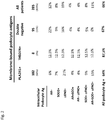
- Three-hundred-thirty-two patients were recruited at the time of diagnosis immediately after having a pathology diagnosis of membranous glomerulonephritis and were followed on clinical ground for at least 12 months; a part had successive serum and urinary controls up to 36 months. Demographic characteristics are reported in Table 2. Criteria for enrollment were (1) a biopsy-based diagnosis of MN, (2) a normal complement profile (3) negative tests for ANA, nDNA, and ANCA and cryoglobulins and the absence of viral markers (hepatitis B surface antigen and HIV).
- Kidney histology was evaluated on Dubosq-Bresil solution-fixed tissues embedded in paraffin, sectioned, and stained with hematoxylin/eosin, Masson trichrome, silver methenamine, and periodic acid-Schiff. Specimens for EM were fixed in Karnovsky solution and processed using standard methods. IgG, IgA, IgM, C1q, C3, and fibrinogen depositions were detected by direct IF staining on frozensections. Blood from the cohort described was obtained the day before renal biopsy and after centrifugation at 1.55 _ g for 15minutes at 4°C, was immediately stored at -80°C. At that time no therapy were started yet.
- Serum was obtained from 50 normal controls recruited at the same institutions that participated to the sample recruitment. They consisted of normal blood donors who had at least one normal urinalysis and normal clinical tests in the prior 6 months (Table 1).
- Antibodies Purified rabbit antiserum against human SOD2 was purchased from Upstate Biotechnologies (Charlottesville, VA); recombinant human Aldose Reductase (H00000231), recombinant human SOD2 (H00002023) and recombinant human ⁇ Enolase were obtained from AbNova (Tapei, Taiwan); anti-human synaptopodin (clone G1D4-IgG1) were obtained from Progen Biotechnik, (Heidelberg, Germany) and mouse mAb anti-human C5b-9 (clone aE11-IgG2a) were from Dako).
- IgG4 Purified mouse mAb to human IgG4 (clone HP6025, IgG1-K) was purchased from Invitrogen (Camarillo, CA).
- IEM donkey anti-goat or anti-rabbit IgG conjugated with 12 nm of colloidal gold particles (Jackson Immunoresearch) was used.
- Anti- PLA2R epitopes The titer of circulating autoantibodies (IgG4) versus versus epitopes of human PLA2r we utilized recombinant and soluble form of CysR, CTLD1 and CTLD6-7 with HA-tag domains containing PLA2R epitopes. CysR and CTLD6-7 were produced in HEK293 cells and CTLD1 in E. Coli. Plates were coated with anti-HA antibody (Sigma-Aldrich) diluted at 1:5000 in 20 mM Tris pH 8.0 (100 ⁇ l/well) at 4°C/overnight or with purified rabbit PLAR1 (100 ng/well) or mouse PLA2R (50 ng/well).
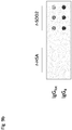
- Anti-AR and Anti-SOD2 and anti - ⁇ enolase IgG4 The titer of circulating anti-SOD2, anti-AR and anti-alphaenolase IgG4 in sera of patients with MN normal control subjects were determined with dot blot utilizing recombinant proteins as fixed antigen for anti-.
- the Dot-Blot essay was done with a Bio-Dot apparatus (Bio-Rad, Hercules, CA, USA) following the instruction manual with minor modifications. All samples and the calibrator serum were diluted with Tris buffered saline (TBS) pH 7.4 (1:100). A calibration curve was prepared by keeping constant the amount of recombinant protein and increasing dilution of specific antibody from 1:500 to 1:32,000. Sera were diluted in the same buffer (1:100) to achieve the desired range of levels, obtained after testing several conditions.
- the nitrocellulose membrane was pre-wetted in TBS and placed on a sheet of Whatman 3 mm filter paper embedded with the same buffer. Constant amounts of protein (300 ng) were placed in the 80 ⁇ l of TBS. After removal of the air bubbles between the two sheets by gentle pressure, the sample template was placed on the nitrocellulose membrane and a vacuum was applied for a few minutes, to fill up the 96 sample wells with 50 mL using a multi-channel pipette. The vacuum was applied until all the samples were adsorbed. The same operation was repeated five times with 150 uL of buffer each, to wash out the non-adsorbed sample.
- the nitrocellulose was then gently removed and saturated with 5% w/v bovine serum albumin (BSA) in TBS. Sera were then left for six hours at room temperature; at the end the membrane was washedsix times in 0.15% v/v Tween-TBS. Incubation with HPR-anti human IgG1-3-4 0.5 ⁇ g/mL in 1% w/v BSA in TBS was performed for two hours room temperature. The membrane was then washed four times, 15 min each, with TBS-T prior to developing the immuno reaction with SuperSignal West Pico Chemiluminescent substrate (Thermo scientific, Rockford, IL, USA).
- BSA bovine serum albumin
- Renal histological sections (3- ⁇ m) were obtained from the original paraffin tissue blocks and used for immunohistochemical analyses.
- a primary antibody anti-Superoxide dismutase 2 (SOD 2, Abcam, rif. ab16956, Cambridge, UK; dilution 1:400) was immunohistochemically tested.
- the histological sections were dewaxed in xylene, rehydrated through graded alcohol, and, after quenching endogenous peroxidase with a 3% H 2 O 2 water solution, incubated with a protein block (Ready to Use DakoCytomation Biotin Blocking System, DakoCytomation California Inc, Carpinteria, CA, USA).
- the slides were incubated for 30 minutes at room temperature, and the reactions were revealed using streptavidin-biotin-peroxidase complex (K0675, LSAB 2 System, DakoCytomation) followed by a solution of 3-3'-diaminobenzidine tetrahydrochloride. In the end, they were counterstained with Mayer hematoxylin, mounted, and coverslipped.
- the negative control procedure consisted of the omission of the primary antibody.
- the Molecular Machines & Industries Cell cut Laser Capture Microdissection system is equipped with a solid-state ultraviolet laser that guarantees precise cutting without damaging the tissue.
- High precision-stage and CCD camera allow identification, documentation, and dissection of multiple regions of interest from the same tissue specimen.
- a total of 25 to 30 glomeruli were microdissected and removed sequentially in separate isolation cap (Nikon Instruments)with special adhesive material in the lid.
- isolation cap Nekon Instruments
- captured tissue was immersed in denaturation buffer and used for proteomic analysis. Sections of human kidney derived from non carcinomatous portions of kidneys removed for renal carcinoma were used as negative control.
- Igs were recovered from glomeruli by means of acid elution as described previously.34 Briefly, after washings with PBS (0.01 M, pH 7.2) groups of 20 glomeruli were incubated overnight with 0.02 M citrate buffer (pH 3.2) at 4°C in a humid atmosphere. After removal of citrate buffer, 0.4 M NaOH was added to achieve a pH of 7.2.
- Odds ratios (OR) and 95% confidence intervals (CI) were tested to determine the association between clinical parameters and protenuria and Fisher's exact test was used to determine the statistically significance.
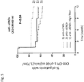
- Kaplan Meier survival curves for remission events were computed according to the level of protenuria (protenuria ⁇ 0.3 gr/die; between 0.3 and 3.5; >3.5). Differences in outcomes were estimated using the log-rank (Mantel-Cox) test. P-values ⁇ 0.05 were considered significant.
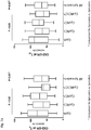
- anti-CysR, anti-C1, anti-C6C7 were also tested with specific ELISA and, on the basis of positiveness , patients were divided in 4 sub-groups: (1) anti-CysR+; (2) antiCysRC1+; (3) anti-CysRC7+ and (4) anti-CysRC1C7+. As expected, circulating levels were very high for the whole panel ( Figure 1 a-f ). 3. Anti-THSD7A. Antibodies against THSD7A were determined by a ELISA ( Figure 1g ), immunofluorescence and then confirmed by western-blot. Only 8 cases matching the three techniques were considered positive ( Figure 2d ); all were negative for anti-hPLA2r.
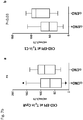
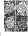
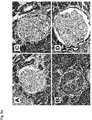
- Renal expression was evaluated by immune-histochemistry ( Figure 3a ) in 6 patients with variable circulating levels of anti-SOD2 antibodies (3 with high and 3 with medium-levels).
- Glomeruli were, in parallel, micro-dissected from biopsies of patients with high circulating anti-SOD2 levels for determining anti-SOD2 IgG4 in micro-eluates.
- SOD2 was undetectable in normal kidneys, comparably expressed in high SOD2 patients and very high in lupus glomeruli.
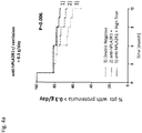
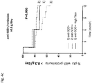
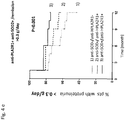
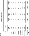
- Anti-SOD2 IgG4 were very high in all the 3 micro-dissected biopsies utilized for analysis ( Figure 3b ). 5-Risk assessment and survival. Proteinuria. Clinical activity of the disease during the follow-up was evaluated on the basis of proteinuria levels in response to treatment, which was defined according to shared guidelines ⁇ Thompson, 2015 #198 ⁇ as complete (proteinuria ⁇ 0.3 gr/day) and partial remission (proteinuria 0.3-3.5 gr/day or reduced by at least 50% from baseline). Persistence of proteinuria >3.5 gr/day was defined as no-remission. The probability of reaching complete or partial remission associated with the presence of antibody in the serum (i.e. Ab+ vs.
- Ab- was calculated in the whole cohort or in subgroups defined on the basis of proteinuria at To (i.e., proteinuria above or below 3.5 gr/day).
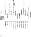
- serum positivity of the following antibodies was associated with the risk of failure to achieve complete remission: anti-hPLA2r (OR 2.87 [95%CI 1.54-5.34]),(OR 2.79 [95%Cl 1.37-5.66]), anti-CysRC1C7 epitopes (2.67 [95%CI 1.03-6.94]) and anti-SOD2 (OR 2.43 [95%CI 1.17-5.04])
- anti-hPLA2r OR 2.87 [95%CI 1.54-5.34]
- anti-CysRC1C7 epitopes (2.67 [95%CI 1.03-6.94]
- anti-SOD2 OR 2.43 [95%CI 1.17-5.04]
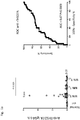
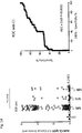
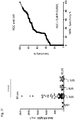
- Kaplan Meier curves were analyzed considering proteinuria as above (complete, partial and no remission) and patients were subdivided in positive or negative to each antibody ( Figure 5a-f ). Analysis relative to the positivity versus anti-PLA2r epitopes was restricted to patients positive for anti-hPLA2r who were, however, the majority. Considering the first outcome (complete remission) patients positive for anti-hPLA2r anti-CysRC6C7 and anti-SOD2 had a worse outcome at T 12 . Combining positivity for anti-hPLA2r and anti-SOD2 gave the best fitting allowing to identify those patients negative to both who presented the best outcome (60% reached remission at T 12 ).
Landscapes
- Health & Medical Sciences (AREA)
- Life Sciences & Earth Sciences (AREA)
- Immunology (AREA)
- Hematology (AREA)
- Engineering & Computer Science (AREA)
- Urology & Nephrology (AREA)
- Molecular Biology (AREA)
- Biomedical Technology (AREA)
- Chemical & Material Sciences (AREA)
- Food Science & Technology (AREA)
- Physics & Mathematics (AREA)
- Cell Biology (AREA)
- Rehabilitation Therapy (AREA)
- Biotechnology (AREA)
- Rheumatology (AREA)
- Medicinal Chemistry (AREA)
- Microbiology (AREA)
- Analytical Chemistry (AREA)
- Biochemistry (AREA)
- General Health & Medical Sciences (AREA)
- General Physics & Mathematics (AREA)
- Pathology (AREA)
- Measuring Or Testing Involving Enzymes Or Micro-Organisms (AREA)
- Peptides Or Proteins (AREA)
Applications Claiming Priority (1)
| Application Number | Priority Date | Filing Date | Title |
|---|---|---|---|
| IT102017000025190A IT201700025190A1 (it) | 2017-03-07 | 2017-03-07 | Metodo in vitro e kit per predire l’andamento clinico e la sensibilita’ ai farmaci nei pazienti con nefropatia membranosa |
Publications (1)
| Publication Number | Publication Date |
|---|---|
| EP3373008A1 true EP3373008A1 (de) | 2018-09-12 |
Family
ID=59337790
Family Applications (1)
| Application Number | Title | Priority Date | Filing Date |
|---|---|---|---|
| EP18159989.5A Withdrawn EP3373008A1 (de) | 2017-03-07 | 2018-03-05 | In-vitro-verfahren und kit zur vorhersage der klinischen resultate in patienten mit membranöser nephropathie |
Country Status (2)
| Country | Link |
|---|---|
| EP (1) | EP3373008A1 (de) |
| IT (1) | IT201700025190A1 (de) |
Cited By (2)
| Publication number | Priority date | Publication date | Assignee | Title |
|---|---|---|---|---|
| CN113899908A (zh) * | 2021-09-22 | 2022-01-07 | 深圳临研医学有限公司 | 磷酸化蛋白质在膜性肾病中的应用 |
| WO2023185957A1 (zh) * | 2022-03-29 | 2023-10-05 | 成都地奥制药集团有限公司 | 抗体、融合蛋白及其用途 |
Citations (2)
| Publication number | Priority date | Publication date | Assignee | Title |
|---|---|---|---|---|
| EP2977758A1 (de) * | 2014-07-24 | 2016-01-27 | Université De Nice Sophia Antipolis | Verfahren und Kits zur Überwachung membranöser Nephropathie |
| WO2017009245A1 (en) * | 2015-07-10 | 2017-01-19 | Chu De Nice | Prognosis and monitoring of membranous nephropathy based on the analysis of pla2r1 epitope profile and spreading |
-
2017
- 2017-03-07 IT IT102017000025190A patent/IT201700025190A1/it unknown
-
2018
- 2018-03-05 EP EP18159989.5A patent/EP3373008A1/de not_active Withdrawn
Patent Citations (2)
| Publication number | Priority date | Publication date | Assignee | Title |
|---|---|---|---|---|
| EP2977758A1 (de) * | 2014-07-24 | 2016-01-27 | Université De Nice Sophia Antipolis | Verfahren und Kits zur Überwachung membranöser Nephropathie |
| WO2017009245A1 (en) * | 2015-07-10 | 2017-01-19 | Chu De Nice | Prognosis and monitoring of membranous nephropathy based on the analysis of pla2r1 epitope profile and spreading |
Non-Patent Citations (3)
| Title |
|---|
| BRUSCHI MAURIZIO ET AL: "Glomerular Autoimmune Multicomponents of Human Lupus Nephritis In Vivo (2): Planted Antigens", JOURNAL OF THE AMERICAN SOCIETY OF NEPHROLOGY, WILLIAMS AND WILKINS, BALTIMORE, MD, US, vol. 26, no. 8, 1 August 2015 (2015-08-01), pages 1905 - 1924, XP009500345, ISSN: 1046-6673, DOI: 10.1681/ASN.2014050493 * |
| C. MURTAS ET AL: "Coexistence of Different Circulating Anti-Podocyte Antibodies in Membranous Nephropathy", CLINICAL JOURNAL OF THE AMERICAN SOCIETY OF NEPHROLOGY, vol. 7, no. 9, 5 July 2012 (2012-07-05), pages 1394 - 1400, XP055409223, ISSN: 1555-9041, DOI: 10.2215/CJN.02170312 * |
| MAURIZIO BRUSCHI ET AL: "Direct characterization of target podocyte antigens and auto-antibodies in human membranous glomerulonephritis: Alfa-enolase and borderline antigens", JOURNAL OF PROTEOMICS, ELSEVIER, AMSTERDAM, NL, vol. 74, no. 10, 10 May 2011 (2011-05-10), pages 2008 - 2017, XP028272518, ISSN: 1874-3919, [retrieved on 20110519], DOI: 10.1016/J.JPROT.2011.05.021 * |
Cited By (3)
| Publication number | Priority date | Publication date | Assignee | Title |
|---|---|---|---|---|
| CN113899908A (zh) * | 2021-09-22 | 2022-01-07 | 深圳临研医学有限公司 | 磷酸化蛋白质在膜性肾病中的应用 |
| CN113899908B (zh) * | 2021-09-22 | 2024-05-28 | 深圳临研医学有限公司 | 磷酸化蛋白质在膜性肾病中的应用 |
| WO2023185957A1 (zh) * | 2022-03-29 | 2023-10-05 | 成都地奥制药集团有限公司 | 抗体、融合蛋白及其用途 |
Also Published As
| Publication number | Publication date |
|---|---|
| IT201700025190A1 (it) | 2018-09-07 |
Similar Documents
| Publication | Publication Date | Title |
|---|---|---|
| Kattah et al. | Anti-phospholipase A2 receptor antibodies in recurrent membranous nephropathy | |
| US9134326B2 (en) | Biomarkers for liver fibrosis | |
| Dou et al. | The accuracy of the anti-phospholipase A2 receptor antibody in the diagnosis of idiopathic membranous nephropathy: a comparison of different cutoff values as measured by the ELISA method | |
| Kohei et al. | Neutrophil gelatinase-associated lipocalin is a sensitive biomarker for the early diagnosis of acute rejection after living-donor kidney transplantation | |
| Yonglitthipagon et al. | Prognostic significance of peroxiredoxin 1 and ezrin-radixin-moesin–binding phosphoprotein 50 in cholangiocarcinoma | |
| NO20110895A1 (no) | Kompetitive S100A9 immunoanalyser | |
| JP2009020049A (ja) | 脳血管疾患の診断方法 | |
| US9494590B2 (en) | Methods of renal cancer detection | |
| Chebotareva et al. | A pilot study of anti‐nephrin antibodies in podocytopaties among adults | |
| CN110716050A (zh) | 抗原组合在制备用于肺癌相关自身抗体检测的试剂盒中的用途以及相应的试剂盒和检测方法 | |
| Mutavhatsindi et al. | Identification of novel salivary candidate protein biomarkers for tuberculosis diagnosis: a preliminary biomarker discovery study | |
| US20120107295A1 (en) | Methods and Compositions for Detecting Pancreatic Disease | |
| Xiang et al. | Establishment of heparin-binding protein time-resolved immunoassay and some potential clinical applications | |
| JP2024507580A (ja) | 間質性肺疾患の処置のためのバイオマーカー | |
| EA034364B1 (ru) | Иммуноанализ для обнаружения хромогранина а | |
| Zhang et al. | Target antigens of membranous nephropathy with syphilis infection | |
| EP3373008A1 (de) | In-vitro-verfahren und kit zur vorhersage der klinischen resultate in patienten mit membranöser nephropathie | |
| Ambe et al. | Improving the preoperative diagnostic accuracy of acute appendicitis. can fecal calprotectin be helpful? | |
| Xia et al. | Clinicopathological phenotype and outcomes of NCAM-1+ membranous lupus nephritis | |
| JP5280214B2 (ja) | 炎症性腸疾患の診断方法 | |
| Infantino et al. | Positive tissue transglutaminase antibodies with negative endomysial antibodies: Unresolved issues in diagnosing celiac disease | |
| Xiang et al. | Development and application of amplified luminescent proximity homogeneous assay for quantitation of heparin-binding protein | |
| Wu et al. | Serum Soluble CD89‐IgA Complexes Are Elevated in IgA Nephropathy without Immunosuppressant History | |
| CN109891241B (zh) | 能够进行糖尿病性肾病的早期病情的特异性诊断的检查方法 | |
| KR102235718B1 (ko) | 방광암 진단 또는 예후 분석용 바이오마커 조성물, 키트 및 이를 이용한 진단 방법 |
Legal Events
| Date | Code | Title | Description |
|---|---|---|---|
| PUAI | Public reference made under article 153(3) epc to a published international application that has entered the european phase |
Free format text: ORIGINAL CODE: 0009012 |
|
| AK | Designated contracting states |
Kind code of ref document: A1 Designated state(s): AL AT BE BG CH CY CZ DE DK EE ES FI FR GB GR HR HU IE IS IT LI LT LU LV MC MK MT NL NO PL PT RO RS SE SI SK SM TR |
|
| AX | Request for extension of the european patent |
Extension state: BA ME |
|
| STAA | Information on the status of an ep patent application or granted ep patent |
Free format text: STATUS: THE APPLICATION IS DEEMED TO BE WITHDRAWN |
|
| 18D | Application deemed to be withdrawn |
Effective date: 20190313 |