EP3376466A1 - Reduction de bruit dans une image volumetrique d'un objet - Google Patents
Reduction de bruit dans une image volumetrique d'un objet Download PDFInfo
- Publication number
- EP3376466A1 EP3376466A1 EP17160756.7A EP17160756A EP3376466A1 EP 3376466 A1 EP3376466 A1 EP 3376466A1 EP 17160756 A EP17160756 A EP 17160756A EP 3376466 A1 EP3376466 A1 EP 3376466A1
- Authority
- EP
- European Patent Office
- Prior art keywords
- volume image
- volume
- image data
- data set
- subsets
- Prior art date
- Legal status (The legal status is an assumption and is not a legal conclusion. Google has not performed a legal analysis and makes no representation as to the accuracy of the status listed.)
- Ceased
Links
Images




Classifications
-
- G—PHYSICS
- G06—COMPUTING OR CALCULATING; COUNTING
- G06T—IMAGE DATA PROCESSING OR GENERATION, IN GENERAL
- G06T5/00—Image enhancement or restoration
- G06T5/70—Denoising; Smoothing
-
- G—PHYSICS
- G06—COMPUTING OR CALCULATING; COUNTING
- G06T—IMAGE DATA PROCESSING OR GENERATION, IN GENERAL
- G06T5/00—Image enhancement or restoration
- G06T5/77—Retouching; Inpainting; Scratch removal
-
- G—PHYSICS
- G06—COMPUTING OR CALCULATING; COUNTING
- G06T—IMAGE DATA PROCESSING OR GENERATION, IN GENERAL
- G06T2207/00—Indexing scheme for image analysis or image enhancement
- G06T2207/10—Image acquisition modality
- G06T2207/10072—Tomographic images
-
- G—PHYSICS
- G06—COMPUTING OR CALCULATING; COUNTING
- G06T—IMAGE DATA PROCESSING OR GENERATION, IN GENERAL
- G06T2207/00—Indexing scheme for image analysis or image enhancement
- G06T2207/20—Special algorithmic details
- G06T2207/20021—Dividing image into blocks, subimages or windows
Definitions
- the invention relates to a method and a de-entrainment device for denoising a volume image data set of an object, in particular with the aid of a medical imaging system.
- the invention also relates to a control device for a medical imaging system or a medical imaging system with such a denoising device.
- the invention relates to a method for creating a base element directory, which can be used for such denoising a volume image data set of an object.
- Imaging systems such as computed tomography, magnetic resonance systems, etc., today play an important role in the medical field.
- the images of the internal organs and structures of the patient produced by the imaging systems are used for the diagnosis of causes of disease, for the planning and execution of operations or for the preparation of therapeutic measures.
- the new generation of computed tomography systems, as well as magnetic resonance systems allow large-volume examinations, which can be a maximum of full-body examinations.
- exposing the patient influences that may pose a burden on the human body depending on the dosage.
- examples include ionizing electromagnetic radiation (eg X-radiation) or particle radiation (eg beta radiation). It is desirable that the necessary radiation exposure for a Image capture for the human body is as low as possible.
- Disadvantage of the prior art is that is measured either with a non-optimal radiation load or a non-optimal noise ratio.
- volume image data record to be de-noiseed is first of all provided.
- Such volume image data sets generally comprise volume image data of a mostly three-dimensional object.
- a computed tomography scan or a magnetic resonance imaging of a human body or its parts may be considered as such a volumetric image data set.
- the volume image data set can also have been created and / or stored in a different form, for example as a data record in the form of a stack of two-dimensional images or as a data set which contains the values for the pixels or voxels of the acquired volume (and their spatial assignment). contains in another way.
- the third dimension of the volume image data record can be a third direction in spatial space.
- At least one dimension can also be a time, for.
- the term "spatial directions", which will be used later, can also encompass a temporal dimension.
- Most applications, however, are directions in space.
- a time component is taken into account, for example a four-dimensional volume image data set, which results from a plurality of spatially consecutively acquired spatial volume image data records.
- the pixels are usually referred to as "pixels” and in at least three-dimensional volume image data, usually as "voxels".
- the term "object” refers to all possible three-dimensional objects, whereby also parts of objects can be called “object”.
- the method according to the invention is suitable for improving recordings of a complete body, but also for improving pictures of a body part or an organ.
- the volume image data need not necessarily come from a human body, but z.
- plants As well as animals or plants, mummified objects or other objects, such as technical items to be tested or the like, created.
- a base element directory is provided.
- Such a base element directory is also often referred to by the person skilled in the German language with the originally English word "dictionary” and is generally a data record which comprises a predetermined set of volume basic elements.
- These volume primitives are also referred to as “primitives” or “primitives” and may represent characteristic primitives of an image. Examples of a volume base element are a cuboid, a sphere, a cube, a cylinder, a cone or a torus.
- volume base elements if they are at least three-dimensional, with one dimension also a time component can be. In particular, higher dimensions than three are possible if, for example, a time component is taken into account.
- the volume base elements are usually stored in raster graphics or the like as groups of pixels with their values. In the following, these at least three-dimensional volume basic elements are also sometimes referred to simply as "basic elements”.
- volume image subsets in English, two-dimensional image blocks are usually called “patch”, for which reason this term is also used as a shorter synonym for a volume image subset), which can also be referred to as "image regions”.
- image regions can also be referred to as "image regions”.
- the volume image data set can be divided into subsets, or the volume image data set can be examined by screening with a mask of a picture element.
- volume image subsets may then be replicated using a linear combination of volume primitives of the primitive dictionary.
- this step could also be described as a kind of "reconstruction” of the corresponding image area with picture elements of the dictionary or as a kind of "decomposition” of the content of the corresponding image area into a linear combination of basic elements.
- This step can be carried out in a numerical optimization method by means of suitable software. Possibilities of such optimization will be described in more detail below.
- such a simulation is carried out at least for a part of the volume image subsets of the volume image data set. Although it is generally preferred that this happens for all volume image subsets, however It may also be advantageous, depending on the case of the application, not to replicate certain subsets, because they z. B. should contain no relevant information to remain unchanged due to a targeted closer examination or because they should not be replicated for other reasons.
- the desired noise-reduced volume image data set is then generated using the linear combination of basic elements of the volume image subsets.
- the noise-reduced volume image data set can be generated by a combination of the replicas of the volume image subsets generated by the linear combination of basic elements.
- a base element dictionary comprising a set of volume primitives is required for performing the denoudation method.
- This base element directory can have been previously created at any time and stored in a memory for later use for denoising various image data.
- a training volume image data record is provided.
- Such a training volume image data set should preferably correspond to the volume image data set to be noise-depleted, i. H. care is taken to ensure that the type of training volume image data set, for example the type of training of the training volume image data set or the recorded object types, is similar to one another.
- volume image data should preferably be used for the training volume image data set.
- the technical nature of the image recording method should be comparable. If recordings are to be made with a device of a certain category, the device with which the image data for the training volume image data record was created could preferably be of the same category, possibly even the same device. If, for example, volume image data of a computer tomograph are to be emptied, the image recordings for the training volume image data record should preferably also originate from computer tomographs. This has the advantage that specific technical features of a recording device can also be found in the training volume image data record.
- a training volume image data set is selected whose image quality is better than that of the volume image data to be de-noiseed, at least in such a way that it has a lower noise than the aforementioned volume image data set intended for denoudation.
- the training volume image data set may have previously been recorded for other medical reasons with a higher radiation dose.
- the object which serves as the basis of the training volume image data record is not identical to the object whose volume image data is denouched. So it is rare in a planned gentle examination of a patient that have previously been made by him recordings with a higher radiation dose. However, then z. B. recordings of other patients, which from others For medical reasons with a higher radiation dose and better image quality have been recorded, are used to create the training volume image data set, it being assumed that the images of human bodies are similar to some individual characteristics by and large.
- image data from such organs can also serve as a basis for the training volume image data set.
- training volume image datasets corresponding to other substances and / or objects could also be used to create special dictionaries for other substances and / or objects.
- the volume image data set of the object to be de-noiseed is based, as mentioned, on measurement data of an imaging medical-technical imaging system, i. H. for example, it was reconstructed from raw data measured with this imaging system.
- the training volume image data set may also include volume image data of a plurality of corresponding objects.
- the use of the volume mappings of several corresponding but not necessarily identical objects in the training volume image data set has the advantage of statistically improving the results.
- the training volume image dataset comprises alternatively or in addition to images of real objects also simulated or calculated volume image data of corresponding objects. In this case it is an advantage if they are simulated / calculated as if they came from a corresponding device.
- volume image subsets of the training volume image data record are formed. This can be done in the same way as described above in the course of the denoudation process.
- volume image subsets Thereafter, basic elements in the volume image subsets are determined and a base element directory is formed from the ascertained basic elements of the volume image subsets. In particular, this is done according to rules for machine learning. A preferred method will be explained later.
- a de-inking device (which could also be termed an image processing device) for denoising a volume image data set, e.g.
- the volume image data of a medical imaging system or an imaging system uses the inventive method for denoising and preferably uses a created with the relevant inventive method base element directory.
- This interface comprises at least one interface by means of which a volume image data set can be provided to the de-inking device.
- This interface may be physically present, such as B. a bus system or an interface for data transmission, in particular USB, LAN, WLAN, or it may be present on the software side and, for. represent a protocol structure.
- the de-inking device further comprises a storage unit containing a base element directory is, which has been created in particular as described above.
- this base element directory can be stored in a database, but also as a file on a memory element or in RAM.
- This primitive directory includes a number of primitives to provide for denoising. This can z. B. primitives, as described above.
- the Entrauschungsvoroplasty invention also includes a Entrauschungsaku according to the invention, which is adapted to form volume image subsets of the volume image data set to replicate at least one volume image subset by means of a linear combination of basic elements of the base element directory and to generate a noise-reduced volume image data set using this linear combination.
- a Entrauschungsaku according to the invention, which is adapted to form volume image subsets of the volume image data set to replicate at least one volume image subset by means of a linear combination of basic elements of the base element directory and to generate a noise-reduced volume image data set using this linear combination.
- the Entrauschungsvoroplasty or their Entrauschungsaku is thus designed to perform as mentioned above, the Entrauschung according to the inventive method.
- the Entrauschungsvoroplasty invention further comprises another interface for passing the denoilled volume image data set.
- another interface for passing the denoilled volume image data set.
- it can also be used for that interface over which the Entrauschungsvoroplasty has received their data, if they can work bidirectionally.
- the Entrauschungsvoroplasty invention may be present as a separate device, by means of which in particular any three-dimensional data can be flushed out, z. B. as a standalone computer or arithmetic unit.
- the Entrauschungsvoroplasty invention also part, physical part or software function, a higher-level device be, z. B. as part of a workstation at a diagnostic station at which a surveyor can edit, view and evaluate the volume image data, or as an integrated part of a control device of a medical imaging system.
- An inventive control device for controlling a medical imaging system is therefore equipped with a Entrauschungsvoroplasty invention.
- a medical imaging system according to the invention preferably a computed tomography system
- a denoising device according to the invention for example by being integrated into the control device or other components of the imaging system.
- a largely software implementation has the advantage that even already existing z.
- two-dimensional Entrauschungsvortechniken or control devices can be retrofitted in a simple way by a software update to work on the inventive way.
- a corresponding computer program product with a computer program which can be loaded directly into a memory device of a noise reduction device or control device of a computer tomography system or as an executable file in a computer system, with program sections in order to carry out all the steps of the relevant method according to the invention.
- Such a computer program product In addition to the computer program, if necessary, additional components such.
- additional components including hardware components, such as hardware keys (dongles, etc.) for using the software.
- a computer-readable medium for example a memory stick, a hard disk or another portable or permanently installed data carrier can be used for transporting to the de-entrainment device or for the control device and / or for storage on or in the control device, on which device the readable and executable by a computer unit of the control device Program sections of the computer program are stored.
- the computer unit can, for. B. for this purpose have one or more cooperating microprocessors or the like.
- the base element directory may have been previously created at any time.
- it may be provided by a provider of the medical imaging system.
- the invention therefore also encompasses a computer-readable medium on which there is at least one base element directory which can be read and executed by a computer unit for denoising a volume image data record.
- it can have corresponding volume base elements which are suitable for denoising such volume image data sets.
- This base element directory has been created in particular according to the inventive method for creating a base element directory.
- the volume image subsets are selected so that adjacent volume image subsets overlap in at least one spatial direction, preferably in at least two spatial directions, more preferably in at least three spatial directions. This has the advantage that in the noisy image in particular those artifacts resulting from transitions between two pixels are minimized.
- a predominant part of the volume picture subsets ie more than 50% of the volume picture subsets, preferably more than 80% of the volume picture subsets, more preferably all volume picture subsets have the same size. If the volume image subsets are in the form of raster graphics, the volume image subsets have, in particular, the same amount of volume image data or, preferably, the same shape. This has the advantage that a consistent simulation of different volume image subsets is possible.
- the volume image subsets are many times smaller than the volume image data set, preferably less than 1/10, in particular less than 1/100 of the volume data set. If the volume image subsets are present in a raster graphic, they preferably comprise at least two pixels in all three spatial directions and / or preferably a maximum of 15 pixels in all three spatial directions.
- weighting factors are assigned to the volume base elements of a linear combination for simulating a volume image subset.
- L a 1 P 1 + a 2 P 2 + ...
- z. B the generation of the noise-reduced volume image data set in dependence on the value of these weighting factors a i and / or the maximum number of basic elements of the individual linear combinations are made.
- an optimization function can preferably be used for simulating a volume image subset by a linear combination, wherein a goal of the optimization function is the optimization of a similarity of the linear combinations with the volume image subset.
- a similarity is indicated quantitatively in an optimization function by means of a defined distance function or a standard representing the similarity.
- the optimization function in the form of a cost function to be minimized, which, for example, in a particularly simple case, may look like this: min ⁇ ⁇ I - L ⁇ 2 2 ⁇
- Another option that may be used additionally or alternatively would be to select a maximum number of basic elements, e.g. B. according to a predetermined maximum value, from the linear combination, wherein the base elements are selected with the highest weighting factors and the other basic elements are discarded. This would result in noise-reduced linear combinations that do not have more basic elements than the maximum value specifies.
- a maximum number of basic elements e.g. B. according to a predetermined maximum value
- Another, alternatively or additionally applicable, preferred method is based on the use of a penalty term in the optimization function, in which the number of basic elements in the linear combination is received.
- a penalty term in which the number of basic elements in the linear combination is received.
- the preferred penalty terms are designed so that an undesirable condition enters the optimization function in a manner that runs counter to the optimization.
- N is the number of basic elements of a linear combination L.
- the number of basic elements of the respective linear combination N is therefore provided here with a weighting factor c squares as a penalty in the optimization function.
- c weighting factor
- each volume image subset I was reconstructed by itself into an optimal linear combination L or by a linear combination L.
- This linear combination L is thus a "noisy volume image subset".
- These entrained volume image subsets or linear combinations L then only have to be combined to form a denoised volume image data set. This can be done by reversing the depleted volume image subsets in the reverse manner as the formation of the volume image subsets from the original volume image data set, d. H. by re-applying the rules used (re) assembled.
- the optimization function could preferably be selected such that simultaneously multiple linear combinations are optimized for different volume image subsets.
- the optimization function preferably comprises a further term, in particular in the form of a possibly weighted, penalty term, by means of which a similarity of a combination of the linear combinations taking into account in the optimization function is taken into account.
- a simultaneous optimization of the linear combinations for all patches (and thus their simultaneous denoising) of a larger volume image area, particularly preferably for the complete volume image data set can take place, with the similarity of the reconstructed therefrom being denuded Volume image data set is taken into account with the original volume image data set in the optimization.
- An example of such an optimization function could be defined for a complete volume image data set X as follows: min ⁇ d ⁇ X - Y ⁇ 2 2 + e ⁇ ⁇ PK ⁇ 2 2 ⁇
- PK is the optimization function for a single patch, here z. B. according to equation (3). But it could also be another optimization feature for a single patch can be used, for.
- the simple function of equation (2) can be used.
- Y is the depleted volume image data set which can be composed from the current linear combinations used in function PK as already described above. To denigrate only a part of the volume image data set, only the image data sets X and Y would have to be selected according to this part.
- d and f are selectable weighting factors to determine which aspect more strongly influences the optimization, the "local similarity" of the patches with the linear combinations, or the "global similarity" of the depleted volume image data to the original volume image data. The optimum values of these weighting factors can be found by tests.
- the creation of a base element directory can be done in a similar manner to the noise removal described above, except that the aforementioned training volume image data sets are used for this purpose.
- a replication of at least a part of the volume image subsets of the training volume image data records can also be carried out by a linear combination of basic elements for the creation of the base element directory, wherein, inter alia, the same or the same optimization functions can be used.
- a decision as to whether a base element is taken over into the base element directory can then be made as a function of the number of times a base element has been used to emulate volume image subsets of a training volume image data set.
- initial start base element directory initial dictionary
- initial dictionary initial start base element directory
- initial dictionary initial dictionary
- the imaging system or the imaging medical technology system 1 is a computed tomography system 1.
- the method can also be used with regard to other imaging systems.
- FIG. 1 shows a schematic representation of a volume image data set X, for example a combination of several sectional images or a picture stack of a computed tomography system 1.
- the object O shown here could for example be an area of the torso of a human.
- the individual slice images could comprise 512x512 pixels and, for example, 40 layers could be stacked on top of each other, just to give a concrete example. But of course, all other sizes are possible.
- the slice images are superimposed in the volume image data set X and the pixels become voxels V in the volume image data set X.
- the distance of the slice images from one another is selected such that the voxels approximate cube-shaped, which is not mandatory.
- a spatial direction could also represent a temporal dimension.
- groups of voxels of the volume image data set X are assigned volume image subsets I (ie, the patches I). This can be done, for example, by copying the respective voxels into individual memory areas or by tracing the volume image data set with a (three-dimensional) mask.
- volume image subsets I for example, 3 x 3 x 3 voxels available, as shown schematically by the arrow in the figure.
- These volume image subsets I preferably overlap one another in all spatial directions, for example, in each case around a voxel.
- a volume image subset I contains z.
- B an array of voxels V with different color values. In computed tomography scans, these are usually simple shades of gray.
- an attempt is made to emulate this arrangement by constructing the volume image subset of a plurality of volume basic elements P 1 -P N , so-called primitives.
- the influence of the individual volume basic elements P 1 , P 2 ,..., P N on the replica is represented by weighting factors a 1 , a 2 ,..., A N.
- volume base elements P 1 , P 2 ,..., P N merely for the sake of clarity.
- the volume image subset I which here shows a cylindrical structure and several points, is subdivided into basic elements P 1 , P 2 ,..., P N , the first base element P 1 here having a cylinder and the remaining base elements P 2 ,..., P N points at different positions included.
- a weighting could, for example, be reproduced by varying the transparency or the gray tones of these picture elements.
- the totality of the selected basic elements P 1 , P 2 ,..., P N then yields the linear combination L, from which the noise-reduced volume image data record Y is created in the further method.
- FIG. 3 shows an exemplary base element directory D.
- the points between two basic elements P are intended to make clear that further basic elements P of this type are contained in the base element directory D.
- the volume base elements of the base element directory D are constructed in a more abstract manner, in particular since this is usually also a matter of pixel or raster graphics. For example, as described above, it may have been developed in a training method from a modified redundant DCT Dictionary or other suitable initial dictionary.
- FIG. 4 now shows a flowchart for a possible flow of a method according to the invention for creating a base element directory.
- a volume image data set is provided as a training volume image data set X '.
- This training volume image data set X ' originates, for example, from computed tomography images which have been recorded in other patients, in particular for medical reasons, with a higher radiation dose and therefore have only low noise.
- the training volume image data set X ' is subdivided into volume data subsets I, which in particular overlap one another, so that the probability is reduced that relevant basic elements P are cut by the decomposition.
- the training determines the basic elements P, which are then included in the base element directory D.
- FIG. 5 shows a flowchart for a possible flow of a method according to the invention for denoising.
- a volume image data record X is made available.
- This volume image data set X originates, for example, from computed tomography images taken in a patient with a comparatively low radiation dose and consequently having a relatively high level of noise.
- volume image data set X ' is subdivided into volume data subsets I, which likewise preferably overlap one another.
- the de-noise is based on a selection of basic elements P from a base element directory D performed. This can preferably take place by one of the methods already explained above, for example by means of equations (2) to (4).
- step IV.2 linear combinations of the basic elements P of the base element directory D are first formed in step IV.2.
- the image information of the linear combination L is then compared in step V.2 with the respective volume image subset I, and then little relevant base elements are removed from the linear combination according to an optimization function.
- steps IV.2 and V.2 are repeated or recursively carried out for this purpose so that ultimately all the desired regions of the volume image data record can be emptied.
- optimization functions as presented in equations (2) or (3) above, can be used here.
- step VI.2 an insouciant image is assembled from the individual linear combinations L, with the linear combinations taking the place of the volume image subsets.
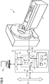
- FIG. 6 shows roughly schematically a computer tomography system 1 ("CT system") with a control device 10 for carrying out the method according to the invention.
- the computer tomography system 1 comprises in a conventional manner a scanner 2 with a gantry in which an x-ray source 3 rotates, which in each case irradiates a patient who is pushed by means of a couch 5 into a measuring space of the gantry, so that the radiation onto one of the x-ray source 3 each opposite detector 4 meets.
- CT system computer tomography system
- the computer tomography system 1 comprises in a conventional manner a scanner 2 with a gantry in which an x-ray source 3 rotates, which in each case irradiates a patient who is pushed by means of a couch 5 into a measuring space of the gantry, so that the radiation onto one of the x-ray source 3 each opposite detector 4 meets.
- FIG. 7 just an example of a CT and the Invention can also be used on any CT designs, for example,
- control device 10 substantially. In principle, such CT systems and associated control devices and the measuring method are known to the person skilled in the art and therefore need not be explained in detail.
- a core component of the control device 10 here is a processor 11, on which various components in the form of software modules are realized.
- the control device 10 further has a terminal interface 14, to which a terminal 20 is connected, via which an operator can operate the control device 10 and thus the computer tomography system 1.
- Another interface 15 is a network interface for connection to a data bus 21 so as to connect to a RIS or PACS, respectively.
- generated image data can be transmitted via this bus 21 before or after denoising to other units, such as diagnostic stations, mass storage (not shown) or the like.
- the scanner 2 can be controlled by the control device 10, that is, z.
- the control device 10 For example, the rotation speed of the gantry, the displacement of the patient couch 5 and the X-ray source 3 itself are controlled to perform a measurement.
- the raw data RD generated thereby are read out of the detector 4.
- the control device 10 has a memory unit 16 in which, inter alia, various measurement protocols MP are stored.
- This measuring control unit 17 controls the scanner 2 via the control interface 13 on the basis of one or more selected measurement protocols MP, which were possibly modified by the operator via the terminal 20, in order to carry out a measurement and to acquire data.
- Another component on the processor 11 is an image data reconstruction unit 18, with which from the data obtained via the data acquisition interface 12 raw data RD, the desired image data, here z. B. to be rooted volume image data X, to be reconstructed.
- a Entrauschungsvoroplasty 19 according to the invention is additionally realized here, which can receive to be rooted volume image data X from the image data reconstruction unit 18, as described above denousted and the entrauschten volume image data Y again makes available.
- this Entrauschungsvoroplasty 19 does not necessarily have to be directly connected to the control device 10 or part of this. It can also be used as an independent device z. B. part of the network realized by means of the data bus 21 or in another unit, such as said diagnostic station or a workstation, be integrated. In this case, for example, to be entrauschenden volume image data X via the data bus 21 to the Entrauschungsvortechnik be sent and the de-noise occurs z. B. in the diagnostic station. Furthermore, the Entrauschungsvortechnisch 19 could also be integrated in the terminal 20 or even be part of the image data reconstruction unit 18.
- the Entrauschungsvoroplasty 19 is in FIG. 7 shown in more detail.
- receives volume image data X it comprises an interface 22.
- This interface 22 can, for example, take over the volume image data X provided by the image data reconstruction unit 18.
- the Entrauschungsvortechnisch 19 includes a memory unit 24, in which the base element directory D is present.
- the base element directory D could also be contained in the memory unit 16, in which case the noise removal device 19 could advantageously access the memory unit 16 directly.
- the denoised data can be output via an interface 24 to other systems for viewing or further processing.
- the denoised data could also be output via the first-mentioned interface 22 if it operates bidirectionally.
- the de-inking device 19 is preferably designed such that not only de-icing is carried out with it, but in particular a creation or modification or supplementation of the base element directory D can be performed.
- volume image data can be produced with a far smaller radiation dose than with conventional systems, since the quality can be considerably improved by the de-noise without losing relevant information content in the volume image data X.
- existing volume image data X can also be edited and thus improved.
- a volume image data record can then be created from the layer stack as described above, or the layer stack is treated like a volume image data record. Then, the three-dimensional denoudation according to the invention takes place and then the two-dimensional sectional images are again viewed from the noise-removed volume image data set.
- a layer stack of three sectional images is sufficient to achieve a significant improvement.
- FIG. 8 shows a sectional view of a layer stack of sectional images across the abdominal cavity of a patient.
- the sectional image was generated by means of a computed tomography system at a relatively low dose and is therefore quite noisy.
- FIG. 9 shows for comparison the picture after a de-noise with the inventive method.
Landscapes
- Physics & Mathematics (AREA)
- General Physics & Mathematics (AREA)
- Engineering & Computer Science (AREA)
- Theoretical Computer Science (AREA)
- Apparatus For Radiation Diagnosis (AREA)
- Image Processing (AREA)
Priority Applications (1)
| Application Number | Priority Date | Filing Date | Title |
|---|---|---|---|
| EP17160756.7A EP3376466A1 (fr) | 2017-03-14 | 2017-03-14 | Reduction de bruit dans une image volumetrique d'un objet |
Applications Claiming Priority (1)
| Application Number | Priority Date | Filing Date | Title |
|---|---|---|---|
| EP17160756.7A EP3376466A1 (fr) | 2017-03-14 | 2017-03-14 | Reduction de bruit dans une image volumetrique d'un objet |
Publications (1)
| Publication Number | Publication Date |
|---|---|
| EP3376466A1 true EP3376466A1 (fr) | 2018-09-19 |
Family
ID=58448294
Family Applications (1)
| Application Number | Title | Priority Date | Filing Date |
|---|---|---|---|
| EP17160756.7A Ceased EP3376466A1 (fr) | 2017-03-14 | 2017-03-14 | Reduction de bruit dans une image volumetrique d'un objet |
Country Status (1)
| Country | Link |
|---|---|
| EP (1) | EP3376466A1 (fr) |
Cited By (1)
| Publication number | Priority date | Publication date | Assignee | Title |
|---|---|---|---|---|
| CN115471477A (zh) * | 2022-09-16 | 2022-12-13 | 先临三维科技股份有限公司 | 扫描数据的去噪方法、扫描方法、装置、设备和介质 |
Citations (2)
| Publication number | Priority date | Publication date | Assignee | Title |
|---|---|---|---|---|
| US8600137B2 (en) | 2010-06-01 | 2013-12-03 | Siemens Aktiengesellschaft | Iterative CT image reconstruction with a four-dimensional noise filter |
| US8605977B2 (en) | 2009-09-03 | 2013-12-10 | Siemens Aktiengesellschaft | Iterative CT image filter for noise reduction |
-
2017
- 2017-03-14 EP EP17160756.7A patent/EP3376466A1/fr not_active Ceased
Patent Citations (2)
| Publication number | Priority date | Publication date | Assignee | Title |
|---|---|---|---|---|
| US8605977B2 (en) | 2009-09-03 | 2013-12-10 | Siemens Aktiengesellschaft | Iterative CT image filter for noise reduction |
| US8600137B2 (en) | 2010-06-01 | 2013-12-03 | Siemens Aktiengesellschaft | Iterative CT image reconstruction with a four-dimensional noise filter |
Non-Patent Citations (3)
| Title |
|---|
| M. ELAD, M. AHARON: "Image Denoising Via Sparse and Redundant Representations Over Learned Dictionaries", IEEE TRANSACTIONS ON IMAGE PROCESSING, vol. 15, no. 12, December 2006 (2006-12-01), pages 3736 - 3745, XP011149944 * |
| M. ELAD; M. AHARON: "Image Denoising Via Learned Dictionaries and Sparse representation", IEEE TRANSACTIONS ON IMAGE PROCESSING, vol. 15, no. 12, 2006, pages 3736 - 3745, XP002769754 |
| MECHLEM KORBINIAN ET AL: "Dictionary-based image denoising for dual energy computed tomography", PROGRESS IN BIOMEDICAL OPTICS AND IMAGING, SPIE - INTERNATIONAL SOCIETY FOR OPTICAL ENGINEERING, BELLINGHAM, WA, US, vol. 9783, 22 March 2016 (2016-03-22), pages 97830E - 97830E, XP060066051, ISSN: 1605-7422, ISBN: 978-1-5106-0027-0, DOI: 10.1117/12.2216749 * |
Cited By (1)
| Publication number | Priority date | Publication date | Assignee | Title |
|---|---|---|---|---|
| CN115471477A (zh) * | 2022-09-16 | 2022-12-13 | 先临三维科技股份有限公司 | 扫描数据的去噪方法、扫描方法、装置、设备和介质 |
Similar Documents
| Publication | Publication Date | Title |
|---|---|---|
| DE102019207238B4 (de) | Bereitstellen eines Ergebnisbilddatensatzes | |
| DE102019215460A1 (de) | Verfahren und Vorrichtung zur Rauschreduktion von Bildaufnahmen | |
| DE102014200303B4 (de) | Verfahren zur Durchführung einer Positronenemissionstomographie in einer Hybridanlage und entsprechende Hybridanlage | |
| DE102013106467A1 (de) | Verfahren und Vorrichtung zur iterativen Rekonstruktion | |
| DE102013103832A1 (de) | Dämpfungskorrektur in der Positronen-Emissions-Tomographie unter Verwendung von Magnetresonanztomographie | |
| EP0938063B1 (fr) | Procédé d'imagerie bidimensionelle de structures pour le diagnostic médical | |
| DE102016218359B3 (de) | Verfahren und Vorrichtung zur Korrektur einer synthetischen Elektronendichtekarte | |
| DE112014000611T5 (de) | Verfahren und Vorrichtung zum Erzeugen eines abgeleiteten Bildes unter Verwendung von Bildern verschiedener Typen | |
| WO2008006451A1 (fr) | Procédé de détermination d'une carte de propriété pour un objet, en particulier pour un être humain, basé sur au moins une première image, en particulier une image par résonance magnétique nucléaire | |
| DE102012110497A1 (de) | Verfahren und Vorrichtung für iterative Rekonstruktion | |
| EP3485457B1 (fr) | Système, en particulier système de résonance magnétique, pour générer des images | |
| DE102005051620A1 (de) | Verfahren zur Rekonstruktion einer tomographischen Darstellung eines Objektes | |
| DE102019208496A1 (de) | Bereitstellen eines Differenzbilddatensatzes und einer trainierten Generatorfunktion | |
| DE102016219887A1 (de) | Verfahren und System zur Nutzung von Messdaten | |
| DE19526234A1 (de) | Verringerung von Artefakten durch z-abhängiges Filtern dreidimensionaler Kegelbündel-Daten | |
| DE112021005075T5 (de) | Lernvorrichtung, -verfahren und -programm, bilderzeugungsvorrichtung, -verfahren und -programm, trainiertes modell, virtuelles bild und aufzeichnungsmedium | |
| DE102020216017A1 (de) | Bereitstellen von korrigierten medizinischen Bilddaten | |
| EP1498851A1 (fr) | Détermination d'une forme tridimensionnelle d'un corps, en particulier d'une structure anatomique, à partir d'images de projection bidimensionnelles | |
| DE102011075917A1 (de) | Verfahren zum Bereitstellen eines 3D-Bilddatensatzes mit unterdrückten Messfeldüberschreitungsartefakten und Computertomograph | |
| DE102024201496A1 (de) | Verfahren und Systeme zum Bereitstellen einer Bilderfassungsinformation eines medizinischen Bildes | |
| DE102019210545A1 (de) | Bereitstellen eines Ergebnisbilddatensatzes und einer trainierten Generatorfunktion | |
| DE112005002311T5 (de) | Bildsegmentierung unter Anwendung isoperimetrischer Bäume | |
| EP3376466A1 (fr) | Reduction de bruit dans une image volumetrique d'un objet | |
| DE112015002831T5 (de) | Tomographische rekonstruktion mehrerer ansichten | |
| DE102017217550A1 (de) | Identifikation von Bildartefakten mittels maschinellen Lernens |
Legal Events
| Date | Code | Title | Description |
|---|---|---|---|
| PUAI | Public reference made under article 153(3) epc to a published international application that has entered the european phase |
Free format text: ORIGINAL CODE: 0009012 |
|
| STAA | Information on the status of an ep patent application or granted ep patent |
Free format text: STATUS: THE APPLICATION HAS BEEN PUBLISHED |
|
| AK | Designated contracting states |
Kind code of ref document: A1 Designated state(s): AL AT BE BG CH CY CZ DE DK EE ES FI FR GB GR HR HU IE IS IT LI LT LU LV MC MK MT NL NO PL PT RO RS SE SI SK SM TR |
|
| AX | Request for extension of the european patent |
Extension state: BA ME |
|
| STAA | Information on the status of an ep patent application or granted ep patent |
Free format text: STATUS: REQUEST FOR EXAMINATION WAS MADE |
|
| 17P | Request for examination filed |
Effective date: 20190205 |
|
| RBV | Designated contracting states (corrected) |
Designated state(s): AL AT BE BG CH CY CZ DE DK EE ES FI FR GB GR HR HU IE IS IT LI LT LU LV MC MK MT NL NO PL PT RO RS SE SI SK SM TR |
|
| STAA | Information on the status of an ep patent application or granted ep patent |
Free format text: STATUS: EXAMINATION IS IN PROGRESS |
|
| 17Q | First examination report despatched |
Effective date: 20190916 |
|
| STAA | Information on the status of an ep patent application or granted ep patent |
Free format text: STATUS: THE APPLICATION HAS BEEN REFUSED |
|
| 18R | Application refused |
Effective date: 20210514 |