EP3377102B1 - Anticorps anti-pd-1 et leurs utilisations thérapeutiques - Google Patents
Anticorps anti-pd-1 et leurs utilisations thérapeutiques Download PDFInfo
- Publication number
- EP3377102B1 EP3377102B1 EP16867095.8A EP16867095A EP3377102B1 EP 3377102 B1 EP3377102 B1 EP 3377102B1 EP 16867095 A EP16867095 A EP 16867095A EP 3377102 B1 EP3377102 B1 EP 3377102B1
- Authority
- EP
- European Patent Office
- Prior art keywords
- antibody
- antibodies
- cells
- human
- nucleic acid
- Prior art date
- Legal status (The legal status is an assumption and is not a legal conclusion. Google has not performed a legal analysis and makes no representation as to the accuracy of the status listed.)
- Active
Links
Images








Classifications
-
- C—CHEMISTRY; METALLURGY
- C07—ORGANIC CHEMISTRY
- C07K—PEPTIDES
- C07K16/00—Immunoglobulins [IG], e.g. monoclonal or polyclonal antibodies
- C07K16/18—Immunoglobulins [IG], e.g. monoclonal or polyclonal antibodies against material from animals or humans
- C07K16/28—Immunoglobulins [IG], e.g. monoclonal or polyclonal antibodies against material from animals or humans against receptors, cell surface antigens or cell surface determinants
- C07K16/2803—Immunoglobulins [IG], e.g. monoclonal or polyclonal antibodies against material from animals or humans against receptors, cell surface antigens or cell surface determinants against the immunoglobulin superfamily
- C07K16/2818—Immunoglobulins [IG], e.g. monoclonal or polyclonal antibodies against material from animals or humans against receptors, cell surface antigens or cell surface determinants against the immunoglobulin superfamily against CD28 or CD152
-
- A—HUMAN NECESSITIES
- A61—MEDICAL OR VETERINARY SCIENCE; HYGIENE
- A61P—SPECIFIC THERAPEUTIC ACTIVITY OF CHEMICAL COMPOUNDS OR MEDICINAL PREPARATIONS
- A61P35/00—Antineoplastic agents
-
- C—CHEMISTRY; METALLURGY
- C07—ORGANIC CHEMISTRY
- C07K—PEPTIDES
- C07K16/00—Immunoglobulins [IG], e.g. monoclonal or polyclonal antibodies
- C07K16/18—Immunoglobulins [IG], e.g. monoclonal or polyclonal antibodies against material from animals or humans
- C07K16/22—Immunoglobulins [IG], e.g. monoclonal or polyclonal antibodies against material from animals or humans against growth factors ; against growth regulators
-
- C—CHEMISTRY; METALLURGY
- C12—BIOCHEMISTRY; BEER; SPIRITS; WINE; VINEGAR; MICROBIOLOGY; ENZYMOLOGY; MUTATION OR GENETIC ENGINEERING
- C12N—MICROORGANISMS OR ENZYMES; COMPOSITIONS THEREOF; PROPAGATING, PRESERVING, OR MAINTAINING MICROORGANISMS; MUTATION OR GENETIC ENGINEERING; CULTURE MEDIA
- C12N15/00—Mutation or genetic engineering; DNA or RNA concerning genetic engineering, vectors, e.g. plasmids, or their isolation, preparation or purification; Use of hosts therefor
- C12N15/09—Recombinant DNA-technology
- C12N15/11—DNA or RNA fragments; Modified forms thereof; Non-coding nucleic acids having a biological activity
- C12N15/62—DNA sequences coding for fusion proteins
-
- A—HUMAN NECESSITIES
- A61—MEDICAL OR VETERINARY SCIENCE; HYGIENE
- A61K—PREPARATIONS FOR MEDICAL, DENTAL OR TOILETRY PURPOSES
- A61K39/00—Medicinal preparations containing antigens or antibodies
- A61K2039/505—Medicinal preparations containing antigens or antibodies comprising antibodies
-
- C—CHEMISTRY; METALLURGY
- C07—ORGANIC CHEMISTRY
- C07K—PEPTIDES
- C07K2317/00—Immunoglobulins specific features
- C07K2317/20—Immunoglobulins specific features characterized by taxonomic origin
- C07K2317/24—Immunoglobulins specific features characterized by taxonomic origin containing regions, domains or residues from different species, e.g. chimeric, humanized or veneered
-
- C—CHEMISTRY; METALLURGY
- C07—ORGANIC CHEMISTRY
- C07K—PEPTIDES
- C07K2317/00—Immunoglobulins specific features
- C07K2317/70—Immunoglobulins specific features characterized by effect upon binding to a cell or to an antigen
-
- C—CHEMISTRY; METALLURGY
- C07—ORGANIC CHEMISTRY
- C07K—PEPTIDES
- C07K2317/00—Immunoglobulins specific features
- C07K2317/70—Immunoglobulins specific features characterized by effect upon binding to a cell or to an antigen
- C07K2317/76—Antagonist effect on antigen, e.g. neutralization or inhibition of binding
-
- C—CHEMISTRY; METALLURGY
- C07—ORGANIC CHEMISTRY
- C07K—PEPTIDES
- C07K2317/00—Immunoglobulins specific features
- C07K2317/90—Immunoglobulins specific features characterized by (pharmaco)kinetic aspects or by stability of the immunoglobulin
- C07K2317/92—Affinity (KD), association rate (Ka), dissociation rate (Kd) or EC50 value
Definitions
- PD-1 Programmed cell death protein 1
- CD279 is a cell surface receptor expressed on immune cells including T cells.
- PD-1 binds two ligands, PD-L1 and PD-L2. Binding of PD-1 to its ligand triggers the PD-1-mediated signaling pathway, which is believed to negatively regulate immune responses.
- PD-1 is an Immunoglobulin (Ig) superfamily member which includes CD28, and CTLA-4.
- Ig Immunoglobulin
- PD-1 and other family members are type I transmembrane glycoproteins containing extracellular Ig domains responsible for ligand binding and a cytoplasmic tail that binds signaling mediator molecules ( Keir ME et al. Annu Rev Immunol. 2008;26:677-704 ).
- the cytoplasmic tail of PD-1 contains two tyrosine-based signaling motifs, an ITIM (immunoreceptor tyrosine-based inhibition motif) and an ITSM (immunoreceptor tyrosine-based switch motif).
- ITIM immunoglobulin-associated tyrosine-based inhibition motif
- ITSM immunoglobulin-associated switch motif
- PD-1 knockout mice developed spontaneous autoimmune diseases including lupus-like proliferative arthritis ( Nishimura H. et al., 1998, Int. Immunol. 10: 1563-1572 ), fatal cardiomyopathy ( Nishimura H. et al., 2001, Science 291: 319-322 ) and type I diabetes ( Wang J. et al., Proc. Natl. Acad. Sci. U.S.A., 2005, 102: 11823-11828 ).
- therapeutic blockade of the PD-1 pathway may be helpful in overcoming immune tolerance.
- Such selective blockade may be of use in the treatment of cancer or infection as well as in boosting immunity during vaccination (either prophylactic or therapeutic).
- Tumors evade immune surveillance by creating an immune suppressive microenvironment.
- Expression of PD-1 and PD-L1 on tumor infiltrating lymphocytes or tumor cells has been found in a number of primary tumor biopsies and is known to contribute to the immune evasion ( Ribas A. Cancer Discov. 2015, 5(9):915-9 ).
- Such tissues include cancers of the lung, liver, ovary, cervix, skin, colon, glioma, bladder, breast, kidney, esophagus, stomach, oral squamous cell, urothelial cell, and pancreas as well as tumors of the head and neck ( Brown et al., J.
- PD-1 functioning as an immune checkpoint, plays an important role in down regulating the immune system by preventing the activation of T-cells, which in turn reduces autoimmunity and promotes self-tolerance.
- the inhibitory effect of PD-1 is accomplished through a dual mechanism of promoting apoptosis in antigen specific T-cells in lymph nodes while simultaneously reducing apoptosis in regulatory T cells.
- PD-1 inhibitors Given its immune suppressive role, PD-1 inhibitors are believed to activate the immune system to attack tumors and are therefore used to treat cancer and other immune related diseases.
- Blockade of the interactions between PD-1 and its ligands enhances tumor-specific CD8 T-cell immunity that is capable of eliminating tumor cells ( Topalian S. et al. Curr Opin Immunol. 2012, 24(2):207-12 ). Indeed, antibodies against PD-1 or its ligand PD-L1 have shown clinical utilities in the treatment of melanoma and lung cancer.
- the present disclosure is based on the development of an anti-PD-1 antibody, SSI-361, which showed high binding affinity to PD-1 and successfully blocked PD-1-mediated signaling and enhanced T cell activation.
- the invention provides an isolated anti-PD-1 antibody which is a full-length IgG4, comprising a heavy chain variable region (V H ) that comprises, or consists of, the amino acid sequence of QVTLKESGPALVKPTQTLTLTCTFSGFS LSTSGTCVSWIRQPPGKALEWLATICWEDSKGYNPSLKSRLTISKDTSKNQAVLT MTNMDPVDTATYYCARREDSGYFWFPYWGQGTLVTVSS (SEQ ID NO: 12); and a light chain variable region (V L ) that comprises or consists of the amino acid sequence of NIQMTQSPSSLSASVGDRVTITCKAGQNVNNYLAWYQQKPGKAPKVLIFNANSLQ TGVPSRFSGSGSG SGTDFTLTISSLQPEDFATYYCQQYNSWTTFGGGTKVEIKR (SEQ ID NO:11).
- V H heavy chain variable region
- anti-PD-1 antibodies Described herein are anti-PD-1 antibodies, nucleic acids encoding such, and uses thereof in enhancing immune responses by activating T cells and treating diseases associated with the PD-1 signaling such as cancer.
- PD-1 is a cell surface receptor expressed on immune cells, including T cells and B cells, that negatively regulates immune responses.
- Human PD-1 is encoded by the PDCD1 gene.
- the amino acid sequence of a human PD-1 is disclosed under GenBank accession number NP_005009.
- an anti-PD-1 antibody (interchangeably used in plural form) is an immunoglobulin molecule capable of specific binding to a PD-1 protein or a fragment thereof, through at least one antigen recognition site, located in the variable region of the immunoglobulin molecule.
- antibody encompasses not only intact (i.e., full-length) polyclonal or monoclonal antibodies, but also antigen-binding fragments thereof (such as Fab, Fab', F(ab') 2 , Fv), single chain (scFv), mutants thereof, fusion proteins comprising an antibody portion, humanized antibodies, chimeric antibodies, diabodies, linear antibodies, single chain antibodies, multispecific antibodies (e.g., bispecific antibodies) and any other modified configuration of the immunoglobulin molecule that comprises an antigen recognition site of the required specificity, including glycosylation variants of antibodies, amino acid sequence variants of antibodies, and covalently modified antibodies.
- An antibody includes an antibody of any class, such as IgD, IgE, IgG, IgA, or IgM (or sub-class thereof), and the antibody need not be of any particular class.
- immunoglobulins can be assigned to different classes. There are five major classes of immunoglobulins: IgA, IgD, IgE, IgG, and IgM, and several of these may be further divided into subclasses (isotypes), e.g., IgG1, IgG2, IgG3, IgG4, IgA1 and IgA2.
- the heavy-chain constant domains that correspond to the different classes of immunoglobulins are called alpha, delta, epsilon, gamma, and mu, respectively.
- the subunit structures and three-dimensional configurations of different classes of immunoglobulins are well known.
- the antibodies to be used in the methods described herein can be murine, rat, human, or any other origin (including chimeric or humanized antibodies).
- the antibody comprises a modified constant region, such as a constant region that is immunologically inert, e.g., does not trigger complement mediated lysis, or does not stimulate antibody-dependent cell mediated cytotoxicity (ADCC). ADCC activity can be assessed using methods disclosed in U.S. Pat. No. 5,500,362 .
- the constant region is modified as described in Eur. J. Immunol. (1999) 29:2613-2624 ; PCT Application No. PCT/GB99/01441 ; and/or UK Patent Application No. 9809951.8 .
- any of the antibodies described herein can be either monoclonal or polyclonal.
- a “monoclonal antibody” refers to a homogenous antibody population and a “polyclonal antibody” refers to a heterogenous antibody population. These two terms do not limit the source of an antibody or the manner in which it is made.
- the antibody disclosed herein specifically binds a target PD-1 antigen.
- An antibody that "specifically binds" (used interchangeably herein) to a target or an epitope is a term well understood in the art, and methods to determine such specific binding are also well known in the art.
- a molecule is said to exhibit "specific binding” if it reacts or associates more frequently, more rapidly, with greater duration and/or with greater affinity with a particular target antigen than it does with alternative targets.
- An antibody “specifically binds" to a target antigen if it binds with greater affinity, avidity, more readily, and/or with greater duration than it binds to other substances.
- an antibody that specifically (or preferentially) binds to a PD-1 epitope is an antibody that binds this PD-1 epitope with greater affinity, avidity, more readily, and/or with greater duration than it binds to other PD-1 epitopes or non-PD-1 epitopes. It is also understood by reading this definition that, for example, an antibody that specifically binds to a first target antigen may or may not specifically or preferentially bind to a second target antigen. As such, “specific binding” or “preferential binding” does not necessarily require (although it can include) exclusive binding. Generally, but not necessarily, reference to binding means preferential binding.
- An anti-PD-1 antibody is an antibody capable of binding to a PD-1 antigen and inhibits the PD-1-mediated signaling pathway, thereby enhancing immune responses such as T cell activation.
- an anti-PD-1 antibody described herein suppresses the PD-1 signaling pathway by at least 20%, at least 40%, at least 50%, at least 75%, at least 90%, at least 100%, or by at least 2-fold, at least 5-fold, at least 10-fold, at least 20-fold, at least 50-fold, at least 100-fold, or at least 1000-fold.
- the binding affinity of an anti-PD-1 antibody to the PD-1 antigen can be less than any of about 100 nM, about 50 nM, about 10 nM, about 1 nM, about 500 pM, about 100 pM, or about 50 pM to any of about 2 pM. Binding affinity can be expressed K D or dissociation constant, and an increased binding affinity corresponds to a decreased K D .
- One way of determining binding affinity of antibodies to PD-1 is by measuring binding affinity of monofunctional Fab fragments of the antibody. To obtain monofunctional Fab fragments, an antibody (for example, IgG) can be cleaved with papain or expressed recombinantly.
- the affinity of an anti-PD-1 Fab fragment of an antibody can be determined by surface plasmon resonance (BIAcore3000 TM surface plasmon resonance (SPR) system, BIAcore, INC, Piscaway N.J.).
- Kinetic association rates (k on ) and dissociation rates (k off ) (generally measured at 25 °C.) are obtained; and equilibrium dissociation constant (K D ) values are calculated as k off /k on .
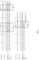
- the anti-PD-1 antibody described herein may bind to the same epitope as SHB-617, the amino acid sequences of the V H and V L chains of which are provided in Fig. 2 and Fig. 3 .
- the antibody may compete against SHB-617 from binding to the PD-1 target antigen.
- Such antibodies may exhibit at least 20% (e.g., 30%, 40%, 50%, 60%, 70%, 80%, 90%, or greater) of the activity of inhibiting a signaling pathway mediated by the PD-1 target antigen as relative to SHB-617.
- Such an anti-PD-1 antibody comprise the same heavy chain CDRs and/or the same light chain CDRs as those of SHB-617, for example, the CDR regions highlighted in Fig. 2 and Fig. 3 , which are determined by the Chothia numbering scheme.
- CDRs Complementarity determining regions
- CDRs are known in the art as referring to non-contiguous sequences of amino acids within antibody variable regions, which confer antigen specificity and binding affinity. In general, there are three (3) CDRs in each heavy chain variable region and three (3) CDRs in each light chain variable region. The precise amino acid sequence boundaries of a given CDR can be readily determined using any of a number of well-known schemes, including those described by Kabat et al. (1991), 5th Ed. Public Health Service, National Institutes of Health, Bethesda, Md.
- CDR complementary determining region
- the anti-PD-1 antibody described herein may comprise a V H chain at least 75% (e.g., 80%, 85%, 90%, 95%, or 98%) identical to the V H chain (mature or precursor) of SHB-617 and/or a V L chain at least 75% (e.g., 80%, 85%, 90%, 95%, or 98%) identical to the V L chain (mature of precursor) of SHB-617.
- Gapped BLAST can be utilized as described in Altschul et al., Nucleic Acids Res. 25(17):3389-3402, 1997 .
- the default parameters of the respective programs e . g ., XBLAST and NBLAST.
- the anti-PD-1 antibody described herein may comprise a V H chain that includes up to 5 (e.g., 1, 2, 3, 4, or 5) amino acid residue variations in the V H CDR regions (V H CDR1, CDR2, and/or CDR3) as compared to the V H CDRs of SHB-617, and/or a V L chain that includes up to 5 (e.g., 1, 2, 3, 4, or 5) amino acid residue variations in the V L CDR regions (V L CDR1, CDR2, and/or CDR3) as compared to the V L CDRs of SHB-617.
- V H chain that includes up to 5 (e.g., 1, 2, 3, 4, or 5) amino acid residue variations in the V H CDR regions (V H CDR1, CDR2, and/or CDR3) as compared to the V L CDRs of SHB-617
- V H CDR1, CDR2, and/or CDR3 amino acid residue variations in the V H CDR regions
- V L CDR1, CDR2, and/or CDR3 amino acid residue
- the anti-PD-1 antibody as described herein may be a chimeric antibody derived from SHB-617.
- a chimeric antibody can include a V H and V L chains derived from SHB-617 (as those described herein) and a heavy constant region and a light constant region from a human antibody.
- Chimeric antibodies refer to antibodies having a variable region or part of variable region from a first species and a constant region from a second species.
- the variable region of both light and heavy chains mimics the variable regions of antibodies derived from one species of mammals (e.g., a non-human mammal such as mouse, rabbit, and rat), while the constant portions are homologous to the sequences in antibodies derived from another mammal such as human.
- amino acid modifications can be made in the variable region and/or the constant region.
- the anti-PD-1 antibody described herein is a humanized antibody derived from SHB-617.
- a "humanized antibody” can be an antibody derived from modifying a non-human antibody to replace certain antibody fragments (e.g., framework regions) with those from a human antibody so as to reduce immunogenicity in humans.
- the humanized antibody described herein can be in any antibody form. In some embodiments, they are intact immunoglobulin molecules (full-length antibodies), including IgG, IgA, IgD, IgE, and IgM.
- the humanized antibodies are antigen binding fragments thereof, e.g., Fab, F(ab') 2 , scFv and Fv. In some instances, they also can be single-chain antibodies or bi-specific antibodies.
- Humanized antibodies can be designed as follows. First, the variable regions of V H and V L of a parent non-human antibody are subjected to three-dimensional molecular modeling analysis following methods known in the art. See, e.g., Queen et al., Proc. Natl. Acad. Sci. USA, 86:10029-10033 (1989 ). Next, framework amino acid residues predicted to be important for the formation of the correct CDR structures are identified using the same molecular modeling analysis. In parallel, human V H and V L chains having amino acid sequences that are homologous to those of the parent non-human antibody are identified from any antibody gene database using the parent V H and V L sequences as search queries. Human V H and V L acceptor genes are then selected.
- the CDR regions within the selected human acceptor genes can be replaced with the CDR regions from the parent non-human antibody or functional variants thereof.
- residues within the framework regions of the parent chain that are predicted to be important in interacting with the CDR regions can be used to substitute for the corresponding residues in the human acceptor genes.
- Such functional variants can comprise a V H chain that comprises an amino acid sequence at least 85% (e.g., 90%, 92%, 94%, 95%, 96%, 97%, 98%, or 99%) identical to that of SSI 361 and/or a V L chain that has an amino acid sequence at least 85% (e.g., 90%, 92%, 94%, 95%, 96%, 97%, 98%, or 99%) identical to that of SSI 361.
- These variants are capable of binding to an PD-1 molecule, particularly a human PD-1 antigen.
- the variants possess similar antigen-binding affinity relative to the exemplary humanized antibody described above (e.g., having a Kd ⁇ 100 ⁇ 10 -9 M).
- the functional variants described above contain one or more mutations (e.g., conservative substitutions) in the FRs of either the V H or the V L as compared to those in SSI-361. Preferably, such mutations do not occur at residues which are predicted to interact with one or more of the CDRs (see Example 2 below). As known in the art, mutations within the FR regions are unlikely to affect the antigen-binding activity of the antibody.
- the functional variants described herein contain one or more mutations (e.g., 1, 2, or 3) within one or more of the CDR regions. Preferably, such functional variants retain the same regions/residues responsible for antigen-binding as the parent, such as the same specificity-determining residues inside the CDRs.
- any of the anti-PD-1 antibodies can be prepared via conventional methodology, e.g., recombination technology. See, e.g., Green et al. (1994) Nature Genetics 7, 13 ; and U.S. Patent Nos. 5,545,806 and 5,569,825 .
- coding sequences of any of the anti-PD-1 antibody V H and V L chains described herein can be linked to the coding sequences of the Fc region of a human immunoglobulin and the resultant gene encoding a full-length antibody heavy and light chains can be expressed and assembled in a suitable host cell, e.g., a plant cell, a mammalian cell, a yeast cell, or an insect cell.
- a suitable host cell e.g., a plant cell, a mammalian cell, a yeast cell, or an insect cell.
- Antigen-binding fragments can be prepared via routine methods.
- F(ab')2 fragments can be produced by pepsin digestion of an full-length antibody molecule, and Fab fragments that can be generated by reducing the disulfide bridges of F(ab')2 fragments.
- Fab fragments that can be generated by reducing the disulfide bridges of F(ab')2 fragments.
- fragments can be prepared via recombinant technology by expressing the heavy and light chain fragments in suitable host cells (e.g., E. coli, yeast, mammalian, plant, or insect cells) and have them assembled to form the desired antigen-binding fragments either in vivo or in vitro.
- a single-chain antibody can be prepared via recombinant technology by linking a nucleotide sequence coding for a heavy chain variable region and a nucleotide sequence coding for a light chain variable region.
- a flexible linker is incorporated between the two variable regions.
- a humanized anti-PD-1 antibody produced as described above can be examined to determine their properties, such as antigen-binding activity and biological function, following routine methods, e.g., those described in Example 3 below.
- nucleic acids encoding any of the anti-PD-1 antibodies described herein, vectors such as expression vectors or recombinant viruses comprising these nucleic acids, and host cells comprising the vectors.
- both the heavy and light chain coding sequences are included in one expression vector.
- Each of the heavy chain coding sequence and the light chain coding sequence may be in operable linkage to a suitable promoter.
- expression of both the heavy chain and the light chain may be driven by the same promoter.
- each of the heavy and light chains of the antibody is cloned in to an individual vector.
- the expression vectors encoding the heavy and light chains can be co-transfected into one host cell for expression of both chains, which can be assembled to form intact antibodies either in vivo or in vitro.
- the expression vector encoding the heavy chain and that encoding the light chain can be introduced into different host cells for expression each of the heavy and light chains, which can then be purified and assembled to form intact antibodies in vitro.
- Suitable host cells include, but are not limited to, mammalian cells (e.g., CHO cells or 293 cells), plant cells, insect cells, yeast cells, or bacterial cells. Recombinant viruses such as oncolytic viruses capable of expressing SSI-361 or its fragments may be used to for therapeutic applications.
- the present disclosure also provides an immunoconjugate comprising any of the anti-PD-1 antibodies described herein and a suitable agent, which can be a therapeutic agent or diagnostic agent.
- a suitable agent which can be a therapeutic agent or diagnostic agent.
- the anti-PD-1 antibody disclosed herein is linked to a cytotoxic agent, a radioisotope, or a drug.
- Methods for preparing such an immunoconjugate are well known in the art. See, e.g., WO 2014/160160 , U.S. Patent Nos. 5,208,020 and 5,416,064 ; and Chari et al., 1992 Cancer Res. 52:127-131 .
- the antibody may be modified with a bifunctional agent such as N-Succinimidyl-3-(2-pyridyldithio) propionate (SPDP) to introduce an active disulfide moiety.
- SPDP N-Succinimidyl-3-(2-pyridyldithio) propionate
- the agent to be conjugated to the antibody may be modified to introduce a thiol-group following routine practice. Reaction with the thiol-containing agent would produce an immunoconjugate in which the antibody and the agent are linked via disulfide bonds.
- anti-PD-1 antibodies described herein can be used as therapeutic agents and diagnostic agents, as well as research tools in biochemistry, molecular biology, and medical researches.
- a disease associated with the PD-1 signaling comprising administering to a subject in need of the treatment an effective amount of any of the anti-PD-1 antibodies described herein.
- Such antibodies can also be used to enhance immune responses such as T cell activation in a subject in need of the treatment.
- a subject e.g., a human patient
- a subject who needs the just-noted treatment is a patient having, suspected of having, or at risk for cancer development.
- any of the anti-PD-1 antibodies described herein can be used for treating PD-L1 positive tumors.
- a human patient suitable for the treatment can be identified by examining whether such a patient carries PD-L1 positive cancer cells.
- a human patient who would be suitable for the treatment described herein can be a cancer patient carrying PD-L1 positive cancer cells.
- treating refers to the application or administration of a composition including one or more active agents to a subject, who has a disorder/disease associated with the signaling pathway mediated by PD-1 ( e . g ., those described herein), a symptom of the disease/disorder, or a predisposition toward the disease/disorder, with the purpose to cure, heal, alleviate, relieve, alter, remedy, ameliorate, improve, or affect the disease/disorder, the symptom of the disease/disorder, or the predisposition toward the disease/disorder.
- PD-1 e . g ., those described herein
- an effective amount refers to the amount of each active agent required to confer therapeutic effect on the subject, either alone or in combination with one or more other active agents. Effective amounts vary, as recognized by those skilled in the art, depending on the particular condition being treated, the severity of the condition, the individual patient parameters including age, physical condition, size, gender and weight, the duration of the treatment, the nature of concurrent therapy (if any), the specific route of administration and like factors within the knowledge and expertise of the health practitioner. These factors are well known to those of ordinary skill in the art and can be addressed with no more than routine experimentation. It is generally preferred that a maximum dose of the individual components or combinations thereof be used, that is, the highest safe dose according to sound medical judgment. It will be understood by those of ordinary skill in the art, however, that a patient may insist upon a lower dose or tolerable dose for medical reasons, psychological reasons or for virtually any other reasons.
- the amount of the anti-PD-1 antibody described herein is effective in suppressing the PD-1 signaling (e.g., reducing the PD-1 signaling by at least 20%, 30%, 50%, 80%, 100%, 200%, 400%, or 500% as compared to a blank control). In other embodiments, the amount of the anti-PD-1 antibody described herein is effective in activating immune responses (e.g., by at least 20%, 30%, 50%, 80%, 100%, 200%, 400%, or 500% as compared to a blank control).
- an initial candidate dosage can be about 2 mg/kg.
- a typical daily dosage might range from about any of 0.1 ⁇ g/kg to 3 ⁇ g/kg to 30 ⁇ g/kg to 300 ⁇ g/kg to 3 mg/kg, to 30 mg/kg to 100 mg/kg or more, depending on the factors mentioned above.
- the treatment is sustained until a desired suppression of symptoms occurs or until sufficient therapeutic levels are achieved to the desired therapeutic effects.
- An exemplary dosing regimen comprises administering an initial dose of about 2 mg/kg, followed by a weekly maintenance dose of about 1 mg/kg of the antibody, or followed by a maintenance dose of about 1 mg/kg every other week.
- other dosage regimens may be useful, depending on the pattern of pharmacokinetic decay that the practitioner wishes to achieve. For example, dosing from one-four times a week is contemplated. In some embodiments, dosing ranging from about 3 ⁇ g/mg to about 2 mg/kg (such as about 3 ⁇ g/mg, about 10 ⁇ g/mg, about 30 ⁇ g/mg, about 100 ⁇ g/mg, about 300 ⁇ g/mg, about 1 mg/kg, and about 2 mg/kg) may be used.
- dosing frequency is once every week, every 2 weeks, every 4 weeks, every 5 weeks, every 6 weeks, every 7 weeks, every 8 weeks, every 9 weeks, or every 10 weeks; or once every month, every 2 months, or every 3 months, or longer.
- the progress of this therapy is easily monitored by conventional techniques and assays.
- the dosing regimen (including the antibody used) can vary over time.
- any of the anti-PD-1 antibody or the encoding nucleic acid can be mixed with a pharmaceutically acceptable carrier to form a pharmaceutical composition for administration to a subject in need of the treatment.
- a pharmaceutically acceptable carrier is compatible with the active ingredient(s) in the composition (and preferably, capable of stabilizing it) and not deleterious to the subject to be treated.
- solubilizing agents such as cyclodextrins, which form more soluble complexes with a anti-PD-1 antibody as described herein, or a nucleic acid encoding such, or more solubilizing agents, can be utilized as pharmaceutical carriers for delivery of the agonist/antagonist.
- Examples of other carriers include colloidal silicon dioxide, magnesium stearate, sodium lauryl sulfate, and D&C Yellow # 10. See, e.g., Remington's Pharmaceutical Sciences, Edition 16, Mack Publishing Co., Easton, Pa (1980 ); and Goodman and Gilman's "The Pharmacological Basis of Therapeutics", Tenth Edition, Gilman, J. Hardman and L. Limbird, eds., McGraw-Hill Press, 155-173, 2001 .
- compositions formulated for therapeutic uses, may be prepared for storage by mixing an agent having the desired degree of purity with optional pharmaceutically acceptable carriers, excipients or stabilizers ( Remington's Pharmaceutical Sciences 16th edition, Osol, A. Ed. (1980 )), in the form of lyophilized formulations or aqueous solutions.
- Acceptable carriers, excipients, or stabilizers are nontoxic to recipients at the dosages and concentrations employed, and include buffers such as phosphate, citrate, and other organic acids; antioxidants including ascorbic acid and methionine; preservatives (such as octadecyldimethylbenzyl ammonium chloride; hexamethonium chloride; benzalkonium chloride, benzethonium chloride; phenol, butyl or benzyl alcohol; alkyl parabens such as methyl or propyl paraben; catechol; resorcinol; cyclohexanol; 3-pentanol; and m-cresol); low molecular weight (less than about 10 residues) polypeptides; proteins, such as serum albumin, gelatin, or immunoglobulins; hydrophilic polymers such as polyvinylpyrrolidone; amino acids such as glycine, glutamine, asparagine, histidine,
- an effective amount of the pharmaceutical composition noted above can be administered to a subject (e.g., a human) in need of the treatment via a suitable route.
- a subject e.g., a human
- a human subject who needs the treatment may be a human patient having, at risk for, or suspected of having a disorder associated with the signaling pathway mediated by PD-1. Such a patient can be identified by routine medical examination.
- the therapeutic polynucleotides and polypeptides described herein can be delivered using gene delivery vehicles.
- the gene delivery vehicle can be of viral or non-viral origin (see generally, Jolly, Cancer Gene Therapy (1994) 1:51 ; Kimura, Human Gene Therapy (1994) 5:845 ; Connelly, Human Gene Therapy (1995) 1:185 ; and Kaplitt, Nature Genetics (1994) 6:148 ).
- Expression of such coding sequences can be induced using endogenous mammalian or heterologous promoters and/or enhancers. Expression of the coding sequence can be either constitutive or regulated.
- Viral-based vectors for delivery of a desired polynucleotide and expression in a desired cell are well known in the art.
- Exemplary viral-based vehicles include, but are not limited to, recombinant retroviruses (see, e.g., PCT Publication Nos. WO 90/07936 ; WO 94/03622 ; WO 93/25698 ; WO 93/25234 ; WO 93/11230 ; WO 93/10218 ; WO 91/02805 ; U.S. Pat. Nos. 5,219,740 and 4,777,127 ; GB Patent No. 2,200,651 ; and EP Patent No.
- alphavirus-based vectors e.g., Sindbis virus vectors, Semliki forest virus (ATCC VR-67; ATCC VR-1247), Ross River virus (ATCC VR-373; ATCC VR-1246) and Venezuelan equine encephalitis virus (ATCC VR-923; ATCC VR-1250; ATCC VR 1249; ATCC VR-532)
- AAV adeno-associated virus
- Non-viral delivery vehicles and methods can also be employed, including, but not limited to, polycationic condensed DNA linked or unlinked to killed adenovirus alone (see, e.g., Curiel, Hum. Gene Ther. (1992) 3:147 ); ligand-linked DNA (see, e.g., Wu, J. Biol. Chem. (1989) 264:16985 ); eukaryotic cell delivery vehicles cells (see, e.g., U.S. Pat. No. 5,814,482 ; PCT Publication Nos. WO 95/07994 ; WO 96/17072 ; WO 95/30763 ; and WO 97/42338 ) and nucleic charge neutralization or fusion with cell membranes.
- polycationic condensed DNA linked or unlinked to killed adenovirus alone see, e.g., Curiel, Hum. Gene Ther. (1992) 3:147
- ligand-linked DNA see, e.g., Wu, J. Bio
- Naked DNA can also be employed.
- Exemplary naked DNA introduction methods are described in PCT Publication No. WO 90/11092 and U.S. Pat. No. 5,580,859 .
- Liposomes that can act as gene delivery vehicles are described in U.S. Pat. No. 5,422,120 ; PCT Publication Nos. WO 95/13796 ; WO 94/23697 ; WO 91/14445 ; and EP Patent No. 0524968 .
- Additional approaches are described in Philip, Mol. Cell. Biol. (1994) 14:2411 , and in Woffendin, Proc. Natl. Acad. Sci. (1994) 91:1581 .
- the pharmaceutical composition described herein, containing an anti-PD-1 antibody or its encoding nucleic acid(s), can be co-administered with a second therapeutic agent.
- the selection of the second therapeutic agent depends on the type of the disease to be treated. For example, if the target disease is cancer, the second agent can be an anti-cancer agent (e.g., Tamoxifen, Taxol, Erlotinib, Dexasone, and Herceptin).
- a sub-therapeutic dosage of either the composition or of the second agent, or a sub-therapeutic dosage of both can be used in the treatment of a subject having, or at risk of developing a disease or disorder associated with the cell signaling mediated by PD-1.
- Target diseases include cancer (e.g., those described herein), infectious diseases such as diseases caused by viral, bacterial, or fungus infection, or other diseases associated with immune deficiency, such as T cell dysfunction.
- Diseases caused by viral infection include, but are not limited to, HAV, HBV, and HCV.
- a "sub-therapeutic dose” as used herein refers to a dosage, which is less than that dosage which would produce a therapeutic result in the subject if administered in the absence of the other agent or agents.
- the sub-therapeutic dose of an agent is one which would not produce the desired therapeutic result in the subject in the absence of the administration of the agents of the invention.
- Therapeutic doses of many agents that are in clinical use are well known in the field of medicine, and additional therapeutic doses can be determined by those of skill without undue experimentation. Therapeutic dosages have been extensively described in references such as Remington's Pharmaceutical Sciences, 22th ed., 2012 ; as well as many other medical references relied upon by the medical profession as guidance for the treatment of diseases and disorders.
- compositions can also be administered via other conventional routes, e.g., administered orally, parenterally, by inhalation spray, topically, rectally, nasally, buccally, vaginally or via an implanted reservoir.
- parenteral as used herein includes subcutaneous, intracutaneous, intravenous, intramuscular, intraarticular, intraarterial, intrasynovial, intrasternal, intrathecal, intralesional, and intracranial injection or infusion techniques.
- injectable depot routes of administration such as using 1-, 3-, or 6-month depot injectable or biodegradable materials and methods.
- Injectable compositions may contain various carriers such as vegetable oils, dimethylactamide, dimethyformamide, ethyl lactate, ethyl carbonate, isopropyl myristate, ethanol, and polyols (glycerol, propylene glycol, liquid polyethylene glycol, and the like).
- water soluble antibodies can be administered by the drip method, whereby a pharmaceutical formulation containing the antibody and a physiologically acceptable excipients is infused.
- Physiologically acceptable excipients may include, for example, 5% dextrose, 0.9% saline, Ringer's solution or other suitable excipients.
- Intramuscular preparations e.g., a sterile formulation of a suitable soluble salt form of the antibody
- a pharmaceutical excipient such as Water-for-Injection, 0.9% saline, or 5% glucose solution.
- the nucleic acid(s) or a vector(s) expressing the antibody can be delivered to a subject by methods, such as that described in Akhtar et al., 1992, Trends Cell Bio. 2, 139 .
- it can be introduced into cells using liposomes, hydrogels, cyclodextrins, biodegradable nanocapsules, or bioadhesive microspheres.
- the nucleic acid or vector can be locally delivered by direct injection or by use of an infusion pump.
- Other approaches include employing various transport and carrier systems, for example through the use of conjugates and biodegradable polymers.
- any of the anti-PD-1 antibody or its encoding nucleic acids can be conjugated with a chaperon agent.
- conjugated means two entities are associated, preferably with sufficient affinity that the therapeutic benefit of the association between the two entities is realized. Conjugated includes covalent or noncovalent bonding as well as other forms of association, such as entrapment of one entity on or within the other, or of either or both entities on or within a third entity (e.g., a micelle).
- the chaperon agent can be a naturally occurring substance, such as a protein (e.g., human serum albumin, low-density lipoprotein, or globulin), carbohydrate (e.g., a dextran, pullulan, chitin, chitosan, inulin, cyclodextrin or hyaluronic acid), or lipid. It can also be a recombinant or synthetic molecule, such as a synthetic polymer, e.g., a synthetic polyamino acid.
- a protein e.g., human serum albumin, low-density lipoprotein, or globulin
- carbohydrate e.g., a dextran, pullulan, chitin, chitosan, inulin, cyclodextrin or hyaluronic acid
- lipid e.g., a recombinant or synthetic molecule, such as a synthetic polymer, e.g.,
- polyamino acids examples include polylysine (PLL), poly L aspartic acid, poly L-glutamic acid, styrene-maleic acid anhydride copolymer, poly(L-lactide-co-glycolied) copolymer, divinyl ether-maleic anhydride copolymer, N-(2-hydroxypropyl) methacrylamide copolymer (HMPA), polyethylene glycol (PEG), polyvinyl alcohol (PVA), polyurethane, poly(2-ethylacryllic acid), N-isopropylacrylamide polymers, and polyphosphazine.
- PLL polylysine
- poly L aspartic acid poly L-glutamic acid
- styrene-maleic acid anhydride copolymer examples include poly(L-lactide-co-glycolied) copolymer, divinyl ether-maleic anhydride copolymer, N-(2-hydroxypropyl) methacrylamide copoly
- the chaperon agent is a micelle, liposome, nanoparticle, or microsphere, in which the oligonucleotide/interfering RNA is encapsulated.
- Methods for preparing such a micelle, liposome, nanoparticle, or microsphere are well known in the art. See, e.g., US Patents 5,108,921 ; 5,354,844 ; 5,416,016 ; and 5,527,5285 .
- the chaperon agent serves as a substrate for attachment of one or more of a fusogenic or condensing agent.
- a fusogenic agent is responsive to the local pH. For instance, upon encountering the pH within an endosome, it can cause a physical change in its immediate environment, e.g., a change in osmotic properties which disrupts or increases the permeability of the endosome membrane, thereby facilitating release of the antisense oligonucleotide into host cell's cytoplasm.
- a preferred fusogenic agent changes charge, e.g., becomes protonated at a pH lower than a physiological range (e.g., at pH 4.5-6.5). Fusogenic agents can be molecules containing an amino group capable of undergoing a change of charge (e.g., protonation) when exposed to a specific pH range.
- Such fusogenic agents include polymers having polyamino chains (e.g., polyethyleneimine) and membrane disruptive agents (e.g., mellittin).
- polymers having polyamino chains e.g., polyethyleneimine
- membrane disruptive agents e.g., mellittin
- Other examples include polyhistidine, polyimidazole, polypyridine, polypropyleneimine, and a polyacetal substance (e.g., a cationic polyacetal).
- a condensing agent interacts with the antisense oligonucleotide, causing it to condense (e.g., reduce the size of the oligonucleotide), thus protecting it against degradation.
- the condensing agent includes a moiety (e.g., a charged moiety) that interacts with the oligonucleotide via, e.g., ionic interactions.
- condensing agents include polylysine, spermine, spermidine, polyamine or quarternary salt thereof, pseudopeptide-polyamine, peptidomimetic polyamine, dendrimer polyamine, arginine, amidine, protamine, cationic lipid, cationic porphyrin, and alpha helical peptide.
- combination therapy embraces administration of these agents (e.g., an anti-PD-1 antibody such as those described herein and a second suitable therapeutic agent) in a sequential manner, that is, wherein each therapeutic agent is administered at a different time, as well as administration of these therapeutic agents, or at least two of the agents, in a substantially simultaneous manner.
- sequential or substantially simultaneous administration of each agent can be affected by any appropriate route including, but not limited to, oral routes, intravenous routes, intramuscular, subcutaneous routes, and direct absorption through mucous membrane tissues.
- the agents can be administered by the same route or by different routes.
- a first agent e.g., an anti- PD-1 antibody
- a second agent e.g., an anti-cancer agent, an anti-infection agent; or an immune modulator
- an agent of the combination selected may be administered by intravenous injection while the other agents of the combination may be administered orally.
- two or more of the agents may be administered by intravenous or subcutaneous injection.
- the term “sequential” means, unless otherwise specified, characterized by a regular sequence or order, e.g., if a dosage regimen includes the administration of an anti- PD-1 antibody and the second agent, a sequential dosage regimen could include administration of the anti-PD-1 antibody before, simultaneously, substantially simultaneously, or after administration of the second agent, but both agents will be administered in a regular sequence or order.
- the term “separate” means, unless otherwise specified, to keep apart one from the other.
- the term “simultaneously” means, unless otherwise specified, happening or done at the same time, i.e. , the agents of the invention are administered at the same time.
- substantially simultaneously means that the agents are administered within minutes of each other (e.g., within 10 minutes of each other) and intends to embrace joint administration as well as consecutive administration, but if the administration is consecutive it is separated in time for only a short period (e.g., the time it would take a medical practitioner to administer two compounds separately).
- concurrent administration and substantially simultaneous administration are used interchangeably.
- Sequential administration refers to temporally separated administration of the agents described herein.
- Combination therapy can also embrace the administration of the agents described herein in further combination with other biologically active ingredients.
- the combination therapy further comprises radiation treatment
- the radiation treatment may be conducted at any suitable time so long as a beneficial effect from the co-action of the combination of the therapeutic agents and radiation treatment is achieved.
- the beneficial effect is still achieved when the radiation treatment is temporally removed from the administration of the therapeutic agents, perhaps by days or even weeks.
- the anti-PD-1 antibody may also be used before or after a surgery or radiotherapy.
- any combination as described herein may be used in any sequence for treating the target disease described herein.
- the combinations described herein may be selected on the basis of a number of factors, which include but are not limited to the effectiveness of inhibiting or preventing the target disease progression, the effectiveness for mitigating the side effects of another agent of the combination, or the effectiveness of mitigating symptoms related to the target disease.
- a combined therapy described herein may reduce any of the side effects associated with each individual members of the combination.
- Suitable agents for co-use with any of the anti-PD-1 antibodies include an antibody binding to a co-stimulatory receptor (e.g., CD137/4-1BB, OX40, CD40, ICOS, CD27, HVEM or GITR), an agent that induces immunogenic cell death (e.g., a chemotherapeutic agent, a radiotherapeutic agent, an anti-angiogenic agent, or an agent for targeted therapies), an agent that inhibits a checkpoint molecule (e.g., CTLA4, LAG3, TIM3, B7H3, B7H4, BTLA, or TIGIT), a cancer vaccine, an agent that modifies an immunosuppressive enzyme (e.g., IDO1 or iNOS), an agent that targets T reg cells, an agent for adoptive cell therapy, or an agent that modulates myeloid cells.
- a co-stimulatory receptor e.g., CD137/4-1BB, OX40, CD40, ICOS, CD27, HVEM or GITR
- kits for use in enhancing immune responses and/or treating diseases associated with the PD-1 signaling can include one or more containers comprising an anti-PD-1 antibody as described herein.
- the kit can comprise instructions for use in accordance with any of the methods described herein.
- the included instructions can comprise a description of administration of the anti-PD-1 antibody to enhance immune responses and/or to treating a target disease as described herein.
- the kit may further comprise a description of selecting an individual suitable for treatment based on identifying whether that individual has a disease associated with the PD-1 signaling, e.g., those described herein.
- the instructions relating to the use of an anti-PD-1 antibody generally include information as to dosage, dosing schedule, and route of administration for the intended treatment.
- the containers may be unit doses, bulk packages (e.g., multi-dose packages) or sub-unit doses.
- Instructions supplied in the kits of the invention are typically written instructions on a label or package insert (e.g., a paper sheet included in the kit), but machine-readable instructions (e.g., instructions carried on a magnetic or optical storage disk) are also acceptable.
- the label or package insert indicates that the composition is used for treating any of the target diseases disclosed herein or alleviating one or more symptoms of such a disease may be provided for practicing any of the methods described herein.
- kits of this invention are in suitable packaging.
- suitable packaging includes, but is not limited to, vials, bottles, jars, flexible packaging (e.g., sealed Mylar or plastic bags), and the like.
- packages for use in combination with a specific device such as an inhaler, nasal administration device (e.g., an atomizer) or an infusion device such as a minipump.
- a kit may have a sterile access port (for example the container may be an intravenous solution bag or a vial having a stopper pierceable by a hypodermic injection needle).
- the container may also have a sterile access port (for example the container may be an intravenous solution bag or a vial having a stopper pierceable by a hypodermic injection needle).
- a sterile access port for example the container may be an intravenous solution bag or a vial having a stopper pierceable by a hypodermic injection needle.
- At least one active agent in the composition is an anti-PD-1 antibody as described herein.
- kits described herein may include one or more additional therapeutic agents such as those described in the "Combination Treatment” above.
- Kits may optionally provide additional components such as buffers and interpretive information.
- the kit comprises a container and a label or package insert(s) on or associated with the container.
- the invention provides articles of manufacture comprising contents of the kits described above.
- Human PD-1 protein (cat#1086-PD, Fc chimera), PD-L1 (cat# 156-B7, Fc chimera), and antigen affinity-purified polyclonal goat IgG against human PD-1 (cat#AF1086) were purchased from R&D Systems (USA). Monoclonal mouse anti-human PD-1 IgG (clone 1116, cat# 16-9989-82) was purchased from eBiosciences (USA). Stable CHO cell line expressing human PD-1 was developed by transfection with full length human PD-1 cDNA (Gene ID: 5133).
- Capture ELISA to detect anti-PD-1 antibodies was carried out by coating plates with streptavidin followed by biotin-PD-1. Antibody samples bound to PD-1 were detected by secondary antibodies (goat anti-mouse or rat IgG) conjugated with peroxidase.
- FACS to detect anti-PD-1 antibodies was performed with CHO-PD-1 cell line. After incubating with antibody samples, CHO-PD-1 cells were detected by PE or FITC labeled secondary antibodies by FACS.
- Blockade of PD-1 and PD-L1 binding was measured by FACS with CHO-PD-1 cells.
- PD-L1 was incubated with CHO-PD-1 cells followed by competition with anti-PD-1 antibody samples. The bound PD-L1 was then detected with anti-human Fc secondary antibody conjugated with Alexa 488.
- mice and Wistar rats were immunized with recombinant human PD-1 protein (R&D, cat#1086-PD, Fc chimera).
- the anti-PD-1 serum titers of the immunized animals were monitored by capture ELISA with biotin-PD-1 and human Fc as counter screen. The positive titers were further confirmed with FACS with CHO-PD-1 cells.
- One mouse and one rat with the highest titers were sacrificed, and spleenocytes were isolated and fused with SP2/0 myeloma cells using standard hybridoma protocol.
- mice hybridomas Twenty 96-well plates of mouse hybridomas were screened by capture ELISA with biotin-PD-1 and identified 334 positive wells. The positive samples were then tested by human Fc counter screen and 85 clones were identified as PD-1 specific. Next, these specific clones were tested for binding to CHO-PD-1 by FACS and top 34 clones were identified for further screening to block PD-L1 binding to CHO-PD-1 by FACS.
- rat hybridomas were screened by capture ELISA with biotin-PD-1 and identified 207 positive wells. The positive samples were then tested by human Fc counter screen and 54 clones were identified as PD-1 specific. Next, these specific clones were tested for binding to CHO-PD-1 by FACS and identified top 30 clones for further screening to block PD-L1 binding to CHO-PD-1 by FACS.
- a PD-1/ PD-L1 blockade assay was used to screen positive hybridoma samples. 24 clones with specific binding to PD-1 and ability to block PD-L1 binding to cellular PD-1 were identified. The top 13 hybridomas (5 rat and 8 mice) were cloned, produced and purified antibodies from 8 single cell clones including 1 rat IgM and 7 mouse IgG hybridomas. These purified antibodies were confirmed to bind human PD-1 protein and cellular PD-1, and block PD-L1 binding to cellular PD-1.
- the antibody variable domains of these confirmed hybridomas were sequenced as follows. Briefly, total RNA was extracted from frozen hybridoma cells and cDNA was synthesized from the RNA using isotype-specific anti-sense primers or universal primers following the technical manual of SuperScript III First-Strand Synthesis System. The antibody fragments of V H and V L were amplified according to the standard operating procedure of RACE of GenScript. Amplified antibody fragments were separately cloned into a standard cloning vector using standard molecular cloning procedures. Colony PCR screening was performed to identify clones with inserts of correct sizes. Five single colonies with correct V H and V L insert sizes were sent for sequencing. The V H and V L genes of five different clones were found nearly identical. The consensus sequence is believed to be the sequence of the antibody produced by the hybridoma.
- the cDNA sequences encoding the anti-PD-1 antibody variable domain sequences were synthesized as chimeras to human IgG4 heavy chain constant regions containing the hinge S228P (EU numbering; Kabat numbering 241) stabilizing mutation ( Angal et., Mol. Immunol 30:105, 1993 ) or human kappa light chain constant region.
- HEK293 transient expression was carried out with plasmids containing the corresponding heavy and light chain sequences.
- These chimeric antibodies were purified by protein A affinity chromatography. The purified antibodies were buffered exchanged to PBS and checked for endotoxin ( ⁇ 5 EU/mg) and monomer (>95%).
- the chimeric antibodies were tested in functional assays for their activity to stimulate human T cell function in vitro.
- Fresh PBMC were isolated from volunteers who were recently immunized with tetanus toxoid vaccines. The cells were re-suspended in PRMI-1640 containing 10% FBS at 1 ⁇ 10 ⁇ 6/ml and plated into 96-well plate, 200 ul/well.
- the testing antibodies were diluted in PBS-BSA and added to the PMBC culture at final concentrations of 0, 0.03, 0.1, 0.3, 1, 3, 10 and 30 ug/ml. After 30 minutes of incubation, tetanus toxoid antigen (Astarte Biologics, cat#1002) was added to the PBMC culture.
- the plate was returned into incubator at 37 °C and 5% CO2 for 7 days.
- the IFN- ⁇ in the culture supernatants were determined by ELISA (eBioscience, 88-7316-88).
- a positive control antibody against human PD-L1 (eBioscience #16-5983) was used.
- the SHB-617 (rat/human IgG4/kappa) chimera showed the highest maximal induction of IFN ⁇ secretion from the activated T cells ( Fig. 1 ).
- SHB-617 was derived from rat IgM. The activity of SHB-617 indicated that its binding domains possess unique activity in blocking PD-1 function.
- SHB-617 was humanized as described herein and the humanized antibody thus obtained were further characterized.
- the amino acid sequences of the lead antibody SHB-617 were analyzed and annotated with the updated Chothia CDR definitions (Al-Lazikani et al, 1997).
- the CDR regions (L1, L2, L3, H1, H2 and H3) in the variable domains are shown in Figure 2 and Figure 3 . Positional numbering is ordinal unless otherwise specified, in which case Chothia 1987 numbering will be used.
- Recombinant full human IgG4/kappa of humanized SHB-617 was constructed with human IgG4 containing the hinge S228P (EU numbering; Kabat numbering 241) stabilizing mutation ( Angal et., Mol. Immunol 30:105, 1993 ) and human kappa light chain constant region.
- the humanized SSI-361 antibody was expressed in and purified from HEK293 cells or CHO cells.
- Flow cells of a Series S CM5 sensor chip were activated with freshly mixed 50 mmol/L NHS and 200 mmol/L EDC for 360 seconds (10 ⁇ L/min). Then, anti-His IgGs, in 10 mmol/L NaAC (pH 5.0) was injected onto the activated flow cell (10 ⁇ L/min) for 300 seconds (10 ⁇ L/min) with HBS-EP+ (10mM HEPES, 150mM NaCl, 3mM EDTA and 0.05% P20, pH 7.4) as the running buffer. The remaining active coupling sites were blocked with a 420 second injection of 1 mol/L ethanolamine (10 ⁇ L/min).
- the PD-1 antibody sample was diluted in PBS to 1ug/ml and was coated in an ELISA plate (Costar, Cat#3590, high binding) 100ul/well, 4 °C overnight.
- the plate was decanted and blocking solution (eBioscience, ELISA/ELISPOT Diluent, Cat# 00-4202-55) was added 200ul/well. After 1 hour incubation at room temperature, the plate was washed with PBST three times.
- Human PD-1-His tag protein (Sino Biological Inc. Cat#10377-H08H-100), or rhesus monkey PD-1-His (Sino Biological Inc.
- Cyno PD-1- Fc tag (Sino Biological Inc. Cat# 90311-C02H) was diluted in PBS to 2ug/ml, and 100ul/well was used to coat an ELISA plate (Costar, Cat#3590, high binding), 4 °C overnight. The plate was decanted and blocking solution (eBioscience, ELISA/ELISPOT Diluent, Cat# 00-4202-55) was added, 200ul/well. After 1 hour incubation at room temperature, the plate was washed with PBST three times.
- the testing PD-1 antibody was diluted in blocking buffer to 0, 0.001, 0.01, 0.1, 1,10ug/ml and added to the plate, 100 ul/well. The plate was incubated 1 hour at room temperature and then washed four times with PBST. Anti- Hu IgG-HRP (goat-anti-human Ig kappa-HRP, Millipore, Cat# AP502P) at 1:4000 dilution was added to the plate, 100 ul/well. The plate was incubated 1 hour at room temperature followed by washing with PBST four times. The TMB substrate solution was added 100 ul/well. The color developed for 15 minutes and 50ul/well of 1M H2SO4 was added to terminate the reaction.
- Anti- Hu IgG-HRP goat-anti-human Ig kappa-HRP, Millipore, Cat# AP502P
- CHO cells over-expressing human PD-1 were harvested by centrifugation at 1000 rpm for 5 minutes. The cells were resuspended in cold PBS-BSA at 5 ⁇ 10 ⁇ 6/ml and aliquoted 100ul/vial.
- PD-1 antibody was diluted in PBS-BSA at 3x (final concentrations at 0, 0.01, 0.1, 1 and 10ug/ml) and 50ul was added to the CHO-PD-1 cells. The cell solutions were mixed and incubated 4°C in dark for 1 hour. The cells were washed with PBS-BSA two times. Secondary antibody conjugates (PE Mouse Anti-Human IgG in staining buffer.
- BD Cat#555787
- 100 ul/vial was added and cells were mixed and incubated 4°C in dark for 1 hour.
- the cells were then washed twice with PBS-BSA followed by fixation and FACS analysis with FACScaliber.
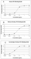
- SSI-361 at 1 ⁇ g/mL ( ⁇ 6.7 nM) concentration saturated human PD-1on the CHO cells over-expressing human PD-1.
- CHO cells over-expressing human PD-1 were harvested by centrifugation at 1000 rpm for 5 minutes. The cells were resuspended in cold PBS-BSA at 5 ⁇ 10 ⁇ 6/ml and aliquoted 100ul/vial.
- PD-1 antibody was diluted in PBS-BSA at 4x (final concentration 1ug/ml) and 50ul was added to the CHO-PD-1 cells.
- 50ul 4x human PD-L1 (Sino Biological Inc. Cat# 10084-H08H-100) was added at final 2ug/ml or 50ul 4x human PD-L2 (Sino Biological Inc. Cat# 10292-H08H-100) at final 1ug/ml.
- the cell solutions were mixed and incubated 4°C in dark for 4 hours.
- the cells were washed with PBS-BSA two times.
- 100u1 2ug/ml anti-His Tag Antibody [iFluor 488] (GenScript,Cat# A01800-100) was added and cells were mixed and incubated 4°C in dark for 1 hour.
- the cells were then washed twice with PBS-BSA followed by fixation and FACS analysis with FACScaliber.
- SSI-361 at 1 ug/ml effectively blocked the binding of human PD-1 with PD-L1 and PD-L2 ( Fig. 8 ).
- Fresh PBMC was isolated from volunteers who were recently immunized with tetanus toxoid vaccines. The cells were resuspended in PRMI-1640 containing 10% FBS at 1 ⁇ 10 ⁇ 6/ml and plated into 96-well plate, 200 ul/well. The testing antibodies were diluted in PBS-BSA and added to the PMBC culture at final concentration of 3 ug/ml. After 30 minutes incubation, tetanus toxoid antigen (Astarte Biologics, cat#1002) was added to the PBMC culture at a final concentration of 1ug/ml. The plate was returned into incubator at 37 °C and 5% CO2 for 7 days.
- the IFN-g in the culture supernatants were determined by ELISA (eBioscience, 88-7316-88).
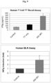
- SSI-361 at 3 ug/ml enhanced tetanus toxoid antigen induced IFN-g secretion ( Fig. 9 ).
- MLR Mixed lymphocyte reaction
- EasySep TM Human T Cell Isolation Kit (Stemcell, Cat# 17951) was used to isolate human T cells. PBMC was diluted in EasySep TM buffer at 5 ⁇ 10 ⁇ 7/ml, and Isolation Cocktail antibodies were added as instructed. After mixing and sitting at room temperature for 5 minutes, RapidSpheres magnetic beads were added, followed by addition of 5 ml EasySep TM buffer. The T cells were harvested after removal of non-T cells with the EasySep TM magnets.
- the allergenic dendritic cells and T cells were resuspended in RPMI-1640 medium at density of 1 ⁇ 10 ⁇ 6/ml and 1 ⁇ 10 ⁇ 5/ml, respectively. 100 ul of each cell types were added into the wells of 96-well culture plate. PD-1 antibodies were added to the culture mix at final concentration of 3 ug/ml. The plate was returned to incubator at 37 °C , 5% CO2 for 5 days. The IFN-g in the culture supernatants were determined by ELISA (eBioscience, 88-7316-88). SSI-361 increased IFG-g secretion from the MLR ( Fig. 9 ).
- PK pharmacokinetic
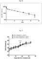
- Anti-Hu IgG-HRP (goat-anti-human Ig kappa-HRP, Millipore, Cat# AP502P) at 1:4000 dilution was added to the plate 100, ul/well. The plate was incubated 1 hour at room temperature followed by washing with PBST four times. The TMB substrate solution was added, 100 ul/well. The color developed for 15 minutes and added 50ul/well of 1M H2SO4 to terminate the reaction. Absorbance at 450nm was determined in FlexStation 3 ELISA reader. SSI-661 concentrations in the samples were calculated from standard curve and shown in Fig. 10 . PK parameters were calculated and listed as in Table 2. Table 2.
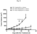
- the objective of this study was to verify the in vivo efficacies of SSI-361 (PD-1 Ab01) along with commercially purchasedantibody Opdivo (nivolumab) PD-1 Ab02 in the treatment of tumors induced by MC38-huPD-L1 cells (colon adenocarcinoma cells transfected with human PD-L1) in human PD-1 knocked-in (hPD-1 KI) mice.
- An exemplary experimental design is provided in Table 3 below: Table 3.
- Table 3 Table 3.
- Experimental Design Group N Treatment Dose (mg/kg) Dosing route Planned Schedule 1 8 PBS 0 i.p. BIW x3wks 2 8 PD-1 Ab01 10 i.p. BIW x3wks 3 8 PD-1 Ab02 10 i.p. BIW x3wks
- the dosing volume is 10 ⁇ l/g.
- mice 24 male hPD-1 KI mice (background strain: C57BL/6), age 7.7 weeks (starting from the drug administration) were used in this study.
- the mice were kept in individually ventilated cage (IVC) systems at 20-24 °C and 30-70% humidity with 2 animals in each cage.
- the animals had free access to irradiation sterilized dry granule food and drinking water, which was sterilized by US and ozone, during the entire study period.
- IVC individually ventilated cage
- MC38-huPD-L1 cells were maintained in vitro as a suspension culture in RPMI-1640 Medium (without phenol red) with concentration of 1 ⁇ 10 7 cells/ml. Each mouse was inoculated subcutaneously into right side with 1 ⁇ 10 6 cells in 0.1ml RPMI-1640 medium for tumor development. Tumor volumes of inoculated animals were checked twice weekly. At the 9th day after inoculation, the animals were randomly assigned into 3 different treatment groups. The average tumor volumes of these animals reached 68.3mm 3 . Each group has 8 animals and two of them were kept in one cage.
- the anti-PD-1 antibodies were administered to the tumor-bearing mice according to the experimental design shown in Table 3. Administration of the anti-PD-1 antibodies began after randomization of the mice into the three study arms, which was denoted as day 0. Treatments were ended on day 17.
- the animals were checked for any effects of tumor growth and treatments on normal behavior such as mobility, visual estimation of food and water consumption, body weight gain/loss (body weights were measured twice weekly or every other day), eye/hair matting and any other abnormal effect. Death and observed clinical signs were recorded on the basis of the numbers of animals within each subset. Twenty-four hours after the 4th dosing, 10ul blood was collected through the tail nip to pre-labeled tubes with 40ul PBS, mixed and stored at -80 °C.
- TGI tumor growth inhibition
- T-C Tn - Cn
- Tn refers to the time (days after treatment) when tumor volume reached n (e.g.,:1000mm 3 ) in the treatment group (days); and Cn refers to the time (days after treatment) when tumor volume reached n(e.g.,:1000mm 3 ) in the control group (days).
- the study was terminated two weeks after the 6 th dosing.
- the animals were euthanized individually or entirely when the following signs were observed: (a) when the animal was moribund, in severe distress or unable to obtain adequate food or water; (b) when body weight continued to drop and body weight loss was >20%; or (c) tumor volume of individual mice reached 3000 mm 3 or MTV of entire group reached 1500 mm 3 .
- the mean and the standard error of the mean (SEM) were calculated for the tumor volume and body weight of each group at each time point. Statistical analyses were carried out using Graph Pad Prism software. Significant differences in tumor growth were determined by Student's t test. Values of P ⁇ 0.05 were considered significant.
- Tumor Volumes Days after Grouping Tumor volume(mm 3 ) Group1 PBS Group2 PD-1 Ab01 10mg/kg Group3 PD-1 Ab02 10mg/kg 0 68.416.0 68.015.7 68.515.6 3 130.4117.5 91.217.4 104.918.3 7 245.0137.6 70.617.8 127.5133.0 10 352.5169.0 38.718.1 112.4149.3 14 486.01108.7 24.8111.5 129.6169.9 17 617.41161.7 24.3113.0 145.7182.9 21 1008.81303.8 28.1117.1 183.61101.7 26 1498.31455.4 34.5119.4 241.81135.8 33 / 62.9115.2 372.11197.8 Table 5.
- both anti-PD-1 antibodies showed significant tumor suppression activities in this study.
- this study verified the therapeutic efficacies of two exemplary anti-PD-1 antibodies using the syngenic MC38-huPD-L1 tumor cell xenograft in hPD-1 KI mice. No significant body weight change was observed in the treated mice during the course of treatment.
- the mean tumor volume in PBS-treated groups reached 1008.8 mm 3 , while both anti-PD-1 antibodies significantly reduced tumor volumes.
- the TGI (%) values for Ab01 and Ab02 were 104.24% and 87.76%, respectively.
- the tumor suppression levels of both antibodies were statistically significant as compared with the vehicle control group (P ⁇ 0.05).
- Example 5 Efficacy of anti-PD-1 antibodies in Treating Tumors Induced by MC38 and B16F1 Cancer Cells in Syngenic Mouse Model
- Transgenic mice having human PD-1 knocked-in were used in this study and C57BL/6 mice were used as controls.
- the hPD-1 KI mice were shown to be responsive to anti-human PD-1 antibody treatment in tumor xenograft models.
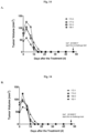
- Figure 14 The hPD-1 KI mice and C57BL/6 mice were kept in individually ventilated cages at 20-24 °C and 30-70% humidity with 1 ⁇ 3 animals in each cage. The animals had free access to irradiation sterilized dry granule food and drinking water, which was sterilized by US and ozone, during the entire study period.
- MC38 cells colon adenocarcinoma cells
- B16F1 cells skin melanoma cells
- the MC38 cells were maintained in vitro as a suspension culture at a concentration of 5 ⁇ 10 6 cells/ml.
- PD-1Mice from Example 4 which were cured by the treatment of PD-1 antibodies as described, as well as control naive mice, were then inoculated subcutaneously into right side with 5 ⁇ 10 5 cells in 0.1ml RPMI-1640 medium for tumor development. Tumor volumes of inoculated animals were checked twice weekly. When the mean volume of the tumor induced by MC38 cells in the C57BL/6 mice reached 110.4 mm 3 , B16F1 cells (2 ⁇ 10 5 cells in 0.1 ml of RPMI-1640 medium) were inoculated subcutaneously into the left side of the same animal for tumor development.
- the animals were checked for any effects of tumor growth and treatments on normal behavior such as mobility, visual estimation of food and water consumption, body weight gain/loss (body weights were measured twice weekly or every other day), eye/hair matting and any other abnormal effect. Death and observed clinical signs were recorded on the basis of the numbers of animals within each subset.
- B16F1 cells were inoculated into left side 9 days after MC38 cell inoculation. Tumor volumes of all animals were checked twice weekly for 24 days. The animals were then euthanized individually or entirely when the following signs were observed: (a) when the animal was moribund, in severe distress or unable to obtain adequate food or water; (b) When body weight continued to drop and body weight loss was >20%; or (c) MTV of entire group reached 1500mm 3 .
- Tumor Volumes Tumor volume(mm 3 ) Days after inoculation Group1 PD-1 Ab01-treatment respond Group2 PD-1 Ab02-treatment respond Group3 C57BL/6 mice MC38 B16F1 MC38 B16F1 MC38 B16F1 MC38 B16F1 0 / 0 ⁇ 0 / 0 ⁇ 0 / 6 / 0 ⁇ 0 / 0 ⁇ 0 / 73.9 ⁇ 4.0 / 8 / 0 ⁇ 0 / 0 ⁇ 0 / 95.1 ⁇ 8.0 / 9 0 0 ⁇ 0 0 ⁇ 0 0 ⁇ 0 0 ⁇ 0 110.4 ⁇ 9.0 0 ⁇ 0 13 4 0 ⁇ 0 010 0 ⁇ 0 0 ⁇ 0 202.5 ⁇ 21.9 0 ⁇ 0 17 8 0 ⁇ 0 010 0 ⁇ 0 4.8 ⁇ 4.8 278.2 ⁇ 31.5 0 ⁇ 0 20 11 0 ⁇ 0 9.6 ⁇ 9.6 0 ⁇ 0 28.5 ⁇ 18.7 329.4 ⁇ 57.3 9.0 ⁇ 6.3 24 15 0+0
Landscapes
- Health & Medical Sciences (AREA)
- Chemical & Material Sciences (AREA)
- Genetics & Genomics (AREA)
- Life Sciences & Earth Sciences (AREA)
- Organic Chemistry (AREA)
- Engineering & Computer Science (AREA)
- Immunology (AREA)
- Molecular Biology (AREA)
- General Health & Medical Sciences (AREA)
- Biomedical Technology (AREA)
- Biochemistry (AREA)
- Biophysics (AREA)
- Medicinal Chemistry (AREA)
- Wood Science & Technology (AREA)
- Bioinformatics & Cheminformatics (AREA)
- General Engineering & Computer Science (AREA)
- Biotechnology (AREA)
- Zoology (AREA)
- Proteomics, Peptides & Aminoacids (AREA)
- Physics & Mathematics (AREA)
- Plant Pathology (AREA)
- Microbiology (AREA)
- Veterinary Medicine (AREA)
- Public Health (AREA)
- Animal Behavior & Ethology (AREA)
- Pharmacology & Pharmacy (AREA)
- Nuclear Medicine, Radiotherapy & Molecular Imaging (AREA)
- General Chemical & Material Sciences (AREA)
- Chemical Kinetics & Catalysis (AREA)
- Peptides Or Proteins (AREA)
- Medicines Containing Antibodies Or Antigens For Use As Internal Diagnostic Agents (AREA)
Claims (10)
- Anticorps anti-PD-1 isolé, qui est une IgG4 de pleine longueur, comprenant :(i) une région variable de chaîne lourde (VH) qui comprend, ou consiste en, la séquence d'acides aminés QVTLKESGPALVKPTQTLTLTCTFSGFSLSTSGTCVSWI RQPPGKALEWLATICWEDSKGYNPSLKSRLTISKDTSKNQAVLTMTNMDPVDT ATYYCARREDSGYFWFPYWGQGTLVTVSS (SEQ ID NO : 12), et(ii) une région variable de chaîne légère (VL) qui comprend, ou consiste en, la séquence d'acides aminés NIQMTQSPSSLSASVGDRVTITCKAGQNVNNYLAWYQ QKPGKAPKVLIFNANSLQTGVPSRFSGSGSGTDFTLTISSLQPEDFATYYCQQYN SWTTFGGGTKVEIKR (SEQ ID NO : 11).
- Anticorps anti-PD-1 isolé selon la revendication 1, comprenant la séquence de la SEQ ID NO : 23 en tant que chaîne lourde, et la séquence de la SEQ ID NO : 24 en tant que chaîne légère.
- Acide nucléique isolé ou jeu d'acides nucléiques, qui code collectivement un anticorps anti-PD-1 selon l'une quelconque des revendications 1 et 2.
- Acide nucléique isolé ou jeu d'acides nucléiques selon la revendication 3, qui est un vecteur ou un jeu de vecteurs, de préférence dans lequel le ou les vecteurs sont des vecteurs d'expression ou des virus recombinés.
- Acide nucléique isolé ou jeu d'acides nucléiques selon l'une quelconque des revendications 3 et 4, qui est i) un seul acide nucléique comprenant une première séquence de nucléotides qui code la VH et une deuxième séquence de nucléotides qui code la VL ; ou ii) un jeu d'acides nucléiques comprenant un premier acide nucléique qui comprend une première séquence de nucléotides codant la VH et un deuxième acide nucléique qui comprend une deuxième séquence de nucléotides codant la VL.
- Cellule hôte comprenant un vecteur ou un jeu de vecteurs comprenant un acide nucléique ou un jeu d'acides nucléiques selon l'une quelconque des revendications 3 à 5, de préférence dans laquelle le vecteur ou le jeu de vecteurs est un ou plusieurs vecteurs d'expression.
- Immunoconjugué comprenant un anticorps anti-PD-1 selon l'une quelconque des revendications 1 et 2 lié à un agent thérapeutique ou diagnostique.
- Composition pharmaceutique comprenant un anticorps anti-PD-1 selon l'une quelconque des revendications 1 et 2, un acide nucléique/jeu d'acides nucléiques selon l'une quelconque des revendications 3 à 5, ou un immunoconjugué selon la revendication 7, et un véhicule pharmaceutiquement acceptable.
- Composition pharmaceutique selon la revendication 8 pour une utilisation dans le traitement d'une maladie associée à la signalisation de PD-1 chez un sujet, dans laquelle la maladie associée à PD-1 est un cancer ou une maladie infectieuse, et dans laquelle il est en éventuellement en outre administré au sujet un autre agent thérapeutique.
- Méthode pour préparer un anticorps anti-PD-1, la méthode comprenant :(i) la culture d'une cellule hôte selon la revendication 6 dans des conditions permettant l'expression de l'anticorps ; et(ii) la collecte de la cellule hôte ou du milieu de culture pour une isolation de l'anticorps.
Applications Claiming Priority (2)
| Application Number | Priority Date | Filing Date | Title |
|---|---|---|---|
| CN201510799538.8A CN106699889A (zh) | 2015-11-18 | 2015-11-18 | 抗pd-1抗体及其治疗用途 |
| PCT/US2016/062407 WO2017087599A1 (fr) | 2015-11-18 | 2016-11-17 | Anticorps anti-pd-1 et leurs utilisations thérapeutiques |
Publications (4)
| Publication Number | Publication Date |
|---|---|
| EP3377102A1 EP3377102A1 (fr) | 2018-09-26 |
| EP3377102A4 EP3377102A4 (fr) | 2019-07-10 |
| EP3377102B1 true EP3377102B1 (fr) | 2023-08-09 |
| EP3377102C0 EP3377102C0 (fr) | 2023-08-09 |
Family
ID=58717802
Family Applications (1)
| Application Number | Title | Priority Date | Filing Date |
|---|---|---|---|
| EP16867095.8A Active EP3377102B1 (fr) | 2015-11-18 | 2016-11-17 | Anticorps anti-pd-1 et leurs utilisations thérapeutiques |
Country Status (6)
| Country | Link |
|---|---|
| US (1) | US10913797B2 (fr) |
| EP (1) | EP3377102B1 (fr) |
| CN (2) | CN106699889A (fr) |
| AU (1) | AU2016356686B2 (fr) |
| CA (1) | CA3005749A1 (fr) |
| WO (1) | WO2017087599A1 (fr) |
Families Citing this family (49)
| Publication number | Priority date | Publication date | Assignee | Title |
|---|---|---|---|---|
| CA2991976A1 (fr) | 2015-07-13 | 2017-01-19 | Cytomx Therapeutics, Inc. | Anticorps anti-pd-1, anticorps anti-pd-1 activables, et leurs procedes d'utilisation |
| MX392711B (es) | 2015-08-11 | 2025-03-24 | Wuxi Biologics Ireland Ltd | Anticuerpos anti-pd-1 novedosos. |
| EP3344656A1 (fr) | 2015-09-01 | 2018-07-11 | Agenus Inc. | Anticorps anti-pd1 et méthodes d'utilisation de ceux-ci |
| KR20250099459A (ko) | 2015-11-03 | 2025-07-01 | 얀센 바이오테크 인코포레이티드 | Pd-1과 특이적으로 결합하는 항체 및 그의 용도 |
| US11583593B2 (en) | 2016-01-14 | 2023-02-21 | Synthis Therapeutics, Inc. | Antibody-ALK5 inhibitor conjugates and their uses |
| JP7461741B2 (ja) | 2016-06-20 | 2024-04-04 | カイマブ・リミテッド | 抗pd-l1およびil-2サイトカイン |
| CN118634323A (zh) | 2016-12-07 | 2024-09-13 | 艾吉纳斯公司 | 抗体和其使用方法 |
| KR102721137B1 (ko) | 2017-05-30 | 2024-10-25 | 브리스톨-마이어스 스큅 컴퍼니 | 항-lag-3 항체 또는 항-lag-3 항체 및 항-pd-1 또는 항-pd-l1 항체를 포함하는 조성물 |
| CA3060984A1 (fr) | 2017-05-30 | 2018-12-06 | Bristol-Myers Squibb Company | Traitement de tumeurs positives pour lag-3 |
| WO2019109238A1 (fr) | 2017-12-05 | 2019-06-13 | Lyvgen Biopharma Co., Ltd. | Anticorps anti-cd137 et leurs utilisations |
| US12398209B2 (en) | 2018-01-22 | 2025-08-26 | Janssen Biotech, Inc. | Methods of treating cancers with antagonistic anti-PD-1 antibodies |
| CN112512572B (zh) | 2018-04-20 | 2024-12-10 | 礼进生物医药科技(上海)有限公司 | 抗cd40抗体及其用途 |
| AU2019288276B2 (en) | 2018-06-20 | 2026-02-12 | Incyte Corporation | Anti-PD-1 antibodies and uses thereof |
| CN116063495A (zh) | 2018-09-28 | 2023-05-05 | 礼进生物医药科技(上海)有限公司 | 具有经过工程化的Fc结构域的抗CD40结合分子及其治疗用途 |
| CN113039201A (zh) * | 2018-09-28 | 2021-06-25 | 礼进生物医药科技(上海)有限公司 | 具有经过工程化的Fc结构域的抗CD137结合分子及其治疗用途 |
| CN111100846A (zh) | 2018-10-26 | 2020-05-05 | 上海元宋生物技术有限公司 | 表达pd-1结合蛋白的溶瘤病毒及其应用 |
| EP3883969A4 (fr) | 2018-11-19 | 2022-11-16 | Biocytogen Pharmaceuticals (Beijing) Co., Ltd. | Anticorps anti-pd-1 et leurs utilisations |
| CN111349162A (zh) * | 2018-12-21 | 2020-06-30 | 神州细胞工程有限公司 | 人源化抗pd-1抗体及其用途 |
| CN111423510B (zh) * | 2019-01-10 | 2024-02-06 | 迈威(上海)生物科技股份有限公司 | 重组抗人pd-1抗体及其应用 |
| WO2020231809A1 (fr) | 2019-05-10 | 2020-11-19 | Lyvgen Biopharma Co., Ltd. | Anticorps anti-cd137 humanisés et leurs utilisations |
| WO2020256721A1 (fr) * | 2019-06-19 | 2020-12-24 | Synthis, Llc | Conjugués anticorps-inhibiteurs d'alk5 et leurs utilisations |
| KR20220030956A (ko) | 2019-07-05 | 2022-03-11 | 오노 야꾸힝 고교 가부시키가이샤 | Pd-1/cd3 이중 특이성 단백질에 의한 혈액암 치료 |
| TW202120550A (zh) | 2019-08-08 | 2021-06-01 | 日商小野藥品工業股份有限公司 | 雙特異性蛋白質 |
| CN114450028A (zh) | 2019-09-22 | 2022-05-06 | 百时美施贵宝公司 | Lag-3拮抗剂治疗的定量空间剖析 |
| EP4054723A1 (fr) | 2019-11-08 | 2022-09-14 | Bristol-Myers Squibb Company | Thérapie par antagoniste de lag-3 contre le mélanome |
| US20230063151A1 (en) * | 2020-01-29 | 2023-03-02 | The Wistar Institute Of Anatomy And Biology | Cd40-l blockade to enhance synthetic antibody therapy |
| MA59827B1 (fr) | 2020-05-26 | 2024-10-31 | Boehringer Ingelheim International Gmbh | Anticorps anti-pd-1 |
| CN115803346A (zh) * | 2020-07-16 | 2023-03-14 | 和铂医药(上海)有限责任公司 | Pd-1抗原结合蛋白及其应用 |
| MX2023001160A (es) * | 2020-07-31 | 2023-02-22 | Jiangsu Hengrui Pharmaceuticals Co Ltd | Composicion farmaceutica del anticuerpo anti-pd-1 y uso del mismo. |
| CA3193421A1 (fr) | 2020-08-28 | 2022-03-03 | Bristol-Myers Squibb Company | Therapie par antagoniste de lag-3 pour le carcinome hepatocellulaire |
| US20240101666A1 (en) | 2020-10-23 | 2024-03-28 | Bristol-Myers Squibb Company | Lag-3 antagonist therapy for lung cancer |
| WO2022103780A1 (fr) * | 2020-11-10 | 2022-05-19 | Lyvgen Biopharma Holdings Limited | Anticorps bispécifiques comprenant des molécules de liaison anti-cd137 |
| WO2022212400A1 (fr) | 2021-03-29 | 2022-10-06 | Juno Therapeutics, Inc. | Méthodes de dosage et de traitement au moyen d'une combinaison d'une thérapie par inhibiteur de point de contrôle et d'une thérapie par lymphocytes car t |
| JP2024522349A (ja) | 2021-05-25 | 2024-06-18 | エーデルワイス イミューン インク | C-x-cモチーフケモカイン受容体6(cxcr6)結合分子及びその使用方法 |
| GB202107994D0 (en) | 2021-06-04 | 2021-07-21 | Kymab Ltd | Treatment of cancer |
| US20240417473A1 (en) | 2021-10-29 | 2024-12-19 | Bristol-Myers Squibb Company | Lag-3 antagonist therapy for hematological cancer |
| IL314050A (en) | 2022-01-26 | 2024-09-01 | Bristol Myers Squibb Co | Combination therapy for hepatocellular carcinoma |
| US20250179174A1 (en) | 2022-02-25 | 2025-06-05 | Bristol-Myers Squibb Company | Combination therapy for colorectal carcinoma |
| CN119562830A (zh) | 2022-06-02 | 2025-03-04 | 百时美施贵宝公司 | 抗体组合物及其使用方法 |
| CN120731228A (zh) | 2022-12-21 | 2025-09-30 | 百时美施贵宝公司 | 肺癌的组合疗法 |
| CN120813375A (zh) | 2023-01-30 | 2025-10-17 | 凯玛布有限公司 | 抗体 |
| EP4687988A2 (fr) * | 2023-03-28 | 2026-02-11 | KIST (Korea Institute of Science and Technology) | Composés thérapeutiques pour inhiber et réduire l'expression de protéines de surface cellulaire |
| EP4704807A1 (fr) | 2023-05-03 | 2026-03-11 | IOX Therapeutics Limited | Compositions liposomales de modulateur de cellules inkt et procédés d'utilisation |
| WO2025145207A1 (fr) | 2023-12-29 | 2025-07-03 | Bristol-Myers Squibb Company | Polythérapie d'inhibiteur de kras et d'agent de déplétion des treg |
| WO2025184211A1 (fr) | 2024-02-27 | 2025-09-04 | Bristol-Myers Squibb Company | Conjugués anticorps anti-ceacam5-médicament |
| US20250361320A1 (en) | 2024-02-27 | 2025-11-27 | Bristol-Myers Squibb Company | Anti-ceacam5 antibodies and uses thereof |
| WO2025245489A1 (fr) | 2024-05-24 | 2025-11-27 | Bristol-Myers Squibb Company | Traitement de tumeurs chez des sujets dont les échantillons sont positifs à fgl-1 |
| US20260115307A1 (en) | 2024-10-31 | 2026-04-30 | Bristol-Myers Squibb Company | Anti-fucosyl-gm1 antibody drug conjugates |
| CN119874914B (zh) * | 2025-01-23 | 2025-09-26 | 百吉生物医药(广州)股份有限公司 | 一种抗程序性死亡受体1的抗体a3及其应用 |
Family Cites Families (46)
| Publication number | Priority date | Publication date | Assignee | Title |
|---|---|---|---|---|
| US4777127A (en) | 1985-09-30 | 1988-10-11 | Labsystems Oy | Human retrovirus-related products and methods of diagnosing and treating conditions associated with said retrovirus |
| IL85035A0 (en) | 1987-01-08 | 1988-06-30 | Int Genetic Eng | Polynucleotide molecule,a chimeric antibody with specificity for human b cell surface antigen,a process for the preparation and methods utilizing the same |
| GB8702816D0 (en) | 1987-02-07 | 1987-03-11 | Al Sumidaie A M K | Obtaining retrovirus-containing fraction |
| US5219740A (en) | 1987-02-13 | 1993-06-15 | Fred Hutchinson Cancer Research Center | Retroviral gene transfer into diploid fibroblasts for gene therapy |
| US5422120A (en) | 1988-05-30 | 1995-06-06 | Depotech Corporation | Heterovesicular liposomes |
| AP129A (en) | 1988-06-03 | 1991-04-17 | Smithkline Biologicals S A | Expression of retrovirus gag protein eukaryotic cells |
| WO1990007936A1 (fr) | 1989-01-23 | 1990-07-26 | Chiron Corporation | Therapies de recombinaison pour infections et troubles hyperproliferatifs |
| US5354844A (en) | 1989-03-16 | 1994-10-11 | Boehringer Ingelheim International Gmbh | Protein-polycation conjugates |
| EP0737750B1 (fr) | 1989-03-21 | 2003-05-14 | Vical, Inc. | Expression de séquences polynucléotidiques exogènes chez un vertébré |
| US5703055A (en) | 1989-03-21 | 1997-12-30 | Wisconsin Alumni Research Foundation | Generation of antibodies through lipid mediated DNA delivery |
| US5108921A (en) | 1989-04-03 | 1992-04-28 | Purdue Research Foundation | Method for enhanced transmembrane transport of exogenous molecules |
| EP0487587A1 (fr) | 1989-08-18 | 1992-06-03 | Chiron Corporation | Retrovirus de recombinaison apportant des constructions de vecteur a des cellules cibles |
| US5585362A (en) | 1989-08-22 | 1996-12-17 | The Regents Of The University Of Michigan | Adenovirus vectors for gene therapy |
| US5527528A (en) | 1989-10-20 | 1996-06-18 | Sequus Pharmaceuticals, Inc. | Solid-tumor treatment method |
| US5208020A (en) | 1989-10-25 | 1993-05-04 | Immunogen Inc. | Cytotoxic agents comprising maytansinoids and their therapeutic use |
| NZ237464A (en) | 1990-03-21 | 1995-02-24 | Depotech Corp | Liposomes with at least two separate chambers encapsulating two separate biologically active substances |
| US5545806A (en) | 1990-08-29 | 1996-08-13 | Genpharm International, Inc. | Ransgenic non-human animals for producing heterologous antibodies |
| AU8507191A (en) | 1990-08-29 | 1992-03-30 | Genpharm International, Inc. | Transgenic non-human animals capable of producing heterologous antibodies |
| WO1993003769A1 (fr) | 1991-08-20 | 1993-03-04 | THE UNITED STATES OF AMERICA, represented by THE SECRETARY, DEPARTEMENT OF HEALTH AND HUMAN SERVICES | Transfert induit par adenovirus de genes vers la voie gastro-intestinale |
| WO1993010218A1 (fr) | 1991-11-14 | 1993-05-27 | The United States Government As Represented By The Secretary Of The Department Of Health And Human Services | Vecteurs comprenant des genes etrangers et des marqueurs selectifs negatifs |
| GB9125623D0 (en) | 1991-12-02 | 1992-01-29 | Dynal As | Cell modification |
| FR2688514A1 (fr) | 1992-03-16 | 1993-09-17 | Centre Nat Rech Scient | Adenovirus recombinants defectifs exprimant des cytokines et medicaments antitumoraux les contenant. |
| JPH07507689A (ja) | 1992-06-08 | 1995-08-31 | ザ リージェンツ オブ ザ ユニバーシティ オブ カリフォルニア | 特定組織のターゲティング方法及び組成物 |
| WO1993025698A1 (fr) | 1992-06-10 | 1993-12-23 | The United States Government As Represented By The | Particules vecteurs resistantes a l'inactivation par le serum humain |
| GB2269175A (en) | 1992-07-31 | 1994-02-02 | Imperial College | Retroviral vectors |
| JPH08503855A (ja) | 1992-12-03 | 1996-04-30 | ジェンザイム・コーポレイション | 嚢胞性線維症に対する遺伝子治療 |
| EP0695169B1 (fr) | 1993-04-22 | 2002-11-20 | SkyePharma Inc. | Liposomes multivesiculaires de cyclodextrine encapsulant des composes pharmacologiques et leurs procedes d'utilisation |
| ATE304604T1 (de) | 1993-06-24 | 2005-09-15 | Frank L Graham | Adenovirus vektoren für gentherapie |
| US6015686A (en) | 1993-09-15 | 2000-01-18 | Chiron Viagene, Inc. | Eukaryotic layered vector initiation systems |
| EP0711829A3 (fr) | 1993-09-15 | 1997-07-09 | Viagene Inc | Vecteurs composés d'alphavirus recombinants |
| DE69434594T2 (de) | 1993-10-25 | 2006-09-21 | Canji, Inc., San Diego | Rekombinante adenoviren-vektor und verfahren zur verwendung |
| JP3002702B2 (ja) | 1993-11-16 | 2000-01-24 | スカイファーム インコーポレーテッド | 活性物質の制御放出を有する小胞 |
| JP4303315B2 (ja) | 1994-05-09 | 2009-07-29 | オックスフォード バイオメディカ(ユーケー)リミテッド | 非交差性レトロウイルスベクター |
| WO1996017072A2 (fr) | 1994-11-30 | 1996-06-06 | Chiron Viagene, Inc. | Vecteurs d'alphavirus de recombinaison |
| AU2998597A (en) | 1996-05-06 | 1997-11-26 | Chiron Corporation | Crossless retroviral vectors |
| US7393531B2 (en) * | 2003-01-21 | 2008-07-01 | Arius Research Inc. | Cytotoxicity mediation of cells evidencing surface expression of MCSP |
| PL2161336T5 (pl) * | 2005-05-09 | 2017-10-31 | Ono Pharmaceutical Co | Ludzkie przeciwciała monoklonalne przeciwko białku Programmed Death 1 (PD-1) oraz sposoby leczenia raka z zastosowaniem samych przeciwciał anty-PD-1 lub w połączeniu z innymi środkami immunoterapeutycznymi |
| KR101586617B1 (ko) | 2007-06-18 | 2016-01-20 | 머크 샤프 앤 도메 비.브이. | 사람 프로그램된 사멸 수용체 pd-1에 대한 항체 |
| PL2342226T3 (pl) * | 2008-09-26 | 2017-01-31 | Dana-Farber Cancer Institute, Inc. | Ludzkie przeciwciała anty-PD-1, PD-L1 i PD-L2 oraz ich zastosowania |
| SI2699264T1 (en) * | 2011-04-20 | 2018-08-31 | Medimmune Llc | Antibodies and other molecules that bind b7-h1 and pd-1 |
| WO2013169693A1 (fr) | 2012-05-09 | 2013-11-14 | Bristol-Myers Squibb Company | Méthodes destinées à traiter le cancer à l'aide d'un polypeptide il-21 et d'un anticorps anti-pd-1 |
| US9498532B2 (en) | 2013-03-13 | 2016-11-22 | Novartis Ag | Antibody drug conjugates |
| CN104250302B (zh) | 2013-06-26 | 2017-11-14 | 上海君实生物医药科技股份有限公司 | 抗pd‑1抗体及其应用 |
| LT3702373T (lt) * | 2013-09-13 | 2022-11-10 | Beigene Switzerland Gmbh | Anti-pd1 antikūnai ir jų naudojimas terapijai ir diagnostikai |
| RU2697522C1 (ru) | 2013-11-25 | 2019-08-15 | СиСиЭйЭм БАЙОТЕРАПЬЮТИКС ЛТД. | Композиции, содержащие анти-сеасам 1 и анти-pd антитела для терапии рака |
| TWI681969B (zh) | 2014-01-23 | 2020-01-11 | 美商再生元醫藥公司 | 針對pd-1的人類抗體 |
-
2015
- 2015-11-18 CN CN201510799538.8A patent/CN106699889A/zh active Pending
-
2016
- 2016-11-17 US US15/777,015 patent/US10913797B2/en active Active
- 2016-11-17 WO PCT/US2016/062407 patent/WO2017087599A1/fr not_active Ceased
- 2016-11-17 AU AU2016356686A patent/AU2016356686B2/en not_active Ceased
- 2016-11-17 EP EP16867095.8A patent/EP3377102B1/fr active Active
- 2016-11-17 CA CA3005749A patent/CA3005749A1/fr active Pending
- 2016-11-17 CN CN201680079355.1A patent/CN108601829B/zh active Active
Also Published As
| Publication number | Publication date |
|---|---|
| CA3005749A1 (fr) | 2017-05-26 |
| US20180346569A1 (en) | 2018-12-06 |
| EP3377102C0 (fr) | 2023-08-09 |
| CN108601829A (zh) | 2018-09-28 |
| US10913797B2 (en) | 2021-02-09 |
| AU2016356686A1 (en) | 2018-06-14 |
| EP3377102A4 (fr) | 2019-07-10 |
| EP3377102A1 (fr) | 2018-09-26 |
| CN106699889A (zh) | 2017-05-24 |
| AU2016356686B2 (en) | 2023-06-15 |
| WO2017087599A1 (fr) | 2017-05-26 |
| CN108601829B (zh) | 2020-04-24 |
Similar Documents
| Publication | Publication Date | Title |
|---|---|---|
| US10913797B2 (en) | Anti-PD-1 antibodies and therapeutic uses thereof | |
| US12269896B2 (en) | Antibodies specific for GUCY2c and uses thereof | |
| TWI851634B (zh) | Muc18特異性抗體 | |
| TWI865456B (zh) | 葉酸受體阿法特異性抗體 | |
| TWI870378B (zh) | Cd44特異性抗體 | |
| CN112771161A (zh) | 滋养层细胞表面抗原2(trop2)特异性抗体 | |
| US12570757B2 (en) | Antibodies against the poliovirus receptor (PVR) and uses thereof | |
| CN115093480A (zh) | 与pd-l1结合的抗原结合蛋白 | |
| US12364770B2 (en) | Antibodies specific to MUC18 | |
| JP2021510533A (ja) | 抗4−1bb抗体、その抗原結合フラグメント、およびその医学的使用 | |
| EP4223777A1 (fr) | Anticorps anti-cd3 et ses utilisations | |
| TW202337908A (zh) | 抗b7-h7抗體或其抗原結合片段及製備方法與應用 | |
| CN119584995A (zh) | 组合疗法 | |
| RU2812113C2 (ru) | Антитела против gucy2c и их применение | |
| RU2815066C2 (ru) | Молекулы, связывающие мезотелин и cd137 | |
| AU2023409023A1 (en) | Combination therapies for the treatment of cancer | |
| TW202523694A (zh) | 抗muc16抗體及其用途 | |
| HK40060091A (en) | Antibodies specific for gucy2c and uses thereof |
Legal Events
| Date | Code | Title | Description |
|---|---|---|---|
| STAA | Information on the status of an ep patent application or granted ep patent |
Free format text: STATUS: THE INTERNATIONAL PUBLICATION HAS BEEN MADE |
|
| PUAI | Public reference made under article 153(3) epc to a published international application that has entered the european phase |
Free format text: ORIGINAL CODE: 0009012 |
|
| STAA | Information on the status of an ep patent application or granted ep patent |
Free format text: STATUS: REQUEST FOR EXAMINATION WAS MADE |
|
| 17P | Request for examination filed |
Effective date: 20180614 |
|
| AK | Designated contracting states |
Kind code of ref document: A1 Designated state(s): AL AT BE BG CH CY CZ DE DK EE ES FI FR GB GR HR HU IE IS IT LI LT LU LV MC MK MT NL NO PL PT RO RS SE SI SK SM TR |
|
| AX | Request for extension of the european patent |
Extension state: BA ME |
|
| DAV | Request for validation of the european patent (deleted) | ||
| DAX | Request for extension of the european patent (deleted) | ||
| A4 | Supplementary search report drawn up and despatched |
Effective date: 20190606 |
|
| RIC1 | Information provided on ipc code assigned before grant |
Ipc: C07K 16/28 20060101ALI20190531BHEP Ipc: C07K 16/46 20060101ALI20190531BHEP Ipc: A61K 39/395 20060101AFI20190531BHEP Ipc: A61P 35/00 20060101ALI20190531BHEP |
|
| REG | Reference to a national code |
Ref country code: HK Ref legal event code: DE Ref document number: 1260926 Country of ref document: HK |
|
| STAA | Information on the status of an ep patent application or granted ep patent |
Free format text: STATUS: EXAMINATION IS IN PROGRESS |
|
| 17Q | First examination report despatched |
Effective date: 20200428 |
|
| GRAP | Despatch of communication of intention to grant a patent |
Free format text: ORIGINAL CODE: EPIDOSNIGR1 |
|
| STAA | Information on the status of an ep patent application or granted ep patent |
Free format text: STATUS: GRANT OF PATENT IS INTENDED |
|
| RAP1 | Party data changed (applicant data changed or rights of an application transferred) |
Owner name: LYVGEN BIOPHARMA Owner name: LYVGEN BIOPHARMA HOLDINGS LIMITED |
|
| RAP1 | Party data changed (applicant data changed or rights of an application transferred) |
Owner name: LYVGEN BIOPHARMA HOLDINGS LIMITED |
|
| INTG | Intention to grant announced |
Effective date: 20230301 |
|
| RIN1 | Information on inventor provided before grant (corrected) |
Inventor name: WANG, YAOLIN Inventor name: DONG, LIQUN Inventor name: WANG, JIEYI |
|
| GRAS | Grant fee paid |
Free format text: ORIGINAL CODE: EPIDOSNIGR3 |
|
| GRAA | (expected) grant |
Free format text: ORIGINAL CODE: 0009210 |
|
| STAA | Information on the status of an ep patent application or granted ep patent |
Free format text: STATUS: THE PATENT HAS BEEN GRANTED |
|
| AK | Designated contracting states |
Kind code of ref document: B1 Designated state(s): AL AT BE BG CH CY CZ DE DK EE ES FI FR GB GR HR HU IE IS IT LI LT LU LV MC MK MT NL NO PL PT RO RS SE SI SK SM TR |
|
| REG | Reference to a national code |
Ref country code: GB Ref legal event code: FG4D |
|
| REG | Reference to a national code |
Ref country code: CH Ref legal event code: EP |
|
| REG | Reference to a national code |
Ref country code: IE Ref legal event code: FG4D |
|
| REG | Reference to a national code |
Ref country code: DE Ref legal event code: R096 Ref document number: 602016081842 Country of ref document: DE |
|
| U01 | Request for unitary effect filed |
Effective date: 20230904 |
|
| U07 | Unitary effect registered |
Designated state(s): AT BE BG DE DK EE FI FR IT LT LU LV MT NL PT SE SI Effective date: 20230908 |
|
| U20 | Renewal fee for the european patent with unitary effect paid |
Year of fee payment: 8 Effective date: 20231127 |
|
| PG25 | Lapsed in a contracting state [announced via postgrant information from national office to epo] |
Ref country code: GR Free format text: LAPSE BECAUSE OF FAILURE TO SUBMIT A TRANSLATION OF THE DESCRIPTION OR TO PAY THE FEE WITHIN THE PRESCRIBED TIME-LIMIT Effective date: 20231110 |
|
| PG25 | Lapsed in a contracting state [announced via postgrant information from national office to epo] |
Ref country code: IS Free format text: LAPSE BECAUSE OF FAILURE TO SUBMIT A TRANSLATION OF THE DESCRIPTION OR TO PAY THE FEE WITHIN THE PRESCRIBED TIME-LIMIT Effective date: 20231209 |
|
| PG25 | Lapsed in a contracting state [announced via postgrant information from national office to epo] |
Ref country code: RS Free format text: LAPSE BECAUSE OF FAILURE TO SUBMIT A TRANSLATION OF THE DESCRIPTION OR TO PAY THE FEE WITHIN THE PRESCRIBED TIME-LIMIT Effective date: 20230809 Ref country code: NO Free format text: LAPSE BECAUSE OF FAILURE TO SUBMIT A TRANSLATION OF THE DESCRIPTION OR TO PAY THE FEE WITHIN THE PRESCRIBED TIME-LIMIT Effective date: 20231109 Ref country code: IS Free format text: LAPSE BECAUSE OF FAILURE TO SUBMIT A TRANSLATION OF THE DESCRIPTION OR TO PAY THE FEE WITHIN THE PRESCRIBED TIME-LIMIT Effective date: 20231209 Ref country code: HR Free format text: LAPSE BECAUSE OF FAILURE TO SUBMIT A TRANSLATION OF THE DESCRIPTION OR TO PAY THE FEE WITHIN THE PRESCRIBED TIME-LIMIT Effective date: 20230809 Ref country code: GR Free format text: LAPSE BECAUSE OF FAILURE TO SUBMIT A TRANSLATION OF THE DESCRIPTION OR TO PAY THE FEE WITHIN THE PRESCRIBED TIME-LIMIT Effective date: 20231110 |
|
| PG25 | Lapsed in a contracting state [announced via postgrant information from national office to epo] |
Ref country code: PL Free format text: LAPSE BECAUSE OF FAILURE TO SUBMIT A TRANSLATION OF THE DESCRIPTION OR TO PAY THE FEE WITHIN THE PRESCRIBED TIME-LIMIT Effective date: 20230809 |
|
| PG25 | Lapsed in a contracting state [announced via postgrant information from national office to epo] |
Ref country code: ES Free format text: LAPSE BECAUSE OF FAILURE TO SUBMIT A TRANSLATION OF THE DESCRIPTION OR TO PAY THE FEE WITHIN THE PRESCRIBED TIME-LIMIT Effective date: 20230809 |
|
| PG25 | Lapsed in a contracting state [announced via postgrant information from national office to epo] |
Ref country code: SM Free format text: LAPSE BECAUSE OF FAILURE TO SUBMIT A TRANSLATION OF THE DESCRIPTION OR TO PAY THE FEE WITHIN THE PRESCRIBED TIME-LIMIT Effective date: 20230809 Ref country code: RO Free format text: LAPSE BECAUSE OF FAILURE TO SUBMIT A TRANSLATION OF THE DESCRIPTION OR TO PAY THE FEE WITHIN THE PRESCRIBED TIME-LIMIT Effective date: 20230809 Ref country code: ES Free format text: LAPSE BECAUSE OF FAILURE TO SUBMIT A TRANSLATION OF THE DESCRIPTION OR TO PAY THE FEE WITHIN THE PRESCRIBED TIME-LIMIT Effective date: 20230809 Ref country code: CZ Free format text: LAPSE BECAUSE OF FAILURE TO SUBMIT A TRANSLATION OF THE DESCRIPTION OR TO PAY THE FEE WITHIN THE PRESCRIBED TIME-LIMIT Effective date: 20230809 Ref country code: SK Free format text: LAPSE BECAUSE OF FAILURE TO SUBMIT A TRANSLATION OF THE DESCRIPTION OR TO PAY THE FEE WITHIN THE PRESCRIBED TIME-LIMIT Effective date: 20230809 |
|
| REG | Reference to a national code |
Ref country code: DE Ref legal event code: R097 Ref document number: 602016081842 Country of ref document: DE |
|
| PLBE | No opposition filed within time limit |
Free format text: ORIGINAL CODE: 0009261 |
|
| STAA | Information on the status of an ep patent application or granted ep patent |
Free format text: STATUS: NO OPPOSITION FILED WITHIN TIME LIMIT |
|
| REG | Reference to a national code |
Ref country code: CH Ref legal event code: PL |
|
| PG25 | Lapsed in a contracting state [announced via postgrant information from national office to epo] |
Ref country code: MC Free format text: LAPSE BECAUSE OF FAILURE TO SUBMIT A TRANSLATION OF THE DESCRIPTION OR TO PAY THE FEE WITHIN THE PRESCRIBED TIME-LIMIT Effective date: 20230809 |
|
| 26N | No opposition filed |
Effective date: 20240513 |
|
| PG25 | Lapsed in a contracting state [announced via postgrant information from national office to epo] |
Ref country code: CH Free format text: LAPSE BECAUSE OF NON-PAYMENT OF DUE FEES Effective date: 20231130 |
|
| GBPC | Gb: european patent ceased through non-payment of renewal fee |
Effective date: 20231117 |
|
| PG25 | Lapsed in a contracting state [announced via postgrant information from national office to epo] |
Ref country code: MC Free format text: LAPSE BECAUSE OF FAILURE TO SUBMIT A TRANSLATION OF THE DESCRIPTION OR TO PAY THE FEE WITHIN THE PRESCRIBED TIME-LIMIT Effective date: 20230809 Ref country code: CH Free format text: LAPSE BECAUSE OF NON-PAYMENT OF DUE FEES Effective date: 20231130 |
|
| REG | Reference to a national code |
Ref country code: IE Ref legal event code: MM4A |
|
| PG25 | Lapsed in a contracting state [announced via postgrant information from national office to epo] |
Ref country code: IE Free format text: LAPSE BECAUSE OF NON-PAYMENT OF DUE FEES Effective date: 20231117 |
|
| PG25 | Lapsed in a contracting state [announced via postgrant information from national office to epo] |
Ref country code: GB Free format text: LAPSE BECAUSE OF NON-PAYMENT OF DUE FEES Effective date: 20231117 |
|
| PG25 | Lapsed in a contracting state [announced via postgrant information from national office to epo] |
Ref country code: IE Free format text: LAPSE BECAUSE OF NON-PAYMENT OF DUE FEES Effective date: 20231117 Ref country code: GB Free format text: LAPSE BECAUSE OF NON-PAYMENT OF DUE FEES Effective date: 20231117 |
|
| U20 | Renewal fee for the european patent with unitary effect paid |
Year of fee payment: 9 Effective date: 20241025 |
|
| PG25 | Lapsed in a contracting state [announced via postgrant information from national office to epo] |
Ref country code: CY Free format text: LAPSE BECAUSE OF FAILURE TO SUBMIT A TRANSLATION OF THE DESCRIPTION OR TO PAY THE FEE WITHIN THE PRESCRIBED TIME-LIMIT; INVALID AB INITIO Effective date: 20161117 |
|
| PG25 | Lapsed in a contracting state [announced via postgrant information from national office to epo] |
Ref country code: HU Free format text: LAPSE BECAUSE OF FAILURE TO SUBMIT A TRANSLATION OF THE DESCRIPTION OR TO PAY THE FEE WITHIN THE PRESCRIBED TIME-LIMIT; INVALID AB INITIO Effective date: 20161117 |
|
| PG25 | Lapsed in a contracting state [announced via postgrant information from national office to epo] |
Ref country code: TR Free format text: LAPSE BECAUSE OF FAILURE TO SUBMIT A TRANSLATION OF THE DESCRIPTION OR TO PAY THE FEE WITHIN THE PRESCRIBED TIME-LIMIT Effective date: 20230809 |
|
| U20 | Renewal fee for the european patent with unitary effect paid |
Year of fee payment: 10 Effective date: 20251110 |