EP3380828B1 - Verfahren zur bestimmung der reaktion eines mikroorganismus auf seine exposition gegenüber einer chemischen verbindung - Google Patents
Verfahren zur bestimmung der reaktion eines mikroorganismus auf seine exposition gegenüber einer chemischen verbindung Download PDFInfo
- Publication number
- EP3380828B1 EP3380828B1 EP16815603.2A EP16815603A EP3380828B1 EP 3380828 B1 EP3380828 B1 EP 3380828B1 EP 16815603 A EP16815603 A EP 16815603A EP 3380828 B1 EP3380828 B1 EP 3380828B1
- Authority
- EP
- European Patent Office
- Prior art keywords
- antibiotic
- bacteria
- interest
- bacterium
- spectra
- Prior art date
- Legal status (The legal status is an assumption and is not a legal conclusion. Google has not performed a legal analysis and makes no representation as to the accuracy of the status listed.)
- Active
Links
Images



Classifications
-
- C—CHEMISTRY; METALLURGY
- C12—BIOCHEMISTRY; BEER; SPIRITS; WINE; VINEGAR; MICROBIOLOGY; ENZYMOLOGY; MUTATION OR GENETIC ENGINEERING
- C12Q—MEASURING OR TESTING PROCESSES INVOLVING ENZYMES, NUCLEIC ACIDS OR MICROORGANISMS; COMPOSITIONS OR TEST PAPERS THEREFOR; PROCESSES OF PREPARING SUCH COMPOSITIONS; CONDITION-RESPONSIVE CONTROL IN MICROBIOLOGICAL OR ENZYMOLOGICAL PROCESSES
- C12Q1/00—Measuring or testing processes involving enzymes, nucleic acids or microorganisms; Compositions therefor; Processes of preparing such compositions
- C12Q1/02—Measuring or testing processes involving enzymes, nucleic acids or microorganisms; Compositions therefor; Processes of preparing such compositions involving viable microorganisms
- C12Q1/18—Testing for antimicrobial activity of a material
-
- G—PHYSICS
- G01—MEASURING; TESTING
- G01N—INVESTIGATING OR ANALYSING MATERIALS BY DETERMINING THEIR CHEMICAL OR PHYSICAL PROPERTIES
- G01N21/00—Investigating or analysing materials by the use of optical means, i.e. using sub-millimetre waves, infrared, visible or ultraviolet light
- G01N21/62—Systems in which the material investigated is excited whereby it emits light or causes a change in wavelength of the incident light
- G01N21/63—Systems in which the material investigated is excited whereby it emits light or causes a change in wavelength of the incident light optically excited
- G01N21/65—Raman scattering
-
- G—PHYSICS
- G01—MEASURING; TESTING
- G01N—INVESTIGATING OR ANALYSING MATERIALS BY DETERMINING THEIR CHEMICAL OR PHYSICAL PROPERTIES
- G01N33/00—Investigating or analysing materials by specific methods not covered by groups G01N1/00 - G01N31/00
- G01N33/48—Biological material, e.g. blood, urine; Haemocytometers
- G01N33/50—Chemical analysis of biological material, e.g. blood, urine; Testing involving biospecific ligand binding methods; Immunological testing
- G01N33/53—Immunoassay; Biospecific binding assay; Materials therefor
- G01N33/569—Immunoassay; Biospecific binding assay; Materials therefor for microorganisms, e.g. protozoa, bacteria, viruses
- G01N33/56911—Bacteria
-
- C—CHEMISTRY; METALLURGY
- C12—BIOCHEMISTRY; BEER; SPIRITS; WINE; VINEGAR; MICROBIOLOGY; ENZYMOLOGY; MUTATION OR GENETIC ENGINEERING
- C12N—MICROORGANISMS OR ENZYMES; COMPOSITIONS THEREOF; PROPAGATING, PRESERVING, OR MAINTAINING MICROORGANISMS; MUTATION OR GENETIC ENGINEERING; CULTURE MEDIA
- C12N11/00—Carrier-bound or immobilised enzymes; Carrier-bound or immobilised microbial cells; Preparation thereof
Definitions
- the invention falls within the scope of the analysis of the sensitivity phenotype of microorganisms to antibiotics. It relates to the determination of the reaction of at least one microorganism of interest to its exposure to an antibiotic implementing a Raman spectroscopy analysis and its applications.
- microorganism covers any microorganism that may react to its exposure to an antibiotic, such as bacteria or yeasts. Although the invention is more specifically described below with reference to bacteria, it is understood that it is not limited thereto.
- This determination is of major interest in the microbiological diagnosis in the fields of health, agri-food, environment, it can be just as important in pharmacology, in the screening of new molecules, especially antibiotics, or in search of cytotoxic compounds present in food products, for example milk.
- This selection of applications is not exhaustive, and in general, the invention can find an application in any field as soon as the question is posed of the reaction of cells to an exposure to a chemical or biological compound.
- the Raman effect is a phenomenon of light diffusion that applies to the vast majority of molecules. Its observation in spectroscopy makes it possible to characterize a molecule, a microorganism, a medium, it is of a simple implementation, it is fast and economic, and has the substantial advantage in biology of not being strongly disturbed by water. and do not require marking or contrast agent.
- the authors used Raman spectroscopy to characterize the sensitivity of E. coli cultures to 15 known antibiotics representing 5 families of antibiotics, to constitute a baseline.
- the method described comprises the following steps, production of an E. coli culture , exposure of a sample of this culture to antibiotics in a concentration of three times the minimum inhibitory concentration (MIC), maintenance of said cells in contact with the antibiotic for at least 30 minutes, harvesting and washing the bacterial cells, taking a cell suspension and treatment for analysis of the cell layers by Raman spectroscopy.
- MIC minimum inhibitory concentration
- the result of the analysis is derived from the average of a multiplicity of spectra obtained for each sample comprising a multitude of cells and integrated in the reference base. Once constituted, this reference base can be exploited to assign an unknown antibiotic to one of said families, depending on the sensitivity of an E. coli culture to that antibiotic.
- the classification results thus obtained make it possible to obtain elements as to the class of the unknown molecule, which can be useful for pharmacological research.
- the results obtained do not provide information related to the susceptibility phenotype of the bacteria studied nor information that is conducive to clinical use.
- the document WO2013 / 093913A1 describes a method for identifying a bacterium in a biological fluid using Raman spectroscopy in particular.
- a sample of a bacterial culture of said biological fluid is subjected to incident light and the resultant light obtained by diffusion is analyzed by Raman spectroscopy.
- the read signal is then interpreted by means of a reference database listing the spectral signature of different microorganisms defined under the same conditions.
- This method may further comprise a step of exposing said bacteria to an antibiotic, the signal then read being an effect of said antibiotic on these bacteria.
- the effect of the antibiotic measured is exerted especially on the viability of bacterial cells or the development of the culture.
- the disadvantage of this method lies in its use of a culture step which not only lengthens the obtaining of a response, but which, in addition, requires mastering an additional step necessary to obtain the expected response.
- WO 03/042406 A2 describes a method for characterizing the mechanism or mechanisms of action of antibiotics on bacteria, implementing a Raman spectroscopy analysis.
- the invention provides a response to this need with a method for determining the reaction of a bacterial strain of interest to its exposure to an antibiotic, requiring no culture, the result of which is accessible in about 2 hours is therefore very fast by compared to state-of-the-art methods (36h-72h), which is also reliable.
- the advantage of the process of the invention compared to the state of the art cited above is that it makes it possible to obtain a relevant signal correlated with the chemical modifications of the microorganisms in response to their exposure to the test compound.
- Raman spectrometry for individual cells. Not only does it dispense with the cell culture step, but it also provides information per cell analyzed. Unlike known methods, the information is not obtained by averaging the analysis results or by obtaining a physically averaged measurement. It is the expression of a result per cell, which leads to a more relevant information in that it makes it possible to detect a variability, and which could be significantly different from that resulting from an average of results which masks any heterogeneity. . Of course, depending on the applications considered, the information can be obtained from the result of several individual cells.
- a method for determining the reaction of a bacterium to its exposure to an antibiotic by Raman spectroscopy, which is suitable for all the applications envisaged above. It can actually be used to characterize the clinical sensitivity profile of a bacterium of interest to an antibiotic, in a biological sample, but it can also be arranged for the screening of antibiotic molecules. Since it does not use a culture step, it is also suitable for non-culturable cell analysis.
- tissue a tissue, a fluid, as well as components of said tissue and fluid.
- the sample may be of human or animal origin such as blood, urine, saliva, breast milk; it can be of plant origin, be a food extract, an extract of the soil ...
- ATB antibiotic
- any modification, for example metabolic which can be detected by Raman spectroscopy compared to the same non-bacterium. exposed to an antibiotic.
- the minimum inhibitory concentration (MIC) of an antibiotic for a given bacterium expressed in ⁇ g / ml, is the smallest concentration of said antibiotic of a range of dilutions capable of stopping bacterial growth.
- a clinical breakpoint is a given concentration of a defined antibiotic for a species, determined by the European Committee for Antimicrobial Susceptibility Testing (EUCAST) on the basis of microbiological criteria and pharmacokinetic and pharmacodynamic data.
- EUCAST European Committee for Antimicrobial Susceptibility Testing
- threshold values two different breakpoints, called threshold values, are defined and thus determine a range of concentrations. When the MIC of a strain tested is below this range, the strain tested is qualified as sensitive, that is to say that it is capable of being inhibited in vivo thus implying a high probability of therapeutic success.
- the bacterium if it is in this range, the bacterium is said intermediate, and if it is beyond this interval, the bacterium is said to be resistant, that is to say that it supports antibiotic concentrations higher than those acceptable in vivo and for which there is a high probability of therapeutic failure.
- signature of the reaction of a microorganism of interest to its exposure to a chemical compound is meant any variation, for example metabolic or constitutional, expressed by said microorganism specifically in response to its contact with said compound and which can be detected by spectrometry Raman.
- signature the result of one or more Raman data processing steps resulting in a signal used to carry out the test. For example, one may choose to subtract an initial state or a condition where the bacteria of interest have not been exposed to the test compound to the spectra acquired on exposed bacteria.
- Preferred variants of a method of the invention are hereinafter presented, they must be considered alone or in combination. They are more suitable for diagnostic applications, and specifically for determining the clinical sensitivity profile of a bacterium to an antibiotic in a biological sample, but as said above the method of the invention is not restricted to such applications .
- the determination of the clinical sensitivity profile of a bacterium to an antibiotic consists first of all in identifying the susceptibility phenotype of a bacterium to an antibiotic.
- more than two fractions of said sample are preferably prepared and at least two fractions are exposed to increasing concentrations, respectively, of the antibiotic.
- at least three fractions of said sample, or even four or five or more, are exposed to increasing concentrations, respectively, of the antibiotic.
- the concentrations of said antibiotic are chosen within a range of values reflecting conventional concentrations of in vitro tests , thus allowing a comparison with the current reference data, for example with the microdilution tests.
- the concentrations of the antibiotic to which the fraction or fractions are respectively exposed are in a range of values including at least one of the values chosen from among the typical MICs and the threshold values of clinical breakpoints for the couple. species / antibiotic tested, or concentration panels used in the reference methods. In a diagnostic application of the process of the invention, these concentrations are therefore specifically chosen as a function of the bacterial / antibiotic pair considered as an example for the E. coli / gentamicin pair for which the typical MIC, or the epidemiological cut-off.
- both clinical breakpoints are 2 and 4 ⁇ g / mL, they can be within a range of values including at least one of the values selected from 1, 2, 4 and 8 and 16 ⁇ g / ml, preferably two or even three or four of these values or even these five values.
- the method of the invention comprises a step of capturing a bacterium of interest by means of a binding partner.
- a binding partner according to the invention specifically or specifically recognizes a bacterium of interest for the capture thereof for its analysis.
- the binding partner may be present in the free state in the medium or may have previously been immobilized on a support. If the attachment of the bacterium on the binding partner takes place in the medium, an immobilization of said partner on the support can be carried out subsequently.
- immobilization on a support is meant a direct or indirect immobilization of said link partner on said support, by any means well known to those skilled in the art.
- a binding partner according to the invention may be of a biological and / or chemical nature.
- a nonspecific or generic capture of cells it may consist of a chemical compound or carry chemical functions which will interact with cells.
- Polymers of the chitosan, poly-L-lysine, polyethylenimine and polyaniline type are illustrative. It may consist of a biological molecule such as selected from proteins, antibodies, antigens, aptamers, phages, phage proteins; this will usually be a specific capture of the cells.
- the binding partner is immobilized on the support and then the bacterium captured by said immobilized binding partner.
- the capture step may be performed on fractions of one or more bacteria exposed to an antibiotic and control, after concentration of said fractions. For example, these fractions are concentrated by centrifugation and then the pellets which are subjected to the capture step are recovered.
- the capture is directly carried out in the biological sample without a separate pellet concentration step.
- the captured bacteria are identified and sorted. This step is carried out for example by imaging or spectrophotometry. Uncaptured bacteria can then be eliminated.
- the antibiotic is in a physiological medium allowing at least to maintain the living bacterium (s) of interest.
- Exposure to the antibiotic is carried out at a culture temperature of the strains considered, from about 18 ° C to about 40 ° C, typically between 28 ° C and 37 ° C for strains of clinical interest.
- Exposure to the antibiotic is carried out during a so-called incubation time much shorter than the time required in the reference methods.
- the incubation time is advantageously at least 10 minutes and at most 4 hours.
- a complete method of processing the spectra at the individual level is preferably performed. It is divided into two major stages, a preprocessing step that consists in processing the spectra to maximize the extraction of a signal of interest and a classification step that allows the actual test of interest to be performed and lead to the result. interest.
- the preprocessing step comprises at least one, preferably two, and more preferably all of the operations mentioned below:
- the suppression of saturated spectra is the first step of pretreatment of the spectra. It is carried out from the raw spectra obtained. The spectra for which more than 20% of the channels of the region of interest have an intensity greater than 99% of the maximum intensity are considered saturated.
- the so-called "cosmic" rays are charged particles of high energy, of solar origin, galactic or extragalactic, which constantly bombard the detector CCD (Charge-Coupled Device). They cause very fine signal peaks that can appear randomly in the spectra.
- a search for peaks is first performed from the calculation of the second derivative of the raw spectrum.
- a comparison of this second derivative and the second derivative of the smoothed rough spectrum then makes it possible to identify the very fine peaks for which the smoothing has significantly reduced the height of the peak, that is to say the cosmic rays.
- the peaks associated with cosmic rays are replaced by a line.
- the method consists in realigning all the spectra with respect to a "reference" constituted by the positions of the 2 peaks 1001 cm -1 and 1126 cm -1 .
- the position of the peaks of the spectra to be realigned is determined from a peak adjustment by a model composed of a Gaussian on an affine background.
- Spectra on individual bacteria generally do not allow realignment by spectrum (too noisy spectra).
- the realignment is therefore preferably carried out from the average spectra of the bacteria (after removal of the background by the SNIP algorithm).
- a comparison between the position of 2 peaks in the average net spectrum to be realigned and the reference values of these same 2 peaks makes it possible to measure the offset.
- the correction found from the bacterial spectra is applied to the environmental spectra of the same date acquired on the material constituting the support.
- the background is subtracted in two stages.
- a first base consisting of a medium spectrum of the material constituting the support, is adjusted for example between 450 and 650 cm -1 to the bacterium spectrum in the case of a glass support because it is a region where there is no that the spectral contribution of glass. This adjustment is made with the constraint of staying under the bacterium spectrum in this region.
- the second step is to subtract a background by the SNIP algorithm.
- the spectra used in deviant search are the normalized net spectra used in the rest of the analysis.
- the search for deviants is applied to a group of spectra corresponding to a strain, a given antibiotic concentration and a given date.
- the method is based on the calculation of the Euclidean distance between each spectrum and the average spectrum of a group of spectra. This suppression of the deviants is carried out twice consecutively. The first round removes the very aberrant spectra that have a significant effect on the average spectrum.
- the choice of the region of interest is important because it is on this region that the spectra will be compared with each other.
- the spectra are measured between 400 and 3080 cm -1 of energy shift.
- the selected region of interest is [650-1750] cm -1 and / or [2800-3050] cm -1 . It is essential to standardize the net signals so that they can be compared directly with one another.
- the normalization interval used is [650-1750] cm -1 or [2800-3050] cm -1 .
- the net signal is divided by the value of the average of the net signal in this interval. It is useful, in an advantageous mode, to subtract a reference state of the bacteria from all the spectra of individual bacteria acquired.
- the reference state used is that consisting of spectra derived from S 0 . This operation makes it possible to overcome variations in the growth conditions, variations of the interface I (see figure 2 ) and extracting a signal called signature related to exposure to varying antibiotic agent concentrations.
- the method finds a strong interest in its application to the determination of the clinical phenotypic profile of a bacterium of interest and in particular to determine its sensitivity or its resistance to an antibiotic, and advantageously determine the MIC of said bacterium said antibiotic .
- each fraction comprise at least 2, preferably at least 5, bacteria of interest to obtain at least 2, preferably at least 5, signals.
- the bacteria compared are substantially at the same stage of growth.
- a microorganism conditioning device as mentioned above, optionally coupled to a spectrometer and a computer for carrying out the method of the invention is also an object of the invention.
- a packaging device of the invention may be a standard microscope slide I (25 mm ⁇ 75 mm ⁇ 1 mm, for example of the reference 631-1551 from VWR). constituting the piece P , two double-sided adhesives serving as fluidic chamber (for example of the reference AB-0577 from Thermo Scientific commonly called "Gene Frame") constituting the joint J and a cover plate object (for example from the reference 0107052 from Marienfeld) constituting the interface I.
- two double-sided adhesives serving as fluidic chamber (for example of the reference AB-0577 from Thermo Scientific commonly called "Gene Frame") constituting the joint J and a cover plate object (for example from the reference 0107052 from Marienfeld) constituting the interface I.
- the interface I can be functionalized by a so-called “generic” capture chemistry which will be based on properties generally encountered in the microorganisms in solution or by a so-called “specific” capture chemistry and based on particular properties of a desired species.
- “generic" capture chemistry can be materialized by the absorption on the coverslip of polycationic molecules (such as polyethylenimine, poly-L-lysine or chitosan, etc.) and the specific capture chemistry can be materialized by the adsorption or coupling of biological molecules such as proteins, antibodies, antigens, aptamers, phage or phage proteins, on the glass surface to allow the capture of a microorganism of interest .
- the conditioning device ensures the physicochemical conditions (temperatures, gas, etc.) enabling the microorganisms of interest to present a metabolic activity.
- the selected packaging device consists of two fluidic chambers and two antibiotic concentrations are tested: c 0 : “Without antibiotic” and c 1 : “resistance test”.
- the objective of this test is to determine if the bacterium is considered resistant within the meaning of the definitions proposed by EUCAST.
- a solution containing the bacteria to be tested is used as a test sample.
- This solution is obtained by suspending 5.10 7 CFU / mL in order to represent a concentration potentially encountered in a clinical sample, for example a urine sample.
- This solution of bacteria is brought into contact with the interface I of the device functionalized by the adsorption of polyethyleneimine (PEI) (generic capture). After a capture time of 10 minutes allowing the bacteria to come into contact with the functionalization, the interface I is washed with a water solution, this optional step to eliminate the surplus of uncaptured bacteria still in solution.
- PEI polyethyleneimine
- the physiological medium selected in this example consists of a poorly enriched mixture of TSB-T Trypcase Soy Broth broth (for example of bioMérieux reference 42100) and 10x PBS (for example obtained from the PBS reference tablets A9162, 0100 brand AppliChem) in a 1: 9 ratio.
- an amount of gentamicin for example, reference G1397-10ML from Sigma-Aldricht
- the solutions of concentrations c 0 and c 1 thus produced are respectively introduced into the chambers C 0 or C 1 .
- the bacteria captured directly from the sample on the surface I are thus exposed, in a suitable medium, to a different concentration of antibiotic depending on the chamber in which they are present.
- the device is then heated to reach a temperature of 37 ° C for two hours and then placed in measurement position on the microspectrometer.
- the identification of the captured bacteria is performed by an automatic procedure based on image analysis, by a conventional particle detection procedure, acquired by means of the microspectrometer camera and a suitable light source. This identification makes it possible to automatically acquire a series of Raman spectra (S 0 and S 1 , respectively) acquired on individual bacteria present in each chamber C 0 and C 1 .
- the number of spectra to be acquired to constitute a data set depends on the level of requirement on the performance of the tests to be performed.
- a complete method of processing spectra at the individual level comprising a preprocessing step comprising all the phases described above to maximize the extraction of a signal of interest and the classification.
- N spectra from S 0 and N spectra from S 1 are drawn without discount among the M spectra available.
- Each of the N spectra derived from S 1 and the N spectra from S 0 is subtracted from the average of N spectra derived from S 0 , these two batches of spectra constituting a "control test sample” and a "test sample of resistance ".
- a reference database obtained from similar experiments made previously at different dates and from different cultures is used in the present example to result in a classifier obtained by means of a Vector Machine of Support (SVM) with radial core.
- SVM Vector Machine of Support
- This classifier is trained to recognize two classes, one "No antibiotic effect” from spectra from conditions without antibiotic and the other "Antibiotic effect” from spectra previously acquired under conditions where the concentration is greater than the MIC of the strain (s) used in the reference database.
- SVM Vector Machine of Support
- This majority allocation is based on the good correlation of the results obtained with the reference methods but can be modified to take into account certain other parameters of the tests. For example, a threshold vote different from the majority where, as soon as the number of bacteria having no effect exceeds 30%, then a "no antibiotic effect" result is conservatively attributed. This threshold can also be adjusted to take into account the incubation time: for example, if the time of exposure to antibiotics is significantly reduced, or when the microorganism tested has a typical doubling time slower, consider a lower threshold to assign a result "antibiotic effect" to this group of spectra. Finally, we could also adopt a more nuanced system where each bacterium is considered in a totally individual way. This latter embodiment may be advantageous if the method of the present invention is used for research purposes.
- the strain can therefore be described as sensitive according to the invention with great confidence on the basis of only a few analyzes of individual bacteria.
- Example 2 A test identical to that of Example 1 is carried out for another strain of Escherichia coli, strain ATCC 35421 called "EC21" to confirm the discriminating nature of the measurement.
- EC10 sensitive strain 6 ⁇ g / mL: 0 ⁇ g / mL; 2 ⁇ g / mL; 4 ⁇ g / mL; 8 ⁇ g / mL and 16 ⁇ g / mL
- EC21 resistant strain 256 ⁇ g / mL: 0 ⁇ g / mL; 4 ⁇ g / mL and 8 ⁇ g / mL.
- the tubes thus obtained are incubated for 2 hours at 37 ° C. with stirring. Centrifugation, at 1200 g for 8 minutes using a centrifuge adapted to the containers used (for example the 8415C Eppendorf brand model) then allows to recover a bacterial pellet on the filter portion of each tube and eliminate the middle.
- the bacterial pellets are respectively resuspended in water to carry out a washing before being again pelleted by centrifugation (1200 g for 10 minutes) always on the filter part of the tube.
- These pellets are distributed on a Marienfield type glass slide constituting the interface I (not functionalized in this configuration) by means of a swab in corresponding chambers denoted C 0 to C 4 .
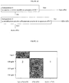
- the chambers are not necessarily isolated from a physical point of view since no exchange is possible between the different conditions. It is thus possible to use a glass slide whose virtual compartments are clearly identified for each concentration as presented in FIG. figure 3 .
- the virtual compartments are defined by delimitations materialized in the present example by a marking previously performed on the lamella on the opposite side to that where the bacteria are deposited.
- the glass slide is then deposited on a "geneframe" constituting the seal J in the previously described conditioning device.
- the identification of the captured bacteria is done by a manual procedure in this example based on the visual analysis by the operator of the image acquired by means of the microspectrometer camera and a light source adapted by an experimenter. This identification makes it possible to acquire a series of at least N Raman spectra acquired on individual bacteria respectively present in each chamber C 0 to C 4 . The number of spectra to be acquired to constitute a data set depends on the level of requirement on the performance of the tests to be performed.
- the data processing mode proposed here is identical to that of Examples 1 and 2: a first pretreatment step followed by a step of classifying the spectra acquired using a classifier previously trained.
- the classifier is trained with a reference base containing "no antibiotic effect" spectra previously acquired in a condition without antibiotic (0 ⁇ g / ml) of amoxicillin of EC10 bacteria and "antibiotic effect” spectra of EC10 bacteria in the presence of 8 ⁇ g / mL of amoxicillin.
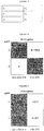
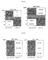
- the results obtained are presented in the matrix of confusion proposed in figure 6 . As before, this matrix makes it possible to demonstrate the robustness of the method by giving the results for a large number of tests.
- an antibiotic molecule known but not belonging to the antibiotic families previously used is used: ciprofloxacin of the family of fluoroquinolones.
- the distance measure obtained is then compared with this threshold value to define the proximity between the selected signature and the difference spectra acquired in the presence of the different concentrations of the molecule deemed unknown. If the distance is below the threshold, the test is positive and an effect of the antibiotic molecule has been detected. If the distance is greater than this threshold then no effect is detected.
- the first which is less strict, makes it possible to detect a significant variation of the tested spectra while the second, which is more strict, makes it possible to qualify a close proximity of the modification with the signature.
- a test of the distance of the spectrum tested at the signature with respect to the first threshold is thus carried out. If the test is not passed, then no effect is detected. If the test is passed, the measured distance is less than the first threshold, so an effect is detected. A second test is then performed using the second threshold and if this test is passed, the "Antibiotic Effect" result is assigned to the complete test.
- Another way to perform an equivalent test would be to perform a test in directly using the Euclidean or other standard of the tested spectrum and comparing it to a threshold of significance chosen to ignore the classical variations related to the measurement mode (sensor noise level, biological variability, etc.) and then perform the test at a single strict threshold. If the standard exceeds a certain threshold then the spectrum is significantly different from a near-zero difference spectrum if there were no changes in the concentration and can then be compared strictly to the reference signature. for example by measuring its distance to the signature in the sense of the standard used.
- a test carried out with an etest allows to measure a MIC of 0.008 ⁇ g / mL which confirms that the result obtained is in accordance with the reference methods [bioMérieux Products ETEST® CI 32 (ref 412311) and VITEK® N233 board (ref 413117)].
- the selected packaging device consists of two fluidic chambers and two antibiotic concentrations are tested: c 0 : “Without antibiotic” and c 1 : “resistance test”.
- the objective of this test is to determine whether the bacterial strain is considered resistant within the meaning of the definitions proposed by EUCAST.
- a solution containing the bacteria to be tested is used as a test sample.
- This solution is obtained by suspending 5.10 7 CFU / mL in order to represent a concentration potentially encountered in a clinical sample, for example a urine sample.
- the bacteria can come from a culture in a liquid medium or a culture in agar medium agar Petri dish.
- This Bacterial solution is brought into contact with the interface I of the functionalized device by the adsorption of polyethylenimine (PEI) (generic capture). After a capture time of 10 minutes allowing the bacteria to come into contact with the functionalization, the interface I is washed with a water solution, this optional step to eliminate the surplus of uncaptured bacteria still in solution.
- PEI polyethylenimine
- the physiological medium used in this example consists of a poorly enriched mixture of BHI Brain Heart Infusion Bouillon (for example of bioMérieux reference 42081) and PBS 10x (in a 1: 9 ratio). After dividing into two fractions of this physiological medium, an amount of oxacillin (for example of the reference O0353 from TCI Europe) is added to each of these fractions, making it possible to obtain a different concentration of gentamicin c 0 or c 1 . The solutions of concentrations c 0 and c 1 thus produced are respectively introduced into the chambers C 0 or C 1 . The bacteria captured directly from the sample on the surface I are thus exposed, in a suitable medium, to a different concentration of antibiotic depending on the chamber in which they are present.
- the device is then heated to reach a temperature of 37 ° C for two hours and then placed in measurement position on the microspectrometer.
- the identification of the captured bacteria is performed by an automatic procedure based on image analysis, by a conventional particle detection procedure, acquired by means of the microspectrometer camera and a suitable light source. This identification makes it possible to automatically acquire a series of Raman spectra (S 0 and S 1 , respectively) acquired on individual bacteria present in each chamber C 0 and C 1 .
- the number of spectra to be acquired to constitute a data set depends on the level of requirement on the performance of the tests to be performed.
- a complete method of processing spectra at the individual level comprising a preprocessing step comprising all the phases described above to maximize the extraction of a signal of interest and the classification.
- N spectra from S 0 and N spectra from S 1 are drawn without discount among the M spectra available.
- Each of the N spectra derived from S 1 and the N spectra from S 0 is subtracted from the average of N spectra derived from S 0 , these two batches of spectra constituting a "control test sample” and a "test sample of resistance ".
- a database is used in this example reference method obtained from similar experiments previously performed at different dates and from different cultures to drive a classifier obtained using a radial core Support Vector Machine (SVM).
- SVM Support Vector Machine
- This classifier is trained to recognize two classes, one "no antibiotic effect” from spectra from conditions without antibiotic and the other "antibiotic effect” from spectra previously acquired under conditions where the concentration is greater than the MIC of the strain (s) used in the reference database.
- SVM Support Vector Machine
- This threshold can also be adjusted to take into account the incubation time: for example, if the time of exposure to antibiotics is significantly reduced, or if the test bacterium has a slower typical doubling time, take account of the incubation time. a lower threshold to assign an "antibiotic effect" result to this group of spectra.
- the strain can therefore be described as sensitive according to the invention with great confidence on the basis of only a few analyzes of individual bacteria.
Landscapes
- Health & Medical Sciences (AREA)
- Life Sciences & Earth Sciences (AREA)
- Chemical & Material Sciences (AREA)
- Engineering & Computer Science (AREA)
- Immunology (AREA)
- Organic Chemistry (AREA)
- Analytical Chemistry (AREA)
- Biochemistry (AREA)
- General Health & Medical Sciences (AREA)
- Physics & Mathematics (AREA)
- Molecular Biology (AREA)
- Wood Science & Technology (AREA)
- Zoology (AREA)
- Proteomics, Peptides & Aminoacids (AREA)
- General Physics & Mathematics (AREA)
- Microbiology (AREA)
- Biotechnology (AREA)
- Pathology (AREA)
- Biomedical Technology (AREA)
- Urology & Nephrology (AREA)
- Hematology (AREA)
- General Engineering & Computer Science (AREA)
- Biophysics (AREA)
- Genetics & Genomics (AREA)
- Toxicology (AREA)
- Nuclear Medicine, Radiotherapy & Molecular Imaging (AREA)
- Bioinformatics & Cheminformatics (AREA)
- Virology (AREA)
- Cell Biology (AREA)
- Tropical Medicine & Parasitology (AREA)
- Food Science & Technology (AREA)
- Medicinal Chemistry (AREA)
- Measuring Or Testing Involving Enzymes Or Micro-Organisms (AREA)
- Investigating, Analyzing Materials By Fluorescence Or Luminescence (AREA)
- Investigating Or Analysing Biological Materials (AREA)
- Apparatus Associated With Microorganisms And Enzymes (AREA)
Claims (16)
- Verfahren zur Bestimmung der Reaktion mindestens einer bestimmten Bakterie auf ihre Exposition gegenüber einem Antibiotikum, das eine Analyse durch Raman-Spektroskopie durchführt und die folgenden Schritte umfasst:Verfügen über eine biologische Probe, die die bestimmten Bakterien enthalten kann,Herstellen von mindestens zwei Fraktionen der Probe, umfassend jeweils eine oder mehrere bestimmte lebende Bakterien,Erfassen in jeder Fraktion mindestens einer bestimmten lebenden Bakterie mit Hilfe eines Bindungspartners,Exponieren mindestens einer der Fraktionen gegenüber mindestens einer Konzentration von mindestens einem gegebenen Antibiotikum, wobei die andere der Fraktionen die Kontrollfraktion ist,Unterziehen der bestimmten Bakterie(n), die in den Fraktionen enthalten ist/sind, an ein einfallendes Licht und Analysieren des sich ergebenden Lichts, erhalten durch Raman-Diffusion durch die bestimmte(n) Bakterie(n) durch Raman-Spektroskopie, um ebenso viele Raman-Spektren als Bakterien zu erhalten,Behandeln der Spektren, um eine Signatur der Reaktion der oder jeder der bestimmten Bakterie(n) beim Exponieren gegenüber dem Antibiotikum und der Kontrolle zu erhalten,Vergleichen der so erhaltenen Signatur pro bestimmter Bakterie mit einer Referenzbasis, die durch die gleichen Bedingungen wie den oben Angegebenen definiert ist, bei verschiedenen Bakterien und mindestens dem Antibiotikum, undDefinieren eines klinischen Profils der Empfindlichkeit der bestimmten Bakterie gegenüber dem Antibiotikum.
- Verfahren nach Anspruch 1, dadurch gekennzeichnet, dass mehr als zwei Fraktionen der Probe hergestellt werden und jeweils mindestens zwei Fraktionen gegenüber zunehmenden Konzentrationen des Antibiotikums exponiert werden.
- Verfahren nach Anspruch 1 oder 2, dadurch gekennzeichnet, dass die Konzentrationen des Antibiotikums, gegenüber denen die Fraktion(en) jeweils exponiert wird/werden, in einem Intervall von Werten enthalten sind, darin eingeschlossen mindestens einen ausgewählten Wert oder Werten, die mindestens ein Intervall bilden, das mindestens einen Wert einschließt, der ausgewählt ist aus den Werten, die für ein Paar Antibiotikum/Art, wie z. B. den epidemiologischen Cutoff, den oder die klinischen Breakpoints oder Konzentrationspaneele, die in den Referenzverfahren verwendet werden, charakteristisch sind.
- Verfahren nach einem der Ansprüche 1 bis 3, dadurch gekennzeichnet, dass die bestimmte(n) Bakterie(n), die in den Fraktionen vorhanden sind, von einem Bindungspartner erfasst werden, der direkt oder indirekt auf einem Träger blockiert wird.
- Verfahren nach Anspruch 4, dadurch gekennzeichnet, dass der Bindungspartner spezifisch mit der/den bestimmten Bakterie(n) interagieren kann und vorzugsweise ausgewählt ist aus Proteinen, Antikörpern, Antigenen, Aptameren, Phagen, Phagenproteinen.
- Verfahren nach einem der Ansprüche 1 bis 5, dadurch gekennzeichnet, dass die erfassten Bakterien ausfindig gemacht und sortiert werden, z. B. durch Bildgebung oder Spektrophotometrie.
- Verfahren nach einem der Ansprüche 1 bis 6, dadurch gekennzeichnet, dass vor dem Unterziehen der bestimmten Bakterie(n) an ein einfallendes Licht die Bakterien, die nicht erfasst wurden, eliminiert werden.
- Verfahren nach Anspruch 7, dadurch gekennzeichnet, dass die Bakterien vor oder nach dem Schritt des Aussetzens gegenüber dem Antibiotikum eliminiert werden.
- Verfahren nach einem der Ansprüche 1 bis 8, dadurch gekennzeichnet, dass nach dem Schritt des Aussetzens der Fraktionen die Fraktionen und die Kontrollfraktion konzentriert werden und sie dann dem Schritt des Erfassens unterzogen werden.
- Verfahren nach einem der Ansprüche 1 bis 9, dadurch gekennzeichnet, dass sich das Antibiotikum in einem physiologischen Medium befindet, das ermöglicht, mindestens die lebende(n) bestimmte(n) Bakterie(n) beizubehalten.
- Verfahren nach einem der Ansprüche 1 bis 10, dadurch gekennzeichnet, dass die Exposition gegenüber dem Antibiotikum bei einer Temperatur von mindestens 18 °C und höchstens 40 °C durchgeführt wird.
- Verfahren nach einem der Ansprüche 1 bis 11, dadurch gekennzeichnet, dass die Exposition gegenüber dem Antibiotikum während eines Zeitraums, bezeichnet als Inkubation, von mindestens 10 Minuten und höchstens 4 Stunden erfolgt.
- Verfahren nach einem der vorhergehenden Ansprüche, dadurch gekennzeichnet, dass, um die Signatur zu erhalten, für jede gegenüber dem Antibiotikum exponierte Fraktion das oder die Raman-Spektren der Kontrollentnahme den entsprechenden Raman-Spektren der Entnahmen entzogen werden.
- Verfahren nach einem der Ansprüche 1 bis 13, dadurch gekennzeichnet, dass der sensible, intermediäre oder resistente Status des bestimmten Stamms gegenüber dem Antibiotikum bestimmt wird.
- Verfahren nach Anspruch 14, dadurch gekennzeichnet, dass die minimale Hemmkonzentration (CMI) des Stamms gegenüber dem Antibiotikum bestimmt wird.
- Verfahren nach Anspruch 14 oder 15, dadurch gekennzeichnet, dass jede Fraktion mindestens 2, vorzugsweise mindestens 5 bestimmte Bakterien umfasst, um mindestens 2, vorzugsweise mindesten 5 Signale zu erhalten.
Applications Claiming Priority (2)
| Application Number | Priority Date | Filing Date | Title |
|---|---|---|---|
| FR1561446A FR3044415B1 (fr) | 2015-11-27 | 2015-11-27 | Procede de determination de la reaction d'un microorganisme a son exposition a un antibiotique |
| PCT/FR2016/053100 WO2017089727A1 (fr) | 2015-11-27 | 2016-11-25 | Procédé de détermination de la réaction d'un microorganisme à son exposition à un composé chimique |
Publications (2)
| Publication Number | Publication Date |
|---|---|
| EP3380828A1 EP3380828A1 (de) | 2018-10-03 |
| EP3380828B1 true EP3380828B1 (de) | 2019-11-06 |
Family
ID=55346010
Family Applications (1)
| Application Number | Title | Priority Date | Filing Date |
|---|---|---|---|
| EP16815603.2A Active EP3380828B1 (de) | 2015-11-27 | 2016-11-25 | Verfahren zur bestimmung der reaktion eines mikroorganismus auf seine exposition gegenüber einer chemischen verbindung |
Country Status (6)
| Country | Link |
|---|---|
| US (1) | US10704075B2 (de) |
| EP (1) | EP3380828B1 (de) |
| JP (1) | JP6912477B2 (de) |
| CN (1) | CN108700521B (de) |
| FR (1) | FR3044415B1 (de) |
| WO (1) | WO2017089727A1 (de) |
Families Citing this family (9)
| Publication number | Priority date | Publication date | Assignee | Title |
|---|---|---|---|---|
| FR3044415B1 (fr) * | 2015-11-27 | 2017-12-01 | Biomerieux Sa | Procede de determination de la reaction d'un microorganisme a son exposition a un antibiotique |
| DE102016113748A1 (de) * | 2016-07-26 | 2018-02-01 | Leibniz-Institut für Photonische Technologien e. V. | Kombiniertes optisch-spektroskopisches Verfahren zur Bestimmung von mikrobiellen Erregern |
| EP3669180B1 (de) | 2017-08-18 | 2024-10-16 | Somadetect Inc. | Verfahren und systeme zur beurteilung eines gesundheitszustands eines stillenden säugers |
| JP7033888B2 (ja) * | 2017-11-08 | 2022-03-11 | 株式会社堀場製作所 | ラマンイメージング装置、ラマンイメージングシステム、ラマンイメージング方法、及び、ラマンイメージング装置用プログラム |
| WO2020183437A1 (en) * | 2019-03-13 | 2020-09-17 | Monash University | Systems and methods for spectral detection of drug-resistant pathogens |
| CN110658178A (zh) * | 2019-09-29 | 2020-01-07 | 江苏拉曼医疗设备有限公司 | 一种用于拉曼光谱的荧光背底扣除方法 |
| FR3103900B1 (fr) * | 2019-11-29 | 2024-07-19 | Univ Du Mans | Méthode d'identification rapide de microorganismes par analyse de matrices excitation-émission |
| EP4125064A1 (de) * | 2021-07-30 | 2023-02-01 | Bacteromic sp. z.o.o. | Verfahren und system zur vorhersage des wachstums von mikroorganismen mittels künstlicher intelligenz |
| CN114544587A (zh) * | 2022-02-15 | 2022-05-27 | 天津医科大学口腔医院 | 一种鉴别牙周致病菌的方法 |
Citations (1)
| Publication number | Priority date | Publication date | Assignee | Title |
|---|---|---|---|---|
| WO2017089427A1 (de) * | 2015-11-23 | 2017-06-01 | Celltool Gmbh | Vorrichtung und verfahren zum analysieren biologischer objekte mit raman spektroskopie |
Family Cites Families (28)
| Publication number | Priority date | Publication date | Assignee | Title |
|---|---|---|---|---|
| US5573927A (en) * | 1992-11-18 | 1996-11-12 | Nelson; Wilfred H. | Antibiotic susceptibility test |
| FR2799281B1 (fr) * | 1999-09-30 | 2002-04-26 | Commissariat Energie Atomique | Procede et dispositif de detection d'une reaction de reconnaissance moleculaire |
| DE10155185A1 (de) * | 2001-11-12 | 2003-05-22 | Synthon Kg | Verfahren zur Charakterisierung und/oder Identifizierung von Wirkmechanismen antimikrobieller Prüfsubstanzen |
| WO2007035814A2 (en) * | 2005-09-20 | 2007-03-29 | Advandx, Inc. | Reagents, methods and kits for classification of fungi and direction of anti-fungal therapy |
| WO2008018933A2 (en) * | 2006-05-03 | 2008-02-14 | The Regents Of The University Of California | Detection of protease and protease activity using a single nanocrescent sers probe |
| US20080220465A1 (en) * | 2007-03-05 | 2008-09-11 | Pocared Diagnostics, Inc. | Antibiotic Sensitivity Testing Method |
| TWI390202B (zh) * | 2007-11-15 | 2013-03-21 | Nat Univ Chung Cheng | The sensing method and system of using nanometer aggregated particles |
| US20110143332A1 (en) * | 2007-11-26 | 2011-06-16 | National Yang-Ming University | Method for identifying microorganism or detecting its morphology alteration using surface enhanced raman scattering (sers) |
| US8519358B2 (en) * | 2008-02-05 | 2013-08-27 | Pocared Diagnostics Ltd. | System for conducting the identification of bacteria in biological samples |
| AU2009289244A1 (en) * | 2008-09-05 | 2010-03-11 | Basf Se | Method for testing substances or substance mixtures and the use thereof |
| KR20110095277A (ko) * | 2008-10-31 | 2011-08-24 | 바이오메리욱스, 인코포레이티드. | 라만 분광법을 이용하여 미생물을 분리, 특성규명, 및/또는 동정하는 방법 |
| JP2010223671A (ja) * | 2009-03-23 | 2010-10-07 | National Yang Ming Univ | 表面増強ラマン散乱(sers)を使用した微生物鑑定またはその形態変化検出方法 |
| US8609364B2 (en) * | 2009-05-07 | 2013-12-17 | bioM{tilde over (e)}rieux, Inc. | Methods for antimicrobial resistance determination |
| DE102009050287B4 (de) * | 2009-10-20 | 2012-07-05 | Friedrich-Schiller-Universität Jena | SERS-Substrat |
| SG181673A1 (en) * | 2009-12-14 | 2012-07-30 | Chiesi Farma Spa | Antibiotic microparticles for inhalation |
| US9329176B2 (en) * | 2010-11-18 | 2016-05-03 | Academia Sinica | Glycopeptide-functionalized nanoparticles arrays for capturing and detecting biomolecules |
| PL2714922T3 (pl) * | 2011-06-03 | 2018-02-28 | Universidade Do Porto | Sposób wykrywania mikroorganizmów opornych na środek terapeutyczny |
| JP2014520575A (ja) * | 2011-08-22 | 2014-08-25 | スペクトラル プラットフォームズ, インコーポレイテッド | 代謝活性の迅速検出 |
| WO2013052915A2 (en) * | 2011-10-05 | 2013-04-11 | Genelux Corporation | Method for detecting replication or colonization of a biological therapeutic |
| EA033790B1 (ru) * | 2011-12-19 | 2019-11-26 | Opticul Diagnostics Ltd | Способ спектрального обнаружения и идентификации микроорганизмов в культуре |
| CA3129741C (en) * | 2013-05-01 | 2023-09-19 | Neoculi Pty Ltd | Compounds and methods of treating infections |
| CN103411955A (zh) * | 2013-08-21 | 2013-11-27 | 天津大学 | 基于拉曼光谱的头孢类抗生素混合溶液的浓度检测方法 |
| FR3028616A1 (fr) * | 2014-11-13 | 2016-05-20 | Commissariat Energie Atomique | Procede d'analyse comprenant la determination d'une position d'une particule biologique. |
| FR3030749B1 (fr) * | 2014-12-19 | 2020-01-03 | Commissariat A L'energie Atomique Et Aux Energies Alternatives | Methode d'identification de particules biologiques par piles d'images holographiques defocalisees |
| FR3044415B1 (fr) * | 2015-11-27 | 2017-12-01 | Biomerieux Sa | Procede de determination de la reaction d'un microorganisme a son exposition a un antibiotique |
| EP3252455A1 (de) * | 2016-05-30 | 2017-12-06 | Biomérieux | Vorrichtung und verfahren zur erfassung eines partikels, das in einer probe enthalten ist |
| JP7154615B2 (ja) * | 2017-01-09 | 2022-10-18 | ポカード・ディアグノスティクス・リミテッド | 単一蛍光膜色素染色およびフローサイトメトリーによる特有のスペクトル強度比分析に基づく迅速抗菌薬感受性試験 |
| US10995358B2 (en) * | 2017-07-13 | 2021-05-04 | Pocared Diagnostics Ltd. | Rapid antibiotic susceptibility test using membrane fluorescence staining and spectral intensity ratio improved by flow cytometry dead to live population ratio |
-
2015
- 2015-11-27 FR FR1561446A patent/FR3044415B1/fr not_active Expired - Fee Related
-
2016
- 2016-11-25 CN CN201680080179.3A patent/CN108700521B/zh active Active
- 2016-11-25 JP JP2018527229A patent/JP6912477B2/ja active Active
- 2016-11-25 WO PCT/FR2016/053100 patent/WO2017089727A1/fr not_active Ceased
- 2016-11-25 EP EP16815603.2A patent/EP3380828B1/de active Active
- 2016-11-25 US US15/779,295 patent/US10704075B2/en active Active
Patent Citations (1)
| Publication number | Priority date | Publication date | Assignee | Title |
|---|---|---|---|---|
| WO2017089427A1 (de) * | 2015-11-23 | 2017-06-01 | Celltool Gmbh | Vorrichtung und verfahren zum analysieren biologischer objekte mit raman spektroskopie |
Also Published As
| Publication number | Publication date |
|---|---|
| US10704075B2 (en) | 2020-07-07 |
| FR3044415A1 (fr) | 2017-06-02 |
| CN108700521B (zh) | 2022-02-25 |
| FR3044415B1 (fr) | 2017-12-01 |
| EP3380828A1 (de) | 2018-10-03 |
| JP6912477B2 (ja) | 2021-08-04 |
| JP2019506590A (ja) | 2019-03-07 |
| US20180355399A1 (en) | 2018-12-13 |
| CN108700521A (zh) | 2018-10-23 |
| WO2017089727A1 (fr) | 2017-06-01 |
Similar Documents
| Publication | Publication Date | Title |
|---|---|---|
| EP3380828B1 (de) | Verfahren zur bestimmung der reaktion eines mikroorganismus auf seine exposition gegenüber einer chemischen verbindung | |
| Cialla-May et al. | Raman spectroscopy and imaging in bioanalytics | |
| Lu et al. | Combination of an artificial intelligence approach and laser tweezers Raman spectroscopy for microbial identification | |
| CN104655840B (zh) | 用于抗微生物剂抗性测定的方法 | |
| Wenning et al. | Identification of microorganisms by FTIR spectroscopy: perspectives and limitations of the method | |
| CN102272602B (zh) | 微生物的分离和鉴定方法 | |
| JP5837420B2 (ja) | 固体又は半固体培地上の微生物のキャラクタリゼーション方法 | |
| CN102203588B (zh) | 利用光谱分离,表征和/或标识微生物的方法 | |
| Hu et al. | Drop‐coating deposition and surface‐enhanced Raman spectroscopies (DCDRS and SERS) provide complementary information of whole human tears | |
| Kotanen et al. | Surface enhanced Raman scattering spectroscopy for detection and identification of microbial pathogens isolated from human serum | |
| EP3257947B1 (de) | Verfahren und system zur identifizierung des gramtyps einer bakterie | |
| EP3728624B1 (de) | Verfahren zur identifizierung von hefe oder bakterien | |
| JP2012507710A (ja) | 同定作用物質を使用した微生物の分離、キャラクタリゼーションおよび/または同定方法 | |
| CN102272585A (zh) | 采用拉曼光谱法分离、表征和/或鉴定微生物的方法 | |
| EP2488847A1 (de) | Verfahren zur optischen detektion gelöster mikrometrischer objekte | |
| Tian et al. | Surface-Enhanced raman analysis of uric acid and hypoxanthine analysis in fractionated bodily fluids | |
| JP2022553332A (ja) | 溶液中の粒子同定のシステムおよび方法 | |
| Dryden et al. | Rapid uropathogen identification using surface enhanced Raman spectroscopy active filters | |
| FR3038620A1 (fr) | Procede de detection d'une presence ou d'une absence d'au moins une premiere zone d'inhibition | |
| JP6182574B2 (ja) | 分光法を使用した微生物の分離、キャラクタリゼーションおよび/または同定方法 | |
| EP1963859A1 (de) | Verfahren zur unterscheidung von wenigstens zwei zellpopulationen und anwendung | |
| Sitjar et al. | Extracting the SERS characteristic features of SARS-CoV-2 and using machine learning to quickly screen positive specimens among hospitalized patients | |
| EP4437493A1 (de) | Verfahren zur bestimmung der empfindlichkeit eines mikroorganismus gegenüber einem antimikrobiellen mittel | |
| EP2997396A1 (de) | Verfahren zur beurteilung, mittels biodosimetrie, der von einer person unter dem einfluss von ionisierender strahlung aufgenommenen strahlendosis | |
| Hobro et al. | Vibrational spectroscopic imaging of pathogens, microorganisms, and their interactions with host systems |
Legal Events
| Date | Code | Title | Description |
|---|---|---|---|
| STAA | Information on the status of an ep patent application or granted ep patent |
Free format text: STATUS: UNKNOWN |
|
| STAA | Information on the status of an ep patent application or granted ep patent |
Free format text: STATUS: THE INTERNATIONAL PUBLICATION HAS BEEN MADE |
|
| PUAI | Public reference made under article 153(3) epc to a published international application that has entered the european phase |
Free format text: ORIGINAL CODE: 0009012 |
|
| STAA | Information on the status of an ep patent application or granted ep patent |
Free format text: STATUS: REQUEST FOR EXAMINATION WAS MADE |
|
| 17P | Request for examination filed |
Effective date: 20180601 |
|
| AK | Designated contracting states |
Kind code of ref document: A1 Designated state(s): AL AT BE BG CH CY CZ DE DK EE ES FI FR GB GR HR HU IE IS IT LI LT LU LV MC MK MT NL NO PL PT RO RS SE SI SK SM TR |
|
| AX | Request for extension of the european patent |
Extension state: BA ME |
|
| DAV | Request for validation of the european patent (deleted) | ||
| DAX | Request for extension of the european patent (deleted) | ||
| GRAP | Despatch of communication of intention to grant a patent |
Free format text: ORIGINAL CODE: EPIDOSNIGR1 |
|
| STAA | Information on the status of an ep patent application or granted ep patent |
Free format text: STATUS: GRANT OF PATENT IS INTENDED |
|
| RIC1 | Information provided on ipc code assigned before grant |
Ipc: C12Q 1/18 20060101ALI20190614BHEP Ipc: G01N 33/569 20060101ALI20190614BHEP Ipc: G01N 21/65 20060101AFI20190614BHEP |
|
| INTG | Intention to grant announced |
Effective date: 20190709 |
|
| GRAS | Grant fee paid |
Free format text: ORIGINAL CODE: EPIDOSNIGR3 |
|
| GRAA | (expected) grant |
Free format text: ORIGINAL CODE: 0009210 |
|
| STAA | Information on the status of an ep patent application or granted ep patent |
Free format text: STATUS: THE PATENT HAS BEEN GRANTED |
|
| AK | Designated contracting states |
Kind code of ref document: B1 Designated state(s): AL AT BE BG CH CY CZ DE DK EE ES FI FR GB GR HR HU IE IS IT LI LT LU LV MC MK MT NL NO PL PT RO RS SE SI SK SM TR |
|
| REG | Reference to a national code |
Ref country code: GB Ref legal event code: FG4D Free format text: NOT ENGLISH |
|
| REG | Reference to a national code |
Ref country code: CH Ref legal event code: EP Ref country code: AT Ref legal event code: REF Ref document number: 1199438 Country of ref document: AT Kind code of ref document: T Effective date: 20191115 |
|
| REG | Reference to a national code |
Ref country code: DE Ref legal event code: R096 Ref document number: 602016023993 Country of ref document: DE |
|
| REG | Reference to a national code |
Ref country code: IE Ref legal event code: FG4D Free format text: LANGUAGE OF EP DOCUMENT: FRENCH |
|
| REG | Reference to a national code |
Ref country code: NL Ref legal event code: MP Effective date: 20191106 |
|
| REG | Reference to a national code |
Ref country code: LT Ref legal event code: MG4D |
|
| PG25 | Lapsed in a contracting state [announced via postgrant information from national office to epo] |
Ref country code: LT Free format text: LAPSE BECAUSE OF FAILURE TO SUBMIT A TRANSLATION OF THE DESCRIPTION OR TO PAY THE FEE WITHIN THE PRESCRIBED TIME-LIMIT Effective date: 20191106 Ref country code: NO Free format text: LAPSE BECAUSE OF FAILURE TO SUBMIT A TRANSLATION OF THE DESCRIPTION OR TO PAY THE FEE WITHIN THE PRESCRIBED TIME-LIMIT Effective date: 20200206 Ref country code: PL Free format text: LAPSE BECAUSE OF FAILURE TO SUBMIT A TRANSLATION OF THE DESCRIPTION OR TO PAY THE FEE WITHIN THE PRESCRIBED TIME-LIMIT Effective date: 20191106 Ref country code: GR Free format text: LAPSE BECAUSE OF FAILURE TO SUBMIT A TRANSLATION OF THE DESCRIPTION OR TO PAY THE FEE WITHIN THE PRESCRIBED TIME-LIMIT Effective date: 20200207 Ref country code: FI Free format text: LAPSE BECAUSE OF FAILURE TO SUBMIT A TRANSLATION OF THE DESCRIPTION OR TO PAY THE FEE WITHIN THE PRESCRIBED TIME-LIMIT Effective date: 20191106 Ref country code: LV Free format text: LAPSE BECAUSE OF FAILURE TO SUBMIT A TRANSLATION OF THE DESCRIPTION OR TO PAY THE FEE WITHIN THE PRESCRIBED TIME-LIMIT Effective date: 20191106 Ref country code: BG Free format text: LAPSE BECAUSE OF FAILURE TO SUBMIT A TRANSLATION OF THE DESCRIPTION OR TO PAY THE FEE WITHIN THE PRESCRIBED TIME-LIMIT Effective date: 20200206 Ref country code: SE Free format text: LAPSE BECAUSE OF FAILURE TO SUBMIT A TRANSLATION OF THE DESCRIPTION OR TO PAY THE FEE WITHIN THE PRESCRIBED TIME-LIMIT Effective date: 20191106 Ref country code: PT Free format text: LAPSE BECAUSE OF FAILURE TO SUBMIT A TRANSLATION OF THE DESCRIPTION OR TO PAY THE FEE WITHIN THE PRESCRIBED TIME-LIMIT Effective date: 20200306 Ref country code: NL Free format text: LAPSE BECAUSE OF FAILURE TO SUBMIT A TRANSLATION OF THE DESCRIPTION OR TO PAY THE FEE WITHIN THE PRESCRIBED TIME-LIMIT Effective date: 20191106 |
|
| PG25 | Lapsed in a contracting state [announced via postgrant information from national office to epo] |
Ref country code: IS Free format text: LAPSE BECAUSE OF FAILURE TO SUBMIT A TRANSLATION OF THE DESCRIPTION OR TO PAY THE FEE WITHIN THE PRESCRIBED TIME-LIMIT Effective date: 20200306 Ref country code: HR Free format text: LAPSE BECAUSE OF FAILURE TO SUBMIT A TRANSLATION OF THE DESCRIPTION OR TO PAY THE FEE WITHIN THE PRESCRIBED TIME-LIMIT Effective date: 20191106 Ref country code: RS Free format text: LAPSE BECAUSE OF FAILURE TO SUBMIT A TRANSLATION OF THE DESCRIPTION OR TO PAY THE FEE WITHIN THE PRESCRIBED TIME-LIMIT Effective date: 20191106 |
|
| PG25 | Lapsed in a contracting state [announced via postgrant information from national office to epo] |
Ref country code: AL Free format text: LAPSE BECAUSE OF FAILURE TO SUBMIT A TRANSLATION OF THE DESCRIPTION OR TO PAY THE FEE WITHIN THE PRESCRIBED TIME-LIMIT Effective date: 20191106 |
|
| REG | Reference to a national code |
Ref country code: CH Ref legal event code: PL |
|
| PG25 | Lapsed in a contracting state [announced via postgrant information from national office to epo] |
Ref country code: ES Free format text: LAPSE BECAUSE OF FAILURE TO SUBMIT A TRANSLATION OF THE DESCRIPTION OR TO PAY THE FEE WITHIN THE PRESCRIBED TIME-LIMIT Effective date: 20191106 Ref country code: RO Free format text: LAPSE BECAUSE OF FAILURE TO SUBMIT A TRANSLATION OF THE DESCRIPTION OR TO PAY THE FEE WITHIN THE PRESCRIBED TIME-LIMIT Effective date: 20191106 Ref country code: LI Free format text: LAPSE BECAUSE OF NON-PAYMENT OF DUE FEES Effective date: 20191130 Ref country code: CZ Free format text: LAPSE BECAUSE OF FAILURE TO SUBMIT A TRANSLATION OF THE DESCRIPTION OR TO PAY THE FEE WITHIN THE PRESCRIBED TIME-LIMIT Effective date: 20191106 Ref country code: CH Free format text: LAPSE BECAUSE OF NON-PAYMENT OF DUE FEES Effective date: 20191130 Ref country code: LU Free format text: LAPSE BECAUSE OF NON-PAYMENT OF DUE FEES Effective date: 20191125 Ref country code: EE Free format text: LAPSE BECAUSE OF FAILURE TO SUBMIT A TRANSLATION OF THE DESCRIPTION OR TO PAY THE FEE WITHIN THE PRESCRIBED TIME-LIMIT Effective date: 20191106 Ref country code: DK Free format text: LAPSE BECAUSE OF FAILURE TO SUBMIT A TRANSLATION OF THE DESCRIPTION OR TO PAY THE FEE WITHIN THE PRESCRIBED TIME-LIMIT Effective date: 20191106 |
|
| REG | Reference to a national code |
Ref country code: DE Ref legal event code: R097 Ref document number: 602016023993 Country of ref document: DE |
|
| REG | Reference to a national code |
Ref country code: AT Ref legal event code: MK05 Ref document number: 1199438 Country of ref document: AT Kind code of ref document: T Effective date: 20191106 |
|
| REG | Reference to a national code |
Ref country code: BE Ref legal event code: MM Effective date: 20191130 |
|
| PG25 | Lapsed in a contracting state [announced via postgrant information from national office to epo] |
Ref country code: SM Free format text: LAPSE BECAUSE OF FAILURE TO SUBMIT A TRANSLATION OF THE DESCRIPTION OR TO PAY THE FEE WITHIN THE PRESCRIBED TIME-LIMIT Effective date: 20191106 Ref country code: SK Free format text: LAPSE BECAUSE OF FAILURE TO SUBMIT A TRANSLATION OF THE DESCRIPTION OR TO PAY THE FEE WITHIN THE PRESCRIBED TIME-LIMIT Effective date: 20191106 Ref country code: MC Free format text: LAPSE BECAUSE OF FAILURE TO SUBMIT A TRANSLATION OF THE DESCRIPTION OR TO PAY THE FEE WITHIN THE PRESCRIBED TIME-LIMIT Effective date: 20191106 |
|
| PLBE | No opposition filed within time limit |
Free format text: ORIGINAL CODE: 0009261 |
|
| STAA | Information on the status of an ep patent application or granted ep patent |
Free format text: STATUS: NO OPPOSITION FILED WITHIN TIME LIMIT |
|
| 26N | No opposition filed |
Effective date: 20200807 |
|
| PG25 | Lapsed in a contracting state [announced via postgrant information from national office to epo] |
Ref country code: IE Free format text: LAPSE BECAUSE OF NON-PAYMENT OF DUE FEES Effective date: 20191125 |
|
| PG25 | Lapsed in a contracting state [announced via postgrant information from national office to epo] |
Ref country code: AT Free format text: LAPSE BECAUSE OF FAILURE TO SUBMIT A TRANSLATION OF THE DESCRIPTION OR TO PAY THE FEE WITHIN THE PRESCRIBED TIME-LIMIT Effective date: 20191106 Ref country code: BE Free format text: LAPSE BECAUSE OF NON-PAYMENT OF DUE FEES Effective date: 20191130 Ref country code: SI Free format text: LAPSE BECAUSE OF FAILURE TO SUBMIT A TRANSLATION OF THE DESCRIPTION OR TO PAY THE FEE WITHIN THE PRESCRIBED TIME-LIMIT Effective date: 20191106 |
|
| PG25 | Lapsed in a contracting state [announced via postgrant information from national office to epo] |
Ref country code: IT Free format text: LAPSE BECAUSE OF FAILURE TO SUBMIT A TRANSLATION OF THE DESCRIPTION OR TO PAY THE FEE WITHIN THE PRESCRIBED TIME-LIMIT Effective date: 20191106 |
|
| PG25 | Lapsed in a contracting state [announced via postgrant information from national office to epo] |
Ref country code: CY Free format text: LAPSE BECAUSE OF FAILURE TO SUBMIT A TRANSLATION OF THE DESCRIPTION OR TO PAY THE FEE WITHIN THE PRESCRIBED TIME-LIMIT Effective date: 20191106 |
|
| PG25 | Lapsed in a contracting state [announced via postgrant information from national office to epo] |
Ref country code: HU Free format text: LAPSE BECAUSE OF FAILURE TO SUBMIT A TRANSLATION OF THE DESCRIPTION OR TO PAY THE FEE WITHIN THE PRESCRIBED TIME-LIMIT; INVALID AB INITIO Effective date: 20161125 Ref country code: MT Free format text: LAPSE BECAUSE OF FAILURE TO SUBMIT A TRANSLATION OF THE DESCRIPTION OR TO PAY THE FEE WITHIN THE PRESCRIBED TIME-LIMIT Effective date: 20191106 |
|
| PG25 | Lapsed in a contracting state [announced via postgrant information from national office to epo] |
Ref country code: TR Free format text: LAPSE BECAUSE OF FAILURE TO SUBMIT A TRANSLATION OF THE DESCRIPTION OR TO PAY THE FEE WITHIN THE PRESCRIBED TIME-LIMIT Effective date: 20191106 |
|
| PG25 | Lapsed in a contracting state [announced via postgrant information from national office to epo] |
Ref country code: MK Free format text: LAPSE BECAUSE OF FAILURE TO SUBMIT A TRANSLATION OF THE DESCRIPTION OR TO PAY THE FEE WITHIN THE PRESCRIBED TIME-LIMIT Effective date: 20191106 |
|
| PGFP | Annual fee paid to national office [announced via postgrant information from national office to epo] |
Ref country code: DE Payment date: 20251118 Year of fee payment: 10 |
|
| PGFP | Annual fee paid to national office [announced via postgrant information from national office to epo] |
Ref country code: GB Payment date: 20251125 Year of fee payment: 10 |
|
| PGFP | Annual fee paid to national office [announced via postgrant information from national office to epo] |
Ref country code: FR Payment date: 20251125 Year of fee payment: 10 |