EP3384010B1 - Utilisation d'une puce microfluidique intégrée dans l'analyse de motilité cellulaire et la prédiction et le pronostic de survie de patients - Google Patents
Utilisation d'une puce microfluidique intégrée dans l'analyse de motilité cellulaire et la prédiction et le pronostic de survie de patients Download PDFInfo
- Publication number
- EP3384010B1 EP3384010B1 EP16871617.3A EP16871617A EP3384010B1 EP 3384010 B1 EP3384010 B1 EP 3384010B1 EP 16871617 A EP16871617 A EP 16871617A EP 3384010 B1 EP3384010 B1 EP 3384010B1
- Authority
- EP
- European Patent Office
- Prior art keywords
- cells
- migratory
- cell
- channels
- channel
- Prior art date
- Legal status (The legal status is an assumption and is not a legal conclusion. Google has not performed a legal analysis and makes no representation as to the accuracy of the status listed.)
- Active
Links
Images








Classifications
-
- G—PHYSICS
- G01—MEASURING; TESTING
- G01N—INVESTIGATING OR ANALYSING MATERIALS BY DETERMINING THEIR CHEMICAL OR PHYSICAL PROPERTIES
- G01N33/00—Investigating or analysing materials by specific methods not covered by groups G01N1/00 - G01N31/00
- G01N33/48—Biological material, e.g. blood, urine; Haemocytometers
- G01N33/50—Chemical analysis of biological material, e.g. blood, urine; Testing involving biospecific ligand binding methods; Immunological testing
- G01N33/53—Immunoassay; Biospecific binding assay; Materials therefor
- G01N33/575—Immunoassay; Biospecific binding assay; Materials therefor for cancer
-
- A—HUMAN NECESSITIES
- A61—MEDICAL OR VETERINARY SCIENCE; HYGIENE
- A61P—SPECIFIC THERAPEUTIC ACTIVITY OF CHEMICAL COMPOUNDS OR MEDICINAL PREPARATIONS
- A61P35/00—Antineoplastic agents
-
- B—PERFORMING OPERATIONS; TRANSPORTING
- B01—PHYSICAL OR CHEMICAL PROCESSES OR APPARATUS IN GENERAL
- B01L—CHEMICAL OR PHYSICAL LABORATORY APPARATUS FOR GENERAL USE
- B01L3/00—Containers or dishes for laboratory use, e.g. laboratory glassware; Droppers
- B01L3/50—Containers for the purpose of retaining a material to be analysed, e.g. test tubes
- B01L3/502—Containers for the purpose of retaining a material to be analysed, e.g. test tubes with fluid transport, e.g. in multi-compartment structures
- B01L3/5027—Containers for the purpose of retaining a material to be analysed, e.g. test tubes with fluid transport, e.g. in multi-compartment structures by integrated microfluidic structures, i.e. dimensions of channels and chambers are such that surface tension forces are important, e.g. lab-on-a-chip
- B01L3/502761—Containers for the purpose of retaining a material to be analysed, e.g. test tubes with fluid transport, e.g. in multi-compartment structures by integrated microfluidic structures, i.e. dimensions of channels and chambers are such that surface tension forces are important, e.g. lab-on-a-chip specially adapted for handling suspended solids or molecules independently from the bulk fluid flow, e.g. for trapping or sorting beads or physically stretching molecules
-
- G—PHYSICS
- G01—MEASURING; TESTING
- G01N—INVESTIGATING OR ANALYSING MATERIALS BY DETERMINING THEIR CHEMICAL OR PHYSICAL PROPERTIES
- G01N33/00—Investigating or analysing materials by specific methods not covered by groups G01N1/00 - G01N31/00
- G01N33/48—Biological material, e.g. blood, urine; Haemocytometers
- G01N33/50—Chemical analysis of biological material, e.g. blood, urine; Testing involving biospecific ligand binding methods; Immunological testing
- G01N33/5005—Chemical analysis of biological material, e.g. blood, urine; Testing involving biospecific ligand binding methods; Immunological testing involving human or animal cells
- G01N33/5008—Chemical analysis of biological material, e.g. blood, urine; Testing involving biospecific ligand binding methods; Immunological testing involving human or animal cells for testing or evaluating the effect of chemical or biological compounds, e.g. drugs, cosmetics
- G01N33/5011—Chemical analysis of biological material, e.g. blood, urine; Testing involving biospecific ligand binding methods; Immunological testing involving human or animal cells for testing or evaluating the effect of chemical or biological compounds, e.g. drugs, cosmetics for testing antineoplastic activity
-
- G—PHYSICS
- G01—MEASURING; TESTING
- G01N—INVESTIGATING OR ANALYSING MATERIALS BY DETERMINING THEIR CHEMICAL OR PHYSICAL PROPERTIES
- G01N33/00—Investigating or analysing materials by specific methods not covered by groups G01N1/00 - G01N31/00
- G01N33/48—Biological material, e.g. blood, urine; Haemocytometers
- G01N33/50—Chemical analysis of biological material, e.g. blood, urine; Testing involving biospecific ligand binding methods; Immunological testing
- G01N33/5005—Chemical analysis of biological material, e.g. blood, urine; Testing involving biospecific ligand binding methods; Immunological testing involving human or animal cells
- G01N33/5008—Chemical analysis of biological material, e.g. blood, urine; Testing involving biospecific ligand binding methods; Immunological testing involving human or animal cells for testing or evaluating the effect of chemical or biological compounds, e.g. drugs, cosmetics
- G01N33/502—Chemical analysis of biological material, e.g. blood, urine; Testing involving biospecific ligand binding methods; Immunological testing involving human or animal cells for testing or evaluating the effect of chemical or biological compounds, e.g. drugs, cosmetics for testing non-proliferative effects
- G01N33/5029—Chemical analysis of biological material, e.g. blood, urine; Testing involving biospecific ligand binding methods; Immunological testing involving human or animal cells for testing or evaluating the effect of chemical or biological compounds, e.g. drugs, cosmetics for testing non-proliferative effects on cell motility
-
- G—PHYSICS
- G01—MEASURING; TESTING
- G01N—INVESTIGATING OR ANALYSING MATERIALS BY DETERMINING THEIR CHEMICAL OR PHYSICAL PROPERTIES
- G01N33/00—Investigating or analysing materials by specific methods not covered by groups G01N1/00 - G01N31/00
- G01N33/48—Biological material, e.g. blood, urine; Haemocytometers
- G01N33/50—Chemical analysis of biological material, e.g. blood, urine; Testing involving biospecific ligand binding methods; Immunological testing
- G01N33/5005—Chemical analysis of biological material, e.g. blood, urine; Testing involving biospecific ligand binding methods; Immunological testing involving human or animal cells
- G01N33/5091—Chemical analysis of biological material, e.g. blood, urine; Testing involving biospecific ligand binding methods; Immunological testing involving human or animal cells for testing the pathological state of an organism
-
- G—PHYSICS
- G01—MEASURING; TESTING
- G01N—INVESTIGATING OR ANALYSING MATERIALS BY DETERMINING THEIR CHEMICAL OR PHYSICAL PROPERTIES
- G01N33/00—Investigating or analysing materials by specific methods not covered by groups G01N1/00 - G01N31/00
- G01N33/48—Biological material, e.g. blood, urine; Haemocytometers
- G01N33/50—Chemical analysis of biological material, e.g. blood, urine; Testing involving biospecific ligand binding methods; Immunological testing
- G01N33/53—Immunoassay; Biospecific binding assay; Materials therefor
- G01N33/575—Immunoassay; Biospecific binding assay; Materials therefor for cancer
- G01N33/57557—Immunoassay; Biospecific binding assay; Materials therefor for cancer of other specific parts of the body, e.g. brain
-
- B—PERFORMING OPERATIONS; TRANSPORTING
- B01—PHYSICAL OR CHEMICAL PROCESSES OR APPARATUS IN GENERAL
- B01L—CHEMICAL OR PHYSICAL LABORATORY APPARATUS FOR GENERAL USE
- B01L2200/00—Solutions for specific problems relating to chemical or physical laboratory apparatus
- B01L2200/06—Fluid handling related problems
- B01L2200/0647—Handling flowable solids, e.g. microscopic beads, cells, particles
- B01L2200/0652—Sorting or classification of particles or molecules
-
- B—PERFORMING OPERATIONS; TRANSPORTING
- B01—PHYSICAL OR CHEMICAL PROCESSES OR APPARATUS IN GENERAL
- B01L—CHEMICAL OR PHYSICAL LABORATORY APPARATUS FOR GENERAL USE
- B01L2300/00—Additional constructional details
- B01L2300/08—Geometry, shape and general structure
- B01L2300/0809—Geometry, shape and general structure rectangular shaped
- B01L2300/0816—Cards, e.g. flat sample carriers usually with flow in two horizontal directions
-
- B—PERFORMING OPERATIONS; TRANSPORTING
- B01—PHYSICAL OR CHEMICAL PROCESSES OR APPARATUS IN GENERAL
- B01L—CHEMICAL OR PHYSICAL LABORATORY APPARATUS FOR GENERAL USE
- B01L2300/00—Additional constructional details
- B01L2300/08—Geometry, shape and general structure
- B01L2300/0861—Configuration of multiple channels and/or chambers in a single devices
- B01L2300/0867—Multiple inlets and one sample wells, e.g. mixing, dilution
-
- G—PHYSICS
- G01—MEASURING; TESTING
- G01N—INVESTIGATING OR ANALYSING MATERIALS BY DETERMINING THEIR CHEMICAL OR PHYSICAL PROPERTIES
- G01N2800/00—Detection or diagnosis of diseases
- G01N2800/70—Mechanisms involved in disease identification
- G01N2800/7023—(Hyper)proliferation
- G01N2800/7028—Cancer
Definitions
- WO 2011/158243 discloses a method of diagnosing a glioma or a breast cancer comprising analyzing an amount or activity of heterogeneous nuclear ribonucleoproteion A2/B1 (hnRNP A2/B1) in a brain cell sample of a subject, wherein an up-regulation in an amount or activity of hnRNP A2/B1 beyond a predetermined threshold with respect to a control sample is indicative of the breast cancer or glioma.
- the method may also be used for staging the cancer and for predicting a patient's prognosis of survival.
- the present invention provides a method for identifying an effective chemotherapeutic treatment regimen in a subject having a tumor, as claimed in claim 10.
- An effective chemotherapeutic treatment regimen is treating a subject with one or more chemotherapeutic agents that will significantly decrease the proportion of migratory cells from the subject, as determined using the apparatus and methods described herein, and therefor inhibit invasiveness of the tumor cells in the subject.
- Multiple devices can be operated in parallel to screen a number of therapeutic agents.
- a method for identifying an agent which inhibits the invasiveness of a cell or population of tumor cells in a sample comprising: a) adding to the integrated microfluidic apparatus a cell or population of cells from the sample and the test agent; b) incubating the cells for a period time; c) imaging the cells in the apparatus for a period of time; d) comparing the images of the cells in the integrated microfluidic apparatus over time; e) identifying a cell or subpopulation of cells in the sample as invasive when the cell or subpopulation of cells migrates past the bifurcation of any of the migratory channels of the apparatus into either of the one or more outlet ends of the migration channels of the integrated microfluidic apparatus and/or exits out of the migration channels; f) comparing the number and/or extent of invasiveness of the cell or subpopulation of cells to the number and/or extent of migration of the cell or subpopulation of cells of e) to the number and/or extent of migration of
- Disclosed herein are methods for identifying a cell or population of cells from a tumor of a subject that are invasive compared to the population of tumor cells as a whole, using an integrated microfluidic apparatus that enables identification of these migratory cells directly from a specimen. These methods and apparatus were first described in International Patent Publication No. WO 2015/009688, filed July 15, 2014 .
- optically transparent materials in some examples allows a wide range of imaging techniques to be used for in situ imaging of migratory and non-migratory cells in the apparatus.
- the apparatus and methods described herein are useful for predicting the invasive propensity of brain tumor cells, predicting survival times and/or risk of recurrence of tumors post-operatively for patients and selecting optimal drugs for personalized therapies.
- the fluid layer of the integrated microfluidic apparatus for use in the present invention comprises at least two channels, each having at least one or more inlets which communicate with a reservoir, and each channel also having at least one or more outlets which communicate with a reservoir.
- the channels can have any dimension within the limits of the depth of the substrate. In some examples, the channels can have dimensions of about 3 ⁇ m to about 15 ⁇ m in height, about 3 ⁇ m to about 50 ⁇ m in width, and about 100 ⁇ m to about 400 ⁇ m in length. There can be any number of channels limited only by the dimensions of the chip used. In some examples, the chip can comprise greater than 200 channels.
- a first channel also termed “a medium channel” which can be filled with any type of biological media or solvent.
- a second channel also termed “a cell channel” which can be filled with any type of biological media or solvent that contains a sample of cells to be assayed.
- the first and second channels are disposed in proximity to each other and are parallel into at least a portion of the two channels in the fluid layer of the substrate.
- the first and second channels have at least one inlet portion which can be the same or have a smaller dimension than the main portion of the first and second channel.
- Each inlet portion is connected to the inlet end of the first and second channel and communicates with the channels.
- Each inlet portion is also in communication with a reservoir wherein media or fluid can be introduced into the inlet of the channel.
- the migratory channel portion is an area where the first channel and second channel are in proximity to each other and are connected by a plurality of migratory channels having at least one inlet and at least one or more outlets.
- the migratory channels are bifurcated at a point distal from the inlet portion of the migratory channel. The bifurcation results in two outlet ends of the migratory channel which communicate with the media channel. These channels are significantly reduced in size, for example, by approximately by a factor of 10, so as to allow one cell body at a time to enter the migratory channel from the cell channel.
- the main portion of the first and second channels has a width of about 400 ⁇ m and a height of about 50 ⁇ m
- the migratory channels have an inlet portion which communicates with the second channel and has a width of about 20 ⁇ m and a height of about 10 ⁇ m.
- the migratory channels can have dimensions of width of about 3 ⁇ m to about 50 ⁇ m, and a height of about 3 ⁇ m to about 15 ⁇ m.
- the one or more outlet portions of the migratory channels can have the same or different widths than the inlet portion of the migratory channel.
- the fluid layer of the substrate is shown generally as (1) and is composed of a polydimethylsiloxane (PDMS) chip molded from a negative replica on a silicon wafer on which photolithography has been used to create a plurality of channels.
- a first channel (2) which has an inlet portion (3) and an outlet portion (4).
- the inlet portion is in communication with three inlets (5), termed “medium inlets” which are channels in the substrate that communicate between the inlet portion (3) and an inlet reservoir (6).
- the first and second channels are between about 10 mm to about 50 mm in length, and have a height/depth of between about 30 ⁇ m to about 100 ⁇ m, and a width of about 100 ⁇ m to about 400 ⁇ m.
- the inlets for the first and second channels have a length of between 2 mm to about 10 mm, a height/depth of between about 30 ⁇ m to about 100 ⁇ m, and a width of about 50 ⁇ m to about 400 ⁇ m.
- the migratory channels have a length of between about 200 to 400 ⁇ m, a height/depth of between about 3 ⁇ m to about 15 ⁇ m, and a width between about 3 ⁇ m to about 50 ⁇ m.
- the apparatus has a coverslip layer (18) which in whole, or in part, is made of an optically transparent substrate. Any optically substrate which is compatible with the fluid layer substrate can be used.
- the coverslip layer is formed of glass. It will be understood by those of ordinary skill that the portion of the coverslip layer that is optically transparent will allow visualization and imaging of the cells in the apparatus in real time.
- the coverslip is bonded using a variety of known means. In an example, the coverslip is bonded to the fluid layer via plasma treatment of about 18 W for a sufficient time, for example, about 2 minutes.
- the fluid layer channels are all treated with a solution of collagen, for example, a solution of about 20 ⁇ g/ml collagen, such as rat tail collagen type I, for about an hour at 37 °C, and then the channels are washed with PBS or similar buffer.
- a solution of collagen for example, a solution of about 20 ⁇ g/ml collagen, such as rat tail collagen type I, for about an hour at 37 °C, and then the channels are washed with PBS or similar buffer.
- extracellular proteins such as laminin, fibronectin, VCAM-1, hyaluronic acid, or gelatin, are used instead of collagen.
- the medium inlet reservoirs (6) and cell inlet reservoir (11) are filled with the same biological medium or buffer.
- other biologically active compounds or molecules can be added to the cell suspension when the cells are introduced into the apparatus, or after the cells have been seeded, to perform a variety of experiments.
- the apparatus and methods described herein include methods for prognostic purposes in which migration of cancer cells to one of the bifurcation branches of the migration channels is associated with a disease state.
- Migration in the device serves as a companion diagnostic with other methods of cancer diagnosis.
- a high migration score indicates that the specific cancer tested has a high propensity to metastasize and indicates that aggressive treatment should be undertaken.
- the inventive methods can be used to predict survivability of a subject with a glioblastoma brain tumor.
- a subject diagnosed with a glioblastoma brain tumor using known means can undergo either a biopsy of the tumor or complete surgical resection of the tumor, or a portion thereof.
- One or more cells from the tumor or biopsy are then suspended in suitable growth media.
- a sample or aliquot of a sufficient number of cells are then added to the microfluidic device as described herein.
- the cells are then imaged over time and the images are observed to determine if any of the cells in the sample have moved from the cell inlet reservoir (11) to the migratory channel (14).
- the term "invasive” refers to a cell or population of cells from a tumor of the subject that display the migratory behavior of migrating into the cell migratory channel (14) of the integrated microfluidic apparatus and migrating past the bifurcation point of the migratory channel (14) or the cells have migrated into the two or more outlet ends (16), or exited out of the outlet ends.
- the inventors have experimental data from brain cancer patients that show that tumors with invasive cells are highly correlated with low survivability and high risk of recurrence ( Fig. 11 ). Without being limited to any particular theory, it is contemplated that these invasive cell subpopulations are tumor or cancer stem cells and highly migratory in vivo.
- a method for identifying the invasiveness of a cancer cell or population of cells in a sample comprising: a) obtaining a cell or population of cancer cells from the sample of a tumor; b) incubating the cells for a period time in a integrative microfluidic apparatus; c) imaging the cells in the apparatus for a period of time; and d) determining whether a cell or subpopulation of cells in the sample are invasive.
- the present invention provides a method for predicting and/or prognosing a brain tumor patient survival and/or recurrence comprising: a) providing a cell or population of cells obtained from the tumor of a patient; b) adding a sufficient sample of the cells from the tumor to the integrated microfluidic apparatus; c) imaging the cells in the integrated microfluidic apparatus for a period of time; d) comparing the images of the cells in the integrated microfluidic apparatus over time; e) determining the number of cells or subpopulation of cells which migrate past the bifurcation of any of the migratory channels of the apparatus into either of the one or more outlet ends of the migration channels of the integrated microfluidic apparatus and/or exits out of the outlet ends of the migration channels; f) identifying the tumor as having low survivability or a high risk of recurrence when the proportion of cells entering through either the wide or narrow outlet ends of the integrated microfluidic apparatus is greater than 5% to about 50%, of all cells
- the migratory or invasive cells migrated into one or the other outlet ends.
- the inventors noted that the proportion of cells which migrated through the narrower of the two outlet ends (3 ⁇ m) also correlated with three-fold less survival time in patients.
- Patients with tumor cell samples having 85% or more wide outlet end migratory cells having greater than two or three-fold survival times over those having less than 85%.
- the apparatus described herein can be used to isolate or separate a cell or subpopulation of cells from a collection of cells in a sample.
- Cells can be separated from the integrated microfluidic apparatus by means of trypsinization or chelation, which allows the cells to detach from the channel walls.
- trypsin or EDTA can be introduced into all of the inlet reservoirs. The cells detach and the flow of the integrated microfluidic apparatus is such that the cells that have migrated through the migratory channels will flow through the first channel and move into the upper outlet reservoir ( Fig. 2A ).
- the migratory cells may be isolated from the integrated microfluidic apparatus and subjected to genomic or proteomic analysis. Such analysis includes, but is not limited to, analysis of gene expression levels using quantitative real-time polymerase chain reaction, RNA sequencing, and surface protein expression levels using flow cytometry.
- biological sample or “biological fluid” includes, but is not limited to, any quantity of cells from a living or formerly living subject. Such cells include, but are not limited to, blood, bone, bone marrow, T-cells, B-cells, fibroblasts, chondrocytes, synovial macrophages, endothelial cells, tumor associated cells, and skin cells.
- the term "subject” refers to any mammal, including, but not limited to, mammals of the order Rodentia, such as mice and hamsters, and mammals of the order Logomorpha, such as rabbits. It is preferred that the mammals are from the order Carnivora, including Felines (cats) and Canines (dogs). It is more preferred that the mammals are from the order Artiodactyla, including Bovines (cows) and Swines (pigs) or of the order Perssodactyla, including Equines (horses). It is most preferred that the mammals are of the order Primates, Ceboids, or Simoids (monkeys) or of the order Anthropoids (humans and apes). An especially preferred mammal is the human.
- mammals of the order Rodentia such as mice and hamsters
- mammals of the order Logomorpha such as rabbits. It is preferred that the mammals are from the order Carnivora, including Felines (cats) and Canines (dogs). It is
- the integrated microfluidic apparatus and methods used may be methods of diagnosis and the migration of cells may be associated with a diseased state, for example, cancer, such as solid tumors.
- cancers such as brain cancers like glioblastoma multiforme, prostate cancer, melanoma, bladder cancer, breast cancer, lymphoma, ovarian cancer, lung cancer, colorectal cancer or head and neck cancer.
- the migration of cells is associated with glioblastoma multiforme.
- migration of cells is associated with an immunological disorder; inflammation; rheumatoid arthritis; cystic fibrosis; or an infection, for example, a viral or bacterial infection.
- the methods of the invention are methods of monitoring prognosis and the migration of cells is associated with the prognosis of glioblastoma multiforme.
- the integrated microfluidic apparatus and methods used are for monitoring drug treatment and the migration of cells is associated with the drug treatment.
- the apparatus and methods used are (e.g., analysis of migration of cells) for the selection of population-oriented drug treatments and/or in prospective studies for selection of dosing, for activity monitoring and/or for determining efficacy endpoints.
- decreased migration upon application of a particular biologically active molecule indicates that that molecule effectively inhibits the movement of migratory or invasive cells.
- the diagnosis can be carried out in a person with or thought to have a disease or condition.
- the diagnosis can also be carried out in a person thought to be at risk for a disease or condition.
- a person at risk is one that has either a genetic predisposition to have the disease or condition or is one that has been exposed to a factor that could increase his/her risk of developing the disease or condition.
- a method for identifying an agent which inhibits the invasiveness of a cell or population of tumor cells in a sample comprising: a) adding to the integrated microfluidic apparatus a cell or population of cells from the sample and the test agent; b) incubating the cells for a period time; c) imaging the cells in the apparatus for a period of time; d) comparing the images of the cells in the integrated microfluidic apparatus over time; e) identifying a cell or subpopulation of cells in the sample as invasive when the cell or subpopulation of cells migrates past the bifurcation of any of the migratory channels of the apparatus into either of the one or more outlet ends of the migration channels of the integrated microfluidic apparatus and/or exits out of the migration channels; f) comparing the number and/or extent of invasiveness of the cell or subpopulation of cells to the number and/or extent of migration of the cell or subpopulation of cells of e) to the number and/or extent of migration of
- Also disclosed, but not claimed, is a method for identifying an agent which inhibits the invasiveness of a cell or population of brain tumor cells in a sample comprising: a) adding to the inlet reservoir of the second channel of the integrated microfluidic apparatus an aliquot of a suspension of a population of cells from the sample and the test agent; b) incubating the cells for a period time to allow the cells to fill the second channel; c) removing any remaining cell suspension from the reservoir of the second channel and washing the inlet of the second channel; d) adding cell media containing the molecule to the one or more reservoirs of the one or more inlets of the first channel; e) imaging the cells in the apparatus for a period of time; f) comparing the images of the cells in the integrated microfluidic apparatus over time and identifying a cell or subpopulation of cells in the sample as invasive when the cell or subpopulation of cells migrates past the bifurcation of any of the migratory channels of the apparatus into either of the one or more outlet ends
- cancer any malignant growth or tumor caused by abnormal and uncontrolled cell division that may spread to other parts of the body through the lymphatic system or the blood stream.
- the cancer can be a metastatic cancer or a non-metastatic (e.g., localized) cancer.
- metastatic cancer refers to a cancer in which cells of the cancer have metastasized, e.g., the cancer is characterized by metastasis of a cancer cells.
- the metastasis can be regional metastasis or distant metastasis, as described herein.
- the apparatus and methods can provide any amount of any level of diagnosis, staging, screening, or other patient management, including treatment or prevention of cancer in a mammal.
- the methods disclosed herein can be used to assess the likelihood of survival and median survival times of patients having brain tumors.
- the brain tumor is a glioblastoma.
- the methods disclosed herein can be used to predict or prognose the survival time of a patient after undergoing a biopsy or resection of the tumor.
- the information is useful to inform the patient as to the status of the cancer. For example, a patient who is diagnosed as having a tumor with a large number of invasive cells, or a greater proportion of cells that enter the wide or narrow exit channels, based on the methods described herein, would be informed of the lower that average survival time expected.
- This information can be used by the patient to determine whether any treatment should be undertaken, or whether a modest or aggressive course of treatment is warranted, depending on the needs of the patient.
- cancers also include but are not limited to adrenal gland cancer, biliary tract cancer; bladder cancer, brain cancer; glioblastoma, breast cancer; cervical cancer; choriocarcinoma; colon cancer; endometrial cancer; esophageal cancer; extrahepatic bile duct cancer; gastric cancer; head and neck cancer; intraepithelial neoplasms; kidney cancer; leukemia; lymphomas; liver cancer; lung cancer (e.g.
- the tumor is a glioblastoma.
- an “active agent or molecule” and a “biologically active agent or molecule” are used interchangeably herein to refer to a chemical or biological compound that induces a desired pharmacological and/or physiological effect, wherein the effect may be prophylactic or therapeutic.
- the terms also encompass pharmaceutically acceptable, pharmacologically active derivatives of those active agents specifically mentioned herein, including, but not limited to, salts, esters, amides, prodrugs, active metabolites, analogs and the like.
- active agent pharmaceutically active agent
- drug drug
- the term includes the active agent per se as well as pharmaceutically acceptable, pharmacologically active salts, esters, amides, prodrugs, metabolites, analogs etc.
- biologically active molecules which can be introduced into the apparatus and used in the methods disclosed herein include, but are not limited to, dyes, including fluorescent, and NIRF dyes, enzymes, and enzyme linked dyes and markers, receptor antagonists or agonists, hormones, growth factors, autogenous bone marrow, antibiotics, antimicrobial agents, and antibodies.
- Non-limiting examples of biologically active agents include following: adrenergic blocking agents, anabolic agents, androgenic steroids, antacids, anti-asthmatic agents, anti-allergenic materials, anti-cholesterolemic and anti-lipid agents, anti-cholinergics and sympathomimetics, anti-coagulants, anticonvulsants, anti-diarrheal, anti-emetics, anti-hypertensive agents, anti-infective agents, anti-inflammatory agents such as steroids, non-steroidal anti-inflammatory agents, anti-malarials, anti-manic agents, anti-nauseants, anti-neoplastic agents, anti-obesity agents, anti-parkinsonian agents, anti-pyretic and analgesic agents, anti-spasmodic agents, antithrombotic agents, anti-uricemic agents, anti-anginal agents, antihistamines, anti-tussives, appetite suppressants, benzophenanthridine alkaloids, biologicals, cardioactive agents, cerebral
- the biologically active molecule is a dye
- the molecule is detected by fluorescence imaging.
- the dyes may be emitters in the visible or near-infrared (NIR) spectrum.
- Known dyes useful in the present invention include carbocyanine, indocarbocyanine, oxacarbocyanine, thüicarbocyanine and merocyanine, polymethine, coumarine, rhodamine, xanthene, fluorescein, boron ⁇ dipyrromethane (BODIPY), Cy5, Cy5.5, Cy7, VivoTag-680, VivoTag-S680, VivoTag-S750, AlexaFluor660, AlexaFluor680, AlexaFluor700, AlexaFluor750, AlexaFluor790, Dy677, Dy676, Dy682, Dy752, Dy780, DyLight547, Dylight647, HiLyte Fluor 647, HiL
- module means that in the presence of the biologically active agent or molecule, the migratory ability of the cell or subpopulation of cells is up regulated or down regulated, such that migration level, or activity is greater than or less than that observed when compared to controls.
- the term “modulate” can mean “inhibit,” but the use of the word “modulate” is not limited to this definition.
- inhibitor means that that in the presence of the biologically active agent or molecule, the migratory ability of the cell or subpopulation of cells is lowered or down regulated when compared to controls.
- the microfluidic device consisted of "Y"-shaped microchannels, with a 20 ⁇ m-wide feeder channel bifurcating to 20 ⁇ m-wide or 3 ⁇ m-wide branches, arrayed between mutually perpendicular cell seeding and cell outlet channels.
- the apparatus was fabricated using multilayer photolithography and replica molding. Photolithography masks were designed using AutoCAD (Autodesk, McLean, VA) and produced by the Photoplot Store (Colorado Springs, CO).
- the master for the device contained a negative mold of the final device and was fabricated using SU-8 3010 positive photoresist (Microchem, Newton, MA).
- SU-8 3010 was spin coated (Single Wafer Spin Processor, Model WS-400A-6NPP-LITE, Laurell Technologies, North Wales, PA) on a cleaned silicon wafer (University Wafer, South Boston, MA) to create a 10 ⁇ m-thick film.
- the film was soft baked on a hot plate and exposed to 170 mJ/cm2 of UV light energy through the chrome-on-glass light field mask using an EVG620 mask aligner (EVG, Austria) to define the microchannels.
- the wafer was baked, post-exposure, to cross link the pattern before development with SU-8 developer.
- a 50 ⁇ m-thick SU-8 3025 film was spun onto the wafer and soft baked.
- a mask defining the medium feed lines was aligned with the channels, and the photoresist was exposed to 250 mJ/cm2 of energy.
- the final master was developed, hard baked, and passivated with a fluorinated silane [(tridecafluoro-1,1,2,2-tetrahydrooctyl)-1-trichlorosilane] (Pfaltz & Bauer, Waterbury, CT) overnight in a vacuum desiccator.
- a fluorinated silane (tridecafluoro-1,1,2,2-tetrahydrooctyl)-1-trichlorosilane] (Pfaltz & Bauer, Waterbury, CT) overnight in a vacuum desiccator.
- PDMS polydimethylsiloxane
- Sylgard® 184 Silicone Elastomer Kit Dow Corning, Midland, MI
- curing at 85 °C for 2 hours.
- Devices were diced, and 6-mm inlet and outlet ports were punched in the PDMS fluid layer.
- the devices and glass coverslips were cleaned with ethanol and DI water and plasma treated for 2 minutes at 18 W (Harrick PDC-32G, Harrick Plasma, Ithaca, NY).
- the device was bonded to the glass slide and coated with 20 ⁇ g/ml rat tail collagen type I (BD, Franklin Lakes, NJ, USA) for 1 hour at 37 °C. Following coating, the channels were washed with DPBS to prepare for cell seeding.
- 20 ⁇ g/ml rat tail collagen type I (BD, Franklin Lakes, NJ, USA) for 1 hour at 37 °C. Following coating, the channels were washed with DPBS to prepare for cell seeding.
- the apparatus termed a “Microchannel Migration Device,” comprises a plurality of Y-shaped microchannels arrayed between cell seeding (second channel) and medium (first channel) lines ( Figure 1A ).
- the microchannels were designed such that 20 ⁇ m base channels bifurcated to 20 ⁇ m and 3 ⁇ m branch channels at a 45° or 65° (from the horizontal) angle (inset, Figure 1A ).
- experiments can be carried out with or without a chemoattractant gradient. If no gradient is desired, growth medium is placed in all four inlet wells, and the topography of the channels is the only driver of migration.
- medium containing a chemoattractant is placed in the uppermost medium inlet well and medium without chemoattractant is placed in the cell inlet and bottom two medium inlet wells, a gradient is formed within the microchannels to induce migration.
- the particular example of the device was formed by bonding a PDMS mold containing the microchannels and medium channels to a glass coverslip ( Figure 1B ).
- Cells were seeded at the bases of the microchannels following gravity-driven flow of suspended cells from the cell inlet well (shown schematically in Figure 1C ).
- Medium was placed in all inlet wells following cell seeding.
- cell seeding and migration were carried out without the need for external pumps or valves, and all flow was driven by gravity. Seeded cells migrated through the channels over the course of the experiment ( Figure 1D ).
- Cell seeding and live cell migration experiments Cells were grown to confluency, trypsinized, and resuspended in serum-free medium at 2 x10 6 cells/ml. 50 ⁇ l of cell suspension was added to the cell inlet well. Cells were incubated in the device for 5-10 minutes at 37 °C to allow initial cell seeding at the base of "Y" channels. The cell suspension was then removed from the cell inlet port. The device was washed with DPBS before the addition of medium to the inlet ports of the device. In select experiments, PI3K activity was inhibited by the addition of 10 ⁇ M LY294002 in the medium through the entire course of migration.
- the migration chamber was moved to a temperature- and CO 2 -controlled stage-top live cell incubator (Okolab, Italy) mounted on the motorized stage of an inverted Nikon Eclipse Ti microscope (Nikon, Tokyo, Japan) with automated controls (NIS-Elements, Nikon). Migrating cells were imaged with a 10x-magnification phase contrast objective every 10 minutes for up to 16 hours.
- Cell position data were used to calculate cell speed over each 10 minute interval, and these speeds were averaged to get an overall average speed for each cell. Additionally, the chemotactic index, defined as the cell displacement divided by the total distance travelled by the cell, was calculated. Cell shape data were used to calculate cell circularity and fit elliptical angle using the Measure function in ImageJ. Statistical significance was assessed with non-paired Student's t-test.
- Migratory cells were further defined as migratory or non-migratory. Migratory cells were defined as those cells which reached the bifurcation in the Y-shaped microchannel; all other cells were defined as non-migratory. Migratory cells were then classified as contact guided or not contact guided. Cells were defined as contact guided if they continued to the branch channel on the side of the base channel on which they were migrating when the bifurcation was reached. Cells that switched walls in the bifurcation region were classified as not contact guided.
- Migratory Cells Cells that had migrated through and exited the channels were washed with a chelator (versene) prior to the addition of 0.25% trypsin to all inlet wells of the device. Hydrodynamic resistance to flow in the narrow microchannels prevented the backflow of cells that had migrated through the microchannels back into the microchannels. Detached cells flowed to the upper outlet well, were collected in culture medium, and were plated in 96-well plates for expansion. Expanded cells were analyzed for the presence of tumor stem cell markers (for example, CD44 or CD271).
- tumor stem cell markers for example, CD44 or CD271.
- ⁇ 300 migratory cells were collected, suspended in 75 ⁇ l of DPBS, mixed with 75 ⁇ l of Matrigel, and injected to the mammary fat pad of an immunodificient mouse. An equal number of control cells that had not migrated through the microchannels were collected and injected in an identical manner. Mice were sacrificed 8 weeks post-injection, and the lungs were histologically analyzed to detect metastases.
- Bifurcating Channels Allow Identification of Migratory Cells: Cell tracking of all MDA-MB-231 cells within the channels revealed two distinct subpopulations: migratory and non-migratory cells ( Figures 2A,B ). 22 ⁇ 3% of human metastatic MDA-MB-231 breast cancer cells were migratory. Interestingly, this subpopulation correlates with the % of MDA-MB-231 (28%) bearing the CD44 + /CD24 - molecular signature 5 that is used to define breast cancer stem cells. Migratory cells, defined as those cells reaching the branch channels, migrated more than twice as fast as non-migratory cells ( Figure 2C ). Migratory cells were also significantly more directional. The chemotactic index of migratory cells increased to 0.91 in comparison to a chemotactic index of 0.37 for nonmigratory cells.
- Circularity a shape factor that decreases as shapes become less circular, was also significantly different between migratory and non-migratory cells.
- Migratory cells were significantly more elongated as they migrated, with a circularity of 0.37.
- Non-migratory cells had an average circularity of 0.58 ( Figure 2F ).
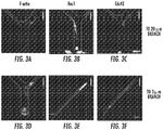
- the apparatus for use in the present invention was used for analysis of cytoskeletal components and intracellular signals via fluorescence microscopy ( Figure 3 ). This was possible because the device was constructed of transparent materials.
- Migratory cells showed increased localization of F-actin to the cell leading edge ( Figure 3A,D ). Actin localization was not seen in non-migratory cells (rounded cell at base of channel, Figure 3D ).
- Rho GTPases Rac1 and Cdc42 were polarized in migratory cells, particularly when these cells reached the 3 ⁇ m-wide branch channel ( Figure 3B,C,E,F ). Non-migratory cells did not exhibit this polarization (for example, cell at channel base in Figure 3B ).
- PI3K Inhibition Promotes Spontaneous Migration of MDA-MB-231 Cells: There is evidence that PI3K signaling is required to stabilize nascent protrusions. New protrusions away from the wall along which a cell is migrating would discourage contact guidance. Therefore, it was investigated whether inhibiting PI3K could promote contact guidance in 200 ⁇ m-long microchannels.
- Migratory cells are more likely to cause metastasis upon orthotopic injection in immunodeficient mice: MDA-MB-231 cells migrated through the device in the absence of a chemotactic cue. Trypsin was added to the all medium inlet wells of the device and caused the migratory cells to become detached and flow to the upper medium outlet well ( Figure 7A ). Resistance to flow through the narrow microchannels prevented detached cells from flowing back into the microchannels ( Figure 7A ; compare cell positions in microchannels before and after removal of migratory cells). Approximately 300 migratory cells were collected in the upper medium outlet well. Trypsin was then added to the cell inlet well. Cells that had not entered the channels flowed to the lower outlet well and were collected.
- Observations using metastatic MDA-MB-231 breast cancer cells were generalized.
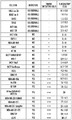
- a panel of cell lines comprised of non-cancerous and non-metastatic (normal) breast epithelial cells, non-metastatic breast cancer cells, and metastatic breast cancer cells was assayed in devices containing 400 ⁇ m-long Y-shaped migration channels ( Figure 8 ).
- an average of 2.6% of migratory cells was observed, with the lowest percentage observed in HMLE Luc 26 cells (0.5 ⁇ 0.8%) and the highest percentage observed in 184A1 cells (5.7 ⁇ 8.0%).
- non-metastatic breast cancer cell lines displayed an average of 20% migratory cells, with a minimum percentage observed in metastatic SUM149 cells (1.2 ⁇ 1.0%) and a maximum observed in metastatic MDA-MB-231 circulating tumor cells (46% ⁇ 18%).
- Examples include: metastatic K-Ras-overexpressing/obscurin-knockdown MCF10A cells (20%), metastatic Bt-549 breast cancer cells (18 ⁇ 3.5%), metastatic MDA-MB-436 breast cancer cells (8.7 ⁇ 0.5%), metastastic MDA-MB-231 breast cancer cells (17 ⁇ 5%), metastatic Hs578t breast cancer cells (16 ⁇ 2%), and metastatic A375 melanoma cells (38 ⁇ 7%).
- the 38 ⁇ 7% of migratory human A375 melanoma cells closely matches the % of A375 cells expressing the cancer stem cell marker CD271.
- the inventors then determined a cutoff range for the percentage of migratory cells observed in a cell line that predicted whether a tested cell line is non-metastatic (if the percentage of migratory cells observed in a cell line was below the cutoff) or metastatic (if the percentage of migratory cells observed in a cell line was above the cutoff).
- a range of cutoff values from 5% to 15% was considered; this range was selected because it was between the average percentage of migratory cells observed in non-metastatic cell lines (2-2.6%) and metastatic cell lines (20%).
- the classification of each cell line predicted by the cutoff value was compared to the known classification of that cell line (listed in Figure 8 ).
- the accuracy (defined as the percentage of classifications correctly predicted by the cutoff compared to those listed in Figure 8 ) was calculated for each cutoff value. For cutoff values of 7-8%, the highest accuracy value was calculated (96%), so this range was chosen as the cutoff between non-migratory and migratory cell lines. For the cutoff range of 7-8%, the sensitivity of the device (defined as the percentage of metastatic cell lines listed in Figure 8 that were predicted to be metastatic by the cutoff) was 90%, and the specificity (defined as the percentage of non-metastatic cell lines listed in Figure 8 that were predicted to be non-metastatic by the cutoff) was 100%.
- Triple negative breast cancer cells display divergent responses to pharmaceutical agents: A panel of triple-negative breast cancer (TNBC) cell lines were assayed in devices containing 400 ⁇ m-long Y-shaped migration channels in the presence or absence of the PI3K inhibitor LY294002 (10 ⁇ M) ( Figure 9 ). PI3K inhibition did not affect the percentage of migratory cells measured for MDA-MB-436 (13 ⁇ 7% migratory for control vs. 18 ⁇ 7% for treated) cells or Hs578t (20 ⁇ 7% migratory for control vs. 15 ⁇ 2% migratory for treated) cells. PI3K inhibition increased migration of MDA-MB-231 (22 ⁇ 3% for control vs. 34 ⁇ 8% for treated) cells. In contrast, PI3K inhibition reduced the migration of Bt549 (32 ⁇ 8% for control vs. 19 ⁇ 1% for treated) cells.
- TNBC triple-negative breast cancer
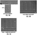
- Metastatic propensity assay is amenable to a wide range of imaging techniques: Numerous imaging techniques were used to image an example in which a PDMS fluidic layer is bonded to a glass coverslip layer. Cells were imaged using lens-free holography ( Figure 10A ), which has an approximately 5 mm x 5 mm field of view. Cells were clearly visible in the channels upon digitally zooming in (inset, Figure 10A ). Additionally, cells in the device were imaged using phase contrast microscopy ( Figure 10B ) and a combination of phase contrast and fluorescence microscopy ( Figure 10C ; a subset of cells was tagged with a fluorescent marker and appears green in the image).
- migratory i.e., cells that entered the migratory channel and entered one of the two outlet ends during the observational interval
- the percentage of migratory cells in the patient sample was then correlated with patient survival time in months.
- a cutoff of 14.5% migratory cells was established by computing the mean % migratory value and separating the patients above or below this mean value.
- the patients' survival times were compared to the 14.6 month standard average, to arrive at a group with high survival (>14.6 mo.) and a low survival group ( ⁇ 14.6 mo.).
- the data was then correlated to the percentage of cells in their sample that were identified as migratory.
- the inventors found a three-fold difference in median survival times of patients with ⁇ 14.5% migratory cells in their sample vs. >14.5% migratory cells.
- Fig 2 shows the same type of analysis as Fig. 1 , but the correlation was to those migratory cells entering the wide (20 ⁇ m) outlet end.
- Fig. 1 shows the same type of analysis as Fig. 1 , but the correlation was to those migratory cells entering the wide (20 ⁇ m) outlet end.
- Fig. 3 is a normalized graph of the data with a cutoff of 1.8%, the inventors did not observe a significant correlation of migratory cells entering the narrow outlet end with low survivability in these patients.
- the median survival time was about two-fold greater for those patients with ⁇ 1.8% migratory cells entering the narrow outlet end.
- the inventors found a clearly significant correlation between survival time and the percentage of cells entered the wide outlet end ( Fig. 4 ). With an 85.7% cutoff, the mean survival time for patients with less wide outlet end entry cells was almost two-fold greater, and patients with >85.7% narrow outlet end entry had significantly lower than average survival times of 8.9 vs. 18.8 months.
- the inventors then analyzed the normalized narrow outlet end entry data in the same manner as Fig. 4 .
- the inventors divided the groups into two cohorts of >14.28% wide outlet end entry, and ⁇ 14.28% wide outlet end entry.
- the Kaplan-Meier survival graph shows a significant difference between each of the curves. When plotted, the data show that patients with the greatest proportion of narrow outlet end entry cells had the longest median survival times (23.3 months), greater than three-fold over the group with the lowest proportion of narrow outlet end entry cells (8.5 months).
- the cutoff and the amount of time during which cell migration is observed can be optimized to allow the methods described herein to identify metastatic and nonmetastatic cell populations from different types of cancer.
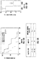
- the accuracy, sensitivity and specificity of the methods are displayed in Figure 17 across a wide range of cutoff values and experiment durations.
- the maximum value for accuracy was 96%, and was observed under three pairs of conditions: 7% cutoff and 13 hours, 8% and 12 hours, and 8% and 13 hours. Under each of these pairs of conditions, the sensitivity is 90% and the specificity is 100%.
Landscapes
- Health & Medical Sciences (AREA)
- Life Sciences & Earth Sciences (AREA)
- Engineering & Computer Science (AREA)
- Chemical & Material Sciences (AREA)
- Immunology (AREA)
- Biomedical Technology (AREA)
- Hematology (AREA)
- Urology & Nephrology (AREA)
- Molecular Biology (AREA)
- General Health & Medical Sciences (AREA)
- Cell Biology (AREA)
- Analytical Chemistry (AREA)
- Physics & Mathematics (AREA)
- Medicinal Chemistry (AREA)
- Biochemistry (AREA)
- Food Science & Technology (AREA)
- Biotechnology (AREA)
- Microbiology (AREA)
- General Physics & Mathematics (AREA)
- Pathology (AREA)
- Tropical Medicine & Parasitology (AREA)
- Toxicology (AREA)
- Bioinformatics & Cheminformatics (AREA)
- Chemical Kinetics & Catalysis (AREA)
- Fluid Mechanics (AREA)
- Dispersion Chemistry (AREA)
- Clinical Laboratory Science (AREA)
- Physiology (AREA)
- Animal Behavior & Ethology (AREA)
- Public Health (AREA)
- Veterinary Medicine (AREA)
- General Chemical & Material Sciences (AREA)
- Nuclear Medicine, Radiotherapy & Molecular Imaging (AREA)
- Pharmacology & Pharmacy (AREA)
- Organic Chemistry (AREA)
- Measuring Or Testing Involving Enzymes Or Micro-Organisms (AREA)
- Apparatus Associated With Microorganisms And Enzymes (AREA)
- Hospice & Palliative Care (AREA)
- Oncology (AREA)
- Investigating Or Analysing Biological Materials (AREA)
Claims (10)
- Un procédé pour prédire et/ou pronostiquer la survie d'un patient atteint d'une tumeur cérébrale glioblastome et/ou la récurrence de celle-ci comprenant :a) mettre à disposition une cellule ou u population de cellules obtenues à partir d'une tumeur cérébrale glioblastome du patient ;b) ajouter un échantillon suffisant des cellules provenant de la tumeur dans un appareil microfluidique intégré, l'appareil microfluidique intégré comprenant un substrat se présentant sous la forme d'une puce comprenant une couche pour fluide (1) et une couche formant lamelle (18), qui sont liées ensemble au moment de l'assemblage final de l'appareil, dans lequel :la couche pour fluide (1) comprend au moins un premier canal (2) et un deuxième canal (8), chacun ayant au moins un ou plusieurs orifices d'introduction (5, 9) qui communiquent avec un réservoir d'orifice d'introduction (6, 11), et chaque canal ayant aussi au moins un ou plusieurs orifices d'évacuation (4, 10) qui communiquent avec un réservoir d'orifice d'évacuation (7, 12) ;les premier et deuxième canaux (2, 8) sont disposés à proximité l'un de l'autre et au moins une portion des premier et deuxième canaux est parallèle dans la couche pour fluide (1) du substrat ;les premier et deuxième canaux (2, 8) sont connectés par une pluralité de canaux migratoires (14) ayant au moins un orifice d'introduction (15) en communication avec le deuxième canal (8) et deux orifices d'évacuation ou plus (16) en communication avec le premier canal (2) ; etla pluralité de canaux migratoires (14) sont bifurqués au niveau d'un point de bifurcation (17) distal relativement à l'au moins un orifice d'introduction (15) du canal migratoire (14) et sont de taille significativement réduite de manière à laisser entrer un corps cellulaire à la fois dans le canal migratoire (14) en provenance du deuxième canal (8) ;c) incuber les cellules pendant une période de temps dans l'appareil microfluidique intégré ;d) former des images des cellules dans l'appareil microfluidique intégré pendant une période de temps ;e) déterminer si une cellule ou sous-population de cellules dans l'échantillon est invasive, dans lequel la cellule ou sous-population de cellules est déterminée comme étant invasive lorsque la cellule ou sous-population de cellules migre dans la pluralité de canaux migratoires (14) de l'appareil de b) au-delà du point de bifurcation (17) ou lorsque la cellule ou sous-population de cellules migre dans la pluralité de canaux migratoires (14) de l'appareil de b) jusque dans les extrémités des deux orifices d'évacuation ou plus (16) ou lorsque la cellule ou sous-population de cellules migre dans la pluralité de canaux migratoires (14) de l'appareil de b) et sort par les extrémités des deux orifices d'évacuation ou plus (16) ; etf) identifier le patient comme ayant une capacité de survie faible ou ayant un risque élevé de récurrence de tumeur cérébrale glioblastome lorsque la proportion de cellules invasives est supérieure à une plage seuil des cellules migratoires.
- Le procédé tel que revendiqué dans la revendication 1, dans lequel l'étape f) comprend le fait d'identifier le patient comme ayant une capacité de survie faible ou un risque élevé de récurrence de tumeur cérébrale lorsque la proportion de cellules invasives est supérieure à 5 à 25 % des cellules dans l'échantillon.
- Le procédé tel que revendiqué dans la revendication 1, dans lequel les extrémités des deux orifices d'évacuation ou plus (16) des canaux migratoires (14) comprennent une extrémité d'orifice d'évacuation large et une extrémité d'orifice d'évacuation étroite, dans lequel dans e), la cellule ou sous-population de cellules est déterminée comme étant invasive lorsque la cellule ou sous-population de cellules migre au-delà du point de bifurcation (17) de n'importe lesquels des canaux migratoires (14) de l'appareil jusque dans les extrémités de l'un ou l'autre des deux orifices d'évacuation ou plus (16) et/ou sort par les extrémités des orifices d'évacuation (16) des canaux de migration (14), et dans lequel f) comprend le fait d'identifier le patient comme ayant une capacité de survie faible ou un risque élevé de récurrence de tumeur cérébrale lorsque la proportion de cellules entrant par soit les extrémités larges, soit les extrémités étroites des deux orifices d'évacuation ou plus (16) de l'appareil est supérieure à 5 % à 50 % de l'ensemble des cellules.
- Le procédé de la revendication 3, dans lequel l'étape f) comprend le fait d'identifier le patient comme ayant une capacité de survie faible ou un risque élevé de récurrence de tumeur cérébrale lorsque la proportion de cellules entrant par soit les extrémités larges, soit les extrémités étroites des deux orifices d'évacuation ou plus (16) de l'appareil est supérieure à 10 % à 25 % de l'ensemble des cellules.
- Le procédé de la revendication 3, dans lequel lorsque la proportion de cellules invasives entrant par les extrémités larges des deux orifices d'évacuation ou plus (16) est de 85 % ou plus, le patient est identifié comme ayant une durée de survie plus de deux à trois fois plus élevée que ceux ayant moins de 85 % de cellules invasives entrant par les extrémités larges des deux orifices d'évacuation ou plus (16).
- Le procédé de n'importe quelle revendication précédente, dans lequel le patient est un humain.
- Le procédé de l'une quelconque des revendications 1 à 6, dans lequel à l'étape d), des images des cellules sont formées pendant 6 heures ou plus.
- Le procédé de l'une quelconque des revendications 1 à 6, dans lequel à l'étape d), des images des cellules sont formées pendant 12 heures.
- Le procédé de l'une quelconque des revendications 1 à 8, dans lequel des images des cellules sont formées à l'aide d'un procédé sélectionné dans le groupe constitué de la microscopie à contraste de phase, en champ clair, à contraste interférentiel différentiel, à fluorescence, confocale et de l'holographie en ligne.
- Utilisation des procédés de n'importe lesquelles des revendications 1 à 9 afin d'identifier un agent thérapeutique optimal pour le traitement du patient.
Priority Applications (1)
| Application Number | Priority Date | Filing Date | Title |
|---|---|---|---|
| EP20208309.3A EP3831934A1 (fr) | 2015-12-02 | 2016-12-02 | Utilisation d'une puce microfluidique integree dans l'analyse de motilite cellulaire et la prediction et le pronostic de survie de patients |
Applications Claiming Priority (2)
| Application Number | Priority Date | Filing Date | Title |
|---|---|---|---|
| US201562262158P | 2015-12-02 | 2015-12-02 | |
| PCT/US2016/064725 WO2017096232A1 (fr) | 2015-12-02 | 2016-12-02 | Utilisation d'une puce microfluidique intégrée dans l'analyse de motilité cellulaire et la prédiction et le pronostic de survie de patients |
Related Child Applications (1)
| Application Number | Title | Priority Date | Filing Date |
|---|---|---|---|
| EP20208309.3A Division EP3831934A1 (fr) | 2015-12-02 | 2016-12-02 | Utilisation d'une puce microfluidique integree dans l'analyse de motilite cellulaire et la prediction et le pronostic de survie de patients |
Publications (3)
| Publication Number | Publication Date |
|---|---|
| EP3384010A1 EP3384010A1 (fr) | 2018-10-10 |
| EP3384010A4 EP3384010A4 (fr) | 2019-04-17 |
| EP3384010B1 true EP3384010B1 (fr) | 2020-11-25 |
Family
ID=58797968
Family Applications (2)
| Application Number | Title | Priority Date | Filing Date |
|---|---|---|---|
| EP16871617.3A Active EP3384010B1 (fr) | 2015-12-02 | 2016-12-02 | Utilisation d'une puce microfluidique intégrée dans l'analyse de motilité cellulaire et la prédiction et le pronostic de survie de patients |
| EP20208309.3A Withdrawn EP3831934A1 (fr) | 2015-12-02 | 2016-12-02 | Utilisation d'une puce microfluidique integree dans l'analyse de motilite cellulaire et la prediction et le pronostic de survie de patients |
Family Applications After (1)
| Application Number | Title | Priority Date | Filing Date |
|---|---|---|---|
| EP20208309.3A Withdrawn EP3831934A1 (fr) | 2015-12-02 | 2016-12-02 | Utilisation d'une puce microfluidique integree dans l'analyse de motilite cellulaire et la prediction et le pronostic de survie de patients |
Country Status (3)
| Country | Link |
|---|---|
| US (2) | US11747338B2 (fr) |
| EP (2) | EP3384010B1 (fr) |
| WO (1) | WO2017096232A1 (fr) |
Families Citing this family (12)
| Publication number | Priority date | Publication date | Assignee | Title |
|---|---|---|---|---|
| KR101987065B1 (ko) * | 2017-08-07 | 2019-06-10 | 주식회사 싸이토젠 | Eml4-alk유전자 변이 분석방법 |
| USD1066733S1 (en) * | 2018-03-22 | 2025-03-11 | Nikon Corporation | Flow channel plate of a cartridge for medical testing equipment |
| USD1016324S1 (en) | 2020-07-08 | 2024-02-27 | NanoCav, LLC | Biological cell processing chip |
| JP1680501S (fr) * | 2020-08-21 | 2021-03-08 | ||
| JP1680214S (fr) * | 2020-08-21 | 2021-03-01 | ||
| JP1680216S (fr) * | 2020-08-21 | 2021-03-01 | ||
| JP1680217S (fr) * | 2020-08-21 | 2021-03-01 | ||
| JP1680215S (fr) * | 2020-08-21 | 2021-03-01 | ||
| US20230357856A1 (en) * | 2020-09-14 | 2023-11-09 | The Johns Hopkins University | Methods and compositions for prognosing glioblastoma or breast cancer |
| EP4056271A1 (fr) | 2021-03-10 | 2022-09-14 | Cline Scientific AB | Quantification de la migration cellulaire et du potentiel métastatique des cellules tumorales |
| USD983405S1 (en) * | 2021-04-15 | 2023-04-11 | Cytoaurora Biotechnologies, Inc. | Microfluidic chip |
| EP4366873A4 (fr) * | 2021-07-08 | 2025-08-06 | Ourotech Inc | Systèmes et procédés de prédiction d'efficacité de traitement |
Citations (1)
| Publication number | Priority date | Publication date | Assignee | Title |
|---|---|---|---|---|
| WO2011158243A2 (fr) * | 2010-06-16 | 2011-12-22 | Yissum Research Development Company Of The Hebrew University Of Jerusalem Ltd. | Procédé de diagnostic et de traitement du cancer |
Family Cites Families (5)
| Publication number | Priority date | Publication date | Assignee | Title |
|---|---|---|---|---|
| EP1327132A4 (fr) | 2000-09-21 | 2004-03-31 | Dna Sciences Inc | Procede et systeme servant a injecter un specimen |
| KR100738071B1 (ko) | 2005-01-21 | 2007-07-12 | 삼성전자주식회사 | 농도구배발생부가 구비된 유전영동 장치, 그를 이용한물질의 분리방법 및 물질 분리의 최적 조건을 탐색하는 방법 |
| US9127530B2 (en) | 2009-08-07 | 2015-09-08 | Schlumberger Technology Corporation | Collision avoidance system with offset wellbore vibration analysis |
| US20120216601A1 (en) | 2011-02-28 | 2012-08-30 | The General Hospital Corporation | Microfluidic devices for analyzing cell motility |
| WO2015009688A1 (fr) * | 2013-07-17 | 2015-01-22 | The Johns Hopkins University | Puce microfluidique pour analyse de motilité cellulaire et méthodes d'utilisation associées |
-
2016
- 2016-12-02 EP EP16871617.3A patent/EP3384010B1/fr active Active
- 2016-12-02 US US15/780,768 patent/US11747338B2/en active Active
- 2016-12-02 WO PCT/US2016/064725 patent/WO2017096232A1/fr not_active Ceased
- 2016-12-02 EP EP20208309.3A patent/EP3831934A1/fr not_active Withdrawn
-
2023
- 2023-08-09 US US18/446,969 patent/US12493037B2/en active Active
Patent Citations (1)
| Publication number | Priority date | Publication date | Assignee | Title |
|---|---|---|---|---|
| WO2011158243A2 (fr) * | 2010-06-16 | 2011-12-22 | Yissum Research Development Company Of The Hebrew University Of Jerusalem Ltd. | Procédé de diagnostic et de traitement du cancer |
Also Published As
| Publication number | Publication date |
|---|---|
| US20200249232A1 (en) | 2020-08-06 |
| EP3831934A1 (fr) | 2021-06-09 |
| US12493037B2 (en) | 2025-12-09 |
| US20240044898A1 (en) | 2024-02-08 |
| EP3384010A4 (fr) | 2019-04-17 |
| US11747338B2 (en) | 2023-09-05 |
| EP3384010A1 (fr) | 2018-10-10 |
| WO2017096232A1 (fr) | 2017-06-08 |
Similar Documents
| Publication | Publication Date | Title |
|---|---|---|
| US12493037B2 (en) | Use of an integrated microfluidic chip for analysis of cell motility and prediction and prognosis of patient survival | |
| US12201977B2 (en) | Microfluidic chip for analysis of cell motility and methods for using same | |
| Rizvi et al. | Flow induces epithelial-mesenchymal transition, cellular heterogeneity and biomarker modulation in 3D ovarian cancer nodules | |
| Minn et al. | Distinct organ-specific metastatic potential of individual breast cancer cells and primary tumors | |
| US10775380B2 (en) | Rare cell isolation device and method of use thereof | |
| JP6404824B2 (ja) | 循環腫瘍細胞のインビトロでの捕捉および解析 | |
| Logun et al. | Microfluidics in malignant glioma research and precision medicine | |
| Zhao et al. | Establishment and characterization of a CTC cell line from peripheral blood of breast cancer patient | |
| Pelosi et al. | What clinicians are asking pathologists when dealing with lung neuroendocrine neoplasms? | |
| Yu et al. | Single cell mechanotype and associated molecular changes in urothelial cell transformation and progression | |
| Chen et al. | Mechanobiology in oncology: basic concepts and clinical prospects | |
| Anjanappa et al. | Individualized breast cancer characterization through single-cell analysis of tumor and adjacent normal cells | |
| Chang et al. | Microfluidic analysis for separating and measuring the deformability of cancer cell subpopulations | |
| EP4042160B1 (fr) | Utilisation des propriétés mécaniques des cellules dans les méthodes de détection du cancer | |
| Sakibuzzaman et al. | Pathology of breast cancer metastasis and a view of metastasis to the brain | |
| Gunel | 218; Meningioma Driver Mutations Determine Their Anatomical Site of Origin | |
| Omenn | Genetic advances will influence the practice of medicine: examples from cancer research and care of cancer patients | |
| WO2017035315A1 (fr) | Procédés et dispositif pour la classification phénotypique de cellules basée sur le comportement migratoire | |
| Datar et al. | Significance of Studying Circulating Tumor Cells | |
| Cheng et al. | A Conserved Geometric Code: Extracellular Matrix Curvature Directs Cell Migration Strategy via Nuclear Mechanosensing | |
| Chislock et al. | DTC-Flow: a flow cytometry-based detection platform for characterizing bone marrow disseminated tumor cells in breast cancer | |
| Crestani et al. | SP2G: an imaging and analysis pipeline revealing the inter and intra-patient migratory diversity of glioblastoma | |
| Yeoman | Assessing Cell Mechanotype as a Prognostic Marker for Metastatic Risk and Recurrence of Cancer | |
| Hasanain et al. | Circulating Tumor Cells as Biomarkers in Pancreatic Cancer | |
| WO2024064866A2 (fr) | Ciblage thérapeutique du cancer basé sur le profilage de cellules tumorales du cancer |
Legal Events
| Date | Code | Title | Description |
|---|---|---|---|
| STAA | Information on the status of an ep patent application or granted ep patent |
Free format text: STATUS: THE INTERNATIONAL PUBLICATION HAS BEEN MADE |
|
| PUAI | Public reference made under article 153(3) epc to a published international application that has entered the european phase |
Free format text: ORIGINAL CODE: 0009012 |
|
| STAA | Information on the status of an ep patent application or granted ep patent |
Free format text: STATUS: REQUEST FOR EXAMINATION WAS MADE |
|
| 17P | Request for examination filed |
Effective date: 20180626 |
|
| AK | Designated contracting states |
Kind code of ref document: A1 Designated state(s): AL AT BE BG CH CY CZ DE DK EE ES FI FR GB GR HR HU IE IS IT LI LT LU LV MC MK MT NL NO PL PT RO RS SE SI SK SM TR |
|
| AX | Request for extension of the european patent |
Extension state: BA ME |
|
| DAV | Request for validation of the european patent (deleted) | ||
| DAX | Request for extension of the european patent (deleted) | ||
| A4 | Supplementary search report drawn up and despatched |
Effective date: 20190320 |
|
| RIC1 | Information provided on ipc code assigned before grant |
Ipc: C12N 5/22 20060101ALI20190314BHEP Ipc: G01N 33/50 20060101ALI20190314BHEP Ipc: C12N 5/09 20100101ALI20190314BHEP Ipc: C12N 5/071 20100101AFI20190314BHEP Ipc: A61P 35/00 20060101ALI20190314BHEP |
|
| STAA | Information on the status of an ep patent application or granted ep patent |
Free format text: STATUS: EXAMINATION IS IN PROGRESS |
|
| 17Q | First examination report despatched |
Effective date: 20191122 |
|
| GRAP | Despatch of communication of intention to grant a patent |
Free format text: ORIGINAL CODE: EPIDOSNIGR1 |
|
| STAA | Information on the status of an ep patent application or granted ep patent |
Free format text: STATUS: GRANT OF PATENT IS INTENDED |
|
| INTG | Intention to grant announced |
Effective date: 20200608 |
|
| RIN1 | Information on inventor provided before grant (corrected) |
Inventor name: QUINONES-HINOJOSA, ALFREDO Inventor name: PAUL, COLIN Inventor name: KONSTANTOPOULOS, KONSTANTINOS |
|
| GRAS | Grant fee paid |
Free format text: ORIGINAL CODE: EPIDOSNIGR3 |
|
| GRAA | (expected) grant |
Free format text: ORIGINAL CODE: 0009210 |
|
| STAA | Information on the status of an ep patent application or granted ep patent |
Free format text: STATUS: THE PATENT HAS BEEN GRANTED |
|
| AK | Designated contracting states |
Kind code of ref document: B1 Designated state(s): AL AT BE BG CH CY CZ DE DK EE ES FI FR GB GR HR HU IE IS IT LI LT LU LV MC MK MT NL NO PL PT RO RS SE SI SK SM TR |
|
| REG | Reference to a national code |
Ref country code: GB Ref legal event code: FG4D |
|
| REG | Reference to a national code |
Ref country code: CH Ref legal event code: EP |
|
| REG | Reference to a national code |
Ref country code: AT Ref legal event code: REF Ref document number: 1338328 Country of ref document: AT Kind code of ref document: T Effective date: 20201215 |
|
| REG | Reference to a national code |
Ref country code: DE Ref legal event code: R096 Ref document number: 602016048815 Country of ref document: DE |
|
| REG | Reference to a national code |
Ref country code: IE Ref legal event code: FG4D |
|
| REG | Reference to a national code |
Ref country code: AT Ref legal event code: MK05 Ref document number: 1338328 Country of ref document: AT Kind code of ref document: T Effective date: 20201125 |
|
| REG | Reference to a national code |
Ref country code: NL Ref legal event code: MP Effective date: 20201125 |
|
| PG25 | Lapsed in a contracting state [announced via postgrant information from national office to epo] |
Ref country code: NO Free format text: LAPSE BECAUSE OF FAILURE TO SUBMIT A TRANSLATION OF THE DESCRIPTION OR TO PAY THE FEE WITHIN THE PRESCRIBED TIME-LIMIT Effective date: 20210225 Ref country code: PT Free format text: LAPSE BECAUSE OF FAILURE TO SUBMIT A TRANSLATION OF THE DESCRIPTION OR TO PAY THE FEE WITHIN THE PRESCRIBED TIME-LIMIT Effective date: 20210325 Ref country code: RS Free format text: LAPSE BECAUSE OF FAILURE TO SUBMIT A TRANSLATION OF THE DESCRIPTION OR TO PAY THE FEE WITHIN THE PRESCRIBED TIME-LIMIT Effective date: 20201125 Ref country code: GR Free format text: LAPSE BECAUSE OF FAILURE TO SUBMIT A TRANSLATION OF THE DESCRIPTION OR TO PAY THE FEE WITHIN THE PRESCRIBED TIME-LIMIT Effective date: 20210226 Ref country code: FI Free format text: LAPSE BECAUSE OF FAILURE TO SUBMIT A TRANSLATION OF THE DESCRIPTION OR TO PAY THE FEE WITHIN THE PRESCRIBED TIME-LIMIT Effective date: 20201125 |
|
| PG25 | Lapsed in a contracting state [announced via postgrant information from national office to epo] |
Ref country code: SE Free format text: LAPSE BECAUSE OF FAILURE TO SUBMIT A TRANSLATION OF THE DESCRIPTION OR TO PAY THE FEE WITHIN THE PRESCRIBED TIME-LIMIT Effective date: 20201125 Ref country code: AT Free format text: LAPSE BECAUSE OF FAILURE TO SUBMIT A TRANSLATION OF THE DESCRIPTION OR TO PAY THE FEE WITHIN THE PRESCRIBED TIME-LIMIT Effective date: 20201125 Ref country code: BG Free format text: LAPSE BECAUSE OF FAILURE TO SUBMIT A TRANSLATION OF THE DESCRIPTION OR TO PAY THE FEE WITHIN THE PRESCRIBED TIME-LIMIT Effective date: 20210225 Ref country code: PL Free format text: LAPSE BECAUSE OF FAILURE TO SUBMIT A TRANSLATION OF THE DESCRIPTION OR TO PAY THE FEE WITHIN THE PRESCRIBED TIME-LIMIT Effective date: 20201125 Ref country code: IS Free format text: LAPSE BECAUSE OF FAILURE TO SUBMIT A TRANSLATION OF THE DESCRIPTION OR TO PAY THE FEE WITHIN THE PRESCRIBED TIME-LIMIT Effective date: 20210325 Ref country code: LV Free format text: LAPSE BECAUSE OF FAILURE TO SUBMIT A TRANSLATION OF THE DESCRIPTION OR TO PAY THE FEE WITHIN THE PRESCRIBED TIME-LIMIT Effective date: 20201125 |
|
| REG | Reference to a national code |
Ref country code: LT Ref legal event code: MG9D |
|
| PG25 | Lapsed in a contracting state [announced via postgrant information from national office to epo] |
Ref country code: HR Free format text: LAPSE BECAUSE OF FAILURE TO SUBMIT A TRANSLATION OF THE DESCRIPTION OR TO PAY THE FEE WITHIN THE PRESCRIBED TIME-LIMIT Effective date: 20201125 |
|
| PG25 | Lapsed in a contracting state [announced via postgrant information from national office to epo] |
Ref country code: SM Free format text: LAPSE BECAUSE OF FAILURE TO SUBMIT A TRANSLATION OF THE DESCRIPTION OR TO PAY THE FEE WITHIN THE PRESCRIBED TIME-LIMIT Effective date: 20201125 Ref country code: LT Free format text: LAPSE BECAUSE OF FAILURE TO SUBMIT A TRANSLATION OF THE DESCRIPTION OR TO PAY THE FEE WITHIN THE PRESCRIBED TIME-LIMIT Effective date: 20201125 Ref country code: EE Free format text: LAPSE BECAUSE OF FAILURE TO SUBMIT A TRANSLATION OF THE DESCRIPTION OR TO PAY THE FEE WITHIN THE PRESCRIBED TIME-LIMIT Effective date: 20201125 Ref country code: CZ Free format text: LAPSE BECAUSE OF FAILURE TO SUBMIT A TRANSLATION OF THE DESCRIPTION OR TO PAY THE FEE WITHIN THE PRESCRIBED TIME-LIMIT Effective date: 20201125 Ref country code: SK Free format text: LAPSE BECAUSE OF FAILURE TO SUBMIT A TRANSLATION OF THE DESCRIPTION OR TO PAY THE FEE WITHIN THE PRESCRIBED TIME-LIMIT Effective date: 20201125 Ref country code: RO Free format text: LAPSE BECAUSE OF FAILURE TO SUBMIT A TRANSLATION OF THE DESCRIPTION OR TO PAY THE FEE WITHIN THE PRESCRIBED TIME-LIMIT Effective date: 20201125 |
|
| REG | Reference to a national code |
Ref country code: CH Ref legal event code: PL |
|
| REG | Reference to a national code |
Ref country code: DE Ref legal event code: R097 Ref document number: 602016048815 Country of ref document: DE |
|
| PG25 | Lapsed in a contracting state [announced via postgrant information from national office to epo] |
Ref country code: MC Free format text: LAPSE BECAUSE OF FAILURE TO SUBMIT A TRANSLATION OF THE DESCRIPTION OR TO PAY THE FEE WITHIN THE PRESCRIBED TIME-LIMIT Effective date: 20201125 Ref country code: DK Free format text: LAPSE BECAUSE OF FAILURE TO SUBMIT A TRANSLATION OF THE DESCRIPTION OR TO PAY THE FEE WITHIN THE PRESCRIBED TIME-LIMIT Effective date: 20201125 |
|
| REG | Reference to a national code |
Ref country code: BE Ref legal event code: MM Effective date: 20201231 |
|
| PLBE | No opposition filed within time limit |
Free format text: ORIGINAL CODE: 0009261 |
|
| STAA | Information on the status of an ep patent application or granted ep patent |
Free format text: STATUS: NO OPPOSITION FILED WITHIN TIME LIMIT |
|
| PG25 | Lapsed in a contracting state [announced via postgrant information from national office to epo] |
Ref country code: AL Free format text: LAPSE BECAUSE OF FAILURE TO SUBMIT A TRANSLATION OF THE DESCRIPTION OR TO PAY THE FEE WITHIN THE PRESCRIBED TIME-LIMIT Effective date: 20201125 Ref country code: IE Free format text: LAPSE BECAUSE OF NON-PAYMENT OF DUE FEES Effective date: 20201202 Ref country code: NL Free format text: LAPSE BECAUSE OF FAILURE TO SUBMIT A TRANSLATION OF THE DESCRIPTION OR TO PAY THE FEE WITHIN THE PRESCRIBED TIME-LIMIT Effective date: 20201125 Ref country code: IT Free format text: LAPSE BECAUSE OF FAILURE TO SUBMIT A TRANSLATION OF THE DESCRIPTION OR TO PAY THE FEE WITHIN THE PRESCRIBED TIME-LIMIT Effective date: 20201125 Ref country code: LU Free format text: LAPSE BECAUSE OF NON-PAYMENT OF DUE FEES Effective date: 20201202 |
|
| 26N | No opposition filed |
Effective date: 20210826 |
|
| PG25 | Lapsed in a contracting state [announced via postgrant information from national office to epo] |
Ref country code: LI Free format text: LAPSE BECAUSE OF NON-PAYMENT OF DUE FEES Effective date: 20201231 Ref country code: CH Free format text: LAPSE BECAUSE OF NON-PAYMENT OF DUE FEES Effective date: 20201231 Ref country code: SI Free format text: LAPSE BECAUSE OF FAILURE TO SUBMIT A TRANSLATION OF THE DESCRIPTION OR TO PAY THE FEE WITHIN THE PRESCRIBED TIME-LIMIT Effective date: 20201125 |
|
| PG25 | Lapsed in a contracting state [announced via postgrant information from national office to epo] |
Ref country code: ES Free format text: LAPSE BECAUSE OF FAILURE TO SUBMIT A TRANSLATION OF THE DESCRIPTION OR TO PAY THE FEE WITHIN THE PRESCRIBED TIME-LIMIT Effective date: 20201125 |
|
| PG25 | Lapsed in a contracting state [announced via postgrant information from national office to epo] |
Ref country code: IS Free format text: LAPSE BECAUSE OF FAILURE TO SUBMIT A TRANSLATION OF THE DESCRIPTION OR TO PAY THE FEE WITHIN THE PRESCRIBED TIME-LIMIT Effective date: 20210325 Ref country code: TR Free format text: LAPSE BECAUSE OF FAILURE TO SUBMIT A TRANSLATION OF THE DESCRIPTION OR TO PAY THE FEE WITHIN THE PRESCRIBED TIME-LIMIT Effective date: 20201125 Ref country code: MT Free format text: LAPSE BECAUSE OF FAILURE TO SUBMIT A TRANSLATION OF THE DESCRIPTION OR TO PAY THE FEE WITHIN THE PRESCRIBED TIME-LIMIT Effective date: 20201125 Ref country code: CY Free format text: LAPSE BECAUSE OF FAILURE TO SUBMIT A TRANSLATION OF THE DESCRIPTION OR TO PAY THE FEE WITHIN THE PRESCRIBED TIME-LIMIT Effective date: 20201125 |
|
| PG25 | Lapsed in a contracting state [announced via postgrant information from national office to epo] |
Ref country code: MK Free format text: LAPSE BECAUSE OF FAILURE TO SUBMIT A TRANSLATION OF THE DESCRIPTION OR TO PAY THE FEE WITHIN THE PRESCRIBED TIME-LIMIT Effective date: 20201125 |
|
| PG25 | Lapsed in a contracting state [announced via postgrant information from national office to epo] |
Ref country code: BE Free format text: LAPSE BECAUSE OF NON-PAYMENT OF DUE FEES Effective date: 20201231 |
|
| P01 | Opt-out of the competence of the unified patent court (upc) registered |
Effective date: 20230515 |
|
| PGFP | Annual fee paid to national office [announced via postgrant information from national office to epo] |
Ref country code: GB Payment date: 20241227 Year of fee payment: 9 |
|
| PGFP | Annual fee paid to national office [announced via postgrant information from national office to epo] |
Ref country code: FR Payment date: 20241226 Year of fee payment: 9 |
|
| PGFP | Annual fee paid to national office [announced via postgrant information from national office to epo] |
Ref country code: DE Payment date: 20241227 Year of fee payment: 9 |