EP3388083A1 - Zusammensetzungen und verfahren zur behandlung von krebs - Google Patents
Zusammensetzungen und verfahren zur behandlung von krebs Download PDFInfo
- Publication number
- EP3388083A1 EP3388083A1 EP18153044.5A EP18153044A EP3388083A1 EP 3388083 A1 EP3388083 A1 EP 3388083A1 EP 18153044 A EP18153044 A EP 18153044A EP 3388083 A1 EP3388083 A1 EP 3388083A1
- Authority
- EP
- European Patent Office
- Prior art keywords
- myo
- nog
- cells
- cell targeting
- targeting molecule
- Prior art date
- Legal status (The legal status is an assumption and is not a legal conclusion. Google has not performed a legal analysis and makes no representation as to the accuracy of the status listed.)
- Withdrawn
Links
Images






Classifications
-
- C—CHEMISTRY; METALLURGY
- C07—ORGANIC CHEMISTRY
- C07K—PEPTIDES
- C07K16/00—Immunoglobulins [IG], e.g. monoclonal or polyclonal antibodies
- C07K16/18—Immunoglobulins [IG], e.g. monoclonal or polyclonal antibodies against material from animals or humans
- C07K16/28—Immunoglobulins [IG], e.g. monoclonal or polyclonal antibodies against material from animals or humans against receptors, cell surface antigens or cell surface determinants
-
- A—HUMAN NECESSITIES
- A61—MEDICAL OR VETERINARY SCIENCE; HYGIENE
- A61K—PREPARATIONS FOR MEDICAL, DENTAL OR TOILETRY PURPOSES
- A61K47/00—Medicinal preparations characterised by the non-active ingredients used, e.g. carriers or inert additives; Targeting or modifying agents chemically bound to the active ingredient
- A61K47/50—Medicinal preparations characterised by the non-active ingredients used, e.g. carriers or inert additives; Targeting or modifying agents chemically bound to the active ingredient the non-active ingredient being chemically bound to the active ingredient, e.g. polymer-drug conjugates
- A61K47/51—Medicinal preparations characterised by the non-active ingredients used, e.g. carriers or inert additives; Targeting or modifying agents chemically bound to the active ingredient the non-active ingredient being chemically bound to the active ingredient, e.g. polymer-drug conjugates the non-active ingredient being a modifying agent
- A61K47/54—Medicinal preparations characterised by the non-active ingredients used, e.g. carriers or inert additives; Targeting or modifying agents chemically bound to the active ingredient the non-active ingredient being chemically bound to the active ingredient, e.g. polymer-drug conjugates the non-active ingredient being a modifying agent the modifying agent being an organic compound
-
- A—HUMAN NECESSITIES
- A61—MEDICAL OR VETERINARY SCIENCE; HYGIENE
- A61K—PREPARATIONS FOR MEDICAL, DENTAL OR TOILETRY PURPOSES
- A61K47/00—Medicinal preparations characterised by the non-active ingredients used, e.g. carriers or inert additives; Targeting or modifying agents chemically bound to the active ingredient
- A61K47/50—Medicinal preparations characterised by the non-active ingredients used, e.g. carriers or inert additives; Targeting or modifying agents chemically bound to the active ingredient the non-active ingredient being chemically bound to the active ingredient, e.g. polymer-drug conjugates
- A61K47/51—Medicinal preparations characterised by the non-active ingredients used, e.g. carriers or inert additives; Targeting or modifying agents chemically bound to the active ingredient the non-active ingredient being chemically bound to the active ingredient, e.g. polymer-drug conjugates the non-active ingredient being a modifying agent
- A61K47/54—Medicinal preparations characterised by the non-active ingredients used, e.g. carriers or inert additives; Targeting or modifying agents chemically bound to the active ingredient the non-active ingredient being chemically bound to the active ingredient, e.g. polymer-drug conjugates the non-active ingredient being a modifying agent the modifying agent being an organic compound
- A61K47/549—Sugars, nucleosides, nucleotides or nucleic acids
-
- A—HUMAN NECESSITIES
- A61—MEDICAL OR VETERINARY SCIENCE; HYGIENE
- A61P—SPECIFIC THERAPEUTIC ACTIVITY OF CHEMICAL COMPOUNDS OR MEDICINAL PREPARATIONS
- A61P35/00—Antineoplastic agents
-
- A—HUMAN NECESSITIES
- A61—MEDICAL OR VETERINARY SCIENCE; HYGIENE
- A61P—SPECIFIC THERAPEUTIC ACTIVITY OF CHEMICAL COMPOUNDS OR MEDICINAL PREPARATIONS
- A61P35/00—Antineoplastic agents
- A61P35/04—Antineoplastic agents specific for metastasis
-
- A—HUMAN NECESSITIES
- A61—MEDICAL OR VETERINARY SCIENCE; HYGIENE
- A61P—SPECIFIC THERAPEUTIC ACTIVITY OF CHEMICAL COMPOUNDS OR MEDICINAL PREPARATIONS
- A61P43/00—Drugs for specific purposes, not provided for in groups A61P1/00-A61P41/00
-
- C—CHEMISTRY; METALLURGY
- C12—BIOCHEMISTRY; BEER; SPIRITS; WINE; VINEGAR; MICROBIOLOGY; ENZYMOLOGY; MUTATION OR GENETIC ENGINEERING
- C12Q—MEASURING OR TESTING PROCESSES INVOLVING ENZYMES, NUCLEIC ACIDS OR MICROORGANISMS; COMPOSITIONS OR TEST PAPERS THEREFOR; PROCESSES OF PREPARING SUCH COMPOSITIONS; CONDITION-RESPONSIVE CONTROL IN MICROBIOLOGICAL OR ENZYMOLOGICAL PROCESSES
- C12Q1/00—Measuring or testing processes involving enzymes, nucleic acids or microorganisms; Compositions therefor; Processes of preparing such compositions
- C12Q1/68—Measuring or testing processes involving enzymes, nucleic acids or microorganisms; Compositions therefor; Processes of preparing such compositions involving nucleic acids
- C12Q1/6876—Nucleic acid products used in the analysis of nucleic acids, e.g. primers or probes
- C12Q1/6883—Nucleic acid products used in the analysis of nucleic acids, e.g. primers or probes for diseases caused by alterations of genetic material
- C12Q1/6886—Nucleic acid products used in the analysis of nucleic acids, e.g. primers or probes for diseases caused by alterations of genetic material for cancer
-
- G—PHYSICS
- G01—MEASURING; TESTING
- G01N—INVESTIGATING OR ANALYSING MATERIALS BY DETERMINING THEIR CHEMICAL OR PHYSICAL PROPERTIES
- G01N33/00—Investigating or analysing materials by specific methods not covered by groups G01N1/00 - G01N31/00
- G01N33/48—Biological material, e.g. blood, urine; Haemocytometers
- G01N33/50—Chemical analysis of biological material, e.g. blood, urine; Testing involving biospecific ligand binding methods; Immunological testing
- G01N33/53—Immunoassay; Biospecific binding assay; Materials therefor
- G01N33/575—Immunoassay; Biospecific binding assay; Materials therefor for cancer
- G01N33/5758—Immunoassay; Biospecific binding assay; Materials therefor for cancer involving compounds serving as markers for tumours, cancers or neoplasias, e.g. cellular determinants, receptors, heat shock/stress proteins, A-protein, oligosaccharides or metabolites
-
- G—PHYSICS
- G01—MEASURING; TESTING
- G01N—INVESTIGATING OR ANALYSING MATERIALS BY DETERMINING THEIR CHEMICAL OR PHYSICAL PROPERTIES
- G01N33/00—Investigating or analysing materials by specific methods not covered by groups G01N1/00 - G01N31/00
- G01N33/48—Biological material, e.g. blood, urine; Haemocytometers
- G01N33/50—Chemical analysis of biological material, e.g. blood, urine; Testing involving biospecific ligand binding methods; Immunological testing
- G01N33/53—Immunoassay; Biospecific binding assay; Materials therefor
- G01N33/575—Immunoassay; Biospecific binding assay; Materials therefor for cancer
- G01N33/5758—Immunoassay; Biospecific binding assay; Materials therefor for cancer involving compounds serving as markers for tumours, cancers or neoplasias, e.g. cellular determinants, receptors, heat shock/stress proteins, A-protein, oligosaccharides or metabolites
- G01N33/5759—Immunoassay; Biospecific binding assay; Materials therefor for cancer involving compounds serving as markers for tumours, cancers or neoplasias, e.g. cellular determinants, receptors, heat shock/stress proteins, A-protein, oligosaccharides or metabolites involving compounds localised on the membrane of tumour or cancer cells
-
- C—CHEMISTRY; METALLURGY
- C12—BIOCHEMISTRY; BEER; SPIRITS; WINE; VINEGAR; MICROBIOLOGY; ENZYMOLOGY; MUTATION OR GENETIC ENGINEERING
- C12Q—MEASURING OR TESTING PROCESSES INVOLVING ENZYMES, NUCLEIC ACIDS OR MICROORGANISMS; COMPOSITIONS OR TEST PAPERS THEREFOR; PROCESSES OF PREPARING SUCH COMPOSITIONS; CONDITION-RESPONSIVE CONTROL IN MICROBIOLOGICAL OR ENZYMOLOGICAL PROCESSES
- C12Q2600/00—Oligonucleotides characterized by their use
- C12Q2600/158—Expression markers
-
- G—PHYSICS
- G01—MEASURING; TESTING
- G01N—INVESTIGATING OR ANALYSING MATERIALS BY DETERMINING THEIR CHEMICAL OR PHYSICAL PROPERTIES
- G01N2333/00—Assays involving biological materials from specific organisms or of a specific nature
- G01N2333/435—Assays involving biological materials from specific organisms or of a specific nature from animals; from humans
- G01N2333/705—Assays involving receptors, cell surface antigens or cell surface determinants
- G01N2333/70585—CD44
-
- G—PHYSICS
- G01—MEASURING; TESTING
- G01N—INVESTIGATING OR ANALYSING MATERIALS BY DETERMINING THEIR CHEMICAL OR PHYSICAL PROPERTIES
- G01N2800/00—Detection or diagnosis of diseases
- G01N2800/50—Determining the risk of developing a disease
Definitions
- the present invention relates to the field of cancer therapy. Specifically, compositions and methods for detecting, prognosing, inhibiting, treating, and/or preventing cancer are disclosed.
- the methods comprise administering to the subject a therapeutically effective amount of at least one Myo/Nog cell targeting molecule and at least one cytotoxic molecule.
- the Myo/Nog cell targeting molecule may be conjugated to the cytotoxic molecule(s).
- the method further comprises at least one other anti-cancer measure, such as the administration of at least one other chemotherapeutic agent, tumor excision, and/or administration of radiation therapy.
- the method comprises detecting Myo/Nog cells in a biological sample obtained from a subject, wherein an increase in Myo/Nog cells in the biological sample and/or localization compared to a corresponding biological sample in a normal subject is indicative of an increased risk of cancer or metastatic potential in the test subject.
- the method comprises contacting the biological sample with at least one Myo/Nog cell targeting molecule conjugated to at least one detection molecule.
- Bone morphogenetic proteins are powerful regulators of proliferation, differentiation and apoptosis in many embryonic and adult tissues ( Bragdon et al. (2011) Cell Signal 23:609-620 ). In postnatal skin, BMP signaling plays a key role in controlling normal epidermal homeostasis, hair follicle growth and melanogenesis ( Botchkarev, V.A. (2003) J. Invest. Dermatol., 120:36-47 ; Owens et al. (2008) J. Invest. Dermatol., 128:783-790 ).
- Targeted knockout of the BMP receptor 1A (BMPR-1A) in the skin inhibits the differentiation of cells that produce the hair shaft ( Yuhki et al.
- BMPs are also involved in tissue remodeling processes such as wound healing, psoriasis and tumorigenesis ( Ming et al. (2004) Genesis 39:10-25 ; Kaiser et al. (1998) J. Invest. Dermatol., 111:1145-1152 ; Blessing et al. (1996) J.
- BMP-6 is elevated in keratinocytes adjacent to sites of hyperproliferation and migration during normal wound healing and is abnormally expressed throughout all suprabasal layers of chronic wounds and psoriatic lesions ( Kaiser et al. (1998) J. Invest. Dermatol., 111:1145-1152 ; Wall et al. (1993) J. Cell Biol., 120:493-502 ). Overexpression of BMP-6 in the skin significantly delays wound healing ( Kaiser et al. (1998) J. Invest. Dermatol., 111:1145-1152 ).
- BMP-6 represses epidermal cell proliferation and the formation of carcinogen-induced skin tumors ( Blessing et al. (1996) J. Cell Biol., 135:227-239 ; Drozdoff et al. (1994) Proc. Natl. Acad. Sci., 91:5528-5532 ; Wach et al. (2001) Oncogene 20:7761-7769 ), while inhibition of BMP signaling via knockout of BMPR-1A or Smad4 results in the formation of tumors derived from hair follicle cells ( Ming et al. (2004) Genesis 39:10-25 ; Blessing et al. (1996) J.
- BMPs The activity of BMPs is regulated, in part, by a number of secreted BMP antagonists, including noggin, chordin, follistatin, and Cerberus/DAN/gremlin ( Bragdon et al. (2011) Cell Signal 23:609-620 ; Walsh et al. (2010) Trends Cell Biol., 20:244-256 ).
- Noggin is expressed in dermal papillae and follicles in anagen phase skin where it promotes new hair growth ( Botchkarev et al. (2001) FASEB J., 15:2205-2214 ).
- Mice lacking noggin display impaired hair follicle induction and morphogenesis ( Botchkarev et al.
- Myo/Nog cells were identified by their expression of mRNA for the skeletal muscle specific transcription factor MyoD, noggin and the G8 cell surface antigen ( Gerhart et al. (2011) Dev. Biol., 359:12-25 ; Gerhart et al. (2006) J. Cell Biol., 175:283-292 ; Gerhart et al. (2009) Dev. Biol., 336:30-41 ; Gerhart et al. (2001) J. Cell Biol., 155:381-392 ; Gerhart et al. (2000) J. Cell Biol., 149:825-834 ; Strony et al. (2005) Gene Expr. Patterns 5:387-395 ).
- Myo/Nog cells express MyoD mRNA and the bone morphogenetic protein inhibitor noggin. In the chick embryo, Myo/Nog cells are required for normal morphogenesis and differentiation and they respond to wounding. Herein, it was determined whether Myo/Nog cells are present in normal mammalian skin and whether they react to epidermal abrasion and tumorigenesis. Myo/Nog cells are the source of noggin in hair follicles. Myo/Nog cells are scarce within the interfollicular dermis and are absent in the epidermis of murine and human skin. Within 24 hours of abrading murine skin, Myo/Nog cells increase in number in the follicles and appear in the dermis surrounding the hair bulb and the wound.
- Myo/Nog cells are also recruited to the stroma of tumors formed from Ras transformed keratinocytes (Ker/Ras). Noggin blocks the inhibitory effect of BMP-4 on Ker/Ras proliferation in vitro.
- Human squamous cell carcinomas and malignant melanomas contain significantly more Myo/Nog cells than basal cell carcinomas.
- Myo/Nog cells are distinct from macrophages, granulocytes and cells expressing alpha smooth muscle actin.
- Myo/Nog cells are key regulators of skin homeostasis and wound healing and are diagnostic and therapeutic targets in skin cancer.
- Myo/Nog cells are indispensable regulators of BMP signaling during chick embryo morphogenesis and differentiation ( Gerhart et al. (2011) Dev. Biol., 359:12-25 ; Gerhart et al. (2006) J. Cell Biol., 175:283-292 ; Gerhart et al. (2009) Dev. Biol., 336:30-41 ).
- the presence of Myo/Nog cells in skin of adult mice and humans was determined. Lineage analyses will reveal whether Myo/Nog cells in the adult mouse are direct descendants of those that emerge in the epiblast of the embryo. Regardless of their origin in adult tissues, Myo/Nog cells are the primary source of noggin in the hair follicle.
- Myo/Nog cells are present in low numbers in the hair follicles and scarce within the dermis.
- Myo/Nog cells differ from multipotent dermal papilla cells that do not express myogenic markers in vivo but are induced to do so in vitro ( Rufaut et al. (2006) J. Cell Physiol., 209:959-966 ; Ben-Yair et al. (2005) Development 132:689-701 ; Lang et al. (2005) Nature 433:884-887 ; Rendl et al. (2005) PLoS Biol., 3:e331 ).
- G8 and ⁇ -SMA were detected in separate populations of cells in mammalian skin, the G8 antigen is downregulated as ⁇ -SMA expression is initiated in cultures of embryonic lens tissue ( Walker et al. (2010) Proc. Natl. Acad. Sci., 107:13730-13735 ). Therefore, Myo/Nog cells are a source of at least some ⁇ -SMA+ myofibroblasts cells in wounded and tumor bearing skin. Myo/Nog cells are likely among the earliest responders to tissue injury and tumorigenesis given their capacity for rapid activation and migration in the epiblast, lens and abraded skin ( Gerhart et al. (2011) Dev. Biol., 359:12-25 ; Walker et al. (2010) Proc. Natl. Acad. Sci., 107:13730-13735 ).
- Myo/Nog cells were more abundant in sections of malignant melanomas and squamous cell carcinomas than basal cell carcinomas, indicating that the extent to which Myo/Nog cells are recruited to tumors correlates with metastatic potential. In melanoma tumors, a few Myo/Nog cells contained melanin within the cytoplasm. Myo/Nog cells may have been induced to synthesize melanin, phagocytosed apoptotic melanoma cells or engulfed melanosomes. Alternatively, Myo/Nog cells, which are capable of fusing to form multinucleated myofibers in vitro ( Gerhart et al. (2001) J.
- Myo/Nog cells are likely to have multiple functions in wounds and tumors. They are the primary source of noggin in normal, abraded and tumor bearing skin. BMPs inhibit keratinocyte proliferation, and when overexpressed in the epidermis, they delay wound healing ( Kaiser et al. (1998) J. Invest. Dermatol., 111:1145-1152 ; Blessing et al. (1996) J. Cell Biol., 135:227-239 ; Drozdoff et al. (1994) Proc. Natl. Acad. Sci., 91:5528-5532 ). In this context, Myo/Nog cells can be important for modulating the regeneration of the epidermis.
- Blocking BMP signaling promotes the development of skin tumors in mice ( Ming et al. (2004) Genesis 39:10-25 ; Qiao et al. (2006) Oncogene 25:207-217 ; Yang et al. (2005) Cancer Res., 65:8671-8678 ; Sharov et al. (2009) Am. J. Pathol., 175:1303-1314 ; Hsu et al. (2008) Lab Invest., 88:842-855 ).
- the ability of noggin to stimulate Ker/Ras proliferation in vitro is consistent with these in vivo results and indicates that Myo/Nog cells may facilitate the growth of skin carcinomas by releasing noggin.
- BMPs stimulate the proliferation of other types of cancer cells in vitro and in vivo ( Thawani et al. (2010) Neurosurgery 66:233-246 ). Therefore, the effect of noggin released by Myo/Nog cells may be tissue and context dependent and ultimately reflect how tumor and Myo/Nog cells integrate their inputs from multiple signaling pathways.
- Myo/Nog cells are capable of synthesizing contractile proteins and expanding and migrating in response to wounding and apoptosis in the chick embryo ( Gerhart et al. (2011) Dev. Biol., 359:12-25 ; Walker et al. (2010) Proc. Natl. Acad. Sci., 107:13730-13735 ; Gerhart et al. (2004) J. Cell Biol., 164:739-746 ; Gerhart et al. (2001) J. Cell Biol., 155:381-392 ; Strony et al. (2005) Gene Expr. Patterns 5:387-395 ; Gerhart et al. (2007) J.
- Myo/Nog cells play a role in mammalian wound repair, as well as the development of neoplastic lesions. Accordingly, Myo/Nog cell are useful targets for diagnosing, prognosing and treating cancer.
- Myo/Nog cells cells that express MyoD, the G8 antigen and the bone morphogenetic protein (BMP) inhibitor noggin (Myo/Nog cells) are associated with cancer cells in a variety of human tumors, including carcinomas, melanomas, and sarcomas. Proliferation of some cancer cells is repressed by BMPs, and therefore, release of noggin by Myo/Nog cells may enhance tumor growth.
- Myo/Nog cells have the capacity to migrate to the leading edge of wounds and direct the movement of epithelial cells, indicating that that Myo/Nog cells may facilitate the invasion of carcinoma cells.
- Myo/Nog cells also can develop into myofibroblast-like cells that can promote tumor progression.
- Myo/Nog cells express the immunoregulatory enzyme indoleamine 2, 3-dioxygenase (IDO), they also can suppress T cell responses and/or contribute to tumor-induced tolerance and immune escape.
- IDO immunoregulatory enzyme indoleamine 2, 3-dioxygenase
- human rhabdomyosarcoma tumors contain a subpopulation of Myo/Nog cells, and a human rhabdomyosarcoma cell line has been determined to express MyoD, G8 and noggin in vitro.
- Myo/Nog cells in human cancers are targeted as adjuvant or direct therapy for reducing tumor burden.
- the invention is applicable to the treatment of multiple types of cancers as adjuvant or direct chemotherapy.
- Administration of, for example, the G8 antibody (such as a humanized version) will lead to the destruction of Myo/Nog cells in human tumors.
- Reduction of Myo/Nog cells with antibody may be accomplished, for example, following binding of endogenous complement or conjugating the antibody with a toxin released in the cytoplasm upon internalization.
- the antibody may also be conjugated with photothermal or radio frequency activated nanoparticles.
- the invention also includes all other methods for targeting Myo/Nog cells or the molecules they release, such as other cell type specific antibodies, drugs and small molecule inhibitors.
- Myo/Nog cells in tumor tissue is a useful prognostic test for tumor grade and potential effectiveness of targeted therapy.
- Myo/Nog cells may be identified and quantified in biopsy tissue and resected tumors (e.g., with a G8 antibody (e.g., a mouse monoclonal antibody)).
- the number of Myo/Nog cells in tissue sections serves as a prognostic indicator of tumor malignancy and an indicator of whether agents that target Myo/Nog cells are an appropriate form of therapy.
- the cancer is sarcoma (e.g., Ewing's sarcoma, Wilm's tumor, osteosarcoma, leiomyosarcoma, and rhabdomyosarcoma (e.g., alveolar or embryonal rhabdomyosarcoma)), a skin cancer, glioblastoma, bladder cancer, breast cancer, kidney cancer, bone cancer, colon cancer, or pancreatic cancer.
- the cancer is Wilm's tumor or a rhabdomyosarcoma.
- Skin cancers include, without limitation, basal cell carcinomas, squamous cell carcinomas, and melanomas.
- the skin cancer is a squamous cell carcinoma or a malignant melanoma.
- the methods may comprise reducing and/or eliminating the Myo/Nog cells in the target tissue or tumor.
- the method comprises administering to a subject in need thereof a therapeutically effective amount of at least one Myo/Nog targeting agent (e.g., antibody) of the instant invention.
- the Myo/Nog targeting agent is conjugated to at least one toxin.
- the method may further comprise the administration of at least one other cancer therapy (simultaneously and/or sequentially (before and/or after)) such as radiation therapy, surgery to remove the tumor, and/or the administration of at least one other chemotherapeutic agent.
- the method may further comprise administering Myo/Nog cells to the patient following treatment (e.g., Myo/Nog cells harvested from normal tissue, particularly autologous Myo/Nog cells).
- Myo/Nog cells are progenitor cells which express MyoD, noggin, and G8 and which can give rise to skeletal muscle, develop into myofibroblasts-like cells, and/or remain as Myo/Nog cells that release Noggin.
- the methods of the instant invention take advantage of the fact that Myo/Nog cells possess different molecules (e.g., at the plasma membrane) than other cells.
- the methods of inhibiting, preventing, and/or treating cancer comprise the administration of at least one molecule comprising at least one Myo/Nog cells targeting moiety optionally with (e.g., conjugated to) at least one cytotoxic molecule).
- the molecule(s) of the instant invention may be contained in at least one pharmaceutically acceptable carrier.
- the targeting moiety is covalently linked to the cytotoxic molecule, optionally through a linking domain.
- the cytotoxic molecule is attached to a molecule having specific affinity for the targeting moiety (e.g., an antibody which recognizes the G8 antibody).
- the targeting moiety and cytotoxic molecule are not covalently linked.
- the method of the instant invention may comprise the administration of the G8 antibody (a Myo/Nog cell targeting molecule) and complement in a pharmaceutical composition or separate pharmaceutical compositions administered sequentially or concurrently.
- Specific targeting of Myo/Nog cells can be achieved by specifically binding cell surface molecules, e.g., with ligands and/or antibodies, which are unique to Myo/Nog cells compared to other cells (e.g., those surrounding the tumor).
- Cell surface targets for Myo/Nog cells include, without limitation, G8 antigen, syndecans (syndecans 1-4 (e.g., syndecan 1); e.g., GenelD Nos: 6382, 6383, 9672, and 6385 and GenBank Accession Nos.
- the cell surface target is G8 antigen ( Gerhart et al. (2001) J. Cell Biol., 155:381-392 ; Gerhart et al. (2004) J. Cell Biol., 164:739-746 ; Strony et al. (2005) Gene Expr. Patterns, 5:387-395 ).
- the cell surface target is IQ motif containing GTPase activating protein 1 (IQGAP1; e.g., Gene ID: 8826; GenBank Accession Nos.: P46940, Q6P1N4, Q05DN7, Q5FWG8, Q96PA3, AK304337, and AK298000; UniProtKB/TrEMBL Nos. F5H7A1, A4QPB0, B4E2M0, and B4DNP4).
- IQGAP1 IQ motif containing GTPase activating protein 1
- the cell surface target is eukaryotic translation initiation factor 3, subunit A (Eif3A; e.g., Gene ID: 8661; GenBank Accession Nos.: NP_003741.1 and NM_003750.2; UniProtKB/Swiss-Prot Nos. Q14152; Nakai et al. (2005) Exp. Cell Res., 309:137-148 ).
- the cell surface target is eukaryotic translation initiation factor 5B (Eif5B; e.g., Gene ID: 9669; GenBank Accession Nos.: NM_015904.3 and NP_056988.3; UniProtKB/TrEMBL Nos.
- the surface target is a variant (e.g., alternative splice variant) or subunit of the above identified proteins.
- the targeting moiety is an antibody which is immunologically specific for an amino acid sequence provided in Figure 8 . Accordingly, the targeting moiety of the instant invention specifically binds a cell surface target of Myo/Nog cells, particularly to the general exclusion of other cells (e.g., epithelial cells), particularly those cells around the tumor/cancer cells to be treated.
- Myo/Nog cells can be eliminated/reduced by coupling the targeting moiety to reagents that induce cell death (e.g., a cytotoxic molecule).
- Cytotoxic molecules include, without limitation, complement (e.g., mouse, rat, rabbit, guinea pig, cow, horse, and human complement; blood/serum fractions containing complement; complement component(s)/protein(s); activators of complement; and the like), nanoparticles and nanotubes (e.g., photothermal or radio frequency activated nanoparticles; heat sensitive carbon nanocrystals; see e.g., Chakravarty et al. (2008) PNAS 105:8697-8702 and Cho et al. (2008) Clin.
- cytoxic antibiotics e.g., calicheamicin
- cationic amphipathic lytic peptides e.g., KLA (amino acid sequence: KLAKLAKKLAKLAK (SEQ ID NO: 4)
- PTP prostate-specific membrane antigen-targeting peptide: CQKHHNYLC (SEQ ID NO: 5)
- radionuclides chemotherapeutic agents, and toxins.
- Toxins can be derived from various sources, such as plants, bacteria, animals, and humans or be synthetic toxins (drugs), and include, without limitation, saprin, ricin (e.g., ricin A), abrin, ethidium bromide, diptheria toxin, Pseudomonas exotoxin, PE40, PE38, saporin, gelonin, RNAse, peptide nucleic acids (PNAs), ribosome inactivating protein (RIP) type-1 or type-2, pokeweed anti-viral protein (PAP), bryodin, momordin, chemotherapeutic agents, and bouganin.
- saprin e.g., ricin A
- abrin ethidium bromide
- diptheria toxin diptheria toxin
- Pseudomonas exotoxin PE40, PE38
- saporin saporin
- gelonin RNAse
- PNAse
- Radionuclides of the instant invention include, without limitation, positron-emitting isotopes and alpha-, beta-, gamma-, Auger- and low energy electron-emitters.
- the radionuclides are alpha-emitters or auger-emitters.
- the radioisotopes include, without limitation: 13 N, 18 F , 32 P, 64 Cu, 66 Ga, 67 Ga, 68 Ga, 67 Cu, 77 Br, 80m Br, 82 Rb, 86 Y, 90 Y, 95 Ru, 97 Ru, 99m Tc, 103 Ru, 105 Ru, 111 In, 113m In, 113 Sn, 121m Te, 122m Te, 125m Te, 123 I, 124 I, 125 I, 126 I, 131 I, 133 I, 165 Tm, 167 Tm, 168 Tm, 177 Lu, 186 Re, 188 Re, 195m Hg, 211 At, 212 Bi, 213 Bi, and 225 Ac.
- the radionuclide containing molecule can be administered with a radiosensitizer.
- the targeting moiety is conjugated to a radiosentizer.
- compositions comprising 1) at least one targeting moiety optionally with (e.g., conjugated to) at least one cytotoxin and 2) at least one pharmaceutically acceptable carrier.
- Such compositions may be administered, in a therapeutically effective amount, to a patient in need thereof for the treatment of cancer.
- the compositions may further comprise one other chemotherapeutic agent.
- the other chemotherapeutic agent may be contained within a separate composition with at least one pharmaceutically acceptable carrier for sequential and/or simultaneous administration.
- Composition(s) of the instant invention may be contained within a kit.
- Chemotherapeutic agents are compounds that exhibit anticancer activity and/or are detrimental to a cell (e.g., a toxin). Suitable chemotherapeutic agents include, but are not limited to: toxins (e.g., saporin, ricin, abrin, ethidium bromide, diptheria toxin, Pseudomonas exotoxin, and others listed above; thereby generating an immunotoxin when conjugated or fused to an antibody); alkylating agents (e.g., nitrogen mustards such as chlorambucil, cyclophosphamide, isofamide, mechlorethamine, melphalan, and uracil mustard; aziridines such as thiotepa; methanesulphonate esters such as busulfan; nitroso ureas such as carmustine, lomustine, and streptozocin; platinum complexes such as cisplatin and carboplatin; bioreductive alkyl
- compositions of the present invention can be administered by any suitable route, for example, by injection (e.g., for local (direct, including to or within a tumor) or systemic administration), oral, pulmonary, topical, nasal or other modes of administration.
- the composition may be administered by any suitable means, including parenteral, intramuscular, intravenous, intraarterial, intraperitoneal, subcutaneous, topical, inhalatory, transdermal, intrapulmonary, intraareterial, intrarectal, intramuscular, and intranasal administration.
- the composition is administered directly to the skin.
- the pharmaceutically acceptable carrier of the composition is selected from the group of diluents, preservatives, solubilizers, emulsifiers, adjuvants and/or carriers.
- the compositions can include diluents of various buffer content (e.g., Tris HCl, acetate, phosphate), pH and ionic strength; and additives such as detergents and solubilizing agents (e.g., Tween 80, Polysorbate 80), anti oxidants (e.g., ascorbic acid, sodium metabisulfite), preservatives (e.g., Thimersol, benzyl alcohol) and bulking substances (e.g., lactose, mannitol).
- buffer content e.g., Tris HCl, acetate, phosphate
- pH and ionic strength e.g., Tris HCl, acetate, phosphate
- additives e.g., Tween 80, Polysorbate 80
- compositions can also be incorporated into particulate preparations of polymeric compounds such as polyesters, polyamino acids, hydrogels, polylactide/glycolide copolymers, ethylenevinylacetate copolymers, polylactic acid, polyglycolic acid, etc., or into liposomes.
- polymeric compounds such as polyesters, polyamino acids, hydrogels, polylactide/glycolide copolymers, ethylenevinylacetate copolymers, polylactic acid, polyglycolic acid, etc., or into liposomes.
- Such compositions may influence the physical state, stability, rate of in vivo release, and rate of in vivo clearance of components of a pharmaceutical composition of the present invention. See, e.g., Remington: The Science and Practice of Pharmacy, 21st edition, Philadelphia, PA. Lippincott Williams & Wilkins. 2005 .
- the pharmaceutical composition of the present invention can be prepared, for example, in liquid form, or can be in dried powder form (e.
- pharmaceutically acceptable carrier includes any and all solvents, dispersion media and the like which may be appropriate for the desired route of administration of the pharmaceutical preparation, as exemplified in the preceding paragraph.
- the use of such media for pharmaceutically active substances is known in the art. Except insofar as any conventional media or agent is incompatible with the molecules to be administered, its use in the pharmaceutical preparation is contemplated.
- the dose and dosage regimen of the molecule of the invention that is suitable for administration to a particular patient may be determined by a physician considering the patient's age, sex, weight, general medical condition, and the specific condition and severity thereof for which the antibody is being administered. The physician may also consider the route of administration, the pharmaceutical carrier, and the molecule's biological activity.
- a suitable pharmaceutical preparation depends upon the method of administration chosen.
- the molecules of the invention may be administered by direct injection into any cancerous tissue or into the area surrounding the cancer.
- a pharmaceutical preparation comprises the molecules dispersed in a medium that is compatible with the cancerous tissue.
- Molecules of the instant invention may also be administered parenterally by intravenous injection into the blood stream, or by subcutaneous, intramuscular, intrathecal, or intraperitoneal injection.
- Pharmaceutical preparations for parenteral injection are known in the art. If parenteral injection is selected as a method for administering the molecules, steps should be taken to ensure that sufficient amounts of the molecules reach their target cells to exert a biological effect.
- the lipophilicity of the molecules, or the pharmaceutical preparation in which they are delivered, may have to be increased so that the molecules can arrive at their target locations. Methods for increasing the lipophilicity of a molecule are known in the art.
- a small form of the antibody may be administered, including but not limited to a Fab fragment, a Dab, an scFv or a diabody, it may be conjugated to a second molecule such as, but not limited to polyethylene glycol (PEG) or an albumin-binding antibody or peptide to prolong its retention (e.g., in blood).
- a second molecule such as, but not limited to polyethylene glycol (PEG) or an albumin-binding antibody or peptide to prolong its retention (e.g., in blood).
- compositions containing a compound of the present invention as the active ingredient in intimate admixture with a pharmaceutical carrier can be prepared according to conventional pharmaceutical compounding techniques.
- the carrier may take a wide variety of forms depending on the form of preparation desired for administration, e.g., intravenous, oral, topical, or parenteral.
- any of the usual pharmaceutical media may be employed, such as, for example, water, glycols, oils, alcohols, flavoring agents, preservatives, coloring agents and the like in the case of oral liquid preparations (such as, for example, suspensions, elixirs and solutions); or carriers such as starches, sugars, diluents, granulating agents, lubricants, binders, disintegrating agents and the like in the case of oral solid preparations (such as, for example, powders, capsules and tablets). Because of their ease in administration, tablets and capsules represent the most advantageous oral dosage unit form in which case solid pharmaceutical carriers are obviously employed. If desired, tablets may be sugar-coated or enteric-coated by standard techniques.
- the carrier will usually comprise sterile water, though other ingredients, for example, to aid solubility or for preservative purposes, may be included.
- injectable suspensions may also be prepared, in which case appropriate liquid carriers, suspending agents and the like may be employed.
- a pharmaceutical preparation of the invention may be formulated in dosage unit form for ease of administration and uniformity of dosage.
- Dosage unit form refers to a physically discrete unit of the pharmaceutical preparation appropriate for the patient undergoing treatment. Each dosage should contain a quantity of active ingredient calculated to produce the desired effect in association with the selected pharmaceutical carrier. Procedures for determining the appropriate dosage unit are well known to those skilled in the art. Dosage units may be proportionately increased or decreased based on the weight of the patient. Appropriate concentrations for alleviation of a particular pathological condition may be determined by dosage concentration curve calculations, as known in the art.
- the appropriate dosage unit for the administration of the molecules of the instant invention may be determined by evaluating the toxicity of the molecules in animal models.
- отно ⁇ dosage unit may be determined based on the results of significant reduction of tumor size and side effects as a result of the treatment.
- Appropriate dosage unit may also be determined by assessing the efficacy of the treatment in combination with other standard chemotherapies.
- the dosage units of the molecules may be determined individually or in combination with each chemotherapy according to greater shrinkage and/or reduced growth rate of tumors.
- the pharmaceutical preparation comprising the molecules of the instant invention may be administered at appropriate intervals, for example, at least twice a day or more until the pathological symptoms are reduced or alleviated, after which the dosage may be reduced to a maintenance level.
- the appropriate interval in a particular case would normally depend on the condition of the patient.
- the methods comprise administering to a patient a targeting moiety of the instant invention (e.g., an agent which specifically targets Myo/Nog cells) conjugated to at least one detectable label.
- a targeting moiety of the instant invention e.g., an agent which specifically targets Myo/Nog cells
- the methods comprise detecting the presence of Myo/Nog cells (e.g., in, among and/or around tumor cells) in a biological sample with the molecules of the instant invention.
- the method comprises incubating a biological sample with at least one antibody of the instant invention, optionally comprising at least one detectable label.
- Detectable labels include, for example, chemiluminescent moieties, bioluminescent moieties, fluorescent moieties, contrast agents, radionuclides, isotopes (e.g., radioisotopes (e.g., 3 H (tritium) and 14 C) or stable isotopes (e.g., 2 H (deuterium), 11 C, 13 C, 17 O and 18 O), optical agents for imaging, and metals (e.g., gold).
- Examples of detectable labels include, without limitation, paramagnetic or superparamagnetic ions for detection by MRI imaging.
- Paramagnetic ions include, without limitation, Gd(III), Eu(III), Dy(III), Pr(III), Pa(IV), Mn(II), Cr(III), Co(III), Fe(III), Cu(II), Ni(II), Ti(III), and V(IV).
- Fluorescent agents include, without limitation, fluorescein and rhodamine and their derivatives.
- Optical agents include, without limitation, derivatives of phorphyrins, anthraquinones, anthrapyrazoles, perylenequinones, xanthenes, cyanines, acridines, phenoxazines and phenothiazines.
- the molecules of the invention may be used to 1) diagnose (e.g., determine an increased risk of) cancer in patient, 2) determine the prognosis of a patient, including A) stage and grade of a tumor (particularly whether the cancer is metastatic or likely to be metastatic) and/or B) its potential sensitivity to Myo/Nog therapy of the instant invention, 3) determine the origin of a tumor, 4) determine the efficacy of a treatment of a patient.
- the molecules are utilized to detect the presence of Myo/Nog cells in a biological sample from a patient.
- the biological sample may include biopsies of various tissues including, without limitation: skin, muscle, breast, prostate, cervical, ovarian, testicular, and pulmonary.
- biological samples include tumor cells, skin cells, muscle cells, blood cells, ovarian cells, prostate cells, breast cells, testicular cells, cervical cells, and lung cells.
- the biological sample may also be a biological fluid.
- Many immunological assays are well known in the art for assaying of biological samples for the presence of a certain protein (e.g., Myo/Nog specific surface protein) including, without limitation: immunoprecipitations, radioimmunoassays, enzyme-linked immunosorbent assays (ELISA), immunohistochemical assays, Western blot and the like.
- the presence of Myo/Nog cells in a biological sample is indicative of the presence of cancer and indicative of metastases, particularly when present in quantities greater than that of normal healthy subjects.
- the loss of Myo/Nog cells in a patient, particularly one undergoing treatment, over time is indicative of remission (i.e., successful treatment), while the lack of change in Myo/Nog cell levels in a patient undergoing treatment is indicative of resistance to the therapy and indicates that a different therapeutic strategy could be employed.
- the gain of Myo/Nog cells in a patient over time can be indicative of recurrence.
- the imaging techniques described hereinabove may be employed to monitor the size of the tumor to determine the efficacy of a treatment. In a particular embodiment of the invention, other cancer diagnostic assays can be performed to confirm the results obtained with the instant invention.
- a biological sample (e.g., a tumor sample) may be obtained from a subject and the presence of Myo/Nog cells determined.
- the number of Myo/Nog cells may be correlated with tumor grade.
- the number of Myo/Nog cells in the biological sample is compared to the number of Myo/Nog cells in a corresponding biological sample from a healthy individual to determine the modulation of Myo/Nog cells in the tumor.
- Subjects comprising the tumor may be treated with agents to modulate the activity of Myo/Nog cells to normal, healthy levels.
- antibody or antibody molecule is any immunoglobulin, including antibodies and fragments thereof, that binds to a specific antigen.
- the term includes polyclonal, monoclonal, chimeric, single domain (Dab) and bispecific antibodies.
- antibody or antibody molecule contemplates recombinantly generated intact immunoglobulin molecules and immunologically active portions of an immunoglobulin molecule such as, without limitation: Fab, Fab', F(ab') 2 , F(v), scFv, scFv 2 , scFv-Fc, minibody, diabody, tetrabody, single variable domain (e.g., variable heavy domain, variable light domain), and bispecific.
- Dabs can be composed of a single variable light or heavy chain domain.
- the instant invention also encompasses Affibody® molecules (Affibody, Bromma, Sweden) and peptabodies ( Terskikh et al. (1997) PNAS 94:1663-1668 ).
- the variable light domain and/or variable heavy domain specific for target molecule on Myo/Nog cells are inserted into the backbone of the above mentioned antibody constructs. Methods for recombinantly producing antibodies are well-known in the art.
- “Fv” is an antibody fragment which contains an antigen-recognition and -binding site. This region consists of a dimer of one heavy- and one light-chain variable domain in tight, non-covalent association.
- variable domains interact to define an antigen-binding site on the surface of the V H -V L dimer.
- the six CDRs confer antigen-binding specificity to the antibody.
- a single variable domain or half of an Fv comprising only three CDRs specific for an antigen
- the Fv polypeptide further comprises a polypeptide linker between the V H and V L domains which enables the scFv to form the desired structure for antigen binding.
- the term "diabodies” refers to small antibody fragments with two antigen-binding sites, which fragments comprise a heavy-chain variable domain (V H ) connected to a light-chain variable domain (V L ) on the same polypeptide chain (V H -V L ).
- V H heavy-chain variable domain
- V L light-chain variable domain
- Diabodies are described more fully in, for example, EP 404,097 ; WO 93/11161 ; and Holliger et al., (1993) Proc. Natl. Acad. Sci. USA, 90: 6444-6448 .
- the antibody is humanized.
- the term “immunologically specific” refers to antibodies that bind to one or more epitopes of a protein or compound of interest, but which do not substantially recognize and bind other molecules in a sample containing a mixed population of antigenic biological molecules.
- the term “specifically binds” refers to the binding of a polypeptide or compound of interest to a target polypeptide or compound while not substantially recognizing and binding other molecules in a sample containing a mixed population of biological molecules.
- a “specific binding pair” comprises a specific binding member and a binding partner which have a particular specificity for each other and which in normal conditions bind to each other in preference to other molecules.
- conjugated refers to the joining by covalent or noncovalent means of two molecules or compounds of the invention.
- the molecules may be joined by a linker domain.
- linker domain refers to a chemical moiety comprising a covalent bond or a chain of atoms that covalently attaches the targeting moiety to the cytotoxin.
- the linker may contain from 0 (i.e., a bond) to about 500 atoms, about 1 to about 100 atoms, or about 1 to about 50 atoms.
- Exemplary linkers may comprise at least one optionally substituted; saturated or unsaturated; linear, branched or cyclic alkyl, alkenyl, or aryl group.
- the linker may also be a polypeptide (e.g., from about 1 to about 20 amino acids).
- radiosensitizer is defined as a molecule administered to animals in therapeutically effective amounts to increase the sensitivity of the cells to radiation. Radiosensitizers are known to increase the sensitivity of cells to the toxic effects of radiation.
- Radiosensitizers include, without limitation, 2-nitroimidazole compounds, and benzotriazine dioxide compounds, halogenated pyrimidines, metronidazole, misonidazole, desmethylmisonidazole, pimonidazole, etanidazole, nimorazole, mitomycin C, RSU 1069, SR 4233, EO9, RB 6145, nicotinamide, 5-bromodeoxyuridine (BUdR), 5-iododeoxyuridine (IUdR), bromodeoxycytidine, fluorodeoxyuridine (FudR), hydroxyurea, cisplatin, and therapeutically effective analogs and derivatives of the same.
- “Pharmaceutically acceptable” indicates approval by a regulatory agency of the Federal or a state government or listed in the U.S. Pharmacopeia or other generally recognized pharmacopeia for use in animals, and more particularly in humans.
- a “carrier” refers to, for example, a diluent, adjuvant, preservative (e.g., Thimersol, benzyl alcohol), anti-oxidant (e.g., ascorbic acid, sodium metabisulfite), solubilizer (e.g., Tween 80, Polysorbate 80), emulsifier, buffer (e.g., Tris HCl, acetate, phosphate), antimicrobial, bulking substance (e.g., lactose, mannitol), excipient, auxilliary agent or vehicle with which an active agent of the present invention is administered.
- Pharmaceutically acceptable carriers can be sterile liquids, such as water and oils, including those of petroleum, animal, vegetable or synthetic origin.
- Water or aqueous saline solutions and aqueous dextrose and glycerol solutions are preferably employed as carriers, particularly for injectable solutions.
- Suitable pharmaceutical carriers are described in Remington: The Science and Practice of Pharmacy, 21st Edition, (Lippincott, Williams and Wilkins; 2005 ); Liberman, et al., Eds., Pharmaceutical Dosage Forms, Marcel Decker, New York, N.Y., 1980 ; and Rowe, et al., Eds., Handbook of Pharmaceutical Excipients (6th Ed.), Pharmaceutical Pr, 2009 .
- the term "prevent” refers to the prophylactic treatment of a subject who is at risk of developing a condition (e.g., cancer) resulting in a decrease in the probability that the subject will develop the condition.
- a condition e.g., cancer
- a “biological sample” refers to a sample of biological material obtained from a subject, preferably a human subject, including a tissue, a tissue sample, a cell sample, a tumor sample, and a biological fluid (e.g., blood, urine, or amniotic fluid).
- a biological fluid e.g., blood, urine, or amniotic fluid
- diagnosis refers to detecting and identifying a disease or disorder in a subject.
- the term may also encompass assessing or evaluating the disease or disorder status (progression, regression, stabilization, response to treatment, etc.) in a patient known to have the disease or disorder.
- the term “prognosis” refers to providing information regarding the impact of the presence of a disease or disorder (e.g., as determined by the diagnostic methods of the present invention) on a subject's future health (e.g., expected morbidity or mortality, the likelihood of getting diabetes, and the risk of cardiovascular disease).
- the term “prognosis” refers to providing a prediction of the probable course and outcome of a disease/disorder or the likelihood of recovery from the disease/disorder.
- treat refers to any type of treatment that imparts a benefit to a patient afflicted with a disease or disorder, including improvement in the condition of the patient (e.g., in one or more symptoms), delay in the progression of the condition, etc.
- Skin was excised from the dorsum of nine FVB seven week-old mice whose follicles were in telogen phase. Anagen phase skin was excised from 14 week old mice. Regenerative epidermal growth was induced by abrasion of the dorsal skin of six FVB mice ( Hayes et al. (2011) Carcinogenesis 32:1340-1348 ). Briefly, a two cm 2 area of the shaved and depilated dry dorsum was gently abraded with a felt wheel on a motor tool, leaving the exposed dermis shiny, pink and bloodless. The abraded and adjacent nonabraded dorsal skin was excised 24 hours later. Tissue was fixed in 4% paraformaldehyde, embedded in paraffin and sectioned at 5 ⁇ m.
- Keratinocytes were retrovirally infected with v-Ras and cultured for 3 days ( Hayes et al. (2006) Oncogene 25:1543-1553 ; Clifford et al. (1995) Cancer Res., 55:1680-1686 ). Ker/Ras cells were subcutaneously (s.c.) injected beneath the dorsal skin of FVB mice (4.0 x 10 6 cells per injection) and observed daily for tumor development until sacrificed five weeks later. Tumors and adjacent skin were excised from 11 mice and embedded in paraffin.
- Human tissue arrays (#SK721, 24 cases/72 cores) were obtained from US Biomax, Inc. (Rockville, MD). Each array contained three 5 ⁇ m sections from five, six and seven cases of basal cell carcinoma, squamous cell carcinoma and malignant melanoma, respectively, and three cases each of normal dermal tissue adjacent to cancer and normal dermal tissue.
- Non-array human skin tissue was obtained from a subject undergoing breast reduction surgery and resected squamous cell carcinoma.
- Sections were double labeled for the G8 antigen and mRNA by incubating with the G8 IgM mAb and goat anti-mouse IgM ⁇ chain antibodies conjugated with DyLight® 488 (Jackson ImmunoResearch, West Grove, PA), followed by incubation in Cy3 labeled 3DNATM dendrimers (Genisphere, LLC, Hatfield, PA) conjugated with an antisense cDNA sequence to MyoD or noggin mRNA ( Gerhart et al. (2006) J. Cell Biol., 175:283-292 ; Gerhart et al. (2009) Dev. Biol., 336:30-41 ; Gerhart et al. (2004) J. Cell Biol., 164:739-746 ).
- mouse MyoD1 (M84918: 5'-GAAACACGGATCATCATAGAAGTCGTCTGCTGTCTC-3'; SEQ ID NO: 1) ( Pinney et al. (1988) Cell 53:781-93 ), human MyoD1 (NM_002478.4: 5'-CTGTCCGGCCTGATTTGTGGTTAAGGA-3'; SEQ ID NO: 2) and mouse noggin (NM_008711.2: 5'-TCTCGTTCAGATCCTTCTCCTTAGGGTCAAA-3'; SEQ ID NO: 3) ( Tonegawa et al. (1998) Dev. Biol., 202:172-182 ).
- Double labeling was also carried out with the G8 mAb and a goat antiserum to mouse noggin (AF719, R&D Systems, Minneapolis, MN), the F4/80 IgG mAb (AbD SeroTec, Oxford, UK), alpha smooth muscle actin (a-SMA) IgG conjugated with fluorescein (Sigma-Aldrich, St.
- the Wilcoxon rank sum test was used to perform pairwise comparisons of the numbers of G8+ cells in sections of normal dermal and tumor tissue in the human tissue arrays.
- the nonparametric Kruskal-Wallis test was applied for comparisons of the number of G8+ cells and the extent of tumor invasion (T value).
- Myo / Nog cells are associated with the hair follicles in murine skin
- Myo/Nog cells were identified in mice in which the skin was in resting, telogen phase by double labeling tissue sections for the G8 antigen and MyoD mRNA, noggin mRNA or noggin protein.
- G8+/MyoD+ and G8+/noggin+ cells were associated with the hair bulb of the follicles ( Fig. 1B and C ).
- 25-50% of the follicles contained two to four Myo/Nog cells which were present within the dermal sheath, dermal papillae and between the epithelial cells of the root sheath ( Fig. 1B ).
- Anagen phase follicles had more Myo/Nog cells than telogen follicles ( Fig.
- Myo/Nog cells were rarely found in the follicles above the hair bulb or in the interfollicular dermis, and none were observed within the epidermis ( Fig. 1D and E ).
- Adipose tissue within the hypodermis and skeletal muscle below the hypodermis contained a few Myo/Nog cells. Less than one percent of the stained cells in double labeled sections contained G8, MyoD mRNA or noggin mRNA alone. Noggin+/G8- cells were the most common of the single labeled populations.
- Myo / Nog cells are present in the stroma of Ker / Ras tumors
- FVB mice were subcutaneously injected with Ker/Ras cells to examine the behavior of Myo/Nog cells in response to carcinoma cells.
- Ker/Ras cells had formed well-encapsulated tumors of various sizes ranging from 100 to 500 mm 3 .
- Myo/Nog cells either completely surrounded the tumor or were clustered in a portion of the tumor stroma beneath the basal layer of Ker/Ras cells ( Fig. 2B-D ). Few Myo/Nog cells had invaded the tumor epithelial compartment. All G8+ cells expressed MyoD and noggin and no cells were labeled for either marker alone ( Fig. 2C and D ).
- the number of Myo/Nog cells was elevated in approximately 75% of the hair follicles overlying Ker/Ras tumors ( Fig. 2E and F ) compared to follicles lateral to the tumors which had approximately the same number of Myo/Nog cells as non-tumor bearing skin.
- Myo/Nog cells were adjacent to TUNEL+ apoptotic cells in the tumor stroma ( FIG. 2B ). A few G8+ cells also stained with TUNEL reagents. No G8+ cells in the tumor stroma were positive for the F4/80 antigen, a marker of the monocyte/macrophage lineage ( Fig. 2G ). The nuclei of Myo/Nog cells were round ( Fig. 2 ), and therefore, G8+ cells did not appear to be granulocytes with segmented nuclei. Ker/Ras tumor sections were also double labeled for G8 and ⁇ -SMA, a marker of myofibroblasts and smooth muscle cells. Neither G8, MyoD nor noggin was detected in ⁇ -SMA+ cells that were adjacent to the basal layer of the tumor and in the walls of blood vessels ( Fig. 2I ).
- Myo / Nog cells are present in normal and tumor bearing human skin
- Myo/Nog cells were associated with the hair bulb in sections of normal human skin ( FIG. 3B ). Myo/Nog cells were rarely detected in the interfollicular dermis and were not found within the epidermis. Myo/Nog cells were also present in human basal cell carcinoma, squamous cell carcinoma and malignant melanoma tumor tissue (Table 1 and Fig. 3 ). All G8+ cells in one tissue array expressed MyoD mRNA ( Fig. 3F and I ). Noggin protein was detected in 73% of the G8+ cells present in three tissue arrays ( Fig. 3D , H, and J). The nuclei of most Myo/Nog cells in all three types of tumors were small and round ( Fig. 3 ).
- Myo/Nog cells were rarely found in the normal dermis, they were present in clusters of various sizes underlying basal cell carcinoma cells ( Fig. 3D ). In squamous cell carcinomas, Myo/Nog cells were mostly concentrated within the stromal compartment; however, some were found among the tumor cells themselves ( Fig. 3E-H ). A greater degree of Myo/Nog cell infiltration among tumor cells was observed in malignant melanomas than carcinomas ( Fig. 3I-K ). In heavily pigmented melanoma tissue, some G8+/MyoD+ and G8+/noggin+ cells appeared to have melanin in the cytoplasm ( Fig. 3J ).
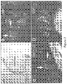
- FIG. 4 shows that Myo/Nog cells express the Pax3/FOXO1 translocation in a mouse model of alveolar rhabdomyosarcoma (ARMS).
- the Pax3:FOXO1 translocation is present in 80% of patients with ARMS.
- Myf6 drives this translocation in mice.
- Fluorescent protein is co-expressed with the translocation and sections were stained with the G8 antibody. Other sections were double labeled for G8 and noggin (inset of Figure 4 ).
- the data indicate that G8-positive cells co-express noggin and a subpopulation of ARMS cells co-expresses the G8 antigen and the Pax3:FOXO1 translocation.
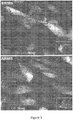
- Figure 5 shows that human rhabdomyosarcoma cells express G8 and noggin. More specifically, human embryonal and alveolar rhabdomyosarcoma cells (ERMS and ARMS, respectively) were stained with antibodies to G8 and noggin. ERMS and ARMS cell cultures contained G8+/noggin+ cells. Notably, the percent of G8+ cells in ERMS was 10% ⁇ 5 and the percent of G8+ cells in ARMS was 91% ⁇ 8. The G8+ ERMS and ARMS cells were killed with G8 antibody and complement ( Figure 6 ). Specifically, ERMS and ARMS cultures were incubated with the G8 antibody, complement that lyses antibody bound cells, or both. The cells were then labeled for G8 and TUNEL. The results presented in Figure 6 demonstrate that treatment with G8 and complement specifically kills cells that express the G8 antigen.
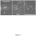
- Figure 7 shows presence of G8-positive cells in various sarcomas. Sections of human osteosarcoma, Wilms and rhabdomyosarcoma tumors were labeled with antibodies to G8 and noggin. G8+/noggin+ cells were a subpopulation within the tumors. Indeed, G8 expressing cells have been identified in osteosarcoma, Wilms, rhabdomyosarcoma, leiomyocarcoma, squamous cell, melanoma, basal cell, and Ewings tumors. The following represent possible embodiments according to the invention:
Landscapes
- Health & Medical Sciences (AREA)
- Life Sciences & Earth Sciences (AREA)
- Chemical & Material Sciences (AREA)
- Engineering & Computer Science (AREA)
- Immunology (AREA)
- General Health & Medical Sciences (AREA)
- Medicinal Chemistry (AREA)
- Molecular Biology (AREA)
- Organic Chemistry (AREA)
- Biochemistry (AREA)
- Animal Behavior & Ethology (AREA)
- Public Health (AREA)
- Veterinary Medicine (AREA)
- Pharmacology & Pharmacy (AREA)
- Bioinformatics & Cheminformatics (AREA)
- Hematology (AREA)
- Biomedical Technology (AREA)
- Urology & Nephrology (AREA)
- Proteomics, Peptides & Aminoacids (AREA)
- Analytical Chemistry (AREA)
- Pathology (AREA)
- Genetics & Genomics (AREA)
- Microbiology (AREA)
- Physics & Mathematics (AREA)
- Biotechnology (AREA)
- Epidemiology (AREA)
- Biophysics (AREA)
- Cell Biology (AREA)
- Chemical Kinetics & Catalysis (AREA)
- General Chemical & Material Sciences (AREA)
- Nuclear Medicine, Radiotherapy & Molecular Imaging (AREA)
- Food Science & Technology (AREA)
- General Physics & Mathematics (AREA)
- Wood Science & Technology (AREA)
- Zoology (AREA)
- Oncology (AREA)
- General Engineering & Computer Science (AREA)
- Hospice & Palliative Care (AREA)
- Medicines That Contain Protein Lipid Enzymes And Other Medicines (AREA)
- Medicines Containing Antibodies Or Antigens For Use As Internal Diagnostic Agents (AREA)
Applications Claiming Priority (3)
| Application Number | Priority Date | Filing Date | Title |
|---|---|---|---|
| US201261608971P | 2012-03-09 | 2012-03-09 | |
| PCT/US2013/030210 WO2013134768A1 (en) | 2012-03-09 | 2013-03-11 | Compositions and methods for treating cancer |
| EP13758403.3A EP2822593A4 (de) | 2012-03-09 | 2013-03-11 | Zusammensetzungen und verfahren zur behandlung von krebs |
Related Parent Applications (1)
| Application Number | Title | Priority Date | Filing Date |
|---|---|---|---|
| EP13758403.3A Division EP2822593A4 (de) | 2012-03-09 | 2013-03-11 | Zusammensetzungen und verfahren zur behandlung von krebs |
Publications (1)
| Publication Number | Publication Date |
|---|---|
| EP3388083A1 true EP3388083A1 (de) | 2018-10-17 |
Family
ID=49117425
Family Applications (2)
| Application Number | Title | Priority Date | Filing Date |
|---|---|---|---|
| EP13758403.3A Withdrawn EP2822593A4 (de) | 2012-03-09 | 2013-03-11 | Zusammensetzungen und verfahren zur behandlung von krebs |
| EP18153044.5A Withdrawn EP3388083A1 (de) | 2012-03-09 | 2013-03-11 | Zusammensetzungen und verfahren zur behandlung von krebs |
Family Applications Before (1)
| Application Number | Title | Priority Date | Filing Date |
|---|---|---|---|
| EP13758403.3A Withdrawn EP2822593A4 (de) | 2012-03-09 | 2013-03-11 | Zusammensetzungen und verfahren zur behandlung von krebs |
Country Status (5)
| Country | Link |
|---|---|
| US (1) | US20150037358A1 (de) |
| EP (2) | EP2822593A4 (de) |
| JP (2) | JP2015511960A (de) |
| CA (1) | CA2866881A1 (de) |
| WO (1) | WO2013134768A1 (de) |
Families Citing this family (4)
| Publication number | Priority date | Publication date | Assignee | Title |
|---|---|---|---|---|
| US20160051693A1 (en) | 2013-03-21 | 2016-02-25 | Genisphere, Llc | Cellular Delivery of DNA Intercalating Agents |
| IL260983B (en) | 2016-02-19 | 2022-07-01 | Genisphere Llc | Nucleic acid carriers and methods of medical use |
| WO2017177140A1 (en) * | 2016-04-07 | 2017-10-12 | Rowan University | Methods and kits for detecting myo/nog cells in human adipose tissue and methods of use thereof |
| US11802151B2 (en) * | 2019-11-04 | 2023-10-31 | Code Biotherapeutics, Inc. | Brain-specific angiogenesis inhibitor 1 (BAI1) antibodies and uses thereof |
Citations (3)
| Publication number | Priority date | Publication date | Assignee | Title |
|---|---|---|---|---|
| EP0404097A2 (de) | 1989-06-22 | 1990-12-27 | BEHRINGWERKE Aktiengesellschaft | Bispezifische und oligospezifische, mono- und oligovalente Rezeptoren, ihre Herstellung und Verwendung |
| WO1993011161A1 (en) | 1991-11-25 | 1993-06-10 | Enzon, Inc. | Multivalent antigen-binding proteins |
| WO2010065920A1 (en) * | 2008-12-04 | 2010-06-10 | Lankenau Institute For Medical Research | Compositions and methods for the treatment and prevention of lens fibrotic diseases |
Family Cites Families (5)
| Publication number | Priority date | Publication date | Assignee | Title |
|---|---|---|---|---|
| WO2004100885A2 (en) * | 2003-05-09 | 2004-11-25 | The Trustees Of The University Of Pennsylvania | Compositions and methods for treating cancer |
| US7445906B2 (en) * | 2003-06-30 | 2008-11-04 | East Carolina University | Method of detecting cancer using delta-catenin |
| BRPI0821417A2 (pt) * | 2007-12-26 | 2015-06-16 | Biotest Ag | Método para diminuir a adesão de células de estroma a células tumorais que expressam cd138 em células tumorais de um indivíduo em necessidade do mesmo |
| BRPI1013990A2 (pt) * | 2009-05-06 | 2019-04-30 | Biotest Ag | método para tratar uma doença , e, combinação anticâncer |
| WO2012145469A1 (en) * | 2011-04-19 | 2012-10-26 | The United States Of America, As Represented By The Secretary, Department Of Health & Human Services | Human monoclonal antibodies specific for glypican-3 and use thereof |
-
2013
- 2013-03-11 WO PCT/US2013/030210 patent/WO2013134768A1/en not_active Ceased
- 2013-03-11 JP JP2014561184A patent/JP2015511960A/ja active Pending
- 2013-03-11 EP EP13758403.3A patent/EP2822593A4/de not_active Withdrawn
- 2013-03-11 US US14/384,102 patent/US20150037358A1/en not_active Abandoned
- 2013-03-11 CA CA2866881A patent/CA2866881A1/en not_active Abandoned
- 2013-03-11 EP EP18153044.5A patent/EP3388083A1/de not_active Withdrawn
-
2017
- 2017-12-01 JP JP2017231847A patent/JP2018083813A/ja active Pending
Patent Citations (3)
| Publication number | Priority date | Publication date | Assignee | Title |
|---|---|---|---|---|
| EP0404097A2 (de) | 1989-06-22 | 1990-12-27 | BEHRINGWERKE Aktiengesellschaft | Bispezifische und oligospezifische, mono- und oligovalente Rezeptoren, ihre Herstellung und Verwendung |
| WO1993011161A1 (en) | 1991-11-25 | 1993-06-10 | Enzon, Inc. | Multivalent antigen-binding proteins |
| WO2010065920A1 (en) * | 2008-12-04 | 2010-06-10 | Lankenau Institute For Medical Research | Compositions and methods for the treatment and prevention of lens fibrotic diseases |
Non-Patent Citations (82)
| Title |
|---|
| "GenBank", Database accession no. AK298000 |
| "GenBank", Database accession no. AK304337 |
| "GenBank", Database accession no. NM_003750.2 |
| "GenBank", Database accession no. NM_015904.3 |
| "GenBank", Database accession no. NP _001006947 |
| "GenBank", Database accession no. NP _003741.1 |
| "GenBank", Database accession no. NP 002989 |
| "GenBank", Database accession no. NP 002990 |
| "GenBank", Database accession no. NP 055469 |
| "GenBank", Database accession no. NP_056988.3 |
| "GenBank", Database accession no. P46940 |
| "GenBank", Database accession no. Q05DN7 |
| "GenBank", Database accession no. Q5FWG8 |
| "GenBank", Database accession no. Q6P1N4 |
| "GenBank", Database accession no. Q96PA3 |
| "Remington: The Science and Practice of Pharmacy, 21st edition,", 2005, PA. LIPPINCOTT WILLIAMS & WILKINS |
| ANDL ET AL., DEVELOPMENT, vol. 131, 2004, pages 2257 - 2268 |
| BARTH ET AL., CURR. STEM CELL RES. THER., vol. 2, 2007, pages 221 - 227 |
| BAYNE ET AL., J. CELL BIOL., vol. 99, 1984, pages 1486 - 1501 |
| BEN-YAIR ET AL., DEVELOPMENT, vol. 132, 2005, pages 689 - 701 |
| BLESSING ET AL., J. CELL BIOL., vol. 135, 1996, pages 227 - 239 |
| BLESSING ET AL., TERATOG. CARCINOG. MUTAGEN., vol. 15, 1995, pages 11 - 21 |
| BOTCHKAREV ET AL., FASEB J., vol. 15, 2001, pages 2205 - 2214 |
| BOTCHKAREV, V.A., J. INVEST. DERMATOL., vol. 120, 2003, pages 36 - 47 |
| BRAGDON ET AL., CELL SIGNAL, vol. 23, 2011, pages 609 - 620 |
| CANCER RES., vol. 14, pages 1310 - 1316 |
| CHAKRAVARTY ET AL., PNAS, vol. 105, 2008, pages 8697 - 8702 |
| CHO ET AL., CLIN, 2008 |
| CLIFFORD ET AL., CANCER RES., vol. 55, 1995, pages 1680 - 1686 |
| DE WEVER ET AL., INT. J. CANCER, vol. 123, 2008, pages 2229 - 2238 |
| DIREKZE ET AL., HEMATOL. ONCOL., vol. 24, 2006, pages 189 - 195 |
| DROZDOFF ET AL., PROC. NATL. ACAD. SCI., vol. 91, 1994, pages 5528 - 5532 |
| GABBIANI, G., J. PATHOL., vol. 200, 2003, pages 500 - 503 |
| GERHART ET AL., DEV. BIOL., vol. 336, 2009, pages 30 - 41 |
| GERHART ET AL., DEV. BIOL., vol. 359, 2011, pages 12 - 25 |
| GERHART ET AL., J. CELL BIOL., vol. 149, 2000, pages 825 - 834 |
| GERHART ET AL., J. CELL BIOL., vol. 155, 2001, pages 381 - 392 |
| GERHART ET AL., J. CELL BIOL., vol. 164, 2004, pages 739 - 746 |
| GERHART ET AL., J. CELL BIOL., vol. 164, pages 739 - 746 |
| GERHART ET AL., J. CELL BIOL., vol. 175, 2006, pages 283 - 292 |
| GERHART ET AL., J. CELL BIOL., vol. 178, 2007, pages 649 - 660 |
| GERHART JACQUELYN ET AL: "Myo/Nog cells in normal, wounded and tumor-bearing skin.", EXPERIMENTAL DERMATOLOGY JUN 2012, vol. 21, no. 6, June 2012 (2012-06-01), pages 466 - 468, XP002783249, ISSN: 1600-0625 * |
| GHARZI ET AL., EXP. DERMATOL., vol. 12, 2003, pages 126 - 136 |
| HAYES ET AL., CARCINOGENESIS, vol. 32, 2011, pages 1340 - 1348 |
| HAYES ET AL., ONCOGENE, vol. 25, 2006, pages 1543 - 1553 |
| HENNINGS ET AL., CELL, vol. 19, 1980, pages 245 - 254 |
| HOLLIGER ET AL., PROC. NATL. ACAD. SCI. USA, vol. 90, 1993, pages 6444 - 6448 |
| HSU ET AL., LAB INVEST., vol. 88, 2008, pages 842 - 855 |
| JAMORA ET AL., NATURE, vol. 422, 2003, pages 317 - 322 |
| JIN HONGKUI ET AL: "MetMAb, the one-armed 5D5 anti-c-Met antibody, inhibits orthotopic pancreatic tumor growth and improves survival", CANCER RESEARCH, AMERICAN ASSOCIATION FOR CANCER RESEARCH, US, vol. 68, no. 11, 1 June 2008 (2008-06-01), pages 4360 - 4368, XP002570057, ISSN: 0008-5472, DOI: 10.1158/0008-5472.CAN-07-5960 * |
| KAISER ET AL., J. INVEST. DERMATOL., vol. 111, 1998, pages 1145 - 1152 |
| KOBIELAK ET AL., PROC. NATL. ACAD. SCI., vol. 104, 2007, pages 10063 - 10068 |
| LANG ET AL., NATURE, vol. 433, 2005, pages 884 - 887 |
| LIBERMAN ET AL.: "Pharmaceutical Dosage Forms", 1980, MARCEL DECKER |
| LUGINI ET AL., CANCER RES., vol. 66, 2006, pages 3629 - 3638 |
| MING ET AL., GENESIS, vol. 39, 2004, pages 10 - 25 |
| MONTANARO ET AL., PROC. NATL. ACAD. SCI., vol. 100, 2003, pages 9336 - 9341 |
| N. SHARMA ET AL: "In the clinic: ongoing clinical trials evaluating c-MET-inhibiting drugs", THERAPEUTIC ADVANCES IN MEDICAL ONCOLOGY, vol. 3, no. 1 Suppl, 1 November 2011 (2011-11-01), pages S37 - S50, XP055095200, ISSN: 1758-8340, DOI: 10.1177/1758834011423403 * |
| NAKAI ET AL., EXP. CELL RES., vol. 309, 2005, pages 137 - 148 |
| OWENS ET AL., J. INVEST. DERMATOL., vol. 128, 2008, pages 783 - 790 |
| PINNEY ET AL., CELL, vol. 53, 1988, pages 781 - 93 |
| PYE ET AL., J. ANAT., vol. 198, 2001, pages 163 - 173 |
| QIAO ET AL., ONCOGENE, vol. 25, 2006, pages 207 - 217 |
| RENDL ET AL., PLOS BIOL., vol. 3, 2005, pages e331 |
| ROWE, ET AL.,: "Handbook of Pharmaceutical Excipients (6th Ed.),", 2009, PHARMACEUTICAL PR |
| RUFAUT ET AL., J. CELL PHYSIOL., vol. 209, 2006, pages 959 - 966 |
| SHAROV ET AL., AM. J. PATHOL., vol. 175, 2009, pages 1303 - 1314 |
| SNEDDON ET AL., PROC. NATL. ACAD. SCI., vol. 103, 2006, pages 14842 - 14847 |
| STRONY ET AL., GENE EXPR. PATTERNS, vol. 5, 2005, pages 387 - 395 |
| TERSKIKH ET AL., PNAS, vol. 94, 1997, pages 1663 - 1668 |
| THAWANI ET AL., NEUROSURGERY, vol. 66, 2010, pages 233 - 246 |
| TONEGAWA ET AL., DEV. BIOL., vol. 202, 1998, pages 172 - 182 |
| WACH ET AL., ONCOGENE, vol. 20, 2001, pages 7761 - 7769 |
| WALKER ET AL., PROC. NATL. ACAD. SCI, vol. 107, 2010, pages 13730 - 13735 |
| WALKER ET AL., PROC. NATL. ACAD. SCI., vol. 107, 2010, pages 13730 - 13735 |
| WALL ET AL., J. CELL BIOL., vol. 120, 1993, pages 493 - 502 |
| WALSH ET AL., TRENDS CELL BIOL., vol. 20, 2010, pages 244 - 256 |
| XU WEI ET AL: "Recombinant immunotoxin anti-c-Met/PE38KDEL inhibits proliferation and promotes apoptosis of gastric cancer cells", JOURNAL OF EXPERIMENTAL & CLINICAL CANCER RESEARCH, BIOMED CENTRAL LTD, LONDON UK, vol. 30, no. 1, 7 July 2011 (2011-07-07), pages 67, XP021104366, ISSN: 1756-9966, DOI: 10.1186/1756-9966-30-67 * |
| YANG ET AL., CANCER RES., vol. 65, 2005, pages 8671 - 8678 |
| YUHKI ET AL., DEVELOPMENT, vol. 131, 2004, pages 1825 - 1833 |
| YUSPA ET AL., EXP. CELL RES., vol. 86, 1974, pages 95 - 105 |
| ZHANG ET AL., STEM CELLS, vol. 24, 2006, pages 2826 - 2839 |
Also Published As
| Publication number | Publication date |
|---|---|
| WO2013134768A1 (en) | 2013-09-12 |
| EP2822593A4 (de) | 2015-10-14 |
| US20150037358A1 (en) | 2015-02-05 |
| EP2822593A1 (de) | 2015-01-14 |
| JP2018083813A (ja) | 2018-05-31 |
| JP2015511960A (ja) | 2015-04-23 |
| CA2866881A1 (en) | 2013-09-12 |
Similar Documents
| Publication | Publication Date | Title |
|---|---|---|
| KR101883280B1 (ko) | 종양 특이 항체 및 그 사용 | |
| US8715675B2 (en) | Vascular targets for detecting, imaging and treating neoplasia or neovasculature | |
| JP7139569B2 (ja) | 中枢神経系における悪性腫瘍の検出及び治療 | |
| ES2902420T3 (es) | Composiciones para el tratamiento del cáncer que expresa ADAM8 | |
| CN106573976B (zh) | 抗cd84抗体、包含所述抗体的组合物及其用途 | |
| US20180043038A1 (en) | Tumor specific antibody conjugates and uses therefor | |
| JP2018083813A (ja) | がんを治療するための組成物及び方法 | |
| WO2003048302A2 (en) | Identifying anti-tumor targets or agents by lipid raft immunization and proteomics | |
| DK2984108T3 (en) | Anti-s100a7 antibodies for the treatment and diagnosis of cancer | |
| US12059429B2 (en) | Hornerin: a novel non-VEGF mediated angiogenic protein expressed in both human and mouse angiogenic endothelial cells and human pancreatic cancer cells | |
| EP4497755A1 (de) | Anti-wnt-11 antikörper | |
| Ogawa et al. | Matrix Metalloproteinase and Tissue Inhibitor of Metalloproteinase in Human Bone Marrow Tissues An Immunohistochemical Study | |
| US20240361324A1 (en) | Point of service method of detecting, diagnosing and following cancer and other proliferative conditions | |
| JP7406784B2 (ja) | 卵巣癌の検出方法 | |
| WO2023276768A1 (ja) | 低酸素バイオマーカー及びその利用 | |
| Meghnani | RAGE (Receptor for Advanced Glycation End Products) in melanoma progression | |
| HK1188128A (en) | Tumor specific antibodies and uses therefor | |
| HK1188128B (en) | Tumor specific antibodies and uses therefor |
Legal Events
| Date | Code | Title | Description |
|---|---|---|---|
| PUAI | Public reference made under article 153(3) epc to a published international application that has entered the european phase |
Free format text: ORIGINAL CODE: 0009012 |
|
| STAA | Information on the status of an ep patent application or granted ep patent |
Free format text: STATUS: THE APPLICATION HAS BEEN PUBLISHED |
|
| AC | Divisional application: reference to earlier application |
Ref document number: 2822593 Country of ref document: EP Kind code of ref document: P |
|
| AK | Designated contracting states |
Kind code of ref document: A1 Designated state(s): AL AT BE BG CH CY CZ DE DK EE ES FI FR GB GR HR HU IE IS IT LI LT LU LV MC MK MT NL NO PL PT RO RS SE SI SK SM TR |
|
| STAA | Information on the status of an ep patent application or granted ep patent |
Free format text: STATUS: THE APPLICATION IS DEEMED TO BE WITHDRAWN |
|
| 18D | Application deemed to be withdrawn |
Effective date: 20190418 |